Anatomical and neurochemical definition of the nucleus of the stria terminalis in japanese quail...
17
Anatomical and Neurochemical Definition of the Nucleus of the Stria Terminalis in Japanese Quail (Coturnix japonica) N. ASTE, 1 J. BALTHAZART, 2 P. ABSIL, 2 R. GROSSMANN, 3 E. MU ¨ LHBAUER, 3 C. VIGLIETTI-PANZICA, 1 AND G.C. PANZICA 1 * 1 Department ofAnatomy, Pharmacology, and Forensic Medicine, University of Torino, 10126 Torino, Italy 2 Laboratory of Biochemistry, University of Lie ` ge, 4020 Lie ` ge, Belgium 3 Institute for Small Animal Research, Federal Research Center of Agriculture, 29223 Celle, Germany ABSTRACT This study in birds provides anatomical, immunohistochemical, and hodological data on a prosencephalic region in which the nomenclature is still a matter of discussion. In quail, this region is located just dorsal to the anterior commissure and extends from the level of the medial part of the preoptic area at its most rostral end to the caudal aspects of the nucleus preopticus medialis. At this caudal level, it reaches its maximal elongation and extends from the ventral tip of the lateral ventricles to the dorsolateral aspects of the paraventricular nucleus. This area contains aromatase-immunoreactive cells and a sexually dimorphic population of small, vasotocinergic neurons. The Nissl staining of adjacent sections revealed the presence of a cluster of intensely stained cells outlining the same region delineated by the vasotocin-immunoreactive structures. Cytoarchitectonic, immunohistochemical, and in situ hybridization data support the notion that this area is similar and is probably homologous to the medial part of the nucleus of the stria terminalis of the mammalian brain. The present data provide a clear definition of this nucleus in quail: They show for the first time the presence of sexually dimorphic vasotocinergic neurons in this region of the quail brain and provide the first detailed description of this region in an avian species. J. Comp. Neurol. 396:141–157, 1998. r 1998 Wiley-Liss, Inc. Indexing terms: vasotocin; aromatase; sex dimorphism; in situ hybridization; bird The bed nucleus of the stria terminalis (BST) has been studied intensively in mammals, whereas its location and characteristics are still a matter of discussion in other classes of vertebrates. Traditionally, this nucleus has been divided into a medial and a lateral subdivision (Krettek and Price, 1978; De Olmos et al., 1985) that consist of several subnuclei (Del Abril et al., 1987; Moga et al., 1989; Hines et al., 1992). The medial subdivision 3 (BSTm) is part of the circuitry that controls mating behavior. It receives inputs from the accessory olfactory bulb and the medial amygdala, and it sends projections to the medial preoptic area and the ventromedial hypothalamus (De Olmos and Ingram, 1972; De Olmos et al., 1978; Krettek and Price, 1978; Weller and Smith, 1982; Simerly and Swanson, 1986, 1988; Akesson et al., 1988; Simerly et al., 1989; Canteras et al., 1995). These regions are involved in the control of sexual motivation and/or performance (for recent reviews, see Segovia and Guillamon, 1993; Van Furth et al., 1995). In contrast, the lateral subdivision (BSTl) is characterized by reciprocal connections with nuclei that are involved in central autonomic regulation (for a review of the literature, see Moga et al., 1989). Several anatomical (Del Abril et al., 1987; Allen and Gorski, 1990; Hines et al., 1992; Segovia and Guillamon, Grant sponsor: MURST; Grant sponsor: CNR; Grant number 97.04308CT04; Grant sponsor: NIMH; Grant number MH 50388; Grant sponsor: European Community; Grant number: CT94–0472; Grant spon- sor: Belgian FRFC; Grant number: 9.4565.96F; Grant sponsor: University of Lie ` ge; Grant sponsor: Government of the French Community of Belgium; Grant number: AC 93/98–171; Grant sponsor: German Research Founda- tion; Grant sponsor: European Science Foundation; Grant number: RG95/203. N. Aste is presently at the Institute of Molecular Neurobiology, Shiga University of Medical Science, Otsu, Shiga, Japan. *Correspondence to: Dr. G.C. Panzica, Department ofAnatomy, Pharma- cology, and Forensic Medicine, Corso M. D’Azeglio 52, 10126 Torino, Italy. E-mail: [email protected] Received 7 July 1997; Revised 30 December 1997; Accepted 10 February 1998 THE JOURNAL OF COMPARATIVE NEUROLOGY 396:141–157 (1998) r 1998 WILEY-LISS, INC.
Transcript of Anatomical and neurochemical definition of the nucleus of the stria terminalis in japanese quail...

Anatomical and Neurochemical Definitionof the Nucleus of the Stria Terminalis
in Japanese Quail (Coturnix japonica)
N. ASTE,1 J. BALTHAZART,2 P. ABSIL,2 R. GROSSMANN,3 E. MULHBAUER,3
C. VIGLIETTI-PANZICA,1 AND G.C. PANZICA1*1Department of Anatomy, Pharmacology, and Forensic Medicine,
University of Torino, 10126 Torino, Italy2Laboratory of Biochemistry, University of Liege, 4020 Liege, Belgium
3Institute for Small Animal Research, Federal Research Center of Agriculture,29223 Celle, Germany
ABSTRACTThis study in birds provides anatomical, immunohistochemical, and hodological data on a
prosencephalic region in which the nomenclature is still a matter of discussion. In quail, thisregion is located just dorsal to the anterior commissure and extends from the level of themedial part of the preoptic area at its most rostral end to the caudal aspects of the nucleuspreopticus medialis. At this caudal level, it reaches its maximal elongation and extends fromthe ventral tip of the lateral ventricles to the dorsolateral aspects of the paraventricularnucleus. This area contains aromatase-immunoreactive cells and a sexually dimorphicpopulation of small, vasotocinergic neurons. The Nissl staining of adjacent sections revealedthe presence of a cluster of intensely stained cells outlining the same region delineated by thevasotocin-immunoreactive structures. Cytoarchitectonic, immunohistochemical, and in situhybridization data support the notion that this area is similar and is probably homologous tothe medial part of the nucleus of the stria terminalis of the mammalian brain. The presentdata provide a clear definition of this nucleus in quail: They show for the first time thepresence of sexually dimorphic vasotocinergic neurons in this region of the quail brain andprovide the first detailed description of this region in an avian species. J. Comp. Neurol.396:141–157, 1998. r 1998 Wiley-Liss, Inc.
Indexing terms: vasotocin; aromatase; sex dimorphism; in situ hybridization; bird
The bed nucleus of the stria terminalis (BST) has beenstudied intensively in mammals, whereas its location andcharacteristics are still a matter of discussion in otherclasses of vertebrates. Traditionally, this nucleus has beendivided into a medial and a lateral subdivision (Krettekand Price, 1978; De Olmos et al., 1985) that consist ofseveral subnuclei (Del Abril et al., 1987; Moga et al., 1989;Hines et al., 1992). The medial subdivision 3 (BSTm) ispart of the circuitry that controls mating behavior. Itreceives inputs from the accessory olfactory bulb and themedial amygdala, and it sends projections to the medialpreoptic area and the ventromedial hypothalamus (DeOlmos and Ingram, 1972; De Olmos et al., 1978; Krettekand Price, 1978; Weller and Smith, 1982; Simerly andSwanson, 1986, 1988; Akesson et al., 1988; Simerly et al.,1989; Canteras et al., 1995). These regions are involved inthe control of sexual motivation and/or performance (forrecent reviews, see Segovia and Guillamon, 1993; VanFurth et al., 1995). In contrast, the lateral subdivision
(BSTl) is characterized by reciprocal connections withnuclei that are involved in central autonomic regulation(for a review of the literature, see Moga et al., 1989).
Several anatomical (Del Abril et al., 1987; Allen andGorski, 1990; Hines et al., 1992; Segovia and Guillamon,
Grant sponsor: MURST; Grant sponsor: CNR; Grant number97.04308CT04; Grant sponsor: NIMH; Grant number MH 50388; Grantsponsor: European Community; Grant number: CT94–0472; Grant spon-sor: Belgian FRFC; Grant number: 9.4565.96F; Grant sponsor: Universityof Liege; Grant sponsor: Government of the French Community of Belgium;Grant number: AC 93/98–171; Grant sponsor: German Research Founda-tion; Grant sponsor: European Science Foundation; Grant number: RG95/203.
N. Aste is presently at the Institute of Molecular Neurobiology, ShigaUniversity of Medical Science, Otsu, Shiga, Japan.
*Correspondence to: Dr. G.C. Panzica, Department of Anatomy, Pharma-cology, and Forensic Medicine, Corso M. D’Azeglio 52, 10126 Torino, Italy.E-mail: [email protected]
Received 7 July 1997; Revised 30 December 1997;Accepted 10 February 1998
THE JOURNAL OF COMPARATIVE NEUROLOGY 396:141–157 (1998)
r 1998 WILEY-LISS, INC.

1993) and neurochemical (Woodhams et al., 1983; Walteret al., 1991) characteristics of the BSTm or of its subdivi-sions are sexually dimorphic and are organized in early lifeby steroid hormones (for review, see Guillamon and Se-govia, 1997). In particular, sex differences that favor malescompared with females affect the number of vasopressin-immunoreactive (VP-ir) neurons and the VP content ofBSTm in the rat brain (De Vries et al., 1994a; Wang et al.,1994). Moreover, the rodent BSTm is an important site oftestosterone aromatization (transformation of testoster-one into 17b-estradiol), as shown by measures of theenzymatic activity and immunocytochemical detection ofthe protein (Roselli, 1991; Roselli and Resko, 1993; Jakabet al., 1993; Shinoda et al., 1994; Tsuruo et al., 1994;Foidart et al., 1995a). Recently, the presence of nitric oxidesynthase has been demonstrated in many of the nucleithat control the rodent mating behavior, including the BST(Hadeishi and Wood, 1996).
In the rat, the BSTm is one element of the pathway thatconnects the vomeronasal organ to the preoptic region-hypothalamus; therefore, it is implicated in the control ofvarious aspects of reproductive behavior that are governedby chemosensory stimuli (Segovia and Guillamon, 1993;Guillamon and Segovia, 1997). Several studies indicatethat lesions aimed at the rat BST disrupt consummatoryand/or motivational aspects of male and female sexualbehavior (Emery and Sachs, 1976; Valcourt and Sachs,1979; Lopez and Carrer, 1982; Claro et al., 1995; Takeo etal., 1995) and impair maternal behavior (Numan andNuman, 1996). Moreover, a recent study suggests that thevolume of the central subdivision of the BST, as identifiedby vasoactive intestinal polypeptide immunoreactivity, isrelated to gender identity in humans (Zhou et al., 1995).However, despite numerous studies investigating morpho-logical and functional characteristics of the BST in mam-mals, the definition of the homologous nucleus in otherclasses of vertebrates is still a matter of controversy.
The Japanese quail is an experimental model that hasbeen employed widely in the study of the neural andendocrine bases of male sexual behavior (for recent re-views, see Balthazart and Foidart, 1993; Panzica et al.,1996b). The action of steroids in the quail preoptic-hypothalamic region is necessary and sufficient to activate
male copulatory behavior (Balthazart and Surlemont,1990a,b); accordingly, this area contains large numbers ofcells that express steroid receptors (Watson and AdkinsRegan, 1988; Balthazart et al., 1989, 1992b). Aromatiza-tion of testosterone into estrogens is a limiting step in theactivation of male quail copulatory behavior; consequently,the distribution and regulation of preoptic aromatase(ARO) activity and ARO-containing neurons have beenintensively studied (for recent reviews, see Balthazart etal., 1996b; Balthazart, 1997). ARO-ir neurons are presentin the nucleus preopticus medialis (POM), the tuberalregion, an area dorsal to the anterior commissure (CA),and a V-shaped structure extending from a region ventralto the lateral septal area to the caudal aspects of the POM(Balthazart et al., 1990a,b, 1997; Foidart et al., 1995b).With the exception of the POM, which is outlined clearlyby ARO-ir cells and can be observed also in Nissl-stainedmaterial, the regions containing ARO-ir cells in the quailbrain do not match exactly the structures that have beendefined previously based on Nissl-stained sections. Theiridentification and nomenclature is therefore somewhatunclear.
Numerous peptidergic elements have also been identi-fied within the quail septopreoptic-hypothalamic region(Panzica et al., 1992a; Aste et al., 1997). In particular,several studies have been devoted to the vasotocin (VT)-irfibers of the POM and of the lateral septum, because thesepeptidergic structures are sexually dimorphic and steroid-sensitive (Viglietti-Panzica et al., 1992, 1994; Panzica etal., 1996a; Aste et al., 1997; for review see Panzica et al.,1997). However, these studies have paid little attention tothe distribution of this neuropeptide in the aforemen-tioned region of the quail brain, which is located betweenthe septum and the caudal aspects of the POM.
In a number of immunocytochemical studies analyzingthe distribution of ARO-ir or VT-ir structures in quail andother avian species (fowl, canary, zebra finch), this regionwas named the nucleus of the stria terminalis (Kiss et al.,1987; Balthazart et al., 1990b; Viglietti-Panzica et al.,1992; Voorhuis and De Kloet, 1992; Foidart et al., 1995b;Shen et al., 1995; Jurkevich et al., 1997). However, the
Abbreviations
AA archistriatum anteriorAc nucleus accumbensAM nucleus anterior medialis hypothalamiARO aromataseBSTl bed nucleus striae terminalis, pars lateralisBSTm bed nucleus striae terminalis, pars medialisCA commissura anteriorCPa commissura palliiDLA nucleus dorsolateralis anterior thalamiE ectostriatumFPL fasciculus prosencephali lateralis (lateral forebrain bundle)GLv nucleus geniculatus lateralis, pars ventralisHA hyperstriatum accessoriumHp hippocampusHV hyperstriatum ventraleir immunoreactiveLA nucleus lateralis anterior thalamiLHy nucleus lateralis hypothalamiLPO lobus paraolfactorius (medial part of the dorsal striatum)LV lateral ventricleN neostriatumnCPa nucleus commissurae pallii
NI neostriatum intermediumOM occipitomesencephalic tractPA paleostriatum augmentatum (lateral part of the dorsal
striatum)POA nucleus preopticus anteriorPOD nucleus preopticus dorsalisPOM nucleus preopticus medialisPP paleostriatum primitivum (dorsal pallidum)PVN nucleus paraventricularisQF tractus quintofrontalisROT nucleus rotundusSCNl nucleus suprachiasmaticus, pars lateralisSCNm nucleus suprachiasmaticus, pars medialisSL nucleus septali lateralisSM nucleus septali medialisTn nucleus taeniaeTPO area temporoparietooccipitalisTSM tractus septomesencephalicusVLT nucleus ventrolateralis thalamiVMN nucleus ventromedialis hypothalamiVP ventral paleostriatum (ventral pallidum)VT vasotocin
142 N. ASTE ET AL.

region identified in these relatively recent immunocyto-chemical studies does not always correspond to the arealabeled the nucleus stria terminalis in atlases of the avianbrain, which are based largely on Nissl-stained material(Stokes et al., 1974; Kuenzel and Masson, 1988; Breazileand Kuenzel, 1993), and some atlases do not even mentionthis structure (Karten and Hodos, 1967; Bayle et al., 1974).Furthermore, different atlases or publications do not makea consistent use of terminology for the cell groups thatconstitute the ventral striatal regions and that include thenucleus accumbens, the BST, and the tuberculum olfacto-rium. The boundaries and exact localization of the BSTand its position relative to the nucleus accumbens, there-fore, have not been defined consistently in different studies(compare Brauth et al., 1978; Reiner et al., 1983; Kitt andBrauth, 1986a,b; Berk, 1987; Anderson and Reiner, 1991;Veenman et al., 1995). There has been also a lack ofdetailed anatomical investigations describing the full ex-tent of this nucleus, which probably explains the discrepan-cies in the nomenclature of this region across and withinthe avian species.
Neurochemical markers provides a useful method toanalyze homology between brain structures (Gahr, 1997).Therefore, the present study was designed to provide adetailed description of this region in quail and attempts toestablish its homology with the mammalian BSTm basedon topographical and neurochemical criteria. Special atten-tion was paid here to the VT-ir and ARO-ir structures thatare clear anatomical markers of the BSTm in mammals.
MATERIALS AND METHODS
Subjects
The experiments described in this paper were carriedout on adult male and female Japanese quail (Coturnixjaponica) that were obtained from local breeders in Bel-gium (Dujardin Farms, Liernu) or Italy (Morini, Modena).Throughout their life, they received water and food adlibitum and were submitted to a photoperiod of 16 hours oflight and 8 hours of darkness. All experimental procedureshave been reviewed by the appropriate authorities and arein compliance with the relevant laws and regulations ofItaly, Germany, and Belgium that govern the treatment ofexperimental animals.
Immunocytochemistry
Seven male and seven female quail were used forimmunocytochemical investigations. They were injectedwith 0.1 ml of heparin solution (30 mg/ml; Sigma, St.Louis, MO) into the wing vein and deeply anaesthetizedwith an overdose of Hypnodil (50 mg/kg; Janssen Pharma-ceutica, Beerse, Belgium). The animals were then perfusedintracardially with a saline solution followed by 4% para-formaldehyde in 0.1 M phosphate buffer, pH 7.2. Thedissected brains were postfixed in the same fixative for 12hours at 4°C and washed with 0.01 M phosphate buffercontaining 0.125 M NaCl, pH 7.2 (PBS). After cryoprotec-tion with a solution containing 20% sucrose in PBS, brainswere rapidly frozen on dry ice and stored at 280°C.
Brains were cut in 30-µm-thick coronal sections thatwere collected in multiwell plates filled with PBS. Adja-cent sections were collected in three separate series, sothat sections placed in the same well were 90 µm apart.One series was stained with toluidine blue for Nissl
material, and two series were submitted to immunocyto-chemical procedures.
Sections were stained by immunocytochemistry for VTand ARO by using procedures that have been described inprevious studies (Aste et al., 1995; Foidart et al., 1995b).Briefly, after a 30-minute wash in PBS containing 0.2%Triton X-100 (PBST) and inhibition of endogenous peroxi-dase activity by an incubation in hydrogen peroxide,sections were incubated with normal serum (VectastainLabtek; Vector Laboratories, Burlingame, CA) for 20minute at room temperature. Adjacent sections were thenincubated overnight at room temperature in a primaryantibody diluted in PBST (anti-VT, 1:8,000; anti-ARO,1:1,000).
The polyclonal anti-VT serum was developed originallyby Dr. D. Gray at the Max Plank Institute of Clinical andPhysiological Research of Bad Nauheim, Germany (Grayand Simon, 1983). The characteristics and specificity ofthis antibody for the detection of quail VT were reported ina previous study (Viglietti-Panzica et al., 1994). Thepolyclonal anti-ARO serum (kindly provided by Dr. N.Harada, Fujita Health University, Toyoake, Japan) wasraised in rabbit against quail recombinant ARO, as de-scribed by Foidart et al. (1995b).
On the next day, sections were incubated for 45 minutesin a secondary biotinylated antibody and for 30 minutes inan avidin-biotin-peroxidase complex (Vectastain EliteLabtek; Vector Laboratories). The peroxidase activity wasrevealed with a solution containing 0.187 mg/ml 38,38-diaminobenzidine and 0.003% hydrogen peroxide in 0.05M Tris-HCl, pH 7.6. Several rinses in PBS were made aftereach step. The sections were mounted on slides, dehy-drated, and coverslipped with Entellan (Merck, Milano,Italy). The sections were photographed with a Zeiss Ax-ioplan microscope (Thornwood, NY) with a Kodak Wratten44 or 75 gelatine filter (Eastman Kodak, Rochester, NY) toincrease the contrast.
In situ hybridization
Five adult male and five adult female quail were killedby decapitation. The brains were quickly dissected andfrozen on dry ice. Coronal sections were cut on a cryostat at20 µm thickness, serially collected on 3-aminopropyltrieth-oxy silane-coated slides, and fixed by immersion for 5minutes in 4% paraformaldehyde in 0.1 M phosphatebuffer, pH 7.2.
After dehydration, the slides were incubated for 2 hoursat 52°C in prehybridization buffer (50% formamide, 5IDenhardt’s solution, 0.75 M NaCl, 25 mM PIPES, 25 mMEDTA, 0.2% sodium dodecylsulfate [SDS], 10 mM DTT,and 250 µg/ml herring sperm DNA). The hybridizationproceeded overnight at 52°C in prehybridization bufferplus 10% sodium dextran sulfate. The AVT-specific probe(a 260-bp cDNA directed toward the 38 distal part of thechicken AVT gene; Hamann et al., 1992) was labeledwith[33P]dCTP by using the random priming method(Megaprime DNA labeling system; Amersham, Braun-schweig, Germany). Approximately 4,000 cpm/µl of[33P]dCTP-labelled probe were used for each section. Thespecificity of this probe for quail AVT mRNA was describedin a previous study (Aste et al., 1996).
Sections were then washed three times in 53 standardsaline citrate (SSC; 3 M NaCl, 0.3 M Na-citrate, pH 7.0),washed three more times in 23 SSC at room temperaturefor 5 minutes, and dried under a vacuum. Slides were
DEFINITION OF THE NUCLEUS OF THE STRIA TERMINALIS IN BIRDS 143

coated with autoradiography emulsion (LM-1; Amersham,Little Chalfont, UK) and exposed for 4 days at 6°C. Thesections were finally counterstained with Toluidine blue tohelp in the identification of brain nuclei. They werephotographed on a Leitz Diaplan microscope (Wetzlar,Germany) by using darkfield illumination.
The nomenclature used in this study is based largely onthe chicken atlas of Kuenzel and Masson (1988) with somemodifications, according to our previous studies on thequail preoptic region (Panzica et al., 1991, 1996b), and onsome recent studies on the pigeon prosencephalon (Veen-man et al., 1995; Medina and Reiner, 1997). The drawingsin Figure 1 summarize the distribution of nuclei in thequail prosencephalon as they are used in this paper.Discussion of some of the choices made for nomenclaturecan be found below (see Results and Discussion).
RESULTS
VT immunoreactivity in males
The septopreoptic-hypothalamic area of male quail con-tains several discrete clusters of VT-ir structures (cells andfibers). In agreement with previous observations on quail(Viglietti-Panzica, 1986; Panzica et al., 1992a, 1996a;Viglietti-Panzica et al., 1992, 1994; Aste et al., 1997), VT-irfibers are observed in the lateral septum, the periventricu-lar hypothalamus, and the tuberal region. In particular, atthe level of the preoptic area, a dense cluster of VT-ir fibersoutlines the whole POM throughout its entire rostral-to-caudal extent (Viglietti-Panzica et al., 1994). VT-ir neu-rons are found in a periventricular position, lining theependymal wall of the third ventricle, close to the pialsurface of the preoptic area and in the nucleus paraven-tricularis. They are also seen more laterally in the lateraland dorsal thalamic areas (see Fig. 2E–H).
In addition, VT-ir cells and fibers are observed in adiscrete area located above and caudal to the anteriorcommissure, where they have never been described indetail previously in the quail brain. This region, as dis-cussed below, corresponds to an identifiable cell cluster inNissl-stained sections (Fig. 2A–D). This cluster has beennamed nucleus of the stria terminalis in a number ofprevious publications, but this identification was neverevaluated critically. It is the purpose of the present paperto provide neurochemical criteria confirming and specify-ing this nomenclature. In this section, we describe the dataand use the term BSTm without making any additionalcomments on the name. In the Discussion, a criticalevaluation of this decision is presented.
VT-ir structures are present in the entire area connect-ing the lateral septum with the medial preoptic region.However, they are distributed heterogeneously, and higherdensities of immunoreactive structures allow one to definethe BSTm. These structures are located ventral to theseptal area and extend in the rostrocaudal direction fromabout 100 µm rostral to the CA to sections located 200–300µm caudal to the caudal edge of this commissure. Justrostral to the CA, a small, dense cluster of VT-ir fibers isobserved ventromedial to the tip of the lateral ventricle. Inslightly more caudal sections, where the first signs of theCA become visible, this cluster expands to form a largerstructure that also contains a few scattered, immunoposi-tive cell bodies (Figs. 2E, 3C,E). When the CA reaches itsfull extension, VT-ir cells are seen in larger numbers abovethe commissure, and the cluster of VT-ir fibers extends
above and below the commissure (Figs. 2F, 3G). Morecaudally, in sections where the CA is reduced or is nolonger visible, numerous vasotocinergic cells and fiberscover an ovoid area that is positioned in oblique manner(dorsolateral-to-ventromedial direction) and border thedorsal edge of the occipitomesencephalic tract (OM) and ofthe lateral forebrain bundle (FPL; Figs. 2G,H, 4B,F, 5B,D).The most rostral aspect of this ovoid structure merges atits ventral edge with the most dorsal and caudal aspects ofthe VT-ir structures, identifying the POM (Figs. 2G, 3G).This cluster of VT-ir cells and fibers suddenly disappearsin more caudal sections. The VT-ir fibers defining theBSTm (Figs. 3E, 4D) are associated only with a fewpunctate structures. These VT-ir fibers are often organizedin basket-like ring structures around immunonegativecells (Fig. 4D).
Scattered VT-ir cells are also present in the most caudalaspects of the POM at the level of the CA (Figs. 2E–G, 3G,4B,C). These cells are similar morphologically to thoseobserved in the BSTm (Figs. 3E, 4D, 5D). Although theVT-ir cells are not distributed homogeneously in theBSTm, and although they increase in number as oneprogresses in rostrocaudal direction (compare Fig. 3E withFig. 5D), their morphological characteristics are similar inthe entire region. These cell bodies are round or slightlyelongated, mainly bipolar, and their diameter rangesbetween 5 µm and 12 µm. The density of the immunoreac-tive material in these cells is variable (Figs. 4C, 5D), but,in general, it is lower than in the magnocellular elementsobserved in the lateral, periventricular, and subpial groups(Fig. 3G; see also Viglietti-Panzica, 1986; Viglietti-Panzicaet al., 1994).
Distribution of VT mRNA in males
Detection of mRNA for VT by using in situ hybridizationrevealed a distribution of large and small elements that isin agreement with a previous study (Aste et al., 1996).Magnocellular elements were observed bordering the thirdventricle wall and were also found in a more lateralposition (Fig. 6B,D). In addition, smaller and weaklylabeled cells were revealed by using in situ hybridizationin the septopreoptic region and had a general distributionthat was similar to that of the VT-ir cells described abovein the BSTm and the POM. In particular, a group of VTgene-expressing cells could be observed easily that corre-sponded topographically to the group of VT-ir cells locatedjust above the CA from the rostral edge of this structure toa level slightly caudal to the end of the POM (Fig. 6B,D).
The small variation in the number of VT-ir cells that hasbeen described in the rostrocaudal extent of the BSTmbased on the immunocytochemistry could also be observedat the level of the mRNA. More VT gene-expressing cellswere observed in the caudal portion (Fig. 6D) than in therostral portion of the BSTm (Fig. 6B). In situ hybridizationalso confirmed the presence of VT gene-expressing cells inthe caudal part of the POM (Fig. 6B,D).
Sexual dimorphism of vasotocinergicstructures
A qualitative sexual dimorphism in the number of VT-ircells could be observed in the entire BSTm. VT-ir cells weredetectable only in males (compare Figs. 3G, 4F, 5B,D, and7A–D), and no VT-ir cell body could be observed in thisarea of the female brain, even after prolonged developmentof the peroxidase reaction.
144 N. ASTE ET AL.
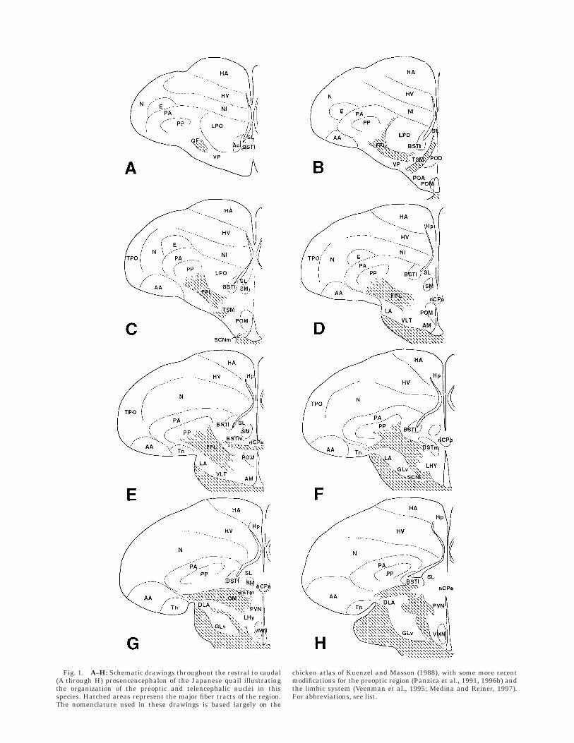
Fig. 1. A–H: Schematic drawings throughout the rostral to caudal(A through H) prosencencephalon of the Japanese quail illustratingthe organization of the preoptic and telencephalic nuclei in thisspecies. Hatched areas represent the major fiber tracts of the region.The nomenclature used in these drawings is based largely on the
chicken atlas of Kuenzel and Masson (1988), with some more recentmodifications for the preoptic region (Panzica et al., 1991, 1996b) andthe limbic system (Veenman et al., 1995; Medina and Reiner, 1997).For abbreviations, see list.
Fig. 2. A–D: Schematic drawings of Nissl-stained sections through-out the male quail septopreoptic-hypothalamic area, including themedial part of the bed nucleus of the stria terminalis (BSTm)throughout its rostrocaudal extent. Shaded areas indicate the pres-ence of Nissl-stained cell clusters. E,F: Schematic distribution of thevasotocin-immunoreactive (VT-ir) cells in the quail septopreopticregion. Small and large dots indicate the parvocellular and magnocel-
lular perikarya, respectively. I–L: Distribution of aromatase (ARO)-ircells (triangles) in the quail septopreoptic region. LV, lateral ventricle;BSTl, lateral part of the bed nucleus of the stria terminalis; nCPa,nucleus of the pallial commissure; POM, medial preoptic nucleus; CA,anterior commissure; FPL, lateral forebrain bundle. For other abbre-viations, see list.
146 N. ASTE ET AL.

Fig. 3. A: Low-power enlargement of a Nissl-stained section of themale quail septopreoptic area. Arrow indicates the rostral pole of thebed nucleus of the stria terminalis (BSTm). The relationship of thisregion (arrow) with the lateral septum, the lateral ventricle, and theanterior commissure can be appreciated. B,C: Consecutive sectionsillustrating the clustered, Nissl-stained cells (B) and vasotocin (VT)-irstructures (C) in the rostral part of the BSTm. The two arrows indicatetwo blood vessels for reference. The VT-ir elements in C (identified bythe dashed line) cover the denser Nissl-stained region that is observed
in B (dashed line). D: High magnification of aromatase (ARO)-irelements in the BSTm that are visible in F. E: High magnification ofthe VT-ir cells and fibers shown in C. Small bipolar elements arevisible (arrows) among the network of positive fibers. F,G: Topographi-cal correspondence between the ARO-ir neurons (F) and VT-ir struc-tures (G) in the medial preoptic nucleus (POM) and in the BSTm inadjacent sections. Asterisks indicate the lateral ventricle and the thirdventricle. CA, anterior commissure; FPL, lateral forebrain bundle. Forabbreviations, see list. Scale bars 5 200 µm in A,B,C, 100 µm in D–G.

Fig. 4. A: Nissl-stained section at the level of the pallial commis-sure (CPa). The medial preoptic nucleus (POM) and the bed nucleus ofthe stria terminalis (BSTm) are at caudal levels, where they merge.B: Adjacent section showing the distribution of vasotocin (VT)-irstructures. C,D: High magnification of VT-ir cells and fibers in thePOM (C) and the BSTm (D). Cell bodies are round and weakly stained.
A broad network of ir fibers is also present. E,F: Consecutive sectionsrepresenting the caudal BSTm and its merging with the POM. Notethe correspondence of the blood vessels (arrows) and their identicalrelationship between the Nissl-stained cluster of cells (E) and theVT-ir structures (F). Asterisk indicates the third ventricle. Scalebars 5 200 µm in A,B,E,F, 100 µm in C,D.

Fig. 5. A–D: Caudal portion of the male quail bed nucleus of thestria terminalis (BSTm). A,C: Distribution of aromatase (ARO)-irelements. B,D: Distribution of vasotocin (VT)-ir elements. Cells andfibers positive for these two markers are distributed in the sameregion. ARO-ir cell bodies are more numerous than VT-ir perikarya,
whereas the number of VT-ir fibers is higher than the number ofARO-ir fibers. Asterisks in B indicate the large VT-ir elements of themagnocellular system. Scale bars 5 200 µm in A,C (also apply to B,D,respectively).
DEFINITION OF THE NUCLEUS OF THE STRIA TERMINALIS IN BIRDS 149

VT-ir fibers also formed a less compact network infemales than in males (compare Figs. 3E and 4D with Fig.7B,D), and VT-ir fibers were often seen surrounding nega-tive cell bodies (compare Fig. 3G,D with Fig. 7B,D) in bothsexes.
A sexual dimorphism affecting the vasotocinergic peri-karya was also observed at the level of gene expression.The sex difference observed at this level, however, was notas dramatic as the difference observed by using immunocy-tochemistry. VT gene-expressing neurons were present inthe BSTm of both sexes (Fig. 6), but, in males, the densityof grains on the autoradiography was obviously higherover each cell than in females, suggesting a sex differencein the amount of expressed mRNA per cell (compare Fig.6A,C with Fig. 6B,D, respectively). This sex difference didnot appear to affect the magnocellular cell bodies.
Distribution of ARO-ir elements
In agreement with previously published studies (Baltha-zart et al., 1990b; Foidart et al., 1994, 1995b), a densecluster of ARO-ir cells was observed in the septopreopticregion in the exact same location as the dense cluster ofVT-ir fibers (Fig. 2I–L). When moving in a rostral-to-caudal direction, this cell group was first observed in
sections just rostral to the CA. It then increased progres-sively in size at the level of the CA (Fig. 2I,J), and, insections caudal to this commissure, the group of ARO-ircells appeared as an oblique structure that extended fromthe ventral tips of the lateral ventricles to the dorsocaudalaspects of the POM, which is also identified by a densecluster of ARO-ir neurons (Fig. 2K). These two clusters ofcells eventually merged to form an extended group ofimmunoreactive cells and then disappeared abruptly(Fig. 2L).
The analysis of consecutive sections that were immuno-stained for VT or ARO indicated a clear topographicaloverlapping between the area covered by the VT-ir struc-tures and the area that contained ARO-ir neurons (com-pare Figs. 3F and 5A, respectively, with Figs. 3G and 5B).ARO-ir cells were distributed uniformly and showed thesame morphological characteristics in the whole area (Fig.3D, anterior BSTm; Fig. 5C, posterior BSTm). The ARO-ircells located in this region and those located in the POMwere observed to merge at the same rostrocaudal levelwhere the fusion of the VT-ir structures occurred (Fig.5A,B). It must be stressed that ARO-ir cells were morenumerous by far than VT-ir cells in the BSTm.
Fig. 6. A–D: Vasotocin (VT) gene-expressing neurons in the bednucleus of the stria terminalis (BSTm) of female (A,C) or male (B,D)Japanese quail. The levels represented in A and B are comparable tothe drawing in Figure 1F. The level represented in C and D corre-sponds to Figure 1H. In the photomicrographs both small, weakly
labeled and large, intensely labeled (asterisks) neurons are visible.Small neurons are present in the BSTm and medial preoptic nucleus(POM), whereas large, intensely labeled neurons belong to the periven-tricular and lateral subdivisions of the magnocellular system. Scalebar 5 200 µm.
150 N. ASTE ET AL.

Analysis of Nissl-stained sections
A comparison of the sections stained by immunocyto-chemistry with the alternate Nissl-stained sections re-vealed the presence of a cluster of Nissl-stained cells thathad never been identified precisely before the topographi-cal data derived from the immunocytochemical studieswere available. This cell group is characterized by a highercell density than the surrounding area and allows one todefine the extension of the BSTm. The cells in this clusterare also stained in a slightly denser manner than in thesurrounding area.
Throughout most of the rostrocaudal extent of the BSTm(Fig. 2A–D), a perfect match was observed between theboundaries of this cell cluster: They could be drawn at lowmagnification based on the distribution of either the VT-irfibers and cells, the ARO-ir cells, or the densely packed,Nissl-stained cells (compare Figs. 3B and 4A,E, respec-tively, with Figs. 3C and 4B,F). However, in the rostralpart of the region, scattered ARO-containing cells werealso present outside the boundaries of the BSTm, and thecluster of Nissl-stained cells was very small and was noteasily recognizable. At the very rostral tip of the BSTm(rostral to the beginning of the CA), sometimes, no match
could be made between the distribution of the denselypacked, Nissl-stained neurons and the distribution ofVT-ir fibers.
When it was observed in Nissl-stained sections, theBSTm first appeared at its most rostral level as a densecell group located above and, to a lesser extent, below theCA (Fig. 2A,B). At this level, the dense cluster of Nissl-stained cells matched perfectly the group of ARO-ir cells,but there was only a partial overlap with VT-ir structures:The correspondence was good in the area dorsal to the CA,but the Nissl-stained cell group located below the CA didnot correspond to matching VT-ir material (Fig. 2A,B,E,F,I,J).
Caudal to the CA, the overlap between Nissl staining,ARO-ir cells, and VT-ir cells and fibers was very reliable.The staining of the BSTm increased gradually in a rostro-caudal gradient, reaching a maximum where the BSTmmerges with the POM (Fig. 4E). Visual inspection of thesections did not reveal significant cytoarchitectural differ-ences between the Nissl-stained cells of the POM andthose of the BSTm (Fig. 4A). When, at their most caudallevel, these two nuclei merge, they constitute a single,oblique structure in which no subdivision can be madebased on cell characteristics (Fig. 4E).
Fig. 7. Vasotocin (VT)-ir structures in the septopreoptic region offemale Japanese quail. A,C: Low-power magnification of the rostral(A) and caudal (C) parts of the bed nucleus of the stria terminalis(BSTm; for comparison with the male, see Figs. 2G and 4B). Noimmunoreactive cell body is visible. Asterisks indicate the intensely
stained magnocellular neurons (for comparison with in situ hybridiza-tion, see Fig. 6A,C). B,D: High magnification of the VT-ir fibersobserved in the female BSTm. Even at this magnification, no cellbodies are visible, but in some cases, positive fibers surround negativecell bodies. Scale bars 5 200 µm in A,C, 100 µm in B,D.
DEFINITION OF THE NUCLEUS OF THE STRIA TERMINALIS IN BIRDS 151

DISCUSSION
The present study describes the location and someneurochemical characteristics of a region of the quailforebrain that has been identified previously by differentnames. Its rostral portion has been named the dorsaldiencephalon, the caudal paleostriatum pars ventralis, orthe septal area (Kiss et al., 1987; Voorhuis et al., 1988;Balthazart et al., 1992a; Voorhuis and De Kloet, 1992;Foidart et al., 1995b). This study also provides the firstevidence for the presence of a sexually dimorphic popula-tion of vasotocinergic neurons within this region of thequail brain. The ARO-ir cells and the VT-ir cells and fibersin this region can be considered as neurochemical markersof a clearly recognizable cluster of Nissl-stained cells thatbear the anatomical characteristics of a nucleus. Thesedata further confirm the existence of a close associationbetween ARO-ir cells and VT-ir fibers that was observedpreviously in the quail brain (Balthazart et al., 1997).
VP (the mammalian homologue of VT in birds) and AROare two neurochemical markers of the rat BSTm. Thisnucleus is also defined as a part of the accessory olfactorypathway due to its afferents, which originate from themedial amygdala. The rat BSTm also shows special topo-graphical relationships with the lateral portion of the CAthat are similar to those of the region investigated here.Therefore, the data presented in this paper stronglysuggest that the cluster of Nissl-stained neurons that islocated dorsal to the CA and that matches the ARO-ir andVT-ir structures fulfills the requirements to be identifiedas the avian homologue of the BSTm.
Similarity of connections andof topographical organization
A detailed description of the olfactory inputs to theseptopreoptic region of the avian brain was proposedoriginally for the pigeon by Zeier and Karten (1971). Inthat study, the authors defined the stria terminalis as aportion of the dorsal occipitomesencephalic tract (OM) thatshowed degenerating fibers ending in a region ventral tothe lateral septum and the lateral ventricle after lesions ofthe posteromedial archistriatum, including the nucleustaeniae. In more recent studies, which were performedmainly in pigeons, the BST has been described as adistinct, small, round region that bulges into the lateralbase of the lateral ventricle throughout nearly the entirerostrocaudal extent of the paraolfactory lobe. At its mostcaudal level, the BST extends laterally and ventrallytoward the archistriatal complex (see drawings and discus-sion in Veenman et al., 1995; Medina and Reiner, 1997).However, this region was previously named the nucleusaccumbens (Karten and Hodos, 1967; Reiner et al., 1983),whereas the name BST was employed to indicate differentparts of the septohypothalamic area or was not used at allin some brain atlases (see above). A recent study (Veenmanet al., 1995; see Fig. 1) presented a graphical description ofthe relative positions of the BST, paraolfactory lobe, andnucleus accumbens in the pigeon basal telencephalon. Thesame authors also emphasized that some neurochemicalcharacteristics are comparable between the pigeon BSTand the mammalian BST (Moga et al., 1989). In particular,both are relatively poor in dopaminergic innervation(Reiner et al., 1994) and in substance P-containing fibersand cells (Reiner et al., 1983), whereas neurotensin fibersand neurons are relatively abundant (Reiner and Carr-
away, 1987). In our previous studies in quail, we alsodemonstrated a paucity of substance P-ir elements as wellas a prominent population of corticotrophin releasing-factor (CRF)-like-positive neurons in the same region(Panzica et al., 1986; Aste et al., 1995). Tract-tracingstudies have also demonstrated a peculiar reciprocal con-nection of pigeon BST with the parabrachial region andwith the nucleus of the solitary tract (Arends et al., 1988;Wild et al., 1990). Comparing these data in birds with thewide literature in mammals (for a list of references, seeMoga et al., 1989), it appears that many of these character-istics are not typical to the entire BST but chiefly to itslateral portion (BSTl). In particular, the BSTl is distin-guished by its reciprocal connections with nuclei involvedin central autonomic regulation, including the parabra-chial nucleus. Moreover, it is characterized by a largepopulation of CRF- and neurotensin-positive neurons (Mogaet al., 1989). Therefore, here, we propose to name thisregion in the pigeon (and the quail homologue) the BSTl,whereas the area that we have described in the presentstudy would be named the BSTm.
The name BST had been used previously to describe thecaudal portion of this region in quail (Balthazart et al.,1990b, 1992a; Panzica et al., 1991, 1994; Foidart et al.,1995b) as well as in other avian species (Kiss et al., 1987;Balthazart et al., 1990b, 1996a; Voorhuis and De Kloet,1992; Shen et al., 1995; Deviche et al., 1996; Jurkevich etal., 1996, 1997). Topographically, the anterior portions ofthe quail and the rat BSTm show similar relationshipswith the lateral ventricle, the septal area, and the CA. Inparticular, the BSTm is partitioned at this level by thelateral edge of the CA, which creates a very characteristicanatomical relationship. Similarly, the caudal portion ofthis nucleus in quail and in mammals forms a structurerunning from the ventral edge of the lateral ventricle tothe dorsolateral aspects of the periventricular region (DeOlmos et al., 1985; present study). Thus, overall, theappearance and connectivity of the BSTm in mammalsand quail are very similar.
Neurochemical criteria
VP has been considered classically to be a useful markerof the medial portion of the BST (De Vries and Buijs, 1983;Miller et al., 1989). However, whether the population ofVP-ir cells and fibers is restricted to the BSTm or is onlymost intense in this region is unclear. In the present study,fibers immunoreactive for VT, the avian homologue of VP,were shown to identify the full extent of the structure thatwe suggest to be homologous to the mammalian BSTm. Wedemonstrated also the presence of VT-ir cells in this brainarea, which provides an additional similarity with themammalian BSTm. The distribution of VT- or VP-ir cellsfollows a similar rostrocaudal gradient in these two nuclei,which are more abundant in their caudal aspects than intheir rostral aspects (Van Leeuwen and Caffe, 1983; VanLeeuwen et al., 1985; present study). This differentialdistribution allows a distinction between a rostral portionof the nucleus (which shows a relatively low content in VP-or VT-ir cells) and a caudal portion (where these neuronsare more abundant).
In the present study, a cluster of intensely Nissl-stainedcells was also observed just ventral to the lateral aspects ofthe CA in a location that is reminiscent of the ventralsubdivision of the rat BSTm (Del Abril et al., 1987). Thisportion of the BSTm contains VP-ir cells in rat (De Vries et
152 N. ASTE ET AL.

al., 1985; Van Leuween et al., 1985). By contrast, neitherVT-ir cells nor VT-expressing neurons were observed inthis specific position in the present study. Therefore, wecannot discard the hypothesis that some other small areasthat do not contain VT-ir elements must also be viewed aspart of the BST.
Furthermore, it must be considered that, although VTand VP can be regarded as a useful markers of the quailand mammalian BSTm, interclass and interspecies differ-ences may also occur. The BST of the male hamster, forexample, contains fewer VP-ir cells than the BST of the rat(Ferris et al., 1995). Conspicuous interspecific differenceshave also been reported in the BST of birds: VT-ir neuronsor VT gene-expressing cells are far more evident in thisregion of the canary, zebra finch, chicken, and junco thanin the quail (Kiss et al., 1987; Voorhuis and De Kloet, 1992;Aste et al., 1996; Deviche et al., 1996; Jurkevich et al.,1997).
Incidentally, it must also be observed that the presentstudy, by using optimized immunocytochemical proce-dures for VT immunocytochemistry, has identified for thefirst time the presence of a small number of VT-ir peri-karya within the boundaries of the POM. The presence ofthese cells was confirmed independently by using in situhybridization. Whether these VT-ir cells located in thePOM and BSTm are the origin of the immunoreactivefibers that outline these two nuclei remains to be analyzedexperimentally with tract-tracing studies combined withimmunocytochemistry.
A high level of ARO activity has been reported to bepresent in the BST of rats (Roselli et al., 1985; for recentreview, see Roselli et al., 1997). Surprisingly, however,immunocytochemical studies have largely failed to iden-tify high numbers of ARO-ir cells in this structure (Sang-hera et al., 1991; Shinoda et al., 1994), with the exceptionof one study by Jakab et al. (1993). This failure must bedue to technical problems associated with the relative lackof specificity of antibodies, because more recent work usingin situ hybridization has confirmed that the rat BSTm canbe identified both during ontogeny and in adulthood by ahigh density of ARO-expressing cells (Lauber and Lichten-steiger, 1994; Wagner and Morrell, 1996). Thus, the pres-ence of a dense cluster of ARO-ir cells in the quail BSTm,as defined here, provides additional evidence for thehomology of this nucleus with the mammalian BSTm.
The presence of ARO-ir cells or VT-ir fibers within theboundaries of a nucleus (as defined by the observation ofNissl-stained sections) had been reported previously onlyfor the POM (Balthazart et al., 1990a; Viglietti-Panzica etal., 1994). Therefore, this nucleus shows considerablesimilarities with the region investigated in this study. Thedefinition of the boundaries between the POM and theBSTm has always represented a difficult question. In ourprevious work, we decided to proceed operationally on thepremise that the POM ends caudally, where it fuses withthe dorsal cell group that we define here as the caudalportion of the BSTm (Panzica et al., 1991). By usingneurochemical markers (ARO and VT), it is impossible todistinguish two separate subpopulations at this level. It ispossible that the dorsocaudal POM and the ventral portionof the BSTm fuse caudally in a single morphologicalstructure that is still composed of two functionally distinctunits. Alternatively the elongated structure might beconsidered to be entirely part of the BSTm. The cytoarchi-tectonic and neurochemical criteria used in the present
study do not permit differentiation between these twointerpretations.
Two other neurochemical markers have been consideredto be typical of the mammalian BST (mostly in the BSTm):galanin (Planas et al., 1994) and nitric oxide synthase(Hadeishi and Wood, 1996). A subpopulation of galanin-expressing neurons is associated specifically with VPneurons in the rat BSTm (Planas et al., 1995). In quail, thedistribution of galanin and its binding sites have beenstudied recently (Azumaya and Tsutsui, 1996), but nodetails have been provided concerning the presence of thispeptide in the region of the BST. In contrast, our study ofthe distribution of nicotinamide adenine dinucleotide phos-phate (NADPH)-diaphorase (the enzymatic activity associ-ated with nitric oxide synthase) in the quail brain (Panzicaet al., 1994) described a population of positive neurons in aregion corresponding to the posterior part of the BSTm(see Fig. 6B in Panzica et al., 1994). In the hamster,NADPH-diaphorase activity is also highly concentrated inthe posterior BSTm (Hadeishi and Wood, 1996).
Sex differences
A comparison between the present data and previousstudies on other avian species indicates that the presenceof a sexual dimorphism in vasotocinergic elements is acommon feature of the BST, although interspecific differ-ences do occur (for reviews, see Jurkevich et al., 1996;Panzica et al., 1997). VT-ir cells or VT gene-expressingcells are present in canary, zebra finch, and fowl in a regioncorresponding to the quail BSTm (Kiss et al., 1987; Voorhuisand De Kloet, 1992; Jurkevich et al., 1997). However,neither VT-ir nor VT gene-expressing neurons were re-ported in the hen (Jurkevich et al., 1997), and no sexualdimorphism in VT-ir structures has been reported in thezebra finch (Voorhuis and De Kloet, 1992). VT-ir cells arepresent in both sexes in the canary, but they are lessabundant in females than in males (Voorhuis et al., 1988).This dimorphism observed in canaries is activational innature, in that testosterone administration to gonadecto-mized females increases the number of vasotocinergicelements of this region to typical male levels (Voorhuis etal., 1988). On the other hand, an analogous hormonaltreatment performed in gonadectomized female quail failscompletely to increase the vasotocinergic immunoreactiv-ity of this region (Viglietti-Panzica et al., 1992). These datasuggest that the sexual dimorphism in VT-ir fibers in quaildepends in this species on an organizational effect ofgonadal hormones (Panzica et al., 1997).
The presence of a sexual dimorphism in the number ofVT-ir neurons and in the density of the in situ hybridiza-tion signal in the quail BSTm provides an additionalcriteria that supports the homology of this region with themammalian BSTm. The number of the vasopressinergicneurons in the rat BST, defined by using in situ hybridiza-tion or immunocytochemistry for the peptide, is higher inmales than in females (De Vries et al., 1985; Miller et al.,1989; De Vries and Al Shamma, 1990). In contrast to ourresults, De Vries et al. (1985) reported the presence ofVP-ir neurons in the BST of females. However, that studyand subsequent investigations on the rat vasopressinergicsystem were based on colchicine-treated animals in whichthe peptidergic concentration in the cell bodies was en-hanced by blocking axonal transport (Van Leeuwen andCaffe, 1983; De Vries et al., 1985; De Vries and Al Shamma,1990). It is possible that the use of colchicine could also
DEFINITION OF THE NUCLEUS OF THE STRIA TERMINALIS IN BIRDS 153
enable us to see these cells in female quail, as suggested bythe fact that cells containing a low level of mRNA weredetected in the female BSTm. To our knowledge, no studyis available so far on the rate of VT gene transcription vs.translation or on the stability of the mRNA for VT in thetwo sexes of quail. The specific absence of VT mRNAtranslation in female quail, therefore, cannot be ruled outcompletely.
Functional significance of the quail BSTm
The vasotocinergic system of the quail BSTm that isdescribed in detail in the present study is known to beexquisitely steroid-sensitive (Viglietti-Panzica et al., 1992).A similar sensitivity to steroids has also been demon-strated for the VT-ir fibers in the lateral septum and in thePOM (Viglietti-Panzica et al., 1992, 1994; Panzica et al.,1996a). All of these studies demonstrated a prominenteffect of testosterone, but it is not known at presentwhether this steroid needs to be aromatized into anestrogen in order to exert its effects, as shown previously inmammals (De Vries and Duetz, 1984; De Vries et al., 1986,1994b; Brot et al., 1993). This may be the case, becausenumerous estrogen receptors containing cells are presentin the area (Watson and Adkins Regan, 1988; Balthazart etal., 1989). However, we do not know at this point whetherthese estrogen receptors are colocalized specifically withVT. The VT-ir innervation of these regions decreaseswhenever a drop in circulating testosterone occurs, eitherafter surgical castration (Viglietti-Panzica et al., 1992,1994), after exposure to a short-day photoperiod, or duringaging (Viglietti-Panzica et al., 1992; Panzica et al., 1996a).This effect can be reversed completely by testosteronereplacement therapy or by exposure to a long-day photope-riod (Viglietti-Panzica et al., 1992, 1994; Panzica et al.,1996a).
The location of VT-ir cells that give rise to the steroid-sensitive fibers innervating the lateral septum, the BSTm,and the POM is unknown. In rat, the vasopressinergicinnervation of the lateral septal area is also sexuallydimorphic, testosterone-sensitive, and affected by aging(for reviews, see De Vries et al., 1994a; De Vries, 1995).These aspects of the septal vasopressinergic innervationcorrelate qualitatively with those of VP-ir or VP gene-expressing cells in the BST and in the medial amygdala, sothat these two nuclei are supposed to be the major sourceof the septal vasopressinergic innervation (De Vries et al.,1984, 1994a). Based on the available data in quail, it isimpossible to identify the cell bodies that are responsiblefor the VT-ir innervation of the POM, lateral septum, andBSTm. VT-ir cells are present in large numbers in thecaudal part of the BSTm and, to a lesser extent, in therostral part of this nucleus and in the POM. Tract-tracingstudies have demonstrated a reciprocal connection be-tween the POM and the lateral septum (Panzica et al.,1992b; Balthazart et al., 1994; Balthazart and Absil,1997), but no evidence is available so far for the existenceof projections from the BSTm to the POM and septum.Additional work should be performed to answer thisquestion.
The sex differences and steroid-induced changes in VTimmunoreactivity closely parallel changes of male sexualbehavior that are observed in the same situations. Thesecorrelations may suggest that VT is actually implicated inthe control of copulatory behavior, as also indicated byhodological and recent pharmacological evidence (Panzica
et al., 1992b; Balthazart et al., 1994; Balthazart and Absil,1997; Castagna et al., 1998). The specific part of the quailvasotocinergic system that is implicated in behavior con-trol, however, has not been identified so far. Electrolyticallesions or testosterone implantation in the POM exertpotent effects on male copulatory behavior in quail, but thesame manipulations aimed at the anterior part of theBSTm do not appear to have major behavioral effects(Balthazart and Ball, 1997). In mammals, the BSTm isimplicated in the regulation of several behavioral systems,including male copulatory behavior and maternal behav-ior. This nucleus receives sensory inputs mainly from theolfactory centers and projects to the medial preoptic area.This projection is supposed to modulate the motivationalaspects of reproductive activities (Emery and Sachs, 1976;Valcourt and Sachs, 1979; Claro et al., 1995). However, thespecific role of the VP circuitry originating in the BST inthe control of these behaviors still remains to be demon-strated.
In quail, the presence of afferent connections from thearchistriatum to the BSTm (Balthazart and Absil, 1997),together with the functional and morphological data col-lected in mammals, suggests a possible participation of theBST in the elaboration of limbic information. The archi-striatum, specifically, the nucleus taeniae (the avian homo-logue of the medial amygdala of mammals), is known toreceive olfactory inputs, at least in pigeon (Reiner andKarten, 1985). The significance of putative olfactory and/orpheromonal inputs to the quail BST, however, remains tobe assessed. Although the importance of the olfactoryperception in birds is still a matter of discussion, somestudies support the hypothesis that olfaction may play amodulatory role in the control of avian reproductive activi-ties or other behaviors (Balthazart and Schoffeniels, 1979;Papi, 1989; Papi, 1990; Burne and Rogers, 1996; Jones andRoper, 1997).
In conclusion, this paper provides a clear anatomicaldefinition and neurochemical characterization of the me-dial portion of the BST in the quail brain and supports thenotion that this nucleus is homologous to the mammalianBSTm. In view of the involvement of the mammalianBSTm in the control of different aspects of reproductiveactivities, it will be important now to determine whetherthis structure plays a similar role in the control of quailreproduction.
ACKNOWLEDGMENTS
N.A. was a 3-month fellow of ESF in Celle.
LITERATURE CITED
Akesson, T.R., R.B. Simerly, and P.E. Micevych (1988) Estrogen-concentrat-ing hypothalamic and limbic neurons project to the medial preopticnucleus. Brain Res. 451:381–385.
Allen, L.S. and R.A. Gorski (1990) Sex difference in the bed nucleus of thestria terminalis of the human brain. J. Comp. Neurol. 302:697–706.
Anderson, K.D. and A. Reiner (1991) Striatonigral projection neurons: Aretrograde labeling study of the percentages that contain substance P orenkephalin in pigeons. J. Comp. Neurol. 303:658–673.
Arends, J.J.A., J.M. Wild, and H.P. Zeigler (1988) Projections of the nucleusof the tractus solitarius in the pigeon. J. Comp. Neurol. 278:405–429.
Aste, N., C. Viglietti-Panzica, A. Fasolo, and G.C. Panzica (1995) Mappingof neurochemical markers in quail central nervous system: VIP- andSP-like immunoreactivity. J. Chem. Neuroanat. 8:87–102.
Aste, N., E. Muhlbauer, and R. Grossmann (1996) Distribution of AVT geneexpressing neurons in the prosencephalon of japanese quail andchicken. Cell Tissue Res. 286:365–373.
154 N. ASTE ET AL.

Aste, N., C. Viglietti-Panzica, J. Balthazart, and G.C. Panzica (1997)Testosterone modulation of peptidergic pathways in the septo-preopticregion of male Japanese quail. Poultry Avian Biol. Rev. 8:9–20.
Azumaya, Y. and K. Tsutsui (1996) Localization of galanin and its bindingsites in the quail brain. Brain Res. 727:187–195.
Balthazart, J. (1997) Steroid control and sexual differentiation of brainaromatase. J. Steroid Biochem. Mol. Biol. 61:323–339.
Balthazart, J. and P. Absil (1997) Identification of catecholaminergic inputsto and outputs from aromatase-containing brain areas of the Japanesequail by tract tracing combined with tyrosine hydroxylase immunocyto-chemistry. J. Comp. Neurol. 382:401–428.
Balthazart, J. and G.F. Ball (1997) Neuroendocrine regulation of appetitiveand consummatory aspects of male sexual behavior in Japanese quail.In S. Harvey, R. Etches (eds): Perspectives in Avian Endocrinology.Bristol, United Kingdom: Journal of Endocrinology Ltd., pp. 241–255.
Balthazart, J. and A. Foidart (1993) Neural bases of behavioral sexdifferences in quail. In M. Haug (ed): The development of sex differencesand similarities in behavior. Amsterdam: Kluwer Academic Pubishers,pp. 51–75.
Balthazart, J. and E. Schoffeniels (1979) Pheromones are involved in thecontrol of sexual behaviour in birds. Naturwissenschaften 66:55–56.
Balthazart, J. and C. Surlemont (1990a) Copulatory behavior is controlledby the sexually dimorphic nucleus of the quail preoptic area. Brain Res.Bull. 25:7–14.
Balthazart, J. and C. Surlemont (1990b) Androgen and estrogen action inthe preoptic area and activation of copulatory behavior in quail.Physiol. Behav. 48:599–609.
Balthazart, J., M. Gahr, and C. Surlemont (1989) Distribution of estrogenreceptors in the brain of the Japanese quail: An immunocytochemicalstudy. Brain Res. 501:205–214.
Balthazart, J., A. Foidart, and N. Harada (1990a) Immunocytochemicallocalization of aromatase in the brain. Brain Res. 514:327–333.
Balthazart, J., A. Foidart, C. Surlemont, A. Vockel, and N. Harada (1990b)Distribution of aromatase in the brain of the Japanese quail, ring dove,and zebra finch: An immunocytochemical study. J. Comp. Neurol.301:276–288.
Balthazart, J., A. Foidart, C. Surlemont, N. Harada, and F. Naftolin (1992a)Neuroanatomical specificity in the autoregulation of aromatase-immunoreactive neurons by androgens and estrogens. An immunocyto-chemical study. Brain Res. 574:280–290.
Balthazart, J., A. Foidart, E.M. Wilson, and G.F. Ball (1992b) Immunocyto-chemical localization of androgen receptors in the male songbird andquail brain. J. Comp. Neurol. 317:407–420.
Balthazart, J., V. Dupiereux, N. Aste, C. Viglietti-Panzica, M. Barrese, andG.C. Panzica (1994) Afferent and efferent connections of the sexuallydimorphic medial preoptic nucleus of the male quail revealed by in vitrotransport of DiI. Cell Tissue Res. 276:455–475.
Balthazart, J., P. Absil, A. Foidart, M. Houbart, N. Harada, and G.F. Ball(1996a) Distribution of aromatase-immunoreactive cells in the fore-brain of zebra finches (Taeniopygia guttata): Implications for the neuralaction of steroids and nuclear definition in the avian hypothalamus. J.Neurobiol. 31:129–148.
Balthazart, J., A. Foidart, P. Absil, and N. Harada (1996b) Effects oftestosterone and its metabolites on aromatase-immunoreactive cells inthe quail brain: Relationship with the activation of male reproductivebehavior. J. Steroid Biochem. Mol. Biol. 56:185–200.
Balthazart, J., P. Absil, C. Viglietti-Panzica, and G.C. Panzica (1997)Vasotocinergic innervation of areas containing aromatase-immunoreac-tive cells in the quail forebrain. J. Neurobiol. 33:45–60.
Bayle, J.D., F. Ramade, and J. Oliver (1974) Stereotaxic topography of thebrain of the quail (Coturnix coturnix japonica). J. Physiol. (Paris)68:219–241.
Berk, M.L. (1987) Projections of the lateral hypothalamus and bed nucleusof the stria terminalis to the dorsal vagal complex in the pigeon. J.Comp. Neurol. 260:140–156.
Brauth, S.E., J.L. Ferguson, and C.A. Kitt (1978) Prosencephalic pathwaysrelated to the paleostriatum of the pigeon (Columba livia). Brain Res.147:205–221.
Breazile, J.E. and W.J. Kuenzel (1993) Systema nervosum centrale. In J.J.Baumel, A.S. King, J.E. Breazile, H.E. Evans, and J.C. Vanden Berge(eds): Handbook of Avian Anatomy: Nomina Anatomicum Avium. Cam-bridge, MA: Nuttall Ornithological Club, pp. 493–554.
Brot, M.D., G.J. De Vries, and D.M. Dorsa (1993) Local implants oftestosterone metabolites regulate vasopressin mRNA in sexually dimor-phic nuclei of the rat brain. Peptides 14:933–940.
Burne, T.H.J. and L.J. Rogers (1996) Responses to odorants by the domesticchick. Physiol. Behav. 60:1441–1447.
Canteras, N.S., R.B. Simerly, and L.W. Swanson (1995) Organization ofprojections from the medial nucleus of the amygdala: A PHAL study inthe rat. J. Comp. Neurol. 360:213–245.
Castagna, C., P. Absil, A. Foidart, and J. Balthazart (1998) Systemic andintracerebroventricular injections of vasotocin inhibit appetitive andconsummatory components of male sexual behavior in Japanese quail.Behav. Neurosci. (in press)
Claro, F., S. Segovia, A. Guilamon, and A. Del Abril (1995) Lesions in themedial posterior region of the BST impair sexual behavior in sexuallyexperienced and inexperienced male rats. Brain Res. Bull. 36:1–10.
De Olmos, J. and W.R. Ingram (1972) The projection fields of the striaterminalis in the rat brain. An experimental study. J. Comp. Neurol.146:303–334.
De Olmos, J., H. Hardy, and L. Heimer (1978) The afferent connections ofthe main and accessory olfactory bulb formation in the rat: Anexperimental and HRP-study. J. Comp. Neurol. 181:213–244.
De Olmos, J., G.F. Alheid, and C.A. Beltramino (1985) Amygdala. In G.Paxinos (ed): The Rat Nervous System, Vol 1. Forebrain and Midbrain.Orlando, FL: Academic Press, pp. 223–334.
De Vries, G.J. (1995) Studying neurotransmitter systems to understand thedevelopment and function of sex differences in the brain: The case ofvasopressin. In P. Micevich and R.P. Hammer (eds): Neurobiologicaleffects of sex steroid hormones. Cambridge, Ma: Cambridge UniversityPress, pp. 254–278.
De Vries, G.J. and H.A. Al Shamma (1990) Sex differences in hormonalresponses of vasopressin pathways in the rat brain. J. Neurobiol.21:686–693.
De Vries, G.J. and R.M. Buijs (1983) The origin of the vasopressinergic andoxytocinergic innervation of the rat brain with special reference to thelateral septum. Brain Res. 273:307–317.
De Vries, G.J. and W. Duetz (1984) Sex steroid effects on the vasopressininnervation of the adult rat brain. Neurosci. Lett. 18(Suppl.):343.
De Vries, G.J., R.M. Buijs, and F.W. Van Leeuwen (1984) Sex differences invasopressin and other neurotransmitter systems in the brain. Progr.Brain Res. 61:185–203.
De Vries, G.J., R.M. Buijs, F.W. Van Leeuwen, A.R. Caffe, and D.F. Swaab(1985) The vasopressinergic innervation of the brain in normal andcastrated rats. J. Comp. Neurol. 233:236–254.
De Vries, G.J., W. Duetz, R.M. Buijs, J. Van Heerikhuize, and J.Y.M.Vreeburg (1986) Effects of androgens and estrogens on the vasopressinand oxytocin innervation of the adult rat brain. Brain Res. 399:296–302.
De Vries, G.J., H.A. Al Shamma, and L. Zhou (1994a) The sexuallydimorphic vasopressin innervation of the brain as a model for steroidmodulation of neuropeptide transmission. In V.N. Luine and C.F.Harding (eds): Hormonal Restructuring of the Adult Brain. Basic andClinical Perspectives. Ann. NY Acad. Sci., Vol. 743. New York: New YorkAcademy of Sciences, pp. 95–120.
De Vries, G.J., Z. Wang, N.A. Bullock, and S. Numan (1994b) Sexdifferences in the effects of testosterone and its metabolites on vasopres-sin messenger RNA levels in the bed nucleus of the stria terminalis ofrats. J. Neurosci. 14:1789–1794.
Del Abril, A., S. Segovia, and A. Guillamon (1987) The bed nucleus of thestria terminalis in the rat: Regional sex differences controlled bygonadal steroids early after birth. Dev. Brain Res. 32:295–300.
Deviche, P., E. Garcıa-Ojeda, L. Plumari, and G.C. Panzica (1996) Vasotocin-ergic innervation in a male passerine bird (Junco hyemalis): Effect ofphotoperiodic condition. Soc. Neurosci. Abstr. 22:1551.
Emery, D.E. and B.D. Sachs (1976) Copulatory behavior in male rats withlesions in the bed nucleus of the stria terminalis. Physiol. Behav.17:803–806.
Ferris, C.F., Y. Delville, M.A. Miller, D.M. Dorsa, and G.J. De Vries (1995)Distribution of small vasopressinergic neurons in golden hamsters. J.Comp. Neurol. 360:589–598.
Foidart, A., A. De Clerck, N. Harada, and J. Balthazart (1994) Aromatase-immunoreactive cells in the quail brain: Effects of testosterone and sexdimorphism. Physiol. Behav. 55:453–464.
Foidart, A., N. Harada, and J. Balthazart (1995a) Aromatase-immunoreac-tive cells are present in mouse brain areas that are known to expresshigh levels of aromatase activity. Cell Tissue Res. 280:561–574.
Foidart, A., J. Reid, P. Absil, N. Yoshimura, N. Harada, and J. Balthazart(1995b) Critical reexamination of the distribution of aromatase-immunoreactive cells in the quail forebrain using antibodies raised
DEFINITION OF THE NUCLEUS OF THE STRIA TERMINALIS IN BIRDS 155

against human placental aromatase and against the recombinant quail,mouse or human enzyme. J. Chem. Neuroanat. 8:267–282.
Gahr, M. (1997) How should brain nuclei be delineated? Consequences fordevelopmental mechanisms and for correlations of area size, neuronnumbers and functions of brain nuclei. Trends Neurosci. 20:58–62.
Gray, D.A. and E. Simon (1983) Mammalian and avian antidiuretichormone: Studies related to possible species variation in osmoregula-tory systems. J. Comp. Physiol. A 151:241–246.
Guillamon, A. and S. Segovia (1997) Sex differences in the vomeronasalsystem. In G.C. Panzica and J. Balthazart (eds): Hormones, Brain, andBehavior. Brain Res. Bull. 44:377–382.
Hadeishi, Y. and R.I. Wood (1996) Nitric oxide synthase in mating behaviorcircuitry of male Syrian hamster brain. J. Neurobiol. 30:480–492.
Hamann, D., N. Hunt, and R. Ivell (1992) The chicken vasotocin gene. J.Neuroendocrinol. 4:505–513.
Hines, M., L.S. Allen, and R.A. Gorski (1992) Sex differences in subregionsof the medial nucleus of the amygdala and the bed nucleus of the striaterminalis of the rat. Brain Res. 579:321–326.
Jakab, R.L., T.L. Horvath, C. Leranth, N. Harada, and F. Naftolin (1993)Aromatase immunoreactivity in the rat brain: Gonadectomy-sensitivehypothalamic neurons and an unresponsive ‘‘limbic ring’’ of the lateralseptum-bed nucleus-amygdala complex. J. Steroid Biochem. Mol. Biol.44:481–498.
Jones, R.B. and T.J. Roper (1997) Olfaction in the domestic fowl: A criticalreview. Physiol. Behav. 62:1009–1018.
Jurkevich, A., S.W. Barth, N. Aste, G.C. Panzica, and R. Grossmann (1996)Intracerebral sex differences in the vasotocin system in birds: Possibleimplication on behavioral and autonomic functions. Hormone Behav.30:673–681.
Jurkevich, A., S.W. Barth, and R. Grossmann (1997) Sexual dimorphism ofarg-vasotocin gene expressing neurons in the telencephalon and dorsaldiencephalon of the domestic fowl. An immunocytochemical and in situhybridization study. Cell Tissue Res. 287:69–77.
Karten, H.J. and W. Hodos (1967) A Stereotaxis Atlas of the Brain of thePigeon (Columba livia). Baltimore: The John Hopkins University Press.
Kiss, J.Z., T.A.M. Voorhuis, J.A.M. Van Eekelen, E.R. De Kloet, and D. DeWied (1987) Organization of vasotocin-immunoreactive cells and fibersin the canary brain. J. Comp. Neurol. 263:347–364.
Kitt, C.A. and S.E. Brauth (1986a) Telencephalic projections from midbrainand isthmal cell groups in the pigeon. I. Locus coeruleus and subcoer-uleus. J. Comp. Neurol. 247:69–91.
Kitt, C.A. and S.E. Brauth (1986b) Telencephalic projections from midbrainand isthmal cell groups in the pigeon. II. The nigral complex. J. Comp.Neurol. 247:92–110.
Krettek, J.E. and J.L. Price (1978) Amygdaloid projections to subcorticalstructures within the basal forebrain and brainstem in the rat and cat.J. Comp. Neurol. 178:225–254.
Kuenzel, W.J. and M. Masson (1988) A Stereotaxic Atlas of the Brain of theChick (Gallus domesticus). Baltimore, London: The Johns HopkinsUniversity Press.
Lauber, M.E. and W. Lichtensteiger (1994) pre and postnatal ontogeny ofaromatase cytochrome P450 messenger ribonucleic acid expression inthe male rat brain studied by in situ hybridization. Endocrinology135:1661–1668.
Lopez, H.S. and H.F. Carrer (1982) Investigations of peripeduncular-hypothalamic pathways involved in the control of lordosis in rat. BrainRes. 253:287–302.
Medina, L. and A. Reiner (1997) The efferent projections of the dorsal andventral pallidal parts of the pigeon basal ganglia, studied with biotinyl-ated dextran amine. Neuroscience 81:773–802.
Miller, M.A., L. Vician, D.K. Clifton, and D.M. Dorsa (1989) Sex differencesin vasopressin neurons in the bed nucleus of the stria terminalis by insitu hybridization. Peptides 10:615–619.
Moga, M.M., C.B. Saper, and T.S. Gray (1989) Bed nucleus of the striaterminalis: Cytoarchitecture, immunohistochemistry, and projection tothe parabrachial nucleus in the rat. J. Comp. Neurol. 283:315–332.
Numan, M. and M.J. Numan (1996) A lesion and neuroanatomical tract-tracing analysis of the role of the bed nucleus of the stria terminalis inretrieval behavior and other aspects of maternal responsiveness in rats.Dev. Psychobiol. 29:23–51.
Panzica, G.C., C. Viglietti-Panzica, A. Fasolo, and F. Vandesande (1986)CRF-like immunoreactive system in the quail brain. J.Hirnforsch.27:539–547.
Panzica, G.C., C. Viglietti-Panzica, F. Sanchez, P. Sante, and J. Balthazart(1991) Effects of testosterone on a selected neuronal population within
the preoptic sexually dimorphic nucleus of the Japanese quail. J. Comp.Neurol. 303:443–456.
Panzica, G.C., N. Aste, C. Viglietti-Panzica, and A. Fasolo (1992a) Neuronalcircuits controlling quail sexual behavior. Chemical neuroanatomy ofthe septo-preoptic region. Poultry Sci. Rev. 4:249–259.
Panzica, G.C., M. Barrese, N. Aste, and C. Viglietti-Panzica (1992b) Directconnections between septum and medial preoptic nucleus of the Japa-nese quail. A DiI study on fixed tissue [abstract]. Neurosci. Lett.43(Suppl.):82.
Panzica, G.C., R. Arevalo, F. Sanchez, J.R. Alonso, N. Aste, C. Viglietti-Panzica, J. Aijon, and R. Vazquez (1994) Topographical distribution ofreduced nicotinamide adenine dinucleotide phosphate (NADPH)-diaphorase in the brain of the Japanese quail. J. Comp. Neurol.342:97–114.
Panzica, G.C., E. Garcıa-Ojeda, C. Viglietti Panzica, N.E. Thompson, andM.A. Ottinger (1996a) Testosterone effects on vasotocinergic innerva-tion of sexually dimorphic medial preoptic nucleus and lateral septumduring aging in male quail. Brain Res. 712:190–198.
Panzica, G.C., C. Viglietti-Panzica, and J. Balthazart (1996b) The sexuallydimorphic medial preoptic nucleus of quail: A key brain area mediatingsteroid action on male sexual behavior. Front. Neuroendocrinol. 17:1–75.
Panzica, G.C., N. Aste, C. Castagna, J. Balthazart, and C. Viglietti-Panzica(1997) Sexual dimorphism, steroid-induced plasticity, and behavioralsignificance of the vasotocinergic innervation of the avian brain. InH.-W. Korf and K.H. Usadel (eds): Neuroendocrinology: Retrospect andPerspectives. Berlin: Springer Verlag, pp. 127–150.
Papi, F. (1989) Pigeons use olfactory cues to navigate. Ethol. Ecol. Evol.1:219–231.
Papi, F. (1990) Olfactory navigation in birds. Experientia 46:352–363.Planas, B., P.E. Kolb, M.A. Raskind, and M.A. Miller (1994) Galanin in the
bed nucleus of the stria terminalis and medial amygdala of the rat: Lackof sexual dimorphism despite regulation of gene expression acrosspuberty. Endocrinology 134:1999–2004.
Planas, B., P.E. Kolb, M.A. Raskind, and M.A. Miller (1995) Sex differencein coexpression by galanin neurons accounts for sexual dimorphism ofvasopressin in the bed nucleus of the stria terminalis. Endocrinology136:727–733.
Reiner, A. and R.E. Carraway (1987) Immunohistochemical and biochemi-cal studies on Lys8-Asn9 Neurotensin8–13 (LANT6)-related peptides inthe basal ganglia of pigeons, turtles, and hamsters. J. Comp. Neurol.257:453–476.
Reiner, A. and H.J. Karten (1985) Comparison of olfactory bulb projectionsin pigeons and turtles. Brain Behav. Evol. 27:11–27.
Reiner, A., H.J. Karten, and A.R. Solina (1983) Substance P: Localizationwithin paleostriatal-tegmental pathways in the pigeon. Neuroscience9:61–85.
Reiner, A., E.J. Karle, K.D. Anderson, and L. Medina (1994) Catecholamin-ergic perikarya and fibers in the avian nervous system. In W.J.A.J.Smeets and A. Reiner (eds): Phylogeny and Development of Catechol-amine Systems in the CNS of Vertebrates. Cambridge UK: CambridgeUniversity Press, pp. 135–181.
Roselli, C.E. (1991) Sex differences in androgen receptors and aromataseactivity in microdissected regions of the rat brain. Endocrinology128:1310–1316.
Roselli, C.E. and J.A. Resko (1993) Aromatase activity in the rat brain:Hormonal regulation and sex differences. J. Steroid Biochem. Mol. Biol.44:499–508.
Roselli, C.E., L.E. Horton, and J.A. Resko (1985) Distribution and regula-tion of aromatase activity in the rat hypothalamus and limbic system.Endocrinology 117:2471–2477.
Roselli, C.E., S.E. Abdelgadir, and J.A. Resko (1997) Regulation of aro-matase gene expression in the adult rat brain. In G.C. Panzica and J.Balthazart (eds): Hormones, Brain, and Behavior. Brain Res. Bull.44:351–357.
Sanghera, M.K., E.R. Simpson, M.J. Mcphaul, G. Kozlowski, A.J. Conley,and E.D. Lephart (1991) Immunocytochemical distribution of aro-matase cytochrome P450 in the rat brain using peptide-generatedpolyclonal antibodies. Endocrinology 129:2834–2844.
Segovia, S. and A. Guillamon (1993) Sexual dimorphism in the vomeronasalpathway and sex differences in reproductive behaviors. Brain Res. Rev.18:51–74.
Shen, P., B.A. Schlinger, A.T. Campagnoni, and A.P. Arnold (1995) An atlasof aromatase mRNA expression in the zebra finch brain. J. Comp.Neurol. 360:172–184.
Shinoda, K., M. Nagano, and Y. Osawa (1994) Neuronal aromatase expres-sion in preoptic, strial, and amygdaloid regions during late prenatal
156 N. ASTE ET AL.

and early postnatal development in the rat. J. Comp. Neurol. 343:113–129.
Simerly, R.B. and L.W. Swanson (1986) The organization of neural inputs tothe medial preoptic nucleus of the rat. J. Comp. Neurol. 246:312–342.
Simerly, R.B. and L.W. Swanson (1988) Projections of the medial preopticnucleus: A Phaseolus vulgaris leucoagglutinin anterograde tract-tracing study in the rat. J. Comp. Neurol. 270:209–242.
Simerly, R.B., B.J. Young, M.A. Capozza, and L.W. Swanson (1989) Estro-gen differentially regulates neuropeptide gene expression in a sexuallydimorphic olfactory pathway. Proc. Natl. Acad. Sci. USA 86:4766–4770.
Stokes, T.M., C.M. Leonard, and F. Nottebohm (1974) The telencephalon,diencephalon, and mesencephalon of the canary, Serinus canaria, instereotaxic coordinates. J. Comp. Neurol. 156:337–374.
Takeo, T., M. Kudo, and Y. Sakuma (1995) Stria terminalis conveys afacilitatory estrogen effect on female rat lordosis reflex. Neurosci. Lett.184:79–81.
Tsuruo, Y., K. Ishimura, H. Fujita, and Y. Osawa (1994) Immunohistochemi-cal localization of aromatase-containing neurons in the rat brain duringpre and postnatal development. Cell Tissue Res. 278:29–39.
Valcourt, R.J. and B.D. Sachs (1979) Penile reflexes and copulatorybehavior in male rats following lesions in the bed nucleus of the striaterminalis. Brain Res. Bull. 4:131–133.
Van Furth, W.R., G. Wolterink, and J.M. Van Ree (1995) Regulation ofmasculine sexual behaviour: Involvement of brain opioids and dopa-mine. Brain Res. Rev. 21:162–184.
Van Leeuwen, F.W. and A.R. Caffe (1983) Vasopressin-immunoreactive cellbodies in the bed nucleus of the stria terminalis of the rat. Cell TissueRes. 228:525–534.
Van Leeuwen, F.W., A.R. Caffe, and G.J. De Vries (1985) Vasopressin cells inthe bed nucleus of the stria terminalis of the rat: Sex differences and theinfluence of androgens. Brain Res. 325:391–394.
Veenman, C.L., J.M. Wild, and A. Reiner (1995) Organization of the avian‘‘corticostriatal’’ projection system: A retrograde and anterograde path-way tracing study in pigeons. J. Comp. Neurol. 354:87–126.
Viglietti-Panzica, C. (1986) Immunohistochemical study of the distributionof vasotocin reacting neurons in avian diencephalon. J. Hirnforsch27:559–566.
Viglietti-Panzica, C., G.C. Anselmetti, J. Balthazart, N. Aste, and G.C.Panzica (1992) Vasotocinergic innervation of the septal region in the
Japanese quail: Sexual differences and the influence of testosterone.Cell Tissue Res. 267:261–265.
Viglietti-Panzica, C., N. Aste, J. Balthazart, and G.C. Panzica (1994)Vasotocinergic innervation of sexually dimorphic medial preoptic nucleusof the male Japanese quail: Influence of testosterone. Brain Res.657:171–184.
Voorhuis, T.A.M. and E.R. De Kloet (1992) Immunoreactive vasotocin in thezebra finch brain (Taeniopigia guttata). Dev. Brain Res. 69:1–10.
Voorhuis, T.A.M., J.Z. Kiss, E.R. De Kloet, and D. De Wied (1988)Testosterone-sensitive vasotocin-immunoreactive cells and fibers in thecanary brain. Brain Res. 442:139–146.
Wagner, C.K. and J.I. Morrell (1996) Distribution and steroid hormoneregulation of aromatase mRNA expression in the forebrain of adultmale and female rats: A cellular level analysis using in situ hybridiza-tion. J. Comp. Neurol. 370:71–84.
Walter, A., J.K. Mai, L. Lanta, and T. Gorcs (1991) Differential distributionof immunohistochemical markers in the bed nucleus of the striaterminalis in the human brain. J. Chem. Neuroanat. 4:281–298.
Wang, Z., W. Smith, D.E. Major, and G.J. De Vries (1994) Sex and speciesdifferences in the effects of cohabitation on vasopressin messenger RNAexpression in the bed nucleus of the stria terminalis in prairie voles(Microtus ochrogaster) and meadow voles (Microtus pennsylvanicus).Brain Res. 650:212–218.
Watson, J.T. and E.K. Adkins Regan (1988) Neuroanatomical localization ofsex steroid-concentrating cells in the Japanese quail (Coturnix ja-ponica): Autoradiography with (3H)-testosterone, (3H)-estradiol, and(3H)-dihydrotestosterone. Neuroendocrinology 49:51–64.
Weller, K.L. and D.A. Smith (1982) Afferent connections to the bed nucleusof the stria terminalis. Brain Res. 232:255–270.
Wild, J.M., H.J. Karten, and B.J. Frost (1990) Projections of the parabra-chial nucleus in the pigeon (Columbia livia). J. Comp. Neurol. 293:499–523.
Woodhams, P.L., G.W. Roberts, J.M. Polak, and T.J. Crow (1983) Distribu-tion of neuropeptides in the limbic system of the rat: The bed nucleus ofthe stria terminalis, septum and preoptic area. Neuroscience 8:677–703.
Zeier, H. and H.J. Karten (1971) The archistriatum of the pigeon: Organiza-tion of afferent and efferent connections. Brain Res. 31:313–326.
Zhou, J.N., M.A. Hofman, L.J.G. Gooren, and D.F. Swaab (1995) A sexdifference in the human brain and its relation to transsexuality. Nature378:68–70.
DEFINITION OF THE NUCLEUS OF THE STRIA TERMINALIS IN BIRDS 157