Goodness, a feminine noun: an outline for a history of benevolence and the feminization of goodness
Upload
nipissinguCategory
view
1download
0
Potential Feminization of Japanese Quail (Coturnix
japonica) Embryos
Due to In Ovo Exposure to Genistein
Steven Piquette
Supervisor: Dr. Dave Hackett
Co-Supervisor: Dr. Reehan Mirza
Nipissing University
April 28, 2011

Acknowledgements
I would like to acknowledge Dr. Dave Hackett for all the help,
time and guidance he has put forth in the development of this
study. I would like to thank Alison Jackson for the extremity of
hours spent in the lab to help with the care and the experimental
protocol. As well I would like to give thanks to Dr. Tony Parks
for letting me use his camera and his time in the process. I
would also want to show appreciation to Geatan Piquette, Matt
Jones, Tina Piché and David Piquette who all helped out with the
experimental process for this study. Lastly, I would like to
recognize the support from the Department of Biology and
Chemistry at Nipissing University and the permission from the ACC
((PR) 2009-04-11-5) to conduct this study.
II

Table of Contents
Acknowledgements……………………………………………………………………………….IITable of Contents………………………………………………………………………………...IIIList of Figures……………………………………………………………………………………IVAbstract…………………………………………………………………………………………...V
Introduction………………………………………………………………………………………..1Material & Methods……………………………………………………………………………….5
Treatments…………………………………………………………………………………5Euthanasia and Preservation………………………………………………………………7Dissection and Evaluation…………………………………………………………………7Statistical Analysis………………………………………………………………………...8
Results……………………………………………………………………………………………..8Sex Ratio…………………………………………………………………………………..8
III

Feminization………………………………………………………………………………9Effects of Increased Exposure…………………………………………………………...10Minimum Concentration of Genistein Causing
Feminization……………………...........11Other Effects of Genistein……………………………………………………………….14
Discussion………………………………………………………………………………………..15Sex Ratio…………………………………………………………………………………15Feminization……………………………………………………………………………..16Effects of Increased Exposure…………………………………………………………...17Minimum Concentration of Genistein Causing
Feminization…………………………...18Other Effects of Genistein……………………………………………………………….19Conclusion……………………………………………………………………………….21
Literature Cited…………………………………………………………………………………..24Appendix A………………………………………………………………………………………28Appendix B………………………………………………………………………………………29
List of Figures
Figure 1: Comparison of Japanese quail (Coturnix japonica) mortality in various treatment
groups……………………………………………………………………………………...8Figure 2: Total individual sex count ratio of male and females Japanese quail (Coturnix japonica) between treatment groups………………………………………………………9
IV

Figure 3: % reproductive system development types of Japanese quail (Coturnix japonica) embryo between treatment groups………………………………………………………...9Figure 4: Graphical representation of the lowest concentration ofgenistein to have a minimal effect on Japanese quail (Coturnix japonica) embryos…………………………………..10Figure 5: A) Visual comparison of the % of normal male developed and % of feminized males Japanese quail (Coturnix japonica) exposed to all treatment groups. B) Visual comparisonof the % of normal female developed and % of over-feminized females exposed to all treatment groups………………………………………………………….11Figure 6: Distribution of Japanese quail (Coturnix japonica) embryosreproductive development between treatment groups)……………………………………………………………….13Figure 7: Comparison of the mean weight (g) of developed Japanesequail (Coturnix japonica)
……………………………………………………………………………………………15Figure A1. Distribution of Japanese quail (Coturnix japonica) embryos with teratogenic deformities between treatment groups……………..………………….…………………28 Figure B1. Example of normal male reproductive anatomy for Japanese quail (Coturnix japonica)………………………………………………………………………………... 29Figure B2. Example of feminized treated male reproductive anatomyfor Japanese quail (Coturnix japonica….
…………………………………………………………………....29Figure B3. Example of feminized treated male reproductive anatomyfor Japanese quail (Coturnix japonica)……………………………………………………………...……….30Figure B4. Example of feminized treated male reproductive anatomyfor Japanese quail (Coturnix japonica)………………………………………………………………………30Figure B5. Example of normal female Japanese quail (Coturnix japonica) reproductive anatomy……………………………………………………………………………….….30Figure B6. Example of over-feminized Japanese quail (Coturnix japonica) female exposed to
treatments………………………………………………………………………..……….30
V

Abstract
Many environmental contaminants such as genistein (40, 5, 7-trihydroxyisoflavone) have the potential to disrupt thevertebrate endocrine system. For humans, it has become anincreasingly popular pharmaceutical and has been increased in ourdiets. Typically found in soybeans and soy products manly used asanimal feed, it is consumed by livestock and can be transferredor accumulated into the resulted animal products, such as meatand eggs. The Japanese quail (Coturnix japonica) egg provides aperfect closed system test organism in which to test suchendocrine disrupting chemicals, due to its embryonic sensitivityand the fact that the presence of estrogen determines thedifferentiation of sexual dimorphism. In this study, genisteinwas examined to determine the minimum concentration forfeminization as an endocrine disrupter. Genistein wasadministered in ovo dissolved in a corn oil vehicle. At the lowconcentration of 0.01µg/egg, genistein feminized 31% of the malesand 6% of the females. The higher concentrations of 0.1µg/egggenistein feminized 58% of males and 15% of females. 1.0µg/egg ofgenistein resulted in 71% male and 23% female feminizationbetween all developed embryos. These results suggest thatgenistein can cause endocrine disruption triggering feminizeddevelopment, where the concentration of 0.1µg/egg resulted inbeing the lowest dose needed to have significant feminization.However, the lowest concentration of 0.01µg/egg genistein stillshowed a noteworthy amount of feminization; implying that the
VI

isoflavones found in meat and eggs can still lead to potentialendocrine disrupting effects to humans if consumed.
VII

In recent years, there has been strong evidence that some
industrial pollutants such as plasticiser contaminants
(xenoestrogens) and natural substances like antioxidant
flavonoids (environmental estrogens) can be accumulated in the
environment (Waring and Harris 2005). Endocrine disrupting
chemicals (EDCs) are defined as “exogenous substances that change
endocrine function and cause adverse effects at the level of the
organism, its progeny, and of populations of the
organisms”(EDSTAC 1998); they have the capability of impacting
endocrine function, which guides the proliferation and
differentiation of cells in many species, including human beings.
Endocrine disrupters (EDs) are compounds that are estrogen or
estrogen-like molecules that alter normal hormone regulation
within the body even in low concentrations (Waring and Harris
2005; Panzica et al. 2009).
Compounds such as genistein (40, 5, 7-trihydroxyisoflavone)
are estrogen mimicking and are the simplest isoflavonoid known as
phytoestrogens. They are produced in legumes, particularly from
soybeans and soy products which are used mainly as animal feed
(Dixon and Ferreira 2002). Its benefit to human beings has made
1

it an increasingly popular pharmaceutical as it has antimicrobial
activity and has been proposed to prevent cancer and
cardiovascular diseases, as well as to relieve post menopausal
difficulties (Dixon and Ferreira 2002). Efforts have been made to
integrate soy base products within human diets because of the
awareness of potential benefits of isoflavones. However, soy is
most consumed as feed for livestock, where it can be transferred
or accumulated into the resulted animal products, such as meat
and eggs (Chapman 1998.). The isoflavones found in meat and eggs
can still lead to potential advantages and disadvantages to
humans if consumed (Lin et al. 2004).
Genistein has the potential to induce endocrine disrupting
effects (Dixon and Ferreira 2002). Genistein and other similar
phytoestrogens can exert both estrogenic and antiestrogenic
activity by competing with estradiol for receptor binding
(Bramlett et al. 2001). Recent studies have shown that genistein
can have adverse effects on other vertebrates including rats,
mice and quail (Panzica et al. 2007). In birds, genistein has
been shown to induce oviduct growth in broiler chicks (Berry et
2
al. 1999) and zebra finches (Millam et al. 2002). Genistein has
been released in the environment via several point and non-point
sources which include sewage treatment plant effluents (Green and
Kelly 2009), agricultural feed, agricultural runoff (Burnison et
al. 2003; Kiparissis et al. 2003), as well as landfill leachate.
Genistein may disrupt endocrine development in wildlife if
ingested over time in sources such as freshwater, marine and
terrestrial food products (Colborn et al. 1993).
According to Herbst and Bern (1981), exposure to estrogenic
substances during critical periods of development can have
adverse consequences on differentiating reproductive systems of
many animals. Several EDCs may interact with estrogen or
androgen receptors (Mura et al. 2009). The most common hormones
guiding this process are the gonadal hormones, such as 17β-
estradiol (E2) or androgens, which play key roles in the
development of primary and secondary sex characteristics in
higher vertebrates (Panzica et al. 2009). Hormonal interactions
with these receptors are needed for sexual determination or
alteration for many species during prenatal periods of embryo
development. For birds, sexual determination of the avian
3

reproductive system is dependent on estrogen levels, which
regulates production of the female phenotype (Waring and Harris
2005). Early studies indicate that estrogen to produce
feminization of the gonads; the removal of the left ovary from
immature chickens resulted in the right gonad developing into a
testis (Benoit 1926). Therefore, if estrogen synthesis is
inhibited during gonadal sex differentiation, genetic females
will develop a male phenotype (Elbrecht and Smith 1992). Early in
life, both male and female bird embryos have pairs of
undifferentiated gonads and Müllerian ducts (MDs), which
differentiate in a sex-dependent manner during gonadogenesis
(Romanoff 1960.). The resulting sex differentiation is identified
asymmetrically when the left gonad and Müllerian duct of the
chick embryo develops into a functional ovary and oviduct, while
the right gonad and duct regress to produce a female bird. In
males, gonads and Wolffian ducts will develop into a symmetrical,
bilateral reproductive system, while the MDs will regress (Berg
et al. 1999). Berg et al. have stated that exposure to estrogen
during early quail development induces dose-dependent
malformation of the Müllerian ducts of both sexes and a
4

feminization of the left testis, which is transformed into an
ovotestis (Berg et al. 1999).
As described above, it is clear that estrogenic compounds
affect the reproductive organs of many species which later affect
their population growth due to reproductive difficulties
(Rochester et al., 2009). The Japanese quail (Coturnix japonica)
offers several advantages as an experimental test subject because
it shares many features with other avian species. One advantage
is that exposure in ovo can be carried out under well-controlled
conditions and at different stages of embryonic development. The
egg offers an enclosed environment for the embryo and controlled
doses of chemicals are easily administered. Other advantages to
using quail include the relatively small sizes of the adults and
eggs. Also, in spite of its small size, the embryo is large
enough for experimental studies (Halldin et al. 2005). Quail are
considered to be a model bird species for experimentation and
they have the advantage of being more representative of wildlife
fauna than other common test subjects, such as chickens
(Abdelnabi et al. 2000).
5
Studies have shown that in rats, a dietary supplement of
genistein given to a pregnant mother was shown to cause a
decrease in food consumption as well as weight in both the mother
and the litter (Flynn et al. 2000). In Japanese quail, genistein
was associated with feminizing dimorphic portions of the brain
(Panzica and Melcangi 2008). Few studies have shown the minimal
amount of phytoestrogens needed to produce estrogenic effects.
Accordingly, this study will focus on whether or not estrogen
mimicking compounds such as genistein can show the effect of an
endocrine disrupter causing feminization. . It is hypothesed that
increasing concentrations of genistein will result in more
pronounced feminization; where the male:female groups of japanese
quail eggs will be skewed from 50:50 towards showing signs of
over-feminization of female embryos and any recognizable males
will have feminized reproductive organs.
The objectives of this experiment will be: I) to determine
whether or not in ovo exposure to genistein will result in a
skewed sex ratio (favouring females) and feminization of male
embryos, II) to determine whether or not the above effects
increase with exposure to greater concentrations of genistein,
6

and III) to determine the minimum concentration of genistein that
has an observable effect on quail embryos. Lin et al. (2004) have
illustrated that dietary genistein is readily transferred from
the mother to the egg and accumulates in the yolk. This implies
dangers of releasing genistein into the environment since it
could easily be ingested by birds such as quail and transferred
to their embryos, causing detrimental effects in wild
populations.
Material and Methods
335 Fertilized Japanese quail eggs were obtained from a
local breeder at Cro Quail Farms Incorporated in St. Anns,
Ontario, Canada. Once received, the eggs were left to aclimate to
room temperature. Afterwards, they were examined for cracks and
other damages. Any organic matter found on the shell was
carefully removed; then eggs were submerged in a diluted 10%
7

provadine solution and rinsed in deionized water for
disinfection. The eggs were incubated at 37.5 oC and at 60%
relative humidity and turned every 3 hours via a Brinsea Ova-Easy
190 Advance digital cabinet incubator with a programmable
automatic egg turning system, along with an adaptable humidity
pump (Berg et al. 2001). After three days of incubation, the eggs
were candled to confirm fertilization and 180 healthy eggs were
chosen to be randomly assigned to 6 intermixed colour coded
treatment groups (n=30 for each treatment group) that were
randomly placed into 6 egg trays (30 eggs/tray).
Treatments
Genistein (4, 5, 7 trihydroxyisoflavone, CAS No. 446-72-0,
purity >99 %) was ordered from LC Laboratories, Woburn, MA, USA;
β-estradiol ((17 β)-Estra-1, 3, 5 (10)-rienne-3, 17-diol, CAS No.
50-28-2) was ordered from Tocris Bioscience, Ellisville, MO, USA;
MAZOLA 100% pure corn oil, used as a carrier for the test
chemicals, was purchased from a department store. The day before
treatment, genistein and β -estradiol were diluted with corn oil
8

to appropriate concentrations for administration (Utsumi and
Yoshimura 2009).
On Day 3 of incubation, the fertilized eggs were removed
from the incubator and placed blunt-side up in egg holders; the
blunt ends were then sandpapered to facilitate adhesion of the
glue and swabbed with 70% ethanol for sterilization. A small hole
was drilled through the outer shell membrane using a Dremel
drill, fitted with a 1/16 inch engraving cutter bit that was
sterilized in 70% ethanol. Eggs were then administered to 1 of 6
treatment group. Treatment 1 which was the control group,
received no injection; Treatment 2 the vehicle control group,
received an injection of 10µl/egg of corn oil; Treatment 3
received 0.01µg/egg of genistein; Treatment 4 received 0.1µg/egg
of genistein; Treatment 5 received 1.0µg/egg of genistein and
finally Treatment 6 received an estrogen dose of 0.01µg/egg of β
-estradiol to produce examples of eggs feminized by estrogen.
A typical quail egg weighs approximately 10g (Berg et al.
1998; Heinz 2006). The injection volume for each egg was 1.0µl/g;
therefore a total solution volume of 10µl was injected into the
air space of each quail egg receiving treatment (Heinz 2006). Ten
9

micro litters of test solution with their respective treatments
were injected into the air chamber through the exposed shell
membrane of each egg using a micropipette (Kamata et al. 2006).
After injection, each hole was sealed with a dab of glue from a
hot glue gun (Mattsson et al. 2008). Using a randomized number
table, the treated and untreated eggs were randomly placed back
in 1 of 6 egg trays and returned to the incubator.
Euthanasia and Preservation
Japanese Quail have an incubation period of approximately
17-18 days until they hatch (Chickscope 1998). The embryos were
dissected on Day 15, 2 days before anticipated hatching for quail
embryos (Berg et al. 2001). Euthanasia is accomplished
instantaneously by injecting an overdose of sodium pentobarbital
(240mg/ml) into the pleuroperitoneal cavity of the quail (CCAC
Guidelines; Close 1997). The euthanized quail embryos were then
fixed in Bouin’s (for about 4 hours). The Bouin’s solution was
rinsed and the fixed quails were preserved in sealed containers
with 70% ethanol for later analysis (Tokita 2003). The preserved
birds were later dissected to determine the gender and degree of
10
feminization, along with size and weight and any other
teratogenic physical deformities or malformations.
Dissection and Evaluation
Each developed quail embryo was then dissected and examined
macroscopically with the aid of a dissecting microscope. The
reproductive organs were inspected to sex the embryos, with
normal males showing a pair of symmetrical testis along with
Wolffian ducts or vas deferens (figure 10). Normal females showed
to have a left ovary and a right vestigial gonad with a left
Müllerian duct and vestigial right Müllerian duct (figure 14).
Feminizations of the quail embryos were shown to have abnormal
Müllerian or Wolffian duct malformation, such as increased length
of right Müllerian duct (longer than 5 mm) or partially retained
Müllerian ducts. Ovotestis formation in males or hyper
feminization of every individual bird was noted (Berg et al.
1999).
Statistical Analysis
11

A descriptive analysis was initiated to assess the
normality and mean of the data. A one way ANOVA was run to
analyze mean weights of quail embryos followed by Tukey’s post
hoc between groups test. A chi-square of independent samples was
run in conjunction with the Pearson chi-square to determine
whether or not there was a significant difference in the data as
a whole. Subsequent pair wise comparisons using the same tests
were run between test groups along with chi-square Goodness-of-
Fit to determine where significant differences exist (>0.05 has
no significant difference, <0.05 has a significant difference).
Version 17 of SPSS was used to run all statistical procedures.
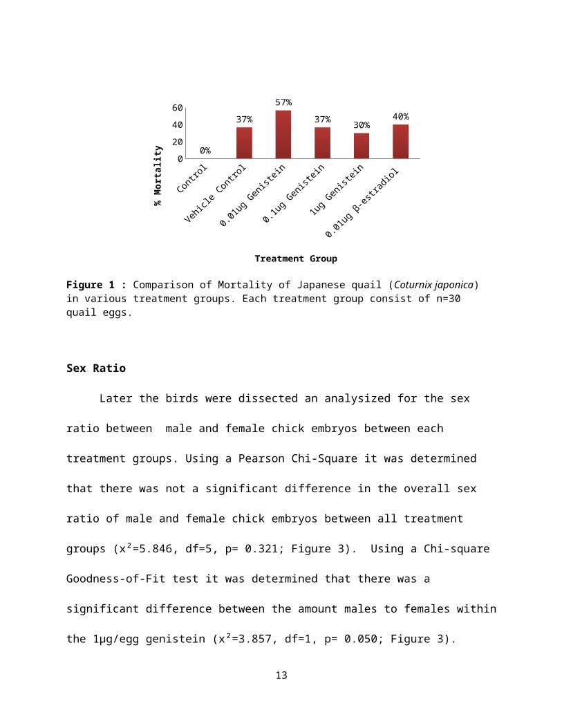
Results
Total hatchability of the fertilized eggs was 67%. The
percent mortality was analyzed between all treatment groups.
Percent mortalities consisted of undeveloped quail eggs. Figure 1
demonstrates that these mortality rates are relatively low and
are well within the normal range of mortality in vehicle control
embryos observed in our laboratory.
12
0204060
0%
37%57%
37% 30%40%
Treatment Group
% Mo
rtal
ity
Figure 1 : Comparison of Mortality of Japanese quail (Coturnix japonica) in various treatment groups. Each treatment group consist of n=30 quail eggs.
Sex Ratio
Later the birds were dissected an analysized for the sex
ratio between male and female chick embryos between each
treatment groups. Using a Pearson Chi-Square it was determined
that there was not a significant difference in the overall sex
ratio of male and female chick embryos between all treatment
groups (x²=5.846, df=5, p= 0.321; Figure 3). Using a Chi-square
Goodness-of-Fit test it was determined that there was a
significant difference between the amount males to females within
the 1µg/egg genistein (x²=3.857, df=1, p= 0.050; Figure 3).
13
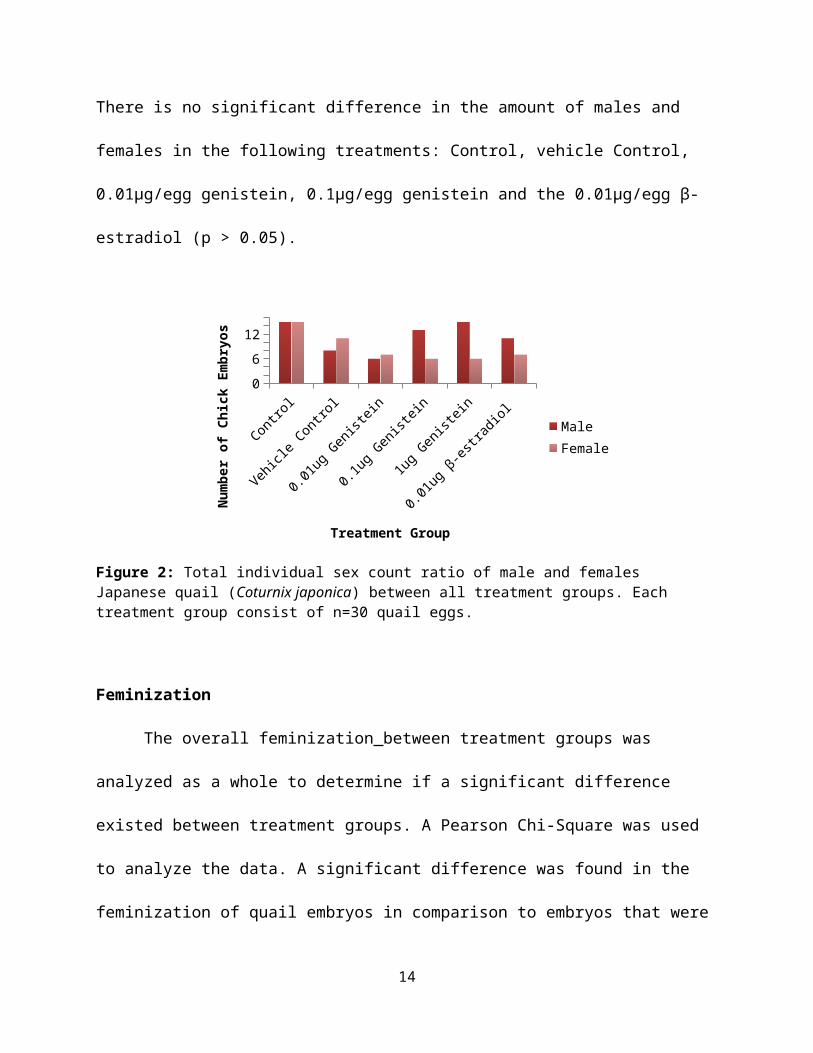
There is no significant difference in the amount of males and
females in the following treatments: Control, vehicle Control,
0.01µg/egg genistein, 0.1µg/egg genistein and the 0.01µg/egg β-
estradiol (p > 0.05).
Control
Vehicle Control
0.01ug Genistein
0.1ug Genistein
1ug Genistein
0.01ug β-estradiol
0612
MaleFemale
Treatment Group
Number of Chick Embryos
Figure 2: Total individual sex count ratio of male and females Japanese quail (Coturnix japonica) between all treatment groups. Each treatment group consist of n=30 quail eggs.
Feminization
The overall feminization between treatment groups was
analyzed as a whole to determine if a significant difference
existed between treatment groups. A Pearson Chi-Square was used
to analyze the data. A significant difference was found in the
feminization of quail embryos in comparison to embryos that were
14
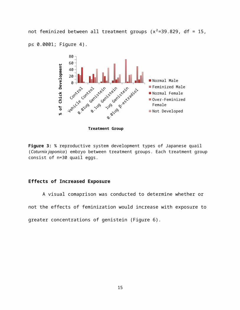
not feminized between all treatment groups (x²=39.829, df = 15,
p≤ 0.0001; Figure 4).
020406080
Normal MaleFeminized MaleNormal FemaleOver-Feminized FemaleNot Developed
Treatment Group
% of
Chi
ck D
evel
opme
nt
Figure 3: % reproductive system development types of Japanese quail (Coturnix japonica) embryo between treatment groups. Each treatment group consist of n=30 quail eggs.
Effects of Increased Exposure
A visual comaprison was conducted to determine whether or
not the effects of feminization would increase with exposure to
greater concentrations of genistein (Figure 6).
15
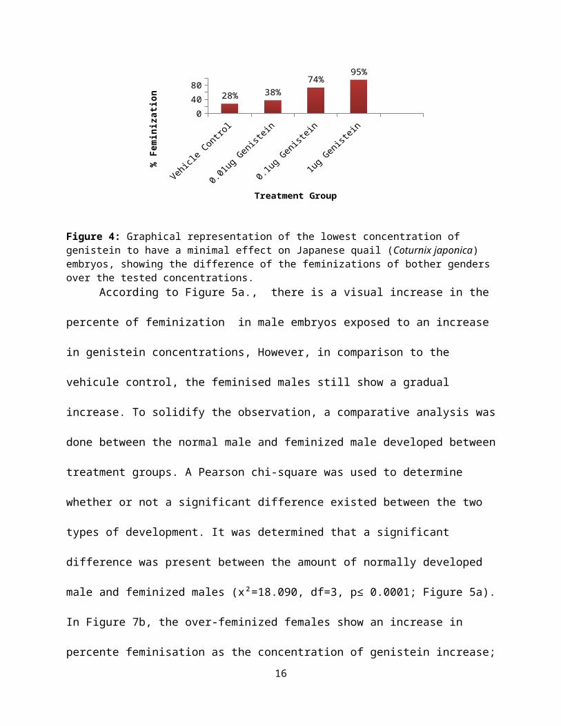
04080
28% 38%74%
95%
Treatment Group
% Fe
mini
zati
on
Figure 4: Graphical representation of the lowest concentration of genistein to have a minimal effect on Japanese quail (Coturnix japonica) embryos, showing the difference of the feminizations of bother gendersover the tested concentrations.
According to Figure 5a., there is a visual increase in the
percente of feminization in male embryos exposed to an increase
in genistein concentrations, However, in comparison to the
vehicule control, the feminised males still show a gradual
increase. To solidify the observation, a comparative analysis was
done between the normal male and feminized male developed between
treatment groups. A Pearson chi-square was used to determine
whether or not a significant difference existed between the two
types of development. It was determined that a significant
difference was present between the amount of normally developed
male and feminized males (x²=18.090, df=3, p≤ 0.0001; Figure 5a).
In Figure 7b, the over-feminized females show an increase in
percente feminisation as the concentration of genistein increase;16

however, in comparison to the vehicle control, the feminization
of female quails does not show a significant increase in the
lower concentration of genistein, but shows an increase at the
highest concentration. A second comparitive analysis was
conducted on the development of normal females and over-feminized
females between all treatment groups. No significant difference
was found between the two development types (x²=7.615, df=3, p=
0.055; Figure 5b). A significant difference was found in the
degree of feminization of quail embryos in correlation to
increased concentrations of Genistein found in both male and
female genders (x²=19.672 df=10, p=0.033).
0
20
40
60
80
Normal MaleFeminized Male
Treatment Group
% of
Qua
il D
evel
opme
nt
01020304050
Normal FemaleOver-Feminized Female
Tireatment Group
% of
Qua
il D
evel
opme
nt
Figure 5: A) Visual comparison of the % of normal male developed and %of feminized males of Japanese quail (Coturnix japonica) exposed to all treatment groups. B) Visual comparison of the % of normal female
17

developed and % of over-feminized females exposed to all treatment groups.
Minimum Concentration of Genistein Causing Feminization
A visual comparison between the normal development and
feminization that occurred in each test group can be seen in
Figure 3, from which the following pair-wise comparisons using a
Pearson chi-square were run. Statistically, there was no
significant difference between the control group and the vehicle
control group (x²=3.402 , df=3, p=0.334; Figure 6a, b). There was
no statistical difference between the vehicle control group and
0.01µg/egg genistein group (x²=5.201 , df=3, p=0.158; Figure 6b,
c). Yet there was a significant difference between the vehicle
control group and 0.1µg/egg genistein group (x²=10.503 , df=3,
p=0.015; Figure 6b, d). The vehicle control group to the 1µg/egg
genistein also had a significant difference (x²=21.840, df=3,
p≤0.0001; Figure 6b, e). ). Between the vehicle control and
0.01µg/egg β-estradiol group there was a significant difference
(x²=8.850 , df=3, p=0.031; Figure 6c, f). In comparing the
0.01µg/egg β-estradiol group to the various concentrations of
genistein it was determined that there was no difference
18

(x²=0.549 , df=3, p=0.908; Figure 6c, d, e, f). The lowest
concentration to have significant feminization is the 0.1µg/egg
genistein group shows and about the same feminizing effect as
0.01ug/egg β-estradiol group. Meaning that genistein has 1/10th
the effect of estradiol.
19

Normal Males
Feminized Males
Normal Females
Feminized Females
0612
Type of Developement
Number of Chick
Embryos
A)
Normal Males
Feminized Males
Normal Females
Feminized Females
048
Type of Developement
Number of Chick
Embryos
B)
Normal Males
Feminized Males
Normal Females
Feminized Females
036
Type of Development
Number of Chic
k Embryos
C)
048
12
Type of Development
Numb
er o
f Ch
ick
Embr
yos
D)
Normal Males
Feminized Males
Normal Females
Feminized Females
0612
Type of Development
Number of Chick
Embryos
E)
Normal Males
Feminized Males
Normal Females
Feminized Females
048
Type of Development
Number of Chick
Embryos
F)
Figure 6: Distribution of Japanese quail (Coturnix japonica) embryos reproductive development between treatment groups. a) Control, b) Vehicle Control, c) 0.01 µg/egg Genistein l, d) 0.1 µg/egg Genistein, e) 1 µg/egg Genistein, f) 0.01 µg/egg β-estradiol.Other Effects of Genistein
20
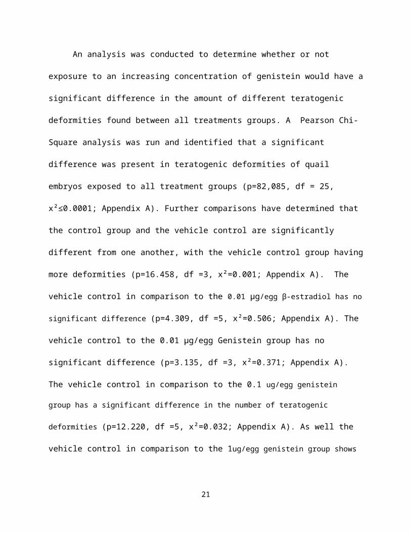
An analysis was conducted to determine whether or not
exposure to an increasing concentration of genistein would have a
significant difference in the amount of different teratogenic
deformities found between all treatments groups. A Pearson Chi-
Square analysis was run and identified that a significant
difference was present in teratogenic deformities of quail
embryos exposed to all treatment groups (p=82,085, df = 25,
x²≤0.0001; Appendix A). Further comparisons have determined that
the control group and the vehicle control are significantly
different from one another, with the vehicle control group having
more deformities (p=16.458, df =3, x²=0.001; Appendix A). The
vehicle control in comparison to the 0.01 µg/egg β-estradiol has no
significant difference (p=4.309, df =5, x²=0.506; Appendix A). The
vehicle control to the 0.01 µg/egg Genistein group has no
significant difference (p=3.135, df =3, x²=0.371; Appendix A).
The vehicle control in comparison to the 0.1 ug/egg genistein
group has a significant difference in the number of teratogenic
deformities (p=12.220, df =5, x²=0.032; Appendix A). As well the
vehicle control in comparison to the 1ug/egg genistein group shows
21

a significant difference in teratogenic deformities (p=22.289, df =5,
x²≤0.0001; Appendix A).
The developed quails were submitted to a mean weight
comparison to determine whether or not different exposure to
treatments would have a significant effect on the body weight of
the Japanese quail embryos. Using a one way ANOVA it was
determined that there was a significant difference in the
bodyweight of the Japanese quail embryos between all treatment
groups (F=5.308, df =5, p ≤ 0.0001; Figure 7). The post-hoc
analysis used by Tukey HSD, showed 1.32 times higher embryo
bodyweight of the control in comparison to 0.01 µg/egg β-
estradiol (p≤0.0001), and 1.26 times more than 0.01ug/egg
Genistein (p=0.008), as well as 1.25 times more than 0.1ug
Genistein (p=0.006) however, there was no difference in
bodyweight between the control and the vehicle control group
(p=0.051), as well as the vehicle control and the treated birds.
22

4.5
5.5
6.5
Treatment Group
Mean
Wei
ght
(g)
Figure 7: Comparison of the mean weight (g) of the developed Japanese quail (Coturnix japonica) embryos after being treated with test solutions.Each treatment group consist of n=30 quail eggs.
Discussion
Sex ratio
For birds, the male is the non-hormonal sex and the presence
of estrogen results in the differentiation of sexual dimorphism
(Lyons 2007). Therefore, estrogen is most important for sex
differentiation in female birds and, without estrogen, female
embryos will develop male sex characteristics (Berg et al. 1998).
This study concluded that there was no difference in the sex
ratio of male and female quail embryos between all treatments
23
groups. However, a dose dependant trend is observed, where higher
production of males versus female quail is noted at
concentrations of 0.1 µg/egg genistein, 1 µg/egg genistein and
0.01 µg/egg β-estradiol. At the 1 µg/egg genistein a significant
difference was found, where there was a greater number of males
than female quails. The increased proportions of male and
intersex individuals in this study could have resulted from the
dual role of genistein as not only an estrogen agonist but also
as an antagonist blocking estrogens action (Green and Kelly
2009). It is possible that the dietary genistein used in our
study resulted in greater numbers of phenotypic males due to the
compound’s weak estrogenic properties, blocking of estrogen
receptors, and inhibition of aromatase and the resulting
decreases in estradiol synthesis (Green and Kelly 2009). This
then suggest that genistein at the tested concentrations in this
study does not act as a significant estrogen mimic, where gender
differentiation would have had a higher frequency of normally
developed females against male development.
Feminization
24
Phytoestrogens are secondary plant compounds, which can act
to mimic estrogen and cause the disruption of estrogenic
responses in organisms (Rochester and Millam 2009) and
phytoestrogens exposure during development may alter adult
reproduction (Millam et al. 2002). The results confirm that males
and females of Japanese quails show feminized or over-feminized
traits in the presence of genistein. Confirming that genistein
can act as an endocrine disrupter causing feminization. which can
lead to behviroural and reproduction difficulties, which may vary
across species. Importantly, disturbances in normal behaviour may
influence the individual fitness and, therefore, assume a real
biological significance in both animal and human ecosystems
(Panzica et al. 2007). Ecologically, plants high in phytoestrogens
have the ability to organizationally disrupt reproductive
endpoints in Japanese quail chicks, because constitute a large
part of the diet of quail and other granivorous wild birds. It is
probable that birds are encountering the levels tested in this
study of estrogenic plant containing genistein in the wild
(Rochester and Millam 2009). According to Rochester and Millam
(2009) showed the effect of seasonal temperatures, such as
25

drought and rainfall would reduce or increase the genistein
concentration produce in these phytoestrogenic plants that would
be regularly be part of Japanese quail wild diet. They then go to
suggest that these plants use phytoestrogens as a chemical
defence against herbivory, which therefore controls the quail and
other wild birds population by effecting their reproduction
endpoints by feminization, behavioural, deformities or just
reduce their fitness as a whole (Rochester and Millam 2009).
Effects of Increased Exposure
Upon further analysis a significant difference was
determined in the amount of normal males in comparison to
feminized males. The normal males regressed as concentration of
genistein became higher, while the amount of feminization
increased. Normal female to over-feminized female had no
statistical difference however, at the highest concentration of
genistein, the effect of feminization are similar with the β-
estradiol group. As one both genders demonstrates an exponential
trends of increased feminization with higher concentration of
genistein. Concentrations of 0.1 µg/egg of genistein yielded
26

similar percentage of overall feminization for both genders than
did 0.01µg/egg β-estradiol, a natural estrogen. Meaning that
genistein is still behaving like a weak form of β-estradiol in
promoting feminizing effects. This study can support the
statement of Herbst and Bern (1981), exposure to estrogenic
substances during critical periods of development can have
adverse consequences on differentiating reproductive systems of
many animals. In correlation to increased concentrations of
genistein, a significant difference was found in the degree of
feminization of quail embryos. More pronounced feminization was
observed as concentration of genistein increase. This tells us
that higher levels of genistein exposure will result in more
harmful endocrine disrupting effects, leading to multiple issues
concerning animal and human health if consumption and
environmental exposure continues to rise through environmental
waste, animal feeds, or other soy products available as the
markets for soy products continue to expand.
Minimum Concentration of Genistein Causing Feminization
27

The developmental trends for both the control group and
vehicle control group typically displayed normal embryo
development. There was no significant difference between them,
which suggests that the vehicle control worked properly. The
occurrence of a multiple feminized chick in both the control
group and vehicle control group are likely a result of avian
farming practices, where there is inherent inbreeding from a
limited gene pool. Such an occurrence was also seen in a study by
Berg et al. (1998) where there was the occurrence of some
feminization in the control group. The 0.01µg/egg β-estradiol
test group was significantly different to the vehicle control
group. It acted primarily as a feminization control to have
something to compare the genistein treatment groups to in terms
of their developed feminized reproductive anatomy. The feminized
development amongst all three genistein treatment groups appeared
to impact both sexes, where few studies have shown the minimal
amount of phytoestrogens needed to produce estrogenic effects.
The lowest concentration 0.01 µg/egg of genistein yielded no
significant results. Exposure to this concentration would be
biologically significant but would not interfere ecologically
28
with reproduction and breeding. Concentration of 0.1 µg/egg and 1
µg/egg of genistein both showed a significant difference in their
feminization of the quail embryos. Lin et al. (2004) showed that
dietary genistein is transferred from the mother to the egg and
accumulates in the yolk. The highest amount of genistein in the
egg yolk obtained in the study was less than 3 µg /egg yolk,
which is low as compared to the amount of supplementation (100
mg/day). This accumulation of genistein within the egg yolk
exceeds the concentration of observed effect for feminization
found in this study. This implies dangers of releasing
genistein into the environment since it could easily be ingested
by birds such as quail and transferred to their embryos, causing
detrimental effects in wild populations.
Other Effects of Genistein
Mortality and body weight mesurements of quail embryos were
not aspects of this study set forth in the objectives. However,
the percent mortality and body weight of quail of every treatment
group were gathered. As expected the control group had no
mortalities and higher mortalities were expected in those groups
29
that received in ovo treatments since they had a hole drilled
into their air cell and a foreign substance introduced to them.
Mortality percentage between all treatments groups remained
practically level with the vehicle control. Thus, identifying
that the major cause of quail mortality while in embryonic
development was the process of which a hole was drilled into the
air cell of the treated eggs.
Exposure to genistein also demonstrated an increase of
teratogenic deformities and a decrease in normal development.
This implies breeding difficulties towards deformed quail, when
time comes for courtship. Courting patterns in Japanese quail
for male birds consiste at least five components: 1) they would
have an increased neck and body tonus, where the posterior is
elevated and the neck is thrust forward and slightly downward.
The male circles the female with head cocked inward toward the
hen. 2) Leg action: the legs straighten and stiffen with the
body being brought up and forward. The leg is stiff during the
strut. 3) Toe walking: the
Bird raises itself on its toes beyond a normal stance and struts
about the female. 4) Vocalization: the courting call is a
30
subdued, hoarse, vibrating call given by the male only when
courting. It is a two-syllable squawk sound which lasts for
several seconds. 5) Feather puffing: most of the body feathers
from the neck downward are fluffed (Farris 1967). With the
occurrences of deformities, males quail will greatly reduce their
individual fitness which is associated in the proper display of
the courting patterns which may not be successful.
To further inspect the treatment protocol the body weight of
the chick embryos were dissected and weighed 2 Days before their
anticipated hatching, yielding a significant difference between
all treatment groups. Further analysis of the trend found in
figure 7 demonstrates what was expected throughout the treatment
groups with a decrease in body weight in treatments where holes
were drilled in the air cell and injected with corn oil., as well
notice that there is a slight increase in body weight at1 µg/egg
of genistein and a decrease in the 0.01µg/egg β-estradiol test
group. This shows us that the 0.01µg/egg β-estradiol group adds
to the effect of drilled hole and an injection of corn oil in the
eggs. However the slight increase in body in the 1 µg/egg of
genistein group can be explained by perhaps laboratory error.
31

We can conclude that corn oil used as a vehicle for genistein did
not increase or reduce the body weight of the quail embryos
rather that the drilling of a hole in the air cell was the
difference in the weight and the increase of mortality in
comparisons to the control group.
Conclusion
Naturally occurring compounds such as genistein is the
isoflavones found in soybeans and soy products and act as weak
estrogens (Warning & Harris 2005). Thus, the impact of genistein
as an endocrine disrupter will vary depending upon a variety of
factors, including the doses that subjects are exposed to, when
in the life cycle of an organism exposure occurs, as well as the
duration of the exposure. Particularly the developmental stages
are typically more vulnerable to disruption than adult stages and
the consequences of foetal exposure may be drastically different
from those of adult exposure (Panzica 2009). Compounds with
endocrine disrupting effect such as genistein can affect any
species and can potentially affect human beings (Warning & Harris
2005).Genistein has been proposed as being protective against
32

breast cancer and peri-menopausal symptoms with many other
benefits. However, they may not necessarily be beneficial to
humans. Consequently, their ingestion and absorption could cause
an increase in local estrogen levels. Where most breast cancers
are, at least initially, estrogen dependent and a surge in levels
would be expected to promote growth (Warning & Harris 2005).
What this study has pursued is the need for tests involving the
effects of environmental relevant genistein whether found through
natural portals or agricultural. Many previous studies have
shown that dietary genistein can be accumulated in areas such as
eggs, mammary milk and other areas in which would be sensitive to
endocrine disrupting effects at early stages of gonadal
development in many different species. This study investigated
the need to find the minimal level at which such a compound would
be able to have a significant endocrine disrupting effect that
would accumulate in Japanese quail eggs in the environment or in
captivity from their exposure to phytoestrogens rich foods.
Future studies, might look at possibly hatching the eggs, to
let the reproductive system developed further, which would permit
33
easier dissections and identification of feminized traits. Having
an assistant identify the reproductive system of quail embryos
while being unaware with which treatment they have been exposed
to and compare results from observation made when knowing which
embryo were exposed to specific treatment. Examining the
behaviour of hatchlings would also give greater insight on the
effect of phytoestrogens, such as genistein in quails. A further
problem is that we do not know whether mixtures of different
endocrine disrupters (the so-called ‘cocktail effect’) are
synergistic and possibly causing further effect of those exposed
at critical stages of development (Warning & Harris 2005). As
well because morphological sex differentiation is a critical
period in early development and is clearly sensitive to the
dietary genistein concentrations (Green and Kelly 2009) future
studies should look in to determining the minimal concentration
of dietary genistein that would produce the concentration of
observed effect found in this study.
Our studies and others have shown that (1) plant-produced
estrogenic chemicals can disrupt reproductive endpoints in birds;
34

(2) dietary (and possibly environmental) levels of phytoestrogens
have disruptive effects on avian reproduction; These facts,
combined with the estrogenic effects documented in our study,
suggest that we can rule out the hypothesis that male-female
groups of Japanese quail eggs will be skewed from 50:50; However,
we can confirm that males and females of Japanese quails will
show feminized or over-feminized traits in the presence of
genistein and that higher concentrations of genistein will result
in more pronounced feminization. Genistein is a highly effective
estrogen mimic, due to the fact that it can cause endocrine
disrupting effects at such low environmental concentrations. But
can also act as an antagonist blocking estrogen action.
Therefore, plant phytoestrogens can exert reproductive effect on
wildlife, especially on developing vertebrates.
Consequently, we can concluded that estrogen mimicking
compounds such as genistein can confirm its effects of as an
endocrine disrupter, causing feminization and that higher
concentrations of this compound will result in a higher frequency
and more pronounced feminization of quail embryos.
35

Literature Cited
36
Abdelnabi, M. A., Bakst, M. R., Woods, J. E., & Ottinger, M. A. (2000). Plasma 17 beta-estradiol levels and ovarian interstitial cell structure in embryonic japanese quail. PoultryScience, 79(4), 564-567.
Benoit J. (1926). Histological study of the right gonad which becomes a testicle in ovariotomized chick. Compt Rend Acad Sci (Paris), (182), 240-3.
Berg, C., Halldin, K., & Brunstrom, B. (2001). Effects of bisphenol A and tetrabromobisphenol A on sex organ development in quail and chicken embryos. Environmental Toxicology and Chemistry, 20(12), 2836-2840.
Berg, C., Holm, L., Brandt, I., & Brunstrom, B. (2001). Anatomical and histological changes in the oviducts of japanese quail, coturnix japonica, after embryonic exposure to ethynyloestradiol. Reproduction, 121(1), 155-165.
Berg, C., Halldin, K., Brunstrom, B., & Brandt, I. (1998). Methods for studying xenoestrogenic effects in birds. Toxicology Letters (Shannon), 102-103(0), 671-676.
Berg, C., Halldin, K., Fridolfsson, A., Brandt, I., & Brunström, B. (1999). The avian egg as a test system for endocrine disrupters: Effects of diethylstilbestrol and ethynylestradiol on sex organ develo. The Science of the Total Environment, 233(1-3), 57-66. doi:DOI: 10.1016/S0048-9697(99)00179-5
Berry, W. D., Zhang, X., & McDaniel, G. R. (1999). Estrogenic effects of the phytoestrogens genistein and daidzein. Poultry Science, (78), 113.
Bramlett, K. S., Wu, Y. F., & Burris, T. P. (2001). Ligands specify coactivator nuclear receptor (NR) box affinity for estrogen receptor subtypes. Molecular Endocrinology, 15(6), 909-922.
Burnison, B. K., Hartmann, A., Lister, A., Servos, M. R., Ternes,T., & Van Der Kraak, G. (2003). A toxicity identification
37
evaluation approach to studying estrogenic substances in hog manure and agricultural runoff. Environmental Toxicology and Chemistry, 22(10), 2243-2250.
Chapman, N. (1998.). Where is the soyfood market headed? The 3rd Annual Soyfoods Symposium Proceeding;Kentucky Soybean Board: Louisville, KY,.
Close, B., Banister, K., Baumans, V., Bernoth, E. M., Bromage, N., Bunyan, J., . . . Warwick, C. (1997). Recommendations foreuthanasia of experimental animals .2. Laboratory Animals, 31(1), 1-32.
Colborn, T., Saal, F. S. V., & Soto, A. M. (1993). Developmental effects of endocrine-disrupting chemicals in wildlife and humans. Environmental Health Perspectives, 101(5), 378-384.
Dixon, R. A., & Ferreira, D. (2002). Genistein. Phytochemistry, 60(3), 205-211. doi:DOI: 10.1016/S0031-9422(02)00116-4
EDSTAC. (1998). Endocrine disruptor screening and testing advisory committee (EDSTAC). (Final Report Washington, DC.: In Environmental Protection Agency.
Elbrecht, A., & Smith, R. G. (1992). Aromatase enzyme-activity and sex determination in chickens. Science, 255(5043), 467-470.
Farris, H. E. (1967). Classical conditioning of courting behaviorin japanese quail coturnix coturnix japonica. Journal of the Experimental Analysis of Behavior, 10(2), 213-&.
Flynn, K. M., Ferguson, S. A., Delclos, K. B., & Newbold, R. R. (2000). Effects of genistein exposure on sexually dimorphic behaviors in rats. Toxicological Sciences, 55(2), 311-319.
Green, C. C., & Kelly, A. M. (2009). Effects of the estrogen mimic genistein as a dietary component on sex differentiationand ethoxyresorufin-O-deethylase (EROD) activity in channel catfish (ictalurus punctatus). Fish Physiology and Biochemistry, 35(3),377-384. doi:10.1007/s10695-008-9260-z ER
38

Gwynne Lyons. (2007). Congenital defects or adverse developmentaleffects in vertebrate wildlife: The wildlife-human connection. Environmental Science and Technology Library, 23(1), 37-87. doi:DOI: 10.1007/1-4020-4831-9_2
Halldin, K., Axelsson, J., & Brunström, B. (2005). Effects of endocrine modulators on sexual differentiation and reproductive function in male japanese quail. Brain Research Bulletin, 65(3), 211-218. doi:DOI: 10.1016/j.brainresbull.2004.11.020
Heinz, G. H., Hoffman, D. J., Kondrad, S. L., & Erwin, C. A. (2006). Factors affecting the toxicity of methylmercury injected into eggs. Archives of Environmental Contamination and Toxicology, 50(2), 264-279. doi:10.1007/s00244-005-1002-y
Herbst, A. L., & Bern, H. A. (1981). Developmental effects of diethylstilbestrol (DES) in pregnancy. In (pp. 203 p.). (New York): Thieme-Stratton.
Kamata, R., Takahashi, S., Shimizu, A., Morita, M., & Shiraishi, F. (2006). In ovo exposure quail assay for risk assessment ofendocrine disrupting chemicals. Archives of Toxicology, 80(12), 857-867. doi:10.1007/s00204-006-0113-1 ER
Kamata, R., Takahashi, S., Shimizu, A., Morita, M., & Shiraishi, F. (2006). In ovo exposure quail assay for risk assessment ofendocrine disrupting chemicals. Archives of Toxicology, 80(12) doi:10.1007/s00204-006-0113-1 ER
Kiparissis, Y., Balch, G. C., Metcalfe, T. L., & Metcalfe, C. D. (2003). Effects of the isoflavones genistein and equol on thegonadal development of japanese medaka (oryzias latipes). Environmental Health Perspectives, 111(9), 1158-1163. doi:10.1289/ehp.5928 ER
Leopold, A. S., Erwin, M., Oh, J., & Browning, B. (1976). Phytoestrogens - adverse effects on reproduction in california quail. Science, 191(4222), 98-100.
39
Lin, F., Wu, J., Abdelnabi, M. A., Ottinger, M. A., & Giusti, M. M. (2004). Effects of dose and glycosylation on the transfer of genistein into the eggs of the japanese quail (coturnix japonica). Journal of Agricultural and Food Chemistry, 52(8), 2397-2403. doi:10.1021/jf034921f
Lin, F., Wu, J., Abdelnabi, M. A., Ottinger, M. A., & Giusti, M. M. (2004). Effects of dose and glycosylation on the transfer of genistein into the eggs of the japanese quail (coturnix japonica). Journal of Agricultural and Food Chemistry, 52(8), 2397-2403. doi:10.1021/jf034921f
Mattsson, A., Mura, E., Brunstrom, B., Panzica, G., & Halldin, K.(2008). Selective activation of estrogen receptor alpha in japanese quail embryos affects reproductive organ differentiation but not the male sexual behavior or the parvocellular vasotocin system. General and Comparative Endocrinology, 159(2-3), 150-157. doi:10.1016/j.ygcen.2008.08.012ER
Millam, J. R., Craig-Veit, C. B., Batchelder, M. E., Viant, M. R., Herbeck, T. M., & Woods, L. W. (2002). An avian bioassay for environmental estrogens: The growth response of zebra finch (taeniopygia guttata) chick oviduct to oral estrogens. Environmental Toxicology and Chemistry, 21(12), 2663-2668.
Millam, J. R., Craig-Veit, C., Batchelder, M. E., Viant, M. R., Herbeck, T. M., & Woods, L. W. (2002). An avian bioassay for environmental estrogens: The growth response of zebra finch (taeniopygia guttata) chick oviduct to oral estrogens. Environmental Toxicology & Chemistry, 21(12), 2663-2668.
Mura, E., Barale, C., Quinn, M. J., Panzica, G., Ottinger, M. A.,& Viglietti-Panzica, C. (2009). Organizational effects of DDEon brain vasotocin system in male japanese quail. Neurotoxicology, 30(3), 479-484. doi:10.1016/j.neuro.2009.01.012
Panzica, G. C., & Melcangi, R. C. (2008). The endocrine nervous system: Source and target for neuroactive steroids. Brain
40

Research Reviews, 57(2), 271-276. doi:10.1016/j.brainresrev.2008.02.002 ER
Panzica, G. C., Mura, E., Miceli, D., Martini, M. A., Gotti, S., & Viglietti-Panzica, C. (2009). Effects of xenoestrogens on the differentiation of behaviorally relevant neural circuits in higher vertebrates. Trends in Comparative Endocrinology and Neurobiology, 1163, 271-278. doi:10.1111/j.1749-6632.2008.03628.x
Panzica, G. C., Viglietti-Panzica, C., Mura, E., Quinn, M. J., Lavoie, E., Palanza, P., & Ottinger, M. A. (2007). Effects ofxenoestrogens on the differentiation of behaviorally-relevantneural circuits. Frontiers in Neuroendocrinology, 28, 179-200. doi:10.1016/j.yfrne.2007.07.001 ER
Rochester, J. R., Klasing, K. C., Stevenson, L., Denison, M. S., Berry, W., & Millam, J. R. (2009). Dietary red clover (trifolium pratense) induces oviduct growth and decreases ovary and testes growth in japanese quail chicks. Reproductive Toxicology, 27(1), 63-71. doi:10.1016/j.reprotox.2008.11.056
Rochester, J. R., & Millam, J. R. (2009). Phytoestrogens and avian reproduction: Exploring the evolution and function of phytoestrogens and possible role of plant compounds in the breeding ecology of wild birds. Comparative Biochemistry & Physiology Part A: Molecular & Integrative Physiology, 154(3), 279-288. doi:10.1016/j.cbpa.2009.06.017
Rochester, J. R., & Millam, J. R. (2009). Phytoestrogens and avian reproduction: Exploring the evolution and function of phytoestrogens and possible role of plant compounds in the breeding ecology of wild birds. Comparative Biochemistry and Physiology, Part A, 154(3), 279-288. doi:10.1016/j.cbpa.2009.06.017
Romanoff AL. (1960.). The avian embryo. . New York, NY, USA.: Macmillan.
Tokita, M. (2003). The skull development of parrots with special reference to the emergence of a morphologically unique cranio-facial hinge. Zoological Science, 20(6), 749-758.
41

University of Illinois at Urbana-Champaign. (1998). Chickscope 1.5: Resources: From egg to chick. Retrieved 3/24/2011, 2011, from http://chickscope.beckman.uiuc.edu/resources/egg_to_chick/procedures.html
Utsumi, T., & Yoshimura, Y. (2009). Sensitive embryonic endpointswith in ovo treatment for detecting androgenic and anti-androgenic effects of chemicals in japanese quail (coturnix japonica). Poultry Science, 88(5), 1052-1059. doi:10.3382/ps.2008-00326
Waring, R. H., & Harris, R. M. (2005). Endocrine disrupters: A human risk? Molecular and Cellular Endocrinology, 244(1-2), 2-9. doi:DOI: 10.1016/j.mce.2005.02.007
Appendix A
42
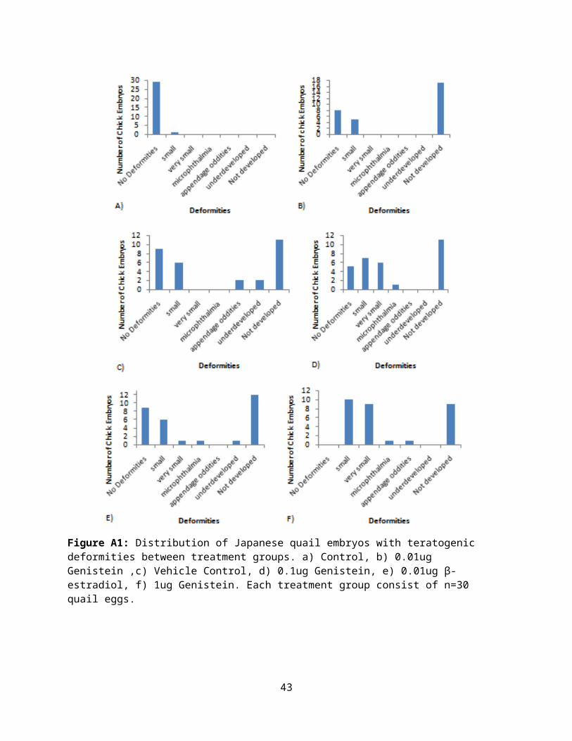
Figure A1: Distribution of Japanese quail embryos with teratogenic deformities between treatment groups. a) Control, b) 0.01ug Genistein ,c) Vehicle Control, d) 0.1ug Genistein, e) 0.01ug β-estradiol, f) 1ug Genistein. Each treatment group consist of n=30 quail eggs.
43

Appendix B
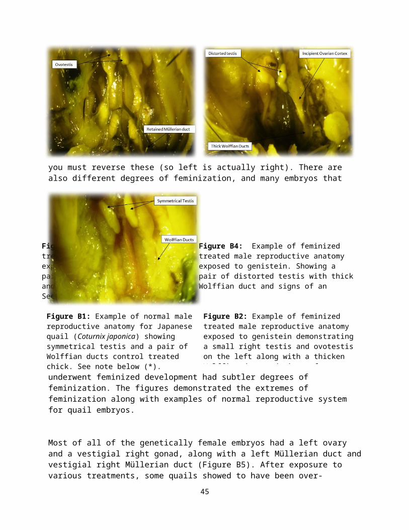
Comparative gonadal observations between normal and feminized embryo development: The appearance of treated and control gonads in genetically male embryos are illustrated in Figures B1, B2, B3, B4. Whereas in untreated and corn oil-treatedembryos, left and right gonads appeared to be symmetrical (FigureB1). After various treatments, feminized chicks showed to have areduced size of the right gonad, while the comparatively much larger left gonad assumed the general appearance of an ovary (ovotestis) (Figure B2). It had a more rugose outline than did the control gonads. Wolffian ducts were usually thicker and
signs of an incipient ovarian cortex could be made out along its lower edge. Some chicks exposed to estradiol and 1 µg/egg genistein treatment was observed to have two swollen testis (ovotestis) and retained Müllerian duct (Figure B3). Higher concentration of genistein also produced deformed testis with ovarian like features, such as thicker Wolffian duct, swollen vasdeferens and distorted and flattened testis (Figure B4).
*Note in interpreting the gonads of reproductive anatomy. The figures are described as if the chick is standing facing away from you. During dissections, the bird is on its back, therefore
44
you must reverse these (so left is actually right). There are also different degrees of feminization, and many embryos that
underwent feminized development had subtler degrees of feminization. The figures demonstrated the extremes of feminization along with examples of normal reproductive system for quail embryos.
Most of all of the genetically female embryos had a left ovary and a vestigial right gonad, along with a left Müllerian duct andvestigial right Müllerian duct (Figure B5). After exposure to various treatments, some quails showed to have been over-
45
Figure B1: Example of normal male reproductive anatomy for Japanese quail (Coturnix japonica) showing symmetrical testis and a pair of Wolffian ducts control treated chick. See note below (*).
Figure B2: Example of feminized treated male reproductive anatomy exposed to genistein demonstratinga small right testis and ovotestison the left along with a thicken Wolffian duct and signs of an
Figure B3: Example of feminized treated male reproductive anatomy exposed to genistein. Showing a pair of swollen testis (ovotestis) and retained left Müllerian duct. See note above (*).
Figure B4: Example of feminized treated male reproductive anatomy exposed to genistein. Showing a pair of distorted testis with thickWolffian duct and signs of an incipient ovarian cortex could be
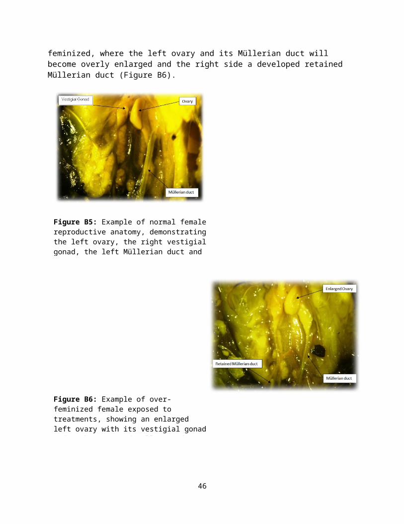
feminized, where the left ovary and its Müllerian duct will become overly enlarged and the right side a developed retained Müllerian duct (Figure B6).
Figure B5: Example of normal femalereproductive anatomy, demonstratingthe left ovary, the right vestigialgonad, the left Müllerian duct and
Figure B6: Example of over-feminized female exposed to treatments, showing an enlarged left ovary with its vestigial gonadand right retained Müllerian duct
46
Copyright © 2022 FDOKUMEN