Fhit tumor suppressor: guardian of the preneoplastic genome
16
Fhit tumor suppressor: guardian of the preneoplastic genome Flavia Pichiorri, Ohio State University Comprehensive Cancer Center, Department of Molecular Virology, Molecular Virology and Medical Genetics. 460 W 12th Avenue, 43210 Columbus, OH, USA Tiziana Palumbo, Ohio State University Comprehensive Cancer Center, Department of Molecular Virology, Molecular Virology and Medical Genetics. 460 W 12th Avenue, 43210 Columbus, OH, USA Sung-Suk Suh, Ohio State University Comprehensive Cancer Center, Department of Molecular Virology, Molecular Virology and Medical Genetics. 460 W 12th Avenue, 43210 Columbus, OH, USA Hiroshi Okamura, Ohio State University Comprehensive Cancer Center, Department of Molecular Virology, Molecular Virology and Medical Genetics. 460 W 12th Avenue, 43210 Columbus, OH, USA Francesco Trapasso, Ohio State University Comprehensive Cancer Center, Department of Molecular Virology, Molecular Virology and Medical Genetics. 460 W 12th Avenue, 43210 Columbus, OH, USA Hideshi Ishii, Department of Gastroenterological Surgery, Osaka University Graduate School of Medicine, Suita, Osaka, 565-087, Japan Kay Huebner, and Ohio State University Comprehensive Cancer Center, Department of Molecular Virology, Molecular Virology and Medical Genetics. 460 W 12th Avenue, 43210 Columbus, OH, USA Carlo M Croce † Director Human Cancer Genetics, Ohio State University, 460 W 12th Avenue, 43210 Columbus, OH, USA Tel.: +1 614 292 4930 Fax: +1 614 292 3558 [email protected] Abstract Environmental agents induce intragenic alterations in the FRA3B/FHIT chromosome fragile site, resulting in fragile FHIT allele loss early in cancer development. Fhit knockout mice are predisposed to tumor development and Fhit gene therapy reduces tumor burden. Repair-deficient cancers are likely to be Fhit-deficient and Fhit-deficient cells show enhanced resistance to ultraviolet C, mitomycin C, camptothecin and oxidative stress-induced cell killing. Loss of Fhit leads to alterations in the DNA damage response checkpoint and contributes to DNA instability. Hsp60/Hsp10 are Fhit interactors, suggesting a direct role for Fhit in stress responses. Fhit also interacts with and stabilizes ferrodoxin reductase (Fdxr), a mitochondrial flavoprotein that © 2008 Future Medicine † Author for correspondence: Director Human Cancer Genetics, Ohio State University, 460 W 12th Avenue, 43210 Columbus, OH, USA Tel.: +1 614 292 4930 Fax: +1 614 292 3558 [email protected]. Financial & competing interests disclosure The authors have no relevant affiliations or financial involvement with any organization or entity with a financial interest in or financial conflict with the subject matter or materials discussed in the manuscript. This includes employment, consultancies, honoraria, stock ownership or options, expert testimony, grants or patents received or pending, or royalties. No writing assistance was utilized in the production of this manuscript. NIH Public Access Author Manuscript Future Oncol. Author manuscript; available in PMC 2012 July 19. Published in final edited form as: Future Oncol. 2008 December ; 4(6): 815–824. doi:10.2217/14796694.4.6.815. NIH-PA Author Manuscript NIH-PA Author Manuscript NIH-PA Author Manuscript
Transcript of Fhit tumor suppressor: guardian of the preneoplastic genome

Fhit tumor suppressor: guardian of the preneoplastic genome
Flavia Pichiorri,Ohio State University Comprehensive Cancer Center, Department of Molecular Virology,Molecular Virology and Medical Genetics. 460 W 12th Avenue, 43210 Columbus, OH, USA
Tiziana Palumbo,Ohio State University Comprehensive Cancer Center, Department of Molecular Virology,Molecular Virology and Medical Genetics. 460 W 12th Avenue, 43210 Columbus, OH, USA
Sung-Suk Suh,Ohio State University Comprehensive Cancer Center, Department of Molecular Virology,Molecular Virology and Medical Genetics. 460 W 12th Avenue, 43210 Columbus, OH, USA
Hiroshi Okamura,Ohio State University Comprehensive Cancer Center, Department of Molecular Virology,Molecular Virology and Medical Genetics. 460 W 12th Avenue, 43210 Columbus, OH, USA
Francesco Trapasso,Ohio State University Comprehensive Cancer Center, Department of Molecular Virology,Molecular Virology and Medical Genetics. 460 W 12th Avenue, 43210 Columbus, OH, USA
Hideshi Ishii,Department of Gastroenterological Surgery, Osaka University Graduate School of Medicine,Suita, Osaka, 565-087, Japan
Kay Huebner, andOhio State University Comprehensive Cancer Center, Department of Molecular Virology,Molecular Virology and Medical Genetics. 460 W 12th Avenue, 43210 Columbus, OH, USA
Carlo M Croce†
Director Human Cancer Genetics, Ohio State University, 460 W 12th Avenue, 43210 Columbus,OH, USA Tel.: +1 614 292 4930 Fax: +1 614 292 3558 [email protected]
AbstractEnvironmental agents induce intragenic alterations in the FRA3B/FHIT chromosome fragile site,resulting in fragile FHIT allele loss early in cancer development. Fhit knockout mice arepredisposed to tumor development and Fhit gene therapy reduces tumor burden. Repair-deficientcancers are likely to be Fhit-deficient and Fhit-deficient cells show enhanced resistance toultraviolet C, mitomycin C, camptothecin and oxidative stress-induced cell killing. Loss of Fhitleads to alterations in the DNA damage response checkpoint and contributes to DNA instability.Hsp60/Hsp10 are Fhit interactors, suggesting a direct role for Fhit in stress responses. Fhit alsointeracts with and stabilizes ferrodoxin reductase (Fdxr), a mitochondrial flavoprotein that
© 2008 Future Medicine†Author for correspondence: Director Human Cancer Genetics, Ohio State University, 460 W 12th Avenue, 43210 Columbus, OH,USA Tel.: +1 614 292 4930 Fax: +1 614 292 3558 [email protected].
Financial & competing interests disclosure The authors have no relevant affiliations or financial involvement with any organizationor entity with a financial interest in or financial conflict with the subject matter or materials discussed in the manuscript. This includesemployment, consultancies, honoraria, stock ownership or options, expert testimony, grants or patents received or pending, orroyalties.No writing assistance was utilized in the production of this manuscript.
NIH Public AccessAuthor ManuscriptFuture Oncol. Author manuscript; available in PMC 2012 July 19.
Published in final edited form as:Future Oncol. 2008 December ; 4(6): 815–824. doi:10.2217/14796694.4.6.815.
NIH
-PA Author Manuscript
NIH
-PA Author Manuscript
NIH
-PA Author Manuscript

transfers electrons from NADPH to cytochrome P450, suggesting a role for Fhit in the modulationof reactive oxygen species production and of genomic damage.
Keywordscarcinogens; DNA instability; ferredoxin reductase; FHIT; HSP; reactive oxygen species; tumorsuppressor gene
Malignant transformation is a multistep process involving numerous genetic changes, whichinclude loss of tumor-suppressor gene function, oncogene activation and alterations ofmodifier genes [1,2]. These genetic changes affect cellular processes such as survival,proliferation and genomic stability. Precancerous cells experience selective pressure toescape from the cell cycle block induced by checkpoint responses to DNA damage, andDNA damage checkpoint genes are frequently mutated to overcome the block and allowneoplastic progression [3,4]. The short arm of human chromosome 3 is a common site ofchromosomal alterations in human malignant disease. In four major regions of 3p (3p25,3p21.3, 3p14.2 and 3p12), allelic losses have been reported in cancers of the kidney, lungand breast, among others [5,6]. The 3p14.2 region is particularly interesting to cancerresearchers because it includes other genetic landmarks, including the most active commonfragile site (the FRA3B locus) [7], a familial kidney cancer-associated breakpoint t(3;8)(p14.2;q24) [8], and a papilloma virus integration site [9]. The familial translocationindicated that a gene important for cancer initiation or progression might be located at3p14.2. In 1996, an intensive search of this genomic region resulted in the identification ofthe fragile histidine triad (FHIT) gene [10,11]. Subsequent studies established that FHIT is atarget of chromosomal rearrangements at 3p14.2 [12]. Loss of Fhit expression is observed inpremalignant lesions of lung, esophagus, cervix and other organs, suggesting that loss ofFhit expression, due to the susceptibility of FHIT/FRA3B to carcinogen damage, plays arole in initial stages of multistep carcinogenesis (Figure 1) [13,14]. Since the FHIT gene isprone to breakage and deletion in precancer or early carcinogenesis, and precancerouslesions and cancers show clonal expansion of cells with specific FHIT gene alterations, itwas proposed that FHIT gene alteration and loss of Fhit function provides a selectiveadvantage for this clonal growth [14]. The abnormal checkpoint responses and genomeinstability of Fhit-deficient cells could clearly contribute to selective growth of precancerouscells with damaged FHIT alleles. For example, carcinogens cause damage at FRA3B,leading to breakage of a FHIT allele with loss of exons 4–6 [13]. Further carcinogenexposure can lead to damage at the second FHIT allele with loss of other exons, 3–5 forexample (illustrated in Figure 1). Loss of the second FHIT allele can lead to total loss of Fhitprotein expression, as observed in many dysplastic lesions (Figure 1). In this review wesummarize new studies describing FHIT alterations in human cancers, from analysis ofFhitdeficient mice to identification of important Fhit biological functions.
FHIT alterations occur in most cancersThe presence of the FHIT gene in the most active common chromosome fragile region hasbeen proposed as an example of a tumor suppressor gene altered by chromosometranslocations and deletions rather than by point mutation; several reports had suggested thatthe FHIT gene was altered in cancer simply because it was in a fragile region and notbecause it had contributed to the clonal expansion [15,16]. If this were the case, it would bedifficult to explain why the FHIT genomic alteration, within all cells of a specific cancer-derived cell line, was identical to the nucleotide. Many cancer cell lines and primary cancersexhibiting hemi- or homo-zygous deletions with end points within the FHIT gene andreduced or absent Fhit expression have been reported [13]. Furthermore, many studies have
Pichiorri et al. Page 2
Future Oncol. Author manuscript; available in PMC 2012 July 19.
NIH
-PA Author Manuscript
NIH
-PA Author Manuscript
NIH
-PA Author Manuscript

reported altered FHIT loci and protein expression in precancerous lesions, suggesting thatFHIT alterations are an early event in carcinogenesis [17]. In esophageal cancer, Mori et al.reported that most of the in situ lesions, 50% of severe and moderate dysplasias and 33% ofmild dysplasias were Fhit negative [18]; in the study of Kitamura et al., reduced Fhitexpression was observed in 68% of in situ and 43.5% of esophageal dysplastic lesions [19].Hao et al. found reduced Fhit expression only in a small fraction of adenomatous colonlesions, but reduced Fhit expression was associated with a greater degree of dysplasia [20].In cervical cancer, Connolly et al. observed reduced or absent Fhit staining in 71% ofinvasive cancers and in 52% of highgrade intraepithelial lesions (HSILs) with invasivecancer [21]. In approximately 85% of bronchial dysphasia there was loss of Fhit expression[22]. In our study of ductal carcinoma in situ (DCIS), reduced Fhit expression was observedin 70% of pure DCIS and 52% of DCIS adjacent-to-invasive tumor cases. In total, 20% ofpure DCIS cases exhibited individual glands of adjacent normal tissue with absence ofexpression [23]. These clinical findings supported the proposal that FHIT inactivationoccurs in the early steps of carcinogenesis in many organs. Several studies have shown thatFHIT alterations are common in environmental carcinogen-related cancers, such as those oflung and esophagus. An association between smoking and loss of Fhit expression or FHITdeletion was shown in lung and esophageal cancers. In fact, alterations in Fhit expression inlung carcinomas were more frequent and occurred earlier than p53 mutations or deregulationof the epidermal growth factor receptor (EGFR) [22]. Interestingly, Fhit inactivation wasalmost twice as frequent in tumors of smokers (75%) than nonsmokers (39%) [22,24]. Thesestudies suggested that loss of Fhit is an early event in the development of lung cancer, andthat predisposing genetic changes have occurred even in normal-appearing epithelium incases heavily exposed to environmental carcinogens. Recent studies point to detection ofFHIT deletions in purified bronchial epithelial cells from sputum as a way to improve earlydetection of lung cancer with 58% sensitivity [25]. Exposure to asbestos and to γ-irradiationduring the Chernobyl accident caused an increase in FHIT inactivation in lung cancer andpreneoplastic bronchial lesions [26,27]. Smoking history and alcohol abuse associated withhigher frequency of loss of Fhit expression was also reported in esophageal cancer [18]. IfFHIT is one of the first targets of carcinogens, the ability of the host to repair this initialdamage or to eliminate cells carrying damage to the FHIT locus may prevent clonalexpansion. In support of this idea, loss of FHIT function is observed more frequently incancers developing in individuals with constitutional alterations to genes involved in DNArepair, such as the BRCA1 and 2 and mismatch repair genes [28–30].
The Fhit-deficient mouse: a model to study the role of Fhit in carcinogen-induced tumors
The mouse Fhit ortholog also encompasses a common fragile site, Fra14A2 on murinechromosome 14, and sustains homozygous deletions in murine cancer cell lines [12];therefore, Fhit-knockout mice have served as models for the study of Fhit function. Toestablish an animal model and to explore the role of Fhit in tumorigenesis, our laboratorydeveloped a mouse strain carrying one or two inactivated Fhit alleles. Fhit+/− and Fhit−/−
mice, although healthy and fertile, showed increased susceptibility to spontaneous andcarcinogen-induced tumors [31,32]. Epidemiological studies have linked exposure tonitrosamines to a high incidence of esophageal cancer [33]; to better understand the role ofFhit in this neoplasia, our laboratory performed a carcinogenesis study with N-nitrosomethyl-benzylamine (NMBA), an environmental nitrosamine [34]. All Fhit+/− micedeveloped several fore stomach tumors, and some developed sebaceous gland tumors aftersix doses of intragastric NMBA, while only 25% of wild-type mice developed tumors. Thetumors were a mixture of benign, in situ and invasive lesions. The NMBA-induced tumorspectrum in Fhit+/− mice was similar to a human syndrome called Muir–Torre syndrome, a
Pichiorri et al. Page 3
Future Oncol. Author manuscript; available in PMC 2012 July 19.
NIH
-PA Author Manuscript
NIH
-PA Author Manuscript
NIH
-PA Author Manuscript

variant of the hereditary nonpolyposis colorectal cancer (HNPCC or Lynch) syndrome,which is caused by inactivation of a mismatch repair gene, usually MSH2. Homozygousdeletions of FHIT were observed in half of the cancer cell lines with a mutant mismatchrepair gene; among nine Msh2-negative human colon cancer cells, eight were negative forFhit, and Fhit loss was reported in 90% of BRCA1- and BRCA2-associated cancers[29,35,36]. These correlations suggest that fragile genes are especially vulnerable todamage-induced alterations in cells with ‘caretaker’ gene deficiencies.
As discussed previously, several studies have observed more frequent FHIT gene deletionsin tumors of smokers than tumors of nonsmokers [14]. Recently D’Agostini et al., to assessthe role of Fhit under controlled experimental conditions, exposed mice to environmentalcigarette smoke (ECS) and evaluated Fhit RNA or protein in the respiratory tract of rodents(Figure 2A) [37]. Confirming previous studies conducted in humans, they found that after 14days of exposure to ECS, there was loss of Fhit protein in bronchial/bronchiolar epitheliumof half of the mice, both wild-type or Fhit+/−. Interestingly, they also found that the oraladministration of N-acetylcysteine (NAC), a well-known antioxidant, attenuated the ECS-related loss of Fhit. Another possible role of Fhit in response to carcinogen wasdemonstrated in the study of Balansky et al., who found that after treatment withbenzo[a]pyrene (B[a]P), a prototypic genotoxic, carcinogenic polycyclic aromatichydrocarbon (PAH), preneoplastic lesions of the uterus were more frequent in Fhit+/− mice[38]. They also found that B6/129 F1 mice underwent spontaneous alopecia areata and hairbulb cell apoptosis, which was greatly accelerated either by Fhit heterozygosity or by B[a]Ptreatment, suggesting that Fhit plays a role in the pathogenesis of alopecia areata.Intriguingly, the oral administration of NAC inhibited occurrence of this inflammatory skindisease. Thus, this thiol compound with anti-inflammatory properties, which can inhibitapoptosis consequent to DNA damage and redox imbalances, also inhibits the stimuli thatcause loss of Fhit protein due to cigarette smoke in the bronchial epithelium of rats, andprevents spontaneous alopecia areata and hair bulb cell apoptosis in Fhit heterozygous mice.Recently, Ishii et al. have shown that in vivo-transplanted, hydroquinone-exposed, Fhit-deficient mouse bone marrow cells escaped the bone marrow suppression exhibited by wild-type bone marrow [39]. After hydroquinone exposure, occurrence of the oxidized base 8-hydroxyguanosine, a marker of DNA damage, was also reduced in Fhit-deficient bonemarrow, as was production of intracellular reactive oxygen species (ROS) (Figure 2B). Also,in this experimental model, treatment with NAC relieved hydroquinone-induced suppressionof colony formation by wild-type hematopoietic cells, suggesting that decreased oxidativedamage to Fhit-deficient cells, relative to wild-type hematopoietic cells, accounts for thesurvival advantage of Fhit-xsdeficient bone marrow. Zanesi et al. reported that 4-methylnitrosamino-1–3-pyridyl-1-butanone induced lung tumors (adenomas andcarcinomas) in 100% of Fhit−/−Vhl+/− mice and adenomas in 40% of Fhit−/− mice by 20months of age [40]. Thus, double deficiency in murine homologues of 3p suppressor genes,including haploinsufficiency of Vhl, predisposes to spontaneous and induced lung cancers,demonstrating that Fhit-deficient mice will be useful, in combination with other 3p tumorsuppressors, in recapitulating a pattern of lung cancer development similar to the humanpattern; such double- or triple-deficient mice will be excellent lung cancer prevention andtherapy models. This summary of effects in several mouse models illustrates the strongcorrelation between FHIT-allele loss and early steps in the neoplastic process. It isespecially interesting that in varied experiments conducted in different laboratories, an anti-inflammatory and ROS scavenging agent such as NAC could inhibit Fhit loss or prevent itsnegative effect.
Pichiorri et al. Page 4
Future Oncol. Author manuscript; available in PMC 2012 July 19.
NIH
-PA Author Manuscript
NIH
-PA Author Manuscript
NIH
-PA Author Manuscript

Fhit function in the stress responseDespite strong evidence of Fhit tumor suppressor function, our knowledge of specific Fhitsignalling pathways and mechanisms involved in its suppressor activity is limited. It is well-known that overexpression of Fhit results in apoptosis in Fhit-deficient cancer cells [41], andrecent studies have demonstrated a role for Fhit in responses to genotoxic damage inducedby ultraviolet C (UVC) light, mitomycin C, camptothecin and ionizing radiation;approximately tenfold more colonies of Fhit-deficient cells survived exposure to high UVCdoses [42,43]. After mitomycin C treatment approximately sixfold, and after UVC treatment3.5-fold more Fhit-positive human cancer cells than Fhit-negative cells had died, and UVC-surviving Fhit−/– cells showed more than fivefold increased mutation frequency.Furthermore, a recent study from our laboratory has shown that p53-negative lung cancercells expressing Fhit are more sensitive to H2O2 treatment compared with Fhit-negative cellsundergoing apoptosis or G2/M arrest when Fhit was present (Figure 2) [44]. After oxidativestress, Fhit-positive cells also produced higher ROS (Figure 2C, D & E) and 8-hydroxyguanosine levels. Fhit-deficent cancer cells show a mild response to oxidative stress,producing less ROS, crucial mediators of chemotherapy-induced cell death, confirming thatFhit deficiency could negatively influence treatment outcome.
It has also been shown that introduction of exogenous Fhit into cells in vitro leads tomodulation of expression of the checkpoint proteins Hus1 and Chk1 at mid-S checkpoint,modulation that led to induction of apoptosis in esophageal cancer cells, but not innoncancerous primary cultures [45]. The results suggested that the DNA-damage-susceptible FRA3B/FHIT chromosome fragile region encodes a protein that is necessary forprotecting cells from accumulation of DNA damage through its role in modulation ofcheckpoint proteins; and the inactivation of Fhit contributes to the accumulation of abnormalcheckpoint phenotypes in cancer development [45,46]. The study showed that when Fhitwas down-modulated in 293 kidney cells, the level of Hus1 and Rad1 proteins was reduced,and experiments further suggested that Fhit protein stabilizes Hus1 protein, preventing itsdegradation by the proteasome pathway.
Together, these studies are consistent with the conclusion that Fhit, as a modifier of stressresponses, can affect stabilization of proteins involved in activation of checkpoints, cell-cycle block or apoptosis.
Fhit protein structureIn contrast to the gene, the ubiquitously expressed FHIT mRNA is only 1.1 kb long andencodes a 146 amino acid chain that forms a protein of 16.8 kD. Fhit is a member of thehistidine triad (HIT) nucleotide-binding protein superfamily, encoding a diadenosinepolyphosphate (ApnA) hydrolase that cleaves substrates such as diadeno sine triphosphate(Ap3A) and diadenosine tetraphosphate (Ap4A) to AMP plus the other nucleo tide [13].Structural studies have shown that Fhit is a dimer that binds two Ap3A substrates, presentinga highly phosphorylated surface, with five phosphate groups and two adenosine moieties[47]. The conserved HIT motif (His-X-His-X-His-X-X, where X is a hydro phobic residue)of Fhit is located near its C-terminal end and the hydrolytic activity of Fhit is lost whenhistidine 96 (H96) is replaced with asparagine (Fhit-H96N), showing that the H96 centralhistidine residue of the triad is essential for Ap3A hydrolase activity [13]; however, theFhitH96N protein suppresses tumorigenicity about as well as wild-type Fhit. This findingled Garrison and colleagues to suggest that the Fhit enzyme–substrate complex might sendthe tumor-suppression signal [49]. Fhit-induced apoptosis in cancer cells was correlated withthe apparent substrate-binding activity (Km) but not the substrate hydrolytic activity (kcat).Recently, it was observed that the sequence DSIY114EEL of Fhit, which fits the consensus
Pichiorri et al. Page 5
Future Oncol. Author manuscript; available in PMC 2012 July 19.
NIH
-PA Author Manuscript
NIH
-PA Author Manuscript
NIH
-PA Author Manuscript

for targets of phosphorylation by Src tyrosine kinase family members, could bephosphorylated in vitro and in vivo [48]. Garrison et al. determined the steadystate Km andkcat values for the Ap3A hydrolase activity of recombinant nonphospho-Fhit, monophospho-Fhit and diphospho-Fhit, and found that the Km and kcat values for monophospho-Fhit anddiphospho-Fhit are lower than for nonphospho-Fhit [49]. Recent studies have shown that aFhit mutant that carries a phenylalanine instead of a tyrosine at position 114 (Y114F), andthus unable to be phosphorylated on tyrosine 114 by Src, does not induce apoptosis incancer cells and prevents Fhit degradation [50,51]. Furthermore, Bianchi et al. have shownthat during the signaling of activated tyrosine kinase receptors after EGF treatment,phosphorylation of Fhit leads to its degradation; the subsequent reduction in Fhit proteinlevel allows transmission of the mitogenic signal [51]. This would suggest a key role forFhit in the balance of proliferation/survival/apoptosis signals, and indicates that Fhitphosphorylation at Y114 may be a key feature of Fhit molecular function.
Identification of Fhit effectors to define Fhit functionOne way to define specific protein signal pathways is through identification of interactingproteins that could be effectors of function. Earlier searches for Fhit-interacting proteinspointed to several candidate proteins, none of which we could confirm as interactors [HUEBNER
K ET AL., OHIO STATE UNIVERSITY, COLUMBUS, OH, USA. UNPUBLISHED DATA] by co-immunoprecipitatonexperiments, including Ubc9, α-tubulin, Mdm2 and β-catenin [52–55]. To readdress thequestion of Fhit protein interactors, and find the molecular basis for the important role ofFhit in cancer prevention, we used adenovirus transduced Fhit-His6 protein for Fhit complexpurification after cross-linking, and Fhit-bound proteins, Hsp60, Hsp10, ferredoxinreductase (Fdxr), malate dehydrogenase (Mdh), electrontransfer flavoprotein (Etfb) andmitochondrial aldehyde dehydrogenase 2 (Adh 2) were identified (Table 1) [44]. Themitochondrial localization of these proteins led to our determination that Fhit localizes tomitochondria, as well as cytosol. We found that Fhit is important for Fdxr stability and thatFhit–Fdxr interaction leads to increased ROS generation through electron leakage from theshuttle system NADPH-cytochrome P450 via ferrodoxin. Using HCT116 cells with one orthree copies of the FDXR gene, we found that cells with only one copy were less susceptibleto Fhit-induced apoptosis and that the level of Fdxr protein was stabilized in the presence ofFhit protein. Furthermore, the finding that Fhit interacts with Hsp ‘stress proteins’ [56], inparticular the chaperone machinery Hsp60/10, suggests that the Hsp complex may beimportant for Fhit stability, correct folding and mitochondrial addressing; it is even possiblethat Fhit itself is part of important stress machinery, able to protect vital proteins such asFdxr from degradation under stress conditions (see model, Figure 3). Intriguingly, Hsp60interacts directly with Fdxr [PICHIORRI F ET AL. OHIO STATE UNIVERSITY, COLUMBUS, OH, USA. UNPUBLISHED
DATA] and we speculate that Hsp60/10 may also mediate the correct folding andmitochondrial import of Fdxr and Mdh [52] and protect them from stress denaturation (seemodel, Figure 3). The finding that Fhit interacts with Fdxr and thereby increases Fdxrstability suggests that Fhit may be part of specific molecular machinery to protect importantproteins from degradation and affecting the cellular response to the damaging effects ofROS.
ConclusionThe FHIT gene was discovered in 1996, and its protein product, Fhit, was shown innumerous studies by laboratories worldwide, to be a tumor suppressor that was reduced inexpression or lost in the majority of cancers. Progress in understanding the Fhit signalpathways was less rapid. Very recently, several pathways affected by Fhit loss, assummarized in this review, have been identified: a stress response pathway; a DNA damageresponse checkpoint pathway; and a role in the production of reactive oxygen species on
Pichiorri et al. Page 6
Future Oncol. Author manuscript; available in PMC 2012 July 19.
NIH
-PA Author Manuscript
NIH
-PA Author Manuscript
NIH
-PA Author Manuscript

exposure to oxidative stress. Participation of Fhit in these pathways suggests reasons whyFhit-deficient cells show increased resistance to certain cytotoxic therapeutic drugs, andsuggest that in normal cells Fhit is involved in the protection of cells from preneoplasticchanges.
Future perspectiveDelineation of direct downstream effectors of the Fhit-suppressor pathway will lead to:
▪ Intensification of mechanistic studies of Fhit function that may influence futurepreventive and therapeutic strategies to activate the Fhit pathway or to specificallytarget Fhit-deficient cancers;
▪ Clarification of mechanisms by which this gene product protects cells fromcarcinogens and modulates sensitivity to external insults.
The finding that ROS generation precedes Fhitmediated apoptosis in lung cancer cells is insatisfying accord with previous reports showing:
▪ That exposure to carcinogens in cigarette smoke was associated with FHIT gene loss;
▪ The importance of Fhit loss as a negative prognostic factor in various clinical settings(for example, assessment of Fhit status in preneoplastic or neoplastic conditions may bepredictive of responses to antioxidant treatments).
Finally, the fact that Fhit interacts with Hsp chaperones and this interaction appears to beimportant for its stability and correct localization in mitochondria, where it can initiateapoptosis through affecting stability of mitochondria respiratory chain proteins, suggests thatdrugs targeted to such chaperones might have efficacy in preneoplastic, neoplastic or otherconditions associated with Fhit loss.
BibliographyPapers of special note have been highlighted as:
▪ of interest
▪▪ of considerable interest
1. Croce CM. Role of chromosome translocations in human neoplasia. Cell. 1987; 49(2):155–156.[PubMed: 3494520] ▪▪ One of the most important reviews explaining the role of chromosometranslocation and how they are related to cancer initiation.
2. Soloman E, Borrow J, Goddart A. Chromosome aberrations and cancer. Science. 1991; 254(5035):1153–1160. [PubMed: 1957167]
3. Bartkova J, Horejsí Z, Koed K, et al. DNA damage response as a candidate anti-cancer barrier inearly human tumorigenesis. Nature. 2005; 434(7035):864–870. [PubMed: 15829956] ▪▪ Reports anelegant study describing how cancer development is associated with DNA replication stress, whichleads to DNA double-strand breaks, including breaks at FRA3B, checkpoint activation and selectivepressure for p53 mutations.
4. Gorgoulis VG, Vassiliou LV, Karakaidos P, et al. Activation of the DNA damage checkpoint andgenomic instability in human precancerous lesions. Nature. 2005; 434(7035):829–830. [PubMed:15829943] ▪▪ Reports an elegant study describing how cancer development is associated with DNAreplication stress, which leads to DNA double-strand breaks, including breaks at FRA3B,checkpoint activation and selective pressure for p53 mutations.
5. Naylor SL, Johnson BE, Minna JD, et al. Loss of heterozygosity of chromosome 3p markers insmall cell lung cancer. Nature. 1987; 329(6138):451–454. [PubMed: 2821400] ▪ One of the firststudies supporting the hypothesis that loss of alleles of chromosome 3p contributes to tumorigenesisin small-cell lung cancer.
Pichiorri et al. Page 7
Future Oncol. Author manuscript; available in PMC 2012 July 19.
NIH
-PA Author Manuscript
NIH
-PA Author Manuscript
NIH
-PA Author Manuscript

6. Hibi K, Takahashi T, Yamakawa K, et al. Three distinct regions involved in 3p deletions in humanlung cancer. Oncogene. 1992; 7(3):445–449. [PubMed: 1347916]
7. Glover TW, Stein CK. Chromosome breakage and recombination at fragile sites. Am. J. Hum.Genet. 1988; 43(3):265–273. [PubMed: 3137811]
8. Glover TW, Coyle-Morris JF, Frederick PL, et al. Translocation t(3;8) (p14.2;q24.1) in renal cellcarcinoma affects expression of the common fragile site at 3p14 (FRA3B) in lymphocytes. CancerGenet. Cytogenet. 1988; 31(1):69–73. [PubMed: 3125959]
9. Rassool FV, McKeithan TW, Neilly ME, et al. Preferential integration of marker DNA into thechromosomal fragile site at 3p14.2: a novel approach to cloning fragile sites. Proc. Natl Acad. Sci.USA. 1991; 88(15):6657–6661. [PubMed: 1862089]
10. Ohta M, Inoue H, Cotticelli MG, et al. The FHIT gene, spanning the chromosome 3p14.2 fragilesite and renal carcinoma-associated t(3;8) breakpoint, is abnormal in digestive tract cancers. Cell.1996; 84(4):587–597. [PubMed: 8598045] ▪▪ Very important paper on the identification andcloning of the human FHIT gene.
11. Zimonjic D, Druck T, Ohta M, et al. Positions of chromosome 3p14.2 fragile sites (FRA3B) withinthe FHIT gene. Cancer Res. 1997; 57(6):1166–1170. [PubMed: 9067288]
12. Huebner K, Croce CM. Cancer and the FRA3B/FHIT fragile locus: it’s a hit. Br. J. Cancer. 2003;88(10):1501–1506. [PubMed: 12771912]
13. Huebner K, Croce CM. FRA3B and other common fragile sites: the weakest links. Nat. Rev.Cancer. 2001; 1(3):214–221. [PubMed: 11902576] ▪▪ Nice review summarizing evidence that theproduct of the FHIT gene is partially or entirely lost in most human cancers, indicating that it has atumor-suppressor function.
14. Pekarsky Y, Zanesi N, Palamarchuk A, Huebner K, Croce CM. FHIT: from gene discovery tocancer treatment and prevention. Lancet Oncol. 2002; 3(12):748–754. [PubMed: 12473516]
15. Iliopoulos D, Guler G, Han SY, et al. Roles of FHIT and WWOX fragile genes in cancer. CancerLett. 2006; 232(1):27–36. [PubMed: 16225988]
16. Le Beau MM, Drabkin H, Glover TW, et al. An FHIT tumor suppressor gene? Genes.Chromosomes Cancer. 1998; 21(13):281–289. [PubMed: 9559339]
17. Ishii H, Ozawa K, Furukawa Y. Alteration of the fragile histidine triad gene early incarcinogenesis: an update. J. Exp. Ther. Oncol. 2003; 3(6):291–296. [PubMed: 14678517]
18. Kuroki T, Tajima Y, Furui J, Kanematsu T. Common fragile genes and digestive tract cancers.Surg. Today. 2006; 36(1):1–5. [PubMed: 16378185]
19. Kitamura A, Yashima K, Okamoto E, et al. Reduced Fhit expression occurs in the early stage ofesophageal tumorigenesis: no correlation with p53 expression and apoptosis. Oncology. 2001;61(3):205–211. [PubMed: 11574776]
20. Hao XP, Willis JE, Pretlow TG, et al. Loss of fragile histidine triad expression in colorectalcarcinomas and premalignant lesions. Cancer Res. 2000; 60(1):18–21. [PubMed: 10646844]
21. Connolly DC, Greenspan DL, Wu R, et al. Loss of Fhit expression in invasive cervical carcinomasand intraepithelial lesions associated with invasive disease. Clin. Cancer Res. 2000; 6(9):3505–3510. [PubMed: 10999736]
22. Sozzi G, Pastorino U, Moiraghi L, et al. Loss of FHIT function in lung cancer and preinvasivebronchial lesions. Cancer Res. 1998; 58(22):5032–5037. [PubMed: 9823304] ▪ Well-conductedstudy describing the loss of FHIT in preinvasive bronchial lesions; suggests a potential use of thisgene in the early detection of lung cancer and in chemopreventive studies as an intermediatebiomarker.
23. Guler G, Uner A, Güler N, et al. Concordant loss of fragile gene expression early in breast cancerdevelopment. Pathol. Int. 2005; 55(8):471–478. [PubMed: 15998374]
24. Mao L, Lee JS, Kurie JM, et al. Clonal genetic alterations in the lungs of current and formersmokers. J. Natl. Cancer Inst. 1997; 89(12):857–862. [PubMed: 9196251]
25. Qiu Q, Todd NW, Li R, et al. Magnetic enrichment of bronchial epithelial cells from sputum forlung cancer diagnosis. Cancer. 2008; 114(4):275–283. [PubMed: 18484646]
26. Pylkkanen L, Wolff H, Stjernvall T, et al. Reduced Fhit protein expression and loss ofheterozygosity at FHIT gene in tumours from smoking and asbestos-exposed lung cancer patients.Int. J. Oncol. 2002; 20(2):285–290. [PubMed: 11788890]
Pichiorri et al. Page 8
Future Oncol. Author manuscript; available in PMC 2012 July 19.
NIH
-PA Author Manuscript
NIH
-PA Author Manuscript
NIH
-PA Author Manuscript

27. Chizhikov V, Chikina S, Gasparian A, et al. Molecular follow-up of preneoplastic lesions inbronchial epithelium of former Chernobyl clean-up workers. Oncogene. 2002; 21(15):2398–2405.[PubMed: 11948423]
28. Ingvarsson S, Agnarsson BA, Sigbjornsdottir BI, et al. Reduced Fhit expression in sporadic andBRCA2-linked breast carcinomas. Cancer Res. 1999; 59(11):2682–2689. [PubMed: 10363992]
29. Mori M, Mimori K, Masuda T, et al. Absence of Msh2 protein expression is associated withalteration in the FHIT locus and Fhit protein expression in colorectal carcinoma. Cancer Res.2001; 61(20):7379–7382. [PubMed: 11606365]
30. Turner BC, Ottey M, Zimonjic DB, et al. The fragile histidine triad/common chromosome fragilesite 3B locus and repair-deficient cancers. Cancer Res. 2002; 62(14):4054–4060. [PubMed:12124341]
31. Zanesi N, Fidanza V, Fong LY, et al. The tumor spectrum in FHIT-deficient mice. Proc. Natl.Acad. Sci. USA. 2001; 98(18):10250–10255. [PubMed: 11517343] ▪ First study describingspontaneous and induced tumor spectra observed in mice with one or both Fhit alleles inactivated;suggested that FHIT is a haplo-insufficient tumor suppressor, so that loss of only one FHIT allelewould predispose to cancer.
32. Zanesi N, Pekarsky Y, Croce CM. A mouse model of the fragile gene FHIT: from carcinogenesisto gene therapy and cancer prevention. Mutat. Res. 2005; 591(1–2):103–109. [PubMed:16085127]
33. Magee PN. The experimental basis for the role of nitroso compounds in human cancer. CancerSurv. 1989; 8(2):207–239. [PubMed: 2696578]
34. Fong LY, Fidanza V, Zanesi N, et al. Muir-Torre-like syndrome in Fhit-deficient mice. Proc. Natl.Acad. Sci. USA. 2000; 97(9):4742–4747. [PubMed: 10758156]
35. Ingvarsson S, Agnarsson BA, Sigbjornsdottir BI, et al. Reduced Fhit expression in sporadic andBRCA2-linked breast carcinomas. Cancer Res. 1999; 59(11):2682–2689. [PubMed: 10363992]
36. Turner BC, Ottey M, Zimonjic DB, et al. The fragile histidine triad/common chromosome fragilesite 3B locus and repair-deficient cancers. Cancer Res. 2002; 62(14):4054–4060. [PubMed:12124341]
37. De Flora S, D’Agostini F, Balansky R, et al. High susceptibility of neonatal mice to molecular,biochemical and cytogenetic alterations induced by environmental cigarette smoke and light.Mutat. Res. 2008; 659(1–2):137–146. [PubMed: 18155953]
38. Balansky R, D’Agostini F, Ganchev G, et al. Influence of FHIT on benzo[a] pyrene-inducedtumors and alopecia in mice: chemoprevention by budesonide and N-acetylcysteine. Proc. NatlAcad. Sci. USA. 2006; 103(20):7823–7828. [PubMed: 16672365]
39. Ishii H, Mimori K, Ishikawa K, et al. Fhit-deficient hematopoietic stem cells survive hydroquinoneexposure carrying precancerous changes. Cancer Res. 2008; 68(10):3662–3670. [PubMed:18483248]
40. Zanesi N, Mancini R, Sevignani C, et al. Lung cancer susceptibility in Fhit-deficient mice isincreased by Vhl haploinsufficiency. Cancer Res. 2005; 65(15):6576–6582. [PubMed: 16061637]
41. Ishii H, Dumon KR, Vecchione A, et al. Potential cancer therapy with the fragile histidine triadgene: review of the preclinical studies. J. Am. Med. Assoc. 2001; 286(19):2441–2449.
42. Ottey M, Han SY, Druck T, et al. Fhit-deficient normal and cancer cells are mitomycin C and UVCresistant. Br. J. Cancer. 2004; 91(9):1669–1677. [PubMed: 15494723]
43. Hu B, Han SY, Wang X, et al. Involvement of the Fhit gene in the ionizing radiation-activatedATR/CHK1 pathway. J. Cell Physiol. 2005; 202(2):518–523. [PubMed: 15389587]
44. Trapasso F, Pichiorri F, Gaspari M, et al. Fhit interaction with ferredoxin reductase triggersgeneration of reactive oxygen species and apoptosis of cancer cells. J. Biol. Chem. 2008; 283(20):13736–13744. [PubMed: 18319262] ▪Describes Fhit molecular interactions and sublocalization.
45. Pichiorri F, Ishii H, Okumura H, Trapasso F, Wang Y, Huebner K. Molecular parameters ofgenome instability: Roles of fragile genes at common fragile sites. J. Cell Biochem. 2008; 104(5):1525–1533. [PubMed: 18393361]
46. Ishii H, Wang Y, Huebner K. A Fhit-ing role in the DNA damage checkpoint response. Cell Cycle.2007; 6(9):1044–1048. [PubMed: 17457056]
Pichiorri et al. Page 9
Future Oncol. Author manuscript; available in PMC 2012 July 19.
NIH
-PA Author Manuscript
NIH
-PA Author Manuscript
NIH
-PA Author Manuscript

47. Campiglio M, Bianchi F, Andriani F, et al. Diadenosines as FHIT-ness instructors. J. Cell Physiol.2006; 208(2):274–281. [PubMed: 16547961]
48. Pekarsky Y, Garrison PN, Palamarchuk A, et al. Fhit is a physiological target of the protein kinaseSrc. Proc. Natl Acad. Sci. USA. 2004; 101(11):3775–3779. [PubMed: 15007172]
49. Garrison PN, Robinson AK, Pekarsky Y, Croce CM, Barnes LD. Phosphorylation of the humanFhit tumor suppressor on tyrosine 114 in Escherichia coli and unexpected steady state kinetics ofthe phosphorylated forms. Biochemistry. 2005; 44(16):6286–6292. [PubMed: 15835917]
50. Semba S, Trapasso F, Fabbri M, et al. Fhit modulation of the Akt–survivin pathway in lung cancercells: Fhit-tyrosine 114 (Y114) is essential. Fhit modulation of the Akt–survivin pathway in lungcancer cells: Fhit-tyrosine 114 (Y114) is essential. Oncogene. 2006; 25(20):2860–2872. [PubMed:16407838]
51. Bianchi F, Magnifico A, Olgiati C, et al. FHIT-proteasome degradation caused by mitogenicstimulation of the EGF receptor family in cancer cells. Proc. Natl Acad. Sci. USA. 2006; 103(50):18981–18986. [PubMed: 17142325]
52. Shi Y, Zou M, Farid NR, Paterson MC. Association of FHIT (fragile histidine triad), a candidatetumour suppressor gene, with the ubiquitin-conjugating enzyme hUBC9. Biochem. J. 2000;352(2):443–448. [PubMed: 11085938]
53. Chaudhuri AR, Khan IA, Prasad V, Robinson AK, Ludueña RF, Barnes LD. The tumor suppressorprotein Fhit. A novel interaction with tubulin. J. Biol. Chem. 1999; 274(34):24378–24382.[PubMed: 10446217]
54. Nishizaki M, Sasaki J, Fang B, et al. Synergistic tumor suppression by coexpression of FHIT andp53 coincides with FHIT-mediated MDM2 inactivation and p53 stabilization in human non-smallcell lung cancer cells. Cancer. Res. 2004; 64(16):5745–5752. [PubMed: 15313915]
55. Weiske J, Albring KF, Huber O. The tumor suppressor Fhit acts as a repressor of β-catenintranscriptional activity. Proc. Natl Acad. Sci. USA. 2007; 104(51):20344–20349. [PubMed:18077326]
56. Levy-Rimler G, Bell RE, Ben-Tal N, Azem A. Type I chaperonins: not all are created equal. FEBSLett. 2002; 529(1):1–5. [PubMed: 12354603]
57. Chen J, Walter S, Horwich AL, Smith DL. Folding of malate dehydrogenase inside the GroEL-GroES cavity. Nat. Struct. Biol. 2001; 8(8):721–728. [PubMed: 11473265]
Pichiorri et al. Page 10
Future Oncol. Author manuscript; available in PMC 2012 July 19.
NIH
-PA Author Manuscript
NIH
-PA Author Manuscript
NIH
-PA Author Manuscript

Executive summary
FHIT alterations occur in most cancers
▪ The FHIT gene is a tumor-suppressor gene altered by chromosome translocationsand deletions.
▪ FHIT alterations are an early event in carcinogenesis.
▪ FHIT alterations are common in environmental carcinogen-related cancers, such asthose of the lung and esophagus.
▪ Loss of FHIT function is observed more frequently in cancers with alterations togenes involved in DNA repair.
The Fhit-deficient mouse: a model to study the role of Fhit in carcinogen-inducedtumors
▪ The mouse Fhit ortholog also encompasses a common fragile site, Fra14A2 onmurine chromosome 14.
▪ Fhit−/− mice, although healthy and fertile, showed increased susceptibility tospontaneous and carcinogen-induced tumors.
▪ The N-nitrosomethyl-benzylamine (NMBA)-induced tumor spectrum in Fhit+/−
mice was similar to a human syndrome known as Muir–Torre syndrome.
▪ Rodents exposed to environmental cigarette smoke showed a loss of Fhit protein inbronchial/bronchiolar epithelium.
▪ Fhit plays a role in the pathogenesis of alopecia areata.
▪ 4-methylnitrosamino-1–3-pyridyl-1-butanone induced lung tumors in 100% ofFhit−/−Vhl+/− mice and adenomas in 40% of Fhit−/− mice.
▪ In different experiments conducted in different laboratories, the anti-inflammatoryagent N-acetylcysteine (NAC) could inhibit Fhit loss or prevent its negative effect.
Fhit function in the stress response
▪ The knowledge of specific Fhit signaling pathways and mechanisms involved in itssuppressor activity is limited.
▪ Overexpression of Fhit results in apoptosis in Fhit-deficient cancer cells.
▪ Fhit-deficient cells are less sensitive to genotoxic damage induced by ultraviolet Clight, mitomycin C, camptothecin and ionizing radiation.
▪ Lung cancer cells expressing Fhit are more sensitive to H2O2 treatment.
▪ After oxidative stress, Fhit-positive cells also produced higher reactive oxygenspecies (ROS) and 8-hydroxyguanosine levels.
▪ Expression of exogenous Fhit in cells in vitro leads to modulation of expression ofcheckpoint proteins Hus1 and Chk1 at the mid-S checkpoint.
Fhit structure
▪ FHIT mRNA is only 1.1 kb long and codes for an 146 amino acid chain that formsa protein of 16.8 kD.
▪ Fhit is a member of the histidine triad (HIT) nucleotide-binding proteinsuperfamily, encoding a diadenosine polyphosphate (ApnA) hydrolase.
Pichiorri et al. Page 11
Future Oncol. Author manuscript; available in PMC 2012 July 19.
NIH
-PA Author Manuscript
NIH
-PA Author Manuscript
NIH
-PA Author Manuscript

▪ The Fhit mutant H96N has shown that the H96 residue is essential for Ap3Ahydrolase activity, but suppresses the tumorigenicity just as well as wild-type FHIT.
▪ The Fhit mutant Y114F, unable to be phosphorylated on tyrosine 114 by Src, doesnot induce apoptosis in cancer cells and prevents Fhit degradation.
Identification of Fhit effectors to define Fhit function
▪ Using adenovirus transduced Fhit-His6, Fhit-linked proteins, Hsp60, Hsp10,ferredoxin reductase (Fdxr), malate dehydrogenase (Mdh), electron-transferflavoprotein (Etfb) and mitochondrial aldehyde dehydrogenase 2 (Adh 2) wereidentified.
▪ Fhit localizes to mitochondria, as well as cytosol.
▪ Fhit is important for Fdxr stability and Fhit–Fdxr interaction.
▪ Fhit interacts with the chaperone machinery Hsp60/10, suggesting that the Hspcomplex may be important for Fhit stability.
▪ The finding that Fhit interacts with Fdxr may be part of specific molecularmachinery to protect important proteins from degradation, and also affects thecellular response to the damaging effects of ROS.
Pichiorri et al. Page 12
Future Oncol. Author manuscript; available in PMC 2012 July 19.
NIH
-PA Author Manuscript
NIH
-PA Author Manuscript
NIH
-PA Author Manuscript
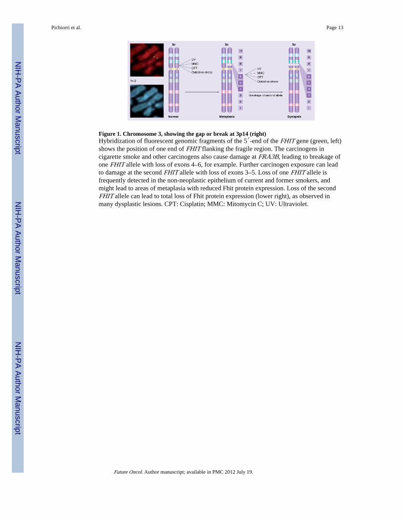
Figure 1. Chromosome 3, showing the gap or break at 3p14 (right)Hybridization of fluorescent genomic fragments of the 5′-end of the FHIT gene (green, left)shows the position of one end of FHIT flanking the fragile region. The carcinogens incigarette smoke and other carcinogens also cause damage at FRA3B, leading to breakage ofone FHIT allele with loss of exons 4–6, for example. Further carcinogen exposure can leadto damage at the second FHIT allele with loss of exons 3–5. Loss of one FHIT allele isfrequently detected in the non-neoplastic epithelium of current and former smokers, andmight lead to areas of metaplasia with reduced Fhit protein expression. Loss of the secondFHIT allele can lead to total loss of Fhit protein expression (lower right), as observed inmany dysplastic lesions. CPT: Cisplatin; MMC: Mitomycin C; UV: Ultraviolet.
Pichiorri et al. Page 13
Future Oncol. Author manuscript; available in PMC 2012 July 19.
NIH
-PA Author Manuscript
NIH
-PA Author Manuscript
NIH
-PA Author Manuscript

Figure 2. Fhit in carcinogen-induced cancer and stress response(A) Table summarizing alterations induced in B6–129(F1) mice, either wildtype or Fhit+/−,by whole-body exposure to ECS for 15 days [36]. *Percentage of mice showing extensiveloss of Fhit within each experimental group; ‡Percentage of mice with the reportedalteration; §Means ± SE among all mice within each experimental group. (B) Detection of 8-OHdG representing the oxidative DNA damage in hydroquinone-exposed transplanted bonemarrow cells; Fko and Wt bone marrow cells were exposed to hydroquinone andtransplanted to recipient mice [38]. (C) Increased green fluorescent DCF signal in H1299Fhit-expressing cells (D1) under stress condition [43]. (D) FACS ana lysis of D1 and E1cell-cycle kinetics at 48 h after oxidative stress treatment; cells were treated with increasingconcentrations of H2O2 (0.25, 0.5 mM) for 4 h. (E) Colony-formation assay of H1299/D1(Fhit+) and H1299/E1 (Fhit−) cells after 5 h treatment with H2O2 at indicated concentrations[43].BMT: Bone marrow transplant; DCF: Dichlorofluorescein diacetate; ECS: Environmentalcigarette smoke; PCNA: Proliferating cell nuclear antigen protein.
Pichiorri et al. Page 14
Future Oncol. Author manuscript; available in PMC 2012 July 19.
NIH
-PA Author Manuscript
NIH
-PA Author Manuscript
NIH
-PA Author Manuscript

Figure 3. Model for the role of Fhit in protection of Fdxr from proteaosomal degradation(A) Non-native Fhit (rough pink ball) and Fdxr (rough green ball) proteins bind to the transring of a Hsp60–Hsp10 complex. End-to-end exchange of Hsp10 results in the encapsulationof the protein substrate in the cis cavity. Release of Hsp10 and substrate proteins, Fhit andFdxr, in the folded conformation (pink and green smooth balls, respectively) formitochondrial addressing; we hypothesized that absence of Fhit leads to enhancedproteasomal degradation of Fdxr [43]. The immunofluorescence microscopy (B–E) wasperformed with antiFhit serum on H1299 Fhit-positive cells; Fhit staining was detectedusing fluorescein isothiocyanate (green) conjugated antirabbit immunoglobulin (IgG);MitoTracker Red staining, which identifies mitochondria, shows partial colocalization withFhit. (E) The yellow color shows the colocalizations points.
Pichiorri et al. Page 15
Future Oncol. Author manuscript; available in PMC 2012 July 19.
NIH
-PA Author Manuscript
NIH
-PA Author Manuscript
NIH
-PA Author Manuscript

NIH
-PA Author Manuscript
NIH
-PA Author Manuscript
NIH
-PA Author Manuscript
Pichiorri et al. Page 16
Tabl
e 1
Can
dida
te F
hit p
rote
in p
artn
ers
isol
ated
fro
m T
rapa
sso
et a
l. [4
4].
Pro
tein
Acc
essi
onno
.M
r(k
Da)
Fun
ctio
n/ca
tego
rySu
bcel
lula
rlo
caliz
atio
n
Hsp
60N
P_00
2147
6060
kD
a he
at s
hock
pro
tein
Cyt
osol
/mito
chon
dria
Mal
ate
dehy
drog
enas
eN
P_00
5909
33C
atal
yzes
the
reve
rsib
le o
xida
tion
of m
alat
e to
oxal
oace
tate
Mito
chon
dria
l mat
rix
Ele
ctro
n tr
ansf
er f
lavo
prot
ein
NP_
0019
7628
Spec
ific
ele
ctro
n ac
cept
or f
or m
itoch
ondr
ial
dehy
drog
enas
esM
itoch
ondr
ial m
atri
x
Hsp
10A
AC
9633
210
10 k
Da
heat
sho
ck p
rote
inC
ytos
ol/m
itoch
ondr
ia
Mito
chon
dria
l ald
ehyd
ede
hydr
ogen
ase
2N
P_00
0681
55Se
cond
enz
yme
of th
e m
ajor
oxi
dativ
e pa
thw
ayof
alc
ohol
met
abol
ism
Mito
chon
dria
l mat
rix
Ferr
edox
in r
educ
tase
P225
7054
Firs
t ele
ctro
n tr
ansf
er p
rote
in in
all
the
mito
chon
dria
l p45
0 sy
stem
sM
itoch
ondr
ial m
atri
x
Future Oncol. Author manuscript; available in PMC 2012 July 19.