Progesterone Receptors Induce FOXO1-dependent Senescence in Ovarian Cancer Cells
Upload
independentCategory
view
2download
0
Cancer Cell
Article
Dissecting the Unique Role of the RetinoblastomaTumor Suppressor during Cellular SenescenceAgustin Chicas,1 Xiaowo Wang,3 Chaolin Zhang,1 Mila McCurrach,1 Zhen Zhao,1 Ozlem Mert,1 Ross A. Dickins,1
Masashi Narita,1 Michael Zhang,1 and Scott W. Lowe1,2,*1Cold Spring Harbor Laboratory, Cold Spring Harbor, NY 11724, USA2Howard Hughes Medical Institute, Cold Spring Harbor, NY 11724, USA3MOE Key Laboratory of Bioinformatics and Bioinformatics Div, TNLIST/Department of Automation, Tsinghua University, Beijing 100084,
China
*Correspondence: [email protected]
DOI 10.1016/j.ccr.2010.01.023
SUMMARY
The RB protein family (RB, p107, and p130) has overlapping and compensatory functions in cell-cyclecontrol. However, cancer-associated mutations are almost exclusively found in RB, implying that RB hasa nonredundant role in tumor suppression. We demonstrate that RB preferentially associates with E2F targetgenes involved in DNA replication and is uniquely required to repress these genes during senescence but notother growth states. Consequently, RB loss leads to inappropriate DNA synthesis following a senescencetrigger and, together with disruption of a p21-mediated cell-cycle checkpoint, enables extensive proliferationand rampant genomic instability. Our results identify a nonredundant RB effector function that maycontribute to tumor suppression and reveal how loss of RB and p53 cooperate to bypass senescence.
INTRODUCTION
Loss-of-function mutations in the retinoblastoma gene product
(RB) or its signaling network are considered requisite for cancer
development; hence, the roles and regulation of RB have been
intensively studied (reviewed in Burkhart and Sage, 2008; Row-
land and Bernards, 2006). The best-characterized RB activity
relates to its ability to control the G1-S transition, where it nega-
tively regulates the E2F family of transcription factors. Cyclin-
dependent kinases (CDKs) activated in response to mitogenic
stimuli phosphorylate and inactivate RB, allowing the released
E2F to transcriptionally activate genes required for cell-cycle
progression. Certain viral oncoproteins bind RB and release
E2F, leading to forced S phase entry. Because spontaneous
mutations in RB may produce similar effects, the ability of RB
to halt cell-cycle transitions is considered central to its tumor
suppressor function. Nevertheless, RB binds other proteins
besides E2F and can regulate processes such as apoptosis,
quiescence, differentiation, and senescence. How these
proteins and processes contribute to the tumor suppressor
activities of RB is poorly understood.
RB is a member of a multigene family consisting also of RBL1
(p107) and RBL2 (p130) (Burkhart and Sage, 2008). Studies using
Significance
The action of RB as a tumor suppressor has been difficult to dep107 and p130. By coupling RNAi technology with a genome-wwe identified a unique and specific activity of RB in repressingWe further show how failure of this activity, when coupled to losand genomic instability. Our study provides a comprehensivedistinct proliferative states and insights into the regulation of c
376 Cancer Cell 17, 376–387, April 13, 2010 ª2010 Elsevier Inc.
both biochemical and genetic approaches have identified
distinct and overlapping functions of each family member
(Classon and Harlow, 2002). Like RB, both p107 and p130
bind E2F proteins and are substrates for phosphorylation by
active cyclin/CDKs (Classon and Harlow, 2002). Furthermore,
p107 and p130 also associate with DNA tumor virus oncopro-
teins and can induce cell-cycle arrest when overexpressed
(Mulligan and Jacks, 1998). Yet, despite the similarities among
the RB proteins in structure and function, somatic mutations
affecting p107 or p130 are rare in human cancers (Burkhart
and Sage, 2008).
In contrast to their action in cell-cycle control, less is known
about how RB proteins influence cellular senescence. Senes-
cent cells exit the cycle irreversibly, acquire a large and flat
morphology, accumulate a senescence-associated b-galactosi-
dase (SA-b-gal), and undergo changes in gene expression linked
to cell-cycle inhibition and inflammation (Campisi and d’Adda di
Fagagna, 2007). In cultured cells, senescence can be triggered
by replicative exhaustion, or in response to activated onco-
genes, DNA damage, or oxidative stress (Courtois-Cox et al.,
2008)., Accordingly, the senescence program acts as a general
antiproliferative stress response and is considered a potent
tumor suppressive mechanism in vivo (reviewed in Narita and
fine, partly because of the redundancy of the related proteinside analysis of gene expression and RB chromatin binding,DNA replication as cells exit the cell cycle into senescence.s of a fail-safe checkpoint, leads to both senescence bypassdata set of the genes controlled by RB family members inellular senescence and RB action in tumor suppression.

Figure 1. RNAi-Mediated Knockdown of RB
But Not p107 or p130 Impairs Ras-Induced
Senescence
(A) Immunoblots of growing IMR90 cells infected
with the indicated shRNAs probed for RB, p107,
or p130. Actin was used as loading control.
(B) Immunoblots of ras-senescent IMR90 cells.
Chromatin-bound fractions were used for the RB,
p107, and p130 blots and Histone H3 was used
as loading control. Whole cell lysates were used
for the p16 and ras blots and actin was used as
loading control.
(C) SA-b-galactosidase staining. The scale bar
represents 100 mM.
(D) DAPI staining to visualize SAHF. Scale bar
represents 10 mM.
(E) Quantification of SAHF (red bars), BrdU incor-
poration (blue bars). Values represent the mean
and standard error (SE) of at least three indepen-
dent experiments. (V) is empty vector, (S) is senes-
cent, (G) is growing, and (Q) is quiescent.
Cancer Cell
Dissecting the Role of RB in Cellular Senescence
Lowe, 2005; Prieur and Peeper, 2008). Indeed, senescent cells
accumulate in benign tumors in mice expressing activated onco-
genes, and in these settings codisruption of genes controlling
senescence regulators lead to malignant progression. Moreover,
certain DNA-damaging chemotherapeutic agents can induce
senescence in tumors, and the integrity of the senescence
program contributes to the antitumor effect of these agents.
The regulation of cellular senescence involves interplay
between the p53 and RB tumor suppressor networks (Cour-
tois-Cox et al., 2008). For example, DNA tumor virus oncopro-
teins that target p53 and RB bypass senescence in cultured cells
(Shay et al., 1991). Although these oncoproteins bind all three RB
family members, acute inactivation of RB is sufficient to promote
proliferation in senescent mouse embryo fibroblasts (MEFs)
(Sage et al., 2003) and prevents SAHF accumulation and coop-
erates with p53 loss to bypass senescence in human diploid
fibroblasts (Narita et al., 2003; Voorhoeve and Agami, 2003).
Based on these observations, we hypothesized that RB must
have targets in senescence that differ from those controlled by
p107 and p130, and that these targets might highlight processes
that mediate its tumor-suppressive effects.
RESULTS
RB Has a Nonredundant Role in Oncogene-InducedSenescenceTo understand the relative contribution of individual RB family
members to various proliferative states, we generated multiple
short-hairpin RNAs (shRNAs) targeting each family member using
Cancer Cell 17, 376–3
the mir-30 design (Silva et al., 2005).
These shRNAs were transduced into
IMR90 human diploid fibroblasts (HDF),
a well-characterized normal human cell
strain that has an intact RB pathway, is
widely used to study both replicative and
oncogene-induced senescence (Narita
et al., 2003; Shay et al., 1991), and can
rapidly and completely transition between
various growth states by simple cell culture manipulations. As
shown in Figure 1A, each shRNA efficiently and specifically
represses its target RB family protein. To elucidate the contribu-
tion of the individual RB family members to ras-induced senes-
cence, we transduced IMR90 cells with shRNAs targeting each
RB family protein together with a retrovirus expressing oncogenic
ras and the populations were selected for cells harboring both
constructs. Nine days postinfection, cell populations were exam-
ined for markers of senescence. For comparison, IMR90 cells ex-
pressing each shRNA were examined in normal growth condi-
tions, or following induction of quiescence by serum withdrawal
or contact inhibition (confluence).
Although each shRNA suppressed its corresponding family
member (Figure 1B), only those targeting RB reduced SA-b-gal
accumulation (Figure 1C) and SAHF formation (Figures 1D and
1E) in cells triggered to senesce. None of the shRNAs were
able to completely bypass senescence; however, a small but
reproducible percentage of shRB-expressing cells (but not
shp107- or shp130-expressing cells) continued to incorporate
BrdU at the 9 day time point (Figure 1E, see below). Identical
results were obtained using two different shRNAs targeting
each gene, suggesting no RNAi off-target effects. Therefore,
most subsequent experiments were performed with the most
potent shRNAs. Consistent with the overlapping functions of
the RB family, none of the shRNAs had any notable impact on
proliferation of growing or quiescent cells (Figures 1E and 5B;
data not shown). Similar results were also observed in WI38
cells, another normal human fibroblast strain (data not shown).
These observations confirm that, in normal human fibroblasts,
87, April 13, 2010 ª2010 Elsevier Inc. 377

Cancer Cell
Dissecting the Role of RB in Cellular Senescence
RB plays a crucial role in cellular senescence that is not shared
with other family members.
Impact of Individual RB Family Members on GeneExpression in Different Growth ConditionsThe RB family proteins function primarily as transcriptional core-
pressors by binding and redundantly modulating the activity of
the E2F transcription factor family (Burkhart and Sage, 2008).
We therefore reasoned that RB might have transcriptional targets
not shared by p107 or p130 during senescence (but not during
quiescence), and that these targets might contribute to the
impaired program observed in cells lacking RB. Previous
attempts to identify specific targets of individual RB family of
proteins have examined growing or quiescent mouse embryo
fibroblasts from the corresponding knockout mice that may
have been susceptible to developmental compensation and
were not comprehensive (Markey et al., 2007). Here, we exam-
ined the impact of acutely inhibiting different pocket proteins on
genome-wide gene expression patterns in normal human cells.
Transcriptional profiling was performed on IMR90 cells in
growing, low serum quiescent (0.1% FBS for 4 days), contact in-
hibited quiescent (5 days after confluency) or senescent condi-
tions expressing shRNAs targeting RB, p107, p130, or control.
RNA from two independent experiments was hybridized to the
Affymetrix U133 Plus 2.0 microarray and the generated data
were processed as described in Experimental Procedures.
Average-linkage hierarchical clustering analysis was performed
to aggregate arrays and genes based on similarities of gene
expression (Eisen et al., 1998). This analysis clustered the
different growth conditions over the effect of the different
shRNAs, indicating that each shRNA has a smaller effect on
global gene expression than the particular growth condition
(see Figure S1A available online).
Senescent cells displayed a global upregulation of antiprolifer-
ative genes, downregulation of growth-promoting genes, and a
pattern of gene expression known as the senescence-associ-
ated secretory phenotype (Coppe et al., 2008). Accordingly,
we observed upregulation of cyclin-dependent kinase inhibitors
(CDKis) and protein metalloproteinases (MMPs), as well as
many cytokines and chemokines (CXCs) in senescent cells
(Figure S1A).
A Nonredundant Role for RB in Repressing Some E2FTarget Genes During SenescenceTo understand how each RB family member influences gene
expression in different growth conditions, we identified differen-
tially expressed genes in the presence of each shRNA. Unique
and reproducible shRNA-dependent changes in gene expres-
sion were observed under all conditions (Figure S1B). Interest-
ingly, senescent cells expressing shRB underwent the most
substantial changes in gene expression as evidenced by the
number of probe sets that were up or downregulated (Fig-
ure S1B). Gene ontology (GO) analysis (Dennis et al., 2003)
showed little overlap in the processes affected by repressing
each of the RB proteins under different conditions (Table S1).
However, we observed a differential but overlapping effect of RB
on gene expression categories depending on the growth state.
Whereas genes upregulated in cells undergoing senescence in
the absence of RB were enriched with ‘‘DNA replication’’ factors
378 Cancer Cell 17, 376–387, April 13, 2010 ª2010 Elsevier Inc.
(Table S1, p < 9.2 3 10�14), those upregulated in growing cells
lacking RB were enriched with ‘‘cell cycle’’ genes (Table S1,
p < 6.12 3 10�8). Using a nonbiased bioinformatics analysis,
we identified the E2F motif, with the consensus sequence
TTTSSCGC (where S represents C or G), as the most enriched
motif in genes upregulated in cells undergoing senescence in
the absence of RB (p < 2.1 3 10�7 with Bonferroni correction)
(Figure S1C). Surprisingly, the E2F motif was only moderately
enriched in the set of genes upregulated in growing cells lacking
RB (p < 0.09 with Bonferroni correction), and not enriched in
gene sets impacted by other conditions or shRNAs (data not
shown). These observations suggest that RB has a unique spec-
ificity in regulating a subset of E2F targets in senescent cells.
RB Represses Distinct Genes Dependingon Cellular ContextTo better characterize the genes and processes controlled by
RB, we grouped genes that were significantly upregulated by
RB loss in senescent, low serum, confluent, and growing condi-
tions (741, 120, 784, and 291, respectively; Table S2) and sub-
jected the corresponding probe sets to hierarchical clustering
(Figure 2A). Consistent with our more global analyses, these
genes often contained E2F binding sites and were highly ex-
pressed in growing but not in quiescent or senescent cells
(Figure 2A; Table S2). Interestingly, most predicted E2F target
genes were contained within two distinct adjacent clusters that
were only divided by the influence of RB in senescent cells.
Whereas the genes in clusters C3 (Figure 2D) were highly ex-
pressed in RB-deficient senescent cells, the genes in cluster
C4 (Figure 2E) remained repressed. Gene ontology analysis re-
vealed that genes in cluster C3 (Table S3; replication cluster)
were enriched for DNA replication factors (p < 3.0 3 10�29
with Benjamini correction), particularly components of the
prereplication complex, whereas genes in cluster C4 (Table S3;
cell-cycle cluster) were enriched for mitotic cell-cycle factors
(p < 1.0 3 10�21 with Benjamini correction). We have confirmed
these results with additional biological replicates (Figure S1D).
Thus RB is uniquely required to repress the transcription of repli-
cation genes during cellular senescence.
Clustering analysis also revealed two other distinct clusters.
One cluster contains a group of genes that were highly ex-
pressed only in senescent cells lacking RB (Figure 2C). The
genes in this cluster (Table S3; cyclin E cluster) are poorly char-
acterized, except for cyclin E1, a bona fide E2F target gene
known to activate several cyclin-dependent kinases. The other
cluster is enriched in many cytokines and chemokines (CXCs)
that are part of the senescence-associated secretory phenotype
(Figure 2B). However, these genes, some of which contribute to
senescence, were still induced in the absence of each RB family
member and, in some cases, their expression further increased
in the absence of RB (data not shown). Thus, whereas RB is
required for some aspects of the senescence program, the
vast majority of changes are RB independent.
Selective High-Affinity Binding of RB to the ‘‘ReplicationFactor’’ Set of E2F TargetsThe expression analysis described above suggests that RB
discriminates between different E2F target genes and growth
states. Previous attempts to detect RB bound to specific E2F
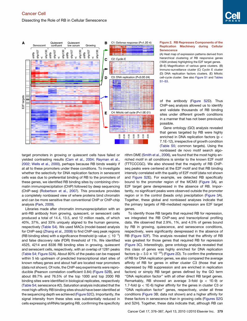
Figure 2. RB Represses Components of the
Replication Machinery during Cellular
Senescence
(A) Heat map of expression patterns derived from
hierarchical clustering of RB responsive genes
(1826 probes) highlighting the E2F target genes.
(B–E) Magnification of various gene clusters. (B)
Immuno-surveillance cluster (C) Cyclin E cluster
(D) DNA replication factors clusters. (E) Mitotic
cell-cycle cluster. See also Figure S1 and Tables
S1–S3.
Cancer Cell
Dissecting the Role of RB in Cellular Senescence
target promoters in growing or quiescent cells have failed or
yielded contrasting results (Cam et al., 2004; Rayman et al.,
2002; Wells et al., 2000), perhaps because RB binds weakly if
at all to these promoters under these conditions. To investigate
whether the selectivity for DNA replication factors in senescent
cells was due to preferential binding of RB to the promoters of
these genes, we identified RB binding sites by combining chro-
matin immunoprecipitation (ChIP) followed by deep sequencing
(ChIP-seq) (Robertson et al., 2007). This procedure provides
a completely nonbiased view of where proteins bind chromatin
and can be more sensitive than conventional ChIP or ChIP-chip
analysis (Park, 2009).
Libraries made after chromatin immunoprecipitation with an
anti-RB antibody from growing, quiescent, or senescent cells
produced a total of 14.4, 13.5, and 12 million reads, of which
40%, 37%, and 33% uniquely aligned to the human genome,
respectively (Table S4). We used MACs (model-based analysis
for ChIP-seq) (Zhang et al., 2008) to find ChIP-seq peak regions
(RB binding sites) with a significance threshold (p value % 10�5)
and false discovery rate (FDR) threshold of 1%. We identified
4525, 4214 and 8336 RB binding sites in growing, quiescent
and senescent cells, respectively, with an overlap of 1281 peaks
(Table S4; Figure S2A). About 80% of the peaks can be mapped
within 5 kb upstream of predicted transcriptional start sites of
known refseq genes and about half are located near promoters
(data not shown). Of note, the ChIP-seq experiments were repro-
ducible (Pearson correlation coefficient 0.84) (Figure S2B), and
about 89.7% and 76.5% of the top 1000 and top 2000 RB
binding sites were identified in biological replicates, respectively
(Table S4; senescence #2). Saturation analysis indicated that the
most high-affinity RB binding sites should have been identified at
the sequencing depth we achieved (Figure S2C). Importantly, the
signal intensity from these sites was substantially reduced in
cells expressing shRNAs targeting RB, confirming the specificity
Cancer Cell 17, 376–3
of the antibody (Figure S2D). Thus
ChIP-seq analysis allowed us to identify
and validate thousands of RB binding
sites under different growth conditions
in a manner that has not been previously
possible.
Gene ontology (GO) analysis revealed
that genes targeted by RB were highly
enriched in DNA replication factors (p <
7.1E-12), irrespective of growth condition
(Table S5; common targets). Using the
nonbiased de novo motif search algo-
rithm DME (Smith et al., 2006), we found that the most highly en-
riched motif in all conditions is similar to the known E2F motif
(TTTCCCGC). We also showed that the majority of RB ChIP-
seq peaks were centered at the E2F motif and that RB binding
intensity correlated with the quality of E2F motif (data not shown
and Figure S2E). For example, we detected RB specifically
bound to the promoter region of the MCM3 (Figure 3A), an
E2F target gene derepressed in the absence of RB. Impor-
tantly, no significant peaks were observed outside the promoter
region or in the control (beads only) precipitation (Figure 3A).
Together, these global and nonbiased analyses indicate that
the primary targets of RB-mediated repression are E2F target
genes.
To identify those RB targets that required RB for repression,
we integrated the RB ChiP-seq and transcriptional profiling
data. We observed that 2.9%, 1%, and 4.5% of genes bound
by RB in growing, quiescence, and senescence conditions,
respectively, were significantly derepressed in the absence of
RB (Figure S2F). This analysis revealed that the affinity of RB
was greatest for those genes that required RB for repression
(Figure 3C). Interestingly, gene ontology analysis revealed that
this class of genes was highly enriched for DNA replication
factors (p < 3.0 3 10�20) (Figure 2D). To confirm the preference
of RB for DNA replication genes, we also compared the average
affinity of RB for genes in either cluster C3 (those that are
depressed by RB suppression and are enriched in replication
factors) or simply RB target genes defined by the GO term
‘‘DNA replication factor’’ with all other direct RB target genes.
Remarkably, RB showed an average 3-fold (p < 1E-9) or
1.7-fold (p < 1E-6) higher affinity for the genes in cluster C3 or
‘‘DNA replication factor’’ genes, respectively, under all three
conditions (Figure 3B; data not shown) and a higher affinity for
these factors in senescence than in growing cells (Figures S2G
and S2H). Together, these data indicate that, although RB can
87, April 13, 2010 ª2010 Elsevier Inc. 379

Figure 3. Selective Binding of RB to the
Promoters of DNA Replication Factors
(A) Binding patterns of RB to the MCM3 gene
shown as custom tracks on the UCSC genome
browser.
(B) Histogram comparing binding of RB-as
measured by read counts-to DNA replication
factors versus other RB targets. Error bars repre-
sent the standard deviation of the read counts.
(C) Correlation of RB binding to gene promoters
and expression of those genes in RB-deficient
cells under different growth conditions. See also
Figure S2 and Tables S4 and S5.
Cancer Cell
Dissecting the Role of RB in Cellular Senescence
bind to genes involved in a variety of processes, DNA replication
genes are uniquely dependent on RB in senescent cells.
Redundancy of the RB Family of Proteinsin Quiescent CellsWe were surprised by the nominal impact of suppressing indi-
vidual RB proteins on E2F target gene expression during quies-
cence (Figures 4A–4C and 2D and 2E), a cell-cycle state thought
to be influenced by the RB family (Burkhart and Sage, 2008).
There is ample evidence that the RB proteins have redundant
and/or compensatory functions (Sage et al., 2000). To test
whether the RB proteins had an overlapping ability to repress
E2F targets in quiescent HDFs, we developed retroviral vectors
that express combination of two or three shRNAs targeting RB,
p107 and p130 in tandem (Figure 4D) and introduced these
into IMR90 cells. Cells expressing each vector efficiently sup-
pressed the targeted proteins, although two or more shRNAs
slightly reduced the efficiency of each individual shRNA
(Figure 4E). Despite this, only cells in which all three RB family
proteins were suppressed showed upregulation of E2F target
genes in quiescent cells (Figure 4E). Therefore, in stark contrast
380 Cancer Cell 17, 376–387, April 13, 2010 ª2010 Elsevier Inc.
to the situation in senescence, the RB
family shares the capacity to suppress
E2F target genes during quiescence.
We tested whether the ability of the
other RB family proteins to compensate
for loss of RB in quiescent HDFs reflected
the ability of these proteins to replace RB
at E2F target gene promoters. Because
p130 is the most prominent RB family
member bound to E2F target genes in
quiescent cells (Balciunaite et al., 2005),
we compared the binding of p130 in the
presence and absence of RB in quiescent
and senescent cells by ChIP-seq. We
found that like RB, the main targets of
p130 are genes containing E2F binding
sites (Figure S3). Importantly, we found
that whereas binding of p130 to many
promoters increased in quiescent cells
lacking RB, no effect was observed in
senescent cells (Figure 4F). For example,
p130 bound the MCM5 gene promoter
with a 1.8-fold greater intensity in quies-
cent cells lacking RB than those expressing RB, whereas the
binding of p130 in senescent cells was not affected by RB
suppression (Figure 4G). Thus, p130 can compensate for RB
loss in quiescent cells by enhancing its binding to E2F target
gene promoters. Together these data suggest that there is
a binding equilibrium between RB and p130 such that at a given
moment either one or the other is bound to particular E2F sites.
Loss of RB shifts the equilibrium toward p130, perhaps explain-
ing why loss of individual RB proteins does not effect expression
of E2F targets in quiescent cells.
RB Represses the DNA Replication Machineryin Cells Undergoing SenescenceAmong the genes derepressed in the absence of RB are key
components of the pre-replication complex (pre-RC), pointing
to a possible defect in the proper shut down of DNA synthesis
in cells undergoing senescence. Assembly of the pre-RC on
origins of replication is a crucial and limiting step in the
synthesis of DNA occurring only once per cell cycle and during
a short window of time prior to S phase (Stillman, 1996). We
therefore tested whether RB-deficient cells inappropriately

Figure 4. Repression of E2F Target Genes Is
Unique for RB in Senescence but Is Redun-
dant in Quiescence
(A–C) Immunoblots and qPCR to measure expres-
sion of (A) MCM2, (B) cyclin A, and (C) cyclin E1 in
growing, quiescent, or senescent cells expressing
the indicated shRNA. qPCR values are averages of
representative experiments done in triplicates.
(D) Schematic diagram of the polycistronic
shRNAs used to knockdown the indicated mem-
bers of the RB family.
(E) Immunoblots probed for the indicated protein
from lysates of quiescent IMR90 cells infected
with the indicated shRNA.
(F) Histograms comparing the binding intensity of
p130 at gene promoters before (non-shRB) and
after RNAi-mediated suppression of RB (shRB)
in quiescent (top) and senescent (bottom) cells.
The shift to the right of zero indicates that
the p130-specific antibody coimmunoprecipitates
more promoter DNA in the absence of RB during
quiescence.
(G) Binding patterns of p130 and RB to the MCM5
gene shown as custom tracks on the UCSC
genome browser. p130 binding to the MCM5 gene
promoter increases 1.8-fold in RB-deficient quies-
cent cells while binding in senescent cells remain
unchanged. See also Figure S3 and Table S6.
Cancer Cell
Dissecting the Role of RB in Cellular Senescence
retained an assembled pre-RC by immunoblotting the chro-
matin-bound fraction of ras-transduced cells for specific repli-
cation factors (Mendez and Stillman, 2000). Although compo-
nents of the pre-RC were bound to chromatin in growing
cells under all conditions, they were not detected in quiescent
cells or in senescent cells lacking p107 or p130 or expressing
a vector control. In contrast, pre-RC components were bound
tightly to chromatin in RB-suppressed cells triggered to sen-
esce (Figure 5A).
Consistent with the above results, experiments examining
BrdU incorporation and DNA content revealed that cells lacking
RB show unscheduled DNA synthesis when entering senes-
cence. Specifically, shRB-expressing cells continued to
incorporate BrdU for at least 4 days longer than control,
shp107-, and shp130-expressing cells upon ras transduction
(Figure 5B). These cells also showed a higher percentage of cells
with 4N and 8N DNA content compared with the other cell pop-
ulations (>2-fold and >7-fold, respectively, Figure 5C). Despite
Cancer Cell 17, 376–3
ongoing DNA replication, shRB-express-
ing cells triggered to senesce did not
expand after postselection day 7 (PS7)
(Figure 7B), indicating that they activated
an additional barrier to proliferation (see
Figure 7). Nevertheless, these results
demonstrate that RB is required to halt
DNA synthesis during cell-cycle exit
into senescence, such that cells lacking
RB undergo unscheduled DNA replica-
tion and, in some cases, endoreplication.
Interestingly, quiescent cells lacking RB
did not display these defects (Figures
5B and 5C), further supporting the unique role of RB in senescent
cells.
RB Repression of Cyclin E1 Is Required to PreventReplication of Senescent CellsOur results argue for a unique role for RB in repressing the repli-
cation machinery, particularly during senescence. However, in
addition to the pre-RC components, loss of RB also led to dere-
pression of the cyclin E1 gene (Figures 2C, 4C, and 5A), a key
regulator of the cell-cycle engine (Sherr and Roberts, 2004).
Although less appreciated, cyclin E1 has also been directly
linked to DNA replication and endoreplication (Zhang, 2007)—
phenotypes observed in senescent cells lacking RB. We there-
fore hypothesized that the aberrant cyclin E expression could
contribute to the unscheduled DNA replication leading to poly-
ploid cells. To test this hypothesis, we introduced tandem
shRNAs capable of repressing cyclin E1 and RB (Figure 6A)
into IMR90 cells together with oncogenic ras. Remarkably,
87, April 13, 2010 ª2010 Elsevier Inc. 381

Figure 5. RB Is Required to Prevent Inap-
propriate DNA Replication in Cells Under-
going Senescence
(A) Immunoblots of chromatin-bound proteins
probed for MCM2, MCM3, ORC1, and cyclin E1.
Blots were normalized by Coomassie blue staining
for histones.
(B) Time course BrdU incorporation assays of
cells undergoing senescence in the absence of
RB, p107, or p130 compared with vector control.
(S/PS3, S/PS5, S/PS7 stand for postselection
day 3, 5, or 7, respectively). Values represent the
mean ± SE of at least three independent experi-
ments.
(C) Cell-cycle profiles of growing (top), quiescent
(middle), or senescent (bottom) IMR90 cells ex-
pressing the indicated shRNA.
Cancer Cell
Dissecting the Role of RB in Cellular Senescence
despite a nominal impact on normal growing cells (data not
shown, see also Geng et al., 2003), suppression of cyclin E1
was sufficient to prevent the aberrant DNA replication and en-
doreplication, otherwise observed in cells undergoing senes-
cence in the absence of RB (Figure 6B). Furthermore, we
rescued the phenotype by reintroducing a shRNA-resistant
cDNAs encoding either wild-type or a kinase-deficient cyclin
E1 (cyclin E1 KD-E) (Sheaff et al., 1997) (Figure S4).
Distinct from its cell-cycle activity, cyclin E1 can directly influ-
ence DNA replication by recruiting MCMs to the pre-RC in
a kinase-independent manner (Geng et al., 2007). To investigate
the requirement for cyclin E1 in promoting assembly of the pre-
RC in cells lacking RB, we used the association between MCM2
and chromatin as a surrogate marker (Geng et al., 2007). Immu-
nofluorescence on pre-extracted samples showed that suppres-
sion of cyclin E1 caused a significant reduction in MCM2-posi-
tive cells (Figure 6C), indicating inefficient loading of MCM2
onto chromatin. Similarly, immunoblotting of chromatin-bound
proteins showed less chromatin-bound MCM2 in cells lacking
cyclin E1 (Figure 6D). These results were not due to a reduction
in MCM2 expression because total MCM2 protein levels were
unchanged (Figure 6E). The levels of MCM2 and MCM3 RNA
also remained unchanged in cells lacking cyclin E1, indicating
that cyclin E is not required for the upregulation of these genes
382 Cancer Cell 17, 376–387, April 13, 2010 ª2010 Elsevier Inc.
in RB-deficient cells (data not shown).
Thus, in senescent cells, RB is required
to repress both replication factors as
well as cyclin E1, which would otherwise
facilitate loading these factors into the
pre-RC leading to aberrant replication
and endoreplication.
RB Loss Triggers a p53/P21-Dependent Checkpoint thatPrevents Escape from SenescenceAlthough loss of RB prevents the appro-
priate shut down of DNA replication in
cells triggered to senesce, these cells
eventually arrest, suggesting the activa-
tion of a second proliferation barrier that
prevents unrestrained proliferation. In
addition to the RB pathway, the p53 tumor suppressor also
contributes to senescence (Courtois-Cox et al., 2008). Interest-
ingly, p53 can respond to replicative stress and (Marusyk and
DeGregori, 2007), accordingly, we noted an increase in p53
ser15 phosphorylation (Figure S5A) and a concomitant increase
in the levels of its target gene p21 in RB-deficient cells triggered
to senesce (Figure 7A).
To test whether p21 was required for this second proliferation
barrier, we introduced shRNAs targeting both RB and p21 into
IMR90 cells together with oncogenic ras. Strikingly, these cells
efficiently bypassed senescence, as assessed by cell prolifera-
tion, BrdU incorporation, and colony formation relative to
controls in both IMR90 and WI38 cells (Figures 7B and 7C, and
data not shown). Furthermore, consistent with their ongoing
proliferation, cells expressing the RB/p21 shRNA derepressed
both the DNA replication E2F targets (MCM3, cyclin E1) and
mitotic E2F targets (cdc2, cyclin A, cyclin B) (Figure 7E,
Figure S5B). Similarly, cells coexpressing shRNAs targeting RB
and p53, or p16 and p21, bypass senescence (data not shown),
which is associated with sustained expression of cell-cycle/
mitotic E2F targets (e.g., cyclin A, B, cdc2), albeit to varying
degrees (Figure 7E). Interestingly, cells cosuppressing p16 and
p21 did not hyperinduce cyclin E, perhaps owing to incomplete
knockdown or a subtly different mechanism of senescence

Figure 6. RB Repression of Cyclin E1 Is
Required to Prevent Replication of Senes-
cent Cells
(A) Immunoblots of lysates from senescent cells
expressing a polycistronic shRNA targeting RB
and cyclin E1 (tan depicted on the top of the figure)
probed for RB and cyclin E1. Growing (G), senes-
cent (S), senescent cells expressing shRNA tar-
geting only RB (shRB/S), or only cyclin E1 (shE1/S)
are used as controls. Actin is used as loading
control.
(B) Cell-cycle profiles of vector control senescent
cells (V/S), senescent cells expressing shRB
(shRB/S), or the tandem shRB/cE1 shRNA (Tan/S).
(C) Immunofluorescence of pre-extracted cells to
measure association of MCM2 with chromatin.
Percentage of MCM2-positive cells is indicated
in the bottom right corner of the images. DAPI
staining was used to visualize the nuclei. At least
200 cells were counted per experiment. Scale
bar represents 100 mm.
(D) Immunoblots of chromatin-bound extracts
from growing (G), quiescent (Q), vector control
senescent cells (V/S), senescent cells expressing
shRB (shRB/S), or the tandem shRB/cE1 shRNA
(Tan/S): two independent samples are shown.
(E) Immunoblots of lysates from senescent cells
expressing the tandem shRB/cE1 shRNA (Tan/S)
and shRNA-resistant cyclin E1 cDNA encoding
either wild-type (WtcE) or kinase-deficient (KDcE)
cyclin E1. Lysates from growing (G), vector control
senescent cells (V/S), senescent cells expressing
shRB (shRB/S), or the tandem shRB/cE1 shRNA
(Tan/S) alone are used as controls. See also
Figure S4.
Cancer Cell
Dissecting the Role of RB in Cellular Senescence
escape. Regardless, in the absence of this proliferation block,
cells cosuppressing RB together with p21 accumulate more
polyploid cells compared with those lacking RB alone
(Figure 7D, Figures S5C–S5E). Therefore, in human fibroblasts,
p21 mediates a checkpoint that is activated in response to RB
loss and serves to limit the consequences of unrestrained DNA
replication. In the absence of this checkpoint, proliferation
continues unabated, thus providing an explanation for how RB
and p53 loss cooperate to bypass senescence.
DISCUSSION
The RB gene family encodes important regulators of cell prolifer-
ation with overlapping and redundant functions, yet only RB is
commonly mutated in human tumors. By performing a series of
genome-wide analyses, we see that RB has a nonredundant
role in binding and repressing E2F target genes that are directly
involved in DNA replication, particularly in senescent cells.
Consequently, in cells triggered to senesce, RB loss leads to
sustained loading of pre-replication complexes, aberrant DNA
replication, and—in the absence of a second cell-cycle check-
point—proliferation and genomic instability. By contrast, RB is
dispensable for proper cell-cycle exit during quiescence
because other RB family members act redundantly to control
key E2F targets. Consistent with our observations, cells
harboring a transcriptionally compromised RB mutant fail to
repress replication targets during oncogene-induced senes-
cence but not cell-cycle exit, including those required for
normal development (Talluri et al., 2010).
Implications for Senescence ControlA hallmark of senescence is its stable cell-cycle arrest coordi-
nated by interplay between the RB and p53 tumor suppressor
networks (Courtois-Cox et al., 2008). Whereas p53 promotes
senescence by transactivating growth inhibitory genes, RB
represses growth-promoting genes, which, in some cases,
may involve heterochromatization of E2F target genes (Narita
et al., 2003; Zhang et al., 2005). Here we see that RB is dispens-
able for many of the gene expression changes that accompany
senescence but, instead, uniquely and specifically represses
a subset of E2F target genes required for DNA replication.
Whether these effects involve the same processes that influence
SAHF formation remains to be determined; nevertheless, they
highlight the importance of repressing DNA replication for proper
execution of the senescence program.
Consistent with a role for p53 in senescence, cells lacking
RB eventually arrest because they engage a second prolifera-
tion barrier mediated by the p21 cyclin-dependent kinase
inhibitor. In response to certain mitogenic oncogenes, p53
activates p21 and triggers senescence in response to replica-
tion stress (Halazonetis et al., 2008), and thus it seems likely
that the aberrant DNA synthesis produced by RB loss exacer-
bates this fail-safe mechanism. Regardless, p21 loss reveals
the full capacity of RB-deficient cells to aberrantly replicate
Cancer Cell 17, 376–387, April 13, 2010 ª2010 Elsevier Inc. 383

Figure 7. RB Loss Triggers a p53/p21
Dependent Checkpoint that Prevents
Escape from Senescence
(A) Immunoblots of lysates from IMR90 senescent
cells expressing the indicated shRNA probed for
p21 or actin.
(B) Growth curve of ras-infected cells expressing
the indicated shRNAs. Counting was initiated at
PS7 (postselection day 7), at which time the vector
control cells are fully growth arrested as measured
by BrdU incorporation. error bars represent the
standard error of at least three independent exper-
iments.
(C) Analysis of PS7 cells for different proliferation
and senescence markers and for the ability to
form colonies at low density. Unlike vector control,
RB, or p21 suppression, suppression of both RB
and p21 leads to an increase in cell proliferation
as measured by BrdU incorporation (20% positive
compared with 2% for either shRB or shp21 alone)
and colony formation. Scale bars for the micro-
graphs showing SA-b-gal staining and BrdU
immunofluorescence represent 100 mM and for
DAPI staining represent 10 mM.
(D) Cell-cycle profiles of ras-infected cells ex-
pressing the indicated shRNA.
(E) Immunoblots of lysates from ras-infected cells
expressing the indicated shRNA probed for the
indicated proteins. See also Figure S5.
Cancer Cell
Dissecting the Role of RB in Cellular Senescence
DNA, thus explaining how RB and p53 cooperate to promote
senescence.
It remains to be determined whether our observations in fibro-
blasts can be extended to other cells types, though it is inter-
esting that, human vulvar intraepithelial neoplasias (VIN) that
have bypassed senescence through inactivation of the RB family
and p53 (Santegoets et al., 2007) also show hyperinduction of
the replication factors studied here (Figure S1E). Furthermore,
the same genetic principles that control senescence in fibro-
blasts also apply to at least some cultured epithelial cells (e.g.,
RB and p53 cooperate to bypass senescence in HMECs and
384 Cancer Cell 17, 376–387, April 13, 2010 ª2010 Elsevier Inc.
keratinocytes) and spontaneous loss of
p16 due to DNA methylation in HMECs
does not completely bypass senescence
but is associated with the accumula-
tion of polyploid cells [Romanov et al.,
2001]).
Insights into RB Action inProliferation ControlBy combining RNAi with genome-wide
gene expression and chromatin binding
analyses, we identified genes that are
direct RB targets and whose regulation
is strictly RB dependent. This compre-
hensive analysis revealed that the
specific targets of RB can vary between
growth states. For example, only
a specific subset of E2F target genes,
particularly those directly involved in
DNA replication, were both bound by
RB and derepressed in RB-deficient cells entering senescence.
Thus our results reveal not only the unique specificity of RB for
E2F replication targets, but also a strict concordance between
the nature of genes regulated by RB and the specific phenotype
associated with RB loss—i.e., unscheduled DNA replication
during senescence but not quiescence. Although previous
studies hinted that RB family proteins can regulate different
E2F targets (Hurford et al., 1997) and that loss of RB can ulti-
mately lead to increased loading of replications complexes on
chromatin (Srinivasan et al., 2007), the selectivity of RB toward
repressing replication genes was not appreciated.

Cancer Cell
Dissecting the Role of RB in Cellular Senescence
By identifying thousands of RB binding sites throughout
the genome, our analyses decisively show that the primary
targets of RB-mediated repression are genes harboring E2F
binding sites and, moreover, reveal that RB preferentially
associates with a subset of E2F targets. By integrating these
data with transcriptional profiling, we further demonstrate that
RB mediates repression through physical association with
these promoters. Still, only a fraction of RB target genes are
derepressed in the absence of RB and only in senescence.
Such a situation has also been observed for similar studies
on the androgen receptor and may suggest compensation or
redundancy in transcriptional control (Massie et al., 2007).
Nevertheless, to our satisfaction, the genes that are sensitive
to RB loss are those genes that show the higher affinity for
RB. The binding of RB to replication factor genes during senes-
cence has not been examined, and this unique combination of
genes and circumstances may explain why RB binding to chro-
matin has previously been difficult to detect (Rayman et al.,
2002).
In human cells, we see that RB is dispensable for cell-cycle
exit into quiescence—instead, any RB family member could
repress E2F target genes in this setting (see also Rayman
et al., 2002); here we show that this redundancy can be ex-
plained, in part, by an increase in p130 recruitment to replication
genes in cells lacking RB. In contrast, studies using mouse
embryo fibroblasts show that acute ablation of Rb in quiescent
cells leads to S phase entry and proliferation (Sage et al.,
2003). Likewise, cre-mediated deletion of Rb in small intestine
enterocytes allows for cell-cycle reentry of quiescent villus cells
(Guo et al., 2009). The apparent discrepancy in our results may
relate to cell type or species differences or the status of the
helix-loop-helix protein HES1, which may play a crucial role in
determining the reversibility of the quiescent state (Sang et al.,
2008). Nevertheless, why other RB family members cannot
always compensate for RB loss remains to be determined,
though we suspect that the stable silencing of RB-target genes
during senescence may require recruitment of an RB-specific
chromatin modifying activity to these promoter of these genes.
Indeed, an RB mutant that is unable to associate with chromatin
modifying enzymes fails to repress DNA replication during onco-
gene-induced senescence but not during quiescence or differ-
entiation (Talluri et al., 2010)
One key RB target in senescence was cyclin E, which was ex-
pressed at low levels in growing cells but strongly upregulated in
RB-suppressed cells triggered to senesce. Because cyclin E
promotes cell-cycle progression by activating CDKs (Zhang,
2007), its selective control by RB stands out from other RB
targets directly involved in DNA replication. However, cyclin E
has additional activities and can directly stimulate DNA replica-
tion by facilitating recruitment of MCM proteins to the pre-RC
(Geng et al., 2007). Similarly, we see that the inappropriate
expression of cyclin E1 in RB-deficient cells leads to sustained
loading of MCM proteins onto chromatin in a CDK-independent
manner, unscheduled DNA replication, and endoreplication.
These results establish connections among the kinase-indepen-
dent function of cyclin E1, loading of MCM complexes onto
chromatin, and endoreplication and highlight how RB acts in
a coordinated way to shut down DNA synthesis as cells enter
senescence.
RB Action in Tumor SuppressionThe importance of RB as a tumor suppressor gene is evident by
the frequency of inactivating mutations that target RB or its
pathway in human cancer; indeed, the RB pathway is thought
to be disabled in virtually all tumor cells (Burkhart and Sage,
2008). Although RB can modulate the G1-S transition, cellular
differentiation, DNA replication, mitosis, apoptosis, chromo-
somal stability, and cellular senescence, it is not clear which of
these functions are crucial for its tumor suppressor activity.
Our results reveal a unique and nonredundant role for RB in
preventing DNA replication in cells undergoing senescence.
Hence, when RB is lost, cells undergo inappropriate DNA repli-
cation, aberrant proliferation, and display genomic instability—
processes that are exacerbated by other checkpoint defects
and contribute to the development of malignant tumors. In this
regard, it is interesting that a key target of RB-mediated repres-
sion in senescence is cyclin E1—a bona fide oncogene whose
overexpression occurs in many human tumor types and is often
associated with poor prognosis (Butt et al., 2008). Because
cellular senescence has been established as a general antiproli-
ferative, stress-response program that acts as a potent barrier to
tumorigenesis, our findings suggest that selective targeting of
DNA replication genes during senescence represents one key
activity of RB in tumor suppression.
EXPERIMENTAL PROCEDURES
A more detailed description of the reagents and experimental procedures used
in this study can be found in the Supplemental Experimental Procedures.
Vectors
The following retroviral vectors were used in this study: pWZL-Hygro
(H-rasV12) (Serrano et al., 1997), pWZL-Blasticidin (H-rasV12) (Narita et al.,
2006). MiR30 design shRNAs targeting, Rb, p107, p130, and cyclin E1 were
subcloned from the pSM2 RNAi codex library vector into MSCV-derived
LMP vector. The polycistronic shRNA vectors were cloned in two steps (see
Supplemental Experimental Procedures). The cyclin E1 cDNA and cyclin E1
kinase-deficient mutant were obtained from Bruce Clurman (Geng et al.,
2007). Cyclin E1 and cyclin E1 mutant cDNAs were made resistant to shRNA
inactivation by modifying the targeted sequence to include seven mismatches
that did not alter the amino acid sequence, using a QuikChange II Site-
Directed Mutagenesis Kit (Stratagene).
Cell Culture and Gene Transfer
Human diploid IMR90 fibroblasts and WI38 (ATCC) were cultured in Dulbec-
co’s modified Eagle’s medium supplemented with 10% fetal bovine serum
and antibiotics. Retroviruses were packed using Phoenix cells (G. Nolan, Stan-
ford University, CA) and infections were performed as described elsewhere
(Narita et al., 2003). The infected population was selected using either 2 mg/
ml puromycin (Sigma) for 2 days, 100 mg/ml hygromycin B (Roche) for
3 days, or Blastacidin (10 mg/ml for 4 days). For coinfection, cells were sequen-
tially selected with Puromycin, Hygromycin, and then Blastacidin. Postselec-
tion day 7 refers to 7 days after the puromycin selection.
Senescence Assays
Ras-induced senescent IMR90 cells (post-selection day 7) were plated on
coverslips coated with 0.1% gelatin and fixed in 4% paraformaldehyde. For
cell-cycle arrest, the cells were labeled with 5-Bromo-20-deoxyuridine (BrdU,
100 mg/ml, Sigma) and 5-fluor-20-deoxyuridine (FdU, 10 mg/ml, Sigma) for
4 hr. Nuclei incorporating BrdU were visualized by immunolabeling with an
anti-BrdU antibody (PharMingen, 1:400) as described previously (Narita
et al., 2003).
Cancer Cell 17, 376–387, April 13, 2010 ª2010 Elsevier Inc. 385
Cancer Cell
Dissecting the Role of RB in Cellular Senescence
Immunoassays
Immunoblotting was carried out as described previously (Narita et al., 2003).
See Supplemental Experimental Procedures for a list of antibodies used. For
immunofluorescence of chromatin-bound MCM2, cells were plated as
described above, preextracted in CSK buffer with 0.3 mg/ml digitonin (Sigma),
and fixed in 4% paraformaldehyde. The MCM2 polyclonal antibody was a gift
from Bruce Stillman (CSHL) and was used at 1/1000 dilution. Alexa Fluor
Conjugates (Molecular Probes) were used as the secondary antibodies, and
DNA was visualized with 40,6-diamidino-2-phenylindole (DAPI) (1 mg/ml). Isola-
tion of chromatin-bound proteins was performed as described elsewhere
(Mendez and Stillman, 2000) with minor modifications (see Supplemental
Experimental Procedures). For flow cytometry, cells were collected, washed
with phosphate-buffered saline (PBS), resuspended in 100 ml PBS plus 900ul
cold methanol and stored at +4�C overnight. Cells were washed in PBS and
resuspended in 500 ml PI/RNase Staining buffer (BD Pharmingen). Data was
collected on LSRII flow cytometer (BD Bioscience) and analyzed with Flowjo
software (TreeStar, Ashland, OR).
Gene Expression Analysis
Total RNA was isolated with the RNeasy Mini Kit (QIAGEN) and was converted
to cDNA with the TaqMan Reverse Transcription Reagents (Applied
Biosystems) and used for quantitative polymerase chain reactions (PCRs) or
utilized for cRNA preparation [Message AmpII (Ambion)] and hybridized to
U133 Plus 2.0 microarray (Affymetrix) according to the manufacturer’s instruc-
tions. Gene-specific primer sets were designed using Primer Express 1.5
(sequences are available from the authors upon request). Real-time PCR
was carried out in triplicate using SYBR Green PCR Master Mix (Applied
Biosystems) on the Roche’s IQ5 ICycler. GAPDH or b-actin served as an
endogenous normalization control. The quantification was done using an
external standard curve made with a serial dilution of one of the RT reactions.
Details on the microarray data analysis are provided in Supplemental Experi-
mental Procedures.
Chip-Seq Analysis
The chromatin immunoprecipitation experiments were done as previously
described (Orlando et al., 1997) with some modifications (see Supplemental
Experimental Procedures). Immunoprecipated DNA was prepared for 1G
sequencing as described elsewhere (Robertson et al., 2007) with some modi-
fication (see Supplemental Experimental Procedures). Further details of the
analysis are found in Supplemental Experimental Procedures.
ACCESSION NUMBERS
All the expression profiling data and genome binding data are available from
the GEO repository (accession number GSE19899).
SUPPLEMENTAL INFORMATION
Supplemental Information includes five figures, six tables, and Supplemental
Experimental Procedures and can be found with this article online at
doi:10.1016/j.ccr.2010.01.023.
ACKNOWLEDGMENTS
With much appreciation we acknowledge Luke Dow for critically reading the
manuscript, James Duffy for help making the figures, Bruce Clurman and
Bruce Stillman for reagents, and members of Lowe laboratory for stimulating
discussions. Z.Z. is a member of the Genetic Program at Stony Brook Univer-
sity and O.M. is a member of the Watson School of Biological Sciences. X.W. is
supported by the National Natural Science Foundation of China (60905013,
60934004). This work was supported by a postdoctoral fellowship from the
National Institutes of Health (to A.C.) (5F32AG027631), the George A. and Mar-
jorie H. Anderson Fellowship (to O.M.), and grant AG16379 from the National
Institutes of Health (to S.W.L). S.W.L is a Howard Hughes Medical Institute
Investigator.
386 Cancer Cell 17, 376–387, April 13, 2010 ª2010 Elsevier Inc.
Received: November 12, 2009
Revised: December 31, 2009
Accepted: February 17, 2010
Published: April 12, 2010
REFERENCES
Balciunaite, E., Spektor, A., Lents, N.H., Cam, H., Te Riele, H., Scime, A., Rud-
nicki, M.A., Young, R., and Dynlacht, B.D. (2005). Pocket protein complexes
are recruited to distinct targets in quiescent and proliferating cells. Mol. Cell.
Biol. 25, 8166–8178.
Burkhart, D.L., and Sage, J. (2008). Cellular mechanisms of tumour suppres-
sion by the retinoblastoma gene. Nat. Rev. Cancer 8, 671–682.
Butt, A.J., Caldon, C.E., McNeil, C.M., Swarbrick, A., Musgrove, E.A., and Su-
therland, R.L. (2008). Cell cycle machinery: Links with genesis and treatment of
breast cancer. Adv. Exp. Med. Biol. 630, 189–205.
Cam, H., Balciunaite, E., Blais, A., Spektor, A., Scarpulla, R.C., Young, R.,
Kluger, Y., and Dynlacht, B.D. (2004). A common set of gene regulatory
networks links metabolism and growth inhibition. Mol. Cell 16, 399–411.
Campisi, J., and d’Adda di Fagagna, F. (2007). Cellular senescence: When bad
things happen to good cells. Nat. Rev. Mol. Cell Biol. 8, 729–740.
Classon, M., and Harlow, E. (2002). The retinoblastoma tumour suppressor in
development and cancer. Nat. Rev. Cancer 2, 910–917.
Coppe, J.P., Patil, C.K., Rodier, F., Sun, Y., Munoz, D.P., Goldstein, J., Nelson,
P.S., Desprez, P.Y., and Campisi, J. (2008). Senescence-associated secretory
phenotypes reveal cell-nonautonomous functions of oncogenic RAS and the
p53 tumor suppressor. PLoS Biol. 6, 2853–2868.
Courtois-Cox, S., Jones, S.L., and Cichowski, K. (2008). Many roads lead to
oncogene-induced senescence. Oncogene 27, 2801–2809.
Dennis, G., Sherman, B., Hosack, D., Yang, J., Gao, W., Lane, H., and Lem-
picki, R. (2003). DAVID: Database for annotation, visualization, and integrated
discovery. Genome Biol. 4, R60.
Eisen, M.B., Spellman, P.T., Brown, P.O., and Botstein, D. (1998). Cluster anal-
ysis and display of genome-wide expression patterns. Proc. Natl. Acad. Sci.
USA 95, 14863–14868.
Geng, Y., Lee, Y.M., Welcker, M., Swanger, J., Zagozdzon, A., Winer, J.D.,
Roberts, J.M., Kaldis, P., Clurman, B.E., and Sicinski, P. (2007). Kinase-
independent function of cyclin E. Mol. Cell 25, 127–139.
Geng, Y., Yu, Q., Sicinska, E., Das, M., Schneider, J.E., Bhattacharya, S.,
Rideout, W.M., Bronson, R.T., Gardner, H., and Sicinski, P. (2003). Cyclin E
ablation in the mouse. Cell 114, 431–443.
Guo, J., Longshore, S., Nair, R., and Warner, B.W. (2009). Retinoblastoma
protein (pRb), but not p107 or p130, is required for maintenance of enterocyte
quiescence and differentiation in small intestine. J. Biol. Chem. 284, 134–140.
Halazonetis, T.D., Gorgoulis, V.G., and Bartek, J. (2008). An oncogene-
induced DNA damage model for cancer development. Science 319,
1352–1355.
Hurford, R.K., Jr., Cobrinik, D., Lee, M.H., and Dyson, N. (1997). pRB and
p107/p130 are required for the regulated expression of different sets of E2F
responsive genes. Genes Dev. 11, 1447–1463.
Markey, M.P., Bergseid, J., Bosco, E.E., Stengel, K., Xu, H., Mayhew, C.N.,
Schwemberger, S.J., Braden, W.A., Jiang, Y., Babcock, G.F., et al. (2007).
Loss of the retinoblastoma tumor suppressor: differential action on transcrip-
tional programs related to cell cycle control and immune function. Oncogene
26, 6307–6318.
Marusyk, A., and DeGregori, J. (2007). Replicational stress selects for p53
mutation. Cell Cycle 6, 2148–2151.
Massie, C.E., Adryan, B., Barbosa-Morais, N.L., Lynch, A.G., Tran, M.G., Neal,
D.E., and Mills, I.G. (2007). New androgen receptor genomic targets show an
interaction with the ETS1 transcription factor. EMBO Rep. 8, 871–878.
Mendez, J., and Stillman, B. (2000). Chromatin association of human origin
recognition complex, cdc6, and minichromosome maintenance proteins
during the cell cycle: Assembly of prereplication complexes in late mitosis.
Mol. Cell. Biol. 20, 8602–8612.

Cancer Cell
Dissecting the Role of RB in Cellular Senescence
Mulligan, G., and Jacks, T. (1998). The retinoblastoma gene family: Cousins
with overlapping interests. Trends Genet. 14, 223–229.
Narita, M., Narita, M., Krizhanovsky, V., Nunez, S., Chicas, A., Hearn, S.A.,
Myers, M.P., and Lowe, S.W. (2006). A novel role for high-mobility group
a proteins in cellular senescence and heterochromatin formation. Cell 126,
503–514.
Narita, M., and Lowe, S.W. (2005). Senescence comes of age. Nat. Med. 11,
920–922.
Narita, M., Nunez, S., Heard, E., Lin, A.W., Hearn, S.A., Spector, D.L., Hannon,
G.J., and Lowe, S.W. (2003). Rb-mediated heterochromatin formation and
silencing of E2F target genes during cellular senescence. Cell 113, 703–716.
Orlando, V., Strutt, H., and Paro, R. (1997). Analysis of chromatin structure by
in vivo formaldehyde cross-linking. Methods 11, 205–214.
Park, P.J. (2009). ChIP-seq: Advantages and challenges of a maturing tech-
nology. Nat. Rev. Genet. 10, 669–680.
Prieur, A., and Peeper, D.S. (2008). Cellular senescence in vivo: A barrier to
tumorigenesis. Curr. Opin. Cell Biol. 20, 150–155.
Rayman, J.B., Takahashi, Y., Indjeian, V.B., Dannenberg, J.H., Catchpole, S.,
Watson, R.J., te Riele, H., and Dynlacht, B.D. (2002). E2F mediates cell cycle-
dependent transcriptional repression in vivo by recruitment of an HDAC1/
mSin3B corepressor complex. Genes Dev. 16, 933–947.
Robertson, G., Hirst, M., Bainbridge, M., Bilenky, M., Zhao, Y., Zeng, T.,
Euskirchen, G., Bernier, B., Varhol, R., Delaney, A., et al. (2007). Genome-
wide profiles of STAT1 DNA association using chromatin immunoprecipitation
and massively parallel sequencing. Nat. Methods 4, 651–657.
Romanov, S.R., Kozakiewicz, B.K., Holst, C.R., Stampfer, M.R., Haupt, L.M.,
and Tlsty, T.D. (2001). Normal human mammary epithelial cells spontaneously
escape senescence and acquire genomic changes. Nature 409, 633–637.
Rowland, B.D., and Bernards, R. (2006). Re-evaluating cell-cycle regulation by
E2Fs. Cell 127, 871–874.
Sage, J., Miller, A.L., Perez-Mancera, P.A., Wysocki, J.M., and Jacks, T.
(2003). Acute mutation of retinoblastoma gene function is sufficient for cell
cycle re-entry. Nature 424, 223–228.
Sage, J., Mulligan, G.J., Attardi, L.D., Miller, A., Chen, S., Williams, B., Theo-
dorou, E., and Jacks, T. (2000). Targeted disruption of the three Rb-related
genes leads to loss of G(1) control and immortalization. Genes Dev. 14,
3037–3050.
Sang, L., Coller, H.A., and Roberts, J.M. (2008). Control of the reversibility of
cellular quiescence by the transcriptional repressor HES1. Science 321,
1095–1100.
Santegoets, L.A., Seters, M., Helmerhorst, T.J., Heijmans-Antonissen, C.,
Hanifi-Moghaddam, P., Ewing, P.C., van Ijcken, W.F., van der Spek, P.J.,
van der Meijden, W.I., and Blok, L.J. (2007). HPV related VIN: highly prolifera-
tive and diminished responsiveness to extracellular signals. Int. J. Cancer 121,
759–766.
Serrano, M., Lin, A.W., McCurrach, M.E., Beach, D., and Lowe, S.W. (1997).
Oncogenic ras provokes premature cell senescence associated with accumu-
lation of p53 and p16INK4a. Cell 88, 593–602.
Shay, J.W., Pereira-Smith, O.M., and Wright, W.E. (1991). A role for both RB
and p53 in the regulation of human cellular senescence. Exp. Cell Res. 196,
33–39.
Sheaff, R.J., Groudine, M., Gordon, M., Roberts, J.M., and Clurman, B.E.
(1997). Cyclin E-CDK2 is a regulator of p27Kip1. Genes Dev. 11, 1464–1478.
Sherr, C.J., and Roberts, J.M. (2004). Living with or without cyclins and cyclin-
dependent kinases. Genes Dev. 18, 2699–2711.
Silva, J.M., Li, M.Z., Chang, K., Ge, W., Golding, M.C., Rickles, R.J., Siolas, D.,
Hu, G., Paddison, P.J., Schlabach, M.R., et al. (2005). Second-generation
shRNA libraries covering the mouse and human genomes. Nat. Genet. 37,
1281–1288.
Smith, A.D., Sumazin, P., Xuan, Z., and Zhang, M.Q. (2006). DNA motifs in
human and mouse proximal promoters predict tissue-specific expression.
Proc. Natl. Acad. Sci. USA 103, 6275–6280.
Srinivasan, S.V., Mayhew, C.N., Schwemberger, S., Zagorski, W., and Knud-
sen, E.S. (2007). RB loss promotes aberrant ploidy by deregulating levels
and activity of DNA replication factors. J. Biol. Chem. 282, 23867–23877.
Stillman, B. (1996). Cell cycle control of DNA replication. Science 274, 1659–
1664.
Talluri, S., Isaac, C.E., Ahmad, M., Henley, S.A., Francis, S.M., Martens, A.L.,
Bremner, R., and Dick, F.A. (2010). A G1 checkpoint mediated by the retino-
blastoma protein that is dispensable in terminal differentiation but essential
for senescence. Mol. Cell. Biol. 30, 948–960.
Voorhoeve, P.M., and Agami, R. (2003). The tumor-suppressive functions of
the human INK4A locus. Cancer Cell 4, 311–319.
Wells, J., Boyd, K.E., Fry, C.J., Bartley, S.M., and Farnham, P.J. (2000). Target
gene specificity of E2F and pocket protein family members in living cells. Mol.
Cell. Biol. 20, 5797–5807.
Zhang, H. (2007). Life without kinase: cyclin E promotes DNA replication
licensing and beyond. Mol. Cell 25, 175–176.
Zhang, R., Poustovoitov, M.V., Ye, X., Santos, H.A., Chen, W., Daganzo, S.M.,
Erzberger, J.P., Serebriiskii, I.G., Canutescu, A.A., Dunbrack, R.L., et al.
(2005). Formation of MacroH2A-containing senescence-associated hetero-
chromatin foci and senescence driven by ASF1a and HIRA. Dev. Cell 8, 19–30.
Zhang, Y., Liu, T., Meyer, C.A., Eeckhoute, J., Johnson, D.S., Bernstein, B.E.,
Nussbaum, C., Myers, R.M., Brown, M., Li, W., and Liu, X.S. (2008). Model-
based analysis of ChIP-Seq (MACS). Genome Biol. 9, R137.
Cancer Cell 17, 376–387, April 13, 2010 ª2010 Elsevier Inc. 387
Copyright © 2022 FDOKUMEN