A negative feedback signaling network underlies oncogene-induced senescence
14
A negative feedback signaling network underlies oncogene- induced senescence Ste ´ phanie Courtois-Cox, 1 Sybil M. Genther Williams, 1 Elizabeth E. Reczek, 1 Bryan W. Johnson, 1 Lauren T. McGillicuddy, 1 Cory M. Johannessen, 1 Pablo E. Hollstein, 1 Mia MacCollin, 2 and Karen Cichowski 1, * 1 Genetics Division, Department of Medicine, Brigham and Women’s Hospital and Harvard Medical School, 458c NRB, 77 Louis Pasteur Avenue, Boston, Massachusetts 02115 2 Molecular Neurogenetics Unit, Massachusetts General Hospital, Charlestown, Massachusetts 02129 *Correspondence: [email protected] Summary Oncogene-induced senescence functions to limit tumor development. However, a complete understanding of the signals that trigger this type of senescence is currently lacking. We found that mutations affecting NF1, Raf, and Ras induce a global negative feedback response that potently suppresses Ras and/or its effectors. Moreover, these signals promote senescence by inhibiting the Ras/PI3K pathway, which can impact the senescence machinery through HDM2 and FOXO. This negative feedback program is regulated in part by RasGEFs, Sprouty proteins, RasGAPs, and MKPs. Moreover, these signals function in vivo in benign human tumors. Thus, the ultimate response to the aberrant activation of the Ras pathway is a multifaceted negative feedback signaling network that terminates the oncogenic signal and participates in the senescence response. Introduction Aberrant activation of the Ras pathway plays an undisputed role in human cancer (Downward, 2003). However, in some primary cells activated Ras induces the accumulation of p53, p16, and ARF and triggers cellular senescence (Serrano et al., 1997). This apparent paradox can be reconciled by the observation that Ras-induced senescence is bypassed by inactivating Rb and p53, suggesting that this response may have evolved as a mechanism of tumor suppression (Serrano et al., 1997). Impor- tantly, recent reports have further demonstrated that oncogene- induced senescence occurs in vivo in response to Ras, Raf, and PTEN mutations in human tumors and in mouse tumor models (Braig et al., 2005; Chen et al., 2005; Collado et al., 2005; Micha- loglou et al., 2005). In each case, a senescent population of cells was detected in benign but not advanced lesions, supporting the in vitro observation that activation of these pathways leads to an initial burst of proliferation followed by cellular senes- cence. Thus, these data suggest that senescence functions as a mechanism of tumor suppression by limiting the development of these benign lesions, in the absence of additional cooperating mutations. However, while these studies support an important biologi- cal role for oncogene-induced senescence, the molecular mechanisms that trigger this response are not completely un- derstood. Certainly some of the signals that contribute to the senescence response have been identified. For example, acti- vation of the Ras/Raf pathway is known to mediate some of its effects through activation of p16 and ARF (Ohtani et al., 2001; Wei et al., 2001; Zhu et al., 1998). However, many additional questions remain unanswered. First, it appears that some cells are sensitive to oncogene-induced senescence, while other cell types are not (Collado et al., 2005; Tuveson et al., 2004), al- though there is no current mechanistic explanation for this ob- servation. In addition, the historical definition of a senescent cell is one that cannot activate immediate-early genes in re- sponse to growth factors (Seshadri and Campisi, 1990). This ob- servation implies that there must be a signal(s) that actively sup- presses early signal transduction events in senescent cells, which presumably contributes to their inability to proliferate. However this aspect of senescence cannot be intuitively ex- plained by the known functions of p53 and Rb. To address these questions, we first developed a system in which we could study cell types that might respond differently to the same oncogenic insult. Specifically, we examined the consequences of inactivating the NF1 tumor suppressor. The NF1-encoded protein, neurofibromin (NF1), is a Ras GTPase- activating protein (GAP) and has been shown to function as SIGNIFICANCE While activation of the Ras pathway generally promotes tumorigenesis, in some benign tumors deregulated oncogenic signaling trig- gers cellular senescence. Consequently, ‘‘oncogene-induced senescence’’ (OIS) is thought to limit the progression of these lesions. However, the molecular mechanisms underlying OIS are not completely understood, and it is unclear why some cells senesce in re- sponse to oncogenic insult while others do not. We have found that ‘‘sensitive cells’’ trigger a negative feedback signaling network, designed to terminate aberrant signals. Moreover, this response actively promotes senescence and is mediated by numerous proteins known to regulate Ras signaling. Finally, this negative feedback program functions in benign human tumors. Thus, these studies provide mechanistic insight into OIS and reveal additional genes that function in tumor suppression. A R T I C L E CANCER CELL 10, 459–472, DECEMBER 2006 ª2006 ELSEVIER INC. DOI 10.1016/j.ccr.2006.10.003 459
-
Upload
independent -
Category
Documents
-
view
1 -
download
0
Transcript of A negative feedback signaling network underlies oncogene-induced senescence
A R T I C L E
A negative feedback signaling network underlies oncogene-induced senescence
Stephanie Courtois-Cox,1 Sybil M. Genther Williams,1 Elizabeth E. Reczek,1 Bryan W. Johnson,1
Lauren T. McGillicuddy,1 Cory M. Johannessen,1 Pablo E. Hollstein,1 Mia MacCollin,2 and Karen Cichowski1,*
1 Genetics Division, Department of Medicine, Brigham and Women’s Hospital and Harvard Medical School, 458c NRB,77 Louis Pasteur Avenue, Boston, Massachusetts 02115
2 Molecular Neurogenetics Unit, Massachusetts General Hospital, Charlestown, Massachusetts 02129*Correspondence: [email protected]
Summary
Oncogene-induced senescence functions to limit tumor development. However, a complete understanding of the signals thattrigger this type of senescence is currently lacking. We found that mutations affecting NF1, Raf, and Ras induce a globalnegative feedback response that potently suppresses Ras and/or its effectors. Moreover, these signals promote senescenceby inhibiting the Ras/PI3K pathway, which can impact the senescence machinery through HDM2 and FOXO. This negativefeedback program is regulated in part by RasGEFs, Sprouty proteins, RasGAPs, and MKPs. Moreover, these signals functionin vivo in benign human tumors. Thus, the ultimate response to the aberrant activation of the Ras pathway is a multifacetednegative feedback signaling network that terminates the oncogenic signal and participates in the senescence response.
Introduction
Aberrant activation of the Ras pathway plays an undisputed rolein human cancer (Downward, 2003). However, in some primarycells activated Ras induces the accumulation of p53, p16, andARF and triggers cellular senescence (Serrano et al., 1997).This apparent paradox can be reconciled by the observationthat Ras-induced senescence is bypassed by inactivating Rband p53, suggesting that this response may have evolved asa mechanism of tumor suppression (Serrano et al., 1997). Impor-tantly, recent reports have further demonstrated that oncogene-induced senescence occurs in vivo in response to Ras, Raf, andPTEN mutations in human tumors and in mouse tumor models(Braig et al., 2005; Chen et al., 2005; Collado et al., 2005; Micha-loglou et al., 2005). In each case, a senescent population of cellswas detected in benign but not advanced lesions, supportingthe in vitro observation that activation of these pathways leadsto an initial burst of proliferation followed by cellular senes-cence. Thus, these data suggest that senescence functions asa mechanism of tumor suppression by limiting the developmentof these benign lesions, in the absence of additional cooperatingmutations.
However, while these studies support an important biologi-cal role for oncogene-induced senescence, the molecular
CANCER CELL 10, 459–472, DECEMBER 2006 ª2006 ELSEVIER INC. DO
mechanisms that trigger this response are not completely un-derstood. Certainly some of the signals that contribute to thesenescence response have been identified. For example, acti-vation of the Ras/Raf pathway is known to mediate some of itseffects through activation of p16 and ARF (Ohtani et al., 2001;Wei et al., 2001; Zhu et al., 1998). However, many additionalquestions remain unanswered. First, it appears that some cellsare sensitive to oncogene-induced senescence, while othercell types are not (Collado et al., 2005; Tuveson et al., 2004), al-though there is no current mechanistic explanation for this ob-servation. In addition, the historical definition of a senescentcell is one that cannot activate immediate-early genes in re-sponse to growth factors (Seshadri and Campisi, 1990). This ob-servation implies that there must be a signal(s) that actively sup-presses early signal transduction events in senescent cells,which presumably contributes to their inability to proliferate.However this aspect of senescence cannot be intuitively ex-plained by the known functions of p53 and Rb.
To address these questions, we first developed a system inwhich we could study cell types that might respond differentlyto the same oncogenic insult. Specifically, we examined theconsequences of inactivating the NF1 tumor suppressor. TheNF1-encoded protein, neurofibromin (NF1), is a Ras GTPase-activating protein (GAP) and has been shown to function as
S I G N I F I C A N C E
While activation of the Ras pathway generally promotes tumorigenesis, in some benign tumors deregulated oncogenic signaling trig-gers cellular senescence. Consequently, ‘‘oncogene-induced senescence’’ (OIS) is thought to limit the progression of these lesions.However, the molecular mechanisms underlying OIS are not completely understood, and it is unclear why some cells senesce in re-sponse to oncogenic insult while others do not. We have found that ‘‘sensitive cells’’ trigger a negative feedback signaling network,designed to terminate aberrant signals. Moreover, this response actively promotes senescence and is mediated by numerous proteinsknown to regulate Ras signaling. Finally, this negative feedback program functions in benign human tumors. Thus, these studies providemechanistic insight into OIS and reveal additional genes that function in tumor suppression.
I 10.1016/j.ccr.2006.10.003 459
A R T I C L E
such in vitro and in vivo (reviewed in Cichowski and Jacks,2001). Loss-of-function mutations in NF1 underlie the familialcancer syndrome neurofibromatosis type I, which is character-ized by the development of a variety of tumorigenic and nontu-morigenic symptoms. Notably, many of the lesions associatedwith NF1 are benign. We and others have previously shownthat Ras is hyperactivated in NF1-deficient mouse cells andhuman tumor cell lines (Basu et al., 1992; Bollag et al., 1996;Cichowski et al., 2003; DeClue et al., 1992; Johannessenet al., 2005; Kim et al., 1995). Accordingly, we reasoned thatacutely inactivating neurofibromin, via an RNAi strategy, wouldprovide a unique means of activating physiological levels ofRas in different cell lines, which cannot be accomplished byectopically expressing an activated Ras protein.
Interestingly, loss of neurofibromin in primary mouse embry-onic fibroblasts (MEFs) resulted in their immortalization, similarto the phenotype of MEFs expressing a single mutant K-ras al-lele (Tuveson et al., 2004). In contrast, acute neurofibromin de-ficiency in primary human cells rapidly triggered senescence.Surprisingly, while Ras, AKT, and ERK activity was sustainedin MEFs upon neurofibromin loss, in primary human cells theseproteins were transiently activated and then quickly suppressedto lower than baseline levels. This negative feedback responsewas not limited to NF1 knockdown (NF1KD) cells, as a mutantRaf allele also induced a rapid and dramatic suppression ofRas and the PI3K/AKT pathway. In both cases, this negativefeedback response appeared to be mediated by several classesof proteins known to suppress Ras signaling. In addition, wefound that the suppression of PI3K in this context can interfacewith Rb and p53 pathways minimally through its effects onHDM2 and FOXO and does so in vivo in benign human tumors.Taken together, these data suggest that one mechanism bywhich a cell protects itself from oncogenic insult is by initiatinga fail-safe negative feedback signaling program that ultimatelypromotes the senescence response. Thus, these negative regu-latory proteins may be unappreciated components functioningto limit the development of benign tumors.
Results
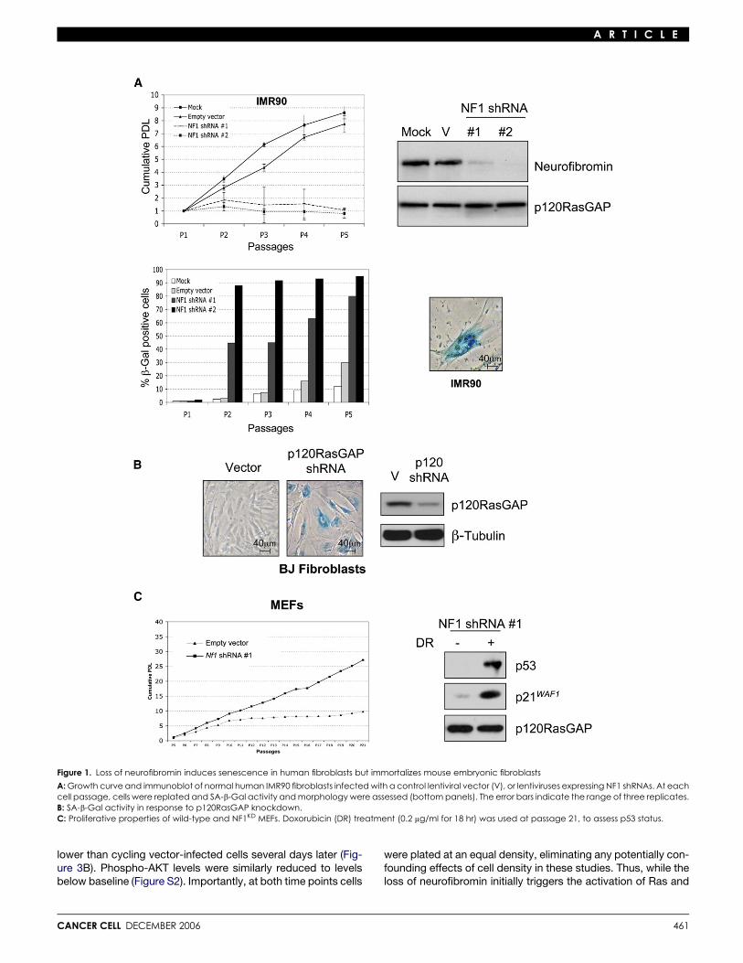
Effects of NF1 loss on primary human and mouse cellsTo investigate mechanisms that contribute to oncogene-in-duced senescence, the effects of neurofibromin deficiencywere compared in several cell systems in which Ras-mediatedsenescence has been well studied (Serrano et al., 1997; Weiand Sedivy, 1999). Human IMR90 or BJ fibroblasts were infectedwith lentiviruses encoding shRNAs recognizing two differentsequences within the NF1 gene. Both constructs suppressedneurofibromin expression and induced senescence within5 days (Figure 1A). Both IMR90 and BJ fibroblasts became sen-escent with roughly the same kinetics, as determined by an ob-served growth arrest, senescence-associated b-galactosidaseactivity (SA-b-Gal), and a flattened cellular morphology (Fig-ure 1A and data not shown). Notably, p120RasGAP shRNAsalso triggered cellular senescence, supporting the conclusionthat senescence in these cells was triggered by endogenousRas activation and was not due to an uncharacterized functionof neurofibromin (Figure 1B).
Because physiological levels of oncogenic Ras have beenreported to immortalize murine fibroblasts, rather than induce
460
senescence (Guerra et al., 2003; Tuveson et al., 2004), we alsoassessed the biological effects of neurofibromin deficiency inprimary MEFs. Consistent with the phenotype conferred by anactivated K-ras allele, Nf1-deficient MEFs did not undergo pre-mature senescence but were immortal. This phenotype was ob-served in cells where neurofibromin expression was abolishedthrough the use of the same shRNA-encoding construct usedin primary human cells, and in several independently derivedNf1-deficient MEF lines (Figure 1C and Figure S1A in the Supple-mental Data available with this article online). Immortalized Nf1-deficient and NF1KD MEFs maintained an intact p53 response todoxorubicin and retained ARF expression (Figure S1B), indicat-ing that these cells did not spontaneously immortalize by acquir-ing defects in these pathways. These results demonstrate thatthe same genetic event can trigger dramatically different biolog-ical responses in primary mouse cells (MEFs) versus primaryhuman fibroblasts. While species-specific differences may con-tribute to these observed effects, based on additional findingsdescribed below, we believe that this observation reflects differ-ent biochemical responses to this aberrant signal in these partic-ular cell types.
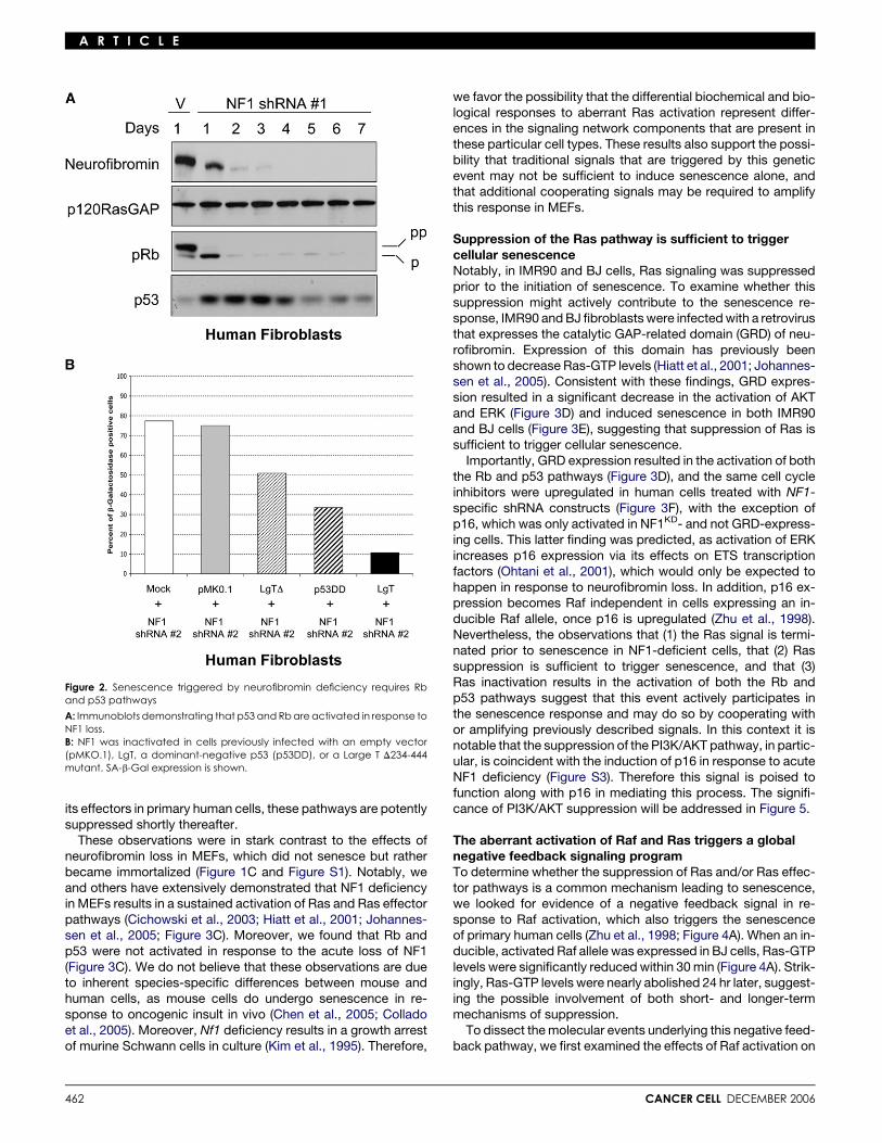
p53 and Rb pathways mediate senescence triggeredby the loss of NF1Senescence in response to Ras activation in human cells is me-diated by the activation of the Rb and p53 pathways (Serranoet al., 1997); therefore, the expression of these proteins was ex-amined in NF1KD cells. Similar to what has been reported in cellsectopically expressing an activated Ras allele, loss of neurofi-bromin induced the accumulation of p53 and the activation ofRb, as demonstrated by reduced levels of hyperphosphorylatedRb (Figure 2A). To assess the relative contribution of these path-ways to the senescence response, Rb and p53 pathways wereinactivated in isolation or together. The p53 pathway was dis-rupted by expressing a dominant-negative p53, and the Rbpathway was inactivated by expressing an SV40 Large T mu-tant, capable of binding and inactivating all three Rb familymembers, but deficient in its ability to bind p53 (Hahn et al.,2002). Inactivation of either the Rb or p53 pathway resulted ina partial decrease in the percentage of senescent cells but, inisolation, was unable to completely override the effects of NF1inactivation (Figure 2B). However, senescence was dramaticallyinhibited in cells expressing the wild-type SV40 Large T antigen,which binds and inactivates Rb family members and p53. Theseresults indicate that both the Rb and p53 pathways mediateNF1-related senescence in human cells.
Ras and Ras effector pathways are rapidly inactivatedin human cells following oncogenic insultTo examine the effects of neurofibromin loss on cell signaling,the activation of ERK and AKT were assessed in human cellsfollowing exposure to NF1-specific, shRNA-expressing lentivi-ruses. Neurofibromin expression was significantly decreasedwithin 2 days after infection (Figure 3A). Concomitant with thisdecrease was an increase in ERK and AKT activity as assessedby their phosphorylation status. Surprisingly, however, the acti-vation of these proteins peaked at day 2 and began to decreaserapidly thereafter. Phospho-AKT and phospho-ERK levels werepotently suppressed by days 3 and 4, respectively. Ras-GTPlevels exhibited similar kinetics: Ras-GTP levels were elevated2 days after infection and were suppressed to levels significantly
CANCER CELL DECEMBER 2006
A R T I C L E
Figure 1. Loss of neurofibromin induces senescence in human fibroblasts but immortalizes mouse embryonic fibroblasts
A: Growth curve and immunoblot of normal human IMR90 fibroblasts infected with a control lentiviral vector (V), or lentiviruses expressing NF1 shRNAs. At eachcell passage, cells were replated and SA-b-Gal activity and morphology were assessed (bottom panels). The error bars indicate the range of three replicates.B: SA-b-Gal activity in response to p120RasGAP knockdown.C: Proliferative properties of wild-type and NF1KD MEFs. Doxorubicin (DR) treatment (0.2 mg/ml for 18 hr) was used at passage 21, to assess p53 status.
lower than cycling vector-infected cells several days later (Fig-ure 3B). Phospho-AKT levels were similarly reduced to levelsbelow baseline (Figure S2). Importantly, at both time points cells
CANCER CELL DECEMBER 2006
were plated at an equal density, eliminating any potentially con-founding effects of cell density in these studies. Thus, while theloss of neurofibromin initially triggers the activation of Ras and
461
A R T I C L E
its effectors in primary human cells, these pathways are potentlysuppressed shortly thereafter.
These observations were in stark contrast to the effects ofneurofibromin loss in MEFs, which did not senesce but ratherbecame immortalized (Figure 1C and Figure S1). Notably, weand others have extensively demonstrated that NF1 deficiencyin MEFs results in a sustained activation of Ras and Ras effectorpathways (Cichowski et al., 2003; Hiatt et al., 2001; Johannes-sen et al., 2005; Figure 3C). Moreover, we found that Rb andp53 were not activated in response to the acute loss of NF1(Figure 3C). We do not believe that these observations are dueto inherent species-specific differences between mouse andhuman cells, as mouse cells do undergo senescence in re-sponse to oncogenic insult in vivo (Chen et al., 2005; Colladoet al., 2005). Moreover, Nf1 deficiency results in a growth arrestof murine Schwann cells in culture (Kim et al., 1995). Therefore,
Figure 2. Senescence triggered by neurofibromin deficiency requires Rband p53 pathways
A: Immunoblots demonstrating that p53 and Rb are activated in response toNF1 loss.B: NF1 was inactivated in cells previously infected with an empty vector(pMKO.1), LgT, a dominant-negative p53 (p53DD), or a Large T D234-444mutant. SA-b-Gal expression is shown.
462
we favor the possibility that the differential biochemical and bio-logical responses to aberrant Ras activation represent differ-ences in the signaling network components that are present inthese particular cell types. These results also support the possi-bility that traditional signals that are triggered by this geneticevent may not be sufficient to induce senescence alone, andthat additional cooperating signals may be required to amplifythis response in MEFs.
Suppression of the Ras pathway is sufficient to triggercellular senescenceNotably, in IMR90 and BJ cells, Ras signaling was suppressedprior to the initiation of senescence. To examine whether thissuppression might actively contribute to the senescence re-sponse, IMR90 and BJ fibroblasts were infected with a retrovirusthat expresses the catalytic GAP-related domain (GRD) of neu-rofibromin. Expression of this domain has previously beenshown to decrease Ras-GTP levels (Hiatt et al., 2001; Johannes-sen et al., 2005). Consistent with these findings, GRD expres-sion resulted in a significant decrease in the activation of AKTand ERK (Figure 3D) and induced senescence in both IMR90and BJ cells (Figure 3E), suggesting that suppression of Ras issufficient to trigger cellular senescence.
Importantly, GRD expression resulted in the activation of boththe Rb and p53 pathways (Figure 3D), and the same cell cycleinhibitors were upregulated in human cells treated with NF1-specific shRNA constructs (Figure 3F), with the exception ofp16, which was only activated in NF1KD- and not GRD-express-ing cells. This latter finding was predicted, as activation of ERKincreases p16 expression via its effects on ETS transcriptionfactors (Ohtani et al., 2001), which would only be expected tohappen in response to neurofibromin loss. In addition, p16 ex-pression becomes Raf independent in cells expressing an in-ducible Raf allele, once p16 is upregulated (Zhu et al., 1998).Nevertheless, the observations that (1) the Ras signal is termi-nated prior to senescence in NF1-deficient cells, that (2) Rassuppression is sufficient to trigger senescence, and that (3)Ras inactivation results in the activation of both the Rb andp53 pathways suggest that this event actively participates inthe senescence response and may do so by cooperating withor amplifying previously described signals. In this context it isnotable that the suppression of the PI3K/AKT pathway, in partic-ular, is coincident with the induction of p16 in response to acuteNF1 deficiency (Figure S3). Therefore this signal is poised tofunction along with p16 in mediating this process. The signifi-cance of PI3K/AKT suppression will be addressed in Figure 5.
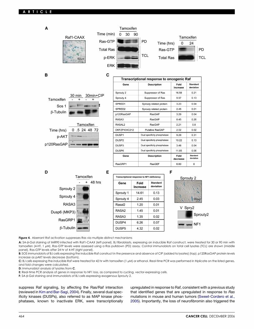
The aberrant activation of Raf and Ras triggers a globalnegative feedback signaling programTo determine whether the suppression of Ras and/or Ras effec-tor pathways is a common mechanism leading to senescence,we looked for evidence of a negative feedback signal in re-sponse to Raf activation, which also triggers the senescenceof primary human cells (Zhu et al., 1998; Figure 4A). When an in-ducible, activated Raf allele was expressed in BJ cells, Ras-GTPlevels were significantly reduced within 30 min (Figure 4A). Strik-ingly, Ras-GTP levels were nearly abolished 24 hr later, suggest-ing the possible involvement of both short- and longer-termmechanisms of suppression.
To dissect the molecular events underlying this negative feed-back pathway, we first examined the effects of Raf activation on
CANCER CELL DECEMBER 2006

A R T I C L E
Figure 3. Negative feedback suppression of theRas pathway underlies cellular senescence
A: Immunoblots of IMR90 lysates showing the dy-namic effects of NF1 loss on pERK and pAKTlevels. Intervening lanes are spliced out where in-dicated, but all time courses represent data fromthe same immunoblot.B: Ras activation in NF1KD versus control cells ondays 2 and 6 (see Experimental Procedures).The long exposure is shown to highlight the de-crease in Ras-GTP levels between vector andNF1 knockdown cells at this latter time point.C: pAKT, Rb, and p53 immunoblots of MEF lysatesin response to NF1 deficiency.D: Immunoblots of lysates from BJ fibroblasts in-fected with retrovirus expressing the GRD of neu-rofibromin, 9 days postinfection.E: SA-b-Gal staining of fibroblasts (IMR90 and BJ)infected with the GRD of neurofibromin.F: Immunoblots of cell lysates from BJ fibroblastsin response to NF1 loss, 6 days postselection.
known Ras regulators: RasGEFs and RasGAPs. Notably, theexchange factor SOS can be phosphorylated by ERK, whichhas been shown to result in its inactivation (Corbalan-Garciaet al., 1996; Dong et al., 1996). We found that SOS becamephosphorylated in this system within 30 min of Raf activation,as demonstrated by a shift in its electrophoretic mobility, andconfirmed by calf intestinal phosphatase (CIP) treatment, whicheliminated the slower migrating species (Figure 4B). In addition,Raf significantly increased the protein levels of p120RasGAPwithin 24–72 hr (Figure 4B). These observations suggestedthat multiple signals, involving posttranslational and perhapstranscriptional events, might be involved in triggering this nega-tive feedback loop.
To obtain a more comprehensive view of the signals partici-pating in this response, a preliminary microarray screen wasperformed on Raf-expressing cells as a means of identifying po-tential candidate genes. From this list of differentially regulatedgenes, those known to regulate Ras signaling were selectedfor real-time PCR and Western analysis (Figures 4C and 4D).Strikingly, several classes of genes, encoding proteins known
CANCER CELL DECEMBER 2006
to suppress Ras signaling at many levels, were shown to beexquisitely sensitive to sustained Raf activation. Among thesecandidates, Sprouty genes (2 and 4) were the most highly upre-gulated in response to Raf activation. Sprouty was originallyidentified in a Drosophila screen as a suppressor of FGF recep-tor signaling and has subsequently been shown to suppress theRas pathway, potentially acting at many levels (reviewed in Kimand Bar-Sagi, 2004). In addition, four RasGAP-encoding genes,including p120RasGAP, were transcriptionally upregulated inresponse to Raf activation, which would also be expected todirectly suppress Ras-GTP levels. RasGRP1, which encodes aRas exchange factor, was also downregulated by more than6-fold in response to Raf activation. Thus, Raf activation ap-pears to suppress Ras-GTP levels directly through a variety ofredundant mechanisms, although because of this redundancywe were not able to experimentally determine which of theseevents are absolutely critical in this system. Interestingly, othersuppressors of Ras signaling known to act on downstream com-ponents were also identified. The Sprouty-related genes Spred-2 and -4 were also highly upregulated and have been shown to
463
A R T I C L E
Figure 4. Aberrant Raf activation suppresses Ras via multiple distinct mechanisms
A: SA-b-Gal staining of IMR90 infected with Raf1-CAAX (left panel). BJ fibroblasts, expressing an inducible Raf construct, were treated for 30 or 90 min withtamoxifen (4-HT, 1 mM). Ras-GTP levels were assessed using a Ras pulldown (PD) assay. Control immunoblots on total cell lysates (TCL) are shown (middlepanel). Ras-GTP levels after 24 hr of 4-HT (right panel).B: SOS immunoblots of BJ cells expressing the inducible Raf construct in the presence and absence of CIP (added to lysates) (top). p120RasGAP protein levelsincrease as pAKT levels decrease (bottom).C: BJ cells expressing the inducible Raf were treated for 42 hr with tamoxifen (1 mM) or ethanol. Real-time PCR was performed in triplicate on the listed genes,and fold changes were calculated.D: Immunoblot analysis of lysates from C.E: Real-time PCR analysis of genes in response to NF1 loss, as compared to cycling, vector-expressing cells.F: SA-b-Gal staining and immunoblots of BJ cells expressing exogenous Sprouty 2.
suppress Raf signaling, by affecting the Ras/Raf interaction(reviewed in Kim and Bar-Sagi, 2004). Finally, several dual spec-ificity kinases (DUSPs), also referred to as MAP kinase phos-phatases, known to inactivate ERK, were transcriptionally
464
upregulated in response to Raf, consistent with a previous studythat identified genes that are upregulated in response to Rasmutations in mouse and human tumors (Sweet-Cordero et al.,2005). Importantly, the loss of neurofibromin also triggered the
CANCER CELL DECEMBER 2006
A R T I C L E
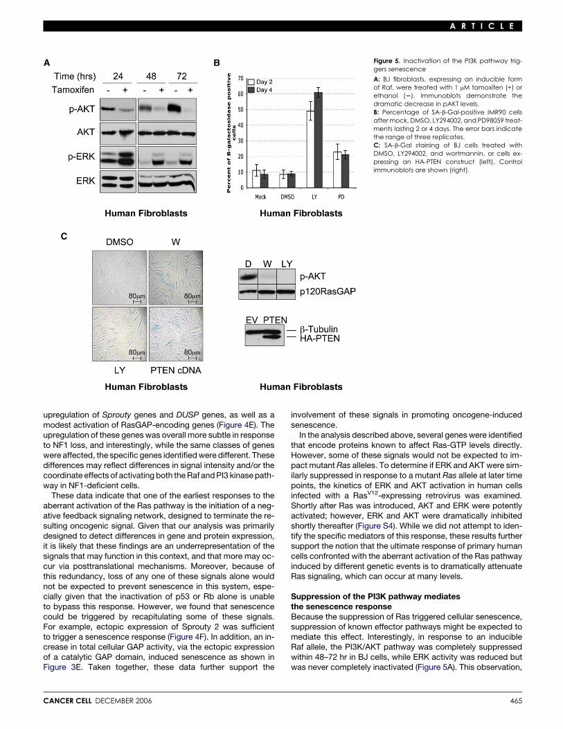
Figure 5. Inactivation of the PI3K pathway trig-gers senescence
A: BJ fibroblasts, expressing an inducible formof Raf, were treated with 1 mM tamoxifen (+) orethanol (2). Immunoblots demonstrate thedramatic decrease in pAKT levels.B: Percentage of SA-b-Gal-positive IMR90 cellsafter mock, DMSO, LY294002, and PD98059 treat-ments lasting 2 or 4 days. The error bars indicatethe range of three replicates.C: SA-b-Gal staining of BJ cells treated withDMSO, LY294002, and wortmannin, or cells ex-pressing an HA-PTEN construct (left). Controlimmunoblots are shown (right).
upregulation of Sprouty genes and DUSP genes, as well as amodest activation of RasGAP-encoding genes (Figure 4E). Theupregulation of these genes was overall more subtle in responseto NF1 loss, and interestingly, while the same classes of geneswere affected, the specific genes identified were different. Thesedifferences may reflect differences in signal intensity and/or thecoordinate effects of activating both the Raf and PI3 kinase path-way in NF1-deficient cells.
These data indicate that one of the earliest responses to theaberrant activation of the Ras pathway is the initiation of a neg-ative feedback signaling network, designed to terminate the re-sulting oncogenic signal. Given that our analysis was primarilydesigned to detect differences in gene and protein expression,it is likely that these findings are an underrepresentation of thesignals that may function in this context, and that more may oc-cur via posttranslational mechanisms. Moreover, because ofthis redundancy, loss of any one of these signals alone wouldnot be expected to prevent senescence in this system, espe-cially given that the inactivation of p53 or Rb alone is unableto bypass this response. However, we found that senescencecould be triggered by recapitulating some of these signals.For example, ectopic expression of Sprouty 2 was sufficientto trigger a senescence response (Figure 4F). In addition, an in-crease in total cellular GAP activity, via the ectopic expressionof a catalytic GAP domain, induced senescence as shown inFigure 3E. Taken together, these data further support the
CANCER CELL DECEMBER 2006
involvement of these signals in promoting oncogene-inducedsenescence.
In the analysis described above, several genes were identifiedthat encode proteins known to affect Ras-GTP levels directly.However, some of these signals would not be expected to im-pact mutant Ras alleles. To determine if ERK and AKT were sim-ilarly suppressed in response to a mutant Ras allele at later timepoints, the kinetics of ERK and AKT activation in human cellsinfected with a RasV12-expressing retrovirus was examined.Shortly after Ras was introduced, AKT and ERK were potentlyactivated; however, ERK and AKT were dramatically inhibitedshortly thereafter (Figure S4). While we did not attempt to iden-tify the specific mediators of this response, these results furthersupport the notion that the ultimate response of primary humancells confronted with the aberrant activation of the Ras pathwayinduced by different genetic events is to dramatically attenuateRas signaling, which can occur at many levels.
Suppression of the PI3K pathway mediatesthe senescence responseBecause the suppression of Ras triggered cellular senescence,suppression of known effector pathways might be expected tomediate this effect. Interestingly, in response to an inducibleRaf allele, the PI3K/AKT pathway was completely suppressedwithin 48–72 hr in BJ cells, while ERK activity was reduced butwas never completely inactivated (Figure 5A). This observation,
465
A R T I C L E
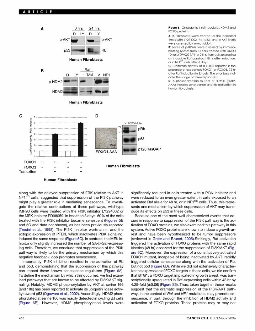
Figure 6. Oncogenic insult regulates HDM2 andFOXO proteins
A: BJ fibroblasts were treated for the indicatedtimes with LY294002. Rb, p53, and p-AKT levelswere assessed by immunoblot.B: Levels of p-HDM2 were assessed by immuno-blotting lysates from BJ cells treated with DMSO(D) or LY294002 (LY) for 24 hr, from cells expressingan inducible Raf construct 48 hr after induction,or in NF1KD cells after 6 days.C: Luciferase activity of a FOXO reporter in thepresence of exogenous FOXO1 or FOXO3, 72 hrafter Raf induction in BJ cells. The error bars indi-cate the range of three replicates.D: A phosphorylation mutant of FOXO1 (FKHR-AAA) induces senescence and Rb activation inhuman fibroblasts.
along with the delayed suppression of ERK relative to AKT inNF1KD cells, suggested that suppression of the PI3K pathwaymight play a greater role in mediating senescence. To investi-gate the relative contributions of these pathways, wild-typeIMR90 cells were treated with the PI3K inhibitor LY294002 orthe MEK inhibitor PD98059. In less than 3 days, 60% of the cellstreated with the PI3K inhibitor became senescent (Figures 5Band 5C and data not shown), as has been previously reported(Tresini et al., 1998). The PI3K inhibitor wortmannin and theectopic expression of PTEN, which inactivates PI3K signaling,induced the same response (Figure 5C). In contrast, the MEK in-hibitor only slightly increased the number of SA-b-Gal-express-ing cells. Therefore, we conclude that suppression of the PI3Kpathway is likely to be the primary mechanism by which thisnegative feedback loop promotes senescence.
Importantly, PI3K inhibition resulted in the activation of Rband p53, demonstrating that the suppression of this pathwaycan impact these known senescence regulators (Figure 6A).To define the mechanism by which this occurred, we first exam-ined pathways that are known to be affected by PI3K/AKT sig-naling. Notably, MDM2 phosphorylation by AKT at serine 166(and 186) has been reported to activate its ubiquitin ligase activ-ity toward p53 (Ogawara et al., 2002). Accordingly, HDM2 phos-phorylated at serine 166 was readily detected in cycling BJ cells(Figure 6B). However, HDM2 phosphorylation levels were
466
significantly reduced in cells treated with a PI3K inhibitor andwere reduced to an even greater extent in cells exposed to anactivated Raf allele for 48 hr, or in NF1KD cells. Thus, this repre-sents one mechanism by which suppression of AKT may trans-duce its effects on p53 in these cells.
Because one of the most well-characterized events that oc-curs in response to suppression of the PI3K pathway is the ac-tivation of FOXO proteins, we also examined this pathway in thissystem. Active FOXO proteins are known to induce a growth ar-rest and have been hypothesized to be tumor suppressors(reviewed in Greer and Brunet, 2005).Strikingly, Raf activationtriggered the activation of FOXO proteins with the same rapidkinetics (48 hr) observed for the suppression of PI3K/AKT (Fig-ure 6C). Moreover, the expression of a constitutively activatedFOXO1 mutant, incapable of being inactivated by AKT, rapidlytriggered cellular senescence along with the activation of Rb,but not p53 (Figure 6D). While we did not extensively character-ize the expression of FOXO targets in these cells, we did confirmthat BTG1, a FOXO target implicated in growth arrest, was tran-scriptionally upregulated in Raf-expressing cells within 48 hr by4.25-fold (60.06) (Figure S5). Thus, taken together these resultssuggest that the dramatic suppression of the PI3K/AKT path-way, in the context of Raf and NF1 mutations, may promote se-nescence, in part, through the inhibition of HDM2 activity andactivation of FOXO proteins. These proteins may or may not
CANCER CELL DECEMBER 2006
A R T I C L E
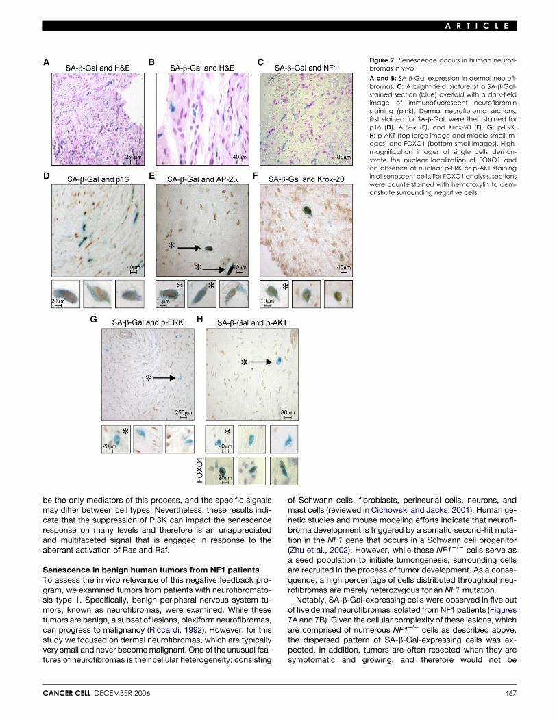
Figure 7. Senescence occurs in human neurofi-bromas in vivo
A and B: SA-b-Gal expression in dermal neurofi-bromas. C: A bright-field picture of a SA-b-Gal-stained section (blue) overlaid with a dark-fieldimage of immunofluorescent neurofibrominstaining (pink). Dermal neurofibroma sections,first stained for SA-b-Gal, were then stained forp16 (D), AP2-a (E), and Krox-20 (F). G: p-ERK.H: p-AKT (top large image and middle small im-ages) and FOXO1 (bottom small images). High-magnification images of single cells demon-strate the nuclear localization of FOXO1 andan absence of nuclear p-ERK or p-AKT stainingin all senescent cells. For FOXO1 analysis, sectionswere counterstained with hematoxylin to dem-onstrate surrounding negative cells.
be the only mediators of this process, and the specific signalsmay differ between cell types. Nevertheless, these results indi-cate that the suppression of PI3K can impact the senescenceresponse on many levels and therefore is an unappreciatedand multifaceted signal that is engaged in response to theaberrant activation of Ras and Raf.
Senescence in benign human tumors from NF1 patientsTo assess the in vivo relevance of this negative feedback pro-gram, we examined tumors from patients with neurofibromato-sis type 1. Specifically, benign peripheral nervous system tu-mors, known as neurofibromas, were examined. While thesetumors are benign, a subset of lesions, plexiform neurofibromas,can progress to malignancy (Riccardi, 1992). However, for thisstudy we focused on dermal neurofibromas, which are typicallyvery small and never become malignant. One of the unusual fea-tures of neurofibromas is their cellular heterogeneity: consisting
CANCER CELL DECEMBER 2006
of Schwann cells, fibroblasts, perineurial cells, neurons, andmast cells (reviewed in Cichowski and Jacks, 2001). Human ge-netic studies and mouse modeling efforts indicate that neurofi-broma development is triggered by a somatic second-hit muta-tion in the NF1 gene that occurs in a Schwann cell progenitor(Zhu et al., 2002). However, while these NF12/2 cells serve asa seed population to initiate tumorigenesis, surrounding cellsare recruited in the process of tumor development. As a conse-quence, a high percentage of cells distributed throughout neu-rofibromas are merely heterozygous for an NF1 mutation.
Notably, SA-b-Gal-expressing cells were observed in five outof five dermal neurofibromas isolated from NF1 patients (Figures7A and 7B). Given the cellular complexity of these lesions, whichare comprised of numerous NF1+/2 cells as described above,the dispersed pattern of SA-b-Gal-expressing cells was ex-pected. In addition, tumors are often resected when they aresymptomatic and growing, and therefore would not be
467
A R T I C L E
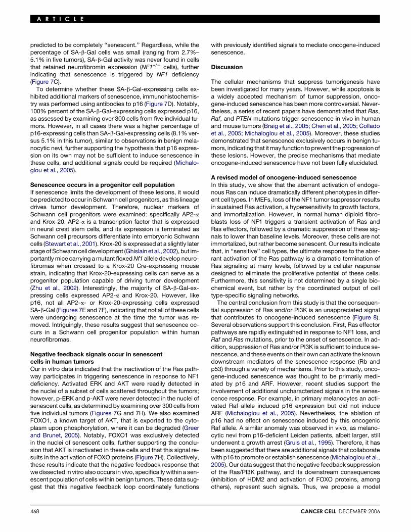
predicted to be completely ‘‘senescent.’’ Regardless, while thepercentage of SA-b-Gal cells was small (ranging from 2.7%–5.1% in five tumors), SA-b-Gal activity was never found in cellsthat retained neurofibromin expression (NF1+/2 cells), furtherindicating that senescence is triggered by NF1 deficiency(Figure 7C).
To determine whether these SA-b-Gal-expressing cells ex-hibited additional markers of senescence, immunohistochemis-try was performed using antibodies to p16 (Figure 7D). Notably,100% percent of the SA-b-Gal-expressing cells expressed p16,as assessed by examining over 300 cells from five individual tu-mors. However, in all cases there was a higher percentage ofp16-expressing cells than SA-b-Gal-expressing cells (8.1% ver-sus 5.1% in this tumor), similar to observations in benign mela-nocytic nevi, further supporting the hypothesis that p16 expres-sion on its own may not be sufficient to induce senescence inthese cells, and additional signals could be required (Michalo-glou et al., 2005).
Senescence occurs in a progenitor cell populationIf senescence limits the development of these lesions, it wouldbe predicted to occur in Schwann cell progenitors, as this lineagedrives tumor development. Therefore, nuclear markers ofSchwann cell progenitors were examined: specifically AP2-aand Krox-20. AP2-a is a transcription factor that is expressedin neural crest stem cells, and its expression is terminated asSchwann cell precursors differentiate into embryonic Schwanncells (Stewart et al., 2001). Krox-20 is expressed at a slightly laterstage of Schwann cell development (Ghislain et al., 2002), but im-portantly mice carrying a mutant floxed Nf1 allele develop neuro-fibromas when crossed to a Krox-20 Cre-expressing mousestrain, indicating that Krox-20-expressing cells can serve as aprogenitor population capable of driving tumor development(Zhu et al., 2002). Interestingly, the majority of SA-b-Gal-ex-pressing cells expressed AP2-a and Krox-20. However, likep16, not all AP2-a- or Krox-20-expressing cells expressedSA-b-Gal (Figures 7E and 7F), indicating that not all of these cellswere undergoing senescence at the time the tumor was re-moved. Intriguingly, these results suggest that senescence oc-curs in a Schwann cell progenitor population within humanneurofibromas.
Negative feedback signals occur in senescentcells in human tumorsOur in vitro data indicated that the inactivation of the Ras path-way participates in triggering senescence in response to NF1deficiency. Activated ERK and AKT were readily detected inthe nuclei of a subset of cells scattered throughout the tumors;however, p-ERK and p-AKT were never detected in the nuclei ofsenescent cells, as determined by examining over 300 cells fromfive individual tumors (Figures 7G and 7H). We also examinedFOXO1, a known target of AKT, that is exported to the cyto-plasm upon phosphorylation, where it can be degraded (Greerand Brunet, 2005). Notably, FOXO1 was exclusively detectedin the nuclei of senescent cells, further supporting the conclu-sion that AKT is inactivated in these cells and that this signal re-sults in the activation of FOXO proteins (Figure 7H). Collectively,these results indicate that the negative feedback response thatwe dissected in vitro also occurs in vivo, specifically within a sen-escent population of cells within benign tumors. These data sug-gest that this negative feedback loop coordinately functions
468
with previously identified signals to mediate oncogene-inducedsenescence.
Discussion
The cellular mechanisms that suppress tumorigenesis havebeen investigated for many years. However, while apoptosis isa widely accepted mechanism of tumor suppression, onco-gene-induced senescence has been more controversial. Never-theless, a series of recent papers have demonstrated that Ras,Raf, and PTEN mutations trigger senescence in vivo in humanand mouse tumors (Braig et al., 2005; Chen et al., 2005; Colladoet al., 2005; Michaloglou et al., 2005). Moreover, these studiesdemonstrated that senescence exclusively occurs in benign tu-mors, indicating that it may function to prevent the progression ofthese lesions. However, the precise mechanisms that mediateoncogene-induced senescence have not been fully elucidated.
A revised model of oncogene-induced senescenceIn this study, we show that the aberrant activation of endoge-nous Ras can induce dramatically different phenotypes in differ-ent cell types. In MEFs, loss of the NF1 tumor suppressor resultsin sustained Ras activation, a hypersensitivity to growth factors,and immortalization. However, in normal human diploid fibro-blasts loss of NF1 triggers a transient activation of Ras andRas effectors, followed by a dramatic suppression of these sig-nals to lower than baseline levels. Moreover, these cells are notimmortalized, but rather become senescent. Our results indicatethat, in ‘‘sensitive’’ cell types, the ultimate response to the aber-rant activation of the Ras pathway is a dramatic termination ofRas signaling at many levels, followed by a cellular responsedesigned to eliminate the proliferative potential of these cells.Furthermore, this sensitivity is not determined by a single bio-chemical event, but rather by the coordinated output of celltype-specific signaling networks.
The central conclusion from this study is that the consequen-tial suppression of Ras and/or PI3K is an unappreciated signalthat contributes to oncogene-induced senescence (Figure 8).Several observations support this conclusion. First, Ras effectorpathways are rapidly extinguished in response to NF1 loss, andRaf and Ras mutations, prior to the onset of senescence. In ad-dition, suppression of Ras and/or PI3K is sufficient to induce se-nescence, and these events on their own can activate the knowndownstream mediators of the senescence response (Rb andp53) through a variety of mechanisms. Prior to this study, onco-gene-induced senescence was thought to be primarily medi-ated by p16 and ARF. However, recent studies support theinvolvement of additional uncharacterized signals in the senes-cence response. For example, in primary melanocytes an acti-vated Raf allele induced p16 expression but did not induceARF (Michaloglou et al., 2005). Nevertheless, the ablation ofp16 had no effect on senescence induced by this oncogenicRaf allele. A similar anomaly was observed in vivo, as melano-cytic nevi from p16-deficient Leiden patients, albeit larger, stillunderwent a growth arrest (Gruis et al., 1995). Therefore, it hasbeen suggested that there are additional signals that collaboratewith p16 to promote or establish senescence (Michaloglou et al.,2005). Our data suggest that the negative feedback suppressionof the Ras/PI3K pathway, and its downstream consequences(inhibition of HDM2 and activation of FOXO proteins, amongothers), represent such signals. Thus, we propose a model
CANCER CELL DECEMBER 2006
A R T I C L E
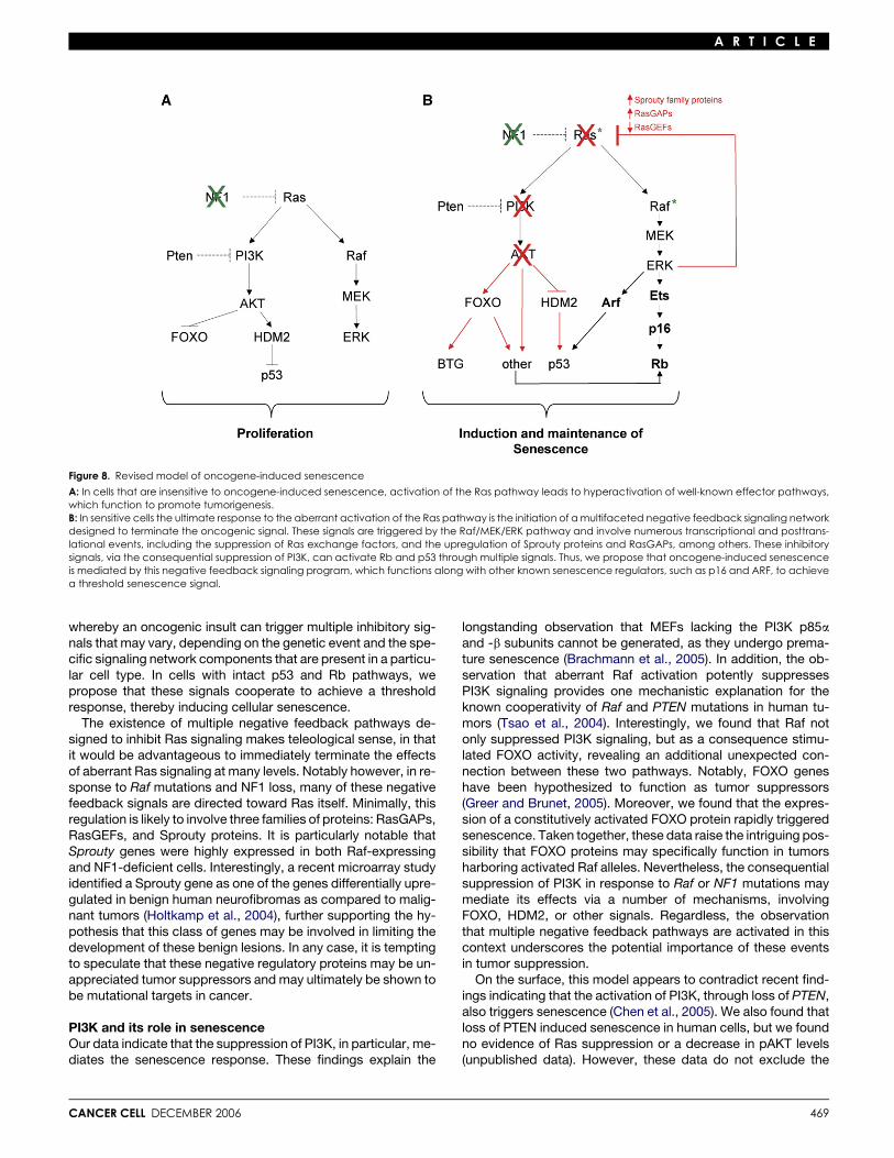
Figure 8. Revised model of oncogene-induced senescence
A: In cells that are insensitive to oncogene-induced senescence, activation of the Ras pathway leads to hyperactivation of well-known effector pathways,which function to promote tumorigenesis.B: In sensitive cells the ultimate response to the aberrant activation of the Ras pathway is the initiation of a multifaceted negative feedback signaling networkdesigned to terminate the oncogenic signal. These signals are triggered by the Raf/MEK/ERK pathway and involve numerous transcriptional and posttrans-lational events, including the suppression of Ras exchange factors, and the upregulation of Sprouty proteins and RasGAPs, among others. These inhibitorysignals, via the consequential suppression of PI3K, can activate Rb and p53 through multiple signals. Thus, we propose that oncogene-induced senescenceis mediated by this negative feedback signaling program, which functions along with other known senescence regulators, such as p16 and ARF, to achievea threshold senescence signal.
whereby an oncogenic insult can trigger multiple inhibitory sig-nals that may vary, depending on the genetic event and the spe-cific signaling network components that are present in a particu-lar cell type. In cells with intact p53 and Rb pathways, wepropose that these signals cooperate to achieve a thresholdresponse, thereby inducing cellular senescence.
The existence of multiple negative feedback pathways de-signed to inhibit Ras signaling makes teleological sense, in thatit would be advantageous to immediately terminate the effectsof aberrant Ras signaling at many levels. Notably however, in re-sponse to Raf mutations and NF1 loss, many of these negativefeedback signals are directed toward Ras itself. Minimally, thisregulation is likely to involve three families of proteins: RasGAPs,RasGEFs, and Sprouty proteins. It is particularly notable thatSprouty genes were highly expressed in both Raf-expressingand NF1-deficient cells. Interestingly, a recent microarray studyidentified a Sprouty gene as one of the genes differentially upre-gulated in benign human neurofibromas as compared to malig-nant tumors (Holtkamp et al., 2004), further supporting the hy-pothesis that this class of genes may be involved in limiting thedevelopment of these benign lesions. In any case, it is temptingto speculate that these negative regulatory proteins may be un-appreciated tumor suppressors and may ultimately be shown tobe mutational targets in cancer.
PI3K and its role in senescenceOur data indicate that the suppression of PI3K, in particular, me-diates the senescence response. These findings explain the
CANCER CELL DECEMBER 2006
longstanding observation that MEFs lacking the PI3K p85aand -b subunits cannot be generated, as they undergo prema-ture senescence (Brachmann et al., 2005). In addition, the ob-servation that aberrant Raf activation potently suppressesPI3K signaling provides one mechanistic explanation for theknown cooperativity of Raf and PTEN mutations in human tu-mors (Tsao et al., 2004). Interestingly, we found that Raf notonly suppressed PI3K signaling, but as a consequence stimu-lated FOXO activity, revealing an additional unexpected con-nection between these two pathways. Notably, FOXO geneshave been hypothesized to function as tumor suppressors(Greer and Brunet, 2005). Moreover, we found that the expres-sion of a constitutively activated FOXO protein rapidly triggeredsenescence. Taken together, these data raise the intriguing pos-sibility that FOXO proteins may specifically function in tumorsharboring activated Raf alleles. Nevertheless, the consequentialsuppression of PI3K in response to Raf or NF1 mutations maymediate its effects via a number of mechanisms, involvingFOXO, HDM2, or other signals. Regardless, the observationthat multiple negative feedback pathways are activated in thiscontext underscores the potential importance of these eventsin tumor suppression.
On the surface, this model appears to contradict recent find-ings indicating that the activation of PI3K, through loss of PTEN,also triggers senescence (Chen et al., 2005). We also found thatloss of PTEN induced senescence in human cells, but we foundno evidence of Ras suppression or a decrease in pAKT levels(unpublished data). However, these data do not exclude the
469
A R T I C L E
possibility that a negative feedback event may occur some-where downstream of AKT. Regardless, these findings indicatethat PI3K may be a critical sensor and regulator of the senes-cence response. Too little or too much PI3K activity can triggersenescence, the former mediated by the mechanisms de-scribed above, the latter perhaps resulting from the inductionof ARF and p53, as has been previously suggested (Chenet al., 2005). Nevertheless, in the context of Raf mutations andNF1 deficiency (in vitro and in vivo), negative feedback signalsdesigned to suppress PI3K signaling appear to predominate;the PI3K/AKT pathway is rapidly suppressed in both instances,and consequential downstream signals are engaged.
Negative feedback signals mediate senescence in vivoFinally, we show that these negative feedback pathways func-tion in vivo in benign human tumors from NF1 patients. Strik-ingly, in senescent cells within human neurofibromas, AKT andERK are completely suppressed and FOXO is activated. Ac-cordingly, we hypothesize that these signals underlie the senes-cence response in these lesions, thereby limiting the growth andprogression of these tumors. It has been suggested that senes-cence might suppress tumor development, if it occurs in a stemor progenitor cell population, resulting in the depletion of thisself-renewing pool of cells (Campisi, 2005). Previous studieshave not attempted to identify or characterize the senescentpopulation of cells in other benign tumors. In this regard it is no-table that the majority of senescent cells in benign human neu-rofibromas expressed both AP2-a and Krox-20, markers of earlySchwann cell progenitors. While the ‘‘cancer stem cell’’ in neu-rofibromas remains to be identified, these observations indicatethat a relatively undifferentiated population of cells can becomesenescent in these benign human tumors. If these cells repre-sent the cancer stem cell or a critical progenitor population,the senescence response may impede tumor development byeliminating the proliferative capacity of this population of cells.However, the challenge in the future will be to determine howa subset of cells escape this self-limiting response.
Experimental procedures
Cell culture and infections
Human IMR90 and BJ fibroblasts were subcultured 1:2 except prior to infec-
tion, when they were split 1:3. Wild-type and Nf12/2 MEFs were generated as
described (Cichowski et al., 2003). Retroviral and lentiviral infections were
performed as described (Johannessen et al., 2005). The following constructs
were in the pBabe retroviral vector: H-RasV12, hNF1-GRD-HA, PTEN, p53DD,
and inducible estrogen receptor-Raf-1 (Raf1:ER). Large T and Large T D234-
444 were in pMKO.1. Sprouty 2 was in pMSCV. The pLKO.1 lentiviral vector
containing the following shRNAs were used: NF1#1, 50-CAACAACTTCAAT
GCAGTCTT-30; NF1#2, 50-TTATAAATAGCCTGGAAAAGG-30; p120RasGAP,
50-GCTGCCTAACTTATCCATCTT-30; PTEN, 50-CGTATACAGGAACAATAT
TG-30.
For experiments in which control cells were compared to shRNA-express-
ing cells, cells were split to an equal cell density 16 hr prior to harvesting. The
following inhibitors were used: 50 mM PD98059 (Calbiochem), 30–50 mM
LY294002 (Calbiochem), and 100 nM wortmannin (Sigma-Aldrich). Wortman-
nin was re-added to the media every 3 hr during the first 12 hr, the media was
changed every 24 hr (and this protocol was repeated daily). Other inhibitors
were added daily with fresh media.
Growth curves
IMR90 fibroblasts were infected and selected for 2 days with puromycin
(2 mg/ml). Cells (2.0 3 105) were seeded in triplicate in 10 cm plates. Cells
were counted and seeded at a density of 2.0 3 105 every 4 days, for five
470
passages. Population doublings (PD) were determined by the following for-
mula: PD = Log (Nf/Ni)/Log2, where Nf = the number of cells counted and
Ni = the number of cells seeded (2.0 3 105). Cumulative PD numbers repre-
sent the sum of PDs from all previous passages. For MEFs, 3.0 3 105 cells
were seeded in 6 cm dishes, counted every 3 days, and reseeded at a density
of 3.0 3 105 (Sage et al., 2000).
SA-b-Gal activity
SA-b-Gal staining was performed as described (Dimri et al., 1995), and per-
centages were assessed by counting at least 300 cells. For human neurofi-
bromas, tumor tissue was obtained immediately after surgery, and small
pieces were fixed for 30 min in 3% paraformaldehyde, incubated as de-
scribed above, and then embedded and sectioned.
Immunoblotting
Western analysis and immunoprecipitations were performed as described
(Johannessen et al., 2005) using the following antibodies: phospho-Akt, total
Akt, phospho-Erk, total ERK, ph-HDM2 (Cell Signaling Technology); protein
kinase Ba/AKT1, actin, b-Tubulin, RASA3 (Sigma-Aldrich); p120RasGAP
(Transduction Laboratories); neurofibromin (UP69: a polyclonal antibody de-
signed to recognize the C terminus of neurofibromin), p53 (PAb 240 clone),
Sprouty 2, Sprouty 4, MKP3, RasGRP1, p16 (Santa Cruz Biotechnologies);
pRB, p16INK4a (BD Pharmingen); SOS1 (Upstate); p53 (DO7) (Signet).
Histology
Histological sections were cut into 5 mm sections from paraffin-embedded tis-
sue and stained with H&E where indicated. Unmasking was performed with
sodium citrate buffer (pH 6.0). Antibody dilutions and preferred buffers are
as follows: 1:100 NF1 antibody (4�C O/N, sc-67, Santa Cruz), 1:50 (TBST)
p16INK4a (Ab-7, MS-1064-P, Neomarkers), 1:100 (PBS) AP-2 a (clone 3B5,
Developmental Studies Hybridoma Bank, University of Iowa), 1:50 (TBST)
Krox-20 (PRB-236P, Covance), 1:50 (TBST) phospho-p44/42 MAPK
(#4376, Cell Signaling Technology), 1:50 (TBST) phospho-AKT (#3787, Cell
Signaling Technology), 1:50 (PBS) FOXO1 (#9462, Cell Signaling Technology).
Antibodies were detected using the Vectastain Elite Universal Kit (PK-6200,
Vector Laboratories).
Ras activation analysis
Ras-GTP levels were detected using a Ras activation assay, following the
manufacturer’s instructions (Upstate Biotechnology).
Luciferase assay
BJ fibroblasts, stably expressing the inducible Raf construct, were seeded
in triplicate in 24-well plates, at a density of 105 cells per well and treated
with ethanol or 1 mM of tamoxifen (Calbiochem). Cells were transfected
with 0.25 mg of the FKHR (FOXO1) or FKHRL1 (FOXO3) plasmid, 0.5 mg of
the Luciferase reporter gene, 0.25 mg of the pRL, and 1 mg of carrier DNA.
Forty-eight hours later, cells were lysed, and one-fifth of the sample was
assayed according to the Promega luciferase protocol.
Real-time PCR
Real-time PCR reactions were performed in triplicate using the Assays-on-
Demand Taqman Gene Expression Assays from Applied Biosystems. Probe
sets and methods are described in Figure S5.
Statistical analysis
All numerical data including error bars represent the mean 6 the standard
deviation.
Human subjects
The Office of Research Protections ruled that this study does not represent
human subjects research due to exemption 32 CFR 219.101(b)4: the re-
search involved collection or study of existing pathological specimens that
are publicly provided without identifiers.
Supplemental data
The Supplemental Data include five supplemental figures and can be found
with this article online at http://www.cancercell.org/cgi/content/full/10/6/
459/DC1/.
CANCER CELL DECEMBER 2006
A R T I C L E
Acknowledgments
We thank Lew Cantley, Ben Neel, Anne Brunet, Reuben Shaw, Sheila
Thomas, and Jeff Engelman for helpful discussions. We thank the Broad In-
stitute RNAi consortium (TRC) and William Hahn for providing shRNA con-
structs and William Hahn for the LgT constructs. We thank Martin McMahon
for the Raf:ER construct, Jeff Engelman for the PTEN shRNA construct, and
Nir Hacohen for the Sprouty 2 construct. The microarray analysis was per-
formed by the Biopolymers Facility at Harvard Medical School. This work
was supported by grants from the DOD (DAMD17-03-1-0350) and the NCI
(R01 CA111754-01).
Received: June 9, 2006Revised: August 9, 2006Accepted: October 3, 2006Published: December 11, 2006
References
Basu, T.N., Gutmann, D.H., Fletcher, J.A., Glover, T.W., Collins, F.S., andDownward, J. (1992). Aberrant regulation of ras proteins in malignant tumourcells from type 1 neurofibromatosis patients. Nature 356, 713–715.
Bollag, G., Clapp, D.W., Shih, S., Adler, F., Zhang, Y.Y., Thompson, P.,Lange, B.J., Freedman, M.H., McCormick, F., Jacks, T., and Shannon, K.(1996). Loss of NF1 results in activation of the Ras signaling pathway andleads to aberrant growth in haematopoietic cells. Nat. Genet. 12, 144–148.
Brachmann, S.M., Yballe, C.M., Innocenti, M., Deane, J.A., Fruman, D.A.,Thomas, S.M., and Cantley, L.C. (2005). Role of phosphoinositide 3-kinaseregulatory isoforms in development and actin rearrangement. Mol. Cell.Biol. 25, 2593–2606.
Braig, M., Lee, S., Loddenkemper, C., Rudolph, C., Peters, A.H., Schlegel-berger, B., Stein, H., Dorken, B., Jenuwein, T., and Schmitt, C.A. (2005). On-cogene-induced senescence as an initial barrier in lymphoma development.Nature 436, 660–665.
Campisi, J. (2005). Senescent cells, tumor suppression, and organismalaging: Good citizens, bad neighbors. Cell 120, 513–522.
Chen, Z., Trotman, L.C., Shaffer, D., Lin, H.K., Dotan, Z.A., Niki, M., Koutcher,J.A., Scher, H.I., Ludwig, T., Gerald, W., et al. (2005). Crucial role of p53-de-pendent cellular senescence in suppression of Pten-deficient tumorigenesis.Nature 436, 725–730.
Cichowski, K., and Jacks, T. (2001). NF1 tumor suppressor gene function:Narrowing the GAP. Cell 104, 593–604.
Cichowski, K., Santiago, S., Jardim, M., Johnson, B.W., and Jacks, T. (2003).Dynamic regulation of the Ras pathway via proteolysis of the NF1 tumor sup-pressor. Genes Dev. 17, 449–454.
Collado, M., Gil, J., Efeyan, A., Guerra, C., Schuhmacher, A.J., Barradas, M.,Benguria, A., Zaballos, A., Flores, J.M., Barbacid, M., et al. (2005). Tumourbiology: Senescence in premalignant tumours. Nature 436, 642.
Corbalan-Garcia, S., Yang, S.S., Degenhardt, K.R., and Bar-Sagi, D. (1996).Identification of the mitogen-activated protein kinase phosphorylation siteson human Sos1 that regulate interaction with Grb2. Mol. Cell. Biol. 16,5674–5682.
DeClue, J.E., Papageorge, A.G., Fletcher, J.A., Diehl, S.R., Ratner, N., Vass,W.C., and Lowy, D.R. (1992). Abnormal regulation of mammalian p21ras con-tributes to malignant tumor growth in von Recklinghausen (type 1) neurofi-bromatosis. Cell 69, 265–273.
Dimri, G.P., Lee, X., Basile, G., Acosta, M., Scott, G., Roskelley, C., Medrano,E.E., Linskens, M., Rubelj, I., Pereira-Smith, O., et al. (1995). A biomarker thatidentifies senescent human cells in culture and in aging skin in vivo. Proc.Natl. Acad. Sci. USA 92, 9363–9367.
Dong, C., Waters, S.B., Holt, K.H., and Pessin, J.E. (1996). SOS phosphory-lation and disassociation of the Grb2-SOS complex by the ERK and JNK sig-naling pathways. J. Biol. Chem. 271, 6328–6332.
CANCER CELL DECEMBER 2006
Downward, J. (2003). Targeting RAS signalling pathways in cancer therapy.Nat. Rev. Cancer 3, 11–22.
Ghislain, J., Desmarquet-Trin-Dinh, C., Jaegle, M., Meijer, D., Charnay, P.,and Frain, M. (2002). Characterisation of cis-acting sequences reveals a bi-phasic, axon-dependent regulation of Krox20 during Schwann cell develop-ment. Development 129, 155–166.
Greer, E.L., and Brunet, A. (2005). FOXO transcription factors at the interfacebetween longevity and tumor suppression. Oncogene 24, 7410–7425.
Gruis, N.A., van der Velden, P.A., Sandkuijl, L.A., Prins, D.E., Weaver-Feld-haus, J., Kamb, A., Bergman, W., and Frants, R.R. (1995). Homozygotesfor CDKN2 (p16) germline mutation in Dutch familial melanoma kindreds.Nat. Genet. 10, 351–353.
Guerra, C., Mijimolle, N., Dhawahir, A., Dubus, P., Barradas, M., Serrano, M.,Campuzano, V., and Barbacid, M. (2003). Tumor induction by an endoge-nous K-ras oncogene is highly dependent on cellular context. Cancer Cell4, 111–120.
Hahn, W.C., Dessain, S.K., Brooks, M.W., King, J.E., Elenbaas, B., Sabatini,D.M., DeCaprio, J.A., and Weinberg, R.A. (2002). Enumeration of the simianvirus 40 early region elements necessary for human cell transformation. Mol.Cell. Biol. 22, 2111–2123.
Hiatt, K.K., Ingram, D.D., Zhang, Y., Bollag, G., and Clapp, D.W. (2001). Neu-rofibromin GTPase-activating protein-related domains restore normalgrowth in Nf12/2 cells. J. Biol. Chem. 276, 7240–7245. Published onlineNovember 15, 2000. 10.1074/jbc.M009202200.
Holtkamp, N., Mautner, V.F., Friedrich, R.E., Harder, A., Hartmann, C., The-allier-Janko, A., Hoffmann, K.T., and von Deimling, A. (2004). Differentially ex-pressed genes in neurofibromatosis 1-associated neurofibromas and malig-nant peripheral nerve sheath tumors. Acta Neuropathol. (Berl.) 107, 159–168.
Johannessen, C.M., Reczek, E.E., James, M.F., Brems, H., Legius, E., andCichowski, K. (2005). The NF1 tumor suppressor critically regulates TSC2and mTOR. Proc. Natl. Acad. Sci. USA 102, 8573–8578.
Kim, H.J., and Bar-Sagi, D. (2004). Modulation of signalling by Sprouty: A de-veloping story. Nat. Rev. Mol. Cell Biol. 5, 441–450.
Kim, H.A., Rosenbaum, T., Marchionni, M.A., Ratner, N., and DeClue, J.E.(1995). Schwann cells from neurofibromin deficient mice exhibit activationof p21ras, inhibition of cell proliferation and morphological changes. Onco-gene 11, 325–335.
Michaloglou, C., Vredeveld, L.C., Soengas, M.S., Denoyelle, C., Kuilman, T.,van der Horst, C.M., Majoor, D.M., Shay, J.W., Mooi, W.J., and Peeper, D.S.(2005). BRAFE600-associated senescence-like cell cycle arrest of humannaevi. Nature 436, 720–724.
Ogawara, Y., Kishishita, S., Obata, T., Isazawa, Y., Suzuki, T., Tanaka, K.,Masuyama, N., and Gotoh, Y. (2002). Akt enhances Mdm2-mediated ubiqui-tination and degradation of p53. J. Biol. Chem. 277, 21843–21850.
Ohtani, N., Zebedee, Z., Huot, T.J., Stinson, J.A., Sugimoto, M., Ohashi, Y.,Sharrocks, A.D., Peters, G., and Hara, E. (2001). Opposing effects of Ets andId proteins on p16INK4a expression during cellular senescence. Nature 409,1067–1070.
Riccardi, V.M. (1992). Neurofibromatosis: Phenotype, Natural History, andPathogenesis, Second Edition (Baltimore: The Johns Hopkins UniversityPress).
Sage, J., Mulligan, G.J., Attardi, L.D., Miller, A., Chen, S., Williams, B., Theo-dorou, E., and Jacks, T. (2000). Targeted disruption of the three Rb-relatedgenes leads to loss of G(1) control and immortalization. Genes Dev. 14,3037–3050.
Serrano, M., Lin, A.W., McCurrach, M.E., Beach, D., and Lowe, S.W. (1997).Oncogenic ras provokes premature cell senescence associated with accu-mulation of p53 and p16INK4a. Cell 88, 593–602.
Seshadri, T., and Campisi, J. (1990). Repression of c-fos transcription and analtered genetic program in senescent human fibroblasts. Science 247, 205–209.
Stewart, H.J., Brennan, A., Rahman, M., Zoidl, G., Mitchell, P.J., Jessen,K.R., and Mirsky, R. (2001). Developmental regulation and overexpression
471
A R T I C L E
of the transcription factor AP-2, a potential regulator of the timing of Schwanncell generation. Eur. J. Neurosci. 14, 363–372.
Sweet-Cordero, A., Mukherjee, S., Subramanian, A., You, H., Roix, J.J.,Ladd-Acosta, C., Mesirov, J., Golub, T.R., and Jacks, T. (2005). An onco-genic KRAS2 expression signature identified by cross-species gene-expres-sion analysis. Nat. Genet. 37, 48–55.
Tresini, M., Mawal-Dewan, M., Cristofalo, V.J., and Sell, C. (1998). A phos-phatidylinositol 3-kinase inhibitor induces a senescent-like growth arrest inhuman diploid fibroblasts. Cancer Res. 58, 1–4.
Tsao, H., Goel, V., Wu, H., Yang, G., and Haluska, F.G. (2004). Genetic inter-action between NRAS and BRAF mutations and PTEN/MMAC1 inactivationin melanoma. J. Invest. Dermatol. 122, 337–341.
Tuveson, D.A., Shaw, A.T., Willis, N.A., Silver, D.P., Jackson, E.L., Chang, S.,Mercer, K.L., Grochow, R., Hock, H., Crowley, D., et al. (2004). Endogenous
472
oncogenic K-ras(G12D) stimulates proliferation and widespread neoplasticand developmental defects. Cancer Cell 5, 375–387.
Wei, S., and Sedivy, J.M. (1999). Expression of catalytically active telomerasedoes not prevent premature senescence caused by overexpression of onco-genic Ha-Ras in normal human fibroblasts. Cancer Res. 59, 1539–1543.
Wei, W., Hemmer, R.M., and Sedivy, J.M. (2001). Role of p14(ARF) in replica-tive and induced senescence of human fibroblasts. Mol. Cell. Biol. 21, 6748–6757.
Zhu, J., Woods, D., McMahon, M., and Bishop, J.M. (1998). Senescence ofhuman fibroblasts induced by oncogenic Raf. Genes Dev. 12, 2997–3007.
Zhu, Y., Ghosh, P., Charnay, P., Burns, D.K., and Parada, L.P. (2002). Neuro-fibromas in NF1: Schwann cell origin and role of tumor environment. Science296, 920–922.
CANCER CELL DECEMBER 2006