Understand the structuring of wheat-legume cakes to promote ...
Upload
independentCategory
view
1download
0
Stress-Induced Legume Root Nodule Senescence.Physiological, Biochemical, and Structural Alterations1
Manuel A. Matamoros, Lisa M. Baird, Pedro R. Escuredo, David A. Dalton, Frank R. Minchin,Inaki Iturbe-Ormaetxe, Maria C. Rubio, Jose F. Moran, Anthony J. Gordon, and Manuel Becana*
Departamento de Nutricion Vegetal, Estacion Experimental de Aula Dei, Consejo Superior de InvestigacionesCientıficas, Apdo 202, 50080 Zaragoza, Spain (M.A.M., P.R.E., I.I.-O., M.C.R., J.F.M., M.B.); Biology
Department, University of San Diego, San Diego, California 92110 (L.M.B.); Biology Department, Reed College,Portland, Oregon 97202 (D.A.D.); and Institute of Grassland and Environmental Research, Plas Gogerddan,
Aberystwyth SY23 3EB, United Kingdom (F.R.M., A.J.G.)
Nitrate-fed and dark-stressed bean (Phaseolus vulgaris) and pea(Pisum sativum) plants were used to study nodule senescence. Inbean, 1 d of nitrate treatment caused a partially reversible declinein nitrogenase activity and an increase in O2 diffusion resistance,but minimal changes in carbon metabolites, antioxidants, and otherbiochemical parameters, indicating that the initial decrease in ni-trogenase activity was due to O2 limitation. In pea, 1 d of darktreatment led to a 96% decline in nitrogenase activity and sucrose,indicating sugar deprivation as the primary cause of activity loss. Inlater stages of senescence (4 d of nitrate or 2–4 d of dark treatment),nodules showed accumulation of oxidized proteins and generalultrastructural deterioration. The major thiol tripeptides of un-treated nodules were homoglutathione (72%) in bean and glutathi-one (89%) in pea. These predominant thiols declined by approxi-mately 93% after 4 d of nitrate or dark treatment, but the loss ofthiol content can be only ascribed in part to limited synthesis byg-glutamylcysteinyl, homoglutathione, and glutathione synthetases.Ascorbate peroxidase was immunolocalized primarily in the in-fected and parenchyma (inner cortex) nodule cells, with large de-creases in senescent tissue. Ferritin was almost undetectable inuntreated bean nodules, but accumulated in the plastids and amy-loplasts of uninfected interstitial and parenchyma cells following 2or 4 d of nitrate treatment, probably as a response to oxidativestress.
Legume N2 fixation is particularly sensitive to environ-mental perturbations, including defoliation, water deficit,continuous darkness, and nitrate fertilization (Vance et al.,1979; Witty et al., 1986; Layzell et al., 1990). In most typesof stress, the initial decrease of nitrogenase activity is as-sociated with a decline in the O2 concentration reaching theinfected cells and bacteroids (Witty et al., 1986; Carroll etal., 1987; Layzell et al., 1990; Escuredo et al., 1996). Prolon-gation of stress induces premature nodule senescence,
which shares some features with natural senescence (nod-ule aging), such as the loss of N2 fixation, the increase inlytic activities, and the formation of green pigments fromleghemoglobin (Lb) (Pfeiffer et al., 1983; Sarath et al., 1986).This stress-induced senescence has been linked to the en-hanced production of oxidants and the lowering of antiox-idant defenses (Escuredo et al., 1996; Gogorcena et al.,1997). Oxidants include inorganic (H2O2) and organic (lip-id) peroxides as well as “catalytic iron,” the fraction of ironin plant tissues capable of catalyzing the generation ofhydroxyl radicals through Fenton reactions (Becana et al.,1998).
A major antioxidant mechanism operating in the nodulecytosol is the ascorbate-GSH cycle, which results ultimatelyin the detoxification of H2O2 at the expense of NAD(P)H.The pathway involves the concerted action of four en-zymes: ascorbate peroxidase (APX), dehydroascorbate re-ductase (DR), monodehydroascorbate reductase (MR), andglutathione reductase (GR), and requires a continuous sup-ply of ascorbate, thiols, and reduced pyridine nucleotides(Dalton et al., 1986, 1992). The initial enzyme of the path-way, APX, may account for up to 1% of the total solubleprotein of nodules (Dalton et al., 1998). The thiol tripeptideGSH (gGlu-Cys-Gly) also participates in the removal ofperoxides through the ascorbate-GSH cycle, but it performsadditional roles in plants, such as the transport and storageof sulfur, the control of redox status, and the detoxificationof heavy metals (Rennenberg, 1995; May et al., 1998). Thesynthesis of GSH involves two ATP-dependent reactionscatalyzed by the enzymes g-glutamylcysteinyl synthetase(gECS) and GSH synthetase (GSHS). Thiol tripeptides areparticularly abundant in the leaves, roots, and seeds oflegumes, where a thiol tripeptide homolog homogluta-thione (hGSH; gGlu-Cys-bAla), may be present in additionto or instead of GSH (Klapheck, 1988). Apparently, a spe-cific hGSH synthetase (hGSHS) catalyzes the second step ofhGSH synthesis in the leaves of some legumes (Macnicol,1987; Klapheck et al., 1988). It is not known whether hGSHand hGSHS are present in the nodules.
An entirely different antioxidant mechanism involvingthe sequestration of catalytic iron by ferritin may alsooperate in the nodules. Plant ferritins are composed of 24subunits and can store up to 4,500 atoms of iron in a safe,
1 This work was supported by grant nos. PB95– 0091 and PB98 –0522 from the Direccion General de Ensenanza Superior e Inves-tigacion Cientıfica (Ministry of Education and Culture, Spain) toM.B., and by fellowships from the Gobierno Vasco (M.A.M.), theEuropean Union (I.I.-O.), and the Ministry of Education and Cul-ture (P.R.E., M.C.R., J.F.M.).
* Corresponding author; e-mail [email protected]; fax34 –976 –575620.
Plant Physiology, September 1999, Vol. 121, pp. 97–111, www.plantphysiol.org © 1999 American Society of Plant Physiologists
97

nontoxic form (Briat and Lobreaux, 1997). In nodules, fer-ritin may also act as an iron reservoir for nitrogenase, Lb,and other iron-proteins, since ferritin protein increasesearly in nodulation and then declines concomitantly withthe increase in nitrogenase activity, heme, and non-hemeiron (Ragland and Theil, 1993). However, little is knownabout ferritin in senescent nodules. This information maybe of considerable interest because nodules are extremelyrich in iron (Ragland and Theil, 1993) and this may becomeavailable (catalytic iron) for Fenton reactions by proteolysisduring nodule senescence, either natural or induced bystress (Becana et al., 1998).
Many other important alterations related to oxygen, car-bon, and nitrogen metabolism occur in nodules duringsenescence, but the precise sequence of these biochemicalchanges is far from clear (Carroll et al., 1987; Layzell et al.,1990; Gordon et al., 1997). There is also a paucity of infor-mation regarding the structural changes involved in stress-induced nodule senescence. Light and electron microscopystudies have been carried out with nodules of detoppedalfalfa (Vance et al., 1979), dark-treated soybean (Cohen etal., 1986), and nitrate-treated lupine (Lorenzo et al., 1990).Nevertheless, legume symbioses differ in their tolerance tostress, and at least some of these differences may be relatedto the growth pattern (indeterminate versus determinate)of nodules (Sprent, 1980). Comparison of the inhibitoryeffects of stress on other legumes may provide insight intothe mechanisms underlying stress tolerance and nodulesenescence.
The general objective of the present study was to ascer-tain the time course of events leading to the loss of functionand structural deterioration of nodules following stressapplication to the plant. The specific objective was to gainfurther information on the role of some important antioxi-dants (APX, thiols, and ferritin) in the protection of legumeN2 fixation against the noxious effects of peroxides, freeradicals, and catalytic iron. Data presented in this paper areintended to complement two previous reports (Escuredo etal., 1996; Gogorcena et al., 1997) in providing an orthogonalcomparison of nitrate- and dark-stress-induced senescencein determinate and indeterminate nodules.
MATERIALS AND METHODS
Plants and Treatments
Nodulated bean (Phaseolus vulgaris L. cv Contender 3Rhizobium leguminosarum bv phaseoli 3622) and pea (Pisumsativum L. cv Lincoln 3 R. leguminosarum bv viciae NLV8)plants were grown in a perlite:vermiculite mixture (2:1) incontrolled-environment chambers as described by Gogor-cena et al. (1997). When bean had reached the late-vegetative stage (30–32 d), pots were divided at randominto three groups receiving 10 mm potassium nitrate for 1,2, or 4 d, and one group of controls receiving N-free nu-trient solution and harvested on the 3rd d. Likewise, whenpea plants had reached the late-vegetative stage (34–36 d),pots were divided at random into three groups placed inthe dark (with otherwise identical conditions) for 1, 2, or4 d, and one group of controls kept in the light and har-
vested on the 3rd d. These control plant harvests werearranged so that the maximum age difference betweentreated and control plants was 2 d. The same protocol wasused to produce dark-treated bean and nitrate-treated peaplants so as to obtain nodules for microscopic studies.Nodules to be used for light and electron microscopicanalyses were fixed immediately upon detachment, as de-scribed under “Light Microscopy Studies.” Nodules to beused for biochemical analyses were flash-frozen in liquidN2 and stored at 280°C for later analysis (within 4–5weeks).
Nodule Activity and Carbon Metabolites
Nitrogenase activity and root respiration of intact, un-disturbed plants were measured simultaneously using aflow-through gas system (Minchin et al., 1983) housed in acontrolled-environment chamber. Root systems weresealed in the growth pots and allowed to stabilize for 18 h.In vivo nitrogenase activity was measured as H2 evolution(Witty and Minchin, 1998) using electrochemical H2 sen-sors (City Technology, Portsmouth, UK) and respiratoryCO2 production was measured using an IR gas analyzer.Measurements were made in air for 5 min and then in a gasstream of 79% (v/v) Ar/21% (v/v) O2. Following exposureto Ar/O2, steady-state conditions were reached after 60 to80 min and the external O2 concentration was then in-creased over the range of 21% to 60% (8.55–24.54 mmol O2
L21) in steps of 5% or 10%. Each increase in O2 took 5 to 6min and was followed by a 20- to 25-min equilibrationperiod. These data were used to calculate the oxygen dif-fusion barrier (ODB) resistance and carbon costs of nitro-genase, as described in Escuredo et al. (1996).
Based on the maximum H2 production under Ar/O2, theelectron allocation coefficient for N2 was 0.67 for controlpea nodules and 0.73 for control bean nodules. The hup-negative genotype of the two Rhizobium strains used inthese studies was confirmed by Southern-blot analysis ofEcoRI-digested total DNA using a hup-specific DNA probeprepared by dioxigenin labeling of cosmid pAL618 con-taining the entire hup gene cluster from Rhizobium legu-minosarum bv viciae strain UPM791 (Leyva et al., 1990).
Lb was determined by a method based on the fluores-cence emitted by the tetrapyrrol ring after removal of ironby hot, saturated oxalic acid (LaRue and Child, 1979) usingmyoglobin (horse skeletal muscle, Calbiochem) as the stan-dard. Protein of the nodule cytosol and bacteroids wasdetermined with a commercial dye (Bio-Rad) using crys-talline BSA (Sigma) as the standard. Total lipids wereextracted from nodules at room temperature essentially asdescribed by Bligh and Dyer (1959).
Carbohydrates were extracted from 0.2 g of nodules with10 mL of boiling 80% (v/v) ethanol. The ethanol-solubleextracts were dried in vacuo at 40°C and the soluble com-pounds redissolved in 4 mL of water. The samples werecentrifuged at 20,000g for 10 min, and the contents of Glc,Fru, and Suc were determined spectrophotometrically at340 nm using enzymatic assays coupled to the formation ofNADH (Gonzalez et al., 1995). Starch was extracted fromthe ethanol-insoluble residue and quantified as the Glc
98 Matamoros et al. Plant Physiol. Vol. 121, 1999

released following digestion with amyloglucosidase (Mac-Rae, 1971).
Catalase and Enzymes of the Ascorbate-GSH Cycle
Antioxidant enzymes were extracted at 4°C from 0.5 g(bean) or 0.25 g (pea) of nodules with a mortar and pestle.Catalase and APX were extracted with 10 mL (bean) or 5mL (pea) of 50 mm potassium phosphate buffer (pH 7.0)containing 0.5% (w/v) PVP-10. DR, GR, and MR wereextracted with 5 mL (bean) or 2.5 mL (pea) of 50 mmpotassium phosphate buffer (pH 7.8) containing 1% (w/v)PVP-10, 0.2 mm Na2EDTA, and 10 mm b-mercaptoethanol.The homogenate was filtered through one layer of Mira-cloth (Calbiochem) and centrifuged at 15,000g for 20 min.
Catalase activity was assayed by following the decom-position of H2O2 at 240 nm (Aebi, 1984). APX and DRactivities were determined by measuring the oxidation ofascorbate at 290 nm (Asada, 1984) and the formation ofascorbate at 265 nm (Nakano and Asada, 1981), respec-tively. MR and GR activities were assayed by monitoringthe oxidation of NADH (Dalton et al., 1992) and NADPH(Dalton et al., 1986) at 340 nm, respectively.
All activities were measured at 25°C in 1-mL reactionmixtures within the linear range. Measurements weremade with a spectrophotometer (Lambda-16, Perkin-Cetus) during the first 1.5 to 3 min with no lag period(except for APX, which was measured after a lag of 40 s).Assays were made using sample volumes ranging from 10(catalase) to 100 mL (GR). Where appropriate, controlsmade by omitting or boiling extracts were run in parallel tocorrect for nonenzymatic rates, and buffers and reagentswere treated with Chelex resin to avoid contamination bytrace amounts of transition metal ions.
Thiols and Thiol Synthetase Activities
GSH and hGSH were extracted from 50 mg of noduleswith 500 mL of 200 mm methanesulfonic acid containing 0.5mm diethylenetriaminepentaacetic acid. After centrifuga-tion at 13,000g for 5 min, the supernatant was derivatizedat pH 8.0 with 2 mm monobromobimane (Fahey and New-ton, 1987). The bimane derivatives of GSH and hGSH wereseparated and quantified by HPLC (Waters) using an ana-lytical C18 column (3.9 3 150 mm; 4 mm; Nova-Pak, Waters)and a 15% (v/v) methanol/0.25% (v/v) acetic acid (pH 3.5)solvent at a constant flow of 1 mL min21. Detection was byfluorescence (model 474 scanning fluorescence detector,Waters) with excitation at 380 nm and emission at 480 nm.Standards of GSH (Sigma) and hGSH (obtained by chem-ical synthesis at the University of Nebraska, Lincoln) wereprocessed identically to the samples. The proportion ofthiol tripeptides present in the oxidized form was deter-mined by an enzymatic recycling procedure using yeast GRto reduce the disulfide forms and vinylpyridine as a thiolblocking agent (Griffith, 1980; Law et al., 1983).
The extraction and assays of gECS, GSHS, and hGSHSactivities were performed by modification of previous pro-tocols, based on the HPLC separation of synthesized gGlu-
Cys, GSH, and hGSH after derivatization with monobro-mobimane (Hell and Bergmann, 1988; Kocsy et al., 1996).
Other Metabolites
Ascorbate was extracted from 0.25 g of nodules with 5mL of 5% (w/v) metaphosphoric acid and quantified by amethod based on the ascorbate-dependent reduction ofiron (III) to iron (II). Formation of the complex betweeniron (II) with 2,29-dipyridyl was measured at 525 nm (Lawet al., 1983).
Pyridine nucleotides were extracted from 30 mg of nod-ules with 23 0.5 mL of 0.1 m NaOH (NADH and NADPH)or with 23 0.5 mL of 5% (w/v) TCA (NAD1 and NADP1)at room temperature. After thorough homogenization for90 to 120 s in an Eppendorf tube, the extracts were boiledfor 6 min, cooled on ice, and centrifuged at 13,000g for 6min at room temperature. The supernatant (25 mL for beanand 50 mL for pea) was made up to 100 mL with NaOH orTCA, and the nucleotides were quantified by the enzymaticcycling method of Matsumura and Miyachi (1980).
Oxidant Damage
Lipid peroxides were extracted from 0.5 g of noduleswith 5 mL of 5% (w/v) metaphosphoric acid and 100 mL of2% (w/v in ethanol) butyl hydroxytoluene (Minotti andAust, 1987). The extract was filtered through one layer ofMiracloth and centrifuged at 15,000g for 20 min. An aliquotof the supernatant was reacted with thiobarbituric acid atlow pH and 95°C and cooled to room temperature. Theresulting thiobarbituric acid-malondialdehyde adduct wasextracted with 1-butanol and quantified by HPLC as de-scribed in detail by Iturbe-Ormaetxe et al. (1998).
Protein carbonyl content was measured by derivatizationwith 2,4-dinitrophenyl-hydrazine as indicated by Levine etal. (1990) with some modifications. Proteins were extractedfrom 0.5 g of nodules with 5 mL of 100 mm potassiumphosphate (pH 7.0), 0.1% (v/v) Triton X-100, 1 mmNa2EDTA, and 2.5 mg each of aprotinin and leupeptin toprevent proteolysis of oxidized proteins during samplepreparation. After precipitation of possible contaminatingnucleic acids in the samples with 1% (w/v) streptomycinsulfate, an aliquot of 0.8 mL of the extracts was reactedwith 0.2 mL of 20 mm dinitrophenylhydrazine (in 2 m HCl),and another aliquot (control) with 0.2 mL of 2 m HCl for1 h, with vigorous shaking every 10 to 15 min. Proteinswere then precipitated with 10% (w/v) TCA, and the pelletwas washed four times with 1:1 (v/v) ethanol:ethyl acetate.Precipitated proteins were solubilized in 6 m guanidine-HCl (pH 4.5) by incubation for 30 min with shaking. Theinsoluble material was removed by centrifugation, and theabsorbance of the hydrazones (derivatized carbonyls) wasmeasured at 370 nm (Levine et al., 1990). To obtain moreaccurate results, the amount of protein to be analyzed forcarbonyl content was adjusted to 0.5 mg in all samples.
Light Microscopy Studies
Prior to immunodetection of relevant proteins at thelight or electron microscope levels, bean and pea nodule
Legume Root Nodule Senescence 99

extracts were subjected to western analysis (Cresswell etal., 1992). Polyclonal antibodies used in this study wereraised in rabbits against nitrogenase (universal antibody tothe ferroprotein of nitrogenase; courtesy of Dr. Paul Lud-den, University of Wisconsin, Madison), APX (soybeannodule cytosol; Dalton et al., 1993, 1998), and ferritin (soy-bean seeds; courtesy of Dr. Elizabeth Theil, Children’sHospital Oakland Research Institute, Oakland, CA).
Nodules to be used for immunodetection of nitrogenaseand APX were fixed overnight in 2% (v/v) paraformalde-hyde, 1.25% (v/v) glutaraldehyde, and 50 mm PIPES (pH7.2). Fixed samples were dehydrated in an ethanol series,embedded in LR White resin, and cut into 1-mm sectionswith a microtome (Reichert Ultracut R, Leica, Deerfield,IL). For light microscopy immunogold staining, slides wereincubated overnight at room temperature in a 1:50 dilutionof antibodies raised against nitrogenase or APX. For bothantigens, the slides were incubated for 1 h with secondaryantibody consisting of affinity-purified goat anti-rabbit IgGconjugated to colloidal gold (Auroprobe, Amersham) at a1:40 dilution. Silver enhancement solution (InsenSEM, Am-ersham) was applied at room temperature and monitoredfor sufficient development (15–25 min) following the pro-tocols of the manufacturer. All sections were counter-stained with 0.5% (w/v) safranin in water for 60 s. Repre-sentative nodule sections were photographed with amicroscope (Laborlux S, Leica) equipped with a camera(FE-2, Nikon).
For light microscopy immunofluorescence staining ofAPX, the secondary antibody consisted of affinity-purifiedgoat anti-rabbit antibodies conjugated to a Cy3 fluorophore(excitation at 550 nm and emission at 570 nm; JacksonImmunoResearch Laboratories, West Grove, PA) at a dilu-tion of 1:300 for 30 min. Sections were viewed with the lightmicroscope equipped with a rhodamine filter.
Electron Microscopy Studies
Ultrastructural studies and immunolocalization of APXand ferritin at the electron microscopy level were carried
out as indicated in Dalton et al. (1993), except that the APXand ferritin antibodies were used at dilutions of 1:500 and1:25, respectively, and grids were counterstained only withuranyl acetate. Nodule sections were obtained with aReichert Ultracut E microtome, and representative micro-graphs for all treatments were taken with a transmissionelectron microscope (model 900T, Zeiss) at 80 kV.
RESULTS
Nitrogenase Activity and Related Parameters
Bean plants were treated with 10 mm nitrate for up to 4 dto progressively induce nodule senescence, which wasmonitored by measuring general markers of metabolic ac-tivity (Table I). One day of nitrate application was suffi-cient to inhibit the in vivo nitrogenase activity by 73% andto increase the ODB resistance by 3-fold. At this stage, thetotal root respiration and carbon cost of nitrogenase did notvary, but there were significant increases in Lb and totalsoluble protein of nodules (Table I). After 2 d with nitrate,the carbon cost of nitrogenase increased 1.7-fold, whereasLb and soluble protein returned to control values. After 4 dthere was a further increase in the carbon cost of nitroge-nase up to 2.4-fold and decreases of 53% and 31% in Lb andsoluble protein, respectively, relative to the control. Anincrease in rhizosphere O2 produced a recovery of nitroge-nase activity at all stages of the nitrate treatment. Maxi-mum activities at increased O2 concentrations relative tothose at 21% O2 were 82%, 211%, 148%, and 130% after 0,1, 2, and 4 d, respectively (Table I).
Dark treatment of pea plants led to a drastic inhibitoryeffect on nodule activity. After only 1 d of dark, in vivonitrogenase activity was almost abolished, whereas totalroot respiration declined by 68% and the ODB resistancewas enhanced by 10-fold (Table I). However, there was noeffect on the carbon cost of nitrogenase, and Lb and solubleprotein only decreased by 29% and 14%, respectively. Pro-longation of dark stress for 1 more d lowered Lb content byan additional 63% but had no effect on total soluble pro-
Table I. Some general markers of metabolic activity in senescing nodules of bean treated with nitrate and of pea treated with darkness for 0,1, 2, and 4 d
Means (n 5 4–8) were compared by one-way analysis of variance and the Duncan’s range test. For each parameter and legume species, meansdenoted by the same letter do not differ significantly at P , 0.05.
ParameterBean Pea
0 d 1 d 2 d 4 d 0 d 1 d 2 d 4 d
Nitrogenase activitya (mmol H2
min21 plant21)1.66a 0.45b 0.29b 0.20b 0.76a 0.03b 0.00b 0.00b
Nitrogenase activity in O2b 1.36a 0.95b 0.43c 0.26c 0.56a 0.04b 0.00b 0.00b
Total root respiration (mmol CO2
min21 plant21)5.24a 4.76a 5.33a 5.59a 3.23a 1.03b 0.63bc 0.46c
ODB resistance (s cm21 3 1026) 0.67a 1.98b 1.87b 1.90b 1.47a 15.01b NDc NDCarbon cost of nitrogenase (mol
CO2 mol21 H2)1.90a 2.10ab 3.30bc 4.56c 2.02a 2.36a ND ND
Lb (nmol g21 fresh wt) 221a 260b 205a 102c 121a 86b 32c 22cSoluble protein (mg g21 fresh wt) 12.4a 16.0b 12.1a 8.6c 14.2a 12.2b 12.2b 12.9aba Maximum H2 production under Ar/21% O2. b Maximum H2 production at increased O2 concentrations following the Ar-induced
decline. c ND, Not determined.
100 Matamoros et al. Plant Physiol. Vol. 121, 1999

tein. There were no further significant changes after 4 d ofdarkness, except for total root respiration, which decreasedby 86% relative to control (Table I). Increasing rhizosphereO2 concentrations above 21% caused a slight rise in nitro-genase activity in pea after 1 d of dark, but did not induceany recovery of activity after 2 and 4 d of continuousdarkness.
Carbohydrates
Nitrate had considerably less effect on nodule carbohy-drates than dark stress. Treatment of bean plants withnitrate led to a progressive decline in Glc, Suc, and starch,whereas Fru increased by 35% after 1 d and declined to60% of the control value after 2 or 4 d (Table II). Thecontents of Glc and Suc decreased by 30% to 40% after 1 dof nitrate application, 47% to 57% after 2 d, and 75% after4 d. Starch was similarly affected after 1 and 2 d withnitrate, but remained at 48% of control after 4 d (Table II).
In contrast, nodules of pea plants dark-treated for 1 dhad lost 97% of their Suc content, along with 69% of Glc,53% of Fru, and 74% of starch (Table II). The contents of Glcand starch remained essentially constant at this low levelfollowing prolongation of the dark treatment to 2 or 4 d,whereas Fru declined by 85% relative to control and Sucvirtually disappeared after 4 d (Table II).
Antioxidant Enzymes
Nitrate application for 1 d had only a minor effect onantioxidant enzyme activities from bean nodules, with theexception of GR activity, which increased by 41% (TableIII). Following 2 d with nitrate, only APX activity haddecreased significantly with respect to control, but after 4 dthere was a general decline in activities of all enzymes of
the ascorbate-GSH cycle. The decreases ranged from ap-proximately 20% for DR and GR to 63% for APX. Catalaseactivity responded quite differently, with no variation after2 d of nitrate supply and a 52% decline after 4 d (Table III).
Likewise, after 1 d of dark stress, pea nodules did notshow apparent changes in antioxidant activities, but after2 d there were decreases of 26% to 32% in APX and GR, andcatalase activity increased by 31% (Table III). Following 4 dof dark treatment, there were major declines in the activi-ties of the enzymes of the ascorbate-GSH cycle: 40% to 50%for APX, GR, and MR, and 82% for DR. In contrast, catalaseactivity exceeded the control value by 44% (Table III).
Antioxidant Metabolites and Nucleotides
Some improvements of an HPLC method based on thederivatization of thiols to form highly fluorescent adducts(Fahey and Newton, 1987) allowed us to quantify sepa-rately GSH and hGSH in nodules (Table IV). Control (un-treated) bean nodules contained 103 nmol GSH and 262nmol hGSH g21 fresh weight, and pea nodules contained698 nmol GSH and 90 nmol hGSH g21 fresh weight. There-fore, GSH was the major thiol (89%) in pea nodules,whereas hGSH predominated (72%) in bean nodules. As-suming uniform distribution and 85% water content innodules, the total thiol tripeptide (GSH 1 hGSH) concen-trations in bean and pea nodules were estimated to be 0.4and 0.9 mm, respectively.
As occurred with antioxidant enzyme activities, nitrateapplication for 1 or 2 d had only a very limited effect on thecontent of antioxidant metabolites and pyridine nucleo-tides in bean nodules (Table IV). After 1 d with nitrate,there were only minor or moderate changes in GSH (37%decrease), ascorbate (20% increase), and NADH andNADP1 (18% decrease). The contents of other nucleotides
Table II. Carbohydrate content in senescing nodules of bean treated with nitrate and of pea treated with darkness for 0, 1, 2, and 4 d
MetaboliteaBean Pea
0 d 1 d 2 d 4 d 0 d 1 d 2 d 4 d
mg g21 fresh wt
Glc 173a 104b 92b 51c 348a 109b 125b 94bFru 89a 120b 53c 52c 70a 33b 11c 10cSuc 4445a 3087b 1902c 1111d 8946a 313b 207b 34cStarch 1095a 652b 466b 521b 8002a 2084b 1526b 1235b
a Statistical analysis of means (n 5 3) was performed as for Table I.
Table III. Antioxidant enzyme activities in senescing nodules of bean treated with nitrate and of pea treated with darkness for 0, 1, 2,and 4 d
EnzymeaBean Pea
0 d 1 d 2 d 4 d 0 d 1 d 2 d 4 d
mmol min21 g21 fresh wt
APX 8.67a 8.62a 6.17b 3.20c 8.91a 7.49b 6.59b 4.49cDR 0.67a 0.77b 0.70ab 0.55c 0.17a 0.13ab 0.09b 0.03cGR 0.41a 0.58b 0.48a 0.33c 0.62a 0.60a 0.42b 0.36bMR 3.20a 3.30a 2.62ab 1.83b 1.65a 1.52a 1.42a 1.00bCatalase 1340a 1390a 1340a 640b 160a 160a 210b 230c
a Statistical analysis of means (n 5 6–8) was performed as for Table I.
Legume Root Nodule Senescence 101

and hGSH remained essentially constant, as did the pro-portion of the disulfide forms of GSH and hGSH. Follow-ing 2 d with nitrate, both GSH and hGSH decreased by28%, but this decrease was not due to simple thiol oxida-tion, since 98% of total thiols were still present in thereduced form. Only after 4 d was there a significant butmodest increase in the proportion of the disulfide formsfrom 2% to 5%. At this stage, GSH and hGSH had declinedby 64% and 85%, respectively, and pyridine nucleotides,except NADPH, by 39% to 56%. In contrast, the ascorbatecontent of nodules treated with nitrate for 4 d was identicalto that of untreated nodules (Table IV).
The effect of prolonged darkness on pea nodule antioxi-dants and nucleotides was also moderate after 1 d, withdecreases in the range of 26% to 37% for ascorbate, NAD1,and NADH (Table IV). After 2 d, all parameters except thehGSH content experienced substantial declines rangingfrom approximately 24% for NADPH to 62% for NADH. Inaddition, the proportion of oxidized thiols rose signifi-cantly, from 14% to 20%. After 4 d, ascorbate and hGSHdeclined by approximately 60% and GSH by 92%. Thenodule content of pyridine nucleotides also decreasedmarkedly, albeit to a lower extent for NAD(H) than forNADP(H). Nevertheless, the NAD1 to NADH and theNADP1 to NADPH ratios were kept at approximately 3and 1, respectively, throughout the dark treatment.
Synthesis of GSH and hGSH
Because the nitrate and dark treatments led to majordeclines of GSH and hGSH in nodules that could not be
accounted for by oxidation to the disulfide forms (TableIV), we decided to investigate whether there is GSH andhGSH synthesis in nodules and, if so, whether this can beimpaired by the stress treatments. Optimization of previ-ous HPLC methods enabled us to assay all the enzymeactivities required for the synthesis of GSH and hGSH innodules (Table V), indicating that there is genuine synthe-sis of thiol tripeptides in the nodule tissue. The first com-mitted step for the synthesis of both tripeptides is cata-lyzed by gECS, and this activity was similar in control(untreated) bean and pea nodules. The second step isthought to be catalyzed by specific synthetases, eitherGSHS or hGSHS. Both activities were present in bean andpea nodules, but hGSHS activity predominated in beannodules, whereas GSHS activity predominated in pea. Thisis in agreement with the major thiol tripeptides found inthe respective nodules (Table IV), which strongly suggeststhat the relative abundance of GSH and hGSH is deter-mined at least in part by the corresponding thiol tripeptidesynthetase activities.
Nitrate supply of bean plants for 2 d led to decreases of40% in gECS activity and 60% in GSHS activity, but had noeffect on hGSHS activity; prolongation of treatment up to4 d led to a decline of 50% to 60% for all three activities ofnodules (Table V). Placement of pea plants in the dark for2 d caused decreases of 40% in GSHS and hGSHS activitiesof nodules; after 4 d, these activities had decreased by 50%to 60%, whereas the gECS activity of nodules had increasedby 55% relative to control (Table V).
Table IV. Antioxidant metabolites and nucleotides in senescing nodules of bean treated with nitrate and of pea treated with darkness for 0,1, 2, and 4 d
MetaboliteaBean Pea
0 d 1 d 2 d 4 d 0 d 1 d 2 d 4 d
nmol g21 fresh wt
Ascorbate 840a 1010b 950b 840a 460a 340b 300b 210cGSHb 103a 65b 75b 37c 698a 570a 378b 58chGSHb 262a 224ab 187b 40c 90a 106a 110a 36bOxidized thiolsc 2a 1a 2a 5b 16a 14a 20b 19bNAD1 32.3a 29.4ab 26.1b 17.8c 29.2a 18.8b 13.6bc 10.2cNADH 3.6a 2.9b 2.5bc 2.2c 11.3a 7.1b 4.3bc 3.3cNADP1 9.3a 7.6b 6.7b 4.1c 4.7a 4.6a 2.9b 2.5bNADPH 5.0a 4.6a 4.6a 4.9a 4.9a 3.9ab 3.7b 3.3b
a Statistical analysis of means (n 5 4–12) was performed as for Table I. b Determined by the HPLC method. c The percentage of thioltripeptides in the disulfide form was determined by the enzymatic method.
Table V. Activities of enzymes involved in GSH and hGSH synthesis in senescing nodules of beantreated with nitrate and of pea treated with darkness for 0, 2, and 4 d
EnzymeaBean Pea
0 d 2 d 4 d 0 d 2 d 4 d
nmol min21 g21 fresh wt
gECS 5.5a 3.3b 2.3c 4.9a 5.5a 7.6bGSHS 1.0a 0.4b 0.4b 18.0a 11.1b 9.3chGSHS 9.8a 9.6a 5.0b 12.0a 7.1b 4.5c
a Statistical analysis of means (n 5 3–4) was performed as for Table I.
102 Matamoros et al. Plant Physiol. Vol. 121, 1999

Oxidant Damage
The contents of lipid peroxides (malondialdehyde) andoxidatively modified proteins (carbonyl groups) were usedas markers of free radical damage in nodules. The responseof both parameters during senescence was, however, dis-tinctly different (Table VI). Nitrate treatment of bean ordark treatment of pea for 4 d caused decreases of 39% to48% in the content of lipid peroxides of nodules. In con-trast, both nitrate and prolonged darkness increased theamount of oxidized proteins in nodules by approximately30% (Table VI).
Immunolocalization of Nitrogenase and APX
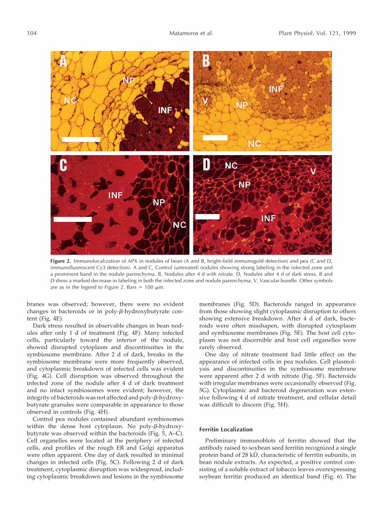
Immunogold localization of nitrogenase with silver en-hancement and dark-field light microscopy indicated thepresence of abundant protein in the infected cells of control(untreated) bean (Fig. 1A) and pea (data not shown) nod-ules. Little or no labeling occurred in nodules that had beenexposed to either of the stress treatments for 4 d (Fig. 1B).Using the same technique but with bright-field light mi-croscopy, APX protein was found to be localized predom-inantly in the endodermis and adjacent cell layers of thenodule parenchyma (inner cortex), as well as in the infectedzone of control bean nodules (Fig. 2A). Using immunoflu-orescence with the secondary antibody conjugated to thefluorophore Cy3, a similar distribution of APX protein wasnoticed in control pea nodules (Fig. 2C). Treatment witheither nitrate or dark led to a substantial decrease in label-ing intensity in both bean and pea nodules (Fig. 2, B andD), in agreement with the observed declines in enzymeactivity (Table III). As expected, bean and pea nodule sec-tions in which rabbit normal serum was used in place of theprimary antibody showed only very sparse backgroundlabeling.
The pattern of immunolocalization of APX at the electronmicroscopy level was similar to that observed at the lightmicroscopy level. No label was evident in negative controls(Fig. 3, A and B), whereas strong labeling was present inthe cytosol of the parenchyma and infected cells of controlbean and pea nodules (Fig. 3, C and D). Label was alsonoted in the cytosol of interstitial cells (Fig. 4B), over thesymbiosomes, and occasionally over the mitochondria andbacteroids (Fig. 4C). Immunolabeling of APX decreased inbean and pea nodule tissue with nitrate and dark stress.The location of labeling was not affected by either of thetwo treatments.
Ultrastructural Studies
Ultrathin sections of representative nodules from nitrate-treated bean and dark-treated pea were also examined byelectron microscopy to follow the structural changes occur-ring during senescence, and complement the physiologicaland biochemical data. In addition, nodules from dark-treated bean and nitrate-treated pea were processed inparallel to allow for orthogonal comparisons.
Infected cells of control (untreated) bean nodules weredensely packed with symbiosomes, each enclosing one tofour bacteroids. Bacteroids contained abundant poly-b-hydroxybutyrate granules, and organelles of the infectedcell, including mitochondria and plastids, were confined tothe periphery of the cell. Bean nodules treated for 1 or 2 dwith nitrate essentially had features similar to control nod-ules (Fig. 4D). After 4 d of nitrate treatment, significantdisruption of the host cytoplasm and of symbiosome mem-
Table VI. Oxidant damage of lipids and proteins in senescing nodules of bean treated with nitrate and of pea treated with darkness for 0, 1,2, and 4 d
ParameteraBean Pea
0 d 1 d 2 d 4 d 0 d 1 d 2 d 4 d
Lipid peroxides 0.27a 0.27a 0.16b 0.14b 1.66a 1.16b 0.88b 1.02bOxidized proteins 16.6a 16.4a 18.6a 22.3b 12.8a 15.3ab 17.3b 16.7b
a Statistical analysis of means (n 5 4–8) was performed as for Table I. Lipid peroxides are expressed in nmol malondialdehyde mg21 lipid andoxidized proteins in nmol carbonyl groups mg21 protein.
Figure 1. Immunolocalization of nitrogenase in bean nodules usingdark-field light microscopy. A, Control (untreated) nodules. Brightspots in the infected zone of this section indicate the presence ofnitrogenase. B, Nodule after 4 d with nitrate. Note the almost com-plete lack of staining in this section. NC, Nodule cortex; NP, noduleparenchyma; INF, infected zone. Bars 5 100 mm.
Legume Root Nodule Senescence 103
branes was observed; however, there were no evidentchanges in bacteroids or in poly-b-hydroxybutyrate con-tent (Fig. 4E).
Dark stress resulted in observable changes in bean nod-ules after only 1 d of treatment (Fig. 4F). Many infectedcells, particularly toward the interior of the nodule,showed disrupted cytoplasm and discontinuities in thesymbiosome membrane. After 2 d of dark, breaks in thesymbiosome membrane were more frequently observed,and cytoplasmic breakdown of infected cells was evident(Fig. 4G). Cell disruption was observed throughout theinfected zone of the nodule after 4 d of dark treatmentand no intact symbiosomes were evident; however, theintegrity of bacteroids was not affected and poly-b-hydroxy-butyrate granules were comparable in appearance to thoseobserved in controls (Fig. 4H).
Control pea nodules contained abundant symbiosomeswithin the dense host cytoplasm. No poly-b-hydroxy-butyrate was observed within the bacteroids (Fig. 5, A–C).Cell organelles were located at the periphery of infectedcells, and profiles of the rough ER and Golgi apparatuswere often apparent. One day of dark resulted in minimalchanges in infected cells (Fig. 5C). Following 2 d of darktreatment, cytoplasmic disruption was widespread, includ-ing cytoplasmic breakdown and lesions in the symbiosome
membranes (Fig. 5D). Bacteroids ranged in appearancefrom those showing slight cytoplasmic disruption to othersshowing extensive breakdown. After 4 d of dark, bacte-roids were often misshapen, with disrupted cytosplasmand symbiosome membranes (Fig. 5E). The host cell cyto-plasm was not discernible and host cell organelles wererarely observed.
One day of nitrate treatment had little effect on theappearance of infected cells in pea nodules. Cell plasmol-ysis and discontinuities in the symbiosome membranewere apparent after 2 d with nitrate (Fig. 5F). Bacteroidswith irregular membranes were occasionally observed (Fig.5G). Cytoplasmic and bacteroid degeneration was exten-sive following 4 d of nitrate treatment, and cellular detailwas difficult to discern (Fig. 5H).
Ferritin Localization
Preliminary immunoblots of ferritin showed that theantibody raised to soybean seed ferritin recognized a singleprotein band of 28 kD, characteristic of ferritin subunits, inbean nodule extracts. As expected, a positive control con-sisting of a soluble extract of tobacco leaves overexpressingsoybean ferritin produced an identical band (Fig. 6). The
Figure 2. Immunolocalization of APX in nodules of bean (A and B, bright-field immunogold detection) and pea (C and D,immunofluorescent Cy3 detection). A and C, Control (untreated) nodules showing strong labeling in the infected zone anda prominent band in the nodule parenchyma. B, Nodules after 4 d with nitrate. D, Nodules after 4 d of dark stress. B andD show a marked decrease in labeling in both the infected zone and nodule parenchyma. V, Vascular bundle. Other symbolsare as in the legend to Figure 2. Bars 5 100 mm.
104 Matamoros et al. Plant Physiol. Vol. 121, 1999

immunoblots also revealed that ferritin accumulated inbean nodules in response to nitrate treatment (Fig. 6).
The soybean antibody, however, exhibited very poorreactivity with pea ferritin (data not shown). Consequently,ferritin was only immunolocalized in bean nodules. Con-trol (untreated) bean nodule sections showed little labelingfor ferritin (Fig. 7A), but this was clearly visible after 2 or4 d of nitrate treatment (Fig. 7, B–D), confirming blotanalysis. After 2 d with nitrate, ferritin was localized in theplastids and amyloplasts of uninfected and infected cells,and occasionally over the bacteroids. The heaviest labelingwas observed in the amyloplasts of the uninfected intersti-tial cells and of the parenchyma cells (Fig. 7, B and C). Insome sections, scattered arrays or small clusters of ferritinparticles could be seen in the amyloplasts without theassistance of gold labeling (Fig. 7B). After 2 d (and espe-cially after 4 d) of nitrate treatment, large ferritin aggre-gates were easily observed in the amyloplasts (Fig. 7, C andD). Quite often, these large deposits did not show anyimmunolabeling, although it was observed in plastids oramyloplasts within the same nodule sections (Fig. 7C),indicating that the antibody was recognizing isolated fer-ritin particles and smaller ferritin deposits.
DISCUSSION
In this work we have obtained simultaneously an arrayof physiological, biochemical, and structural data for bean
and pea nodules induced to senesce by treating the plantswith nitrate or prolonged darkness. This allowed us tomonitor the progress of nodule senescence in a controlledmanner and to discern more readily between the reversibleand irreversible stages of stress application.
Nitrate-Induced Legume Nodule Senescence
Nitrate had a two-stage effect on bean nodules. In thefirst stage (1–2 d), there were major declines in the in vivonitrogenase activity and increases in the ODB resistanceand carbon cost of nitrogenase, but only moderate or nochanges in the nodule content of carbohydrates, antioxi-dants, and pyridine nucleotides; furthermore, there weresignificant increases in Lb, soluble protein, and ascorbate.At this early stage, there were no detectable changes in thenodules at the ultrastructural level. Therefore, it wouldappear that the decrease of nitrogenase activity after 1 to2 d may be attributed to O2 limitation at the ODB level andnot to biochemical factors such as degradation of nitroge-nase or Lb, oxidative damage of cell components, or sugardeprivation of host cells or bacteroids. However, the causalrelationship between nitrogenase activity and ODB opera-tion cannot be fully examined by the present data, and it ispossible that nitrogenase activity is decreased by an as yetunknown mechanism that results in closure of the ODB.
O2 limitation through the ODB was confirmed by thepartial recovery of nitrogenase activity in nitrate-treated
Figure 3. Immunogold localization of APX in control (untreated) nodules. A and B, Negative controls (normal rabbitantiserum in place of APX antibody) of bean and pea nodules, respectively, showing no label in the cytosol or organellesof infected cells. C and D, Bean and pea nodule sections, respectively, showing APX label over the cytosol of infected cells.b, Bacteroid; w, cell wall; m, mitochondrion; p, plastid. The bar in A 5 0.2 mm for all panels.
Legume Root Nodule Senescence 105

plants upon increasing the concentration of rhizosphericO2. This recovery, and the absence of structural and bio-chemical damage, indicate that at this early stage the effectof nitrate was still reversible. In contrast, in the second
stage (4 d), there was an almost complete loss of bothnitrogenase activity and protein, along with a marked de-cline in Lb, soluble protein, sugars, antioxidants (exceptascorbate), and nucleotides. There was also a general de-
Figure 4. Ultrastructural changes and APX localization in bean nodules after nitrate and dark treatments. A, Negative controlshowing no label in the cytosol, organelles, or symbiosome of a control (untreated) nodule. B and C, Control nodulesshowing APX localization in uninfected and infected cells. Label is seen frequently in the cytosol of both uninfected (B,arrowhead) and infected (C, arrowhead) cells, over the symbiosome, and occasionally over mitochondria (C, doublearrowhead) and bacteroids (C, asterisk). D, Detail of infected cell of the nodule after 2 d of nitrate treatment. Cells andbacteroids are similar in appearance to controls. E, Detail of infected cell of the nodule after 4 d with nitrate showing theabsence of symbiosome membrane, but little change in bacteroids or in poly-b-hydroxybutyrate granules. F, Detail ofinfected cells of the nodule after 1 d of dark stress showing disruption of the cytoplasm (arrowhead). Symbiosomemembranes are disrupted in both of the cells shown. G, Infected cells of nodule after 2 d of dark stress showing lesions inthe symbiosome membrane and the cytoplasmic disruption observed throughout the nodule. H, Nodule following 4 d ofdark stress showing general disruption of infected and uninfected cells. However, most bacteroids remain intact. m,Mitochondrion; p, plastid; px, peroxisome; and u, uninfected cell. Bars 5 0.4 mm.
106 Matamoros et al. Plant Physiol. Vol. 121, 1999

terioration of the nodule ultrastructure, in particular ofsymbiosome membranes, and an accumulation of oxidizedproteins. This stage of nitrate inhibition would thereforeappear to be essentially irreversible. However, these conclu-sions await final verification through recovery experiments.
The effects of nitrate on bean and pea nodules can becompared with the limited information available for other
legumes. In lupine and clusterbean nodules, nitrogenaseactivity (assayed in both cases with a closed system) wasinhibited by 35% after 3 to 5 d with 20 mm nitrate, andstructural degradation of lupine nodules was only recog-nizable after 10 d (Lorenzo et al., 1990; Swaraj et al., 1993).In bean and pea nodules, nitrogenase activity (assayed inboth cases with a flow-through gas system) was inhibited
Figure 5. Ultrastructural changes and APX localization in pea nodules after nitrate and dark treatments. A, Negative controlshowing no label in the cytosol, wall, or symbiosome of an untreated nodule. B, Infected cell of untreated nodule showingdistribution of APX label over the cytosol and symbiosome. Label was also observed in uninfected cells and occasionallyover mitochondria. C, Infected cell of nodule after 1 d of dark stress showing only minor disruption of symbiosomemembrane (arrowhead). D, Low-magnification micrograph of cell after 2 d of dark stress showing the extent of cytoplasmicdisruption and bacteroids with various degrees of breakdown. E, Infected cell of the nodule after 4 d of dark stress showingdisruption of symbiosome membranes and misshapen, disrupted bacteroids. F and G, Infected cells of nodules after 2 d withnitrate showing some cell plasmolysis (F, arrowhead), discontinuity in the symbiosome membrane, and misshapenbacteroids (G, arrowhead). H, Infected cell of the nodule after 4 d with nitrate showing the extensive cytoplasmic andbacteroid degeneration characteristic of this stage. A, B, C, E, F, and H, Bars 5 0.4 mm. D and G, Bars 5 2 mm.
Legume Root Nodule Senescence 107

by approximately 85% after only 2 d with 10 mm nitrate.Structural data revealed differences in the progression ofsenescence between pea and bean nodules. In pea nodulesthe plasmolysis of host cells and the disruption of symbio-some membranes and bacteroids were already evident af-ter 2 d, while in bean nodules, nitrate had little effect on theshape or poly-b-hydroxybutyrate content of bacteroidseven after 4 d. These data suggest that pea nodules areparticularly sensitive to nitrate.
Dark-Induced Legume Nodule Senescence
Prolonged darkness severely affected pea nodule metab-olism. After only 1 d of dark there were major effects ontotal root respiration, ODB resistance, and carbon costs ofnitrogenase. In addition there were moderate decreases inLb and in some antioxidants and nucleotides; however, themost affected parameters were by far the in vivo nitroge-
nase activity and the nodule Suc content, which decreasedby 97%. The almost complete depletion of Suc, the majorcarbon and energy source for host cell metabolism, alongwith the substantial decreases in other carbohydrates, is themost likely cause for the limitation of nitrogenase activityin dark-treated pea. This conclusion is supported by im-munoblots of nitrogenase showing no loss in the proteinafter 1 d of dark in both bean (Gogorcena et al., 1997) andpea (data not shown), and is also consistent with the find-ing that isolated bacteroids from dark-treated soybean re-tained 50% of the initial nitrogenase activity (Sarath et al.,1986) and fully recovered upon addition of succinate (Car-roll et al., 1987). After 2 d of dark, pea nodules showeddrastic decreases in Lb and carbohydrates, moderate de-creases of many antioxidants, accumulation of modifiedproteins, and evident symptoms of structural deterioration.After 4 d of dark, there was a general collapse of metabo-lism and extensive structural damage.
There are some significant differences in the response ofother legume nodules to dark stress. Thus, inhibition ofnitrogenase activity was much less dramatic in soybean(Sarath et al., 1986) and clusterbean (Swaraj et al., 1994)than it was in bean (Gogorcena et al., 1997) and pea (thiswork). Also, 2 d of dark had no significant effect on Lb ortotal soluble protein in nodules of soybean (Pfeiffer et al.,1983; Gordon et al., 1993), clusterbean (Swaraj et al., 1994),or bean (Gogorcena et al., 1997). However, in pea nodules,the same treatment caused a 74% decline in Lb but only a14% decline in total soluble protein, which is indicative ofa relatively high sensitivity of pea Lb to degradation com-
Figure 7. Electron micrographs of bean nodules showing ferritin localization. A, Control (untreated) nodule section showingscant deposition of gold label over infected and uninfected cells. B, Detail of parenchyma cell after 2 d with nitrate showingferritin localization in plastids and amyloplasts. A linear array of ferritin particles (arrowhead) is visible in the amyloplast.C, Detail of parenchyma cell after 2 d with nitrate showing large ferritin deposits not labeled with gold (arrowhead) as wellas disperse labeling of ferritin particles (double arrowhead) in the amyloplast. D, Amyloplast from a parenchyma cell after4 d with nitrate. A large aggregate of ferritin (arrowhead) is present, but did not label with gold. b, Bacteroid; p, plastid; ps,peribacteroid space; s, starch grain; v, vacuole; w, cell wall. Bars 5 0.4 mm.
Figure 6. Western analysis of ferritin in bean nodules using chemi-luminiscence detection. Lanes 0, 1, 2, and 4 were loaded withsoluble extracts (20 mg protein) from nodules treated with nitrate for0, 1, 2 and 4 d. Two extracts obtained independently were loaded foreach treatment. Lane T was loaded with a soluble extract (20 mgprotein) of transgenic tobacco leaves overexpressing soybean ferritin(Van Wuytswinkel et al., 1998), which served as a positive control. Inall lanes a single band at 28 kD, characteristic of ferritin subunits,was observed.
108 Matamoros et al. Plant Physiol. Vol. 121, 1999
pared with other cytosolic proteins. This conclusion is con-sistent with an early observation that exposure of peaplants to 3 d of dark was sufficient to induce greening ofnodules and breakdown of 50% of total heme (Roponen,1970). Because the pathway for Lb degradation in vivo islargely unknown, it can only be speculated that the rapidloss of pea Lb in dark-stressed nodules is due to a partic-ularly rapid activation or decompartmentation of proteaseslocated in the infected cells that display a high affinity forLb, especially at the acidic intracellular pH of senescingnodules (Pladys et al., 1991).
Thiols, APX, and Ferritin in Senescent Legume Nodules
Three important antioxidants, GSH and APX, which arecritical for the operation of the ascorbate-GSH cycle, andferritin, which is critical for the control of the cellularconcentration of catalytic iron, have been studied in furtherdetail to extend earlier studies on the mechanism of stress-induced nodule senescence.
An enzymatic method (Griffith, 1980) was previouslyemployed to estimate the concentration of GSH in nodules.Because this method could not distinguish between GSHand hGSH (Klapheck, 1988), and because it was uncertainwhether hGSH was present in nodules, we estimated theconcentration of total tripeptides in pea and bean nodulesas 0.9 mm (Escuredo et al., 1996; Gogorcena et al., 1997).Using HPLC, we found in this work that hGSH is presentin nodules at variable concentrations: 0.12 mm GSH and0.31 mm hGSH for bean nodules, and 0.82 mm GSH and0.11 mm hGSH for pea nodules. Thus, the enzymaticmethod tends to overestimate the thiol content in planttissues having primarily hGSH, because the reaction ofyeast GR with hGSH is faster than with GSH (Klapheck,1988). For pea nodules, which mainly contain GSH, bothmethods yielded similar results.
An obvious question raised in this work is why hGSH isthe major thiol in bean nodules. Although this cannot beanswered at present, our results show that the relativeabundance of GSH and hGSH in nodules is likely to bedictated by the presence of specific tripeptide synthetases:hGSHS in bean nodules and GSHS in pea nodules. Thisconclusion is reinforced by the partial purification of dis-tinct enzymes from pea (GSHS) and mungbean (hGSHS)leaves (Macnicol, 1987), which contain only GSH andhGSH, respectively (Klapheck, 1988). The fact that hGSH isthe only thiol tripeptide present in the leaves of somelegumes also implies that the corresponding chloroplastshave a functional ascorbate-hGSH pathway to avoid pho-tooxidative damage, and that hGSH and GSH share at leastsome antioxidative role in vivo.
The drastic decreases of the major thiols after 4 d oftreatment (85% hGSH in bean nodules and 92% GSH in peanodules) cannot be accounted for by oxidation to the di-sulfide forms or by the 50% to 60% decline in GSHS andhGSHS activities. Nor can it be ascribed to a limitation ofgECS activity, either directly (the activity only declined by60% in bean nodules and increased moderately in peanodules) or through the availability of Cys (the content ofthis substrate decreased by 50%–60% in both bean and pea
nodules after 4 d of treatment). The lack of evidence for amarked inhibition of thiol synthesis, together with the ex-tensive Lb degradation and oxidative reactions takingplace in stressed nodules that are manifest by a loss of Lbheme and accumulation of oxidized proteins, may providea clue for the large decline in GSH and hGSH. These thiolsmay be consumed by nonenzymatic reactions with acti-vated oxygen species or by enzymatic degradation, withthe possible formation of mixed disulfides between thiolsand proteins (Rennenberg, 1995). These aspects of thiolcatabolism in nodules, and in plants in general, remainvirtually unexplored.
Another antioxidant, APX, is critical for the disposal ofH2O2 in nodules. The APX protein was predominantlylocated to the parenchyma and infected zone, confirming aprevious report in which APX protein and mRNA wereshown to be enhanced in the parenchyma and infected cellsof alfalfa nodules (Dalton et al., 1998). In the same study,APX protein was also found to be increased in the noduleparenchyma and infected zone of several determinate nod-ules (Dalton et al., 1998). Our electron microscopy studiescorroborate the heterogeneous distribution of APX withinnodules and indicate that the protein is very abundant inthe cytosol of infected cells. The observation of occasionallabeling of APX in the mitochondria would explain thedetection of enzyme activity in purified mitochondria fromsoybean nodules (Dalton et al., 1993). The activity andcontent of APX protein largely decreased in senescent nod-ules, which may cause a lowering in protection againstH2O2 generated by the respiratory activity of the noduleparenchyma and infected cells.
Finally, we conducted studies to localize ferritin in beannodules and to determine the changes in ferritin contentduring senescence. Ferritin was barely detectable in controlnodules but accumulated in nodules treated with nitratefor 2 or 4 d. The very low content of ferritin protein foundin mature, untreated bean nodules is in agreement with theobservation that ferritin accumulates in young soybeannodules but declines in mature nodules, when iron storageis apparently no longer required (Bergersen, 1963; Raglandand Theil, 1993). Ferritin was predominantly found in theplastids and amyloplasts of interstitial cells in the infectedzone and in the parenchyma cells. The subcellular locationof ferritin in bean nodules is fully consistent with otherreports showing ferritin particles in plastids and amylo-plasts of soybean, alfalfa, and lupine nodules (Bergersen,1963; Lucas et al., 1998), in amyloplasts of soybean cellcultures (Briat and Lobreaux, 1997), and in chloroplastsand other plastids of several plants (Seckback, 1982).
The scattered arrays and large deposits of ferritin parti-cles observed after 2 or 4 d with nitrate closely resemble,respectively, the F-2 and F-3 types described by Seckback(1982). The latter category is defined as paracrystallineferritin arrangements (sometimes including small zones ofcrystalline structure), such as those observed in plastids ofiron-treated Xanthium without the assistance of gold label-ing. Our finding that the paracrystalline deposits offerritin-like material did not label as densely as would beexpected with the soybean antibody requires further inves-tigation, but similar ferritin structures, clearly immunola-
Legume Root Nodule Senescence 109
beled, accumulate in the cortex of senescing soybean andlupine nodules (Lucas et al., 1998).
Ferritin synthesis in plants is regulated by iron and isinduced by various adverse conditions, including ironoverload, which lead to oxidative stress (Briat and Lob-reaux, 1997). Recent experiments with de-rooted maizeplantlets have shown that H2O2 induces ferritin mRNAaccumulation in the presence of low iron concentrationsand that this effect is prevented by pretreatment of plant-lets with antioxidants, indicating that the induction of fer-ritin gene expression in this system requires an oxidativestep (Briat and Lobreaux, 1997). Based on these observa-tions, our results showing ferritin protein accumulation insenescent bean nodules may be interpreted as a response tooxidative stress. This oxidative stress is evidenced by theaccumulation of damaged proteins in nitrate-treated beannodules and is probably a consequence of the lowering ofantioxidant activities and the release of catalytic iron fromproteins (Becana et al., 1998). Further work is needed toestablish the mechanism for ferritin induction in nitrate-treated bean nodules and to determine whether a similarphenomenon occurs in other legume nodules under differ-ent types of stress.
ACKNOWLEDGMENTS
We thank Shannon Joyner (Reed College), Caron James (Insti-tute of Grassland and Environmental Research), and Gloria Rodrı-guez (Estacion Experimental de Aula Dei) for technical assistance.We are considerably indebted to Tomas Ruiz-Argueso (Univer-sidad Politecnica de Madrid) for the Southern analysis of hupgenes in the two Rhizobium strains used in this study, to PaulLudden (University of Wisconsin, Madison) for providing nitro-genase antibody, to Elizabeth Theil (Children’s Hospital OaklandResearch Institute, Oakland, CA) and Jean-Francois Briat (Univer-site de Montpellier II, France) for providing ferritin antibodies andhelp with western analysis, and to Chris Davitt and Valerie Lynch-Holm (Washington State University, Pullman) for sectioning andembedding samples for light microscopy.
Received April 16, 1999; accepted June 2, 1999.
LITERATURE CITED
Aebi H (1984) Catalase in vitro. Methods Enzymol 105: 121–126Asada K (1984) Chloroplasts: formation of active oxygen and its
scavenging. Methods Enzymol 105: 422–429Becana M, Moran JF, Iturbe-Ormaetxe I (1998) Iron-dependent
oxygen free radical generation in plants subjected to environ-mental stress: toxicity and antioxidant protection. Plant Soil 201:137–147
Bergersen FJ (1963) Iron in the developing soybean nodule. AustJ Biol Sci 16: 916–919
Bligh EG, Dyer WJ (1959) A rapid method of total lipid extractionand purification. Can J Biochem Physiol 37: 911–917
Briat JF, Lobreaux S (1997) Iron transport and storage in plants.Trends Plant Sci 2: 187–193
Carroll BJ, Hansen AP, McNeil DL, Gresshoff PM (1987) Effect ofoxygen supply on nitrogenase activity of nitrate- and dark-stressed soybean (Glycine max [L.] Merr.) plants. Aust J PlantPhysiol 14: 679–687
Cohen HP, Sarath G, Lee K, Wagner FW (1986) Soybean rootnodule ultrastructure during dark-induced stress and recovery.Protoplasma 132: 69–75
Cresswell A, Gordon AJ, Mytton LR (1992) The physiology andbiochemistry of cultivar-strain interactions in the white clover-Rhizobium symbiosis. Plant Soil 139: 47–57
Dalton DA, Baird LM, Langeberg L, Taugher CY, Anyan WR,Vance CP, Sarath G (1993) Subcellular localization of oxygendefense enzymes in soybean (Glycine max [L.] Merr.) root nod-ules. Plant Physiol 102: 481–489
Dalton DA, Joyner SL, Becana M, Iturbe-Ormaetxe I, ChatfieldJM (1998) Enhanced antioxidant defenses in the peripheral celllayers of legume root nodules. Plant Physiol 116: 37–43
Dalton DA, Langeberg L, Robbins M (1992) Purification andcharacterization of monodehydroascorbate reductase from soy-bean root nodules. Arch Biochem Biophys 292: 281–286
Dalton DA, Russell SA, Hanus FJ, Pascoe GA, Evans HJ (1986)Enzymatic reactions of ascorbate and glutathione that preventperoxide damage in soybean root nodules. Proc Natl Acad SciUSA 83: 3811–3815
Escuredo PR, Minchin FR, Gogorcena Y, Iturbe-Ormaetxe I, Klu-cas RV, Becana M (1996) Involvement of activated oxygen innitrate-induced senescence of pea root nodules. Plant Physiol110: 1187–1195
Fahey RC, Newton GL (1987) Determination of low-molecular-weight thiols using monobromobimane fluorescent labeling andhigh-performance liquid chromatography. Methods Enzymol143: 85–96
Gogorcena Y, Gordon AJ, Escuredo PR, Minchin FR, Witty JF,Moran JF, Becana M (1997) N2 fixation, carbon metabolism, andoxidative damage in nodules of dark-stressed common beanplants. Plant Physiol 113: 1193–1201
Gonzalez EM, Gordon AJ, James CL, Arrese-Igor C (1995) Therole of sucrose synthase in the response of soybean nodules todrought. J Exp Bot 46: 1515–1523
Gordon AJ, Minchin FR, Skøt L, James CL (1997) Stress-induceddeclines in soybean N2 fixation are related to nodule sucrosesynthase activity. Plant Physiol 114: 937–946
Gordon AJ, Ougham HJ, James CL (1993) Changes in levels ofgene transcripts and their corresponding proteins in nodules ofsoybean plants subjected to dark-induced stress. J Exp Bot 44:1453–1460
Griffith OW (1980) Determination of glutathione and glutathionedisulfide using glutathione reductase and 2-vinylpyridine. AnalBiochem 106: 207–212
Hell R, Bergmann L (1988) Glutathione synthetase in tobaccosuspension cultures: catalytic properties and localization.Physiol Plant 72: 70–76
Iturbe-Ormaetxe I, Escuredo PR, Arrese-Igor C, Becana M (1998)Oxidative damage in pea plants exposed to water deficit orparaquat. Plant Physiol 116: 173–181
Klapheck S (1988) Homoglutathione: isolation, quantification andoccurrence in legumes. Physiol Plant 74: 727–732
Klapheck S, Zopes H, Levels HG, Bergmann L (1988) Propertiesand localization of the homoglutathione synthetase from Phaseo-lus coccineus leaves. Physiol Plant 74: 733–739
Kocsy G, Brunner M, Ruegsegger A, Stamp P, Brunold C (1996)Glutathione synthesis in maize genotypes with different sensi-tivities to chilling. Planta 198: 365–370
LaRue TA, Child JJ (1979) Sensitive fluorometric assay for leghe-moglobin. Anal Biochem 92: 11–15
Law MY, Charles SA, Halliwell B (1983) Glutathione and ascorbicacid in spinach (Spinacia oleracea) chloroplasts. Biochem J 210:899–903
Layzell DB, Hunt S, Palmer GR (1990) Mechanisms of nitrogenaseinhibition in soybean nodules: pulse-modulated spectroscopyindicates that nitrogenase activity is limited by O2. Plant Physiol92: 1101–1107
Levine RL, Garland D, Oliver CN, Amici A, Climent I, Lenz A,Ahn B, Shaltiel S, Stadtman ER (1990) Determination of car-bonyl content in oxidatively modified proteins. Methods Enzy-mol 186: 464–478
Leyva A, Palacios JM, Murillo J, Ruiz-Argueso T (1990) Geneticorganization of the hydrogen uptake (hup) cluster of Rhizobiumleguminosarum. J Bacteriol 172: 1647–1655
110 Matamoros et al. Plant Physiol. Vol. 121, 1999
Lorenzo C, Lucas MM, Vivo A, de Felipe MR (1990) Effect ofnitrate on peroxisome ultrastructure and catalase activity innodules of Lupinus albus L. cv. Multolupa. J Exp Bot 41: 1573–1578
Lucas MM, Van de Sype G, Herouart D, Hernandez MJ, PuppoA, de Felipe MR (1998) Immunolocalization of ferritin in deter-minate and indeterminate legume root nodules. Protoplasma204: 61–70
Macnicol PK (1987) Homoglutathione and glutathione synthetasesof legume seedlings: partial purification and substrate specific-ity. Plant Sci 53: 229–235
MacRae JC (1971) Quantitative measurement of starch in verysmall amounts of leaf tissue. Planta 96: 101–108
Matsumura H, Miyachi S (1980) Cycling assay for nicotinamideadenine dinucleotides. Methods Enzymol 69: 465–470
May MJ, Vernoux T, Leaver C, Van Montagu M, Inze D (1998)Glutathione homeostasis in plants: implications for environmen-tal sensing and plant development. J Exp Bot 49: 649–667
Minchin FR, Witty JF, Sheehy JE, Muller M (1983) A major errorin the acetylene reduction assay: decreases in nodular nitroge-nase activity under assay conditions. J Exp Bot 34: 641–649
Minotti G, Aust SD (1987) The requirement for iron (III) in theinitiation of lipid peroxidation by iron (II) and hydrogen perox-ide. J Biol Chem 262: 1098–1104
Nakano Y, Asada K (1981) Hydrogen peroxide is scavenged byascorbate-specific peroxidase in spinach chloroplasts. Plant CellPhysiol 22: 867–880
Pfeiffer NE, Malik NSA, Wagner FW (1983) Reversible dark-induced senescence of soybean root nodules. Plant Physiol 71:393–399
Pladys D, Dimitrijevic L, Rigaud J (1991) Localization of a pro-tease in protoplast preparations in infected cells of French beannodules. Plant Physiol 97: 1174–1180
Ragland M, Theil EC (1993) Ferritin (mRNA, protein) and ironconcentrations during soybean nodule development. Plant MolBiol 21: 555–560
Rennenberg H (1995) Processes involved in glutathione metabo-lism. In RM Wallsgrove, ed, Amino Acids and Their Derivativesin Higher Plants. Cambridge University Press, Cambridge, UK,pp 155–171
Roponen I (1970) The effect of darkness on the leghemoglobincontent and amino acid levels in the root nodules of pea plants.Physiol Plant 23: 452–460
Sarath G, Pfeiffer NE, Sodhi CS, Wagner FW (1986) Bacteroidsare stable during dark-induced senescence of soybean root nod-ules. Plant Physiol 82: 346–350
Seckback J (1982) Ferreting out the secrets of plant ferritin: areview. J Plant Nutr 5: 369–394
Sprent JI (1980) Root nodule anatomy, type of export product andevolutionary origin in some Leguminosae. Plant Cell Environ 3:35–43
Swaraj K, Laura JS, Bishnoi NR (1993) Nitrate induced nodulesenescence and changes in activities of enzymes scavengingH2O2 in clusterbean (Cyamopsis tetragonaloba Taub.). J PlantPhysiol 141: 202–205
Swaraj K, Laura JS, Bishnoi NR (1994) Dark treatment effects onnitrogen fixation and enzymes associated with scavenging hy-drogen peroxide in clusterbean nodules. Plant Physiol Biochem32: 115–119
Vance CP, Heichel GH, Barnes DK, Bryan JW, Johnson LE (1979)Nitrogen fixation, nodule development, and vegetative re-growth of alfalfa (Medicago sativa L.) following harvest. PlantPhysiol 64: 1–8
Van Wuytswinkel O, Vansuyt G, Grignon N, Fourcroy P, Briat JF(1998) Iron homeostasis alteration in transgenic tobacco overex-pressing ferritin. Plant J 17: 93–97
Witty JF, Minchin FR (1998) Methods for the continuous measure-ment of O2 consumption and H2 production by nodulated le-gume root systems. J Exp Bot 49: 1041–1047
Witty JF, Minchin FR, Skøt L, Sheehy JE (1986) Nitrogen fixationand oxygen in legume root nodules. Oxf Surv Plant Mol Cell Biol3: 275–314
Legume Root Nodule Senescence 111
Copyright © 2022 FDOKUMEN