Intragenomic diversity of Rhizobium leguminosarum bv. trifolii clover nodule isolates
15
RESEARCH ARTICLE Open Access Intragenomic diversity of Rhizobium leguminosarum bv. trifolii clover nodule isolates Andrzej Mazur 1* , Grażyna Stasiak 1 , Jerzy Wielbo 1 , Agnieszka Kubik-Komar 2 , Monika Marek-Kozaczuk 1 and Anna Skorupska 1 Abstract Background: Soil bacteria from the genus Rhizobium are characterized by a complex genomic architecture comprising chromosome and large plasmids. Genes responsible for symbiotic interactions with legumes are usually located on one of the plasmids, named the symbiotic plasmid (pSym). The plasmids have a great impact not only on the metabolic potential of rhizobia but also underlie genome rearrangements and plasticity. Results: Here, we analyzed the distribution and sequence variability of markers located on chromosomes and extrachromosomal replicons of Rhizobium leguminosarum bv. trifolii strains originating from nodules of clover grown in the same site in cultivated soil. First, on the basis of sequence similarity of repA and repC replication genes to the respective counterparts of chromids reported in R. leguminosarum bv. viciae 3841 and R. etli CFN42, chromid-like replicons were distinguished from the pool of plasmids of the nodule isolates studied. Next, variability of the gene content was analyzed in the different genome compartments, i.e., the chromosome, chromid-like and ‘other plasmids’. The stable and unstable chromosomal and plasmid genes were detected on the basis of hybridization data. Displacement of a few unstable genes between the chromosome, chromid-like and ‘other plasmids’, as well as loss of some markers was observed in the sampled strains. Analyses of chosen gene sequences allowed estimation of the degree of their adaptation to the three genome compartments as well as to the host. Conclusions: Our results showed that differences in distribution and sequence divergence of plasmid and chromosomal genes can be detected even within a small group of clover nodule isolates recovered from clovers grown at the same site. Substantial divergence of genome organization could be detected especially taking into account the content of extrachromosomal DNA. Despite the high variability concerning the number and size of plasmids among the studied strains, conservation of the location as well as dynamic distribution of the individual genes (especially replication genes) of a particular genome compartment were demonstrated. The sequence divergence of particular genes may be affected by their location in the given genome compartment. The ‘other plasmid’ genes are less adapted to the host genome than the chromosome and chromid-like genes. Background Rhizobia are widely occurring soil bacteria that are able to establish nitrogen-fixing symbioses with legumes. Bacterium-plant interaction is a complex process in which specific plant and bacterial signals are exchanged resulting in formation of nodules, where rhizobia in the form of bacteroids fix nitrogen [1-3]. Rhizobial genomes are large and multipartite, com- posed of a single circular chromosome and a set of large plasmids [4-6]. The genes responsible for nodulation (nod) and nitrogen-fixation (nif-fix) are either carried by large plasmids (pSym) or are incorporated in the chro- mosome as symbiotic islands [7,8]. Large genomes of Rhizobiaceae and Bradyrhizobiaceae (above 6-9 Mb) are considered more ecologically advantageous in an envir- onment that is scarce in nutrients but diverse as regards carbon and energy sources. These genomes are dispro- portionately enriched in regulation and transport genes and in genes involved in secondary metabolism in * Correspondence: [email protected] 1 Department of Genetics and Microbiology, Maria Curie-Skłodowska University, Akademicka 19, 20-033 Lublin, Poland Full list of author information is available at the end of the article Mazur et al. BMC Microbiology 2011, 11:123 http://www.biomedcentral.com/1471-2180/11/123 © 2011 Mazur et al; licensee BioMed Central Ltd. This is an Open Access article distributed under the terms of the Creative Commons Attribution License (http://creativecommons.org/licenses/by/2.0), which permits unrestricted use, distribution, and reproduction in any medium, provided the original work is properly cited.
-
Upload
independent -
Category
Documents
-
view
4 -
download
0
Transcript of Intragenomic diversity of Rhizobium leguminosarum bv. trifolii clover nodule isolates

RESEARCH ARTICLE Open Access
Intragenomic diversity of Rhizobiumleguminosarum bv. trifolii clover nodule isolatesAndrzej Mazur1*, Grażyna Stasiak1, Jerzy Wielbo1, Agnieszka Kubik-Komar2, Monika Marek-Kozaczuk1 andAnna Skorupska1
Abstract
Background: Soil bacteria from the genus Rhizobium are characterized by a complex genomic architecturecomprising chromosome and large plasmids. Genes responsible for symbiotic interactions with legumes are usuallylocated on one of the plasmids, named the symbiotic plasmid (pSym). The plasmids have a great impact not onlyon the metabolic potential of rhizobia but also underlie genome rearrangements and plasticity.
Results: Here, we analyzed the distribution and sequence variability of markers located on chromosomes andextrachromosomal replicons of Rhizobium leguminosarum bv. trifolii strains originating from nodules of clovergrown in the same site in cultivated soil. First, on the basis of sequence similarity of repA and repC replicationgenes to the respective counterparts of chromids reported in R. leguminosarum bv. viciae 3841 and R. etli CFN42,chromid-like replicons were distinguished from the pool of plasmids of the nodule isolates studied. Next, variabilityof the gene content was analyzed in the different genome compartments, i.e., the chromosome, chromid-like and‘other plasmids’. The stable and unstable chromosomal and plasmid genes were detected on the basis ofhybridization data. Displacement of a few unstable genes between the chromosome, chromid-like and ‘otherplasmids’, as well as loss of some markers was observed in the sampled strains. Analyses of chosen genesequences allowed estimation of the degree of their adaptation to the three genome compartments as well as tothe host.
Conclusions: Our results showed that differences in distribution and sequence divergence of plasmid andchromosomal genes can be detected even within a small group of clover nodule isolates recovered from cloversgrown at the same site. Substantial divergence of genome organization could be detected especially taking intoaccount the content of extrachromosomal DNA. Despite the high variability concerning the number and size ofplasmids among the studied strains, conservation of the location as well as dynamic distribution of the individualgenes (especially replication genes) of a particular genome compartment were demonstrated. The sequencedivergence of particular genes may be affected by their location in the given genome compartment. The ‘otherplasmid’ genes are less adapted to the host genome than the chromosome and chromid-like genes.
BackgroundRhizobia are widely occurring soil bacteria that are ableto establish nitrogen-fixing symbioses with legumes.Bacterium-plant interaction is a complex process inwhich specific plant and bacterial signals are exchangedresulting in formation of nodules, where rhizobia in theform of bacteroids fix nitrogen [1-3].
Rhizobial genomes are large and multipartite, com-posed of a single circular chromosome and a set of largeplasmids [4-6]. The genes responsible for nodulation(nod) and nitrogen-fixation (nif-fix) are either carried bylarge plasmids (pSym) or are incorporated in the chro-mosome as symbiotic islands [7,8]. Large genomes ofRhizobiaceae and Bradyrhizobiaceae (above 6-9 Mb) areconsidered more ecologically advantageous in an envir-onment that is scarce in nutrients but diverse as regardscarbon and energy sources. These genomes are dispro-portionately enriched in regulation and transport genesand in genes involved in secondary metabolism in
* Correspondence: [email protected] of Genetics and Microbiology, Maria Curie-SkłodowskaUniversity, Akademicka 19, 20-033 Lublin, PolandFull list of author information is available at the end of the article
Mazur et al. BMC Microbiology 2011, 11:123http://www.biomedcentral.com/1471-2180/11/123
© 2011 Mazur et al; licensee BioMed Central Ltd. This is an Open Access article distributed under the terms of the Creative CommonsAttribution License (http://creativecommons.org/licenses/by/2.0), which permits unrestricted use, distribution, and reproduction inany medium, provided the original work is properly cited.

comparison with medium-and small-size genomecontaining bacteria [9].“Core” and “accessory” components of Rhizobium
genomes can be distinguished. Chromosomes with con-served gene content and order (synteny) are consideredas core. Accordingly, plasmids constitute the accessorygenome. Plasmids are more flexible than the chromo-somes, as defined by more frequent gene gains andlosses, even in the same species. They are heterogeneousin size and gene content and lack synteny even in clo-sely related species, except for genes involved in plasmidreplication and symbiotic properties [6,10,11]. In somespecies, such as Rhizobium leguminosarum, plasmidsmay comprise up to 35% of the total genome [6,7].Rhizobial plasmids are maintained in the cells via
repABC cassettes, comprising genes required for activesegregation (repAB) and initiation of replication (repC)[12]. The presence of several repABC operons within asingle genome, which are subjected to individual selec-tion pressure and divergence, could be the key elementof the existence of different plasmid incompatibilitygroups in cells and could drive the rearrangement ofgene organization and of their functions [11,13-15]. Itwas proposed that repABC plasmids coexisting in thesame strain most probably emerged by separate eventsof lateral transfer, which required evolution of differentincompatibility groups allowing simultaneous residenceof plasmids equipped with a similar replication/partitionsystem in a single bacterial species [12]. Thus, thedegree of divergence of the plasmid replication appara-tus, whose sequence is subject to strong evolutionarypressure and determines the ability to evade incompat-ibility between plasmids [13], and horizontal gene trans-fers are potential forces that shaped rhizobial genomes.Recently, some (not only rhizobial) extrachromosomal
replicons that have properties distinct from both chro-mosome and plasmids were reported and named “chro-mids” [16]. Chromids are characterized by presence ofsome important genes essential for growth under allconditions, with nucleotide composition and codonusage similar to the chromosome of the parental strain,and, by contrast, plasmid replication and partition sys-tems [16].Furthermore, recent analyses of Rhizobium etli strains
[11] showed that this species has a pangenomic struc-ture. By definition, a pangenome “determines the coregenome, which consists of genes shared by all thestrains studied and probably encoding functions relatedto the basic biology and phenotypes of the species” [17].The basis of the pangenome concept emerged from anobservation that each newly sequenced genome enrichedthe pool of species-specific genes with new ones [17,18].This makes it possible to detect, besides the core gen-omes, the dispensable genomes composed of both
chromosomal and plasmid genes, present only in someof the strains, which contribute to the species diversityand allow adaptation to new ecological niches and aspecific environment. Despite the overall genomic diver-gence, R. etli pangenome comprises a core genomecomposed of both chromosomal and plasmid sequences,as well as highly conserved symbiosis-related genes onthe pSym plasmid. The unusual variability observed inrhizobial genomes may further result from several typesof alterations, such as point mutations, deletions, ampli-fication of DNA, and from intragenome re-assortmentof sequences [19-21].The aim of this study was to evaluate the divergence
of genomes of a small population of R. leguminosarumbv. trifolii (Rlt) nodule isolates from clover plants grownin the same site in cultivated soil. Like the other mem-bers of the genus Rhizobium, the Rlt genomes were par-titioned into the chromosome and several largeplasmids, one of which carried symbiosis-related genes.The variability of the genome architecture involved notonly the number and size of the plasmids, but also thelocation of specific genes on the particular replicons.Distribution of repABC operon markers and other genesin the three genome compartments: the chromosome,chromid-like and ‘other plasmids’ was assessed. Wefound “stable” genes that were permanently located in aspecific genome compartment, as well as “unstable”ones, which were detected in different replicons of thesampled strains. Sequences of selected chromosome andplasmid genes were subjected to an assessment of adap-tation to a particular genome compartment by analysesof codon usage and codon adaptation index. A potentialevolutionary pathway of Rlt strains was proposed on thebasis of gene sequences and their distribution.
MethodsR. leguminosarum bv. trifolii (Rlt) strains129 R. leguminosarum isolates were obtained fromnodules of red clover (Trifolium pratense L. cv. Dajana)growing in sandy loam (N:P:K 0.157:0.014:0.013%).Plants were grown on 1 m2 plot for six weeks betweenMay and June 2008. Afterwards, ten randomly chosenclover plants growing in each other’s vicinity were har-vested, the nodules were collected, surface-sterilized,crushed and their content plated on 79CA medium [22].Strains isolated from the nodules were purified by suc-cessive streaking of single colonies and pure cultureswere used in further experiments.
DNA methodsStandard techniques were used for labeling of DNA,Southern hybridization and agarose gel electrophoresis[23]. DNA probes for Southern hybridizations wereobtained by PCR amplification with RtTA1 genomic
Mazur et al. BMC Microbiology 2011, 11:123http://www.biomedcentral.com/1471-2180/11/123
Page 2 of 15

Table 1 Primers and probes used in this study
RtTA1repliconname
Probename
Probe description Primers GenBankaccession
no
cha pssL(Pss-I)
1300 bp of Pss-I region encoding part of putative flippase PssL pssLFw5’-TCTTATCCGCCACGAATCTCC-3’pssLRw5’-GCCAGGTGAAGGGCCAGCGCCAACT-3’
DQ384110
rfbADBC(Pss-V)
2956 bp of Pss-V region encoding lipopolysaccharide biosynthesisproteins
rfbAFw5’-TCGAGATAGGTGCGGTTGACGTCG-3’rfbCRw5’-GCAGGGCAACGCTGGTGCGCTGC-3’
DQ679959
bioA 445 bp fragment encoding adenosylmethionine-8-amino-7-oxononanoate aminotransferase
bioA35’-CCTCGTCGAAGATCAGAAGG-3’bioA55’-TCTACACCAACTCCGGTTCC-3’
DQ535896
rpoH2 487 bp fragment encoding RNA polymerase sigma factor rpoH2Fw5’-TGGTGCAGGAGGGCTATGTT-3’rpoH2Rw5’-TCGCGTTCGTTGAGATGTTTC-3’
DQ366597
dnaC 624 bp fragment encoding DNA helicase dnaCFw5’-CAGCCCGGCATTTTCACC-3’dnaCRw5’-CTGCGGCCGTTTATCGTC-3’
DQ855524
dnaK 645 bp fragment encoding heat shock protein 70 family dnaKFw15’-CGTCATCACCGTTCCCGCCTACTT-3’dnaKRw15’-TTGCCGAACAGCTGCTTGACGACT-3’
DQ535895
exoR 416 bp fragment encoding negative regulator ofexopolysaccharide synthesis
exoRFw5’-GTTGCCGCCTGCCTGAGATGAAC-3’exoRRw5’-GAGAGCAGCGCGTTGACGAAGAAG-3’
DQ347956
rrl 1135 bp fragment comprising rRNA genes rrl and rrs-rrl intergenicspacer
FGPS14905’-TGCGGCTGGATCACCTCCTT-3’FGPL1325’-CCGGGTTTCCCCATTCGG-3’
DQ639765
lpxQ 850 bp fragment encoding lipid A oxidase lpxQFw5’-GACGGCAAATTTCAGCGGCACATA-3’lpxQRw5’-GGCGGCTGAGCAACACTTACCAA-3’
DQ836933
stbB 423 bp fragment encoding plasmid stability protein stbBFw15’-ATGATCGTTCTCGATACGAATGTGATTTC-3’stbBRw15’-TCAGCCGTCTTCAAACGGGTTT-3’
FJ230890
fixGH 539 bp fragment encoding nitrogen fixation cation transportproteins
fix2Fw5’-GCGGATTTCGTGCCCCTTTATGGA-3’fix2Rw5’-TCTCTGCGGAATGGCTACACG-3’
DQ314612
pRleTA1d prc 442 bp fragment encoding C-terminal tail-specific proteaseprecursor
prcFw5’-CGGCTTGCGCTTTTGTAATCCTG-3’prcRw5’-CGCTGTTTGGTATCGGTGCTGTGC-3’
EF107512
hlyD 620 bp fragment encoding type I secretion membrane fusion hlyDFw5’-CGAAGTCCGCGCCCGTGTG-3’hlyDRw5’-TCGCGACCTTGACCTTGATGG-3’
EF123039
repAd 1467 bp fragment encoding putative replication/partition proteinof pRleTA1d
repAdFw5’-CGCCGTGCCGCCATTTGA-3’repAdRw5’-GGACTCCAGAGCCCGATCGTAGGTTC-3’
FJ592234
repCd 578 bp fragment encoding putative replication protein ofpRleTA1d
repCdFw5’-CCGGACGAGCAAAGACTGAAACAA-3’repCdRw5’-GACCGAGAGCCCGAATTTTTGTGT-3’
FJ592234
Mazur et al. BMC Microbiology 2011, 11:123http://www.biomedcentral.com/1471-2180/11/123
Page 3 of 15

Table 1 Primers and probes used in this study (Continued)
pRleTA1c lpsB2 740 bp fragment encoding dTDP-glucose 4,6-dehydratase, O-antigen biosynthesis protein
lpsBFw5’-GCACATCCGTAAAGCCAGGGTCAA-3’lpsBRw5’-GCGGGTTATATCAGGATGTGTCAG-3’
DQ677348
orf16,orf17,otsB
2191 bp fragment encoding component of ABC transporter Orf16of AraC family, transcriptional regulator Orf17, trehalose-phosphatase OtsB
orf16Fw5’-AAGCTTCTTCATTTCCGCGACGAAGCCC-3’ostBRw5’-AAGCTTGGCGGTGCCTTGGCACTG-3’
FJ237527
tauA-orf14
5 kb fragment encoding taurine uptake protein TauA and flavinmonooxygenase/reductase Orf14 protein
tauAFw5’-CCCGAGATGGAGGCGAGGTAAAAG-3’orf14Rw5’-TTGGCAAGGCAGACGAGGAGAAG-3’
ED797712ED797713
repAc 433 bp fragment encoding replication/partition protein ofpRleTA1c
repAcFw5’-GATTTGCGTGAAYGYCGACCA-3’repAcRw5’-AGGTGGATTGATGTCGTCGTCTTG-3’
EU555187
repCc 1417 bp fragment encoding replication protein of pRleTA1c repCcFw5’-AGTTTTTGGCGCCGTTTTGGTGAG-3’repCcRw5’-TATCTGACCGAGGCTGCTAACCAC-3’
EU555187
pRleTA1b pssM(Pss-III)
440 bp fragment encoding surface polysaccharide biosynthesisprotein of Pss-III region
MnewFw5’-CGCAACACCGGGATTTCTG-3’MnewRw5’-TCGGCGGGTATGGCGTGAT-3’
DQ417329
nadA 582 bp fragment encoding quinolinate synthetase nadAFw5’-GCGCACAACTATCAGACACCGGAGAT-3’nadARw5’-GCGACATTGTCGCTCATCGAGCATT-3’
DQ521662
minD 589 bp fragment encoding septum site-determining protein minDFw5’-ATGATGGGGAAAGTGATCGTCGTCACGTC-3’minDRw5’-CGAGCAGCGGGATGGACAGG-3’
JF920043
hutI 577 bp fragment encoding imidazolonepropionase protein hutIFw5’-CGGCGGCGGCATCGTCTCCT-3’hutIRw5’-CCACCGGCGGCTTCTGCTTTTCAT-3’
JF920044
pcaG 344 bp fragment encoding protocatechuate 3,4-dioxygenaseprotein
pcaGFw5’-CGGCGTCGCGATGGTCAA-3’pcaGRw5’-CGGCGTTGGCCTCCGTCTC-3’
JF920045
repAb 1309 bp fragment encoding replication/partition protein ofpRleTA1b
repAbFw5’-ATGCGGATCGTGCTGTCGTAGA-3’repAbRw5’-GCCGCGGCCAACTCCTG-3’
FJ592235
repCb 932 bp fragment encoding putative replication protein ofpRleTA1b
repCbFw5’-GGGAGCGCCTGACACTTTGCC-3’repCbRw5’-GGAAGCAGGGTTTGAAGCATCGTA-3’
FJ592235
pRleTA1a nodA 662 bp encoding fragment of acyltransferase nodulation protein nodA-15’-TGCRGTGGAARNTRNNCTGGGAAA-3’nodA-25’-GGNCCGTCRTCRAAWGTCARGTA-3’
AY904443
nifNE 649 bp fragment encoding nitrogenase MoFe cofactor biosynthesisproteins
NifNFw5’-CCGGTCGGCGCATCTGTTCC-3’NifNRw5’-GGTGCGCTGCCAATACTCCAT-3’
DQ471906
thiC 478 bp encoding fragment of thiamine biosynthesis protein thi35’-GGCCGGGGTTTTCGCGGATGGCGA-3’thi55’-TTCCGGCTGAGGACTGGGTCTCCAAT-3’
DQ535897
acdS 890 bp encoding fragment 1-aminocyclopropane-1-carboxylatedeaminase
acdSFw5’-GTTCGAACGCTACCCGCTCACCTT-3’acdSRw5’-TCCCCTGCATCGACTTTCCCTCAT-3’
EU700492
Mazur et al. BMC Microbiology 2011, 11:123http://www.biomedcentral.com/1471-2180/11/123
Page 4 of 15

DNA as template and appropriate primers (Table 1).The probes were labeled with non-radioactive DIGDNA Labeling and Detection Kit (Roche). Southernblotting, gel pretreatment and capillary transfers weredone using standard procedures [23]. Hybridizationswere performed at high stringency at 42°C using 50%formamide in pre-hybridization and hybridizationsolutions. Analyses of the plasmid content of the 129isolates were performed as described by Eckhardt [24].
Preparation of high molecular weight DNA and PFGEconditionsThe plugs were formed with 5 ml 48 h culture of Rltstrains, which after centrifugation were resuspended inTE buffer and mixed with 2% LMP agarose (Sigma).Agarose embedded cells were incubated with TE andlysozyme (1.5 mg/ml) for 16 h at 37°C, and then in celllysis buffer (1% sodium lauryl sarcosine, 50 mM EDTA,50 mM Tris-HCl pH 8.0) supplemented with proteinaseK (0.5 mg/ml) at 37°C for additional 48 h. The protei-nase K was inactivated by PMSF (0.4 mg/ml) at 37°C for1 h. Plugs were washed tree times (30 min) with TEbuffer and finally stored in TE at 4°C. PFGE was per-formed with the contour-clamped homogenous electricfield mode with the Bio-Rad system (model CHEF-DRIII). DNA samples were separated in 1% Megabaseagarose gels (Bio-Rad) in 1 × TAE buffer, refrigerated at12-14°C, with switch time 100-300 seconds, angle 106°,voltage gradient 3 V/cm for 48 h. Estimation of plasmidsize was performed with BIO-PROFIL BioGene (Vilber-Lourmat, France), using R. leguminosarum bv. viciaestrain 3841 [6], R. leguminosarum bv. trifolii TA1[25,26] and Sinorhizobium meliloti 1021 [4].
Computer assisted analysesSequence data were analyzed with Lasergene analysissoftware (DNASTAR, Inc). Data base searches weredone with the BLAST and FASTA programs at theNational Centre for Biotechnology Information(Bethesda, Md) and European Bioinformatic Institute(Hinxton, UK). For the DNA sequences multiple align-ments Clustal-W algorithm was used [27]. Codon usageof sequenced genes was calculated using ACUA [28].Codon adaptation index (CAI) was calculated with cai
program [29]. In codon usage discriminant analyses withtwo grouping methods were applied to studiedsequences: (a) based on the localization of genes indefined part of the rhizobial genome (three groups:chromosome, chromid-like, and other plasmids), or (b)based on the origin of the genes (13 groups-each forone strain). The results of this multivariate analysis giveus the information about separation of studied groupson the basis of discriminant functions i.e. linear combi-nations of studied variables maximizing distancesbetween groups and orthogonal to each other [30].For every grouping method set of variables included
the relative frequency of alternative codons (for thesame aminoacids), leading to the investigation of 59variables (omitting stop codons and codons for methio-nine and tryptophan, which have no alternatives).Complete discriminant analysis was performed but from
among many obtained results we focused on Chi-squaredtest providing the number of statistically significant discri-minant functions, squared Mahalanobis distances betweenthe group centroids (taking into account the correlationbetween variables), scatterplots of discriminant scores i.e.cases located in the property space formed by first two dis-criminant functions [31] as well as the classification tablecontaining information about the number and percent ofcorrectly classified cases in each group.The application of discriminant analysis was preceded
by tolerance test, which enable us to remove redundantvariables out of the model [32]. The tolerance tests wereperformed using Classify/Discriminant unit of SPSS soft-ware (SPSS for Windows version 10.0, 1999, SPSS Inc.,Chicago, IL, USA) while other results were obtainedusing Discriminant Function Analysis units of STATIS-TICA software system (Statistica version 6, 2001, Stat-Soft Inc., Tulsa, OK, USA).
Nucleotide sequence accession numbersThe following GenBank accession numbers were given tothe nucleotide sequences determined in this study. FordnaC GQ374266-GQ374277, dnaK GQ374278-GQ374289,exoR GQ374290-GQ374301, fixGH GQ374302-GQ374313,hlyD GQ374314-GQ374325, lpsB GQ374326-GQ374337,nadA GQ374338-GQ374349, nifNE GQ374350-GQ374361,nodA GQ374362-GQ374373, prc GQ374374-GQ374385,
Table 1 Primers and probes used in this study (Continued)
repAa 774 bp fragment encoding replication/partition protein ofpRleTA1a
repAaFw5’-TCCTYYGCTGCGAAGATTACACG-3’repAaRw5’-CGCGCCCAGGGTCAGATAGC-3’
HM032068
repCa 773 bp fragment encoding putative replication protein ofpRleTA1a
repCaFw5’-GGGATYGCCGCCTACCGTGACAGT-3’repCaRw5’-GCATAWCGGCCGACCCTCCCTACA-3’
HM032068
a -ch-chromosome.
Mazur et al. BMC Microbiology 2011, 11:123http://www.biomedcentral.com/1471-2180/11/123
Page 5 of 15

rpoH2 GQ374386-GQ374397, thiC GQ374398-GQ374409,minD JF920043, hutI JF920044, pcaG JF920045
ResultsStrain selection based on variable genomic organizationA group of 23 isolates was selected from among a col-lection of 129 R. leguminosarum bv. trifolii (Rlt) isolatesrecovered from nodules of ten clover plants grown inthe vicinity of each other in cultivated soil. The maincriterion of strain selection, beside the ability of effectivenodulation of clover (Trifolium pratense), was their dif-ferent plasmid pattern obtained by Eckhardt’s lysis pro-cedure (Figure 1A). The strains harbored from 3 to 6plasmids whose size, as assessed by PFGE analysis ofhigh molecular weight (HMW) genomic DNA, rangedapproximately from 150 kb to 1380 kb (Table 2, Figure 1B).The plasmids will be referred to as pRlea to pRlef through-out this report. The isolates that differed in the plasmidpattern were assumed to be distinct strains. In all thestrains studied, the single symbiotic plasmid (pSym), withaverage molecular weight of 361 kb (ranging from 260 kbto 500 kb) was identified by Southern hybridization withnodA and nifNE probes, derived from the R. legumino-sarum bv. trifolii TA1 (RtTA1) laboratory strain [26]. A setof 24 strains (including RtTA1) with a highly variable num-ber and size of plasmids was chosen for further hybridiza-tion assays. Noteworthy is the presence of very largeplasmids with molecular weight above 1.0 Mb, identified ina majority of the sampled strains (Figure 1).Average molecular weight (m.w.) of all the plasmids in
each of the 23 isolates was calculated as 2.815 Mb (ran-ging from 1.89 to 3.25 Mb). With regard to the averagegenome size ~7.145 Mb of recently sequenced R. legu-minosarum bv. trifolii WSM2304 (Rlt2304) andWSM1325 (Rlt1325) [33,34], in which extrachromoso-mal replicons constitute 34% and 36%, respectively, theextrachromosomal DNA content in our strains was cal-culated to range from 26% to 45% (an average ~39%).
Similarity of replication-partition genes in the plasmidpool of selected strainsOne of the methods to assess the phylogenetic related-ness among plasmids is to compare their replication sys-tems. Thus, at the beginning of our study, similarityand/or diversity of replication regions between the plas-mids of the nodule isolates were examined. Recently,the replication systems of four plasmids (pRleTA1a-pRleTA1d), each equipped with repABC genes, wereanalyzed in RtTA1 [35]. An experimental approachcomprising a series of Southern hybridizations withrepA and repC genes derived from plasmids pRleTA1a-pRleTA1d of RtTA1 as molecular probes was used(Table 1). The repA and repC genes were PCR amplifiedfrom the RtTA1 genome and probed against PFGE-
separated HMW DNA of the sampled strains. Thechoice of two different genes from each of the replica-tion system identified in RtTA1 as molecular probesseemed to be justified by lack of single universal phylo-genetic history within the repABC operon and by RepAand RepB evolution, partially independent from RepC[13].Distribution of the given rep marker was assessed with
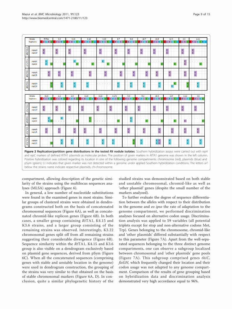
regard to its location in one of the extrachromosomalreplicons of the tested strains. repA and repC genes ofthe largest pRleTA1d were jointly detected on the lar-gest plasmids in all the sampled Rlt strains (Figure 2).Similarly, repA and repC of the pRleTA1b jointly hybri-dized to one of the plasmids of different size in all theRlt strains. In contrast, repA and repC of the pRleTA1cwere rarely localized together (4 of 23 strains). The repAof the pRleTA1c was not similar to any of the plasmidsin most of the sampled strains, but repC hybridized fre-quently (19 of 23 strains) to pSym plasmids. repA andrepC of pRleTA1a (pSym) commonly showed sequencesimilarity to non-symbiotic plasmids of the sampledstrains and only exceptionally hybridized to symbioticones (Figure 2).RepABC of pRleTA1d and pRleTA1b display similarity
with replication systems of the extrachromosomal repli-cons, which were recently described as chromids[16,35]. Within the group of closely related strainsRtTA1, R. leguminosarum bv. viciae 3841 (Rlv), R. etliCFN42 (Rhe), RltWSM2304 and RltWSM1325 clustersof replicons carrying the most similar replication sys-tems can be distinguished. They comprise pRleTA1d-pRL12-p42f-pRLG201-pR132501 and pRleTA1b-pRL11-p42e-pRLG202-pR132502, respectively. Therefore, detec-tion of positive hybridization signals with probes derivedfrom rep genes of RtTA1 chromid-like replicons (i.e.pRleTA1b or pRleTA1d) to any of the replicons of thesampled strains allowed regarding those as a chromid-like. Based on the similarity of replication-partitiongenes detected in our assays, we divided the replicons ofthe studied strains into three genome compartments:chromosome, chromid-like and ‘other plasmids’ (i.e.those replicons which gave a hybridization signal withmolecular probes originating from repA and repC genesof pRleTA1a or pRleTA1c, as well as those that gave nosignal with any rep probes of RtTA1 replication genes).The compartment designated ‘other plasmids’ also com-prised pSym. Such replicon division was taken into con-sideration in the subsequent analyses of distribution ofother markers in the studied strains.
Variability of chromosomal and plasmid marker locationIn further studies, the extent of gene content diversity inthe sampled nodule isolates was examined. We aimed toestimate whether, besides repA and repC displacement
Mazur et al. BMC Microbiology 2011, 11:123http://www.biomedcentral.com/1471-2180/11/123
Page 6 of 15

events, we could demonstrate changes in the location ofthe chromosomal and plasmid genes. The same experi-mental approach was used, i.e. a series of Southernhybridizations with different genes with a well-definedchromosomal or plasmid location in RtTA1 (Table 1)[36].For assays of chromosomal marker variability, essential
bacterial genes were chosen: rpoH2, dnaK, dnaC, rrn,lpxQ as well as genes that are not essential or withunspecified essentiality but chromosomal in RtTA1, i.e.bioA, stbB, exoR, pssL (Pss-I) and rfbADBC (Pss-V)(Table 1). In addition, location of fixGH genes wasassayed, even though they are known to be plasmid
located on the sequenced RltWSM2304, RltWSM1325[33,34], Rlv [6] and Rhe [5] genomes, but chromosomalin RtTA1 [36].A majority of the studied genes (rpoH2, dnaK, dnaC,
rrn, lpxQ, bioA, stbB, exoR and pssL) were located onthe chromosome in all the sampled strains, showingconsiderable conservation of chromosomal markers (Fig-ure 3). Exceptionally, the Pss-V region was identified onthe chromosome of the K3.6, K5.4 and RtTA1 but itwas missing in the other strains (Figure 3) Moreover,fixGH symbiosis-related genes, which were chromoso-mal in the RtTA1, K3.6, K4.15 and K5.4 strains, werelocated mainly in the genome compartment designated
Figure 1 Plasmid profiles of selected R. leguminosarum bv. trifolii nodule isolates. (A) Profiles obtained in Eckhardt-type agarose gelelectrophoresis; stars colored in green indicate pSym plasmids. Lanes: 1-RtTA1; 2-Rlv 3841; 3-K2.2; 4-K2.4; 5-K2.9; 6-K3.6; 7-K3.8; 8-K3.12; 9-K3.16;10-K3.22; 11-K4.11; 12-K4.13; 13-K4.15; 14-K4.16; 15-K4.17; 16-K5.6; 17-K8.7; 18-K9.2; 19-K9.8; 20-K10.7; 21-K10.8, 22-K12.5 (B) PFGE separatedreplicons of Rlt nodule isolates further submitted to hybridization assays. The names of plasmids of Rlv 3841 strain, used as molecular weightmarkers were shown [6]. Molecular weight of Rlv 3841 plasmids is: 870, 684, 488, 353, 152, 147.5 kb. The letters on the respective bands ofparticular plasmids of individual strains indicates the plasmid name, e.g., “a” indicates pRlea plasmid. Lanes: 1-Rlv 3841; 2-RtTA1; 3-K2.4; 4-K3.12; 5-K3.16; 6-K4.13; 7-K4.17; 8-K5.6; 9-K9.2; 10-K10.4; 11-K3.8; 12-K4.11; 13-K8.7; 14-K9.8; 15-Rlv 3841; 16-RtTA1; 17-K2.2; 18-K2.9; 19-K3.6; 20-K3.22; 21-K5.4,22-K10.7, 23-K10.8, 25-K3.13, 26-K4.15.
Mazur et al. BMC Microbiology 2011, 11:123http://www.biomedcentral.com/1471-2180/11/123
Page 7 of 15

as ‘other plasmids’ (pSym to be exact) in the remainingstrains. The variable location of fixGH genes which werefound on the chromosome, pSyms and chromid-likereplicons (K12.5) could be accounted for by location ofthese genes on the putative genomic island flanked by18 bp repeats in R. leguminosarum and R. etli [10,37].Southern hybridizations with probes comprising markers
previously identified on different RtTA1 replicons [36],such as prc and hlyD of pRleTA1d; lpsB2, orf16-orf17-otsB,tauA and orf14 genes cluster of pRleTA1c; nadA and pssM(surface polysaccharide synthesis region Pss-III) of pRle-TA1b, were carried out. These analyses demonstrated thatpRleTA1d markers were almost always jointly detected inthe largest chromid-like replicons (only in K3.22 and K5.4they are separated between distinct chromid-like repli-cons). pRleTA1c markers in almost all (21 out of 23) of thesampled strains were located in the genome compartmentdesignated as ‘other plasmids’ (Figure 3). From amongmarkers of pRleTA1b, nadA, minD, hutI and pcaG hadalways chromid-like location, while the pssM gene waslocated in the chromosome of 19 strains, in chromid-likereplicons of four strains including RtTA1, and was absentin the genome of K3.22 strain, respectively (Figure 3).
Besides the symbiotic genes nodA and nifNE used foridentification of pSym plasmids, stability of thiC andacdS (Table 1) of the pRleTA1a symbiotic plasmid (ipsofacto described as markers of the ‘other plasmids’ pool)was examined (Figure 3). Only thiC was identified in allthe strains, however, located in different genomic com-partments: most frequently on the chromosome (18 of23 strains), and in the ‘other plasmids’ (5 strains). TheacdS gene was detected in 14 of 23 strains, in each caseon pSym (Figure 3). The thiC gene, similarly to fixGHI,showed high variability in location; however, its putativemobile element location is unknown [38]. thiC wasreported as plasmid located in sequenced genomes ofRlv [6], Rlt2304 [33] and Rhe [5].As a result, genes with a stable location in specific
genome compartments in all the strains, as well asunstable genes with variable, strain-dependent distribu-tion were distinguished (Figure 4). Stable markers foreach compartment of the sampled strains were estab-lished i.e. chromosomal: rpoH2, exoR, dnaK, dnaC, bioA,rrn, lpxQ, pssL and stbB; chromid-like: prc, hlyD, nadA,minD, hutI and pcaG; ‘other plasmids’: otsB, lpsB2(exceptionally chromid-like in K3.6), tauA and orf14(exceptionally chromid-like in K3.12) including nodAand nifNE symbiosis-related genes of pSym (Figure 4).Loss of some of the examined markers was noticed, i.e.Pss-V from the chromosome, pssM from chromid-likereplicons, and acdS from the ‘other plasmids’ (pSym).Only two of the sampled strains, i.e. K3.6 and K5.4, con-tained all the studied markers, while others lacked atleast one of the genes.A dendrogram demonstrating similarity of the strains
was constructed with the UPGMA clustering methodbased on markers distribution among their differentgenome compartments. It showed one K3.6 strainapparently split from the others (Figure 5), and twogroups of clustered strains: a small one, includingRtTA1, K5.4 and K4.15, and a large one comprising theremaining strains, which was further subdivided intotwo smaller subgroups of strains with identical markerdistribution (Figure 5).
Sequence divergence of chromosomal and plasmid genesTo assess the overall phylogenetic similarity of thesampled strains, several genes from a subset of 12 differ-ent strains displaying divergent plasmid profiles (plusRtTA1) were partially sequenced and analyzed. Thesequenced genes comprised exclusively chromosomal(dnaC, dnaK, exoR, rpoH2), chromid-like replicons(hlyD, prc, nadA), and ‘other plasmid’ markers (nodA,nifNE) as well as those with unstable location found indifferent genome compartments (fixGH, thiC, lpsB2).Afterwards, phylogenetic trees were constructed basedon concatenated sequences of a distinct genome
Table 2 Plasmid number and size of R. leguminosarumbv. trifolii strains determined by PFGE
Rlt strains Plasmid size (kb)
pRlef pRlee pRled pRlec pRleb pRlea
RtTA1 808 653 603 476*
K3.8 1110 640 570 370*
K3.13 1210 610 590 350* 240
K3.16 915 570 520 270* 200
K3.22 1350 510 420 310* 185
K8.7 1110 710 560 330*
K9.8 1250 710 580 260*
K10.7 1180 710 565 430*
K10.8 1120 670 600 460*
K12.5 1220 670 580 395* 270
K3.6 840 620 430*
K4.11 1060 610 560 350* 190 150
K4.15 770 705 640 500*
K2.2 1230 650 630 440*
K2.4 1250 720 570 320*
K4.13 1240 650 630 420* 310
K4.16 1380 680 585 320*
K4.17 1140 700 600 330* 250
K5.4 780 690 650 335*
K9.2 1140 730 620 340* 250
K10.4 1130 700 570 290*
K2.9 1240 810 590 375* 180
K3.12 1210 700 630 400*
K5.6 1060 635 610 290*
*-symbiotic plasmids.
Mazur et al. BMC Microbiology 2011, 11:123http://www.biomedcentral.com/1471-2180/11/123
Page 8 of 15
compartment, allowing description of the genetic simi-larity of the strains using the multilocus sequences ana-lyses (MLSA) approach (Figure 6).In general, a low number of nucleotide substitutions
were found in the examined genes in most strains. Simi-lar groups of clustered strains were obtained in dendro-grams constructed both on the basis of concatenatedchromosomal sequences (Figure 6A), as well as concate-nated chromid-like replicon genes (Figure 6B). In bothcases, a smaller group containing RtTA1, K4.15 andK3.6 strains, and a larger group consisting of theremaining strains was observed. Interestingly, K3.22chromosomal genes split off from all remaining strainssuggesting their considerable divergence (Figure 6B).Sequence similarity within the RtTA1, K4.15 and K3.6group is also visible on a dendrogram exclusively basedon plasmid gene sequences, derived from pSym (Figure6C). When all the concatenated sequences (comprisinggenes with stable and unstable location in the genome)were used in dendrogram construction, the grouping ofthe strains was very similar to that obtained on the basisof stable chromosomal markers (Figure 6A, D). In con-clusion, quite a similar phylogenetic history of the
studied strains was demonstrated based on both stableand unstable chromosomal, chromid-like as well as‘other plasmid’ genes (despite the small number of themarkers analyzed).To further evaluate the degree of sequence differentia-
tion between the alleles with respect to their distributionin the genome and eo ipso the rate of adaptation to thegenome compartment, we performed discriminationanalyses focused on alternative codon usage. Discrimina-tion analysis was applied to 59 variables (all potentialtriplets except for stop and non-alternative codons Met,Trp). Genes belonging to the chromosome, chromid-likeand ‘other plasmids’ differed substantially with respectto this parameter (Figure 7A). Apart from the well-sepa-rated sequences belonging to the three distinct genomecompartments, one can observe a subgroup localizedbetween chromosomal and ‘other plasmids’ gene pools(Figure 7A). This subgroup comprised genes thiC,fixGH, which frequently changed their location and theircodon usage was not adapted to any genome compart-ment. Comparison of the results of gene grouping basedon hybridization data and discrimination analysisdemonstrated very high accordance equal to 96%.
Figure 2 Replication/partition gene distributions in the tested Rlt nodule isolates. Southern hybridization assays were carried out with repAand repC markers of defined RtTA1 plasmids as molecular probes. The position of given markers in RtTA1 genome was shown in the left column.Positive hybridization was colored regarding its location in one of the following genome compartments: chromosome (red), plasmids (blue) andpSym (green); (-) indicates that given marker was not detected within a genome under applied Southern hybridization conditions. The letters a-fbelow the strains name indicate respective plasmids, ch-chromosome.
Mazur et al. BMC Microbiology 2011, 11:123http://www.biomedcentral.com/1471-2180/11/123
Page 9 of 15

Figure 3 Distribution of replicon specific genes in the tested Rlt nodule isolates. Southern hybridization assays were carried out withseveral chromosome and plasmid markers of RtTA1 as molecular probes. The position of a given markers in RtTA1 genome was shown in theleft column. Positive hybridization was colored regarding its location in one of the following genome compartments of Rlt isolates: chromosome(red), chromid-like (violet), plasmids (blue) and pSym (green); (-) indicates that given marker was not detected within a genome under appliedSouthern hybridization conditions. The letters a-f below the strains name indicate respective plasmids, ch-chromosome.
Mazur et al. BMC Microbiology 2011, 11:123http://www.biomedcentral.com/1471-2180/11/123
Page 10 of 15

The discrimination analysis of codon usage performedon individual strains harboring the set of the testedgenes (13 groups of sequences) revealed only minor dif-ferences between the resultant groups and almost noaccordance (31%) with the grouping performed on thebasis of hybridization. However, some level of similaritybetween the strains can be demonstrated. As a conse-quence, one more discrimination analysis of codonusage was done, and the strains were divided into three
groups: (i) K3.22, (ii) RtTA1, K3.6, K4.15 and (iii) all theremaining strains (Figure 7B). This resulted in 92%accordance between codon usage-based and strain-dependent grouping of sequences (Figure 7B and Figure6D). It was concluded that codon usage was not signifi-cantly influenced by the individual strains but may becharacteristic for the group of strains.Finally, the Codon Adaptation Index (CAI) of the
sequences studied was calculated. The CAI can be usedto “evaluate the extent to which selection has beeneffective in molding the pattern of codon usage” [29] aswell as to compare the codon usage of foreign genesversus that of highly expressed native genes [13]. Here,we applied CAI analyses to assess the degree of adapta-tion of sequenced genes to the host by comparing theobtained CAI values with those of genes encoding ribo-somal proteins in R. leguminosarum. The calculated CAIvalues for each sequence were arbitrarily grouped andsubsequently submitted to ANOVA evaluation, whichmeasures the significance of differences between groups.CAI values can range from 0 (reflecting use of synon-ymous codons) to 1 (reflecting the strongest bias wherecodon usage is equal to that in the ribosomal protein-encoding genes) [13].The CAI values ranged from 0.849 (dnaC-chromoso-
mal gene) to 0.554 (nodA-symbiotic gene). The fixG andthiC had the CAI equal to 0.676 and 0.673, respectively,suggesting weaker adaptation to their genome compart-ments and further confirming their unstable location asindicated in hybridization analyses. We did not find sig-nificant differences with respect to the CAI values calcu-lated for the particular strains, but strains RtTA1, K4.15,K3.6, and K3.22 previously observed as most divergenthad a high average CAI of the studied sequences (from0.722 to 0.718), possibly indicating good adaptation ofthe genes to the host. Finally, the CAI values were eval-uated according to the location of genes in the differentgenome compartments (Table 3). The CAI values ofgenes located on the chromosome and chromid-likereplicons were high and significantly differed from eachother. The genes located on the ‘other plasmids’ (includ-ing pSym) had the lowest CAI values significantly differ-ent from the former ones. These results demonstratedweaker adaptation of plasmid genes to the host genomein comparison to the chromosome and chromid-likegenes.
DiscussionThree genome compartments that differed genetically andfunctionally can be distinguished in the nodule populationof R. leguminosarum bv. trifolii: the chromosome, chro-mid-like and ‘other plasmids’ including pSym. Chromid-like replicons were distinguished in Southern analyses onthe basis of repA and repC sequence similarity to RtTA1
Figure 4 Overall genes distribution in three genomecompartments: chromosome, chromid-like and ‘other plasmids’in Rlt isolates. Southern hybridizations were carried out with RtTA1markers of specified localization as probes. The arrows indicateinstability of some markers location in the given genomecompartments. Asterisk indicates genes exceptionally localized onchromid-like replicon. Yellow area indicates genes detected in alltested strains.
Figure 5 The dendrogram showing similarity of Rlt noduleisolates and RtTA1 strain. The dendrogram was constructed onthe basis of marker distribution among different genomecompartments using UPGMA clustering method.
Mazur et al. BMC Microbiology 2011, 11:123http://www.biomedcentral.com/1471-2180/11/123
Page 11 of 15

and to the respective replication genes of such repliconsdescribed in the sequenced genomes of R. leguminosarumbv. viciae, R. etli and R. leguminosarum bv. trifolii [16].The chosen name “chromid-like” (as opposed to simply“chromid”) was the result of data scarcity concerning theirgene content, insufficient to justify the name “chromid”[16]. Moreover, it is known that genes of the repABCoperon are peculiar genetic markers because of the com-plex phylogeny of particular genes within the operon,whose evolutionary history could not be strictly connectedwith other genes of particular replicons [13].In the study of the distribution of several chromoso-
mal and plasmid markers within a group of 23 noduleisolates, stable genes permanently located in a specific
R. leguminosarum bv. trifolii genome compartment:chromosome, chromid-like and ‘other plasmids’ includ-ing pSym were distinguished. Unstable genes (fixGH,thiC, acdS, pssM and Pss-V region) that changed theirlocation at various rates or were lost from the genomewere also detected. Only two of the sampled 23 strainspossessed all the studied markers. A majority of strainsdiffered in the gene content and gene distribution, sup-porting the hypothesis of the pangenomic structure ofR. leguminosarum, in which each strain of a given spe-cies contains, besides the core genome, additionalgenetic information specific for the strain [11,17,18,39].The distribution of the plasmid replication-partition
genes was even more dynamic than that of genes not
Figure 6 The sequence similarity dendrograms of Rlt nodule isolates and RtTA1 strain. The dendrograms were constructed with UPGMAclustering method based on the chosen sequences of the given genome compartment: (A) concatenated chromosomal gene sequences; (B)chromid-like replicons’genes; (C) ‘other plasmids’ genes; (D) all gene sequences (stable and unstable) located in different genome compartments.
Mazur et al. BMC Microbiology 2011, 11:123http://www.biomedcentral.com/1471-2180/11/123
Page 12 of 15

connected with replication. Independent transfer eventsof repA and repC genes of the putative repABC operonwere frequently observed, especially in the ‘other plas-mids’ compartment, which confirmed different evolu-tionary pathways for various elements of the repABCoperon, recently evidenced in Alphaproteobacteria [13].Such considerable dynamics of replication/partition genedistribution in Rhizobium may account for changes inthe plasmid number and, consequently, gene contentobserved in the sampled population. Beside thedynamics of replication/partition gene distribution,some level of conservation of replication genes, espe-cially those of chromid-like replicons, was also observed.It was reflected in positive hybridizations with pRle-TA1d and pRleTA1b derived rep probes, to the respec-tive replicons of Rlt strains. One could speculate thatthe conservation of replication genes of chromid-likereplicons may be related with their distinct properties e.g. stability. However, the gene content rather than theproperties of the replication system, resulting e.g. from
conservation of replication genes, seem to be crucial forreplicon stability [40].Redistribution of genes between the different genome
compartments could further trigger their sequencedivergence under different selective pressures [13,15,41].Examination of sequence divergence of several stableand unstable chromosomal and plasmid genes showed alow level of substitutions in genes of all the compart-ments. Nearly identical nucleotide sequences of nifNEmarkers were found in different pSym plasmids of thestudied population (Figure 6C), confirming the corecharacter of symbiotic genes and their high conserva-tion, despite the overall genome differentiation [11].The extent of gene adaptation to a given compartment
in the host genome was assessed by analyses of alterna-tive codon usage. Three groups of well separated geneswere obtained corresponding to the chromosome, chro-mid-like and ‘other plasmids’ genome compartments(Figure 7A) with 96% accordance with hybridizationdata. In conclusion, the sequence divergence of particu-lar genes may be affected by their location in the givengenome compartment. When all the sequences of theindividual strains studied were subjected to a discrimi-nation analysis, we obtained good separation of K3.22and a group of strains related to RtTA1 (Figure 7B) thatformed the outermost branch in the phylogenic tree.The remaining strains were randomly mixed with eachother but apparently separated from K3.22 and TA1-related strains, which suggested no differences in codonusage within the main group.
Figure 7 Markers grouping obtained in discrimination analyses. (A) Grouping was carried out regarding frequency of alternative codonusage. Symbols used: red squares-chromosome markers (ch), blue triangles-chromid-like replicons’ markers (cd), green circles-’other plasmid’markers (including pSym markers) (p). (B) Strains grouping observed in discrimination analyses regarding frequency of alternative codon usage ofthe tested gene set.
Table 3 The Codon Adaptation Index (CAI) of geneslocated in genome compartments in Rlt nodule isolates
Gene location Number of sequences Average CAI
Chromosome 66 0.767 ± 0.062 a
Chromid-like 42 0.732 ± 0.065 b
Other plasmids 61 0.645 ± 0.061 cd
Values followed by the various letters are significantly different: b (P < 0.05)and cd P < 0.001.
± Standard deviation (SD).
Mazur et al. BMC Microbiology 2011, 11:123http://www.biomedcentral.com/1471-2180/11/123
Page 13 of 15

The CAI analyses of the evaluated sequences con-firmed good adaptation of chromosomal and chromid-like genes (high CAI values) to host genomes and lowerCAI values for ‘other plasmids’ genes. The CAI valuesalso reflect the level of transcriptional and translationalactivity of particular genes [29]. While the activity ofmost of the chromosomal and chromid-like genes couldbe considered at least to some extent constitutive, the‘other plasmids’ and especially symbiosis-related genesare expressed only transiently in the symbiotic stage[42]. Therefore, in the Rhizobium model, the differencesin codon usage in translation reflect the balancebetween the selection pressure and random mutationsin the functionally differentiated genome compartments.The differences in codon usage and CAI values betweenthe genome compartments are most likely a conse-quence of differential gene expression and adaptabilityto optimal codon usage in host genomes [42].
ConclusionOur study showed that, even within a small rhizobialpopulation of clover nodule isolates, substantial divergenceof genome organization can be detected especially takinginto account the content of extrachromosomal DNA.Despite the high variability with regard to the number andsize of plasmids among the studied strains, conservation ofthe location as well as the dynamic distribution of the indi-vidual genes (especially replication genes) of a particulargenome compartment was demonstrated. The sequencedivergence of particular genes may be affected by theirlocation in the given genome compartment. The ‘otherplasmid’ genes are less adapted to the host genome thanthe chromosome and chromid-like genes.
Acknowledgements and FundingThis work was supported by Grant No. N N301 028734 from Ministry ofScience and Higher Education of Poland.
Author details1Department of Genetics and Microbiology, Maria Curie-SkłodowskaUniversity, Akademicka 19, 20-033 Lublin, Poland. 2Chair of AppliedMathematics and Informatics, Lublin University of Life Sciences, Akademicka13, 20-950 Lublin, Poland.
Authors’ contributionsAM designed and coordinated the study and drafted the manuscript. GSconducted the bulk of the experiments: carried out analyses of Rlt plasmidcontent by Eckhardt’ method and PFGE, prepared and labeled the molecularprobes and provided Southern hybridization. JW helped in statisticalanalyses, contributed to interpretation of data and preparation of themanuscript. AKK performed the statistical analysis. MMK collected rhizobialstrains and helped with Rlt plasmid analyses. AS provided scientific guidanceand discussion and prepared final version of manuscript. All authors haveread and approve of this final manuscript.
Received: 18 February 2011 Accepted: 30 May 2011Published: 30 May 2011
References1. Jones KM, Kobayashi H, Davies BW, Taga ME, Walker GC: How symbionts
invade plants: the Sinorhizobium-Medicago model. Nat Rev Microbiol 2007,5:619-633.
2. Masson-Boivin C, Giraud E, Perret X, Batut J: Establishing nitrogen-fixingsymbiosis with legumes: how many rhizobium recipes? Trends Microbiol2009, 17:458-466.
3. Perret X, Staehelin C, Broughton W: Molecular basis of symbioticpromiscuity. Microbiol Mol Biol Rev 2000, 64:180-201.
4. Galibert F, et al: The composite genome of the legume symbiontSinorhizobium meliloti. Science 2001, 293:668-672.
5. González V, Santamaría RI, Bustos P, Hernández-González I, Medrano-Soto A,Moreno-Hagelsieb G, Janga SC, Ramírez MA, Jiménez-Jacinto V, Collado-Vides J, Dávila G: The partitioned Rhizobium etli genome: genetic andmetabolic redundancy in seven interacting replicons. Proc Natl Acad SciUSA 2006, 103:3834-3839.
6. Young JPW, et al: The genome of Rhizobium leguminosarum hasrecognizable core and accessory components. Genome Biol 2006, 7:R34.
7. Palacios R, Newton WE: Genomes and genomics of nitrogen-fixingorganisms.Edited by: Palacios R, Newton WE. Dordrecht, The Netherlands:Springer; 2005:.
8. Sullivan JT, Trzebiatowski JR, Cruickshank RW, Gouzy J, Brown SD, Elliot RM,Fleetwood DJ, McCallum NG, Rossbach U, Stuart GS, Weaver JE, Webby RJ,De Bruijn FJ, Ronson CW: Comparative sequence analysis of thesymbiosis island of Mesorhizobium loti strain R7A. J Bacteriol 2002,184:3086-3095.
9. Konstantinidis KT, Tiedje JM: Trends between gene content and genomesize in prokaryotic species with larger genomes. Proc Natl Acad Sci USA2004, 101:3160-3165.
10. Crossman LC, Castillo-Ramírez S, McAnnula C, Lozano L, Vernikos GS,Acosta JL, Ghazoui ZF, Hernández-González I, Meakin G, Walker AW,Hynes MF, Young JPW, Downie JA, Romero D, Johnston AWB, Dávila G,Parkhill J, González V: A common genetic framework for a diverseassembly of plasmids in the symbiotic nitrogen fixing bacteria. PLoS ONE2008, 7:e2567.
11. González V, Acosta JL, Santamaría RI, Bustos P, Fernández JL,Hernández González IL, Díaz R, Flores M, Palacios R, Mora J, Dávila G:Conserved symbiotic plasmid DNA sequences in the multirepliconpangenomic structure of Rhizobium etli. Appl Environ Microbiol 2010,76:1604-1614.
12. Cevallos MA, Cervantes-Rivera R, Gutiérrez-Ríos RM: The repABC plasmidfamily. Plasmid 2008, 60:19-37.
13. Castillo-Ramírez S, Vázquez-Castellanos JF, González V, Cevallos MA:Horizontal gene transfer and diverse functional constrains within acommon replication-partitioning system in Alphaproteobacteria: therepABC operon. BMC Genomics 2009, 10:536.
14. Cevallos MA, Porta H, Izquierdo J, Tun-Garrido C, García-de-los-Santos A,Dávila G, Brom S: Rhizobium etli CFN42 contains at least three plasmidsof the repABC family: a structural and evolutionary analysis. Plasmid 2002,48:104-116.
15. Fondi M, Bacci G, Brilli M, Papaleo MC, Mengoni A, Vaneechoutte M,Dijkshoorn L, Fani R: Exploring the evolutionary dynamics of plasmids:the Acinetobacter pan-plasmidome. BMC Evol Biol 2010, 10:59.
16. Harrison PW, Lower RPJ, Kim NKD, Young JPW: Introducing the bacterial‘chromid’: not a chromosome, not a plasmid. Trends Microbiol 2010,18:141-147.
17. Tettelin H, Riley D, Cattuto C, Medini D: Comparative genomics: thebacterial pan-genome. Curr Opin Microbiol 2008, 12:472-477.
18. Medini D, Donati C, Tettelin H, Masignani V, Rappuoli R: The microbialpangenome. Curr Opin Genet Dev 2005, 15:589-594.
19. Flores M, Morales L, Avila A, González V, Bustos P, García D, Mora Y, Guo X,Collado-Vides J, Piñero D, Dávila G, Mora J, Palacios R: Diversification ofDNA sequences in the symbiotic genome of Rhizobium etli. J Bacteriol2005, 187:7185-7192.
20. Guerrero G, Peralta H, Aguilar A, Díaz R, Villalobos MA, Medrano-Soto A,Mora J: Evolutionary, structural and functional relationships revealed bycomparative analysis of syntenic genes in Rhizobiales. BMC Evol Biol 2005,5:55.
21. Rocha EPC: Evolutionary patterns in prokaryotic genomes. Curr OpinMicrobiol 2008, 11:454-460.
Mazur et al. BMC Microbiology 2011, 11:123http://www.biomedcentral.com/1471-2180/11/123
Page 14 of 15

22. Vincent JM: A manual for the practical study of root nodule bacteria.International biological program handbook no.15 Oxford, UK: BlackwellScientific Publications Ltd; 1970.
23. Sambrook J, Fritsch EF, Maniatis T: Molecular Cloning: A LaboratoryManual. Cold Spring Harbor, NY, USA: Cold Spring Harbor Laboratory Press;,2 1989.
24. Eckhardt T: A rapid method for the identification of plasmiddeoxyribonucleic acid in bacteria. Plasmid 1978, 1:584-588.
25. Chakravorty AK, Żurkowski W, Shine J, Rolfe BG: Symbiotic nitrogenfixation: molecular cloning of Rhizobium genes involved inexopolysaccharide synthesis and effective nodulation. J Mol Appl Genet1982, 1:585-596.
26. Król J, Mazur A, Marczak M, Skorupska A: Syntenic arrangements of thesurface polysaccharide biosynthesis genes in Rhizobium leguminosarum.Genomics 2007, 89:237-247.
27. Thompson JD, Higgins DG, Gibson TJ: CLUSTAL W: improving thesensitivity of progressive multiple sequence alignment throughsequence weighting, position-specific gap penalties and weight matrixchoice. Nucl Acids Res 1994, 22:4673-4680.
28. Vetrivel U, Arunkumar V, Dorairaj S: ACUA: A software tool for automatedcodon usage analysis. Bioinformation 2007, 2:62-63.
29. Sharp PM, Li WH: The codon adaptation index-a measure of directionalsynonymous codon usage bias, and its potential applications. Nucl AcidsRes 1987, 15:1281-1295.
30. McLachlan GJ: Discriminant analysis and statistical pattern recognition.Hoboken, New Jersey: John Wiley & Sons Inc; 1992.
31. Dillon WR, Goldstein M: Multivariate analysis. Methods and applications.New York: John Wiley & Sons Inc; 1984.
32. Klecka WR: Discriminant analysis. Thousand Oaks, CA: Sage Publications;1980, Quantitative Applications in the Social Sciences Series, No. 19..
33. Reeve W, O’Hara G, Chain P, Ardley J, Bräu L, Nandesena K, Tiwari R,Malfatti S, Kiss H, Lapidus A, Copeland A, Nolan M, Land M, Ivanova N,Mavromatis K, Markowitz V, Kyrpides N, Melino V, Denton M, Yates R,Howieson J: Complete genome sequence of Rhizobium leguminosarumbv. trifolii strain WSM2304, an effective microsymbiont of the SouthAmerican clover Trifolium polymorphum. Stand Genomic Sci 2010, 2:66-76.
34. Reeve W, O’Hara G, Chain P, Ardley J, Bräu L, Nandesena K, Tiwari R,Copeland A, Nolan M, Han C, Brettin T, Land M, Ovchinikova G, Ivanova N,Mavromatis K, Markowitz V, Kyrpides N, Melino V, Denton M, Yates R,Howieson J: Complete genome sequence of Rhizobium leguminosarumbv. trifolii strain WSM1325, an effective microsymbiont of annualMediterranean clovers. Stand Genomic Sci 2010, 2:347-356.
35. Mazur A, Majewska B, Stasiak G, Wielbo J, Skorupska A: repABC-basedreplication systems of Rhizobium leguminosarum bv. trifolii TA1 plasmids:incompatibility and evolutionary analyses. Plasmid .
36. Król J, Mazur A, Marczak M, Skorupska A: Physical and genetic map ofRhizobium leguminosarum bv. trifolii TA1 and its application incomparison of closely related rhizobial genomes. Mol Genet Genomics2008, 279:107-121.
37. González V, Bustos P, Ramírez-Romero MA, Medrano-Soto A, Salgado H,Hernández-González I, Hernández-Celis JC, Quintero V, Moreno-Hagelsieb G,Girard L, Rodríguez O, Flores M, Cevallos A, Collado-Vides J, Romero D,Dávila G: The mosaic structure of the symbiotic plasmid of Rhizobiumetli CFN42 and its relation to other symbiotic genome compartments.Genome Biol 2003, 4:R36.
38. Miranda-Ríos J, Morera C, Taboada H, Dávalos A, Encarnación S, Mora J,Soberón M: Expression of thiamin biosynthetic genes (thiCOGE) andproduction of symbiotic terminal oxidase cbb3 in Rhizobium etli. JBacteriol 1997, 179:6887-6893.
39. Brom S, Girard L, García-de los-Santos A, Sanjuan-Pinilla JM, Olivares J,Sanjuan J: Conservation of plasmid-encoded traits among bean-nodulating Rhizobium species. Appl Environ Microbiol 2002, 68:2555-2561.
40. Landeta C, Dávalos A, Cevallos MA, Geiger O, Brom S, Romero D: Plasmidswith a chromosome-like role in Rhizobium. J Bacteriol 2011,193:1317-1326.
41. Slater SC, et al: Genome sequences of three Agrobacterium biovars helpelucidate the evolution of multichromosome genomes in bacteria. JBacteriol 2009, 191:2501-2511.
42. Peixoto L, Zaval A, Romero H, Musto H: The strength of translationalselection for codon usage varies in the three replicons of Sinorhizobiummeliloti. Gene 2003, 320:109-116.
doi:10.1186/1471-2180-11-123Cite this article as: Mazur et al.: Intragenomic diversity of Rhizobiumleguminosarum bv. trifolii clover nodule isolates. BMC Microbiology 201111:123.
Submit your next manuscript to BioMed Centraland take full advantage of:
• Convenient online submission
• Thorough peer review
• No space constraints or color figure charges
• Immediate publication on acceptance
• Inclusion in PubMed, CAS, Scopus and Google Scholar
• Research which is freely available for redistribution
Submit your manuscript at www.biomedcentral.com/submit
Mazur et al. BMC Microbiology 2011, 11:123http://www.biomedcentral.com/1471-2180/11/123
Page 15 of 15