Removal of Divalent Cations Induces Structural Transitions in Red Clover Necrotic Mosaic Virus,...
13
Published Ahead of Print 18 August 2006. 2006, 80(21):10395. DOI: 10.1128/JVI.01137-06. J. Virol. Orlova, Timothy S. Baker and Steven A. Lommel Tim L. Sit, Charles L. Brooks, Albert M. Mikhailov, Elena V. Michael B. Sherman, Richard H. Guenther, Florence Tama, Potential Mechanism for RNA Release , Revealing a Necrotic Mosaic Virus Red Clover Structural Transitions in Removal of Divalent Cations Induces http://jvi.asm.org/content/80/21/10395 Updated information and services can be found at: These include: SUPPLEMENTAL MATERIAL Supplemental material REFERENCES http://jvi.asm.org/content/80/21/10395#ref-list-1 This article cites 49 articles, 9 of which can be accessed free at: CONTENT ALERTS more» articles cite this article), Receive: RSS Feeds, eTOCs, free email alerts (when new http://journals.asm.org/site/misc/reprints.xhtml Information about commercial reprint orders: http://journals.asm.org/site/subscriptions/ To subscribe to to another ASM Journal go to: on April 3, 2014 by guest http://jvi.asm.org/ Downloaded from on April 3, 2014 by guest http://jvi.asm.org/ Downloaded from
-
Upload
independent -
Category
Documents
-
view
2 -
download
0
Transcript of Removal of Divalent Cations Induces Structural Transitions in Red Clover Necrotic Mosaic Virus,...
Published Ahead of Print 18 August 2006. 2006, 80(21):10395. DOI: 10.1128/JVI.01137-06. J. Virol.
Orlova, Timothy S. Baker and Steven A. LommelTim L. Sit, Charles L. Brooks, Albert M. Mikhailov, Elena V. Michael B. Sherman, Richard H. Guenther, Florence Tama, Potential Mechanism for RNA Release
, Revealing aNecrotic Mosaic VirusRed CloverStructural Transitions in
Removal of Divalent Cations Induces
http://jvi.asm.org/content/80/21/10395Updated information and services can be found at:
These include:
SUPPLEMENTAL MATERIAL Supplemental material
REFERENCEShttp://jvi.asm.org/content/80/21/10395#ref-list-1This article cites 49 articles, 9 of which can be accessed free at:
CONTENT ALERTS more»articles cite this article),
Receive: RSS Feeds, eTOCs, free email alerts (when new
http://journals.asm.org/site/misc/reprints.xhtmlInformation about commercial reprint orders: http://journals.asm.org/site/subscriptions/To subscribe to to another ASM Journal go to:
on April 3, 2014 by guest
http://jvi.asm.org/
Dow
nloaded from
on April 3, 2014 by guest
http://jvi.asm.org/
Dow
nloaded from
JOURNAL OF VIROLOGY, Nov. 2006, p. 10395–10406 Vol. 80, No. 210022-538X/06/$08.00�0 doi:10.1128/JVI.01137-06Copyright © 2006, American Society for Microbiology. All Rights Reserved.
Removal of Divalent Cations Induces Structural Transitions inRed Clover Necrotic Mosaic Virus, Revealing a Potential
Mechanism for RNA Release�†Michael B. Sherman,1* Richard H. Guenther,2 Florence Tama,3 Tim L. Sit,2 Charles L. Brooks,3
Albert M. Mikhailov,4 Elena V. Orlova,5 Timothy S. Baker,1‡ and Steven A. Lommel2
Department of Biological Sciences, Purdue University, West Lafayette, Indiana 479071; Department of Plant Pathology, NorthCarolina State University, Raleigh, North Carolina 276952; Department of Molecular Biology, Scripps Research Institute,La Jolla, California 920373; Institute of Crystallography, Russian Academy of Sciences, Moscow 119333, Russia4; and
School of Crystallography, Birkbeck College, London WC1E 7HX, United Kingdom5
Received 1 June 2006/Accepted 9 August 2006
The structure of Red clover necrotic mosaic virus (RCNMV), an icosahedral plant virus, was resolved to 8.5Å by cryoelectron microscopy. The virion capsid has prominent surface protrusions and subunits with a clearlydefined shell and protruding domains. The structures of both the individual capsid protein (CP) subunits andthe entire virion capsid are consistent with other species in the Tombusviridae family. Within the RCNMVcapsid, there is a clearly defined inner cage formed by complexes of genomic RNA and the amino termini of CPsubunits. An RCNMV virion has approximately 390 � 30 Ca2� ions bound to the capsid and 420 � 25 Mg2�
ions thought to be in the interior of the capsid. Depletion of both Ca2� and Mg2� ions from RCNMV leads tosignificant structural changes, including (i) formation of 11- to 13-Å-diameter channels that extend throughthe capsid and (ii) significant reorganization within the interior of the capsid. Genomic RNA within nativecapsids containing both Ca2� and Mg2� ions is extremely resistant to nucleases, but depletion of both of thesecations results in nuclease sensitivity, as measured by a significant reduction in RCNMV infectivity. Theseresults indicate that divalent cations play a central role in capsid dynamics and suggest a mechanism for therelease of viral RNA in low-divalent-cation environments such as those found within the cytoplasm of a cell.
The Tombusviridae family consists of small, icosahedralplant viruses that are transmitted through the soil and infecttheir hosts via the root system (34). Given that these virusesmust survive harsh environments, it is not surprising that theypossess unusually stable and robust capsids. This high degreeof virion stability raises the question of how the intracellularenvironment triggers disassembly or minimally exposes the ge-nome for translation. Structural analyses of several plant vi-ruses have revealed that maintenance of a stable capsid con-formation is dependent on the presence of divalent cationsbound to the capsid (25, 33, 36). It has been a longstandinghypothesis that swelling and other conformational changes in-duced by ion extraction from virions are critical for a produc-tive viral life cycle (13). For Tomato bushy stunt virus (TBSV),the type species of the genus Tombusvirus within the Tombus-viridae family, it was hypothesized that Ca2� ions would bereleased from viral capsids within infected cells, leading tovirion swelling sufficient to expose the viral genome.
Crystallographic studies of TBSV (28) revealed a capsidformed by 180 chemically identical capsid protein (CP) sub-
units in three quasiequivalent conformations (A, B, and C).Each CP subunit is composed of three distinct structural do-mains, which include the RNA-interacting (R), shell (S), andprotruding (P) domains. The conformational differences thatdistinguish the A, B, and C subunits are localized within thehinge regions between the respective S and P domains. Thesehinges point either down (in A-B dimers) or up (in C-Cdimers). In addition, the loop that connects the R and S do-mains (the arm) is ordered in C subunits but disordered in Aand B subunits. A similar structural organization was observedin Turnip crinkle virus (TCV) (50) and Carnation mottle virus(CarMV) (25), which are two other species in the Tombusviri-dae family and whose capsid structures have been solved to aresolution of �3 Å. All three viruses share a phylogeneticallyrelated CP, produce virions with capsids possessing T�3 ico-sahedral symmetry (9), and are �350 Å in diameter. Neitherthe packaged RNA nor the amino termini of the CP weresufficiently ordered to be resolved in these crystallographicstudies.
Despite the lack of evidence of an organized genome withinthe Tombusviridae family members studied so far, a number ofother RNA viruses, whose structures have been determined byX-ray crystallography and cryoelectron microscopy (cryoEM),have partially ordered genomes inside their capsids. The frac-tion of ordered RNA visualized in viruses varies significantly.For example, in Cowpea chlorotic mottle virus there is very littleordered RNA (40). Conversely, in the insect-infecting Noda-viridae family, Flock house virus and Pariacoto virus bothpackage a substantial fraction of their RNA genomes withindodecahedral cages (45). Satellite Tobacco mosaic virus repre-
* Corresponding author. Present address: Department of Biochem-istry and Molecular Biology, University of Texas Medical Branch atGalveston, Galveston, TX 77555. Phone: (409) 772-6310. Fax: (409)772-4298. E-mail: [email protected].
‡ Present address: Departments of Chemistry & Biochemistry andMolecular Biology, University of California—San Diego, La Jolla, CA92093.
† Supplemental material for this article may be found at http://jvi.asm.org/.
� Published ahead of print on 18 August 2006.
10395
on April 3, 2014 by guest
http://jvi.asm.org/
Dow
nloaded from

sents an extreme example, because essentially all of the ge-nome within the virion is ordered and has been resolved in theX-ray structure (19, 20).
Red clover necrotic mosaic virus (RCNMV; genus Diantho-virus, family Tombusviridae) is morphologically similar toTBSV, CarMV, and TCV with virions �350 Å in diametercomposed of 23% nucleic acid and 77% protein (26). However,RCNMV is significantly different by virtue of its RNA genome,which is split between two nonhomologous RNAs, a 3.9-kbpolycistronic RNA-1 and a 1.5-kb monocistronic RNA-2 (21).Furthermore, the RCNMV genome is larger than that of anyother monopartite member of the family Tombusviridae yet thevirions are all similar in size. Recent virion characterizationstudies revealed that RCNMV virions exist as two distinct yetmorphologically indistinguishable populations, with one pop-ulation containing four copies of RNA-2 and a second, biolog-ically active population containing one copy each of RNA-1and RNA-2 (4). Such distinguishing features of the RCNMVvirion have stimulated us to examine the RCNMV structure tobetter understand what makes it unique among members ofthe family Tombusviridae.
We have used cryoEM and three-dimensional (3D) imagereconstruction to analyze the structure of RCNMV in its nativeform, as well as its form after removal of divalent cations.Though the native RCNMV capsid structure is similar to thatof other viruses in the taxon, the genomic RNA of RCNMV isordered and organized as an internal, dodecahedral cage. Asdemonstrated by spectroscopic analysis, RCNMV contains fi-nite amounts of Ca2� and Mg2� ions. Selective extraction ofthese ions alters the capsid conformation and generates chan-nels that expose the genomic RNA and likely provide a pathfor its exit into the cytosol of infected cells. This study suggestshow divalent cations help stabilize RCNMV virions and howthey may act as a switch to initiate disassembly.
MATERIALS AND METHODS
RCNMV propagation and purification. Nicotiana clevelandii plants (4 to 6weeks old) were rub inoculated with infectious RCNMV RNA transcripts (53)and maintained in a greenhouse at 20 to 24°C. Virions were purified frominfected plants 7 to 10 days postinoculation (dpi) as previously described (53).Virion concentrations were determined by UV spectroscopy with an extinctioncoefficient (1 mg/ml, 1-cm light path at 260 nm) of 6.46 (21) and reaffirmed withthe Coomassie Plus Protein Assay Reagent (Pierce Biotechnology, Rockford,IL). A typical RCNMV purification yielded 70 to 100 �g of virions per g ofinfected tissue.
Determination of virion Ca2� and Mg2� ion contents. The Ca2� and Mg2� ionconcentrations for purified RCNMV virions were determined by direct aspira-tion atomic absorption spectroscopy (14) with a Perkin-Elmer (Wellesley, MA)model 3110 spectrophotometer. Ca2� and Mg2� ion absorptions were monitoredat 422.7 and 285.2 nm, respectively. Standards were prepared from dry CaCO3
and Mg(C2H3O2)2. The reported ion concentrations are the average of fourdeterminations (duplicate measurements for two different viral preparations).The uncertainty is the standard deviation of the four determinations. A 2.5-�gsample of purified RCNMV virions was suspended directly in 200 �l of 0.2 Msodium acetate, pH 5.3. Ca2� and Mg2� ions were selectively removed throughchelation by addition of an equal volume of EGTA or EDTA, respectively, to afinal concentration of 10 mM and incubated overnight at 20°C. The EGTA orEDTA was removed by exchanging the solution three times with a 100-kDa limitCentricon filter unit (Millipore, Billerica, MA). To determine if divalent cationsleach out of RCNMV virions under low-cation conditions (pseudophysiologicalconditions), virions were dialyzed in a Pierce Slide-A-Lyzer 10K MWCO dialysiscassette against two 1-liter exchanges of 18-m� deionized water over a 24-hincubation period at 20°C.
Infectivity bioassay. The infectivities of treated virion preparations were as-sayed by inoculation onto 6-week-old Chenopodium quinoa plants, a local-lesionhost for RCNMV. Each treatment was prepared in a 200-�l reaction volume. Asappropriate, 4 �g of purified virions, 10 U of RNase (RNase A, T1, or V1;Ambion, Austin, TX) and either EGTA or EDTA (10 mM final concentration)were added to the inoculation buffer (10 mM sodium phosphate, pH 7.2). Allsamples were incubated for 4 h at room temperature prior to inoculation. Foreach treatment, 20 �l of the reaction mixture was applied to each of fourcarborundum-dusted leaves per plant by rub inoculation. Plants were monitoredfor local lesion development, which generally first appeared at 4 dpi, but lesionswere counted at 7 dpi. The percent infectivity for each treatment was calculatedby comparing the average number of lesions obtained per leaf versus that ob-tained for untreated virions.
DLS. Dynamic light-scattering (DLS) data on virion size were collected ona Malvern 1000ES Zetasizer (Malvern Instruments, Worcestershire, UnitedKingdom). Forty micrograms of purified virions was suspended in 1 ml of 10mM Tris-HCl, pH 7.0, unless otherwise noted. DLS measurements wereconducted at 20°C, and the data were analyzed with a nonnegative least-squares algorithm (15).
cryoEM. For cryoEM, an �1-mg/ml RCNMV virus suspension in 10 mMTris-HCl, pH 6.5 to 6.8, and, when appropriate, a 10 mM final concentration ofEGTA or EDTA, were plunge-frozen over holes in a holey carbon film (R2/2Quantifoil; Micro Tools GmbH, Jena, Germany). The cryogrids were held at�174°C in a Gatan 626 Cryo-Holder (Pleasanton, CA). RCNMV micrographswere recorded on Kodak SO-163 film (Kodak, Rochester, NY) at 300 keV witha dose of �20 electrons/Å2 in an FEI CM300 FEG microscope as describedelsewhere (30) at a nominal magnification of �47,000. Focusing was performedoff axis at a magnification of �105,000 in an area adjacent to the one selected forimaging.
Image processing. Images were digitized with a PhotoScan microdensitometer(Z/I Imaging, Huntsville, AL) with a 7-�m step size. The interactive graphicsprogram ROBEM (http://cryoem.ucsd.edu/programs.shtm) was used to box im-ages of individual RCNMV virions and to estimate the defocus and other char-acteristics of the contrast transfer function (CTF) for each micrograph. The CTFwas determined by analyzing an incoherent average of the Fourier transforms ofall of the particle images from each micrograph (55). CTF correction in imageswith Wiener-type filtering (5, 51) was performed before orientation or originsearching and refinement with the stand-alone program CTFCOR.
An ab initio model of the virus was obtained with the IMAGIC-5 package (49).The particle images were aligned and grouped into classes by multivariate sta-tistical analysis and classification (47). Images making up a class were averagedto generate a characteristic view with an enhanced signal-to-noise ratio. Theangular-reconstitution technique (46) was then used to determine the relativeorientations of the views; these orientations were then used to calculate 3D maps(39). After several iterations of alignment, classification, and orientation deter-mination, the 3D reconstruction was deemed reliable to a resolution of 18Å.
Subsequent refinement to a higher resolution made use of a modified versionof the PFT protocol (54). This eventually led to a 3D reconstruction of nativeRCNMV at a resolution of 8.5Å. The resolution at each stage of both IMAGIC-5and PFT refinement was estimated by Fourier shell correlation (37, 48).
For native, untreated RCNMV virions (RCNMVNAT), micrographs rangingfrom 0.6 to 2.9 �m underfocus were chosen for processing. A total of 5,069RCNMV virion images were selected from 56 micrographs. The final 3D recon-struction included data from 2,546 virion images. The resolution of the 3Dreconstruction steadily improved during the refinement process and reached 8.5Å after the final cycle by a conservative 0.5 FSC criterion (35).
For Ca2� ion-depleted virions (RCNMV�Ca), 4,447 virion images were se-lected from 19 micrographs (defocus range, 0.6 to 2.1 �m underfocus). Theprocessing was performed analogous to RCNMVNAT. The final RCNMV�Ca 3Dmap was computed from 1,652 virion images and achieved an estimated resolu-tion of about 9 Å.
For divalent-cation-depleted virions (RCNMV�Ca/�Mg), 1,878 virion imageswere selected from 23 micrographs, where the underfocus ranged between 0.77and 4.2 �m. The 18-Å model of RCNMVNAT obtained in IMAGIC-5 was usedas an initial template for orientation and origin searching of the RCNMV�Ca/�Mg
images by the PFT protocol. Refinement eventually led to a final 3D map(resolution of 16.5 Å) computed from 1,001 virion images.
Difference maps of the RCNMVNAT, RCNMV�Ca, and RCNMV�Ca/�Mg recon-structions were computed in ROBEM or IMAGIC-5. For each difference map,the higher-resolution map was low-pass filtered to the lower resolution limit ofthe other map and the magnification and contrast of one map were scaled to bestmatch those of the other map so the largest differences would most likely reflectgenuine changes in the structures. The density and difference maps were surface
10396 SHERMAN ET AL. J. VIROL.
on April 3, 2014 by guest
http://jvi.asm.org/
Dow
nloaded from
rendered and displayed with a threshold of 1 sigma in IRIS Explorer (NumericalAlgorithms Group, Oxford, United Kingdom). Atomic and pseudoatomicmodels were displayed in PyMOL (DeLano Scientific, San Francisco, CA).
Sequence similarity search, alignment, and fitting of pseudoatomic model intocryoEM maps. Sequences similar to that of RCNMV CP (Swiss-Prot primaryaccession number P22955) were searched for with BLAST on the ExPASywebsite (http://www.expasy.org). Sequence alignments of the S and P domainsseparately relative to TBSV (P11795) were performed with T-COFFEE (http://www.ch.embnet.org/software/TCoffee.html) (32). Homology modeling with thecrystal structure of TBSV CP (PDB entry 2TBV) as the template was performedwith SWISS-MODEL (http://swissmodel.expasy.org) (38). Separate homologymodels of the S and P domains were fitted interactively into the cryoEM mapswith the O program (18). Situs (52) was then used to refine the fit of these modelsinto the cryoEM maps.
For RCNMV�Ca/�Mg virions, the conformations of the CP models needed tobe adjusted to produce models that better fit the reconstructed density map. Toaccomplish this, we used the normal-mode flexible-fitting (NMFF) procedure(42), which relies on elastic-network normal-mode modeling (43) to fit eachcapsid subunit independently. In the elastic-network description, the cutoff wasset to 8 Å, the rotations-translations of blocks method was used for the normal-mode analysis (41), and a total of 26 normal modes with the lowest frequencieswere explored by the NMFF procedure. Only the C atom coordinates were usedfor the flexible fitting, and all atom models were constructed from the originalhomology models by energy minimization with the C trace obtained fromNMFF as a constraint. Energy minimization was carried out with the CHARMMpackage (7).
RESULTS
RCNMV divalent-cation content. Crystallographic studieshave demonstrated that Ca2� ions influence the conformationof plant RNA viruses. Within the resolved crystal structures ofseveral species of the family Tombusviridae, the number ofcoordinated Ca2� ions varies from 60 to 360 per capsid (24, 27,28). Atomic absorption spectroscopy was used to determinethat 390 30 Ca2� ions are present per RCNMV virion (Table1). This corresponds to the presence of slightly more than twoCa2� ions per CP subunit, which is comparable to the numberobserved in TBSV (28). In addition, there are 420 25 Mg2�
ions per RCNMV virion (Table 1). Although the role andlocation of Mg2� ions in RCNMV are unknown, we hypothe-size that they are associated with the viral RNA inside thecapsid.
An experiment was performed to mimic the host cell envi-ronment with respect to divalent-cation concentrations and toverify the hypothesis that loss of these ions from virions would
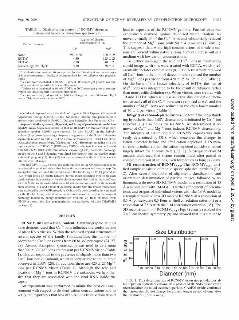
lead to exposure of the RCNMV genome. Purified virus wasexhaustively dialyzed against deionized water. Dialysis re-moved virtually all of the Ca2� ions and substantially reducedthe number of Mg2� ions (only 50 4 remained) (Table 1).This suggests that, while high concentrations of divalent cat-ions are present within native virions, they can diffuse out in amedium with low cation concentrations.
To further investigate the role of Ca2� ions in maintainingcapsid integrity, virions were treated with EGTA, which pref-erentially chelates calcium ions (8). EGTA treatment removedall Ca2� ions to the limit of detection and reduced the numberof Mg2� ions per virion from 420 25 to 125 28 (Table 1).On the basis of the known selectivity of EGTA, the loss ofMg2� ions was interpreted to be the result of diffusion ratherthan nonspecific chelation (8). When virions were treated with10 mM EDTA, which is a less selective divalent-cation chela-tor, virtually all of the Ca2� ions were removed as well and thenumber of Mg2� ions was reduced to the even lower numberof 40 2 per virion (Table 1).
Integrity of cation-depleted virions. To test if the long-stand-ing hypothesis that TBSV disassembly is initiated by Ca2� ionremoval (13) also holds for RCNMV, we determined if re-moval of Ca2� and Mg2� ions induces RCNMV disassembly.The integrity of cation-depleted RCNMV capsids was indi-rectly determined by DLS, which measures the changes invirion diameter before and after cation depletion. DLS mea-surements indicated that the cation-depleted capsids remainedlargely intact for at least 24 h (Fig. 1). Subsequent cryoEManalysis confirmed that virions remain intact after partial orcomplete removal of cations, even for periods as long as 7 days.
3D reconstruction of RCNMVNAT. The RCNMVNAT vitri-fied sample consisted of monodisperse spherical particles (Fig.2). After several iterations of alignment, classification, andorientation determination of particle images, followed by re-finement, a de novo 3D RCNMV model at a resolution of 18Å was obtained with IMAGIC. Further refinement of orienta-tions and origins of individual virions with the 18-Å model asa template resulted in a 3D map of RCNMV at a resolution of8.5 Å (conservative 0.5 Fourier shell correlation criterion) or aresolution of 7.5 Å with the 0.14 correlation criterion (35). The3D reconstruction of RCNMVNAT (Fig. 3) clearly resolved theT�3 icosahedral symmetry (9) and showed that it is similar to
FIG. 1. DLS determination of RCNMV virion size populations af-ter depletion of divalent cations. DLS profiles of RCNMV virions wererecorded after the noted treatment periods. CryoEM results confirmedthat virion size did not change for a much longer period of time afterthe treatment (up to a week).
TABLE 1. Divalent-cation content of RCNMV virions asdetermined by atomic absorption spectroscopy
Virion treatment
Avg no. of divalentcations/viriona SD
Ca2� Mg2�
None 390 30 420 25EGTAb �30 125 28EDTAc �30 40 2Dialysis against H2Od �30 50 4
a Divalent-cation concentrations reported for each treatment are the averagesof four measurements (duplicate determinations for two different viral prepara-tions).
b Virions were incubated in 10 mM EGTA at 20°C overnight prior to concen-tration and desalting with Centricon filter units.
c Virions were incubated in 10 mM EDTA at 20°C overnight prior to concen-tration and desalting with Centricon filter units.
d Virions were dialyzed against two 1-liter exchanges of 18-m� deionized H2Oover a 24-h incubation period at 20°C.
VOL. 80, 2006 STRUCTURE OF RCNMV REVEALED BY CRYOELECTRON MICROSCOPY 10397
on April 3, 2014 by guest
http://jvi.asm.org/
Dow
nloaded from

that known for other species in the family Tombusviridae, i.e.,TBSV (28), TCV (50), and CarMV (25). The close resem-blance between RCNMVNAT and TBSV is supported by thesimilarity of their respective CP sequences (Fig. 4). The diam-eter of the RCNMVNAT virion is 366 Å, and the capsid shell,which comprises the entangled S domains of the three CPsubunit conformations (A, B, and C), is �30 Å thick. Ninetydistinct protrusions, each extending �37 Å above the capsidshell, arise from tight associations between pairs of neighbor-ing P domains contributed by 30 C-C subunit homodimers and60 A-B subunit heterodimers. The C-C protrusions are locatedat the icosahedral twofold axes, and the A and B subunits arearranged around the fivefold and threefold axes of symmetry,respectively (Fig. 3A). Each subunit has an overall L shape,with the bottom bar of the L corresponding to the S domainthat forms the floor of the capsid shell and the vertical bar ofthe L corresponding to the P domain protrusions. The protru-sions are approximately elliptical in cross section with dimen-sions of �50 Å (C-C) and 47 Å (A-B) along the long axis and31 Å along the short axis. Each protrusion has a pair of dimpleson either side (Fig. 3A) with an �13-Å-wide S-shaped ridge atthe top (highest particle radius).
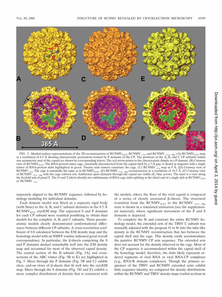
A prominent internal feature of the reconstruction is a cage-like density distribution lying between radii of 87 and 117 Å(Fig. 3B). This inner cage is composed of elongated slabs ofdensity that appear to associate as trimers (one trimer isshaded green in Fig. 3B). The cage and the capsid have differ-ent symmetries; the capsid is composed of 180 CP subunitsarranged with T�3 quasisymmetry, whereas the cage is com-posed of 60 slabs arranged with T�1 icosahedral symmetry. Atthe twofold axes, slabs are oriented in an antiparallel fashionand are separated by �50 Å. Similar cage-like structures havebeen observed in several other RNA viruses and virus-likeparticles (12, 20, 23, 29, 40, 44, 45).
Homology model of RCNMVNAT CP and fitting into densitymap. An RCNMVNAT CP homology model was constructed tohelp better understand the structural organization of the virus.This model was created with SWISS-MODEL and the TBSVcrystal structure because its CP had the highest sequence sim-ilarity to RCNMV among the solved Tombusviridae structures(Fig. 4). In creating S and P domain homology models for eachof the RCNMV A, B, and C CP subunits, the C TBSV CPsubunit was first subdivided into separate S, P, and extended-arm domains and the sequence for each of these pieces was
FIG. 2. Cryoelectron micrograph of RCNMV virions embedded in vitreous ice. The characteristic bumpy appearance of virions is a conse-quence of the radially extended P domains of the CP subunits.
10398 SHERMAN ET AL. J. VIROL.
on April 3, 2014 by guest
http://jvi.asm.org/
Dow
nloaded from
separately aligned to the RCNMV sequence, followed by ho-mology modeling for individual domains.
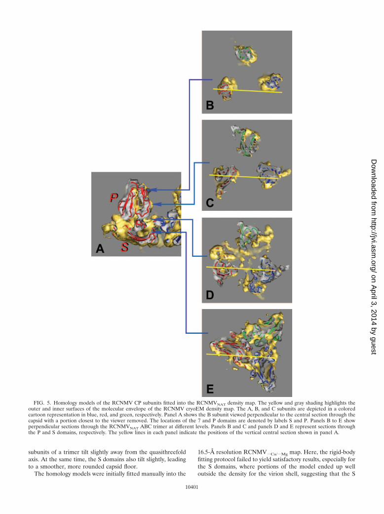
Each domain model was fitted as a separate rigid body(with Situs) to the A, B, and C subunit densities in the 8.5 ÅRCNMVNAT cryoEM map. The separated S and P domainsfor each CP subunit were reunited postfitting to obtain finalmodels for the complete A, B, and C subunits. These pseudo-atomic models clearly demonstrated conformational differ-ences between different CP subunits. A cross-correlation coef-ficient of 0.8 calculated between the EM density map and thehomology model with an NMFF routine indicated good overallcorrespondence. In particular, the �-sheets comprising the Sand P domains docked remarkably well into the EM densitymap and accounted for most of the observed capsid density.The central section of the B subunit (Fig. 5A) and cross-sections of the ABC trimer (Fig. 5B to E) are highlighted inFig. 5. Slices through the P domains (Fig. 5B and C) exhibitclear, end-on views of �-sheets that fit well into the cryoEMmap. Slices through the S domains (Fig. 5D and E) exhibit amore complex distribution of density that is consistent with
the models, where the floor of the viral capsid is composedof a series of closely associated �-sheets. The structuraltransition from the RCNMVNAT to the RCNMV�Ca/�Mg
state is shown in a simulated animation (see the supplemen-tal material), where significant movement of the P and Sdomains is depicted.
To complete the fit and construct the entire RCNMV ho-mology model, the extended arm of the TBSV C subunit wasmanually adjusted with the program O to fit into the tube-likedensity in the RCNMV reconstruction that lies between thecapsid shell and the cage. This density easily accommodatesthe putative RCNMV CP arm sequence. The extended armdoes not account for the density observed in the cage. Most ofthe CP sequence is accommodated within the capsid shell ofthe homology model; therefore, the slabs likely represent or-dered segments of viral RNA or viral RNA-CP complexes(e.g., RNA-R domain complexes). Though the primary se-quences of the TBSV and RCNMV extended arms exhibitlittle sequence identity, we compared the density distributionswithin the RCNMV and TBSV density maps (radial sections in
FIG. 3. Shaded-surface representations of the 3D reconstructions of RCNMVNAT, RCNMV�Ca, and RCNMV�Ca/�Mg. (A) RCNMVNAT mapat a resolution of 8.5 Å showing characteristic protrusions formed by P domains of the CP. The positions of the A, B, and C CP subunits withinone asymmetric unit of the capsid are shown by corresponding letters. The red arrow points to the characteristic dimple in a P domain. (B) Cutawayview of RCNMVNAT. The RNA-protein inner cage, essentially disconnected from the capsid shell by a 7-Å gap, is shown in magenta with a singletrimer of RNA-protein slabs highlighted in green. Twenty such trimers constitute the cage. (C) RCNMV�Ca map at 9 Å. (D) Cutaway view ofRCNMV�Ca. The cage is essentially the same as in RCNMVNAT. (E) RCNMV�Ca/�Mg reconstruction at a resolution of 16.5 Å. (F) Cutaway viewof RCNMV�Ca/�Mg with the cage colored red. Additional open channels through the capsid are visible (E, blue arrow). The inset is a view alongthe fivefold axis of panel F. The O and T labels identify two subdomains of RNA cage slabs splitting at the distal end of a single slab in RCNMVNATor RCNMV�Ca.
VOL. 80, 2006 STRUCTURE OF RCNMV REVEALED BY CRYOELECTRON MICROSCOPY 10399
on April 3, 2014 by guest
http://jvi.asm.org/
Dow
nloaded from
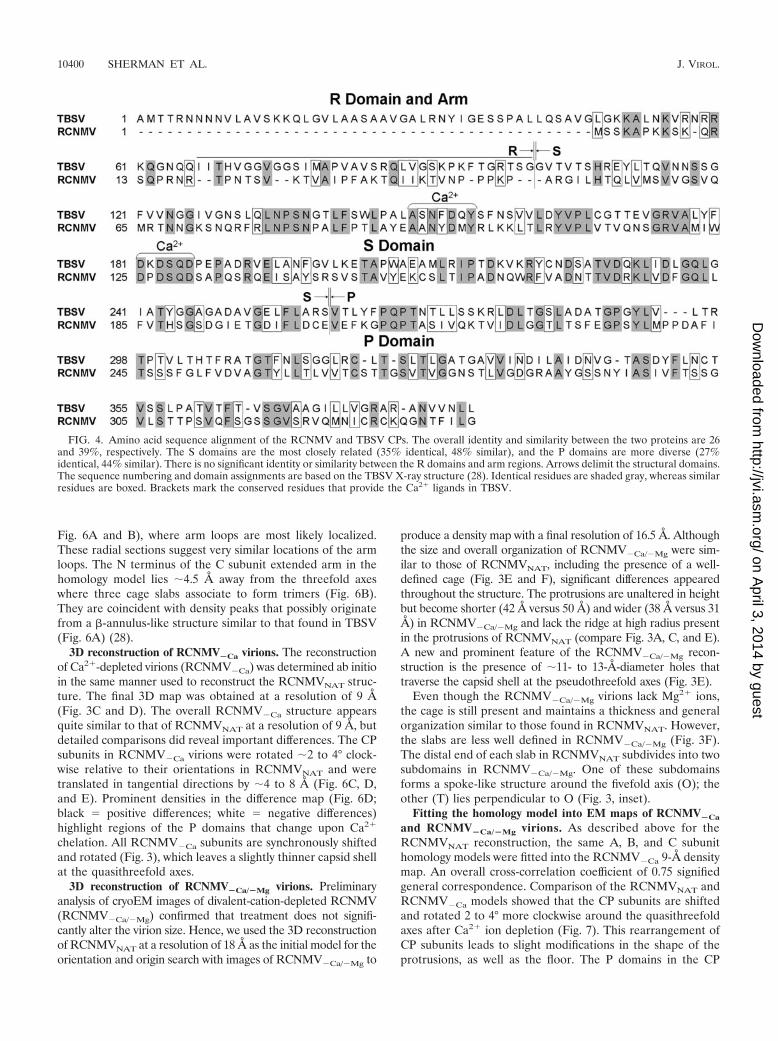
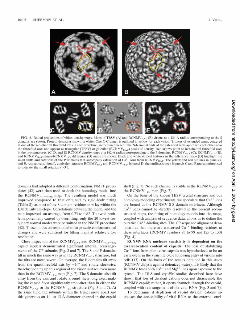
Fig. 6A and B), where arm loops are most likely localized.These radial sections suggest very similar locations of the armloops. The N terminus of the C subunit extended arm in thehomology model lies �4.5 Å away from the threefold axeswhere three cage slabs associate to form trimers (Fig. 6B).They are coincident with density peaks that possibly originatefrom a �-annulus-like structure similar to that found in TBSV(Fig. 6A) (28).
3D reconstruction of RCNMV�Ca virions. The reconstructionof Ca2�-depleted virions (RCNMV�Ca) was determined ab initioin the same manner used to reconstruct the RCNMVNAT struc-ture. The final 3D map was obtained at a resolution of 9 Å(Fig. 3C and D). The overall RCNMV�Ca structure appearsquite similar to that of RCNMVNAT at a resolution of 9 Å, butdetailed comparisons did reveal important differences. The CPsubunits in RCNMV�Ca virions were rotated �2 to 4° clock-wise relative to their orientations in RCNMVNAT and weretranslated in tangential directions by �4 to 8 Å (Fig. 6C, D,and E). Prominent densities in the difference map (Fig. 6D;black � positive differences; white � negative differences)highlight regions of the P domains that change upon Ca2�
chelation. All RCNMV�Ca subunits are synchronously shiftedand rotated (Fig. 3), which leaves a slightly thinner capsid shellat the quasithreefold axes.
3D reconstruction of RCNMV�Ca/�Mg virions. Preliminaryanalysis of cryoEM images of divalent-cation-depleted RCNMV(RCNMV�Ca/�Mg) confirmed that treatment does not signifi-cantly alter the virion size. Hence, we used the 3D reconstructionof RCNMVNAT at a resolution of 18 Å as the initial model for theorientation and origin search with images of RCNMV�Ca/�Mg to
produce a density map with a final resolution of 16.5 Å. Althoughthe size and overall organization of RCNMV�Ca/�Mg were sim-ilar to those of RCNMVNAT, including the presence of a well-defined cage (Fig. 3E and F), significant differences appearedthroughout the structure. The protrusions are unaltered in heightbut become shorter (42 Å versus 50 Å) and wider (38 Å versus 31Å) in RCNMV�Ca/�Mg and lack the ridge at high radius presentin the protrusions of RCNMVNAT (compare Fig. 3A, C, and E).A new and prominent feature of the RCNMV�Ca/�Mg recon-struction is the presence of �11- to 13-Å-diameter holes thattraverse the capsid shell at the pseudothreefold axes (Fig. 3E).
Even though the RCNMV�Ca/�Mg virions lack Mg2� ions,the cage is still present and maintains a thickness and generalorganization similar to those found in RCNMVNAT. However,the slabs are less well defined in RCNMV�Ca/�Mg (Fig. 3F).The distal end of each slab in RCNMVNAT subdivides into twosubdomains in RCNMV�Ca/�Mg. One of these subdomainsforms a spoke-like structure around the fivefold axis (O); theother (T) lies perpendicular to O (Fig. 3, inset).
Fitting the homology model into EM maps of RCNMV�Ca
and RCNMV�Ca/�Mg virions. As described above for theRCNMVNAT reconstruction, the same A, B, and C subunithomology models were fitted into the RCNMV�Ca 9-Å densitymap. An overall cross-correlation coefficient of 0.75 signifiedgeneral correspondence. Comparison of the RCNMVNAT andRCNMV�Ca models showed that the CP subunits are shiftedand rotated 2 to 4° more clockwise around the quasithreefoldaxes after Ca2� ion depletion (Fig. 7). This rearrangement ofCP subunits leads to slight modifications in the shape of theprotrusions, as well as the floor. The P domains in the CP
FIG. 4. Amino acid sequence alignment of the RCNMV and TBSV CPs. The overall identity and similarity between the two proteins are 26and 39%, respectively. The S domains are the most closely related (35% identical, 48% similar), and the P domains are more diverse (27%identical, 44% similar). There is no significant identity or similarity between the R domains and arm regions. Arrows delimit the structural domains.The sequence numbering and domain assignments are based on the TBSV X-ray structure (28). Identical residues are shaded gray, whereas similarresidues are boxed. Brackets mark the conserved residues that provide the Ca2� ligands in TBSV.
10400 SHERMAN ET AL. J. VIROL.
on April 3, 2014 by guest
http://jvi.asm.org/
Dow
nloaded from
subunits of a trimer tilt slightly away from the quasithreefoldaxis. At the same time, the S domains also tilt slightly, leadingto a smoother, more rounded capsid floor.
The homology models were initially fitted manually into the
16.5-Å resolution RCNMV�Ca/�Mg map. Here, the rigid-bodyfitting protocol failed to yield satisfactory results, especially forthe S domains, where portions of the model ended up welloutside the density for the virion shell, suggesting that the S
FIG. 5. Homology models of the RCNMV CP subunits fitted into the RCNMVNAT density map. The yellow and gray shading highlights theouter and inner surfaces of the molecular envelope of the RCNMV cryoEM density map. The A, B, and C subunits are depicted in a coloredcartoon representation in blue, red, and green, respectively. Panel A shows the B subunit viewed perpendicular to the central section through thecapsid with a portion closest to the viewer removed. The locations of the 7 and P domains are denoted by labels S and P. Panels B to E showperpendicular sections through the RCNMVNAT ABC trimer at different levels. Panels B and C and panels D and E represent sections throughthe P and S domains, respectively. The yellow lines in each panel indicate the positions of the vertical central section shown in panel A.
10401
on April 3, 2014 by guest
http://jvi.asm.org/
Dow
nloaded from
domains had adopted a different conformation. NMFF proce-dures (42) were then used to dock the homology model intothe RCNMV�Ca/�Mg map. The resulting model was muchimproved compared to that obtained by rigid-body fitting(Table 2), as most of the S domain residues now lay within theEM density envelope. Correlations between the model and themap improved, on average, from 0.73 to 0.82. To avoid prob-lems potentially caused by overfitting, only the 20 lowest-fre-quency normal modes were permitted in the NMFF procedure(42). These modes corresponded to large-scale conformationalchanges and were sufficient for fitting maps at relatively lowresolution.
Close inspection of the RCNMVNAT and RCNMV�Ca/�Mg
capsid models demonstrated significant internal rearrange-ments of the CP subunits (Fig. 3 and 7). The S and P domainstilt in much the same way as in the RCNMV�Ca structure, butthe tilts are more severe. On average, the P domains tilt awayfrom the quasithreefold axis by �10° and rotate clockwise,thereby opening up this region of the virion surface even morethan in the RCNMV�Ca map (Fig. 7). The S domains also tiltaway from the axis and rotate around their long axes, mak-ing the capsid floor significantly smoother than in either theRCNMVNAT or the RCNMV�Ca structure (Fig. 3 and 7). Atthe same time, the subunits within the trimer move apart andthis generates an 11- to 13-Å diameter channel in the capsid
shell (Fig. 7). No such channel is visible in the RCNMVNAT orthe RCNMV�Ca map (Fig. 7).
On the basis of the known TBSV crystal structure and ourhomology-modeling experiments, we speculate that Ca2� ionsare bound at the RCNMV S-S domain interfaces. AlthoughCa2� ions cannot be directly resolved in the present recon-structed maps, the fitting of homology models into the maps,coupled with analysis of sequence data, allows us to define theputative Ca2�-binding sites. The CP sequence alignment dem-onstrates that there are conserved Ca2�-binding residues atthese interfaces (RCNMV residues 93 to 99 and 125 to 130)(Fig. 4).
RCNMV RNA nuclease sensitivity is dependent on thedivalent-cation content of capsids. The loss of stabilizingCa2� ions from plant virus capsids was hypothesized to be anearly event in the virus life cycle following entry of virions intocells (13). On the basis of the results obtained in this study(RCNMV dialysis against deionized water), it is likely that theRCNMV loses both Ca2� and Mg2� ions upon exposure to thecytosol. The DLS and cryoEM studies described here haveshown that loss of divalent cations does not disassemble theRCNMV capsid; rather, it opens channels through the capsid,coupled with rearrangement of the viral RNA (Fig. 3 and 7).
To determine if depletion of capsid divalent cations in-creases the accessibility of viral RNA to the external envi-
FIG. 6. Radial projections of virion density maps. Maps of TBSV (A) and RCNMVNAT (B) virions at a 124-Å radius corresponding to the Sdomains are shown. Protein density is shown in white. One C-C dimer is outlined in yellow for each virion. Trimers of extended arms, centeredat one of the icosahedral threefold axes in each structure, are outlined in red. The N-terminal ends of the extended arms approach each other nearthe threefold axes and appear as triangular (TBSV) or globular (RCNMVNAT) peaks of density. Red arrows point to icosahedral threefold axesin the two structures. (C, D, and E) RCNMV density maps at a 162-Å radius corresponding to the P domains. RCNMVNAT (C), RCNMV�Ca (E),and RCNMVNAT-minus-RCNMV�Ca difference (D) maps are shown. Black and white striped features in the difference maps (D) highlight thesmall shifts and rotations of the P domains that accompany extraction of Ca2� ions from RCNMVNAT. The yellow and red outlines in panels Cand E, respectively, identify equivalent areas in RCNMVNAT and RCNMV�Ca. In panel D, the outlines shown in panels C and E are superimposedto indicate the small rotation (�3°).
10402 SHERMAN ET AL. J. VIROL.
on April 3, 2014 by guest
http://jvi.asm.org/
Dow
nloaded from

FIG. 7. Close-up views of interfaces of the A, B, and C CP subunits in pseudoatomic homology models. The loops and -helices that form thelining of the quasithreefold axis move slightly away from the axis when Ca2� ions are depleted and significantly farther when both Ca2� and Mg2�
are depleted, resulting in the opening of an 11- to 13-Å channel through the capsid shell in RCNMV�Ca/�Mg virions. Electrostatic-potentialdiagrams show the charge distributions of the inner (middle row) and outer (lower row) surfaces. The concentration of positive charge just outsidethe cavities in RCNMVNAT and RCNMV�Ca or the channel in RCNMV�Ca/�Mg may serve to attract and position the RNA for exit through thecharged channel. The outer surface of the capsid in the region surrounding the local threefold axis is predominantly negatively charged, whichmight help repel RNA as it emerges from the surface. (A to C) Close-up views of a portion of the A, B, and C subunit interfaces in thepseudoatomic homology models for the native RCNMVNAT (A), RCNMV�Ca (B), and RCNMV�Ca/�Mg (C) structures. The loops and -helicesthat form the lining of the local threefold axis move slightly from the axis when Ca2� ions are chelated (B) and significantly farther when both Ca2�
and Mg2� ions are chelated (C), opening an 11- to 13-Å channel through the capsid shell. (D to I) Electrostatic-potential representations of CPtrimers showing charge distributions on the outer (D to F) and inner (G to I) surfaces for RCNMVNAT (D and G), RCNMV�Ca (E and H), andRCNMV�Ca/�Mg (F and I). Red represents negative charges, whereas positively charged residues are shown in blue. On the inner capsid surface,the concentration of positive charge just outside the cavities in RCNMVNAT (G) and RCNMV�Ca (H) or the channel in RCNMV�Ca/�Mg (I) mayserve to attract and position the RNA for exit through the negatively charged channel (not shown). The outer surface of the capsid in the regionsurrounding the local threefold axis is predominantly negatively charged, which might help repel RNA as it emerges from the surface and thereforemay facilitate the binding of ribosomes to the protruding ends.
VOL. 80, 2006 STRUCTURE OF RCNMV REVEALED BY CRYOELECTRON MICROSCOPY 10403
on April 3, 2014 by guest
http://jvi.asm.org/
Dow
nloaded from

ronment, the infectivity of divalent-cation-depleted RCNMVwas determined after nuclease treatment. There was noobserved difference in infectivity between treated and un-treated RCNMVNAT virions (Table 3), suggesting that theRCNMV genome is fully protected within virion capsids con-taining the full complement of divalent cations. Depletion ofCa2� ions resulted in a 33 to 81% reduction in infectivity afternuclease treatment, with the amount of reduction dependenton the nuclease used (Table 3). Infectivity was further reducedby nuclease treatment after both Ca2� and Mg2� ions wereremoved. Only a slight decrease (�11%) in the infectivity ofRCNMV�Ca versus RCNMV�Ca/�Mg was observed afterRNase V1 treatment compared to a more-than-50% decreaseobserved with RNases A and T1 (Table 3). Interestingly, bothsingle-strand-specific RNases (A and T1) and a double-strand-specific RNase (V1) also cause a decrease in the infectivity ofcation-depleted virions. To confirm that nuclease digestionoccurred in vitro, RNA was extracted from nuclease-treated,cation-depleted virions and shown by electrophoresis to bedigested (data not shown). The loss of infectivity after nucleasetreatment suggests that removal of divalent cations from thecapsid causes both single- and double-stranded portions ofthe RCNMV RNA genome to become exposed to the envi-ronment.
DISCUSSION
Structural analysis of RCNMV provides additional evidencethat viruses in the Tombusviridae family have a conserved mor-phology despite differences in their amino acid sequences. Thissuggests that the assembly and disassembly mechanisms ofspecies in the family Tombusviridae are, in general, similar,with possible individual variations on a common theme. Theunusually robust virions of TBSV, RCNMV, and CarMV hintat a trigger factor that causes at least weakening, if not com-plete disassembly, of the capsid, followed by genome leakagein the cytosol and production of new virions. The RCNMVstructures with different divalent-cation contents obtained inthe present study revealed significant conformational rear-rangements upon cation depletion with channels opening inthe capsid. These channels could provide conduits for the viralRNA to exit the capsid and become available for replication.
CP sequence comparisons among the diverse members ofthe Tombusviridae family revealed significant variation in thenumber of residues that comprise the arms and R domains,with the longest sequences belonging to TBSV and Tobacconecrosis virus. The putative R domain and arm regions of theRCNMV CP are 52 residues shorter than the correspondingregions in TBSV (Fig. 4) and may correlate with the necessity
to package a larger genome (�5.3 to 5.8 kb in RCNMV versus�4.9 kb in TBSV). This hypothesis for structure conservationhas been recently confirmed by the ability of a series of TBSVCP N-terminal deletion mutants to form virus-like particles(16). Coincidentally, a 52-residue TBSV deletion mutant (CP-N 52) produces particles which are morphologically very sim-ilar to wild-type TBSV virions, and this resembles the situationwith the shorter RCNMV CP. The major structural differencesbetween RCNMV and TBSV can be partially attributed to theshorter CP amino terminus in RCNMV.
One of the most striking features of the RCNMV virion 3Dreconstruction is the cage, a structure that is composed of 60slabs organized as 20 trimers, each with a prominent knob at itscenter (Fig. 3). The cage and capsid shell are separated by a 7-to 9-Å gap. The pseudoatomic model of the RCNMV CPsubunit accounts for most of the densities in the capsid shelland also the connecting densities corresponding to the ex-tended arms. Altogether, with the exception of 14 to 16 N-terminal residues, the model accounts for the entire CP se-quence. The N-terminal residues may be part of the cage slabs.Each slab is �16 Å thick and �25 Å wide in cross-section. Afeature this large can easily accommodate a single-strandedRNA (ssRNA) fragment complexed with the N-terminal partcorresponding to the R domain of CP. Excluded-volume esti-mates suggest that the cage accounts for as much as 35 to 40%of the total RCNMV RNA genome; the remaining genomicRNA lies inside the cage and appears to be more looselypacked and is most likely disordered or does not conform toicosahedral symmetry.
The presence of an ordered genome has been described fora number of viruses. Within the family Tombusviridae, cryoEMreconstructions of TBSV (1) and Hibiscus chlorotic ringspotvirus (12) revealed the presence of a layer inside the virioncapsid tentatively formed by the CP R domains complexedwith genomic RNA. However, these regions in TBSV andHibiscus chlorotic ringspot virus appeared diffuse with no char-acteristic discernible details like the discrete slabs observed inthe RCNMV cage. This lack of an ordered inner structure inTBSV does not correlate with infectivity since a TBSV CPdeletion mutant lacking the 30 N-proximal residues of theRNA-binding domain forms virus-like particles which vary insize yet are just as infectious as wild-type virions (11).
In TBSV, a �-annulus formed by three interlocking ex-tended arms (28) is located at the threefold axis inside the
TABLE 2. Correlations between pseudoatomic models andRCNMV�Ca/�Mg map before and after application
of NMFF procedure
CP subunitCorrelation coefficient
Before NMFF After NMFF
A 0.76 0.85B 0.67 0.82C 0.76 0.80
TABLE 3. Local-lesion infectivity assay of chelator-treatedRCNMV virions on C. quinoa after exposure to nucleases
Chelatortreatmenta
% Infectivity after treatmentb with:
No RNase RNase A RNase T1 RNase V1
Mocktreatmentc
0 0 0 0
None 100 (53 14) 100 (53 2) 100 (53 11) 100 (45 13)EGTA 106 (56 6) 19 (10 3) 49 (26 4) 67 (30 6)EDTA 91 (48 4) 9 (5 1) 23 (12 5) 56 (25 6)
a Four micrograms of purified RCNMV virions was treated with or without achelator, followed by nuclease digestion prior to inoculation onto C. quinoaleaves.
b Infectivity is expressed as a percentage of the number of lesions formed perleaf by treated virions versus untreated virions. The average number of lesionsobserved and standard deviation from four leaves are shown in parentheses.
c Mock treatment was done with buffer but no RCNMV virions.
10404 SHERMAN ET AL. J. VIROL.
on April 3, 2014 by guest
http://jvi.asm.org/
Dow
nloaded from
capsid shell. Each arm extends away from the annulus andconnects to a corresponding S domain. Similar density featuresat the same threefold location are observed in the RCNMVNAT
(Fig. 6A, red arrows and red outlines) and RCNMV�Ca maps.The �-annuli in the RCNMV density map appear as knobs atthe center of the slab trimer, and the arms appear as well-defined, curved tubes of density that run from the knob towardthe capsid shell (data not shown). On the basis of the sequencealignment of TBSV and RCNMV (Fig. 4), RCNMV appears tolack a globular R domain like that known to be present inTBSV. In RCNMV, the N-terminal sequence is predominantlybasic and we believe that up to 16 N-terminal residues interactwith ordered RNA and are involved in the formation of theslabs.
In addition to determining the structure of the RCNMVNAT
capsid, this study has focused on the role of Ca2� and Mg2�
ions in RCNMV 3D organization and assembly. Atomic ab-sorption spectroscopy showed that each virion contains signif-icant amounts of divalent cations. When native virions wereexposed to low Ca2� or Mg2� ion conditions, such as exhaus-tive dialysis against deionized water, these cations wereleached from the virions. To probe their role in RCNMVcapsid stability, structural and functional studies were per-formed with divalent cations selectively removed from virionsby the chelating agents EGTA (Ca2�) and EDTA (Ca2� andMg2�). Comparative analysis of the structures obtained hasshown that removal of Ca2� ions induces movements of the Sand P domains (Fig. 3 and 7). The removal of both Ca2� andMg2� ions triggers more global rearrangement in the RCNMVstructure (Fig. 3 and 7). These data indicate that Ca2� ions arenot solely responsible for structural dynamics of RCNMV viri-ons; Mg2� ions also significantly contribute to the rearrange-ments.
Upon loss of divalent cations, each of the 180 P domainsexhibited increased flexibility and they reoriented by 30 ormore degrees (Fig. 3). As a result, the P-P interfaces arechanged significantly. Increased mobility of the P domainsappears to explain, at least in part, why the resolution of theRCNMV�Ca/�Mg map was limited to 16.5 Å. Nevertheless, useof the NMFF procedure provided a means to quantitativelymodel the changes observed in the cryoEM map. The flexiblefitting procedure showed dramatic changes in S domains upondivalent-cation depletion that cannot be described in termsof simple rotations and translations. Instead, the S domainsappear to adopt a conformation different from that seen inRCNMVNAT or RCNMV�Ca virions. These conformationalchanges in the P and S domains are responsible for forming the11- to 13-Å-diameter channels at the quasithreefold axes (Fig.3 and 7). The differences were especially prominent for both-helices and for the loop (R165 to D174) lining the axis. In itsnative conformation, loop I158 to G181 lies completely outsideof the density envelope in the RCNMV�Ca/�Mg map whenfitted as a rigid body. The NMFF modeling experimentsstrongly suggest that refolding of this loop occurs (Table 2). Itis noteworthy in this context that the swollen TBSV structuredid not exhibit any significant CP subunit refolding (33). Theconformational changes observed in RCNMV upon loss ofdivalent cations most likely results from the combined effect ofCa2� and Mg2� ions. Loss of Ca2� alone does not significantlyalter virion integrity. Removal of Ca2� seems to initiate struc-
tural changes such as small-scale movements of the P and Sdomains and the slight increase in intersubunit distances at thequasithreefold axes of the capsid, where a channel appearsafter both cations are lost. This fact alone suggests that theRCNMV cage is tightly linked to the capsid and might exert aforce when cations leave the virions, leading to opening of thechannels. Additionally, our experiments with cation depletiondemonstrated that pH changes used in earlier experimentswith TBSV (13) are not necessary for the channels opening inRCNMV.
Depletion of both Ca2� and Mg2� ions also leads to moresignificant changes in the structure of RCNMV (Fig. 3). Weassume that Mg2� ions bind to the ssRNA genome, neutraliz-ing its charge and aiding in its condensation in virions. Loss ofMg2� ions could create a charge imbalance and lead to theobserved splitting of the cage slabs (Fig. 3, inset).
The channels that arise at the quasithreefold axes in theRCNMV�Ca/�Mg virions are too constricted to permit nucle-ases in their native form (�55 Å in diameter) to penetrate thecapsid and to cleave the packaged RNA genome. However,these channels are sufficient in size to permit ssRNA (11 to 13Å in diameter) to leak from the capsid. Such leakage wouldcorrelate with increased nuclease sensitivity of RCNMV�Ca/�Mg
to single-strand-specific RNases (Table 3). The inner part ofthe channel (the entrance) includes a few basic residues but ispredominantly lined with negatively charged residues (Fig. 7).The termini of the genomic RNAs might be attracted to theentrance of the channel, but repulsive forces within the chan-nel would facilitate transit of the RNA to the cytosol. Thechanges observed in both the cage and the outer shell ofRCNMV�Ca/�Mg may point to the actual RNA release mech-anism that occurs for RCNMVNAT in vivo. Interestingly, stud-ies of Cowpea chlorotic mottle virus have similarly suggestedthat viral RNA release by free diffusion occurs through chan-nels at the quasithreefold axes (17). Channels similar to thosein RCNMV were observed in a recent cryoEM reconstructionof TBSV virions that were depleted of divalent cations, fol-lowed by a rise in pH to 7.5 (1). Free-Ca2� and -Mg2� ionconcentrations are typically in the millimolar range in soil andgroundwater (3, 6). In contrast, they range between submicro-molar and micromolar levels in the cytosol. These low levelsare maintained and regulated by several enzymes (22, 31).Such low cation concentrations could trigger leaching of Ca2�
and Mg2� ions from RCNMV virions as they enter cells and inturn would produce the conformational changes that openchannels for RNA to exit. In addition, it is also known that acellular response to stress and infection is to increase cytosoliccalcium (2). Further, once a cell begins apoptosis, regulationof cytosolic calcium is lost (10), ensuring an increased supplyof calcium for virion formation late in the infection cycle.
ACKNOWLEDGMENTS
We thank Y. Ji and C. Xiao, and R. Ashmore for use of theirimage-processing programs.
This research was supported in part by NIH grant GM-33050 toT.S.B., NSF grant MCB-0077964 to S.A.L. and T.L.S., a grant from theW. M. Keck Foundation to the Purdue Structural Biology Group forthe purchase of a CM 300 FEG microscope, and a Purdue Universityreinvestment grant to the Structural Biology Group.
VOL. 80, 2006 STRUCTURE OF RCNMV REVEALED BY CRYOELECTRON MICROSCOPY 10405
on April 3, 2014 by guest
http://jvi.asm.org/
Dow
nloaded from
REFERENCES
1. Aramayo, R., C. Merigoux, E. Larquet, P. Bron, J. Perez, C. Dumas, P.Vachette, and N. Boisset. 2005. Divalent ion-dependent swelling of TomatoBushy Stunt Virus: a multi-approach study. Biochim. Biophys. Acta 1724:345–354.
2. Atkinson, M. M., L. D. Keppler, E. W. Orlandi, C. J. Baker, and C. F.Mischke. 1990. Involvement of plasma membrane calcium influx in bacterialinduction of the K/H and hypersensitive responses in tobacco. Plant Physiol.92:215–221.
3. Barber, S. A. 1995. Soil nutrient bioavailability, p. 271. Wiley-Interscience,New York, N.Y.
4. Basnayake, V. R., T. L. Sit, and S. A. Lommel. 2006. The genomic RNApackaging scheme of Red clover necrotic mosaic virus. Virology 345:532–539.
5. Bowman, V. D., E. S. Chase, A. W. Franz, P. R. Chipman, X. Zhang, K. L.Perry, T. S. Baker, and T. J. Smith. 2002. An antibody to the putative aphidrecognition site on cucumber mosaic virus recognizes pentons but not hexons.J. Virol. 76:12250–12258.
6. Briggs, J. C., and J. F. Ficke. 1977. Quality of rivers of the United States,1975 water year—based on the National Stream Quality Accounting Net-work (NASQAN). U.S. Geological Survey water-supply paper 1540-C.
7. Brooks, B. R., R. E. Bruccoleri, B. D. Olafson, D. J. States, S. Swaminathan,and M. Karplus. 1983. CHARMM: a program for macromolecular energy,minimization, and dynamics calculations. J. Comput. Chem. 4:187–217.
8. Caldwell, P. C. 1970. Calcium chelation and buffers, p. 10–16. In A. W.Cuthbert (ed.), Calcium and cellular function. Macmillan, London, UnitedKingdom.
9. Caspar, D. L., and A. Klug. 1962. Physical principles in the construction ofregular viruses. Cold Spring Harbor Symp. Quant. Biol. 27:1–24.
10. Clough, S. J., K. A. Fengler, I. C. Yu, B. Lippok, R. K. Smith, Jr., and A. F.Bent. 2000. The Arabidopsis dnd1 “defense, no death” gene encodes amutated cyclic nucleotide-gated ion channel. Proc. Natl. Acad. Sci. USA97:9323–9328.
11. Desvoyes, B., and H. B. Scholthof. 2002. Host-dependent recombination of aTomato bushy stunt virus coat protein mutant yields truncated capsid sub-units that form virus-like complexes which benefit systemic spread. Virology304:434–442.
12. Doan, D. N., K. C. Lee, P. Laurinmaki, S. Butcher, S. M. Wong, and T.Dokland. 2003. Three-dimensional reconstruction of hibiscus chlorotic ring-spot virus. J. Struct. Biol. 144:253–261.
13. Durham, A. C., D. A. Hendry, and M. B. Von Wechmar. 1977. Does calciumion binding control plant virus disassembly? Virology 77:524–533.
14. Fishman, M. J., and S. C. Downs. 1966. Methods for analysis of selectedmetals in water by atomic absorption: U.S. Geological Survey water-supplypaper 1540-C, p. C26–C28.
15. Hallett, F. R., J. Watton, and P. Krygsman. 1991. Vesicle sizing: numberdistributions by dynamic light scattering. Biophys. J. 59:357–362.
16. Hsu, C., P. Singh, W. Ochoa, D. J. Manayani, M. Manchester, A. Schneemann,and V. S. Reddy. 2006. Characterization of polymorphism displayed by the coatprotein mutants of tomato bushy stunt virus. Virology 349:222–229.
17. Isea, R., C. Aponte, and R. Cipriani. 2004. Can the RNA of the cowpeachlorotic mottle virus be released through a channel by means of free dif-fusion? A test in silico. Biophys. Chem. 107:101–106.
18. Jones, T. A., J. Y. Zou, S. W. Cowan, and Kjeldgaard. 1991. Improvedmethods for building protein models in electron density maps and the loca-tion of errors in these models. Acta Crystallogr. A 47(Pt. 2):110–119.
19. Larson, S. B., J. Day, A. Greenwood, and A. McPherson. 1998. Refinedstructure of satellite tobacco mosaic virus at 1.8 Å resolution. J. Mol. Biol.277:37–59.
20. Larson, S. B., and A. McPherson. 2001. Satellite tobacco mosaic virus RNA:structure and implications for assembly. Curr. Opin. Struct. Biol. 11:59–65.
21. Lommel, S. A. 1983. Ph.D. dissertation. University California, Berkeley.22. Luan, S., J. Kudla, M. Rodriguez-Concepcion, S. Yalovsky, and W. Gruissem.
2002. Calmodulins and calcineurin B-like proteins: calcium sensors for specificsignal response coupling in plants. Plant Cell 14(Suppl.):S389–S400.
23. Lucas, R. W., S. B. Larson, M. A. Canady, and A. McPherson. 2002. Thestructure of tomato aspermy virus by X-ray crystallography. J. Struct. Biol.139:90–102.
24. Montelius, I., L. Liljas, and T. Unge. 1988. Structure of EDTA-treatedsatellite tobacco necrosis virus at pH 6.5. J. Mol. Biol. 201:353–363.
25. Morgunova, E. Y., Z. Dauter, E. Fry, D. I. Stuart, V. Y. Stel’mashchuk, A. M.Mikhailov, K. S. Wilson, and B. K. Vainshtein. 1994. The atomic structure ofCarnation Mottle Virus capsid protein. FEBS Lett. 338:267–271.
26. Musil, M., and J. Gallo. 1982. Serotypes of red clover necrotic mosaic virus.I. Characterization of three serotypes. Acta Virol. 26:497–501.
27. Oda, Y., K. Saeki, Y. Takahashi, T. Maeda, H. Naitow, T. Tsukihara, and K.Fukuyama. 2000. Crystal structure of tobacco necrosis virus at 2.25 Å reso-lution. J. Mol. Biol. 300:153–169.
28. Olson, A. J., G. Bricogne, and S. C. Harrison. 1983. Structure of tomato busystunt virus. IV. The virus particle at 2.9 Å resolution. J. Mol. Biol. 171:61–93.
29. Opalka, N., M. Tihova, C. Brugidou, A. Kumar, R. N. Beachy, C. M.Fauquet, and M. Yeager. 2000. Structure of native and expanded sobemo-viruses by electron cryo-microscopy and image reconstruction. J. Mol. Biol.303:197–211.
30. Orlova, E. V., M. B. Sherman, W. Chiu, H. Mowri, L. C. Smith, and A. M.Gotto. 1999. Three-dimensional structure of low density lipoproteins byelectron cryomicroscopy. Proc. Natl. Acad. Sci. USA 96:8420–8425.
31. Pittman, J. K., and K. D. Hirschi. 2003. Don’t shoot the (second) messenger:endomembrane transporters and binding proteins modulate cytosolic Ca2�
levels. Curr. Opin. Plant Biol. 6:257–262.32. Poirot, O., K. Suhre, C. Abergel, E. O’Toole, and C. Notredame. 2004.
3DCoffee@igs: a web server for combining sequences and structures into amultiple sequence alignment. Nucleic Acids Res. 32:W37–W40.
33. Robinson, I. K., and S. C. Harrison. 1982. Structure of the expanded state oftomato bushy stunt virus. Nature 297:563–568.
34. Rochon, D., K. Kakani, M. Robbins, and R. Reade. 2004. Molecular aspectsof plant virus transmission by olpidium and plasmodiophorid vectors. Annu.Rev. Phytopathol. 42:211–241.
35. Rosenthal, P. B., and R. Henderson. 2003. Optimal determination of particleorientation, absolute hand, and contrast loss in single-particle electron cryo-microscopy. J. Mol. Biol. 333:721–745.
36. Rossmann, M. G., C. Abad-Zapatero, J. W. Erickson, and H. S. Savithri.1983. RNA-protein interactions in some small plant viruses. J. Biomol.Struct. Dyn. 1:565–579.
37. Saxton, W. O., and W. Baumeister. 1982. The correlation averaging of aregularly arranged bacterial cell envelope protein. J. Microsc. 127:127–138.
38. Schwede, T., J. Kopp, N. Guex, and M. C. Peitsch. 2003. SWISS-MODEL: anautomated protein homology-modeling server. Nucleic Acids Res. 31:3381–3385.
39. Serysheva, I. I., E. V. Orlova, W. Chiu, M. B. Sherman, S. L. Hamilton, andM. van Heel. 1995. Electron cryomicroscopy and angular reconstitution usedto visualize the skeletal muscle calcium release channel. Nat. Struct. Biol.2:18–24.
40. Speir, J. A., S. Munshi, G. Wang, T. S. Baker, and J. E. Johnson. 1995.Structures of the native and swollen forms of cowpea chlorotic mottle virusdetermined by X-ray crystallography and cryo-electron microscopy. Struc-ture 3:63–78.
41. Tama, F., F. X. Gadea, O. Marques, and Y. H. Sanejouand. 2000. Building-block approach for determining low-frequency normal modes of macromol-ecules. Proteins 41:1–7.
42. Tama, F., O. Miyashita, and C. L. Brooks III. 2004. Flexible multi-scalefitting of atomic structures into low-resolution electron density maps withelastic network normal mode analysis. J. Mol. Biol. 337:985–999.
43. Tama, F., and Y. H. Sanejouand. 2001. Conformational change of proteinsarising from normal mode calculations. Protein Eng. 14:1–6.
44. Tang, L., K. N. Johnson, L. A. Ball, T. Lin, M. Yeager, and J. E. Johnson.2001. The structure of pariacoto virus reveals a dodecahedral cage of duplexRNA. Nat. Struct. Biol. 8:77–83.
45. Tihova, M., K. A. Dryden, T. V. Le, S. C. Harvey, J. E. Johnson, M. Yeager,and A. Schneemann. 2004. Nodavirus coat protein imposes dodecahedralRNA structure independent of nucleotide sequence and length. J. Virol.78:2897–2905.
46. van Heel, M. 1987. Angular reconstitution: a posteriori assignment of pro-jection directions for 3D reconstruction. Ultramicroscopy 21:111–124.
47. van Heel, M., and J. Frank. 1981. Use of multivariate statistics in analysingthe images of biological macromolecules. Ultramicroscopy 6:187–194.
48. van Heel, M., and G. Harauz. 1986. Resolution criteria for three dimensionalreconstructions. Optik 73:119–122.
49. van Heel, M., G. Harauz, and E. V. Orlova. 1996. A new generation of theIMAGIC image processing system. J. Struct. Biol. 116:17–24.
50. Wei, N., L. A. Heaton, T. J. Morris, and S. C. Harrison. 1990. Structure andassembly of turnip crinkle virus. VI. Identification of coat protein bindingsites on the RNA. J. Mol. Biol. 214:85–95.
51. Wiener, N. 1949. Extrapolation, interpolation, and smoothing of station-ary time series with engineering applications. The MIT Press, Cambridge,Mass.
52. Wriggers, W., and S. Birmanns. 2001. Using Situs for flexible and rigid-bodyfitting of multiresolution single-molecule data. J. Struct. Biol. 133:193–202.
53. Xiong, Z. G., and S. A. Lommel. 1991. Red clover necrotic mosaic virusinfectious transcripts synthesized in vitro. Virology 182:388–392.
54. Zhang, X., S. B. Walker, P. R. Chipman, M. L. Nibert, and T. S. Baker. 2003.Reovirus polymerase lambda 3 localized by cryo-electron microscopy ofvirions at a resolution of 7.6 Å. Nat. Struct. Biol. 10:1011–1018.
55. Zhou, Z. H., S. Hardt, B. Wang, M. B. Sherman, J. Jakana, and W. Chiu.1996. CTF determination of images of ice-embedded single particles using agraphics interface. J. Struct. Biol. 116:216–222.
10406 SHERMAN ET AL. J. VIROL.
on April 3, 2014 by guest
http://jvi.asm.org/
Dow
nloaded from