A Genetic Basis for a Postmeiotic X Versus Y Chromosome Intragenomic Conflict in the Mouse
15
A Genetic Basis for a Postmeiotic X Versus Y Chromosome Intragenomic Conflict in the Mouse Julie Cocquet 1,2,3 *, Peter J. I. Ellis 4 , Shantha K. Mahadevaiah 5 , Nabeel A. Affara 4 , Daniel Vaiman 1,2,3 , Paul S. Burgoyne 5 1 Inserm, U1016, Institut Cochin, Paris, France, 2 CNRS, UMR8104, Paris, France, 3 Universite ´ Paris Descartes, Sorbonne Paris Cite ´ , Paris, France, 4 Department of Pathology, Mammalian Molecular Genetics Group, University of Cambridge, Cambridge, United Kingdom, 5 Division of Stem Cell Biology and Developmental Genetics, MRC National Institute for Medical Research, London, United Kingdom Abstract Intragenomic conflicts arise when a genetic element favours its own transmission to the detriment of others. Conflicts over sex chromosome transmission are expected to have influenced genome structure, gene regulation, and speciation. In the mouse, the existence of an intragenomic conflict between X- and Y-linked multicopy genes has long been suggested but never demonstrated. The Y-encoded multicopy gene Sly has been shown to have a predominant role in the epigenetic repression of post meiotic sex chromatin (PMSC) and, as such, represses X and Y genes, among which are its X-linked homologs Slx and Slxl1. Here, we produced mice that are deficient for both Sly and Slx/Slxl1 and observed that Slx/Slxl1 has an opposite role to that of Sly, in that it stimulates XY gene expression in spermatids. Slx/Slxl1 deficiency rescues the sperm differentiation defects and near sterility caused by Sly deficiency and vice versa. Slx/Slxl1 deficiency also causes a sex ratio distortion towards the production of male offspring that is corrected by Sly deficiency. All in all, our data show that Slx/Slxl1 and Sly have antagonistic effects during sperm differentiation and are involved in a postmeiotic intragenomic conflict that causes segregation distortion and male sterility. This is undoubtedly what drove the massive gene amplification on the mouse X and Y chromosomes. It may also be at the basis of cases of F1 male hybrid sterility where the balance between Slx/ Slxl1 and Sly copy number, and therefore expression, is disrupted. To the best of our knowledge, our work is the first demonstration of a competition occurring between X and Y related genes in mammals. It also provides a biological basis for the concept that intragenomic conflict is an important evolutionary force which impacts on gene expression, genome structure, and speciation. Citation: Cocquet J, Ellis PJI, Mahadevaiah SK, Affara NA, Vaiman D, et al. (2012) A Genetic Basis for a Postmeiotic X Versus Y Chromosome Intragenomic Conflict in the Mouse. PLoS Genet 8(9): e1002900. doi:10.1371/journal.pgen.1002900 Editor: Michael W. Nachman, University of Arizona, United States of America Received March 20, 2012; Accepted July 1, 2012; Published September 13, 2012 Copyright: ß 2012 Cocquet et al. This is an open-access article distributed under the terms of the Creative Commons Attribution License, which permits unrestricted use, distribution, and reproduction in any medium, provided the original author and source are credited. Funding: This work was supported by INSERM and Fondation pour la Recherche Medicale (FRM fellowship to JC), the Medical Research Council (MRC, U117532009 to PSB), and Biotechnology and Biological Sciences Research Council grant BB/F007434/1 (to NAA and PJIE). The funders had no role in study design, data collection and analysis, decision to publish, or preparation of the manuscript. Competing Interests: The authors have declared that no competing interests exist. * E-mail: [email protected] Introduction Transmission distorters (TDs), also known as segregation distorters or meiotic drivers, are genetic elements that are transmitted to the next generation with a higher frequency than the expected 1:1 Mendelian inheritance ratio. TDs have the tendency to accumulate in low recombination regions where tight linkage allows cooperation between TDs and responder genes to evolve, as seen in the mouse t-complex [1] [for recent reviews see [2,3]]. The non-recombining region of the heteromorphic sex chromosomes is the largest genomic example of recombination suppression [4], with the consequent potential for TDs to arise and distort the population sex ratio. Theory predicts that an unlinked suppressor of the sex ratio distortion (whether autosomal or on the other sex chromosome) would rapidly be selected for to restore the Fisherian 1:1 sex ratio [5]. A subsequent evolutionary arms race between the distorter and its suppressor may follow and lead to repeated bouts of amplification of the genes involved in this intragenomic conflict [6]. In Drosophila, the X- and Y-encoded multicopy genes Stellate and Suppressor of Stellate are believed to illustrate the genomic conflict theory since deletions of Su(Ste) locus lead to a derepression of Stellate associated with a distorted sex ratio towards an excess of females; but to date it remains unclear whether or not Stellate is a transmission distorter [7,8]. Intrage- nomic conflicts over sex chromosome transmission are predicted to have influenced genome structure, gene expression and speciation [2,3]. Several cases of sex chromosome transmission distortion have been reported in the literature but they mostly concern Drosophila species [2,9–14] and remain poorly charac- terized in mammals. Sex ratio segregation distortion may be more frequent than observed as the distortion is often masked by the presence of a suppressor in wild-type (WT) populations [2,9–15]. In the mouse, the existence of an intragenomic conflict between X- and Y-linked genes has long been suggested: males with a partial deletion of the male specific region of the Y long arm (MSYq) produce offspring with a sex ratio skewed towards females [16], suggesting that MSYq encodes a factor(s) suppressing sex ratio distortion. MSYq consists of multicopy gene families, present in ,60 to 100 copies [17–24], many of which possess X-linked multicopy homologous genes [20,25,26]. This has been considered a manifestation of a conflict between an X-encoded TD and a Y- encoded suppressor that remain to be identified [16,20,26,27]. PLOS Genetics | www.plosgenetics.org 1 September 2012 | Volume 8 | Issue 9 | e1002900
-
Upload
sorbonne-paris-cite -
Category
Documents
-
view
5 -
download
0
Transcript of A Genetic Basis for a Postmeiotic X Versus Y Chromosome Intragenomic Conflict in the Mouse

A Genetic Basis for a Postmeiotic X Versus YChromosome Intragenomic Conflict in the MouseJulie Cocquet1,2,3*, Peter J. I. Ellis4, Shantha K. Mahadevaiah5, Nabeel A. Affara4, Daniel Vaiman1,2,3,
Paul S. Burgoyne5
1 Inserm, U1016, Institut Cochin, Paris, France, 2 CNRS, UMR8104, Paris, France, 3 Universite Paris Descartes, Sorbonne Paris Cite, Paris, France, 4 Department of Pathology,
Mammalian Molecular Genetics Group, University of Cambridge, Cambridge, United Kingdom, 5 Division of Stem Cell Biology and Developmental Genetics, MRC National
Institute for Medical Research, London, United Kingdom
Abstract
Intragenomic conflicts arise when a genetic element favours its own transmission to the detriment of others. Conflicts oversex chromosome transmission are expected to have influenced genome structure, gene regulation, and speciation. In themouse, the existence of an intragenomic conflict between X- and Y-linked multicopy genes has long been suggested butnever demonstrated. The Y-encoded multicopy gene Sly has been shown to have a predominant role in the epigeneticrepression of post meiotic sex chromatin (PMSC) and, as such, represses X and Y genes, among which are its X-linkedhomologs Slx and Slxl1. Here, we produced mice that are deficient for both Sly and Slx/Slxl1 and observed that Slx/Slxl1 hasan opposite role to that of Sly, in that it stimulates XY gene expression in spermatids. Slx/Slxl1 deficiency rescues the spermdifferentiation defects and near sterility caused by Sly deficiency and vice versa. Slx/Slxl1 deficiency also causes a sex ratiodistortion towards the production of male offspring that is corrected by Sly deficiency. All in all, our data show that Slx/Slxl1and Sly have antagonistic effects during sperm differentiation and are involved in a postmeiotic intragenomic conflict thatcauses segregation distortion and male sterility. This is undoubtedly what drove the massive gene amplification on themouse X and Y chromosomes. It may also be at the basis of cases of F1 male hybrid sterility where the balance between Slx/Slxl1 and Sly copy number, and therefore expression, is disrupted. To the best of our knowledge, our work is the firstdemonstration of a competition occurring between X and Y related genes in mammals. It also provides a biological basis forthe concept that intragenomic conflict is an important evolutionary force which impacts on gene expression, genomestructure, and speciation.
Citation: Cocquet J, Ellis PJI, Mahadevaiah SK, Affara NA, Vaiman D, et al. (2012) A Genetic Basis for a Postmeiotic X Versus Y Chromosome Intragenomic Conflictin the Mouse. PLoS Genet 8(9): e1002900. doi:10.1371/journal.pgen.1002900
Editor: Michael W. Nachman, University of Arizona, United States of America
Received March 20, 2012; Accepted July 1, 2012; Published September 13, 2012
Copyright: � 2012 Cocquet et al. This is an open-access article distributed under the terms of the Creative Commons Attribution License, which permitsunrestricted use, distribution, and reproduction in any medium, provided the original author and source are credited.
Funding: This work was supported by INSERM and Fondation pour la Recherche Medicale (FRM fellowship to JC), the Medical Research Council (MRC,U117532009 to PSB), and Biotechnology and Biological Sciences Research Council grant BB/F007434/1 (to NAA and PJIE). The funders had no role in study design,data collection and analysis, decision to publish, or preparation of the manuscript.
Competing Interests: The authors have declared that no competing interests exist.
* E-mail: [email protected]
Introduction
Transmission distorters (TDs), also known as segregation
distorters or meiotic drivers, are genetic elements that are
transmitted to the next generation with a higher frequency than
the expected 1:1 Mendelian inheritance ratio. TDs have the
tendency to accumulate in low recombination regions where tight
linkage allows cooperation between TDs and responder genes to
evolve, as seen in the mouse t-complex [1] [for recent reviews see
[2,3]]. The non-recombining region of the heteromorphic sex
chromosomes is the largest genomic example of recombination
suppression [4], with the consequent potential for TDs to arise and
distort the population sex ratio. Theory predicts that an unlinked
suppressor of the sex ratio distortion (whether autosomal or on the
other sex chromosome) would rapidly be selected for to restore the
Fisherian 1:1 sex ratio [5]. A subsequent evolutionary arms race
between the distorter and its suppressor may follow and lead to
repeated bouts of amplification of the genes involved in this
intragenomic conflict [6]. In Drosophila, the X- and Y-encoded
multicopy genes Stellate and Suppressor of Stellate are believed to
illustrate the genomic conflict theory since deletions of Su(Ste) locus
lead to a derepression of Stellate associated with a distorted sex ratio
towards an excess of females; but to date it remains unclear
whether or not Stellate is a transmission distorter [7,8]. Intrage-
nomic conflicts over sex chromosome transmission are predicted
to have influenced genome structure, gene expression and
speciation [2,3]. Several cases of sex chromosome transmission
distortion have been reported in the literature but they mostly
concern Drosophila species [2,9–14] and remain poorly charac-
terized in mammals. Sex ratio segregation distortion may be more
frequent than observed as the distortion is often masked by the
presence of a suppressor in wild-type (WT) populations [2,9–15].
In the mouse, the existence of an intragenomic conflict between
X- and Y-linked genes has long been suggested: males with a
partial deletion of the male specific region of the Y long arm
(MSYq) produce offspring with a sex ratio skewed towards females
[16], suggesting that MSYq encodes a factor(s) suppressing sex
ratio distortion. MSYq consists of multicopy gene families, present
in ,60 to 100 copies [17–24], many of which possess X-linked
multicopy homologous genes [20,25,26]. This has been considered
a manifestation of a conflict between an X-encoded TD and a Y-
encoded suppressor that remain to be identified [16,20,26,27].
PLOS Genetics | www.plosgenetics.org 1 September 2012 | Volume 8 | Issue 9 | e1002900
We have previously shown that the MSYq-encoded multicopy
gene Sly (Sycp3-like Y-linked) represses the postmeiotic expression of
X and Y genes [19]. Sly-deficient males – also known as shSLY
males since they carry a short hairpin RNA-expressing transgene
which triggers the specific degradation of Sly transcripts by RNA
interference – present a remarkable up-regulation of sex chromo-
some genes in postmeiotic germ cells (spermatids) associated with a
loss of repressive epigenetic marks, such as trimethylated histone
H3 (H3K9me3) and CBX1 [19]. SLY therefore limits sex
chromosome expression via the recruitment/maintenance of
repressive epigenetic marks to post meiotic sex chromatin (PMSC)
and has been proposed to associate with the sex chromosomes
through its Cor1 domain – a domain thought to mediate
chromatin interactions (Conserved Domain Database from the
National Center for Biotechnology Information, http://www.ncbi.
nlm.nih.gov/Structure/cdd/cddsrv.cgi?uid=147120).
Interestingly, Slx and Slxl1, two multicopy X-linked genes
related to Sly [25] have been co-amplified with Sly during the
evolution of the mouse genome [18,20] and are among the genes
that are up-regulated when Sly expression is reduced/absent [19].
Using a strategy of transgenically-delivered short hairpin RNA
similar to the one previously used to disrupt the function of Sly, we
have recently produced Slx/Slxl1-deficient mice (also known as
shSLX mice). This study has shown that Slx/Slxl1 are indispens-
able for normal sperm differentiation, and that Slx/Slxl1 deficiency
leads to the deregulation of a number of autosomal genes [28].
Moreover, both SLY and SLXL1 proteins have now been shown
to interact with the acrosomal protein DKKL1 [21,29].
In the present study we show that SLX/SLXL1 and SLY
proteins have antagonistic effects on gene expression for both the
sex chromosomal genes deregulated in shSLY and the set of
autosomal genes deregulated in shSLX, and furthermore have
antagonistic effects on offspring sex ratio. Our data demonstrate
that Slx/Slxl1 and Sly are involved in a postmeiotic intragenomic
conflict; we propose this phenomenon has had a strong impact on
the structure and epigenetic regulation of the sex chromosomes,
and may also have influenced the evolution of hybrid sterility in
the mouse lineage.
Results
In Sly-deficient mice, SLX/SLXL1 proteins relocate to thenuclear sites vacated by SLY proteins
In normal males, SLX/SLXL1 proteins are located in the
cytoplasmic compartment of spermatids [28], whereas SLY is
additionally detected in the spermatid nucleus where it has been
shown to colocalize with the X and Y chromosomes [19]. When
performing immunofluorescence detection of SLX/SLXL1 pro-
teins on spermatids devoid of SLY protein (i.e. on shSLY testicular
sections), we observed an augmented SLX/SLXL1 signal in the
cytoplasm compared to controls (WT) – confirming up-regulation
at the protein level – and some signal in shSLY round spermatid
nuclei that was not visible in WT (Figure 1A). The presence of
SLX/SLXL1 proteins in shSLY spermatid nuclei was confirmed
by Western blot analyses of nuclear fractions (Figure 1B). We then
investigated in more detail the nuclear localization of SLX/
SLXL1 in the context of Sly deficiency. The vast majority of
shSLY spermatid nuclei showed a strong SLX/SLXL1 signal
(280/369, 76%) (Figure 1C and Figure S1). This signal colocalized
with the postmeiotic sex chromatin (PMSC, i.e. the X or the Y
chromosome since spermatids are haploid) in 96.5% of round
spermatids (82/85; 32/34 for X-bearing and 50/51 for Y-bearing
spermatids). In comparison, 84% of WT round spermatid nuclei
(265/316) did not have any SLX/SLXL1 signal. The nuclear
SLX/SLXL1 signal observed in the remaining ,16% of WT
round spermatid was very weak when compared to the nuclear
signal in shSLY round spermatids but appeared to colocalize with
the PMSC in the majority of the cases (Figure S1).
In addition to colocalizing with the PMSC, foci of SLX/SLXL1
proteins were observed outside the sex chromatin, reminiscent of
the SLY signal present in the nucleus of WT spermatids [19]. We
have since established that these ‘ectopic’ SLY sites include a
,14 Mb cluster of 7 Speer genes on chromosome 5 that are up-
regulated in shSLY spermatids. As a result, SLY immunofluores-
cence followed by fluorescent hybridization of a Speer DNA probe
(DNA FISH) showed that, in the majority of WT round
spermatids (107/136, 78.5%), SLY protein colocalized with the
Speer DNA FISH signal (Figure 1D). We next looked at SLX/
SLXL1 proteins in Sly-deficient round spermatids and observed
that they colocalized with the Speer gene cluster in 73% of the cases
(130/178) (Figure 1D). Thus, in the absence of SLY, SLX/SLXL1
proteins colocalize with the sex chromatin and with the autosomal
Speer gene cluster, mimicking the pattern observed for SLY protein
in WT spermatids.
Transgenic delivery of shSLX and shSLY short hairpinRNAs leads to a dramatic reduction in Slx/Slxl1 and SlyRNA and protein levels
We then wondered if the localization of SLX/SLXL1 proteins
to the PMSC in the absence of SLY also affects postmeiotic sex
chromosome gene expression. To address this question, we
generated males that were deficient for SLX/SLXL1 and SLY
proteins: we produced males carrying shSLY (Sly specific short
hairpin RNA) transgene [19] together with one or two shSLX
(Slx/Slxl1 specific short hairpin RNA) transgenes, shSLX1 and/or
shSLX2 [28]. Firstly, we checked the efficiency of Slx/Slxl1 and Sly
knockdowns in round spermatids from males carrying shSLX1
and shSLY transgenes (hereafter named shSLX1shSLY males).
The reduction in Slx/Slxl1 transcript level was similar in
shSLX1shSLY males and in shSLX1 siblings, while Sly knock-
down was even stronger in shSLX1shSLY males compared to
shSLY siblings (Figure 2A and 2B). Sly transcript quantification
included both alternative splice variants (Sly1 and Sly2) [21] which
Author Summary
Both copies of a gene have normally an equal chance ofbeing inherited; however, some genes can act ‘‘selfishly’’ tobe transmitted to .50% of offspring: a phenomenonknown as transmission distortion. Distorting genes on theX or Y chromosome leads to an excess of female/maleoffspring respectively. This then sets up a ‘‘genomicconflict’’ (arms race) between the sex chromosomes thatcan radically affect their gene content. Male mice that havelost part of their Y produce .50% female offspring andshow over-activation of multiple genes on the X, providingstrong circumstantial evidence for distortion. Here, wedemonstrate the existence of a genomic conflict regulatedby the genes Slx/Slxl1 and Sly, present in ,50 to 100copies on the X and Y chromosomes respectively. SLX/SLXL1 and SLY proteins have antagonistic effects on sexchromosome expression in developing sperm and skewthe offspring sex-ratio in favor of females/males. Interest-ingly, while deficiency of either gene alone leads to severefertility problems, fertility is improved when both genesare deficient. We believe that the conflict in which Slx/Slxl1and Sly are involved led to the amplification of X and Ygenes and may have played an important role in mousespeciation.
XY Intragenomic Conflict in the Mouse
PLOS Genetics | www.plosgenetics.org 2 September 2012 | Volume 8 | Issue 9 | e1002900

XY Intragenomic Conflict in the Mouse
PLOS Genetics | www.plosgenetics.org 3 September 2012 | Volume 8 | Issue 9 | e1002900

were knocked-down with the same efficiency [19]. No SLY1
protein could be detected in shSLY or in shSLX1shSLY tissues
(Figure 2D–2E). To date it remains unclear whether Sly2
transcripts are translated since anti-SLY1 antibody cannot detect
SLY2 protein [21]. The discrepancy between transcript and
protein levels is likely due to the presence of non-coding Sly
transcripts, as previously observed [19]. Reduction in SLX and
SLXL1 proteins was similar in shSLX1shSLY males and in
shSLX1 siblings (Figure 2C). We also produced shSLX1/2shSLY
males that carry the two shSLX transgenes along with the shSLY
transgene. As expected, shSLX1/2shSLY males showed a very
efficient knockdown of Slx and Slxl1 (Figure S2A); Sly knockdown
in these males was similar to that in shSLX1shSLY males (Figure
S2B). Thus, the combination of shSLX and shSLY transgenes
gives an efficient knockdown of Slx/Slxl1 and Sly genes; the
resulting transgenic males are therefore deficient for Slx/Slxl1 and
Sly transcripts and proteins (hereafter named Slx/y-deficient
males).
Figure 1. SLX/SLXL1 proteins behave similarly to SLY in its absence. A) Immunofluorescence detection of SLX/SLXL1 protein (green) in wild-type (WT) and Sly-deficient (shSLY) testicular sections. DAPI (blue) was used to stain nuclei and lectin-PNA (red) was used to stain acrosomes in orderto determine tubular stage. The inset represents a 36 magnification. Pictures were taken using the same image capture parameters. Scale barindicates 10 mm. B) Western blot detection of SLX/SLXL1 proteins in nuclear extracts from shSLY and WT round spermatids. SLY1 antibody was usedon the same extracts to confirm the absence of SLY protein in the shSLY nuclear fraction. Sly gene encodes two alternative splice variants (Sly1 andSly2) which are predicted to be translated into a long and a short protein isoform (SLY1 and SLY2), but only SLY1 proteins have been detected so farand it remains unclear whether Sly2 transcripts are translated [21]. LAMIN-B1 detection was used as a loading control. C) Immunofluorescencedetection of SLX/SLXL1 protein (green) in shSLY and WT round spermatid nuclei. DAPI (blue) was used to stain nuclei. X and Y chromosome paintingwere performed sequentially. A strong SLX/SLXL1 signal was observed in the majority of shSLY spermatid nuclei (76%). This signal colocalized witheither sex chromosome in 96.5% of the cases. No signal could be detected in the majority of WT round spermatid nuclei (84%). D)Immunofluorescence detection of SLY1 (pink) or SLX/SLXL1 (green) protein in WT or shSLY round spermatids. Hybridization with a DNA probedetecting Speer gene cluster was subsequently performed, followed by Y chromosome painting. DAPI (white or blue) was used to stain nuclei. SLY1protein colocalized with Speer gene cluster in 78.5% of WT spermatids while SLX/SLXL1 proteins colocalized with Speer gene cluster in 73% of shSLYspermatids.doi:10.1371/journal.pgen.1002900.g001
Figure 2. The combination of shSLX and shSLY transgenes produces an efficient knockdown of Slx/Slxl1 and Sly genes. A–B) Real timePCR quantification of Slx/Slxl1 (Slx-all primers) (A) and Sly (Sly1 and Sly2 variants) (B) transcript levels in WT, shSLX1, shSLY and shSLX1shSLY roundspermatids. The y-axis indicates the level of expression compared to WT after normalization with Acrv1 (2DDCt 6 standard errors). The reduction in Slx/Slxl1 transcript level was similar in shSLX1shSLY males and in shSLX1 siblings. As observed before [19], Slx/Slxl1 transcript level was found increased inshSLY males. One asterisk indicates significant difference from WT (p,0.05; t test on DDCt values). Sly knockdown was even stronger in shSLX1shSLYmales compared to shSLY siblings [two asterisks indicate significant difference between shSLX1shSLY and shSLY (p = 0.02; t test on DDCt values)]. C–E) Western blot detection of SLY1, SLX and SLXL1 proteins in nuclear and cytoplasmic fractions from WT, shSLY, shSLX1 and shSLX1shSLY roundspermatids. LAMIN B1 and ACTIN detection were used as loading controls for nuclear and cytoplasmic fractions, respectively. No SLY1 protein couldbe detected in shSLY or in shSLX1shSLY samples.doi:10.1371/journal.pgen.1002900.g002
XY Intragenomic Conflict in the Mouse
PLOS Genetics | www.plosgenetics.org 4 September 2012 | Volume 8 | Issue 9 | e1002900

In Sly-deficient spermatids, SLX/SLXL1 proteins increasesex chromosome gene expression associated with areduction of H3K9me3 marks on PMSC
We then performed microarray transcriptome analyses on Slx/
y-deficient purified round spermatids and compared these results
to those obtained from Sly-deficient and from WT round
spermatids (Figure 3 and Figure S3). The up-regulation of X-
and Y-encoded spermatid transcripts was significantly less
pronounced in Slx/y-deficient males than in Sly-deficient males
(Figure 3A–3C). Specifically, 222 genes showed a greater than 1.5
fold-increase in Sly-deficient spermatids relative to WT, and 196 of
them were corrected to some degree by the additional Slx/Slxl1
deficiency (i.e. in Slx/y-deficient spermatids). As a Y-encoded gene,
Sly itself is affected by Slx/Slxl1 knockdown and thus is expressed at
a lower level in Slx/y-deficient males than in Sly-deficient males
(Figure 2B and Figure S3). The microarray findings were
confirmed for several representative X and Y genes by real time
PCR (Figure 3B and Figure S2C). These opposite effects of Sly and
Slx/Slxl1 deficiency show that, in the absence of SLY protein,
SLX/SLXL1 proteins localize to PMSC where they increase sex
chromosome gene expression; when both SLX/SLXL1 and SLY
proteins are reduced/absent in PMSC (in Slx/y-deficient males),
the level of X- and Y- encoded transcripts is closer to the WT
value. It is worth noting that while Slx/Slxl1 deficiency significantly
reduces the up-regulation of XY genes induced by Sly deficiency, it
does not bring expression all the way back down to WT levels.
This may indicate that Slx/Slxl1 knockdown is not sufficient to
fully compensate for the effect of Sly deficiency; alternatively it may
be that in the WT MF1 laboratory strain, the combined effect of
the presence of both SLX/SLXL1 and SLY is a net reduction of
XY expression level, thus leading to a net increase when both
genes are deficient.
The up-regulation of X- and Y-encoded spermatid genes in Sly-
deficient spermatids has been shown to be concurrent with a
diminution of the repressive epigenetic marks (such as H3K9me3)
normally associated with PMSC [19]. We therefore decided to
study these repressive marks in Slx/y-deficient spermatids, and
observed that H3K9me3 staining on PMSC (as compared to
H3K9me3 chromocenter staining) was significantly higher
(p = 0.00003) in Slx/y-deficient spermatids than in Sly-deficient
spermatids (average staining intensity: 0.59 and 0.51 respectively),
and closer to but significantly different from the WT value
(average staining intensity in WT: 0.65, p = 0.003) (Figure 3D and
Figure S4 for quantification). Therefore Slx/Slxl1 deficiency
partially compensates the loss of H3K9me3 marks induced by
Sly deficiency. These results correlate with the global effect of Slx/
Slxl1 transcript knockdown on sex chromosome expression and
suggest that SLX/SLXL1 and SLY proteins compete in sperma-
tids for access to PMSC where they have activator and repressive
effects respectively, at the whole-chromosome level.
We then compared the transcriptomes of WT, Slx/Slxl1-
deficient and Slx/y-deficient spermatids. This revealed a 10%
reduction in Y transcription in Slx/Slxl1-deficient spermatids
compared to WT that was not seen in an earlier study [28]
(Figure 3A–3B). This reduction is congruent with our observation
of some SLX/SLXL1 proteins in a small number of WT
spermatid nuclei (Figure 1B and Figure S1); this small fraction
of SLX/SLXL1 proteins most likely increases sex chromosome
gene expression in the nucleus of WT spermatids, while the loss of
these proteins leads to a slight reduction of XY expression in Slx/
Slxl1-deficient spermatids. A faint reduction of expression was
observed for some X genes (for instance Actrt1, see Figure 3B) but
this did not significantly differ from the WT value.
Sly knockdown corrects the gene deregulation inducedby Slx/Slxl1 deficiency
We have previously shown that Slx/Slxl1 deficiency leads to
delay in spermatid elongation and sperm release, associated with
the deregulation (principally the up-regulation) of 115 genes, the
majority of which are located on the autosomes. Given that SLX/
SLXL1 proteins are almost entirely cytoplasmic in wild type, we
proposed that these transcriptional changes were a manifestation
of ‘‘cytoplasmic’’ defects, rather than a direct effect of SLX/
SLXL1 proteins on autosomal gene expression; for instance, an as
yet unidentified cytoplasmic partner of SLX/SLXL1 could
mediate the transcriptional changes that are necessary for normal
spermatid elongation, or it may be that the transcriptional changes
seen reflect an altered cellular proportion of different step
spermatids in shSLX [28]. In the present study, we compared
microarray results from Slx/Slxl1-deficient and Slx/y-deficient
spermatids and, surprisingly, observed that most of the genes
deregulated by Slx/Slxl1 deficiency were less affected in Slx/y-
deficient spermatids (111/115 genes, Figure 3E and Figure S5).
Therefore, Sly knockdown corrects the deregulation of autosomal
genes induced by Slx/Slxl1 (with autosomal gene expression values
close to WT levels in Slx/y-deficient spermatids; Figure 3E and
Figure S5). These results show that SLX/Y proteins have opposite
regulatory effects on autosomal gene expression as well as on sex
chromosome gene expression.
Slx/y-deficient males have better reproductiveparameters and overall fertility than males that aredeficient for either Slx/Slxl1 or for Sly
Our microarray results demonstrate that the deregulation of sex
chromosome-linked or autosomal genes observed in Sly-deficient
or in Slx/Slxl1-deficient spermatids respectively, is corrected in
Slx/y-deficient spermatids; we therefore compared the reproduc-
tive parameters of Slx/y-deficient males with those from males that
are singly deficient for either Slx/Slxl1 or for Sly. Firstly, Slx/y-
deficient males had significantly improved sperm numbers
(Table 1). This was particularly striking for the comparison
between shSLX1/2 and shSLX1/2shSLY males: shSLX1/2 males
had dramatically reduced spermatozoa numbers but the addition
of shSLY transgene to this genotype increased the number of
sperm produced ,50-fold (Table 1). Low sperm count in shSLX
males was attributed to the apoptosis of delayed elongating
spermatids [28]. We therefore analyzed spermatid elongation
delay and apoptosis in Slx/y-deficient males and in their Slx/Slxl1-
deficient siblings. Remarkably, while Slx/Slxl1-deficient males
presented a high number of delayed and apoptotic elongating
spermatids, Slx/y-deficient models did not significantly differ from
WT (Figure 4A–4B). The spermatozoa morphology of Slx/y-
deficient males was also much improved compared to that of Slx/
Slxl1- or Sly-deficient males (Figure 4C and Figure S6). Finally, we
compared the fertility of Slx/y-deficient males with Slx/Slxl1- or
Sly-deficient siblings: Slx/y-deficient males had overall better
fertility than males that are deficient for either Slx/Slxl1 or Sly,
with reproductive parameters close to WT values (Table 1).
Strikingly, the addition of Sly deficiency was able to reverse the
sterility observed in Slx/Slxl1 deficient-males (line shSLX1/2)
(Table 1). All in all, males that were deficient for both Slx/Slxl1
and Sly had considerably better reproductive parameters than
males that were deficient for Slx/Slxl1 or Sly alone.
These analyses show that Sly deficiency almost completely
rescues the defects and gene deregulation induced by Slx/Slxl1
deficiency, while Slx/Slxl1 knockdown only partially rescues those
subsequent to Sly deficiency. This may be due to a different
XY Intragenomic Conflict in the Mouse
PLOS Genetics | www.plosgenetics.org 5 September 2012 | Volume 8 | Issue 9 | e1002900

Figure 3. SLX/SLXL1 and SLY have opposite effects on gene expression and on the recruitment/maintenance of H3K9me3 on thesex chromatin. A) Representation of the microarray results obtained for Slx/Slxl1-deficient (shSLX1), Sly-deficient (shSLY) and Slx/y-deficient(shSLX1shSLY) compared to WT spermatids. B) Real time PCR quantification showed that transcript levels of X-encoded (Actrt1 and 1700008I05Rik)and of Y-encoded genes (Ssty1 and Zfy2) were lower in shSLX1shSLY than in shSLY spermatids. Transcript levels were also lower in shSLX1 spermatidscompared to WT spermatids. The y-axis indicates the level of expression compared to WT after normalization with Acrv1 (2DDCt 6 standard errors).One asterisk indicates significant difference from corresponding shSLY or WT value (t test on DDCt values; p,0.05). C) Graphic representation of theexpression ratio relative to WT for the 222 genes showing greater than 1.5 fold-change in shSLY. Most genes affected by shSLY are sex-linked and up-regulated [19]. The majority of them (196/222) were corrected to some degree by addition of the shSLX transgene (in shSLX1shSLY). For 46 of thesegenes, the difference between shSLY and shSLX1shSLY was itself statistically significant. D) Immunofluorescence detection of H3K9me3 (green) inWT, shSLY and shSLX1/2shSLY round spermatid nuclei. DAPI (white or blue) was used to stain nuclei. The round DAPI-dense structure is thechromocenter. The less DAPI-dense structure at the periphery of the chromocenter is the postmeiotic sex chromatin (PMSC) and is indicated by anarrow. Pictures were taken using the same image capture parameters. Note the decreased H3K9me3 signal on the sex chromatin of Sly-deficientspermatids; this is almost completely restored by Slx/Slxl1 deficiency (in shSLX1/2shSLY spermatids). See also Figure S4. E) Graphic representation ofthe expression ratio relative to WT for the 115 genes showing greater than 1.5 fold-change in shSLX1. Most genes affected by shSLX1 are autosomaland up-regulated [28]. Note that almost all of them (111/115) have a lower fold change in shSLX1shSLY than in shSLX1. For 91 of these genes, thedifference between shSLX1 and shSLX1shSLY was itself statistically significant.doi:10.1371/journal.pgen.1002900.g003
XY Intragenomic Conflict in the Mouse
PLOS Genetics | www.plosgenetics.org 6 September 2012 | Volume 8 | Issue 9 | e1002900
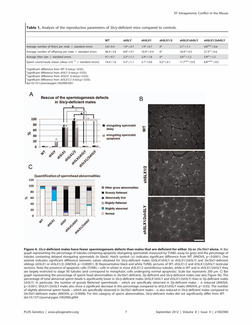
Figure 4. Slx/y-deficient males have fewer spermiogenesis defects than males that are deficient for either Sly or Slx/Slxl1 alone. A) Bargraph representing the percentage of tubules containing apoptotic elongating spermatids measured by TUNEL assay (in grey) and the percentage oftubules containing delayed elongating spermatids (in black). Hatch symbol (#) indicates significant difference from WT (ANOVA, p,0.0001). Oneasterisk indicates significant difference between values obtained for Slx/y-deficient males (shSLX1shSLY or shSLX1/2shSLY) and Slx/Slxl1-deficientsiblings (shSLX1 or shSLX1/2) (ANOVA, p,0.00001). B) Representative black and white TUNEL pictures of WT, shSLX1/2 and shSLX1/2shSLY testicularsections. Note the presence of apoptotic cells (TUNEL+ cells in white) in most shSLX1/2 seminiferous tubules, while in WT and in shSLX1/2shSLY theyare largely restricted to stage XII tubules (and correspond to metaphasic cells undergoing normal apoptosis). Scale bar represents 200 mm. C) Bargraph representing the percentage of sperm head abnormalities in Slx/Slxl1-deficient, Sly-deficient and Slx/y-deficient males (see also Figure S6). Thepercentage of total abnormal sperm heads is significantly lower in Slx/y-deficient males (shSLX1shSLY and shSLX1/2shSLY) than in Sly-deficient males(shSLY). In particular, the number of grossly flattened spermheads – which are specifically observed in Sly-deficient males – is reduced (ANOVA,p,0.001). ShSLX1/2shSLY males also show a significant decrease in this percentage compared to shSLX1shSLY males (ANOVA, p,0.03). The numberof slightly abnormal sperm heads – which are specifically observed in Slx/Slxl1-deficient males – is also reduced in Slx/y-deficient males compared toSlx/Slxl1-deficient males (ANOVA, p,0.0008). For this category of sperm abnormalities, Slx/y-deficient males did not significantly differ from WT.doi:10.1371/journal.pgen.1002900.g004
Table 1. Analysis of the reproductive parameters of Slx/y-deficient mice compared to controls.
WT shSLY shSLX1 shSLX1/2 shSLX1shSLY shSLX1/2shSLY
Average number of litters per male 6 standard errors 5.860.4 1.9*60.7 1.9*60.7 0* 3.1c61.1 4.8a,b,c60.6
Average number of offspring per male 6 standard errors 48.463.8 8.8*64.7 10.4*64.4 0* 18.4*69.3 27.3*c66.6
Average litter size 6 standard errors 9.160.7 2.2*61.1 2.4*61.8 0* 3.6*c61.2 5.8*c61.2
Sperm count/cauda (mean values x1026 6 standard errors) 14.461.6 5.5*61.1 2.1*60.6 0.2*60.1 11.7a,b,c60.9 8.8*a,b,c60.5
*significant difference from WT (t-test,p,0.02);asignificant difference from shSLY (t-test,p,0.03);bsignificant difference from shSLX1 (t-test,p,0.02);csignificant difference from shSLX1/2 (t-test,p,0.03).doi:10.1371/journal.pgen.1002900.t001
XY Intragenomic Conflict in the Mouse
PLOS Genetics | www.plosgenetics.org 7 September 2012 | Volume 8 | Issue 9 | e1002900

knockdown efficiency: indeed, no SLY1 protein can be detected in
Slx/y-deficient samples while some SLX/SLXL1 proteins remain
(Figure 2C–2E).
Slx/Slxl1 deficiency causes a sex ratio distortion in favorof males that is restored by Sly deficiency
We previously reported a tendency of an excess of females in the
progeny of Sly-deficient males (7.7% excess of females, Chi-square
p = 0.0569) [19]. While analyzing the fertility of our transgenic
lines, we observed that Slx/Slxl1-deficient males (i.e. shSLX1)
yielded an offspring sex ratio of 40% (74/187) female progeny,
compared to a ratio of 51% (234/461) in WT siblings. This
represents a statistically significant sex ratio distortion of 11% in
favour of male offspring (Chi-square p = 0.006). Importantly, a
normal sex ratio was restored by the addition of Sly deficiency: Slx/
y-deficient males produced an offspring sex ratio of 50% (103/208)
female progeny that did not differ from WT and was also
significantly different from the offspring sex ratio of shSLX1 males
(Chi-square p = 0.03). These data show that both Slx/Slxl1 and Sly
affect the transmission of X- and Y-bearing gametes, Slx/Slxl1
favouring X transmission while Sly favours Y transmission.
Discussion
SLX/SLXL1 and SLY proteins have antagonistic effects onthe expression of two distinct sets of genes
In recent years, we have identified the transcriptional conse-
quences of Sly and Slx/Slxl1 deficiency, and related these to the
observed phenotypes in terms of spermatid development, sperm
morphology and offspring sex ratio [19,28]. Remarkably, we now
show that in dual shRNA knockdown models where both genes
are deficient, the transcriptional and phenotypic consequences of
the individual knockdown are dramatically ameliorated, correcting
the X/Y/Speer up-regulation and sperm shape abnormalities seen
in Sly-deficient spermatids; the autosomal gene up-regulation,
spermatid elongation delay and apoptosis, and sperm shape
abnormalities seen in Slx/Slxl1-deficient spermatids; and improv-
ing fertility in both cases.
Strikingly, however, two different and almost entirely non-
overlapping sets of genes are affected by the mutual antagonism of
SLX/SLXL1 and SLY. In this discussion, we refer to ‘‘Group 1’’
genes as the set of X/Y/Speer genes up-regulated in Sly-deficient
spermatids and (partially) corrected in the dual knockdown, and
‘‘Group 2’’ genes as the set of metabolism-related autosomal genes
up-regulated in Slx/Slxl1-deficient spermatids and (almost fully)
corrected in the dual knockdown.
Group 1 genes: Nuclear consequences of antagonismbetween SLX/SLXL1 and SLY
Sly regulates the epigenetic repression of post meiotic sex
chromatin (PMSC) and a few specific autosomal genes such as the
Speer cluster. In the nucleus, SLY appears to act via the
recruitment/maintenance of the repressive heterochromatin
marks CBX1 and H3K9me3, which consequently limits the
expression of X and Y genes in spermatids, among which are its
X-linked homologs Slx and Slxl1 [19]. Here, we show that, in the
absence of SLY, SLX/SLXL1 proteins relocate to the nuclear sites
(both sex-linked and autosomal) vacated by SLY proteins. It is
unlikely that SLX/SLXL1 nuclear localization in Sly-deficient
spermatids is solely a consequence of increased SLX/SLXL1
protein abundance, since there is no clear enrichment in nuclear
SLX/SLXL1 proteins in spermatids of transgenic mice overex-
pressing SLX or SLXL1 (our unpublished preliminary data).
Moreover, in the double transgenic model (Slx/y-deficient
males) where SLX/SLXL1 family members are also reduced/
absent, XY gene expression, Speer expression and the intensity of
H3K9me3 marks on the sex chromatin are closer to normal
values. This indicates that SLX and/or SLXL1 have consequences
both for transcriptional activity and for histone modification when
present on sex chromatin, and that these are directly opposed to
the effects of SLY. We therefore propose that SLX/SLXL1 and
SLY proteins compete for access to nuclear sites in spermatids,
where they act as positive and negative transcriptional regulators
respectively. We cannot at this point say precisely where the
competition occurs: it may be directly at the level of chromatin
binding within the nucleus, or SLX/SLXL1 and SLY may
compete for access to factors affecting nuclear import. We note
that SLX and SLXL1 proteins lack nuclear localization signals
(NLS) while SLY NLS is mutated/truncated [22,25]; as such they
probably depend on other interacting factors to enter the nucleus.
It also remains possible that the competition is mediated indirectly:
for example, SLY could affect SLX/SLXL1 intracellular locali-
zation via regulating the expression of a third factor controlling
SLX/SLXL1 access to the nuclear sites.
Group 2 genes: Cytoplasmic consequences ofantagonism between SLX/SLXL1 and SLY
Slx/Slxl1 deficiency has been shown to increase the level of
,100 autosomal transcripts which code for proteins of the
cytoskeleton and the extracellular matrix, or are implicated in
various cytoplasmic processes (i.e. energy production, lipid
metabolism, ubiquitin-mediated degradation, etc.) [28]. These
transcriptional effects are corrected in Slx/y-deficient males,
suggesting that these changes may also be manifestations of the
same nuclear/chromatin regulatory antagonism exhibited by
Group 1 genes, perhaps via relocation of repressive factors from
sex chromatin to autosomal locations and vice versa. There are,
however, three significant objections to this interpretation. Firstly,
as noted previously, in WT spermatids SLX/SLXL1 are
predominantly cytoplasmic proteins, and the levels in the nucleus
are almost undetectable: it is hard therefore to see i) how Slx/Slxl1
knockdown could directly induce widespread transcriptional
changes, ii) what would then be the function of the abundant
SLX/SLXL1 proteins in the cytoplasm. Secondly, this interpre-
tation would require not only that SLX/SLXL1 act simulta-
neously as transcriptional activators of Group 1 genes and as
transcriptional repressors of Group 2 genes, but that SLY has the
reverse effect in both cases: it is challenging to imagine a
mechanism that could explain this. Thirdly, if both Group 1 and
Group 2 gene effects are a manifestation of the changing balance
of SLX/Y proteins in the nucleus and/or of a relocation of
repressive factors from the sex chromosomes to autosomes, then
both groups of genes would be expected to change together. This
is not the case: Group 1 genes are affected in shSLY but not in
shSLX, and Group 2 genes vice versa.
For this reason, we favour our existing interpretation that
Group 2 gene deregulation is a manifestation of the spermiogen-
esis defects occasioned by cytoplasmic Slx/Slxl1 deficiency (i.e.
spermatid elongation delay and apoptosis, reduced sperm count,
abnormal head to tail connections of the spermatozoa and male
infertility) [28], and is not a direct effect of SLX/SLXL1 proteins
on autosomal gene transcription. Given that the (cytoplasmic)
spermiogenesis defects are corrected in the dual mutant, it stands
to reason that the secondary expression changes follow the same
pattern. We therefore propose that, in addition to the nuclear
effects on Group 1 genes, SLY protein has a cytoplasmic role,
opposing that of SLX/SLXL1. SLY proteins have been shown to
XY Intragenomic Conflict in the Mouse
PLOS Genetics | www.plosgenetics.org 8 September 2012 | Volume 8 | Issue 9 | e1002900

be present in both the spermatid nucleus and cytoplasm [19,21].
Intriguingly, a recent report indicates that the acrosomal
(cytoplasmic) protein DKKL1, which we previously identified as
a binding partner of SLY1 [21], also interacts with SLXL1 [29].
We have performed additional experiments and observed that all
SLX/Y family members (i.e. SLY1, SLY2, SLX and SLXL1) can
interact with DKKL1 (Figure S7). Therefore, SLX/SLXL1 and
SLY proteins could compete for interaction with (a) common
partner(s) in the cytoplasm, and this competition could be at the
basis of the opposite effects of SLX/SLXL1 and SLY on
spermiogenesis and autosomal gene expression. A combined
model proposing how SLX/SLXL1 and SLY proteins have
antagonistic effects in both the spermatid nucleus and cytoplasm is
presented in Figure 5.
We recognize that under our preferred model, it is difficult to
explain the directionality of the expression changes seen in shSLX
relative to WT, which was predominantly up-regulation of
autosomal genes with comparatively few down-regulated genes
[28]. A potential explanation for this lies in the spermatid
developmental delay resulting in delayed spermatid elongation in
shSLX. This could potentially skew the round spermatid
population in shSLX testes towards earlier stages, i.e. proportion-
ally more step 1 spermatids and fewer step 7–8 spermatids. Since
there is a progressive transcriptional shutdown throughout
spermatid development as chromatin is repackaged in preparation
for nuclear condensation, this would thus manifest in shSLX as a
selective up-regulation of those genes expressed specifically in early
stage round spermatids (which in turn is plausible given the
annotated functional categories for these Group 2 genes). Testing
this interpretation will require further experiments on fractionated,
staged sub-populations of round spermatids.
The mouse X and Y chromosomes are involved in anintragenomic conflict that is regulated by Slx/Slxl1 andSly
Irrespective of the precise molecular mechanism(s) underlying the
antagonistic effects of SLX/SLXL1 and SLY, our results demon-
strate that both genes have an effect on offspring sex ratio. In
particular, comparing shSLX (where Sly is still present) to the dual
knockdown, there is a significant excess of males; and when
comparing shSLY (where Slx/Slxl1 are still present) to the dual
mutant, there is a trend towards excess of females. Thus, the net
effect of these genes on inheritance is for X-linked family members
to favour X chromosome transmission, and Y-linked members to
favour Y chromosome transmission, constituting a prima facie
genomic conflict. Such a conflict was first postulated in the 1990s
following observations that male mice with a partial deletion of the Y
long arm produce an excess of female offspring, however supporting
evidence has not been forthcoming until recently [16,20,26,27]. The
present study demonstrates that such a conflict exists between the
sex chromosome-linked Sycp3-related genes. An intragenomic
conflict is often not visible under normal conditions (i.e. in a WT
population) [11,13] and here the positive effect of Slx/Slxl1 on sex
chromosome transcription was uncovered by the production of mice
that are deficient for both Sly and Slx/Slxl1; similarly, the effects of
Slx/Slxl1 deficiency are also corrected in the dual mutant, although
the molecular mechanisms involved are less clear.
Can sex ratio distortion be directly attributed to Slx/Slxl1and Sly?
Under the distorter/responder model exemplified by the t
complex [1], both Slx/Slxl1 and Sly are transmission distorters in
that changes in their expression levels lead to a distortion of the sex
ratio. However, it is unlikely that they are directly responsible for
mediating the transmission skew (i.e. responder genes). Indeed, the
physiological mechanism of the skew in the present model is an
asymmetry in fertilizing ability between X and Y sperm [30]. This
implies an underlying molecular/functional asymmetry, namely
the presence of a responder gene product which is not evenly
shared between X and Y sperm. Both of the known mammalian
examples of transmission ratio distortion depend on non-sharing of
gene products (both transcript and protein) between sister
spermatids: Spam1 in the case of Rb(6.16) and Rb(6.15)
translocation heterozygotes, and TcrSmok in the case of driving t
haplotypes [31–33]. We note that SLX/Y proteins appear to be
similarly expressed in X- and Y-bearing spermatids. It therefore
seems likely that the distortion in Yq deleted mice and in
shSLXshSLY transgenic models is mediated by an as yet
unidentified sex-linked gene or gene(s) (i.e. the responder), for
which Slx/Slxl1 and Sly are competing regulators via their global
effects on sex chromatin expression. Among the deregulated genes,
a few appear as promising candidates, such as the X-encoded
homolog of Tcp11, which is one of the genes involved in the t-
complex transmission distortion, albeit as a distorter rather than a
responder [34], and Alkbh7, since another Alkbh gene has recently
been found to cause sex ratio distortion [35].
However, there may be several linked genes involved, at least
one of which is likely to evade transcript sharing. In view of this
possibility, it is worth noting that both regulators of the conflict
have a global effect on sex chromatin; this is an efficient way to
control multiple sex chromosome-linked distorters and/or re-
sponders simultaneously. The ease of identifying the responder(s)
will depend on how directly SLX/Y regulate them and how many
there are. Finally, it is possible that autosomal factors also
contribute to the regulation of sex-linked transmission distortion.
We note that historically, Slx appeared on the X before Sly
appeared on the Y, and its distorting effect on sex ratio may have
subsequently been countered by a combination of Sly-mediated
repression and other autosomal genes being selected to favour a
balanced sex ratio [20].
The intragenomic conflict in which Slx/Slxl1 and Sly areinvolved has influenced the structure of the mouse sexchromosomes
In the mouse lineage, there has been a remarkable amplification
of spermatid-expressed sex chromosome genes (all of which fall into
Group 1 identified above), and which has had a dramatic influence
on the structure of the mouse sex chromosomes. This expansion
occurred subsequent to the appearance of Sly, but was not
accompanied by a matching increase in XY transcript levels [20].
It is therefore very likely that essential sex-linked spermatid-
expressed genes have become amplified in order to maintain a
steady expression in the face of the enhancement of Sly-mediated
repression and in a sense constitute a ‘‘collateral damage’’ arising
from the conflict between Sly and Slx/Slxl1 that we unravel here.
Interestingly, the Speer gene cluster is one of the autosomal gene
families that have experienced the largest rodent-specific expansions
[36] and is also repressed by SLY. Slx/Slxl1 and Sly competition may
therefore have led to the amplification of reproductive genes outside
the sex chromosomes as well as on them.
The intragenomic conflict in which Slx/Slxl1 and Sly areinvolved may have played an important role in mousespeciation
F1 hybrid sterile males produced by asymmetric crosses
between M. m. musculus and M. m. domesticus display sperm
XY Intragenomic Conflict in the Mouse
PLOS Genetics | www.plosgenetics.org 9 September 2012 | Volume 8 | Issue 9 | e1002900

Figure 5. Model presenting how SLX/SLXL1 and SLY proteins have antagonistic effects in the spermatid nucleus and cytoplasm. Inthe spermatids, SLX/SLXL1 (yellow lozenges) and SLY (pink triangles) proteins have antagonistic effects i) in the nucleus, on the expression of XYgenes and of a few autosomal genes, such as Speer (group 1 genes); ii) in the cytoplasm, on the regulation of metabolic processes which secondarilycauses a deregulation of ,100 autosomal genes (group 2 genes). i) In WT, SLY proteins are located in both the nucleus and cytoplasm, while SLX/SLXL1 proteins are almost exclusively in the cytoplasm. The nuclear fraction of SLY proteins colocalizes with the sex chromosomes and the autosomalSpeer gene cluster, and represses their expression. A very small fraction of SLX/SLXL1 proteins also appears to colocalize with the sex chromatin. InSly-deficient spermatids (shSLY), SLX/SLXL1 proteins relocate to the nuclear sites (both sex-linked and autosomal) vacated by SLY proteins; however,SLX/SLXL1 proteins have an opposite effect to that of SLY, and activate XY gene expression. This is associated with a reduction in the repressiveepigenetic mark H3K9me3 on the sex chromatin (purple octagon), and produces sperm differentiation defects such as spermhead abnormalities,shedding delay, motility defects and subsequent male infertility. In Slx/Slxl1-deficient spermatids (shSLX), the absence of SLX/SLXL1 nuclear proteinshas only minor effect on gene regulation, since it does not change SLY localization profile. There is only a slight reduction in XY transcription,congruent with the idea that SLX/SLXL1 is a transcription activator sharing the targets of SLY when present in the nucleus. In the double knock-down(shSLXshSLY), Slx/Slxl1 deficiency almost fully abrogates the effects of Sly knockdown: in shSLXshSLY spermatids, group 1 gene expression andrepressive epigenetic marks are close to WT values. This is correlated with a rescue of SLY-dependent sperm differentiation defects. In sum, in thenucleus, the experimental observations indicate that SLX/SLXL1 competes with SLY at the level of sex chromatin regulation: SLY acts as a repressorwhile SLX/SLXL1 acts as a positive regulator. ii) Slx/Slxl1 deficiency induces various spermiogenic defects (such as spermatid elongation delay andapoptosis, reduced sperm count, abnormal head to tail connections of the spermatozoa and subsequent male infertility) associated with an up-regulation of ,100 autosomal genes which code for proteins of the cytoskeleton, the extracellular matrix, or implicated in various metabolicprocesses (i.e. group 2 genes). Since SLX/SLXL1 proteins are predominantly cytoplasmic in WT spermatids, we propose that this gene deregulation isa manifestation of the spermiogenesis defects occasioned by Slx/Slxl1 deficiency, and not a direct effect of SLX/SLXL1 proteins on autosomal gene
XY Intragenomic Conflict in the Mouse
PLOS Genetics | www.plosgenetics.org 10 September 2012 | Volume 8 | Issue 9 | e1002900

differentiation defects and wide-spread overexpression of X-
encoded spermiogenic genes [37]. Intriguingly, this only occurs
in males with a M. m. musculus X chromosome and M. m. domesticus
Y and autosomal chromosomes [38]. These males have an excess
of Slx/Slxl1 copies compared to Sly copies, since the M. m.
domesticus X and Y chromosomes carry ,40 to 60 copies of Slx/
Slxl1 and Sly, while XY encoded Sycp3-related genes have been
more amplified in M. m. musculus, with .100 of Slx/Slxl1 and Sly
on the X and Y [18,20]. Our data show that a balance between
Slx/Slxl1 and Sly expression exists in wild-type populations and
that disruption of this balance can cause male infertility. In light of
these data, we propose that deficiency in the number of Sly copies
compared to Slx/Slxl1 copies contributes to F1 male hybrid
sterility (see Figure 6) in some of these crosses. This would explain
the observed over-expression of X-encoded spermiogenic genes
observed in some F1 hybrid males [37] and subsequent sperm
differentiation defects and infertility. The observation that F1
males born from the reciprocal cross domesticus x musculus are
reproductively normal [39] does not necessarily challenge this
model. These males have an excess of Sly copies compared to Slx/
Slxl1 copies and, according to our model, could be considered as
Slx/Slxl1-deficient mice and thus display some spermiogenic
defects. This however depends critically on the mechanism of
the antagonistic effects of SLY and SLX/SLXL1 in the cytoplasm,
and on the threshold of copy number imbalance required to
trigger abnormal spermatogenesis and/or sex ratio skewing. Given
that autosomal genes will be selected to maintain a balanced sex
ratio, the Slx/Sly conflict may well be ‘‘buffered’’ to some extent by
epistatic interactions with autosomal genes.
We have observed that mice with a partial knockdown of Slx/
Slxl1 (shSLX1 or shSLX2) have comparatively minor spermio-
genic defects compared to mice with a severe knock-down
(shSLX1/2) [28]. We also note that laboratory strain X
chromosomes (including MF1 mice which were used in the
present study) are predominantly derived from a domesticus
background [40,41], yet are paired in these strains with a musculus
transcription. In the case of Sly deficiency, group 2 gene expression is unaffected; however, in the double knock-down, Sly deficiency corrects SLX/SLXL1-dependent phenotypes which abrogates the subsequent group 2 gene up-regulation. This means that SLY protein has a cytoplasmic role,opposing that of SLX/SLXL1. This antagonism could be mediated via interaction with (a) common partner(s) in the cytoplasm; the absence ofcompetition between SLX/SLXL1 and SLY proteins in the dual knockdown model would explain the absence of defects. In sum, SLX/SLXL1 and SLYproteins apparently compete in the cytoplasm for the regulation of spermiogenic processes. The functional role of SLX/SLXL1 could be to prevent theaccess of SLY to cytoplasmic proteins that are necessary for spermiogenesis.doi:10.1371/journal.pgen.1002900.g005
Figure 6. Model comparing Slx:Sly copy number imbalance in natural and laboratory mouse strains to Slx:Sly gene expressionimbalance in shRNA knockdown models. A. A model for how Slx/Slxl1:Sly imbalance affects sperm shape, offspring sex ratio and fertility. B.Approximate copy number ratio of Slx/Slxl1 and Sly in the reciprocal crosses studied by Good et al. [37] based on an estimate of ,100 copies of eachgene in musculus and ,50 in domesticus, in the WT laboratory strain MF1 YRIII which has a domesticus X and autosomes but a musculus-derived Y [40–42], and in the two natural mutants from the same background studied by us and others [18,20]. C. The relative magnitude of Group 1 and Group 2transcriptional responses seen in the various shRNA/deletion models on the MF1 YRIII background. The double and triple shRNA models show apartial Group 1 response, but no Group 2 response. Importantly, in this model, the shSLX1 and shSLX1/2 phenotypes are expected to fall outside therange of variation seen in the natural mutant and reciprocal cross males, since they are on a background which has already a deficiency in Slx/Slxl1copy number compared to Sly (50:100). We emphasise that the effects of Slx/Slxl1:Sly imbalance are only one contributor to hybrid sterility: spermshape and testis size QTLs on the musculus X map to distinct locations and show different interactions with the domesticus autosomes and Ychromosome [50].doi:10.1371/journal.pgen.1002900.g006
XY Intragenomic Conflict in the Mouse
PLOS Genetics | www.plosgenetics.org 11 September 2012 | Volume 8 | Issue 9 | e1002900

Y chromosome YRIII [42]. Thus laboratory strains are intrinsically
comparable to the reciprocal cross. Our shSLX models therefore
involve skewing the balance of SLX/SLXL1 and SLY even
further, to pathogenic effect (see Figure 6). In this light it is
intriguing that WT MF1 males have lower XY gene transcription
than Slx/y deficient males: might this reflect the fact that
laboratory strains are inherently ‘‘overdosed’’ for Sly relative to
Slx/Slxl1 by virtue of their hybrid origin?
Male hybrid sterility is a complex trait involving several X-
linked loci (as demonstrated by the mapping of several quantitative
trait loci – QTL – on the X chromosome [38,43,44]) as well as
autosomal factors [45,46]). It is worth noting that among the four
non-overlapping X-chromosome-linked QTL associated with
abnormal spermheads and hybrid sterility, one encompasses Slx
(0–37.1 Mb), the other, Slxl1 (47.9–81.8 Mb) [38]. Interestingly, it
has been shown that one of the autosomal loci linked to hybrid
sterility, Prdm9, encodes a histone H3 lysine 4 methyltransferase
involved in the silencing of the sex chromosomes during meiosis
(Meiotic Sex Chromosome Inactivation). It therefore epigeneti-
cally represses multiple X-chromosome loci, some of which part of
the hybrid sterility gene network, and epistatic interactions
between Prdm9 and multiple X and autosomal loci have been
shown to cause asymmetric hybrid male sterility associated with a
disruption of MSCI and thus a de-repression of the X
chromosome [43,46]. However, Prdm9 does not appear to be
involved in the X-chromosome up-regulation and sterility
observed in F1 hybrid males studied by Good et al. [37].
Taken together, the genetic basis of reproductive isolation in
mice is complex, and disruption of the transcriptional regulation of
the X seems to contribute to the evolution of hybrid male sterility.
The antagonistic effects of Slx/Slxl1 and Sly at the transcriptional
and phenotypic level, in particular the effects on postmeiotic XY
gene regulation, may therefore be among the important elements
contributing to the evolution of hybrid sterility between mouse
species. The production of F1 males with a transgene-derived
increased Sly expression or with a knockdown of Slx/Slxl1
expression should help address this question.
In conclusion, we have demonstrated that the mouse X and Y
chromosomes are involved in an intragenomic conflict that is
regulated by the multicopy genes Slx/Slxl1 and Sly. SLX/SLXL1
and SLY proteins compete during sperm differentiation, and
notably have opposite effects on the regulation of sex chromosome
gene expression. Disruption of Slx/y balance causes sex ratio
distortion, sperm differentiation defects and male infertility. To the
best of our knowledge, our work is the first characterization of a
conflict over sex chromosome transmission in mammals and
provides further evidence to support the hypothesis that
intragenomic conflicts can have major consequences on gene
regulation, genome structure and speciation.
Materials and Methods
Generation and breeding of transgenic miceshSLY (aka sh367), shSLX1 and shSLX1/2 males were
produced and maintained as described before [19,28]. To produce
shSLX1shSLY and shSLX1/2shSLY double transgenic mice,
shSLX1 females were mated to shSLY or to shSLYshSLX2
transgenic males. Double transgenic females were then mated to
MF1 XYRIII males (see [19]) to maintain the stock, since shSLY
males are subfertile and give progeny only rarely. Two-month-old
males single or double transgenic for sh367 (shSLY), shSLX1 or
shSLX1/2 transgenes, as well as their non-transgenic siblings
(WT) were processed for all the analyses presented here. Animal
procedures were in accordance with the United Kingdom Animal
Scientific Procedures Act 1986 and were subject to local ethical
review.
Elutriation of spermatidsFractions enriched in round spermatids (.90%) were obtained
from the above described transgenic and control (WT) males as
described previously [19]. Each sample has been purified from a
pool of testes obtained from 2 to 5 males.
TransfectionThe coding sequence of mouse Dkkl1 and Slx cDNA were
amplified by PCR and cloned into a C-terminal Myc-tagged
pCMV vector; the coding sequence of mouse Slx, Slxl1, Sly1 and
Sly2 cDNA were amplified by PCR and cloned into a N-terminal
Flag tagged pCMV vector using EcoRI and NotI restriction sites
(see Table S1 for a full list of primers). Co-transfections of
HEK293 or COS cells were performed in 6-well plates using
1.5 mg of each DNA and 5 ml of Lifofectamine (Invitrogen)
following the manufacturer’s instructions. Proteins were extracted
24 hours post transfection in 200 ml of Lysis buffer (25 mM NaCl,
10 mM Tris-HCl, 5 mM EDTA, 0.1%NP-40) and immunopre-
cipitated as described below.
Protein analysesNuclear and cytoplasmic protein extracts were obtained as
follow. The powder obtained from two adult testes crushed on dry
ice was homogenized in a glass pestle with 1 mL of lysis buffer
(0.6 M Sucrose, 10 mM Hepes pH 7.7, 25 mM KCl, 2 mM
EDTA, 0.5 mM EGTA and protease inhibitors). After the
addition of 0.2% NP40, the lysate was centrifuged for 15 minutes
at 800 g. The supernatant corresponded to the cytoplasmic
fraction. The pellet was washed twice with 1 mL of lysis buffer
and then resuspended in 100 ml of nuclear protein extraction
buffer (20 mM Hepes pH 7.7, 1.5 mM MgCl2, 0.2 mM EDTA,
25% glycerol and protease inhibitors) plus 10 ml of 4 M NaCl.
After 30 minutes of homogenization at 4uC, the samples were
centrifuged for 30 minutes at 11000 g; the supernatant corre-
sponded to the nuclear protein extract. A pellet of ,16107 round
spermatids was extracted following the same protocol using 250 ml
of lysis buffer and 50 ml of nuclear protein extraction buffer.
Whole testicular protein extraction was performed as described
previously [19]. For immunoprecipitation experiments, proteins
extracted from transfected cells were first pre-cleared with protein
A/G sepharose for 1 hour at 4uC. They were then incubated
overnight with Protein G- or Protein A- sepharose which had been
covalently bound to MYC (Santa Cruz Biotechnology) or FLAG
(Sigma) antibody beforehand (see [21] for a detailed protocol).
Western blot experiments were performed as described previously
[19]. Membranes were incubated overnight with anti-SLX/
SLXL1 [28] diluted at 1/3000, anti-SLY1 [21] at 1/3000, anti–
b-actin (Sigma) at 1/50000, or anti-LAMIN B1 (Santa Cruz
Biotechnology) at 1/1000, anti-FLAG (Sigma) at 1/1000, or anti-
MYC (Santa Cruz Biotechnology) at 1/1000. Detection by
chemiluminescence was carried out after incubation with the
corresponding secondary antibody coupled to peroxidase, as
described by the manufacturer (Millipore).
Immunofluorescence and TUNELImmunofluorescence experiments were performed on testis
material fixed in 4% buffered paraformaldehyde and sectioned as
described before [25]. DAPI (49,6-diamidino-2-phenylindole) was
used to stain nuclei (Vectashield DAPI, Vectorlab). Alexa Fluor
594-conjugated peanut agglutinin lectin (Invitrogen), which stains
XY Intragenomic Conflict in the Mouse
PLOS Genetics | www.plosgenetics.org 12 September 2012 | Volume 8 | Issue 9 | e1002900

the developing acrosome of spermatids, was used to stage the testis
tubules. For the analysis of apoptotic elongating spermatids and
delayed elongating spermatids, approximately 150 tubules were
counted per individual (4 to 6 individuals per genotype). The
percentage of tubules containing apoptotic elongating spermatids
was determined on testis sections fluorescently stained using an in
situ cell death detection kit (TUNEL, terminal deoxynucleotidyl-
transferase dUTP nick end labeling) as described by the
manufacturer (Roche Diagnostics, Indianapolis, IN). The percent-
age of tubules containing delayed elongating spermatids (i.e. stage
I to VIII tubules containing elongating spermatids) was assessed on
testis sections fluorescently stained by H4K12Ac antibody
(Millipore, Bedford, MA), a known marker of stage 9–12
elongating spermatids.
Antibody detection, chromosome painting, and DNA–FISH on surface-spread testicular cells
Antibody detection was performed on surface-spread testicular
cells following a protocol described previously [19] adapted from
Barlow et al. [47]. Incubation with the primary antibody (anti-
SLY1 [17], anti-SLX/SLXL1 [28] or anti-H3K9me3 [Upstate]
diluted 1/100) was carried out over-night in a humid chamber at
37uC. DNA-FISH, then chromosome painting were performed
after antibody detection as described previously [48]. Speer DNA-
FISH was carried out using mouse BACs RP23-212A20 and
RP24-310N20 (CHORI). As a control for specificity (see Figure
S1), SLX/SLXL1 antibody was preabsorbed with 8 mg of SLX
immunogenic peptide or with 8 mg of a noncompeting peptide
(SLY peptide). For the quantification of H3K9me3 signal over the
PMSC, the chromocenter domain was defined using the
corresponding black and white DAPI picture. Then, H3K9me3
signal outside this chromocenter domain was measured and
normalized to that of H3K9me3 signal over the chromocenter for
each cell (100 cells per genotype), using Metamorph and ImageJ
(See Figure S4). Slides corresponding to 3 individuals per genotype
were coded and randomized before the analysis; the analysis was
therefore carried out blind as to genotype.
Analysis of sperm head morphologyFor the quantification of spermhead abnormalities, sperm
collected from the initial caput epididymis were suspended in
phosphate-buffered saline. The suspension was smeared on slides
(two slides per individual) and fixed in 3:1 methanol:acetic acid.
The slides were then dipped in 0.4% Photoflo for 2 min, air dried
and stained on a plate heated at 60uC with one drop of 50% silver
nitrate mixed with one drop of 2% gelatin (Sigma). The slides were
coded and randomized. Sperm scoring was carried out ‘blind’ as to
genotype (4 to 6 individuals per genotype) and 100 sperm per slide
were classified into 6 categories on the basis of the type and
severity of abnormalities observed, using criteria described by
Yamauchi et al. [49] and in Figure S6. In the text and figures,
spermheads from category N were termed ‘‘normal’’; category 1S,
‘‘slightly abnormal’’; category 2S, ‘‘slightly flattened’’; category
3G, ‘‘abnormally thin’’; category 4G, ‘‘grossly flattened’’ and
categories 5G to 8G were pooled and named ‘‘other gross
abnormalities’’ (cf. Figure S6).
Fertility testing and sex ratio of the offspringTo assess fertility and obtain sex ratio data from the offspring,
five males of each genotype were mated with MF1 WT females
over a period of six months.
Real-time PCR and microarray analysesReal-time Reverse Transcription-Polymerase Chain Reaction
(RT-PCR) and microarray analyses were performed as previously
described on RNA extracted from 2-month old testis or from
round spermatids obtained after elutriation [19] (cf. Table S1 for a
list of the primers used in this study). Real-time RT-PCR
experiments were performed in parallel for all the genotypes
described in this study, with between 3 to 5 individuals per
genotype. For the microarray analysis, three shSLX1, three
shSLY, three shSLX1shSLY and four wild type spermatid batches
were analyzed (Illumina BeadChip, mouse whole-genome array,
v2). These data thus include and extend our previously-reported
results for shSLX1 round spermatids and for shSLY round
spermatids in previous analyses [19,28], which collectively used
two shSLY, two shSLX1 and four WT spermatid batches. Data
normalization and calculation of FDR-adjusted p values was
carried out in BeadStudio (Illumina) as previously described
[19,28]. The full data set has been uploaded to GEO, accession
number GSE39109.
Statistical analysisFor comparisons of the incidence of sperm head abnormalities,
differences between genotypes were assessed by ANOVA after
angular transformation of percentages, using the General Linear
Models ANOVA provided by NCSS statistical data analysis
software. The same test was applied to the frequency of abnormal
head-tail connections, TUNEL positive elongating spermatids,
delayed elongating spermatids (assessed by H4K12Ac staining)
and H3K9me3 quantification. Student’s t test was used to
compare the data obtained for fecundity, sperm number and
real-time PCR (performed on the DCt values). A Chi-square test
was used for sex ratio data. Microarray results were analyzed as
described in Figure S3 and Figure S5.
Supporting Information
Figure S1 Immunofluorescence detection of SLX/SLXL1
proteins in spermatids. A) Representative pictures of the detection
of SLX/SLXL1 proteins (green) by immunofluorescence in shSLY
and WT round spermatid nuclei (surface spread technique). DAPI
(blue) was used to stain nuclei. X and Y chromosome painting
were performed sequentially. SLX/SLXL1 proteins are detected
in shSLY spermatid nuclei in 76% of the cases. No signal could be
detected in the majority of WT round spermatid nuclei (84%). The
nuclear SLX/SLXL1 signal observed in the remaining ,16% of
WT round spermatid is very weak compared to the nuclear signal
in shSLY round spermatids. B) Control of the specificity of SLX/
SLXL1 immunofluorescence signal (green) in surface-spread
round spermatid nuclei. DAPI (in blue) was used to stain nuclei.
Left Panel: SLX/SLXL1 proteins (in green) were observed in Sly-
deficient (shSLY) round spermatid nuclei. Note in the picture the
presence of a flattened sperm head, characteristic of shSLY
testicular spread. When the antibody was preabsorbed with SLX/
SLXL1 peptide, the signal disappeared. When the antibody was
preabsorbed with a noncompeting peptide (SLY), SLX/SLXL1
signal was maintained. Right Panel: No signal was observed in the
majority of WT round spermatids, in round spermatids deficient
for SLX/SLXL1 proteins (shSLX1/2) and those deficient for both
SLY and SLX/SLXL1 (shSLX1/2shSLY). All these controls
demonstrate the specificity of the nuclear signal obtained with
SLX/SLXL1 antibody in shSLY round spermatids.
(TIF)
Figure S2 Characterization of shSLX1/2shSLY males. We
produced males carrying an shSLY transgene together with one
XY Intragenomic Conflict in the Mouse
PLOS Genetics | www.plosgenetics.org 13 September 2012 | Volume 8 | Issue 9 | e1002900

or two shSLX transgenes (i.e. shSLX1 and/or shSLX2). ShSLY
males carry an shRNA-expressing transgene, which triggers the
specific degradation of Sly transcripts via RNA interference [19].
Similarly, shSLX transgenic mice express Slx/Slxl1-specific
shRNA and display a decrease in whole testis Slx/Slxl1 transcript
levels estimated as ,68% and ,59% for transgenic lines shSLX1
and shSLX2, and ,83% for shSLX1/2 double transgenics [28].
A–B) Real time PCR quantification of Slx/Slxl1 (A) and Sly (B)
transcript levels in WT, shSLY, shSLX2shSLY, shSLX1shSLY
and shSLX1/2shSLY whole testes. The y-axis indicates the level
of expression compared to WT (2DDCt 6 standard errors). The
combination of shSLY transgene with any shSLX transgene
yielded an efficient knockdown of Sly and of Slx/Slxl1 [one asterisk
indicates significant difference from WT (p,0.02; t test on DDCt
values)]. The combination of shSLY transgene and two shSLX
transgenes (i.e. shSLX1/2shSLY) produced a more pronounced
decrease in Slx/Slxl1 expression [two asterisks indicate significant
difference between shSLX1/2shSLY and shSLX2shSLY or
shSLX1shSLY (p,0.02; t test on DDCt values)]. C) Real time
PCR quantification showed that transcript levels of X-encoded
(Actrt1 and 1700008I05Rik) and of Y-encoded genes (Ssty1 and
Zfy2) were lower in shSLX1/2shSLY than in shSLY whole testes
[one asterisk indicates significant difference between shSLY and
shSLX1/2shSLY values (p,0.05; t test on DDCt values)]. The y-
axis indicates the level of expression compared to WT (2DDCt 6
standard errors).
(TIF)
Figure S3 Comparison of the microarray results obtained for
shSLY and shSLX1shSLY round spermatids. List of the 222 genes
showing greater than 1.5 fold-change in Sly-deficient spermatids
(i.e. shSLY) relative to WT; 196 of them were corrected to some
degree by addition of the shSLX transgene (in shSLX1shSLY). For
46 of these genes, the difference between shSLY and
shSLX1shSLY was itself statistically significant.
(PDF)
Figure S4 Measurement of the intensity of H3K9me3 staining
over PMSC in surface-spread spermatids. (A) Graph representing
the distribution of the H3K9me3 PMSC signal intensity per
spermatid as classified by categories in WT (red), Sly-deficient
(shSLY, blue) and Slx/y-deficient mice (shSLX1/2shSLY, green).
The average values obtained for WT, shSLY and shSLX1/
2shSLY are respectively: 0.65, 0.51 and 0.59. B) Graph
representing the H3K9me3 PMSC signal intensity in each
spermatid, ranked by increasing intensity, in WT (red), Sly-
deficient (shSLY, blue) and Slx/y-deficient mice (shSLX1/2shSLY,
green). The median values obtained for WT, shSLY and shSLX1/
2shSLY are respectively: 0.64, 0.52 and 0.58.
(TIF)
Figure S5 Comparison of the microarray results obtained for
shSLX1 and shSLX1shSLY round spermatids. List of the 115
genes showing greater than 1.5 fold-change in Slx/Slxl1-deficient
spermatids (i.e. shSLX1) relative to WT; 111 of them were
corrected to some degree by addition of the shSLY transgene (in
shSLX1shSLY). For 91 of these genes, the difference between
shSLX1 and shSLX1shSLY was itself statistically significant.
(PDF)
Figure S6 Further analyses of the sperm morphology of Slx/y-
deficient males. A) Bar graph representing the percentage of
spermatozoa with abnormal head to tail connections. Slx/Slxl1-
deficient spermatozoa (i.e. shSLX1 or shSLX2) displayed a
significant increase of abnormal head to tail connections
compared to WT (represented by the hatch symbol, p,0.001;
ANOVA). This was significantly rescued by the addition of
shSLY transgene with values from shSLX1shSLY, shSLX2shSLY
and shSLX1/2shSLY males undistinguishable from WT (AN-
OVA, p.0.4) but significantly different from shSLX1 and
shSLX2 values (represented by a star; ANOVA, p,0.05). B)
Pictures and diagrams of the different categories of sperm heads
used for the quantification, as previously described by Yamauchi
et al. [49]. N: normal sperm. Category 1S: slightly abnormal
sperm heads with changes in the caudal part. Category 2S: heads
with slightly flattened acrosome. Category 3G: heads which are
thinner and more pointed. Category 4G: heads with grossly
flattened acrosome (more severe version of 2S). Categories 5G to
8G were pooled and represent other gross abnormalities. 5G:
head-neck junction located on the ventral side. 6G–8G: heads
without the curve normally observed in sperm, with remnants of
the hook (6G) or with an oval shape (7G and 8G). Scale bar
represents 5 mm. C) Detailed quantification of sperm head
abnormalities in all the genotypes analyzed in the present study.
D) Quantification of normal spermheads. Hatch symbol (#)
indicates significant difference from WT (ANOVA, p,0.0001).
One asterisk indicates significant improvement between other
genotypes (ANOVA, p,0.02). Note the increase in the
percentage of normal spermheads in Slx/y-deficient
(shSLX1shSLY, shSLX2shSLY and shSLX1/2shSLY) compared
to Sly-deficient male (shSLY). ShSLX1/2shSLY males also show
a significant increase in the percentage of normal spermheads
compared to shSLX1shSLY and shSLX2shSLY males. E)
Quantification of grossly flattened spermhead abnormalities.
These abnormalities are specifically observed in Sly-deficient
males. Hatch symbol (#) indicates significant difference from WT
(ANOVA, p,0.0001). One asterisk indicates significant improve-
ment between other genotypes (ANOVA, p,0.03). Note the
decrease in the percentage of grossly flattened spermheads in Slx/
y-deficient (shSLX1shSLY, shSLX2shSLY and shSLX1/2shSLY)
compared to Sly-deficient males (shSLY). ShSLX1/2shSLY males
also show a significant decrease in this percentage compared to
shSLX1shSLY and shSLX2shSLY males. According to those
results, the genotypes can be classified in order of decreasing
severity of their spermhead phenotype, as follow:
shSLY.shSLX1shSLY = shSLX2shSLY.shSLX1/2shSLY.
This is in clear correlation with the intensity of Slx/Slxl1 knock-
down and thus demonstrates that Slx/Slxl1 knockdown rescues
Sly-deficiency. F) Quantification of slightly abnormal spermhead
abnormalities. These abnormalities are specifically observed in
Slx/Slxl1-deficient males. Hatch symbol (#) indicates significant
difference from WT (ANOVA, p,0.04). One asterisk indicates
significant difference between Slx/y-deficient and Slx/Slxl1-
deficient males (ANOVA, p,0.0008). Note the decrease in the
percentage of slightly thin spermheads in Slx/y-deficient
(shSLX1shSLY, shSLX2shSLY and shSLX1/2shSLY) compared
to Slx/Slxl1-deficient males (shSLX1 or shSLX2). For this
category of sperm abnormalities, Slx/y-deficient males did not
significantly differ from WT. This shows that Sly deficiency
rescues the spermhead abnormalities observed in Slx/Slxl1-
deficient males.
(TIF)
Figure S7 SLX, SLXL1, SLY1, and SLY2 proteins can interact
with the cytoplasmic protein DKKL1. A) FLAG antibody
detection of extracts from COS cells transfected with DKKL1-
MYC and either FLAG-SLY1, FLAG-SLY2, FLAG-SLX, FLAG-
SLXL1, before (INPUT) and after immunoprecipitation (IP) with
MYC antibody. A non-specific FLAG-tagged control was not
immunoprecipitated by DKKL1-MYC (data not shown).
(TIF)
XY Intragenomic Conflict in the Mouse
PLOS Genetics | www.plosgenetics.org 14 September 2012 | Volume 8 | Issue 9 | e1002900

Table S1 List of the primers used in the study.
(PDF)
Acknowledgments
We thank the NIMR procedural and biological services for production of
transgenic mice and maintenance of mouse stock; T. Bates, O. Ojarikre,
and A. Rattigan for help with mouse breeding, genotyping and elutriation;
C. Reitter and Cambridge Genomic Services for help with microarray
expression analysis; N. Vernet, M. Ward, and J. Turner for helpful
discussion and critical reading of the manuscript.
Author Contributions
Conceived and designed the experiments: JC PSB. Performed the
experiments: JC PJIE SKM. Analyzed the data: JC PJIE DV. Contributed
reagents/materials/analysis tools: NAA DV. Wrote the paper: JC PJIE
PSB.
References
1. Lyon MF (1984) Transmission ratio distortion in mouse t-haplotypes is due tomultiple distorter genes acting on a responder locus. Cell 37: 621–628.
2. Meiklejohn CD, Tao Y (2009) Genetic conflict and sex chromosome evolution.
Trends Ecol Evol 25: 215–223.
3. Werren JH (2011) Selfish genetic elements, genetic conflict, and evolutionary
innovation. Proc Natl Acad Sci U S A 108 Suppl 2: 10863–10870.
4. Hurst LD, Pomiankowski A (1991) Causes of sex ratio bias may account forunisexual sterility in hybrids: a new explanation of Haldane’s rule and related
phenomena. Genetics 128: 841–858.
5. Hamilton WD (1967) Extraordinary sex ratios. A sex-ratio theory for sex linkage
and inbreeding has new implications in cytogenetics and entomology. Science156: 477–488.
6. Partridge L, Hurst LD (1998) Sex and conflict. Science 281: 2003–2008.
7. Hurst LD (1996) Further evidence consistent with Stellate’s involvement in
meiotic drive. Genetics 142: 641–643.
8. Belloni M, Tritto P, Bozzetti MP, Palumbo G, Robbins LG (2002) Does Stellate
cause meiotic drive in Drosophila melanogaster? Genetics 161: 1551–1559.
9. Faulhaber SH (1967) An abnormal sex ratio in Drosophila simulans. Genetics56: 189–213.
10. Cazemajor M, Landre C, Montchamp-Moreau C (1997) The sex-ratio trait in
Drosophila simulans: genetic analysis of distortion and suppression. Genetics147: 635–642.
11. Tao Y, Hartl DL, Laurie CC (2001) Sex-ratio segregation distortion associatedwith reproductive isolation in Drosophila. Proc Natl Acad Sci U S A 98: 13183–
13188.
12. Tao Y, Araripe L, Kingan SB, Ke Y, Xiao H, et al. (2007) A sex-ratio meioticdrive system in Drosophila simulans. II: an X-linked distorter. PLoS Biol 5:
e293. doi:10.1371/journal.pbio.0050293
13. Phadnis N, Orr HA (2009) A single gene causes both male sterility and
segregation distortion in Drosophila hybrids. Science 323: 376–379.
14. Jaenike J (2001) Sex chromosome meiotic drive. Annu Rev Ecol Syst 32: 25–49.
15. Sandler L, Novitski E. (1957) Meiotic drive as an evolutionary force. Am Nat 41:105–110.
16. Conway SJ, Mahadevaiah SK, Darling SM, Capel B, Rattigan AM, et al. (1994)Y353/B: a candidate multiple-copy spermiogenesis gene on the mouse Y
chromosome. Mammalian Genome 5: 203–210.
17. Alfoldi JE (2008) Sequence of the Mouse Y Chromosome [Thesis (Ph. D.)].Cambridge: Massachusetts Institute of Technology. 93 p.
18. Scavetta RJ, Tautz D (2010) Copy number changes of CNV regions inintersubspecific crosses of the house mouse. Mol Biol Evol 27: 1845–1856.
19. Cocquet J, Ellis PJ, Yamauchi Y, Mahadevaiah SK, Affara NA, et al. (2009) The
multicopy gene Sly represses the sex chromosomes in the male mouse germlineafter meiosis. PLoS Biol 7: e1000244. doi:10.1371/journal.pbio.1000244
20. Ellis PJ, Bacon J, Affara NA (2011) Association of Sly with sex-linked gene
amplification during mouse evolution: a side effect of genomic conflict in
spermatids? Hum Mol Genet 20: 3010–3021.
21. Reynard LN, Cocquet J, Burgoyne PS (2009) The multi-copy mouse geneSycp3-like Y-linked (Sly) encodes an abundant spermatid protein that interacts
with a histone acetyltransferase and an acrosomal protein. Biol Reprod 81: 250–257.
22. Ellis PJ, Ferguson L, Clemente EJ, Affara NA (2007) Bidirectional transcriptionof a novel chimeric gene mapping to mouse chromosome Yq. BMC Evol Biol 7:
171.
23. Toure A, Grigoriev V, Mahadevaiah SK, Rattigan A, Ojarikre OA, et al. (2004)A protein encoded by a member of the multicopy Ssty gene family located on the
long arm of the mouse Y chromosome is expressed during sperm development.
Genomics 83: 140–147.
24. Toure A, Clemente EJ, Ellis P, Mahadevaiah SK, Ojarikre OA, et al. (2005)Identification of novel Y chromosome encoded transcripts by testis transcrip-
tome analysis of mice with deletions of the Y chromosome long arm. GenomeBiol 6: R102.
25. Reynard LN, Turner JM, Cocquet J, Mahadevaiah SK, Toure A, et al. (2007)Expression analysis of the mouse multi-copy X-linked gene Xlr-related, meiosis-
regulated (Xmr), reveals that Xmr encodes a spermatid-expressed cytoplasmicprotein, SLX/XMR. Biol Reprod 77: 329–335.
26. Ellis PJI, Clemente EJ, Ball P, Toure A, Ferguson L, et al. (2005) Deletions on
mouse Yq lead to upregulation of multiple X- and Y-linked transcripts in
spermatids. Human Molecular Genetics 14: 2705–2715.
27. Ellis PJ, Affara NA (2006) Spermatogenesis and sex chromosome gene content:an evolutionary perspective. Hum Fertil (Camb) 9: 1–7.
28. Cocquet J, Ellis PJ, Yamauchi Y, Riel JM, Karacs TP, et al. (2010) Deficiency in
the multicopy Sycp3-like X-linked genes Slx and Slxl1 causes major defects inspermatid differentiation. Mol Biol Cell 21: 3497–3505.
29. Zhuang XJ, Hou XJ, Liao SY, Wang XX, Cooke HJ, et al. (2011) SLXL1, anovel acrosomal protein, interacts with DKKL1 and is involved in fertilization in
mice. PLoS ONE 6: e20866. doi:10.1371/journal.pone.0020866
30. Ward MA, Burgoyne PS (2006) The Effects of Deletions of the Mouse YChromosome Long Arm on Sperm Function - Intracytoplasmic Sperm Injection
(ICSI) - Based Analysis. Biol Reprod 74: 652–658.31. Martin-DeLeon PA, Zhang H, Morales CR, Zhao Y, Rulon M, et al. (2005)
Spam1-associated transmission ratio distortion in mice: elucidating themechanism. Reprod Biol Endocrinol 3: 32.
32. Veron N, Bauer H, Weisse AY, Luder G, Werber M, et al. (2009) Retention of
gene products in syncytial spermatids promotes non-Mendelian inheritance asrevealed by the t complex responder. Genes Dev 23: 2705–2710.
33. Ellis PJ, Yu Y, Zhang S (2011) Transcriptional dynamics of the sexchromosomes and the search for offspring sex specific antigens in sperm.
Reproduction. 142: 609–619.
34. Fraser LR, Hosseini R, Hanyalogou A, Talmor A, Dudley RK (1997) TCP-11,the product of a mouse t-complex gene, plays a role in stimulation of
capacitation and inhibition of the spontaneous acrosome reaction. Mol ReprodDev 48: 375–382.
35. Nordstrand LM, Svard J, Larsen E, Nilsen A, Ougland R, et al. (2010) Micelacking Alkbh1 display sex-ratio distortion and unilateral eye defects. PLoS ONE
5: e13827. doi:10.1371/journal.pone.0013827
36. Church DM, Goodstadt L, Hillier LW, Zody MC, Goldstein S, et al. (2009)Lineage-specific biology revealed by a finished genome assembly of the mouse.
PLoS Biol 7: e1000112. doi:10.1371/journal.pbio.100011237. Good JM, Giger T, Dean MD, Nachman MW (2010) Widespread over-
expression of the X chromosome in sterile F1 hybrid mice. PLoS Genet 6:
e1001148. doi:10.1371/journal.pgen.100114838. Good JM, Dean MD, Nachman MW (2008) A complex genetic basis to X-linked
hybrid male sterility between two species of house mice. Genetics 179: 2213–2228.
39. Good JM, Handel MA, Nachman MW (2008) Asymmetry and polymorphism of
hybrid male sterility during the early stages of speciation in house mice.Evolution 62: 50–65.
40. Yalcin B, Nicod J, Bhomra A, Davidson S, Cleak J, et al. (2010) Commerciallyavailable outbred mice for genome-wide association studies. PLoS Genet 6:
e1001085. doi:10.1371/journal.pgen.100108541. Yang H, Wang JR, Didion JP, Buus RJ, Bell TA, et al. (2011) Subspecific origin
and haplotype diversity in the laboratory mouse. Nat Genet 43: 648–655.
42. Mardon G, Mosher R, Disteche CM, Nishioka Y, McLaren A, et al. (1989)Duplication, deletion, and polymorphism in the sex-determining region of the
mouse Y chromosome. Science 243: 78–80.43. Storchova R, Gregorova S, Buckiova D, Kyselova V, Divina P, et al. (2004)
Genetic analysis of X-linked hybrid sterility in the house mouse. Mamm
Genome 15: 515–524.44. Oka A, Mita A, Takada Y, Koseki H, Shiroishi T (2010) Reproductive isolation
in hybrid mice due to spermatogenesis defects at three meiotic stages. Genetics186: 339–351.
45. Forejt J (1996) Hybrid sterility in the mouse. Trends in Genetics 12: 412–417.46. Mihola O, Trachtulec Z, Vlcek C, Schimenti JC, Forejt J (2009) A mouse
speciation gene encodes a meiotic histone H3 methyltransferase. Science 323:
373–375.47. Barlow AL, Benson FE, West SC, Hult’n MA (1997) Distribution of the RAD51
recombinase in human and mouse spermatocytes. EMBO Journal 16: 5207–5215.
48. Turner JM, Mahadevaiah SK, Ellis PJ, Mitchell MJ, Burgoyne PS (2006)
Pachytene asynapsis drives meiotic sex chromosome inactivation and leads tosubstantial postmeiotic repression in spermatids. Dev Cell 10: 521–529.
49. Yamauchi Y, Riel JM, Wong SJ, Ojarikre OA, Burgoyne PS, et al. (2009) Liveoffspring from mice lacking the Y chromosome long arm gene complement. Biol
Reprod 81: 353–361.50. Campbell P, Good JM, Dean MD, Tucker PK, Nachman MW (2012) The
Contribution of the Y Chromosome to Hybrid Male Sterility in House Mice.
Genetics.
XY Intragenomic Conflict in the Mouse
PLOS Genetics | www.plosgenetics.org 15 September 2012 | Volume 8 | Issue 9 | e1002900