Chromosome abnormalities in two benign adipose tumors
7
Chromosome Abnormalities in Two Benign Adipose Tumors Avery A. Sandberg, Zenon Gibas, Elizabeth Saren, Frederick P. Li, Janusz Limon, and Cameron K. Tebbi ABSTRACT: Two histologically benign adipose tumors were found to have clonal karyotypic changes. Del(4), del(6), and inv(13) were present in a fibrolipoma, and t(7;8) in a lipoblastoma. Addi- tional studies are needed of the frequency and malignant potential of lipomas with cytoge- netic abnormalities. INTRODUCTION Clonal cytogenetic abnormalities have been detected in diverse cancers, whereas, data are scanty on the karyotypes of clinically and histologically benign tumors. We wish to report two histologically benign tumors of adipose tissue (lipoblastoma, fibrolipoma) with chromosome changes usually associated with malignant neo- plasms. CASE REPORTS Case I The patient is a 2-year-old white boy with a gradually enlarging mass in the left anterior thigh. Physical examination revealed a hard, nontender, 8 x 3.5 cm mass in the midthigh. The left thigh measured 28 cm in circumference and the right 25 cm. Past history revealed a DPT immunization injection at the tumor site a year ago. Remainder of the medical history, physical examination, and laboratory tests were within normal limits. Surgery on July 20, 1984 revealed a large multilobulated, encapsulated, lipomatous mass attached to the vastus muscles (Fig. 1). The mass was excised in two parts, which measured 7 x 5 × 3 cm and 5 × 3 x 2 cm. Cut sections revealed a grayish-yellow mucoid surface. Histologic sections showed streaks of fibrous tissue separating nodules of mature fatty tissue that contained lipoblasts (Fig. 2). Spindle and stellate cells surrounded by a myxoid stroma were present without pleomorphism or mitotic activity. The diagnosis at both Roswell From the Roswell Park Memorial Institute (A. A. S., Z. G., J. L., C. K. T.), Buffalo, NY, the American Oncologic Hospital (E. S.), Philadelphia, PA, and the National Cancer Institute (F. P. L.), Boston, MA. Address requests for reprints to Dr. Avery A. Sandberg, Roswell Park Memorial Institute, 666 Elm Street, Buffalo, NY 14263. Received July 26, 1985; accepted September 11, 1985. 55 © 1986 Elsevier Science Publishing Co., Inc. Cancer Genet Cytogenet 22:55-61 (1986) 52 Vanderbilt Ave., New York, NY 10017 0165-4608/86/$03.50
-
Upload
independent -
Category
Documents
-
view
0 -
download
0
Transcript of Chromosome abnormalities in two benign adipose tumors

Chromosome Abnormalities in Two Benign Adipose Tumors
Avery A. Sandberg, Zenon Gibas, Elizabeth Saren, Frederick P. Li, Janusz Limon, and Cameron K. Tebbi
ABSTRACT: Two histologically benign adipose tumors were found to have clonal karyotypic changes. Del(4), del(6), and inv(13) were present in a fibrolipoma, and t(7;8) in a lipoblastoma. Addi- tional studies are needed of the frequency and malignant potential of lipomas with cytoge- netic abnormalities.
INTRODUCTION
Clonal cytogenetic abnormal i t ies have been detected in diverse cancers, whereas, data are scanty on the karyotypes of c l in ica l ly and his tological ly benign tumors. We wish to report two his tological ly benign tumors of adipose t issue (l ipoblastoma, fibrolipoma) with chromosome changes usual ly associated with mal ignant neo- plasms.
CASE REPORTS
Case I
The pat ient is a 2-year-old white boy with a gradual ly enlarging mass in the left anterior thigh. Physical examinat ion revealed a hard, nontender , 8 x 3.5 cm mass in the midthigh. The left thigh measured 28 cm in circumference and the right 25 c m .
Past his tory revealed a DPT immuniza t ion inject ion at the tumor site a year ago. Remainder of the medical history, phys ica l examinat ion, and laboratory tests were wi th in normal l imits. Surgery on July 20, 1984 revealed a large mul t i lobula ted , encapsulated, l ipomatous mass at tached to the vastus muscles (Fig. 1). The mass was excised in two parts, which measured 7 x 5 × 3 cm and 5 × 3 x 2 cm. Cut sections revealed a grayish-yel low mucoid surface. Histologic sections showed streaks of fibrous t issue separat ing nodules of mature fatty t issue that contained l ipoblasts (Fig. 2). Spindle and stellate cells sur rounded by a myxoid stroma were present wi thout p leomorph i sm or mitot ic activity. The diagnosis at both Roswell
From the Roswell Park Memorial Institute (A. A. S., Z. G., J. L., C. K. T.), Buffalo, NY, the American Oncologic Hospital (E. S.), Philadelphia, PA, and the National Cancer Institute (F. P. L.), Boston, MA.
Address requests fo r reprints to Dr. Avery A. Sandberg, Roswell Park Memorial Institute, 666 Elm Street, Buffalo, NY 14263.
Received July 26, 1985; accepted September 11, 1985.
5 5
© 1986 Elsevier Science Publishing Co., Inc. Cancer Genet Cytogenet 22:55-61 (1986) 52 Vanderbilt Ave., New York, NY 10017 0165-4608/86/$03.50

Fig
ure
1
Sec
tion
of
lipo
blas
tom
a of
cas
e 1
show
ing
stra
nds
of f
ibro
us t
issu
e se
para
ting
nod
ules
of
mat
ure
fatt
y ti
ssue
als
o co
ntai
ning
lip
obla
sts.
Spi
ndle
and
ste
llat
e ce
lls
surr
ound
ed b
y m
yxoi
d st
rom
a ar
e al
so p
rese
nt.
No
pleo
mor
phis
m o
r m
itot
ic
acti
vity
app
ears
to
be p
rese
nt.
(x 1
00)
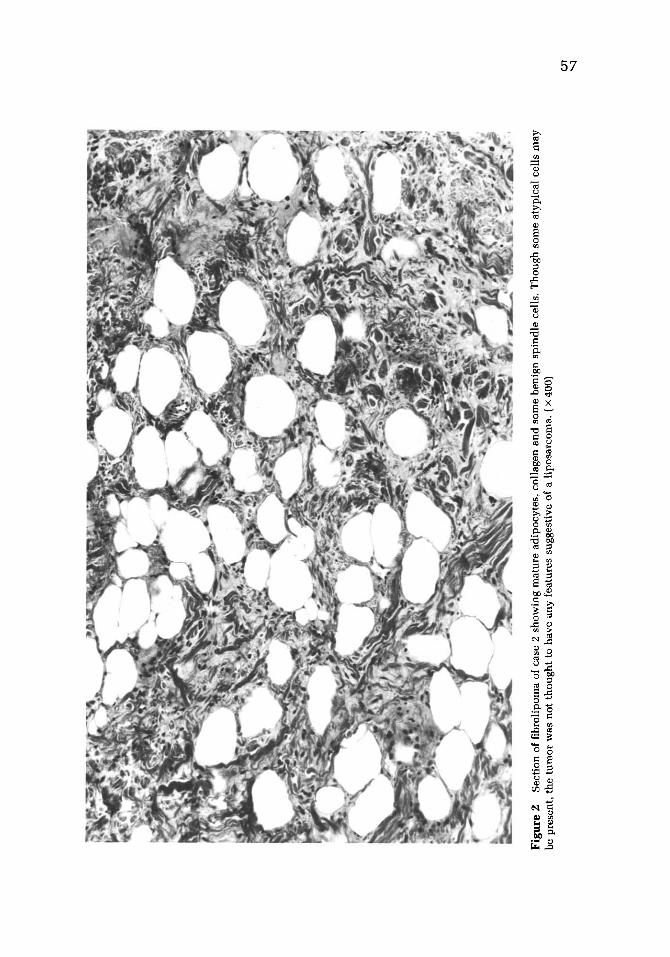
Fig
ure
2
Sec
tion
of
fibr
olip
oma
of c
ase
2 sh
owin
g m
atur
e ad
ipoc
ytes
, co
llag
en a
nd
som
e be
nign
spi
ndle
cel
ls.
Tho
ugh
som
e at
ypic
al c
ells
may
be
pre
sent
, th
e tu
mor
was
not
th
ou
gh
t to
hav
e an
y fe
atur
es s
ugge
stiv
e of
a l
ipos
arco
ma.
( ×
400
)

5 8 A .A. Sandberg et al.
Park Memorial Institute (Dr. John E. Fisher) and Hospital for Sick Children of To- ronto, Canada (Dr. Kent Maucer) was lipoblastoma. The patient was discharged fol- lowing an uneventful hospital course and has been free of disease for the last 9 months.
Case 2
A 56-year-old white male patient was noted to have an asymptomatic enlarging posterior neck mass in June 1984. Physical examination showed an 8 × 8 cm firm, fixed, nontender mass with no other abnormal physical and laboratory findings. The patient had prior peptic ulcer disease and no family historv of cancer.
The tumor was completely excised 1 month later at Jeans Hospital in Phila- delphia. Pathology examination revealed a 6.5 × 5.5 × 2 cm mass of adipose tissue that contained a 4 × 2.5 cm nodule. Microscopically, the lesion was a fibro- l ipoma with mature adipocytes, collagen, and benign fibroblastic cells. No mitotic figures were seen (Dr. Sandra Fisher). The diagnosis of fibrolipoma was confirmed at the Armed Forces Institute of Pathology (Dr. F. M. Enzinger). The patient was seen in follow-up at 1 and 4 months after surgery and had no evidence of a recur- rence.
MATERIALS AND METHODS
The tumor samples were disaggregated mechanically by mincing with curved scis- sors and digested at 37°C for 4 -6 hours with 0.8% collagenase solution (Collagenase II, Cooper Biomedical, Malvern, PA). The resulting cell suspensions were used to establish primary cultures. The cells were suspended in RPMI 1640 medium sup- plemented with 17% FCS, L-glutamine and antibiotics (penicillin 100 micron/ml, streptomocyin 100 ~g/ml, and gentamycin 50 ~g/ml), and distributed into 75 cm 2 culture flasks. The cultures were maintained until an adequate number of prolifer- ating cells became available. The cells were either exposed to colcemid (0.01 ~g/ ml) for 17 hours or synchronized with methotrexate; they were harvested after ex- posure to hypotonic solution, centrifuged, and fixed in a 3:1 solution of methanol/ acetic acid [3]. Slides were prepared in a conventional way and G-banding was performed according to the method of Yunis [4].
RESULTS
Case 1
The tumor cells were cultured for 3 days and adequate chromosome preparations were obtained after prolonged exposure to colcemid. Among 30 metaphases, 19 contained the modal number of 46 chromosomes. A smaller population (six cells) had 49 chromosomes. The remaining five metaphases showed intermediate or hy- podiploid chromosome counts. All metaphases analyzed were characterized by a 7p+ marker chromosome. Detailed analysis of 10 metaphases revealed two clones within the tumor. All metaphases showed a translocation between chromo- somes #7 and #8 as the sole abnormality. The karyotype was: 46,XY,t(7;8) (7qter--~7p22::8q12-~8qter). In three cells, in addition to these changes, + 8, + 9 and +12 were also observed (Fig. 3). The karyotype of this minor clone was 49,XY,t(7;8)(p22;q12), + 8, + 9, + 12. The constitutional karyotype in this patient was normal (46,XY).

Abnormalities in Two Adipose Tumors 59
Figure 3 G-banded karyotype of the minor clone with 49 chromosomes from the lipoblas- toma of case 1. M1 and M2 represent a product of a translocation between chromosomes #7 and #8. Two normal #8 homologs and additional copies of chromosome #9 and #12 are also present.
Case 2
The tumor cells were cultured for 5 days, and processed either by exposure to col- cemid or methotrexate synchronization. Cultures synchronized with methotrexate yielded more favorable chromosome preparations and were used for detailed anal- ysis. Thirty-six metaphases were counted and the modal chromosome number was 46. Seventeen of 20 karyotypes analyzed in detail showed identical chromosome changes: 46,XY,del(4)(q23),del(6)(q13),inv(13)(q12q32) (Figs. 4 and 5). The remain- ing three metaphases showed random losses of chromosomes, but all of them con- tained the structural rearrangements of chromosomes #4, #6, and #13. Cells har- vested after prolonged exposure to colcemid also showed the 4 q - and 6 q - markers, even though these preparations were inferior by comparison.
DISCUSSION
Tumors of adipose tissues arise in diverse anatomic sites and display a wide spec- trum of growth patterns [5]. Benign lipomas grow slowly and do not metastasize, whereas undifferentiated liposarcomas can enlarge rapidly at primary and distant

60 A . A . Sandberg et al.
i
iW
m
~ ii ~ , , , . . . . . . . .
2"1 22 X Y
Figure 4 G-banded karyotype of the fibrolipoma from case 2. M1, M2, and M3 represent del(4)(q23), del(6)(q13), and inv(13)(q12q32), respectively.
sites. Histologic features of these tumors, par t icular ly grade and mitot ic activity, are predict ive of c l inical behavior and pat ient survival. However, the nomencla ture of adipose tumors is complex, and the boundary between benign and malignant le- sions is imprecise [5, 6].
Clonal abnormali t ies have been ident if ied in chromosome studies of diverse can- cers, inc luding sarcomas [1, 7]. In the present s tudy chromosome changes were found in two adipose tumors classified as benign neoplasms on histologic review by expert pathologists. The cytogenetic findings might be due to cul ture condi t ions that selected for the few undiscernable mal ignant cells wi th in the p redominan t ly benign tumor masses. In meningioma, another connect ive t issue tumor with a usu- al ly benign cl inical course, near ly 50% of lesions have anomal ies of chromosome #22 ( 2 2 q - or - 2 2 ) ; recurrences tend to show addi t ional chromosome changes [8].
Figure 5 Paracentric inver- sion of chromosomes #13 in case 2. Each inverted chromo- some (M3) is shown next to the normal chromosome #13 from the same metaphase.
i i i , i i i i 13 M3 13 M3 13 M3 13 M3

Abnormal i t ies in Two Adipose Tumors 61
Among frankly mal ignant neoplas ias such as acute leukemia, chromosomal abnor- mali t ies during the pre leukemic phase can antedate morphologic t ransformation of the neoplas t ic cells [1, 2]. The chromosome findings in our patients, therefore, might be an early indicator of t ransi t ion to a mal ignant state. Alternat ively, the abnormal karyotypes might be a characterist ic of some truly benign l ipomas. Few reports on benign l ipomas and l iposarcomas are available for compar ison with the cytogenetic rearrangements in our pat ients [7]. This s tudy demonstrates the tech- nical feasibil i ty of cytogenetic studies of wel l-different iated adipose tumors, and addi t ional lesions should be examined.
The cl inical significance of clonal abnormali t ies in connect ive t issue tumors is unknown. Among acute leukemias, associat ions have been identif ied of specific chromosomal rearrangements wi th morphology [t(15;17) in acute promyelocyt ic leukemia[, and with prognosis [favorable wi th t(8;21) and +21] [9]. Chromosomal studies have a role in the work-up and management of acute leukemias, and should be evaluated for ut i l i ty in the sarcomas. Our pat ients are under obsrvat ion for re- current disease which can be s tudied for karyotypic progression.
Supported in part by Contrast No. N01-CP-21032 from the National Cancer Institute, National Institutes of Health.
REFERENCES
1. Sandberg AA (1980): The Chromosomes in Human Cancer and Leukemia. Elsevier North- Holland, Inc., New York, pp. 776.
2. Sandberg AA (1984): Chromosomal alterations associated with neoplasia. Transplant Proc XVI: 366-369.
3. Gibas LM, Gibas Z, Sandberg AA (1984): Technical aspects of cytogenetic analysis of hu- man solid tumors. Karyogram 10:25-27.
4. Yunis JJ (1981): New chromosome techniques in the study of human neoplasia. Hum Pathol 12:540-549.
5. Enzinger FM, Weiss SW (1983): Soft Tissue Tumors. C.V. Mosby Co., St. Louis. 6. Evans HL, Soule EH. Winkelmann RK (1979): Atypical lipoma, atypical intramuscular li-
poma, and well differentiated retroperitoneal liposarcoma: A reappraisal of 30 cases for- merly classified as well differentiated liposarcoma. Cancer 43:574-584.
7. Becher R, Wake N, Gibas Z, Sandberg AA (1984): Chromosome changes in soft tissue sar- comas. J Natl Cancer Inst 72:823-831.
8. Zang KD (1982): Cytological and cytogenetical studies on human meningioma. Cancer Ge- net Cytogenet 6:249-274.
9. Fourth International Workshop on Chromosomes in Leukemia, (1982) (1984): Cancer Genet Cytogenet 11:251-360.