Congenital brain abnormalities: Pictorial essay - JournalAgent
Upload
independentCategory
view
0download
0
1988;48:175-180. Cancer Res Constance A. Griffin, Anita L. Hawkins, Roger J. Packer, et al. Chromosome Abnormalities in Pediatric Brain Tumors
Updated version
http://cancerres.aacrjournals.org/content/48/1/175
Access the most recent version of this article at:
E-mail alerts related to this article or journal.Sign up to receive free email-alerts
Subscriptions
Reprints and
To order reprints of this article or to subscribe to the journal, contact the AACR Publications
Permissions
To request permission to re-use all or part of this article, contact the AACR Publications
on March 9, 2014. © 1988 American Association for Cancer Research. cancerres.aacrjournals.org Downloaded from on March 9, 2014. © 1988 American Association for Cancer Research. cancerres.aacrjournals.org Downloaded from
[CANCER RESEARCH 48. 175-180. January 1, 1988]
Chromosome Abnormalities in Pediatrie Brain Tumors1
Constance A. Griffin,2 Anita L. Hawkins, Roger J. Packer, Lucy B. Rorke, and Beverly S. Emanuel3
Department of Pediatrics, Division of Genetics [C. A. G., A. L. H. B. S. E.], Neurology [K. J. P.], Pathology [L. B. R.J, and Oncology [R. J. P.J, The University ofPennsylvania School of Medicine and The Children's Hospital of Philadelphia
ABSTRACT
Recurrent, site-specific chromosome translocations and other cytoge-netic abnormalities are being described in ever-increasing numbers and
types of human tumors. Primary brain tumors are the most commonpediatrie solid tumor and differ from those of adults in both histologyand clinical behavior. We examined chromosomes from 21 primarypediatrie brain neoplasms grown in short-term tissue culture, including 6
astrocytomas, 10 primitive neuroectodermal tumors, and 5 other tumors.Karyotypes from 3 of 5 astrocytomas were abnormal, as were those of 9of 10 primitive neuroectodermal tumors. Numerical abnormalities werefound in 6 tumors and structural aberrations in 12 tumors. Deletions,additions, and translocations involving the short arm of chromosome 1were observed in 5 tumors, with chromosome breakpoints ranging fromIpl to Ip3. An isochromosome of the long arm of 17, i(17q) was themost frequent site-specific structural abnormality, found in 1 anaplastic
astrocytoma and 2 recurrent cerebellar primitive neuroectodermal tumors, one with islands of anaplastic astrocytoma. These results differfrom reported chromosome studies of adult brain tumors, suggesting thatpediatrie brain tumors may differ from those of adults when examined atthe genetic level. Additional chromosomal and molecular studies of braintumors from children are warranted to define these differences.
INTRODUCTION
Recurrent, site-specific chromosomal translocations andother nonrandom cytogenetic abnormalities are being found inever-increasing numbers and types of human tumors. Molecularstudies have demonstrated that the 9;22 translocation ofchronic myelogenous leukemia results in production of anaberrant bcr-abl fusion protein (1-4). While the cytogeneticallysimilar chromosomal translocation in acute lymphocytic leukemia differs at the molecular level (5, 6), it too results inexpression of several unique aW-derived tyrosine kinases which,however, are distinct from the aberrant protein of chronicmyelogenous leukemia (7). In lymphomas, the 8; 14 and variant2;8 and 8;22 translocations of Burkitt's lymphoma result in c-
myc gene deregulation (8-10). These results strongly suggest
that specific chromosome rearrangements are important intumor initiation or maintenance. Similar clues regarding thelocation of genes important in solid tumor pathobiology arenow being sought by cytogenetic examination of solid tumorspecimens.
Primary brain tumors are the most common solid tumoroccurring in children (11). Pediatrie brain tumors differ fromthose of adults in both histology and clinical behavior (12).Comparison of chromosomal findings of pediatrie and adultbrain tumors may delineate additional significant differencesbetween tumors of children and adults. Here we report resultsof chromosome analysis of 21 primary pediatrie brain neoplasms and show that analysis of cytogenetic changes in these
Received 5/15/87; revised 9/2/87: accepted 10/2/87.The costs of publication of this article were defrayed in part by the payment
of page charges. This article must therefore be hereby marked advertisement inaccordance with 18 U.S.C. Section 1734 solely to indicate this fact.
1Supported in part by NIH Grants CA 09485 and GM 32492.2 Present address: The Johns Hopkins Oncology Center, 600 N. Wolfe St..
Baltimore. MD 21205.3To whom requests for reprints should be addressed, at The Children's
Hospital of Philadelphia. 34th St. and Civic Center Blvd. Philadelphia. PA19104.
tumors can quite often be accomplished using current optimalsolid tumor protocols.
METHODS
All tumors from children undergoing diagnostic or therapeutic surgery for brain tumors at The Children's Hospital of Philadelphia duringa 10-mo period were eligible for cytogenetic study when sufficientexcess tissue remained after that required for analysis by the patholo-gists. Tumors were placed in transport media (RPMI 1640 with 15%fetal calf serum) immediately upon removal at surgery and were broughtto the cytogenetics laboratory where they were usually processed withinl h of arrival. One specimen (Case 7, a medulloblastoma metastatic tobone marrow) was sent from an outside hospital and cultured thefollowing day.
Tumors were dissected aseptically into 1- to 2-mm pieces. Specimenswhich were not easily dispersed mechanically were further disaggregated in 0.8% collagenase type II (Cooper Biomedicai) in RPMI 1640for 2-5 h until reduced to small clumps of cells. Cells were thencentrifuged to remove them from collagenase and seeded into 25 cm '
tissue culture flasks in RPMI 1640 (GIBCO) with 15% fetal calf serum(GIBCO), 50 units penicillin, 100 ¿igstreptomycin, 2 HIMglutamineand cultured at 37°Cin 5% CO2/95% air. At least two cultures from
each specimen were initiated whenever possible.Cultures were observed daily for evidence of cell growth and har
vested for metaphase chromosome preparations as soon as mitotic cellswere observed. Cells were exposed to colcemid, 0.01-0.03 ^g/ml for 1-24 h depending on the number of mitotic cells seen, treated withprewarmed 0.075 M KC1 for 30 min at 37°C,and fixed in 3 changes of
3:1 methanol:acetic acid. Slides were made, air-dried, and aged at least1 wk at room temperature before staining with Wright's stain and G-
banding with trypsin (13) or phosphate buffer (14). At least 20 meta-phases and 4 karyotypes from each tumor were analyzed wheneverpossible.
The pathological classification of all tumors was performed by oneof us (L. B. R.) except for the tumor from Patient 7, which was sentfrom an outside hospital. Classification was based on the revision ofbrain tumor nomenclature described at the Pediatrie Brain TumorWorkshop of 1984 (15). Two of these classes are further explainedhere. Atypical teratoid tumors consist of a mixture of neuroepithelial,epithelial, and mesenchymal cells of various types and in varyingproportions. They also generally contain a population of cells similarto those found in rhabdoid tumors. They do not resemble any of theordinary germ cell tumors and because of their mixed cellular elementswere given the unusual name of atypical teratoid tumors. To date theyseem to be a tumor of infancy. PNETs4 are a group of central nervous
system tumors which occur most commonly in childhood and areprimarily composed of primitive or undifferentiated neuroepithelialcells. Much histológica!heterogeneity is often present. The term PNET,not otherwise specified, describes poorly differentiated neuroepithelialcells, while PNET with astrocytes, rosettes, etc. describes the presenceof more differentiated cells within the tumor.
RESULTS
Twenty-one brain tumors were received for chromosomeanalysis during the period of study. Histológica! subtypes included 6 astrocytomas, 10 PNETs, 2 atypical teratoid tumors,1 mixed glioma, 1 ependymoma, and 1 meningioma. We have
4The abbreviation used is: PNET. primitive neuroectodermal tumor.
175
on March 9, 2014. © 1988 American Association for Cancer Research. cancerres.aacrjournals.org Downloaded from

CHROMOSOMES OF PEDIATRIC BRAIN TUMORS
included Case 10 in the group of PNETs. This was a recurrenttumor which had features of anaplastic astrocytoma; 7 yr earlierat initial presentation the tumor appeared to be a PNET withislands of anaplastic astrocytoma. Thirteen tumors were newlydiagnosed, 6 were recurrent disease, and 2 were second primarytumors (pineolblastomas in children with hereditary retinoblas-tomas).
No metaphases were obtained in 3 tumors, in 6 we wereunable to detect abnormalities, and 12 had abnormal karyotypeswith or without normal cells present. The chromosomes of 3 of5 astrocytomas were abnormal, as were those from 9 of the 10PNETs. Karyotype results are summarized in Tables 1-3.
The majority of tumors were near diploid. Two tumors had2 distinct populations of cells (Cases 2 and 7), while 2 tumors(Cases 10 and 13) had only a hypotetraploid cell population.Five tumors had clonal numerical abnormalities, with additionsand losses of apparently normal chromosomes. Additions ofchromosome 7 were noted in 2 tumors, both anaplastic astrocytomas, and gains of chromosomes 3, 10 and 13 were eachnoted once. Loss of chromosome 16 was observed in 2 tumors,an anaplastic astrocytoma and an ependymoma, and losses of
chromosomes 6, 10, and 14 were each noted once.Twelve tumors had structural chromosome abnormalities; 5
tumors had more than one. The most frequently nonrandomstructural abnormality was an isochromosome for the long armof 17, i(17q). This abnormality was found in 3 tumors, includinga newly diagnosed anaplastic astrocytoma (Fig. 1), a recurrenttumor, which at the time of original diagnosis was classified aPNET with islands of anaplastic astrocytoma and at recurrenceappeared to be predominantly an anaplastic astrocytoma(Fig. 2), and a cerebellar PNET (recurrent medulloblastoma)(Fig. 3). An i(17q) was never the solitary abnormal finding.
Deletions, additions, or translocations involving the shortarm of chromosome 1 were observed in 5 tumors. The breakpoints varied between Ipl and Ip3 and in one case could notbe assigned with greater specificity than to the involved regionbecause of the quality of chromosome banding obtained. Involvement of Ip was observed in three astrocytomas: additionof unidentified material to Ip3 (Case 10); a der(l)t(l;14)(p36;q24) in Case 15; and a der(lqlSq) in Case 12. An ependymoma from Case 18 contained an extra copy of a deleted 1,a Ip- chromosome which appeared to be deleted within Ipl
IM MIW im unFig. I. Karyotype (near tetraploid) of a
newly diagnosed anaplastic astrocytoma froma 7-yr-old male (Patient 13). 88.XXYY, -2,-3, +7, +7. -8. -9, +10, -15, -16, -16,i(l7q), i(l7q), -20, -22, +markers. Arrows,¡(17q).
lili lililà III KMHill K58MM10 11 12
•IH »*•*13 14 15
» atti MMTT16 17 18
19
M«
20 mar
••*« e** I} i|
21 22 X Y
ufiMI«m»
Fig. 2. Karyotype (near tetraploid) of abrain tumor with features of anaplastic astrocytoma from a 13-yr-old male (Patient 10),occurring 6 yr after resection of a PNET withislands of anaplastic astrocytoma. 79.XXYY,-3, -4, -4, -5, -5, -10, -10, -11, -11, -13,-13, -15, -17, -21, -22. IP+, lp+, 4q+.5p+, der(7), der(7), 9p-, 11p-, i( 17q). +mark-ers. Arrows, structurally abnormal chromo-
fgii «5*0«UM«««»••*<m*6 7/ / 8 9 ^ 10 tf 12
«•**«* f«ft **** $** A«fcft13 14 15 17 18
19 20nTTmar
21 22
on March 9, 2014. © 1988 American Association for Cancer Research. cancerres.aacrjournals.org Downloaded from
CHROMOSOMES OF PEDIATRIC BRAIN TUMORS
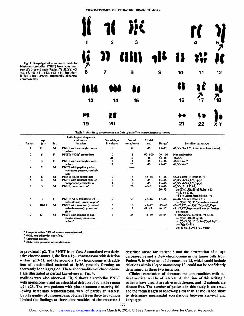
ii ii 5 fiFig. 3. Karyotype of a recurrent medullo-
blastoma (cerebellar PNET) from bone marrow of a 3-yr-old male (Patient 7). 55.XY, +3,+8, +8, +8, +11, +13, +13, +14, 5q+. 6q-,i(17q), 18q+. Arrows, structurally abnormalchromosomes.
mil i»»«8
^^
9 10 11 12
HÃœm M II If i|13 14 15
716 17 '18
ifit1920Table
1 Results of chromosome analysis of primitive neuroectoaermaitumorsAgePatient
(yr)1
212
53
34
25
8647
38
39
10/1210
13SexMFFMMMMFMMPathological
diagnosisandtumorlocationPNET
with astrocytes; cerebellumPNET;
NOS;"cerebellumPNET
with astrocytes; cerebellumPNET
with papillaryade-nomatouspattern; cerebel
lumPNET;NOS;cerebellumPNET
with unusualcellularcomponent;cerebellumPNET;
bonemarrow0PNET;
NOS (trilateral retinoblastoma); pinealregion'PNET
with rosettes(trilateralretinoblastoma);pineal re
gion*PNETwith islands ofana-plastic
astrocytoma; cerebellum''No.
ofdaysinculture222811031612241No.
ofmetaphases385431123none146192650101924Modalno.4690-10046464645-46454546-5142-4645-4745-4778-80Range"43-4790-10042-4845-4645-4741-4645-4645-4645-4650-5542-4645-4740-4778-84*»
«f}•2122 XYStemline
karyotype46,XY/46,XY,
+mar (randomlosses)Not
analysable46.XX46,XX,6q-?46,XX,6q-?46,X
Y,der( 1)t( 1;?)(q44;?)45,XY,-6/45,XY,3q-;-645,XY,-6/45,XY,3q-;-646,XY/51,XY,+3,der(5)t(
1;5)(q2 1;q35),6q-,+ 13,+13,+i(I7q),+i(
17q),der( 18)t( 18;?)(q2 1;?)42-46,XXdel(l)(p13-21),der(
1)t( 1;?)(p36;?)(randomlosses)45-47.XY,der( 1)t( 1;?)(q44;?),Dq+45-47,XY,Dq+
(could not befurtherdelineated)78-80.XXYY,
der(l)t(l;?)(p3;?).der(4)t(l;4)(q21;q35),der(5)t(5;?)(P13;?),
inv(7)(pl3qll).del(9)(p13-21),del(l
I)(pl3),+i(17q), +mar" Range in which 75% of counts were observed.* NOS, not otherwise specified.' Recurrent disease.J Child with previous retinoblastoma.
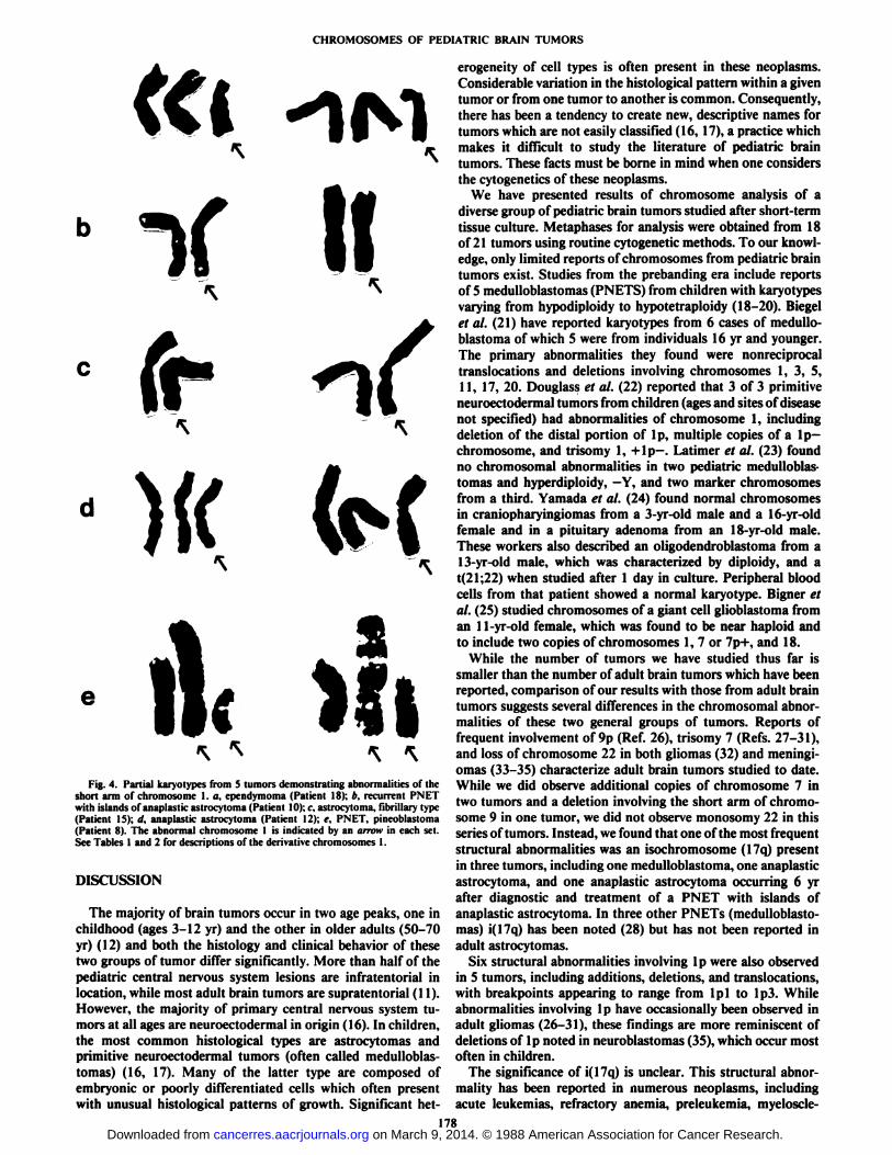
or proximal Ip2. The PNET from Case 8 contained two derivative chromosomes 1, the first a Ip- chromosome with deletionwithin Ipl3-21, and the second a lp+ chromosome with addition of unidentified material at Ip36, possibly forming anaberrantly banding region. These abnormalities of chromosome1 are illustrated as partial karyotypes in Fig. 4.malities were also observed. Fig. 5 shows a cerebellar PNETwith monosomy 6 and an interstitial deletion of 3q in the regionq24-q26. The two patients with pineoblastoma occurring following hereditary retinoblastoma were of particular interest,but the quality of chromosomes obtained from these two tumorslimited the findings to those abnormalities of chromosome 1
described above for Patient 8 and the observation of a lq+chromosome and a Dq+ chromosome in the tumor cells fromPatient 9. Involvement of chromosome 13, which could includedeletions within 13q or monosomy 13, could not be confidentlydetermined in these two instances.
Clinical correlation of chromosome abnormalities with patient survival will be of interest. At the time of this writing 5patients have died, 3 are alive with disease, and 12 patients aredisease free. The number of patients in this study is too smalland the mean length of follow-up (less than 12 mo) is too shortto determine meaningful correlations between survival andkaryotype.
177
on March 9, 2014. © 1988 American Association for Cancer Research. cancerres.aacrjournals.org Downloaded from
CHROMOSOMES OF PEDIATR1C BRAIN TUMORS
«I -1M\ «
v\
fr\
Ì«e
Fig. 4. Partial karyotypes from 5 tumors demonstrating abnormalities of theshort arm of chromosome 1. a, ependymoma (Patient 18); b. recurrent PNETwith islands of anaplastic astrocytoma (Patient 10); r. astrocytoma, fibrillary type(Patient 15); it. anaplaslic astrocytoma (Patient 12); c. PNET, pineoblastoma(Patient 8). The abnormal chromosome I is indicated by an arrow in each set.See Tables 1 and 2 for descriptions of the derivative chromosomes I.
DISCUSSION
The majority of brain tumors occur in two age peaks, one inchildhood (ages 3-12 yr) and the other in older adults (50-70yr) (12) and both the histology and clinical behavior of thesetwo groups of tumor differ significantly. More than half of thepediatrie central nervous system lesions are infratentorial inlocation, while most adult brain tumors are supratentorial (11).However, the majority of primary central nervous system tumors at all ages are neuroectodermal in origin (16). In children,the most common histológica! types are astrocytomas andprimitive neuroectodermal tumors (often called medulloblas-tomas) (16, 17). Many of the latter type are composed ofembryonic or poorly differentiated cells which often presentwith unusual histológica! patterns of growth. Significant het
erogeneity of cell types is often present in these neoplasms.Considerable variation in the histológica! pattern within a giventumor or from one tumor to another is common. Consequently,there has been a tendency to create new, descriptive names fortumors which are not easily classified (16,17), a practice whichmakes it difficult to study the literature of pediatrie braintumors. These facts must be borne in mind when one considersthe cytogenetics of these neoplasms.
We have presented results of chromosome analysis of adiverse group of pediatrie brain tumors studied after short-termtissue culture. Metaphases for analysis were obtained from 18of 21 tumors using routine cytogenetic methods. To our knowledge, only limited reports of chromosomes from pediatrie braintumors exist. Studies from the prebanding era include reportsof 5 medulloblastomas (PNETS) from children with karyotypesvarying from hypodiploidy to hypotetraploidy (18-20). Biege!et al. (21) have reported karyotypes from 6 cases of medullo-blastoma of which 5 were from individuals 16 yr and younger.The primary abnormalities they found were nonreciprocaltranslocations and deletions involving chromosomes 1, 3, 5,11, 17, 20. Douglass et al. (22) reported that 3 of 3 primitiveneuroectodermal tumors from children (ages and sites of diseasenot specified) had abnormalities of chromosome 1, includingdeletion of the distal portion of 1p, multiple copies of a 1p—chromosome, and trisomy 1, + lp—.Lati nier et al. (23) found
no chromosomal abnormalities in two pediatrie medulloblastomas and hyperdiploidy, —Y,and two marker chromosomes
from a third. Yamada et al. (24) found normal chromosomesin craniopharyingiomas from a 3-yr-old male and a 16-yr-oldfemale and in a pituitary adenoma from an 18-yr-old male.These workers also described an oligodendroblastoma from a13-yr-old male, which was characterized by diploidy, and at(21;22) when studied after 1 day in culture. Peripheral bloodcells from that patient showed a normal karyotype. Bigner etal. (25) studied chromosomes of a giant cell glioblastoma froman 11-yr-old female, which was found to be near haploid andto include two copies of chromosomes 1, 7 or 7p+, and 18.
While the number of tumors we have studied thus far issmaller than the number of adult brain tumors which have beenreported, comparison of our results with those from adult braintumors suggests several differences in the chromosomal abnormalities of these two general groups of tumors. Reports offrequent involvement of 9p (Ref. 26), trisomy 7 (Refs. 27-31),and loss of chromosome 22 in both gliomas (32) and meningi-omas (33-35) characterize adult brain tumors studied to date.While we did observe additional copies of chromosome 7 intwo tumors and a deletion involving the short arm of chromosome 9 in one tumor, we did not observe monosomy 22 in thisseries of tumors. Instead, we found that one of the most frequentstructural abnormalities was an isochromosome (17q) presentin three tumors, including one medulloblastoma, one anaplasticastrocytoma, and one anaplastic astrocytoma occurring 6 yrafter diagnostic and treatment of a PNET with islands ofanaplastic astrocytoma. In three other PNETs (medulloblastomas) i(17q) has been noted (28) but has not been reported inadult astrocytomas.
Six structural abnormalities involving Ip were also observedin 5 tumors, including additions, deletions, and translocations,with breakpoints appearing to range from Ipl to Ip3. Whileabnormalities involving Ip have occasionally been observed inadult gliomas (26-31), these findings are more reminiscent ofdeletions of Ip noted in neuroblastomas (35), which occur mostoften in children.
The significance of i(17q) is unclear. This structural abnormality has been reported in numerous neoplasms, includingacute leukemias, refractory anemia, preleukemia, myeloscle-
178on March 9, 2014. © 1988 American Association for Cancer Research. cancerres.aacrjournals.org Downloaded from
CHROMOSOMES OF PEDIATR1C BRAIN TUMORS
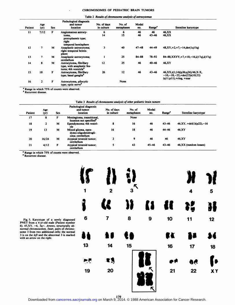
Table 2 Results of chromosome analysis ofastrocytomas
PatientAge(yr) Sex
Pathological diagnosisand tumor
locationNo. of daysin culture
No. ofmetaphases
ModalRange" Stemline karyotype
11 7/12 F Angiomatous astrocy- 6 6toma, 14 15protoplasmic type;righttemporal hemisphere
12 7 M Anaplastic astrocytoma; 3 40right temporal hemisphere
13 7 M Anaplastic astrocytoma; 1 25cerebellum
14 8 M Astrocytoma, fibrillary 12 25type, with anaplastic features; 4th ventricle*
15 10 F Astrocytoma, fibrillary 26 12type; basal ganglia*
16 2 F Astrocytoma, pilocytic Nonetype; optic nerve*
46 46 46,XX46 43-46 46.XX
47-48 44-49 48,XY,+2,+7,-14,der(lql5q)
84-88 78-92 84-88,XXYY,+7,+ 10,-16,i(17q),i(17q)
46 40-46 46,XY
46 43-46 46,XX,t( 1; 14)(p36;q24)/46,X-X,-10,-18,-22,+der(22)t( 18;22)<qll;p
°Range in which 75% of counts were observed.* Recurrent disease.
Table 3 Results of chromosome analysis of other pediatrie brain tumors
Patient1718192021Age(yr)821316/244/12SexFMMMFPathological
diagnosisand tumor
locationMeningioma,
transitional.locationnotspecified*Ependymoma;
4lh ventricleMixed
glioma,epen-dymo-oligodendrogli-oma;
cerebellumAtypicalteratoidtumor;cerebellumAtypical
teratoidtumor;cerebellumNo.
of daysin culture81625No.
ofmetaphasesNone1618943Modalno.46464645-46Range"43-4644-464643-46Stemlinekaryotype46,XY,
+del(l)(p22),-1646,XY46.XY46,XX
(random losses)
a Range in which 75% of counts were observed.* Recurrent disease.
Fig. 5. Karyotype of a newly diagnosedPNET from a 4-yr-old male (Patient number6). 45,XY, —6,3q—.Arrows, structurally abnormal chromosomes. Inset, pairs of chromosome 3 from two additional cells: the normal3 is on the left and the abnormal 3 is markedwith an arrow on the right.
(r n1 2
I tt
«i13
19
M14
««20
8
il
15
II10
II16
»*
21
M if4 5
IÕ 3*11
M17
it22
12
179on March 9, 2014. © 1988 American Association for Cancer Research. cancerres.aacrjournals.org Downloaded from
CHROMOSOMES OF PEDIATRIC BRAIN TUMORS
rosis, lymphomas, and most frequently in the progressive phaseof chronic myelogenous leukemia (36). It has also been observedless frequently in solid tumors, including carcinomas of thecolon, cervix, prostate, ovary, and, interestingly, retinoblastoma(36). At present, it is not known whether a dosage effect ofextra copies of genes from 17q may contribute to disease inthese cases, but the roles of oncogenes c-erb- Al at 17qll-12(37)and neu at 17q21-22 (37) and of growth hormone at 17q22-34
(37) must be considered. Alternatively, loss of genes present on17p could represent the significant event. Perhaps presence ofi(17q) indicates aggressive or progressive disease. Collection ofsurvival data from cases with i(17q) will be required to verifythis suggestion.
That the clinical course of disease correlates with specificchromosome abnormalities present in tumors has been shownmost convincingly for subgroups of leukemias where specifictranslocations offer independent prognostic information inchildhood acute lymphocytic leukemia (38, 39) and insubgroups of acute myelocytic leukemias (40). Whether chromosomal characterization of brain tumors in children will alsoprovide prognostic predictors for response to therapy and overall survival remains to be defined.
Study of additional tumors is warranted. Description of specific chromosome abnormalities, determination of genes present at specific chromosome regions involved in such abnormalities, and determination of the clinical relevance of such observations all remain to be elucidated. Because a diversity ofpathological classifications is used in the diagnosis of pediatriebrain tumors, investigators should keep an open mind whencomparing results of chromosome analyses between differenthistológica! types. As much clinical and pathological data aspossible should be included with chromosome reports so thatgenetic subgroups of these tumors can be identified and compared.
ACKNOWLEDGMENTS
The authors thank Kathy Bonner for expeditious distribution oftumor specimens and Drs. Anna Meadows and Narayan Shah forproviding some of the tumor specimens.
REFERENCES
1. Gale, G. P., and Canaani, E. An 8 kilobase ahi RNA transcript in chronicmyelogenous leukemia. Proc. Nati. Acad. Sci. USA, 81: 5648-5652, 1984.
2. Collins, S. J., Kulsoniski, I., Mijoslin, I., and Grondine, M. T. Alteredtranscription of the c-abl oncogene in K562 and other chronic myelogenousleukemia cells. Science (Wash. DC), 225: 72-74, 1984.
3. Shtivelman, E., Lifshitz, lt.. Gale, R. P., and Canaani. E. Fused transcript ofuhi and her genes in chronic myelogenous leukemia. Nature (Lond.), f/5:550-554, 1985.
4. Heisterkamp, N., Stam, K., Groffen, J., deKlein, A., and Grosveld, G.Structural organization of the hir gene and its role in the Ph translocation.Nature (Lond.), 315: 758-761, 1985.
5. Erikson, J., Griffin, C., ar-Rushdi, A., Valtieiri, M., Hoxie, J., Finan, J.,Emanuel, B. S., Rovera, G., Nowell, P. C., and Croce, C. M. Heterogeneityof chromosome 22 breakpoint in Philadelphia-positive (Ph*) acute lymphocytic leukemia. Proc. Nati. Acad. Sci. USA, 83:1807-1811, 1986.
6. Cannizzaro, L. A., Nowell, P. C., Belasco, J. B., Croce, C. M., and Emanuel,B. S. The breakpoint in 22qll in a case of Ph-positive acute lymphocyticleukemia interrupts the immunoglobulin light chain gene cluster. CancerGenet. Cytogenet., 18: 173-177, 1985.
7. Clark, S. S., McLaughlin, J., Crist, W. M., Champlin, R., and Witte, O. N.Unique forms of the uhi tyrosine kinase distinguish Ph'-positive CML fromPh'-positive ALL. Science (Wash. DC), 235: 85-87, 1987.
8. Erikson, J., ar-Rushdi, A., Drwinga, H. L., Nowell, P. C., and Croce, C. M.Transcript ioiial activation of the c-myc oncogene in Burkitt lymphoma. Proc.Nati. Acad. Sci. USA, 80: 820-824, 1983.
9. Taub, R., Kelly, K., Battey, J., Latt, S., Lenoir, G. M., Tantravahi, U., Tu,Z., and Leder, P. A novel alteration in the structure of an activated c-mycgene in a variant t(2;8) Burkitt lymphoma. Cell, 37: 511-520, 1984.
10. Croce, C., Thierfelder, W., Erikson, J., Nishikura, K., Finan, J., Lenoir, G.M., and Nowell, P. C. Transcriptional activation of an unrearranged anduntranslocated c-myc oncogene by translocation of a C-lambda locus inBurkitt lymphoma cells. Proc. Nati. Acad. Sci. USA, 80:6922-6926, 1983.
11. Cohen, M. E., and Duffner, P. K. Brain Tumors in Children. Principles ofDiagnosis and Treatment. New York: Raven Press, 1984.
12. De Vita, V. T., Hollinan. S., and Rosenberg, S. A. (eds). Cancer: Principlesand Practice of Oncology. Philadelphia: J. B. Lippincott Co., 1982.
13. Seabright, M. A rapid banding technique for human chromosomes. Lancet,2:971-972. 1972.
14. Cannizzaro, L. A., and Emanuel, B. S. An improved method for G-bandingchromosomes after in situ hybridization. Cytogenet. Cell Genet., 38: 308-309, 1984.
15. Rorke, L. B.. Gilles, F. H., Davis, R. L., and Becker, L. E. Revision of theWorld Health Organization classification of brain tumors for childhood braintumors. Cancer (Phila.), 56: 1869-1886, 1985.
16. Gilles, F. H. Classifications of childhood brain tumors. Cancer (Phila.), 56:1850-1857. 1985.
17. Rorke, L. B. The cerebellar medulloblastoma and its relationship to primitiveneuroectodermal tumors. J. Neuropathol. Exp. Neural., 42: 1-15, 1983.
18. Mark, J. Chromosomal characteristics of neurogenic tumors in children.Acta Cytol.. 14: 510-518. 1970.
19. Lubs, H. A., Jr., Salmon, J. H.. and Flanigan, S. Studies of a glial tumorwith multiple minute chromosomes. Cancer (Phila.), 19: 591-599, 1966.
20. Cox, D. Chromosome studies in 12 solid tumors from children. Br. J. Cancer,22:402-414, 1968.
21. Biegel. J. A., Bigner, J. H., Mark, J., Friedman, H. S., and Bigner, D. D.Karyotypic findings in 6 cases of human medulloblastoma. Proc. Am. Assoc.Cancer Res., 27: I38A, 1986.
22. Douglass, E. C., Green, A. A., Hayes. F. A., Etcubanas, E.. Horowitz, M.,and Wilimas, J. A. Chromosome 1 abnormalities: a common feature ofpediatrie solid tumors. J. Nati. Cancer Inst., 75: 51-54, 1985.
23. Latimer, F. R., AlSaadi, A. A., and Robbins, T. O. Cytogenetic studies ofhuman brain tumors and their clinical significance. J. Neuro. Oncol., 4: 287-291, 1987.
24. Yamada, K., Kondo, T., Yoshioka, M., and Oami, H. Cytogenetic studies intwenty human brain tumors: association of no. 22 chromosome abnormalitieswith tumors of the brain. Cancer Genet. Cytogenet., 2: 293-307, 1980.
25. Bigner, S. H., Mark, J., Schuld, S. C., Jr., Eng, L. F., and Bigner, D. D. Aserially transplantable human giant cell glioblastoma that maintains a nearhaploid stem line. Cancer Genet. Cytogenet., 18: 141-154, 1985.
26. Bigner, S., Mark, J., and Bigner, D. Specific abnormalities of 9p in malignanthuman gliomas. Cancer Genet. Cytogenet., /9.-183, 1986.
27. Bigner, S. H., Mark, J., Mahaley, M. S., and Bigner, D. D. Patterns of theearly, gross chromosomal changes in malignant human gliomas. Hereditas,101: 103-113, 1984.
28. Bigner, S., and Mark, J. Chromosomes and chromosomal progression ofhuman gliomas in vivo, in vitro, and in athymic mice. Prog. Exp. TumorRes., 27:67-82, 1984.
29. Al-Saadi, A., and Latimer, F. Cytogenetic and clinical correlations in neoplasms of the human nervous system. Proc. Am. Assoc. Cancer Res., 24: 10,1983.
30. Shapiro, J., Yung, W.-K., and Shapiro, W. Isolation, karyotype, and clonalgrowth of heterogeneous subpopulations of human malignant gliomas. Cancer Res., 41: 2349-2359, 1981.
31. Shapiro. J., and Shapiro, W. R. Clonal tumor cell heterogeneity. Prog. Exp.Tumor Res., 27:49-66. 1984.
32. Rey, J., Bello, J., deCampos, J., Benitez, J., Ayuso, M., and Valcarcel, E.Chromosome studies in 2 human brain tumors. Cancer Genet. Cytogenet.,«.-159-165,1983.
33. Saadi, A. A., and Latimer, F. Nonrandom chromosomal abnormalities inhuman brain tumors. Am. J. Hum. Genet., 32: 61A, 1980.
34. Zang, K. Cytological and cytogenetical studies on human meningioma.Cancer Genet. Cytogenet., 6: 249-274, 1982.
35. Brodeur, G. M., Green, A. A., Hayes, F. A., Williams, K. J., Williams, D.L., and Tsiatis, A. A. Cytogenetic features of human neuroblastomas andcell lines. Cancer Res., 41:4678-4686, 1981.
36. Mitelman, F. Catalog of Chromosome Aberrations in Cancer. New York:Alan R. Liss, 1985.
37. Human Gene Mapping 8 (1985): Eighth International Workshop on HumanGene Mapping. Cytogenet. Cell Genet., 40: 1985.
38. Seeker-Walker, L. M. The prognostic implications of chromosomal findingsin acute lymphoblastic leukemia. Cancer Genet. Cytogenet., //: 233-248,1984.
39. Third International Workshop on Chromosomes in Leukemia. 1980. CancerGenet. Cytogenet., 4:111-137, 1982.
40. Fourth International Workshop on Chromosomes in Leukemia. 1982. Cancer Genet. Cytogenet., //: 251-350, 1984.
180
on March 9, 2014. © 1988 American Association for Cancer Research. cancerres.aacrjournals.org Downloaded from
Copyright © 2022 FDOKUMEN