Structural Brain Abnormalities in Temporomandibular Disorders

Tumorigenesis and Neoplastic Progression
Molecular and Cytogenetic Characterization ofPlexiform Leiomyomata Provide Further Evidence forGenetic Heterogeneity Underlying Uterine Fibroids
Jennelle C. Hodge,*† Bradley J. Quade,†‡
Mark A. Rubin,†‡ Elizabeth A. Stewart,*Paola Dal Cin,†‡ and Cynthia C. Morton*†‡
From the Department of Obstetrics and Gynecology and
Reproductive Biology,* and the Department of Pathology,‡
Brigham and Women’s Hospital, Boston; and Harvard Medical
School,† Boston, Massachusetts
The plexiform variant of uterine leiomyomata (UL) isnamed for its ribbons or nests of smooth muscle cellsthat have a rounded, epithelioid shape caused bytheir entrapment in abundant extracellular matrix.Plexiform UL are currently classified as epithelioidsmooth muscle tumors alongside the less predictable,“true” epithelioid tumors (ie, leiomyoblastomas).Karyotypes of six plexiform UL cases were studied,and their abnormalities were found to differ fromthose of leiomyoblastomas. Analyses using real-time polymerase chain reaction, immunohisto-chemistry , and fluorescence in situ hybridizationdemonstrated elevated mRNA and protein levels ofthe architectural factor HMGA2 and, in some cases ,increased DNA copy number. Four of these plexi-form UL were profiled with Affymetrix human U133plus 2.0 expression arrays. Cluster analysis usinggenes previously shown to discriminate benign andmalignant uterine smooth muscle tissues revealedthat the plexiform tumors form an isolated group inthe benign branch. This is in contrast to an earlierfinding in which another variant , cellular UL char-acterized by loss of a portion of the short arm ofchromosome 1, clustered with malignant leiomyosar-comas. These results provide additional evidence of ge-netic heterogeneity underlying UL of various histologi-cal types. We further suggest that plexiform UL shouldbe classified among tumors with extensive hyaliniza-tion rather than with “true” epithelioid smooth mus-cle neoplasms. (Am J Pathol 2008, 172:1403–1410; DOI:
10.2353/ajpath.2008.071102)
Uterine leiomyomata (UL), frequently referred to as fi-broids, are the most common tumors of the female genitaltract.1 Although considered benign, UL cause a range ofsymptoms including urinary incontinence, constipation,abdominal pain, abnormal uterine bleeding, and im-paired fertility.2,3 As a result of such high morbidity, ULare the most frequent indication for hysterectomy, causeapproximately one in five visits to a gynecologist, andrequire annual expenditures of greater than 2.1 billionhealth care dollars in the US (based on data from the year2000).4–6
Approximately 25 to 40% of UL have simple and non-random cytogenetic abnormalities.7,8 One of the mostcommon of these aberrations is a t(12;14)(q15;q23-24),9
which leaves the coding sequence for the high mobilitygroup (HMG) protein family member HMGA2 intact butup-regulates its expression in UL.10–12 HMGA2, a non-histone component of chromatin and architectural factor,functions to influence transcription and thereby affectdiverse cellular processes such as differentiation andproliferation,13–16 and has most recently been associatedwith height in humans.17
UL arise from the uterine myometrium and typicallyare comprised of fascicles of smooth muscle cells withabundant pink cytoplasm and uniform spindle-shapednuclei. In contrast, an uncommon variant of UL, theplexiform type, is named for its nonfascicular compo-nent of small ribbons, branching strands, or nests ofrounded smooth muscle cells. No significant nuclearpleomorphism, mitotic activity, or necrosis is associ-ated with either. In plexiform UL, abundant collagen-rich matrix is present between the cords of cells en-trapping them, resulting in loss of the typical spindle
Supported by the National Institutes of Health (grants RO1HD046226,RO1CA78895, and T32GM007748 to C.C.M.).
Accepted for publication February 6, 2008.
Supplementary material for this article can be found on http://ajp.amjpathol.org.
Address reprint requests to Cynthia C. Morton, Ph.D., Brigham andWomen’s Hospital, Department of Obstetrics and Gynecology and Re-productive Biology, 77 Avenue Louis Pasteur, New Research BuildingRoom 160, Boston, MA 02115. E-mail: [email protected].
The American Journal of Pathology, Vol. 172, No. 5, May 2008
Copyright © American Society for Investigative Pathology
DOI: 10.2353/ajpath.2008.071102
1403

shape and gain of an epithelioid (rounded) appear-ance.18 Consequently, plexiform UL have been classi-fied with smooth muscle tumors composed of cells with“true” epithelioid differentiation that were once referred toas leiomyoblastoma.19 Although leiomyoblastoma mayrecur or metastasize,20,21 plexiform UL have been widelyreported to be clinically benign.19,22
In addition to the problematic classification with poten-tially malignant leiomyoblastoma, plexiform UL canpresent some diagnostic challenges because of theirgross appearance, which is characterized by a yellowishcomplex nodular composition that is distinctly differentfrom the whorled grayish pattern of typical UL.23,24 Thiscan raise the level of concern for malignancy sufficientlyto cause submission of additional tissue sections for anal-yses such as cytogenetics before arriving at a patholog-ical diagnosis. Also, the presence of multiple plexiformUL has been reported to create an infiltrative pattern thatmay be confused with low-grade endometrial stromalsarcoma,25,26 and concern that the single Indian-file ar-rangement of the smooth muscle cells in plexiform ULmay be mistaken for a lobular breast cancer metastasishas previously been raised.24
The majority of evidence indicates that plexiform ULoriginate from smooth muscle cells,25,27,28 particularlymyofibroblasts.29 These neoplasms are often small, his-torically referred to as plexiform tumorlets, and are usu-ally encountered as an incidental finding in hysterectomyspecimens. Even when small enough to be called a tu-morlet, they can be recognized as plexiform based inpart on the amplification of extracellular matrix, whichsuggests that the abundant accumulation of connectivetissue is an intrinsic and distinctive property of thesetumors. Plexiform UL can occur as a solitary tumor ormore rarely in a group, usually are found in the presenceof typical UL, and have no known predilection for ana-tomical (ie, subserosal, intramural, or submucosal)location.
This study reports cytogenetic and expression analy-ses of plexiform UL, confirming their benign nature anddemonstrating an increased level of HMGA2 expression.The findings have implications for plexiform UL patholog-ical classification and support the evolving recognition ofgenetic heterogeneity in the pathobiology of UL.
Materials and Methods
Clinical Material and Histology
Four plexiform UL were identified by a gynecologicalpathologist through analysis of hematoxylin and eosin(H&E)-stained tissue sections of all samples within a tis-sue bank of more than 100 consented, premenopausal,25- to 50-year-old women who underwent myomectomyor hysterectomy at Brigham and Women’s Hospital be-tween 2003 and 2007 (cases 1 to 4). Two additionalplexiform samples were obtained from the Division ofWomen’s and Perinatal Pathology at Brigham and Wom-en’s Hospital, one ascertained through the clinical cyto-genetics service (case 5) and the other was an archivalspecimen (case 6); diagnoses were confirmed by inde-pendent review of stained tissue sections. Each of thesesix tumors was shown to consist of smooth muscle tissuewith plexiform features comprising �50% of the totaltissue mass unless otherwise noted. All six plexiform ULwere disaggregated, cultured, and karyotyped by GTGbanding according to established protocols.8 Race wasself-reported, and no GnRH agonists were used beforesurgery.
Fluorescence in Situ Hybridization (FISH)
End-sequenced and FISH-verified bacterial artificialchromosomes (BACs)30 were selected using the Univer-sity of California Santa Cruz Biotechnology GenomeBrowser and Database (http://genome.ucsc.edu)31 andobtained from the RP11 library (BACPAC Resource Cen-ter at the Children’s Hospital Oakland Research Institute,Oakland, CA) or the CTD library (Invitrogen, Carlsbad,CA). DNA was isolated from bacterial cultures following astandard protocol consisting of alkaline lysis, neutraliza-tion, and ethanol precipitation.
Probe sets used include an intragenic HMGA2 BAC(RP11-185D13) combined with a commercial probe forthe centromere of chromosome 12 (CEP 12) (Abbott Mo-lecular/Vysis Inc., Des Plaines, IL). Two split-apart probesets were developed to detect intragenic HMGA2 rear-rangements, one from BACs RP11-299L9 (5� HMGA2)and RP11-427K2 (3� HMGA2), and the other from cos-
Table 1. Clinical Features of Uterine Leiomyomata with Plexiform Histology
Case no.Accession
no. Histopathology Tumor size (cm)Total number
of tumors Race
Age atsurgery
(yr) Menstrual cycle*
Gene ExpressionOmnibus (GEO)
identifier†
1 ST04-074F-2 Plexiform 4 � 3.5 � 3 2 White 44 Menstruation GSM2411692 ST06-015F Focal plexiform 8.5 � 3 � 3 11 Black 34 Secretory GSM2411703 ST06-004F-1 Plexiform 19.5 � 14.5 � 10 2 White 46 Menstruation GSM2411714 ST05-001F-2 Focal plexiform 9.3 � 7.6 � 7.2 1 Asian 53 Perimenopausal GSM2411725 ST07-001 Focal plexiform 11.5 TNTC White 59 Menopausal N.D.
6 ST03-0414 Plexiform N.D. 13 White 26 N.D. N.D.
N.D., not determined; TNTC, too numerous to count.*Based on day one of last menstrual period relative to surgery date (days 1 to 5, menstruation; 6 to 14, proliferative; 14 to 28�, secretory).†http://www.ncbi.nlm.nih.gov/geo/.
1404 Hodge et alAJP May 2008, Vol. 172, No. 5
mids 142H1 (5� HMGA2) and 27E12 (3� HMGA2).12 Thekaryotypic abnormality t(12;14)(q15;q23-24), which canbe found in typical spindle cell UL, was detected byfusion signals of probes RP11-185D13 located at 12q15and CTD-3225F7 at 14q24.
FISH was performed as previously described,32 withthe exception that three 50-�m sections from the archivalparaffin sample ST03-0414 were deparaffinized to isolatenuclei. This was accomplished using 5-minute washes ofxylene, 100% ethanol twice, 80% ethanol, and then 50%ethanol followed by incubation in distilled water overnightat 4°C. The sample was then treated with collagenase XI(Sigma, St. Louis, MO) for 2 hours at 37°C, washed withHanks’ balanced salt solution, incubated with 0.05% tryp-sin/ethylenediaminetetraacetic acid (Gibco/Invitrogen,Carlsbad, CA) for 1 hour at 37°C, washed, and resus-pended in Hanks’ balanced salt solution before applying
onto a glass coverslip and baking overnight at 50°C.Slides prepared from sample ST03-0414 were incubatedin the HYBrite denaturation/hybridization system (AbbottMolecular/Vysis Inc.) for 10 minutes at 95°C.
Immunohistochemistry
Detection of HMGA2 protein in formalin-fixed, paraffin-embedded plexiform and myometrial tissue sections in-volved pressure cooker heat-induced antigen retrieval for2 minutes in citrate buffer followed by a 20-minute cooldown, a 5-minute 0.05 mol/L Tris/Tween 20 wash, a5-minute peroxidase block (DAKO, Carpinteria, CA), anda 5-minute Tris incubation. A 1:2000 dilution of a primarypolyclonal anti-HMGA2 antibody (Biocheck Inc., FosterCity, CA) was used for 40 minutes. The Envision Plus
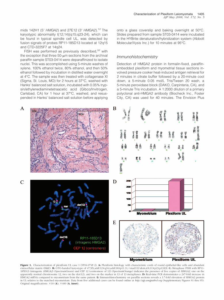
Figure 1. Characterization of plexiform UL case 1 (ST04-074F-2). A: Plexiform histology with characteristic cords of round epithelial-like cells and abundantextracellular matrix (H&E). B: GTG-banded karyotype of 47,XX,add(12)(q24),add(18)(p11.3),�mar[11]/idem,t(8;11)(p23;p13)[3]. C: Metaphase FISH with RP11-185D13 (intragenic HMGA2) (SpectrumGreen) and CEP 12 (centromere of 12) (SpectrumOrange) indicates the presence of five copies of HMGA2, one on theapparently normal chromosome 12, two on the der(12), and two on the marker in 13 of 13 metaphases. D: Real-time PCR demonstrates a 247-fold increase inHMGA2 mRNA compared to myometrium from the same patient. E: Immunohistochemistry on paraffin sections reveals a 3.7-fold elevation of HMGA2 proteinin UL relative to the matched myometrium. Data from five additional cases can be found online at http://ajp.amjpathol.org (Supplementary Figures S1 thru S5).Original magnifications: �10 (A); �400 (A, inset).
Characterization of Plexiform Leiomyomata 1405AJP May 2008, Vol. 172, No. 5

detection system (DAKO) was then applied, including a30-minute incubation with goat anti-rabbit immunoglobulinconjugated to a horseradish peroxidase-labeled polymerfollowed by a 5-minute exposure to the substrate diamino-benzidine to produce a brown precipitate visible by micros-copy. Hematoxylin was used as the counterstain. All stepswere performed at room temperature unless otherwisenoted. HMGA2 protein expression (brown) versus back-ground (blue) staining was evaluated using a semiauto-mated image analysis system (ACISII; Chromavision, SanJuan Capistrano, CA).33 HMGA2 staining for each plexiformsample is expressed as a fold change compared tomatched myometrium from the same patient.
RNA Isolation and Quantitative Real-TimePolymerase Chain Reaction
A portion of each sample was frozen in liquid nitrogenimmediately after surgical removal. RNA was isolatedfrom cases 1 to 4 from both the tumor and the corre-sponding myometrial samples using the RNeasy fibroustissue kit (Qiagen, Valencia, CA). Real-time PCR wasperformed as previously described,10 using the standardcurve method and normalizing the level of HMGA2 ineach tissue to that of GAPDH. HMGA2 expression foreach plexiform RNA sample is shown as a fold changecompared to myometrium from the same patient.
Transcriptional Profiling
RNA isolated from the plexiform UL of cases 1 to 4 washybridized to Affymetrix Human U133 Plus 2.0 GeneChipoligonucleotide expression microarrays (Santa Clara,CA) using standard protocols at the Harvard MedicalSchool–Partners Health Care Center for Genetics andGenomics. These expression profiles were comparedwith those previously acquired for myometrium, UL withtypical histology, UL with loss of the short arm of chro-mosome 1 and hypercellularity or atypia, and uterineleiomyosarcomas using the Affymetrix HuFL microar-ray34,35 as follows. Descriptions of the U133 Plus 2.0 andHuFL microarrays (GPL570 and GPL80 files, respec-
tively) were downloaded from the National Center forBiotechnology Information (NCBI) Gene Expression Om-nibus (http://www.ncbi.nlm.nih.gov/geo/). For each probeset in the GPL80 table, value(s) of the numerical EntrezGene field were used to query the GPL570 table. If morethan one probe set was identified in the GPL570 table,the corresponding expression values were averaged andthe data for genes represented on both microarrays weredeposited into a single database table. Finally, the result-ing raw data for the newly and previously analyzed sam-ples were normalized as described.35 These normalizedexpression data were deposited at the NCBI Gene Ex-pression Omnibus; the series entry number is GSE9511and the specific accession identifiers for PLEX1 to PLEX4are listed under cases 1 to 4 in Table 1. Expressionprofiles of plexiform UL were compared to those previ-ously determined for a collection of normal myometriumand benign and malignant uterine smooth muscle tumorsusing a subset of 134 genes (ie, those represented onboth Affymetrix microarrays used in this study) by hierar-chical cluster analysis using the statistical software pack-age SYSTAT, version 10.2 (Systat Software, Inc., Rich-mond, CA) with the previously described parameters.35
Results
Six UL cases were identified by histopathological analy-sis of H&E-stained tissue sections as having focal ordiffuse plexiform features. These tumors ranged in sizefrom 4 to 19.5 cm and presented in women of varyingrace, age, and menstrual cycle status (Table 1). Case 1has the classic plexiform histology and a complex karyo-type of 47,XX,add(12)(q24),add(18)(p11.3),�mar[11]/idem,t(8;11)(p23;p13)[3] (Figure 1). Follow-up with meta-phase FISH with the intragenic HMGA2 probe RP11-185D13 and the chromosome 12 centromeric probe CEP12 revealed five copies of HMGA2: one on the apparentlynormal chromosome 12, two on the der(12), and two onthe marker chromosome. Real-time PCR and immunohis-tochemistry of paraffin sections revealed a 247-fold in-crease in HMGA2 mRNA and a 3.7-fold elevation ofHMGA2 protein in the tumor relative to its matched myo-
Table 2. Karyotype, FISH, and HMGA2 mRNA and Protein Expression of Uterine Leiomyomata with Plexiform Histology
Case no. Accession no. GTG-banded karyotype
1 ST04-074F-2 47,XX,add(12)(q24),add(18)(p11.3),idem,t(8;11)(p23;p13)�3�
2 ST06-015F 46,XX,t(12;14)(q15;q24)�6�
3 ST06-004F-1 46,XX,add(16)(p13.3)�13�4 ST05-001F-2 47,XX,�mar�2�/46,XX�9�5 ST07-001 48,XX,add(12)(p1?3),�19,�mar�cp5�
6 ST03-0414 46,XX,der(4)inv(4)(p?12q?21),del(11)(q13),der(14)t(12;14)(q15;q24)�7�
N.D., not determined.
1406 Hodge et alAJP May 2008, Vol. 172, No. 5

metrium. These results as well as those of five additionalplexiform UL cases are summarized in Table 2.
The microarray expression profile of this plexiform ULand others (cases 1 to 4) were included in a hierarchicalcluster analysis using 134 probe sets that were previ-ously identified by comparison of myometrium, typical(spindle cell) UL, and leiomyosarcoma (LMS) expressionsignatures to differentiate benign from malignant uterinesmooth muscle tissues.35 Also included in the analysiswere the expression profiles of two cases of another typeof UL variant, namely cellular UL with partial loss of theshort arm of chromosome 1, which were previouslyshown to cluster within the malignant group.34 In thecurrent analysis, the four plexiform tumors were found tocluster together on a separate node within the benignbranch on the sample dendogram (Figure 2A) and matrixplot (Figure 2B). Genes with increased expression inplexiform UL and LMS relative to benign myometrium andtypical UL are UBC, RDBP, CYC1, COX5B, CKS1B, DSS1,and TMSB10 whereas those with decreased expressioninclude PIPPIN, ENPP1, GRIA2, CRMP1, TGFB3, NBL1,LAMA3, MORF4L2, KANK, CTNND1, SETDB1, TACR2,GSTM4, APOD, TIMP3, DPP6, DVL3, VCL, TPM4, GTF2I,THRSP, ID2, STAT4, and MMP2. In a separate analysis in
which the entire expression profiles of the plexiform ULwere directly compared to those of typical UL, the mostup-regulated gene in plexiform UL was the �2 chain fortype I collagen (COL1A2), with a 361-fold increase.
The karyotype for case 2 (see Supplementary FigureS1 at http://ajp.amjpathol.org) is 46,XX,t(12;14)(q15;q24)[6]. This was supported by a metaphase FISH usingan intragenic HMGA2 probe RP11-185D13 and chromo-some 12 centromere probe CEP 12 that indicated thepresence of one copy of HMGA2 on the apparently nor-mal chromosome 12 and translocation of the other copyto the der(14). Surprisingly, metaphase FISH with a split-apart HMGA2 probe set of RP11-299L9 (5� HMGA2) andRP11-427K2 (3� HMGA2) showed the presence of oneintact copy of HMGA2 and one split copy of HMGA2. Torefine the rearrangement, a split-apart HMGA2 probe setof cosmid 142H1 (5� HMGA2) and cosmid 27E12 (3�HMGA2) was used and suggested that one copy ofHMGA2 is disrupted within a region that includes the 3�UTR to the beginning portion of intron 3. Real-time PCRdemonstrated a 66,467-fold increase in HMGA2 mRNAand paraffin section immunohistochemistry showed a4.1-fold elevation of HMGA2 protein relative to thematched myometrium.
Table 2. Continued
No. of HMGA2signals by
metaphase FISH
No. of HMGA2signals by
interphase FISH
t(12;14)(q15;q23–24) detected
by FISH
Intragenic HMGA2break detected
by FISH
HMGA 2 mRNA(fold-changecomparedto matched
myometrium)
HMGA2 protein(fold-changecomparedto matched
myometrium)
1 on normal 12 5 No No 247 3.62 on add(12q)2 on marker1 on normal 12 2 Yes Yes 66,467 4.11 on der(14)1 on each normal 12 2 No No 2017 51 on each normal 12 2 No N.D. 568 01 on normal 12 N.D. N.D. Yes N.D. N.D.3 on add(12p)4 on markerN.D. 3 Yes No N.D. N.D.
Figure 2. Expression analysis of UL with plexi-form histology. A: Comparison of the expressionprofiles of four plexiform UL to a previously de-fined smooth muscle tumor expression signatureusing hierarchical clustering. The horizontal lengthof each arm reflects the relatedness of clusters.Samples are indicated by green terminal segmentsfor benign myometrium (MYO) and UL with nor-mal histology (LEIO), by red terminal segments formalignant leiomyosarcoma (LMS), by orange ter-minal segments for UL with cellular histology andloss of 1p (TUMOR 6 and TUMOR 7), and by blueterminal segments for UL with plexiform histology(PLEX1 through PLEX4 for cases 1 to 4, respec-tively). Although cellular UL have previously beenshown to cluster with malignant LMS, plexiformUL form their own subcluster within the benignbranch. B: Matrix plot displaying clustering ofsamples (columns) by oligonucleotide sets (rows).The color of each rectangle denotes the corre-sponding range of normalized (by SD) expressionvalues as shown at the bottom right.
Characterization of Plexiform Leiomyomata 1407AJP May 2008, Vol. 172, No. 5
The karyotype in case 3 (see Supplementary Figure S2at http://ajp.amjpathol.org) of 46,XX,add(16)(p13.3)[12]as well as the follow-up FISH studies do not suggest achromosomal alteration of HMGA2. Metaphase FISH withRP11-185D13 (intragenic HMGA2) and CEP 12 (centro-mere of 12) indicate the presence of two apparentlynormal chromosomes 12. Metaphase FISH with a split-apart HMGA2 probe set of RP11-299L9 (5� HMGA2) andRP11-427K2 (3� HMGA2) shows the presence of twointact copies of HMGA2. Interphase FISH with RP11-185D13 (intragenic HMGA2) and CTD-3225F7 (chromo-some 14q24) does not produce a fusion signal, consis-tent with exclusion of a cryptic t(12;14)(q15;q24)rearrangement. Real-time PCR, however, demonstrated a2017-fold increase in HMGA2 mRNA compared to thematched myometrium and immunohistochemistry on par-affin sections showed a fivefold elevation of HMGA2 pro-tein in UL relative to the matched myometrium.
Case 4 (see Supplementary Figure S3 at http://ajp.amjpathol.org) had focal areas of plexiform features com-prising �25% of the tissue examined. Although the remain-der of the tumor could not be strictly classified as beingplexiform, there was marked, diffuse deposition of extracel-lular matrix material. The karyotype was 47,XX,�mar[2]/46,XX[9]. Metaphase FISH with RP11-185D13 (intragenicHMGA2) and CEP 12 (centromere of 12) indicates the pres-ence of two apparently normal chromosomes 12 and noexpression of HMGA2 on the marker chromosome. Similarto case 3, evidence of HMGA2 involvement was found byreal-time PCR, which demonstrated a 568-fold increase inHMGA2 mRNA compared to matched myometrium. In-creased HMGA2 protein expression, however, could not beconfirmed by immunohistochemistry in stained sections.
The complex karyotype of case 5 (see SupplementaryFigure S4 at http://ajp.amjpathol.org), 46,XX,der(?4)?inv(4)(p?12q?21),del(11)(q13),der(14)t(12;14)(q15;q24)[7], involves the 12q15 region where HMGA2 is located.Interphase FISH with RP11-185D13 (intragenic HMGA2)and CEP 12 (centromere of 12) indicates the presence ofthree copies of HMGA2 and two copies of the chromo-some 12 centromere. Interphase FISH with RP11-185D13(intragenic HMGA2) and CTD-3225F7 (chromosome14q24) to detect t(12;14)(q15;q24) rearrangements dem-onstrates the presence of two copies of HMGA2 and athird copy involved in a t(12;14) as shown by a fusionsignal. Interphase FISH with a split-apart HMGA2 probeset of RP11-299L9 (5� HMGA2) and RP11-427K2 (3�HMGA2) reveals three intact copies of HMGA2. Becauseof limited material, further investigation was not feasible.
Case 6 (see Supplementary Figure S5 at http://ajp.amjpathol.org) has a karyotype of 48,XX,add(12)(p1?3),�19,�mar[cp5]. Metaphase FISH with the split-apartHMGA2 probe set of RP11-299L9 (5� HMGA2) and RP11-427K2 (3� HMGA2) plus CEP 12 (centromere of 12) indi-cates the presence of eight copies of HMGA2. One intactHMGA2 copy is seen on the presumably normal chromo-some 12, two intact copies and one split-apart copy onthe der(12), and two intact and two split-apart copies onthe marker. Because of limited material, no further anal-yses were possible.
Discussion
This study presents cytogenetic and expression analysesof six plexiform UL. The results suggest these tumorshave been misclassified amid neoplasms with “true” ep-ithelioid differentiation and should instead be groupedamong those with extensive hyalinization. This separationwould emphasize their prognostic distinction.
The six plexiform UL studied were highly divergent insize and occurred in women of varying race, age, andmenstrual cycle status. The unifying finding in each of thesix cases was increased expression of HMGA2, a chro-matin architectural factor that regulates multiple DNA-dependent activities such as gene transcription andplays a role in cell proliferation and differentiation.15,36 Theelevation in HMGA2 mRNA ranged from 284- to more than66,000-fold and the rise in immunodetectable proteinranged from zero- to fivefold in plexiform UL relative totheir adjacent myometria. The lack of detectable proteinexpression in case 4 is discordant with the 568-fold in-crease in mRNA, and may represent an artifact becauseof archival storage. In comparison, chromosomal rear-rangement at the HMGA2 locus and aberrant proteinexpression are found in 10% and 27% of typical (spindlecell) UL, respectively.9,37 The elevation in HMGA2 ex-pression in the plexiform UL appears to result from mul-tiple different mechanisms, including both structural(cases 1, 2, 5, and 6) and submicroscopic (cases 3 and4). Of note, there is a striking similarity in the karyotypesof cases 1 and 5, both including a der(12) and invertedduplicated marker derived from chromosome 12, whichmay indicate a similar pathogenetic mechanism for theirplexiform phenotype.
Interestingly, the karyotype of the plexiform UL in case6, which includes two apparently normal chromosomes12 plus a der(14)t(12;14)(q15;q24) without the reciprocalder(12), closely resembles that of two cases of intrave-nous leiomyomatosis; IVL is a rare smooth muscle prolif-eration that invades vascular spaces but is not clinicallymalignant.38,39 Of note, a plexiform pattern has beenobserved in some cases of IVL. Although this suggeststhat significant HMGA2 overexpression is necessary, it isnot likely to be sufficient to cause the plexiform pheno-type in UL. Further illustration of this observation can befound by inclusion of expression signatures of four of theplexiform UL in a cluster analysis using a previouslyreported gene set that resolves the spectrum of uterinesmooth muscle tissues (myometrium, typical UL, andleiomyosarcoma) into benign versus malignant groups.35
In Figure 2, all plexiform UL clustered into a distinctbranch within the benign group. This node did not in-clude a histologically normal UL (Leio240) with a t(12;14)(q15;q23-q24) and proven aberrant HMGA2 expres-sion.35 In fact, nearly 17% of karyotypically abnormal ULhave a t(12;14)(q15;q23-q24) associated with signifi-cantly increased HMGA2 expression,9,10 and most ofthese tumors have typical, nonplexiform histology.
A similar cluster analysis using the same gene list waspreviously performed for two cases of another histologi-cal variant of UL, the cellular type with loss of 1p, whichdiffers from plexiform UL by the extent of cellularity and
1408 Hodge et alAJP May 2008, Vol. 172, No. 5
extracellular matrix.34 These cases, TUMOR 6 andTUMOR 7 in Figure 2, grouped with the leiomyosarcomason the malignant branch. Thus, microarray expressiondata support the clinical observations that plexiform ULare benign and illustrate that UL are not a single diseaseprocess.
The characteristic pseudo-epithelioid/rounded ap-pearance of the plexiform subgroup of UL is secondary tomatrix deposition and constrictive pressure, suggestingthey are created through their remarkable capacity tosynthesize extracellular matrix.18 In fact, the most signif-icantly up-regulated gene in plexiform UL compared totypical UL is one of the type I collagen chain genes,COL1A. Plexiform UL are also generally solitary and be-nign with an excellent clinical prognosis19; only a singlecase of a malignant plexiform tumor with increased mi-totic activity has been reported.40 This is in contrast toother variants of epithelioid leiomyomata, those with“true” epithelioid differentiation historically calledleiomyoblastoma, which have been known to invade lo-cally, metastasize, and recur.20,21 In a series of five ofthese nonplexiform epithelioid UL, multiple karyotypic ab-normalities were reported ranging from normal in onetumor to one tumor with complex changes including t(10;12)(q22;q15) and del(7)(q21.2q31.2).41 The stemlinet(10;12) in this latter tumor has breakpoints correspond-ing to the bands in which MYST4 (MORF) and HMGA2reside, and the secondary deletion of 7q includes thecritical 7q22 region characteristically deleted in UL.32,42
The karyotypes in these nonplexiform epithelioid tumors,however, did not overlap with those of the plexiform ULfound in the present study. Despite the malignant poten-tial of leiomyoblastoma, the current classification systemcombines them with plexiform UL into a single group.19
In conclusion, because of the lower threshold for clin-ical suspicion of malignancy for “true” epithelioid tumorsand based on both clinical observation and the cytoge-netic and molecular analyses presented here, plexiformUL may most appropriately be classified among tumorswith extensive hyalinization rather than those with “true”epithelioid differentiation. In addition, efforts to dissectthe molecular mechanisms of UL should include stratifi-cation by histopathology and, ideally, by karyotype andrace.
Acknowledgments
We thank Dr. Christopher Fletcher for his facilitation ofHMGA2 immunohistochemistry and Christopher Lafargueand Zuned Khalifa in Dr. Mark Rubin’s laboratory fortechnical assistance in quantitation of the resultingstained slides.
References
1. Cramer SF, Patel A: The frequency of uterine leiomyomas. Am J ClinPathol 1990, 94:435–438
2. Rein MS, Nowak RA: Biology of uterine myomas and myometrium invitro. Semin Reprod Endocrinol 1992, 10:310–319
3. Coronado GD, Marshall LM, Schwartz SM: Complications in preg-
nancy, labor, and delivery with uterine leiomyomas: a population-based study. Obstet Gynecol 2000, 95:764–769
4. Hartmann KE, Birnbaum H, Ben-Hamadi R, Wu EQ, Farrell MH, Spal-ding J, Stang P: Annual costs associated with diagnosis of uterineleiomyomata. Obstet Gynecol 2006, 108:930–937
5. Lepine LA, Hillis SD, Marchbanks PA, Koonin LM, Morrow B, KiekeBA, Wilcox LS: Hysterectomy surveillance—United States, 1980–1993. MMWR CDC Surveill Summ 1997, 46:1–15
6. Flynn M, Jamison M, Datta S, Myers E: Health care resource use foruterine fibroid tumors in the United States. Am J Obstet Gynecol2006, 195:955–964
7. Nibert M, Heim S: Uterine leiomyoma cytogenetics. Genes Chromo-som Cancer 1990, 2:3–13
8. Rein MS, Friedman AJ, Barbieri RL, Pavelka K, Fletcher JA, MortonCC: Cytogenetic abnormalities in uterine leiomyomata. Obstet Gy-necol 1991, 77:923–926
9. Meloni AM, Surti U, Contento AM, Davare J, Sandberg AA: Uterineleiomyomas: cytogenetic and histologic profile. Obstet Gynecol 1992,80:209–217
10. Gross KL, Neskey DM, Manchanda N, Weremowicz S, Kleinman MS,Nowak RA, Ligon AH, Rogalla P, Drechsler K, Bullerdiek J, MortonCC: HMGA2 expression in uterine leiomyomata and myometrium:quantitative analysis and tissue culture studies. Genes ChromosomCancer 2003, 38:68–79
11. Schoenberg Fejzo M, Ashar HR, Krauter KS, Powell WL, Rein MS,Weremowicz S, Yoon SJ, Kucherlapati RS, Chada K, Morton CC: Trans-location breakpoints upstream of the HMGIC gene in uterine leiomyo-mata suggest dysregulation of this gene by a mechanism different fromthat in lipomas. Genes Chromosom Cancer 1996, 17:1–6
12. Schoenmakers EF, Wanschura S, Mols R, Bullerdiek J, Van denBerghe H, Van de Ven WJ: Recurrent rearrangements in the highmobility group protein gene. HMGI-C, in benign mesenchymal tu-mours. Nat Genet 1995, 10:436–444
13. Grosschedl R, Giese K, Pagel J: HMG domain proteins: architecturalelements in the assembly of nucleoprotein structures. Trends Genet1994, 10:94–100
14. Wolffe AP: Architectural transcription factors. Science 1994,264:1100–1101
15. Reeves R: Molecular biology of HMGA proteins: hubs of nuclearfunction. Gene 2001, 277:63–81
16. Ligon AH, Moore SD, Parisi MA, Mealiffe ME, Harris DJ, Ferguson HL,Quade BJ, Morton CC: Constitutional rearrangement of the architec-tural factor HMGA2: a novel human phenotype including overgrowthand lipomas. Am J Hum Genet 2005, 76:340–348
17. Weedon MN, Lettre G, Freathy RM, Lindgren CM, Voight BF, Perry JR,Elliott KS, Hackett R, Guiducci C, Shields B, Zeggini E, Lango H,Lyssenko V, Timpson NJ, Burtt NP, Rayner NW, Saxena R, Ardlie K,Tobias JH, Ness AR, Ring SM, Palmer CN, Morris AD, Peltonen L,Salomaa V, Smith GD, Groop LC, Hattersley AT, McCarthy MI, Hirsch-horn JN, Frayling TM: A common variant of HMGA2 is associated withadult and childhood height in the general population. Nat Genet2007, 39:1245–1250
18. Nucci MR, Quade BJ: Uterine mesenchymal tumors, chapter 20.Diagnostic Gynecologic and Obstetric Pathology. Edited by CrumCP, Lee KR. Frankfurt, Elsevier Saunders, 2005
19. Kurman RJ, Norris HJ: Mesenchymal tumors of the uterus. VI. Epithe-lioid smooth muscle tumors including leiomyoblastoma and clear-cellleiomyoma: a clinical and pathologic analysis of 26 cases. Cancer1976, 37:1853–1865
20. Lavin P, Hajdu SI, Foote FW Jr: Gastric and extragastric leio-myoblastomas: clinicopathologic study of 44 cases. Cancer 1972,29:305–311
21. Kyriazis AP, Kyriazis AA: Uterine leiomyoblastoma (epithelioid leiomy-oma) neoplasm of low-grade malignancy. A histopathologic study.Arch Pathol Lab Med 1992, 116:1189–1191
22. Evans HL, Chawla SP, Simpson C, Finn KP: Smooth muscle neo-plasms of the uterus other than ordinary leiomyoma. A study of 46cases, with emphasis on diagnostic criteria and prognostic factors.Cancer 1988, 62:2239–2247
23. Mazur MT, Kraus FT: Histogenesis of morphologic variations in tu-mors of the uterine wall. Am J Surg Pathol 1980, 4:59–74
24. Nagel H, Brinck U, Luthje D, Fuzesi L: Plexiform leiomyoma of theuterus in a patient with breast carcinoma: case report and review ofthe literature. Pathology 1999, 31:292–294
Characterization of Plexiform Leiomyomata 1409AJP May 2008, Vol. 172, No. 5
25. Kaminski PF, Tavassoli FA: Plexiform tumorlet: a clinical and patho-logic study of 15 cases with ultrastructural observations. Int J GynecolPathol 1984, 3:124–134
26. Seidman JD, Thomas RM: Multiple plexiform tumorlets of the uterus.Arch Pathol Lab Med 1993, 117:1255–1256
27. Goodhue WW, Susin M, Kramer EE: Smooth muscle origin of uterineplexiform tumors. Arch Pathol 1974, 97:263–268
28. Nunez-Alonso C, Battifora HA: Plexiform tumors of the uterus: ultra-structural study. Cancer 1979, 44:1707–1714
29. Fisher ER, Paulson JD, Gregorio RM: The myofibroblastic nature ofthe uterine plexiform tumor. Arch Pathol Lab Med 1978, 102:477–480
30. Cheung VG, Nowak N, Jang W, Kirsch IR, Zhao S, Chen XN, FureyTS, Kim UJ, Kuo WL, Olivier M, Conroy J, Kasprzyk A, Massa H,Yonescu R, Sait S, Thoreen C, Snijders A, Lemyre E, Bailey JA, BruzelA, Burrill WD, Clegg SM, Collins S, Dhami P, Friedman C, Han CS,Herrick S, Lee J, Ligon AH, Lowry S, Morley M, Narasimhan S,Osoegawa K, Peng Z, Plajzer-Frick I, Quade BJ, Scott D, Sirotkin K,Thorpe AA, Gray JW, Hudson J, Pinkel D, Ried T, Rowen L, Shen-OngGL, Strausberg RL, Birney E, Callen DF, Cheng JF, Cox DR, DoggettNA, Carter NP, Eichler EE, Haussler D, Korenberg JR, Morton CC,Albertson D, Schuler G, de Jong PJ, Trask BJ: Integration of cytoge-netic landmarks into the draft sequence of the human genome. Na-ture 2001, 409:953–958
31. Karolchik D, Baertsch R, Diekhans M, Furey TS, Hinrichs A, Lu YT,Roskin KM, Schwartz M, Sugnet CW, Thomas DJ, Weber RJ, HausslerD, Kent WJ: The UCSC Genome Browser Database. Nucleic AcidsRes 2003, 31:51–54
32. Moore SD, Herrick SR, Ince TA, Kleinman MS, Cin PD, Morton CC,Quade BJ: Uterine leiomyomata with t(10;17) disrupt the histoneacetyltransferase MORF. Cancer Res 2004, 64:5570–5577
33. Bismar TA, Demichelis F, Riva A, Kim R, Varambally S, He L, Kutok J,Aster JC, Tang J, Kuefer R, Hofer MD, Febbo PG, Chinnaiyan AM,
Rubin MA: Defining aggressive prostate cancer using a 12-genemodel. Neoplasia 2006, 8:59–68
34. Christacos NC, Quade BJ, Dal Cin P, Morton CC: Uterine leiomyo-mata with deletions of Ip represent a distinct cytogenetic subgroupassociated with unusual histologic features. Genes Chromosom Can-cer 2006, 45:304–312
35. Quade BJ, Wang TY, Sornberger K, Dal Cin P, Mutter GL, Morton CC:Molecular pathogenesis of uterine smooth muscle tumors from tran-scriptional profiling. Genes Chromosom Cancer 2004, 40:97–108
36. Bustin M: Regulation of DNA-dependent activities by the functionalmotifs of the high-mobility-group chromosomal proteins. Mol Cell Biol1999, 19:5237–5246
37. Klotzbucher M, Wasserfall A, Fuhrmann U: Misexpression of wild-typeand truncated isoforms of the high-mobility group I proteins HMGI-Cand HMGI(Y) in uterine leiomyomas. Am J Pathol 1999, 155:1535–1542
38. Quade BJ, Dal Cin P, Neskey DM, Weremowicz S, Morton CC: Intra-venous leiomyomatosis: molecular and cytogenetic analysis of acase. Mod Pathol 2002, 15:351–356
39. Dal Cin P, Quade BJ, Neskey DM, Kleinman MS, Weremowicz S, MortonCC: Intravenous leiomyomatosis is characterized by a der(14)t(12;14)(q15;q24). Genes Chromosom Cancer 2003, 36:205–206
40. Kuroda H, Konishi I, Nanbu K, Mandai M, Komatsu T, Yamamoto S, Yam-abe H, Kida A, Mori T: Malignant plexiform tumor of the uterus: an unusualvariant of epithelioid leiomyosarcoma. Gynecol Oncol 1996, 63:270–275
41. Karaiskos C, Pandis N, Bardi G, Sfikas K, Tserkezoglou A, Fotiou S,Heim S: Cytogenetic findings in uterine epithelioid leiomyomas. Can-cer Genet Cytogenet 1995, 80:103–106
42. Sargent MS, Weremowicz S, Rein MS, Morton CC: Translocations in7q22 define a critical region in uterine leiomyomata. Cancer GenetCytogenet 1994, 77:65–68
1410 Hodge et alAJP May 2008, Vol. 172, No. 5
Copyright © 2022 FDOKUMEN