
Amniotic fluid and its abnormalities Amniotic fluid Origin ...
13
2015-2016 /4 th year Amniotic fluid and its abnormalities د. زﯾﻨﺐ ﻋﺒﺪ اﻷﻣﯿﺮ ﺟﻌﻔﺮ1 Amniotic fluid and its abnormalities Amniotic fluid Origin The first 10 weeks The amnion 10-16 weeks Transudate of the fetal serum via the skin and umbilical cord After 16 weeks Kidney and lungs Functions of the amniotic fluid: 1. Guards the fetus against mechanical shocks. 2. Allows plenty of room for fetal movement. 3. Maintain the temperature of the fetus. 4. May be a source of fetal nutrients. 5. In early pregnancy it is essential for fetal lung development. 6. A valuable source for analysis of fetal tissues and fluids. 7. Equalizes the pressure exerted during uterine contractions. 8. During labor, the amniotic fluid contained in the bag of fore water forms a fluid wedge which, with uterine contractions, dilate the internal os and the cervical canal. 9. When the membranes rupture during labor, fluid flushes the lower genital tract which is aseptic and bacteriostatic. Characteristics of the amniotic fluid 1. Color: It is slightly turbid, but may stain a green or brown color if any meconium has passed into it. 2. Constituents: At term the amniotic fluid has a specific gravity of 1010. It contain 99% water. The osmolality, sodium, urea and creatinine content is not significantly different from the maternal serum. This suggest that amniotic fluid is an ultra- filtrate of maternal serum. The solid constituents of the fluid are:
-
Upload
khangminh22 -
Category
Documents
-
view
1 -
download
0
Transcript of Amniotic fluid and its abnormalities Amniotic fluid Origin ...

2015-2016 /4th year Amniotic fluid and its abnormalities د. زینب عبد األمیر جعفر
1
Amniotic fluid and its abnormalities
Amniotic fluid Origin
The first 10 weeks The amnion 10-16 weeks Transudate of the fetal serum via the skin and
umbilical cord After 16 weeks Kidney and lungs
Functions of the amniotic fluid:
1. Guards the fetus against mechanical shocks. 2. Allows plenty of room for fetal movement. 3. Maintain the temperature of the fetus. 4. May be a source of fetal nutrients. 5. In early pregnancy it is essential for fetal lung development. 6. A valuable source for analysis of fetal tissues and fluids. 7. Equalizes the pressure exerted during uterine contractions. 8. During labor, the amniotic fluid contained in the bag of fore water
forms a fluid wedge which, with uterine contractions, dilate the internal os and the cervical canal.
9. When the membranes rupture during labor, fluid flushes the lower genital tract which is aseptic and bacteriostatic.
Characteristics of the amniotic fluid
1. Color:
It is slightly turbid, but may stain a green or brown color if any meconium has passed into it.
2. Constituents:
At term the amniotic fluid has a specific gravity of 1010. It contain 99% water. The osmolality, sodium, urea and creatinine content is not significantly different from the maternal serum. This suggest that amniotic fluid is an ultra-filtrate of maternal serum.
The solid constituents of the fluid are:
2015-2016 /4th year Amniotic fluid and its abnormalities د. زینب عبد األمیر جعفر
2
1. Lanugos hair. 2. Epithelial cells. 3. Sebaceous material from fetal skin. 4. Amniotic epithelial cells.
3. Volume:
At 10 weeks: 30 ml At 20 weeks: 300 ml At 30 weeks: 600 ml 32-38 weeks: 750- 1000 ml 40 weeks: 800 ml 42 weeks: < 500 ml
? By using ultrasound examination.
1. Measurement of the deepest vertical pool of the fluid (DP) which is free of limbs & without cord. Normal value is 2-8 cm.
2. Amniotic fluid index (AFI):This is calculated by adding the vertical depths of the largest pocket in each of four equal uterine quadrants without cord
If the AFI < 5 cm mean Oligohydramnios.
If the AFI > 25cm mean polyhydramnios.
Oligohydramnios Oligohydramnios means reduced amniotic fluid to less than 200 mL at term
A deepest pool of <2cm AFI is less than 5 cm (<10th centile). The incidence of oligohydramnios varies between 1 and 3 percent anhydramnios means absence of amniotic fluid.

2015-2016 /4th year Amniotic fluid and its abnormalities د. زینب عبد األمیر جعفر
3
Causes of oligohydramnios
1. Fetal 1) Congenital anomalies
i. Renal agenesis. ii. Obstructed uropathy. iii. Renal dysplasia. iv. Polycystic/ multicystic kidney.
2) Chromosomal abnormalities 3) IUGR 4) Fetal death 5) Postmaturity 6) Spontaneous rupture of the membranes (SROM) 7) Premature rupture of the membranes (PROM) 8) Intrauterine infection
2. Placenta a) Abruption b) Placental insufficiency
c) Twin-twin transfusion d) Amnion nodosum :failure of secretion by the cells of the
amnion covering the placenta).
3. Maternal: a) Hypertension
b) Preeclampsia
c) Diabetes
4. Drugs
i. Prostaglandin synthase inhibitors
ii. Angiotensin-converting enzyme inhibitors
5. Idiopathic
2015-2016 /4th year Amniotic fluid and its abnormalities د. زینب عبد األمیر جعفر
4
Management of oligohydramnios
1) History : • Post term pregnancy • Presence of twins • hypertension or preeclampsia • Leakage of fluid(rupture membrane) • congenital infection • Fetal movement • Drugs
2) Physical examination I. abdominal examination:
a. Small for date uterus b. Fetal parts are easily felt and hard c. Malpresentations
II. Check for PPROM (sterile speculum examination to look for Pool, Nitrazine and Fern Tests for Rupture of Membranes )
3) Diagnosis: If suspected spontaneous rupture of membranes (SROM): CRP, FBC
and vaginal swabs should be taken. High resolution ultrasonography including Doppler to assess:
1. Degree of oligohydramnios. 2. Presence of IUGR. 3. Presence of Abnormality of the urinary tract
malformation and see the Presence or absence of the kidneys, Enlarged or cystic kidneys
4. Presence of twin and TTTS
Amniocentesis: in case of suspected chromosomal anomalies
Complications of oligohydramnios
1. Fetal 1. Abortion 2. High fetal mortality. 3. lung hypoplasia if oligohydramnios occurs <22wks
2015-2016 /4th year Amniotic fluid and its abnormalities د. زینب عبد األمیر جعفر
5
4. skeletal deformities (limb abnormalities) e.g. alteration in shape of the skull, wry neck, club foot, or even amputation of the limb, if prolonged oligohydramnios before 22wks has a very poor prognosis
5. Cord compression 2. maternal:
1) Prolonged labor due to inertia 2) Increased operative interference (Increased cesarean section
rate) due to malpresentation or fetal distress .The sum effect may lead to increased maternal morbidity.
Treatment Management targets the underlying etiology. Initially, an evaluation for fetal anomalies and growth is essential.
1. A pregnancy complicated by oligohydramnios and fetal-growth restriction, close fetal surveillance is important because the risk of fetal asphyxia and death is high. Manage aggressively according to umbilical artery Doppler and CTG .with early delivery unless lethal anomalies are present.
2. If fetal renal tract abnormality: Refer to fetal medicine center 3. If SROM
I. If SROM at 34–36 or more weeks: Induce labor unless CS is indicated for another reason.
II. If SROM before 34–36wks Give prophylactic oral erythromycin. Monitor for signs of infection (4-hourly temperature and pulse).
Daily CTG. SERIAL USG – Monitor growth,AFI,BPP
Consider induction by 34–36wks
4. If apparently isolated oligohydramnios Intervention is not usual if umbilical artery Doppler’s are
normal. oligohydramnios Near term probably need no treatment
and their babies are likely to be born healthy
2015-2016 /4th year Amniotic fluid and its abnormalities د. زینب عبد األمیر جعفر
6
Infusion of normal saline into the amniotic sac: by infusing normal saline into the amniotic sac .
1. Sever oligohydromnios 2. Mid trimester (allow lung maturity &prevent
complications mainly pulmonary hypoplasia.)
Benefits of Infusion of normal saline:
1. Decreases cord compression (decreases variable decelerations in labor)
2. Dilutes meconium(is found to improve neonatal outcome)
Polyhydramnios: Amniotic fluid volume (>95th centile for gestational age Amniotic fluid index >25 cm
a largest vertical pocket is >8 cm
polyhydramnios is defined as a state where liquor amnii exceeds 2000mL.
polyhydramnios occurs in 0.4 – 0.5% of pregnancies.
Classification
1. Mild polyhydramnios: DP= 8-12 cm ,AFI(25-29.9)cm 2. Moderate polyhydramnios: DP = 12-15 cm, AFI (30-34.9)cm 3. Severe polyhydramnios: DP > 15cm , AFI =35 cm or more
1. Chronic polyhydramnios: Characterized by gradual accumulation of amniotic fluid
2015-2016 /4th year Amniotic fluid and its abnormalities د. زینب عبد األمیر جعفر
7
2. Acute polyhydramnios (extremely rare): Characterized by sudden and rapid accumulation of amniotic fluid before 20 weeks of pregnancy. It is usually associated with monozygotic twins with TTS or chorioangioma of the placenta.
Causes of polyhydramnios 1. Idiopathic: (35 – 66%) 2. Fetal : 20%
inability to swallow or absorb amniotic fluid i. Gastrointestinal abnormalities that cause
obstruction of fluid transit through the GIT e.g. duodenal atresia and esophageal atresia / tracheo-oesophageal fistula.
ii. Neurological impairment of swallowing mechanism e.g. anencephaly and muscular dystrophies.
iii. Heart defect of the fetus. iv. Other rare abnormalities(e.g.) facial obstruction
Increased fetal urine production. 1) Twin – twin transfusion syndrome. This
complication affects identical twin pregnancies, in which one baby gets too much blood flow and the other gets too little blood. This is due to connections between the blood vessels in their shared placenta. The recipient fetus will develop polyhydramnios, which is related to polyuria.
2) Fetal hydrops 3. Maternal:
1. Infectious conditions: Toxoplasmosis , parvovirus, herpes simplex, rubella or cytomegalovirus infections, causing various abnormalities
2. Maternal Diabetes Mellitus (gestational or pre-existing): 3. Maternal Rhesus Disease
4. Placental causes: 1) Chorioangiomas 2) Arterio-venous fistula
2015-2016 /4th year Amniotic fluid and its abnormalities د. زینب عبد األمیر جعفر
8
Management: History
• Enlarged abdomen • Difficulty in ambulation. • Congenital anomalies in her previous child and previous hx of
polyhydramnios • In severe cases, she may have dyspnea or even remain in the sitting
position for easier breathing, fatigue and palpitations can occur. • indigestion ,Constipation and Heartburn • Edema of the legs, varicosities in the legs or vulva and hemorrhoids • In the rare cases of acute polyhydramnios, there is abdominal pain and
vomiting.
Physical examination
SIGNS:
The patient may be in a dyspneic state in the lying down position. Evidence of preeclampsia (edema, hypertension and proteinuria) may
be present. Varicose veins
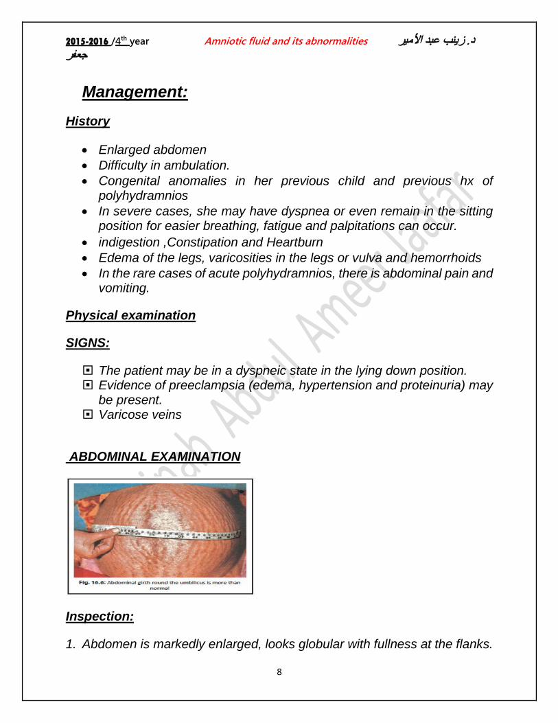
ABDOMINAL EXAMINATION
Inspection:
1. Abdomen is markedly enlarged, looks globular with fullness at the flanks.

2015-2016 /4th year Amniotic fluid and its abnormalities د. زینب عبد األمیر جعفر
9
2. The skin is tense, shiny with large striae 3. Sometimes there is edema of the abdominal wall and vulva.
Palpation:
• The abdomen is larger than date(and higher fundal height) • Fluid thrill can be elicited in all directions over the uterus. • External ballottement can be elicited more easily. • Difficulty to feel the fetal parts; so also the presentation or the position. • The fetus is unduly mobile and the presentation is unstable.
Auscultation:
Fetal heart sound is not heard distinctly, although its presence can be picked up by Doppler ultrasound.
Investigations
1. Ultrasound examination to assess: The degree of polyhydramnios. Presence of multiple pregnancy. Any fetal abnormalities (Specially the central nervous system,
gastrointestinal system and musculoskeletal system). to note the lie and presentation
2. Blood: • ABO and Rh grouping — Rhesus isoimmunisation may cause hydrops
fetalis and fetal ascites. • Postprandial sugar and if necessary glucose tolerance test.
3. Amniotic fluid: Estimation of • alpha fetoprotein (which is markedly elevated in the presence of a fetus
with an open neural tube defect) • Fetal specimens for karyotyping and viral infections
Differential Diagnosis
(1) Twins (2) Pregnancy with huge ovarian cyst (3) Maternal ascites
2015-2016 /4th year Amniotic fluid and its abnormalities د. زینب عبد األمیر جعفر
10
Complications of polyhydramnios
1. Maternal: During pregnancy 1. Preeclampsia (25%) 2. Preterm labor either spontaneous or induced. 3. Preterm premature rupture of membranes. 4. Malpresentation and persistence of floating head 5. Accidental hemorrhage(Placental abruption) due to decrease in
the suface area of the emptying uterus beneath the placenta, following sudden escape of liquor amnii
2. Maternal :During labor: 1) Early rupture of the membranes. 2) Cord prolapse. 3) Uterine inertia. 4) Increased operative delivery due to malpresentation. 5) Retained placenta, postpartum hemorrhage and shock. The
postpartum hemorrhage is due to uterine atony.
3. Puerperium: (1) Sub involution (2) Increased puerperal morbidity due to infection resulting from
increased operative interference and blood loss. 4. Fetal: perinatal mortality -about 50%. The deaths are mostly due to :
a. pre-maturity b. Congenital abnormality c. cord prolapse d. hydrops fetalis e. Effects of increased operative delivery and accidental hemorrhage.
Treatment The first step is identifying the etiology of the abnormal volume of
amniotic fluid, treatment is directed in most situations to the underlying cause

2015-2016 /4th year Amniotic fluid and its abnormalities د. زینب عبد األمیر جعفر
11
1. Mild Polyhydramnios:
The patient with mild polyhydramnios& has no symptoms and without any evidence of fetal abnormalities.It is commonly found in midtrimester. usually requires no treatment, except extra bed rest for a few days. The excess liquor is expected to be diminished as pregnancy advances.
2. Severe Polyhydramnios: The patient should be shifted to tertiary hospital equipped to deal with
“high risk” patients. Use of steroids is recommended to enhance fetal lung maturity if
preterm delivery is anticipated. Schedule weekly or twice weekly perinatal visits Perform serial ultrasonography to determine the AFI and document
fetal growth. If the pregnancy is still premature, the following therapeutic
options can be used: 1. Therapeutic amniocentesis (amnioreduction):
Slow decompression is done at the rate of about 500 ml per hour and the amount of fluid to be removed should be sufficient enough to relieve the mechanical distress. Ordinarily, it should not exceed 1–1.5 liter. Because of slow decompression, chance of accidental hemorrhage is less but liquor amnii may again accumulate, for which the procedure may have to be repeated. Amniotic fluid can be tested for fetal lung maturity.
The goal is to restore amnionic fluid volume to upper normal range and subsequent amnioreduction procedures may be required as often as weekly or even semiweekly
Complications of amniocentesis:
1) Premature rupture of membranes. 2) Chorioamnionitis. 3) Placental abruption 4) preterm labor 5) Perforation of the amnion separating monochorionic diamniotic
twins. 6) The fluid is often re-accumulated quickly.

2015-2016 /4th year Amniotic fluid and its abnormalities د. زینب عبد األمیر جعفر
12
2. Medication :( Prostaglandin synthetase inhibitors)
1) Indomethacin: • Used following the amniocentesis to maintain normal AF volume
without exposing the fetus to the risks of serial invasive procedures • The dose is 50- 100mg/ day
Maternal side – effects
1. Nausea 2. Esophageal reflux 3. Gastritis 4. emesis
Fetal side – effects
1. Premature closure of the ductus arteriosus 2. Cerebral vasoconstriction in the fetus. 3. increased risk of intraventricular hemorrhage 4. Impaired renal function of the fetus.
These risks are increased with progress of pregnancy. For this reason treatment should be stopped by 32-35 week
During indomethacin therapy
1. We monitor AF volume 2 – 3 times per week. The drug is tapered when there is a reduction in AF volume, and stopped when polyhydramnios is no longer severe.
2. We obtain fetal echocardiographic evaluations at intervals to examine ductal flow. If the fetus is found to have major malformations incompatible with life, delivery may be considered.
2) Sulindac: • So it may be a safer alternative to Indomethacin. Has lesser effect on fetal
urine output & the ductus arteriosus.The dose is 200mg every 12 hours.
3. Polyhydramnios with Fetal Congenital Anomaly

2015-2016 /4th year Amniotic fluid and its abnormalities د. زینب عبد األمیر جعفر
13
Termination of pregnancy is to be done irrespective of the duration of pregnancy.
Tense polyhydramnios associated with a congenitally malformed fetus could be first relieved by a slow decompression amniocentesis followed by induction of labor by vaginal PGE2 gel insertion followed by low rupture of membranes and use of oxytocics.
4. Twin–twin transfusion syndrome is best managed in a fetal medicine center, usually with laser ablation of placental anastomoses.
5. Intrapartum Management (during labor): Usual management is: 1) Internal examination should be done soon after the rupture of the membranes
to exclude cord prolapse. 2) If the uterine contraction becomes sluggish, oxytocin infusion may be started, if
not contraindicated. 3) To prevent postpartum hemorrhage, intravenous methergin 0.2 mg should be
given with the delivery of the anterior shoulder. 4) One must remain vigilant following the birth of the baby for retained placenta,
postpartum hemorrhage and shock. 5) Baby should be thoroughly examined for any congenital anomaly.
6.If unstable or transverse lie at term, admit to hospital: CS if labor ensues with an abnormal lie
The end