Clinical significance of the presence of amniotic fluid 'sludge' in asymptomatic high-risk patients...
22
Clinical significance of the presence of amniotic fluid ‘sludge’ in asymptomatic high-risk patients for spontaneous preterm delivery Juan Pedro Kusanovic, MD 1 , Jimmy Espinoza, MD 1,2 , Roberto Romero, MD 1,3 , Luís F. Gonçalves, MD 1,2 , Jyh Kae Nien, MD 1 , Eleazar Soto, MD 1 , Nahla Khalek, MD 2 , Natalia Camacho, MD 2 , Israel Hendler, MD 2 , Pooja Mittal, MD 2 , Lara A. Friel, MD 1,2 , Francesca Gotsch, MD 1 , Offer Erez, MD 1 , Nandor G. Than, MD 1 , Shali Mazaki-Tovi, MD 2 , Mary L. Schoen, RDMS 1 , and Sonia Hassan, MD 1,2 1Perinatology Research Branch, NICHD/NIH/DHHS, Bethesda, Maryland and Detroit, Michigan, USA 2Wayne State University/Hutzel Hospital, Department of Obstetrics and Gynecology, Detroit, Michigan, USA 3Center for Molecular Medicine and Genetics, Wayne State University, Detroit, Michigan, USA Abstract Objective—To determine the clinical significance of the presence of amniotic fluid (AF) ‘sludge’ among asymptomatic patients at high-risk for spontaneous preterm delivery. Study design—This retrospective case-control study included 281 patients with (n=66) and without (n=215) AF ‘sludge’, who underwent transvaginal ultrasound between 13 and 29 completed weeks of gestation. Patients with threatened preterm labor, multiple gestation, fetal anomalies, placenta previa, and uterine contractions were excluded. Results—The prevalence of AF ‘sludge’ in the study population was 23.5% (66/281). The rates of spontaneous preterm delivery at <28, <32, <35, and <37 weeks of gestation were 14.7% (29/197), 21.3% (46/216), 28.7% (62/216), and 42.1% (91/216), respectively. Patients with ‘sludge’ had: (1) a higher rate of spontaneous preterm delivery at <28 weeks [46.5% (20/43) vs. 5.8% (9/154), p<0.001], <32 weeks [55.6% (25/45) vs. 12.3% (21/171), p<0.001], and <35 weeks [62.2% (28/45) vs. 19.9% (34/171), p<0.001]; (2) a higher frequency of clinical chorioamnionitis [15.2% (10/66) vs. 5.1% (11/215), p=0.007], histologic chorioamnionitis [61.5% (40/65) vs. 28% (54/193), p<0.001] and funisitis [32.3% (21/65) vs. 19.2% (37/193), p=0.03]; (3) a higher frequency of preterm PROM [(39.4% (26/66) vs. 13.5% (29/215), p<0.001], lower gestational age at preterm PROM [24.7 weeks (22.3-28.1) vs. 32.3 weeks (27.7-34.8); p<0.001]; and (4) shorter ultrasound-to-delivery [‘sludge’ positive: 127 days (95% CI: 120-134) vs. ‘sludge’ negative: 161 days (95% CI: 153-169), p<0.001] and ultrasound-to-preterm PROM intervals [‘sludge’ positive: 23 days (95% CI: 7-39) vs. ‘sludge’ negative: 57 days (95% CI: 38-77), p=0.003] than those without ‘sludge’. AF ‘sludge’ was an independent explanatory variable for the occurrence of spontaneous preterm delivery at <28, <32, and <35 weeks, preterm PROM, MIAC, and histologic chorioamnionitis. Moreover, the combination of a cervical length <25 mm and ‘sludge’ confers an odds ratio of 14.8 and 9.9 for spontaneous preterm delivery at <28 and <32 weeks, respectively. Conclusions—AF ‘sludge’ is an independent risk factor for spontaneous preterm delivery, preterm PROM, MIAC, and histologic chorioamnionitis in asymptomatic patients at high risk for spontaneous Address correspondence to: Roberto Romero, M.D., Perinatology Research Branch, NICHD, NIH, DHHS, Wayne State University/ Hutzel Women’s Hospital, 3990 John R, Box 4, Detroit, MI 48201, USA, Telephone (313) 993-2700, Fax: (313) 993-2694, e-mail: [email protected]. NIH Public Access Author Manuscript Ultrasound Obstet Gynecol. Author manuscript; available in PMC 2008 May 21. Published in final edited form as: Ultrasound Obstet Gynecol. 2007 October ; 30(5): 706–714. NIH-PA Author Manuscript NIH-PA Author Manuscript NIH-PA Author Manuscript
-
Upload
independent -
Category
Documents
-
view
3 -
download
0
Transcript of Clinical significance of the presence of amniotic fluid 'sludge' in asymptomatic high-risk patients...

Clinical significance of the presence of amniotic fluid ‘sludge’ inasymptomatic high-risk patients for spontaneous pretermdelivery
Juan Pedro Kusanovic, MD1, Jimmy Espinoza, MD1,2, Roberto Romero, MD1,3, Luís F.Gonçalves, MD1,2, Jyh Kae Nien, MD1, Eleazar Soto, MD1, Nahla Khalek, MD2, NataliaCamacho, MD2, Israel Hendler, MD2, Pooja Mittal, MD2, Lara A. Friel, MD1,2, FrancescaGotsch, MD1, Offer Erez, MD1, Nandor G. Than, MD1, Shali Mazaki-Tovi, MD2, Mary L.Schoen, RDMS1, and Sonia Hassan, MD1,2
1Perinatology Research Branch, NICHD/NIH/DHHS, Bethesda, Maryland and Detroit, Michigan, USA
2Wayne State University/Hutzel Hospital, Department of Obstetrics and Gynecology, Detroit, Michigan, USA
3Center for Molecular Medicine and Genetics, Wayne State University, Detroit, Michigan, USA
AbstractObjective—To determine the clinical significance of the presence of amniotic fluid (AF) ‘sludge’among asymptomatic patients at high-risk for spontaneous preterm delivery.
Study design—This retrospective case-control study included 281 patients with (n=66) andwithout (n=215) AF ‘sludge’, who underwent transvaginal ultrasound between 13 and 29 completedweeks of gestation. Patients with threatened preterm labor, multiple gestation, fetal anomalies,placenta previa, and uterine contractions were excluded.
Results—The prevalence of AF ‘sludge’ in the study population was 23.5% (66/281). The rates ofspontaneous preterm delivery at <28, <32, <35, and <37 weeks of gestation were 14.7% (29/197),21.3% (46/216), 28.7% (62/216), and 42.1% (91/216), respectively. Patients with ‘sludge’ had: (1)a higher rate of spontaneous preterm delivery at <28 weeks [46.5% (20/43) vs. 5.8% (9/154),p<0.001], <32 weeks [55.6% (25/45) vs. 12.3% (21/171), p<0.001], and <35 weeks [62.2% (28/45)vs. 19.9% (34/171), p<0.001]; (2) a higher frequency of clinical chorioamnionitis [15.2% (10/66)vs. 5.1% (11/215), p=0.007], histologic chorioamnionitis [61.5% (40/65) vs. 28% (54/193), p<0.001]and funisitis [32.3% (21/65) vs. 19.2% (37/193), p=0.03]; (3) a higher frequency of preterm PROM[(39.4% (26/66) vs. 13.5% (29/215), p<0.001], lower gestational age at preterm PROM [24.7 weeks(22.3-28.1) vs. 32.3 weeks (27.7-34.8); p<0.001]; and (4) shorter ultrasound-to-delivery [‘sludge’positive: 127 days (95% CI: 120-134) vs. ‘sludge’ negative: 161 days (95% CI: 153-169), p<0.001]and ultrasound-to-preterm PROM intervals [‘sludge’ positive: 23 days (95% CI: 7-39) vs. ‘sludge’negative: 57 days (95% CI: 38-77), p=0.003] than those without ‘sludge’. AF ‘sludge’ was anindependent explanatory variable for the occurrence of spontaneous preterm delivery at <28, <32,and <35 weeks, preterm PROM, MIAC, and histologic chorioamnionitis. Moreover, the combinationof a cervical length <25 mm and ‘sludge’ confers an odds ratio of 14.8 and 9.9 for spontaneouspreterm delivery at <28 and <32 weeks, respectively.
Conclusions—AF ‘sludge’ is an independent risk factor for spontaneous preterm delivery, pretermPROM, MIAC, and histologic chorioamnionitis in asymptomatic patients at high risk for spontaneous
Address correspondence to: Roberto Romero, M.D., Perinatology Research Branch, NICHD, NIH, DHHS, Wayne State University/Hutzel Women’s Hospital, 3990 John R, Box 4, Detroit, MI 48201, USA, Telephone (313) 993-2700, Fax: (313) 993-2694, e-mail:[email protected].
NIH Public AccessAuthor ManuscriptUltrasound Obstet Gynecol. Author manuscript; available in PMC 2008 May 21.
Published in final edited form as:Ultrasound Obstet Gynecol. 2007 October ; 30(5): 706–714.
NIH
-PA Author Manuscript
NIH
-PA Author Manuscript
NIH
-PA Author Manuscript
preterm delivery. Furthermore, the combination of ‘sludge’ and a short cervix confers a higher riskfor spontaneous preterm delivery at <28 and <32 weeks than that of a short cervix alone.
Keywordsamniotic fluid ‘sludge’; preterm labor; transvaginal ultrasound; spontaneous preterm delivery;microbial invasion of the amniotic cavity; MIAC; chorioamnionitis; funisitis; preterm prematurerupture of membranes; PPROM
INTRODUCTIONThe sonographic finding of dense aggregates of particulate matter in the amniotic fluid closeto the internal cervical os, known as amniotic fluid (AF) ‘sludge’, is associated with impendingpreterm delivery, microbial invasion of the amniotic cavity (MIAC), and histologicchorioamnionitis in patients with spontaneous preterm labor and intact membranes.1 Similarobservations were recently reported among patients with a history of preterm delivery orthreatened preterm labor.2
Intra-amniotic infection is sometimes chronic in nature.3-5 Indeed, accumulating evidenceindicates that asymptomatic patients with a positive amniotic fluid culture at the time of mid-trimester amniocentesis have a higher risk of adverse pregnancy outcome including fetal lossand/or preterm delivery.3-5 To the extent that AF ‘sludge’ may represent clusters of bacteriaand inflammatory cells,1 it is possible that the presence of AF ‘sludge’ may represent chronicintra-amniotic infection.
AF ‘sludge’ has been observed in some asymptomatic patients with risk factors for pretermdelivery. However, the clinical implications of the presence of this ultrasonographic findingin these patients are unknown. The objective of this study was to determine the clinicalsignificance of amniotic fluid ‘sludge’ in asymptomatic patients at high-risk for spontaneouspreterm delivery.
METHODSStudy design
A retrospective case-control study was conducted by searching our clinical database and digitallibrary of ultrasound images from consecutive patients at high-risk for spontaneous pretermdelivery between March 2002 and November 2005. Inclusion criteria were the following: 1)Transvaginal ultrasound (TVUS) of the cervix between 13 and 29 completed weeks ofgestation; 2) history of spontaneous preterm delivery; 3) prior mid-trimester loss; 4) shortcervix, defined as a mid-trimester cervical length <25 mm on TVUS; 5) mullerian ductanomalies; and/or 6) history of cone biopsy. Exclusion criteria were: 1) threatened pretermlabor in the index pregnancy; 2) multiple gestations; 3) fetal congenital anomalies; 4) placentaprevia; and 5) presence of irregular uterine contractions.
All patients provided written informed consent before participating in the study. The use ofclinical and ultrasound data for research purposes was approved by the Institutional ReviewBoards of Wayne State University and the National Institute of Child Health and HumanDevelopment (NICHD/NIH/DHHS).
Definitions and study proceduresGestational age was determined by the last menstrual period or by ultrasound in case theultrasonographic determination of gestational age was not consistent with the menstrual datingby >2 weeks. Spontaneous preterm parturition was defined by the presence of regular uterine
Kusanovic et al. Page 2
Ultrasound Obstet Gynecol. Author manuscript; available in PMC 2008 May 21.
NIH
-PA Author Manuscript
NIH
-PA Author Manuscript
NIH
-PA Author Manuscript

contractions and cervical changes that led to delivery before 37 completed weeks of gestation.Preterm PROM was diagnosed by sterile speculum examination confirming pooling ofamniotic fluid in the vagina (with nitrazine and ferning tests when necessary) before 37 weeksof gestation and in the absence of labor. Vaginal bleeding of uterine origin was diagnosed bysterile speculum examination confirming the presence of blood coming through the externalcervical os.
Amniotic fluid collection was performed by transabdominal amniocentesis underultrasonographic guidance for clinical indications (e.g. chromosomal analysis) or to determinethe microbiologic state of the amniotic cavity in patients with a transvaginal cervical length<25 mm, because of previous observations demonstrated an association between a short cervixand histologic chorioamnionitis,6 which subsequently have been also linked to sub-clinicalintra-amniotic infection.7 Amniotic fluid was transported to the laboratory in a sterile cappedplastic syringe and cultured for aerobic and anaerobic bacteria, as well as genital Mycoplasmas.White blood cell (WBC) count, glucose concentration, and Gram stain for microorganismswere performed in amniotic fluid shortly after collection using methods previously described.8-10
Microbial invasion of the amniotic cavity and intra-amniotic inflammation (IAI) were definedas a positive amniotic fluid culture for microorganisms and a WBC count >50 cells/mm3,10respectively. Clinical chorioamnionitis was diagnosed according to the criteria proposed byGibbs et al11 including maternal temperature of ≥37.8°C and two or more of the followingcriteria: 1) uterine tenderness; 2) malodorous vaginal discharge; 3) maternal leukocytosis(WBC ≥15,000 cells/mm3); 4) maternal tachycardia (>100 beats/min); and 5) fetal tachycardia(≥160 beats/min). The diagnosis of histologic chorioamnionitis was based on the presence ofinflammatory cells in the chorionic plate and/or chorioamniotic membranes. Acute funisitiswas diagnosed by the presence of neutrophils in the wall of the umbilical vessels and/orWharton’s jelly using the criteria previously described.12
Composite severe neonatal morbidity was defined in the presence of one or more of thefollowing neonatal complications: 1) sepsis or suspected sepsis; 2) assisted ventilation due torespiratory distress syndrome; 3) bronchopulmonary dysplasia; 4) intraventricular hemorrhage(IVH); and 5) necrotizing enterocolitis. Neonatal sepsis was diagnosed in the presence of apositive blood culture. Suspected neonatal sepsis was diagnosed in the absence of a positiveblood culture, but in the presence of clinical, radiographic or laboratory parameters suspiciousfor infection, such as a WBC count <5,000 cells/mm3, polymorphonuclear leukocyte count of<1,800 cells/mm3, ratio of immature neutrophils to total neutrophils >0.2, and/or a positiveaspirate for polymorphonuclear leukocytes >5 per high power field. The diagnosis ofrespiratory distress syndrome required the presence of respiratory grunting and retracting,increased oxygen requirement and diagnostic radiographic and laboratory findings in theabsence of evidence for other causes of respiratory disease.13 Bronchopulmonary dysplasiawas diagnosed if the neonate required oxygen and ventilatory therapy for more than 28 daysduring the first 2 months of life, had typical radiographic changes, and/or had dysplasia of thebronchopulmonary tree at autopsy.13 Intraventricular hemorrhage was diagnosed byultrasonographic examination of the neonatal head. Necrotizing enterocolitis was diagnosedin the presence of abdominal distention and feeding intolerance for at least 24 hours (vomitingor increased gastric residual), with clear radiologic evidence of intramural air, perforation,meconium plug syndrome or definite surgical or autopsy findings of necrotizing enterocolitis.13
Sonographic assessment of the cervixTransvaginal ultrasounds were conducted with commercially available two-dimensional (2D)and three-dimensional (3D) ultrasound systems (Acuson Sequoia, Siemens Medical Systems,
Kusanovic et al. Page 3
Ultrasound Obstet Gynecol. Author manuscript; available in PMC 2008 May 21.
NIH
-PA Author Manuscript
NIH
-PA Author Manuscript
NIH
-PA Author Manuscript

Mountain View, CA and Voluson 730, GE Healthcare, Milwaukee, WI) equipped withendovaginal transducers with frequency ranges of 5-7.5 MHz and 5-9 MHz, respectively. Allsonographic examinations of the cervix were performed by Registered Diagnostic MedicalSonographers using a technique previously described,14 and reviewed by a perinatologist.Amniotic fluid ‘sludge’ was defined as the presence of dense aggregates of particulate matterin proximity to the internal cervical os (Figure 1). Two experienced sonographers, blinded toclinical outcome, reviewed the 2D images and 3D volume datasets of the cervix. Amnioticfluid ‘sludge’ was considered present when identified by both sonographers.
Statistical analysisOutcome variables included: 1) spontaneous preterm delivery at <28 weeks, <32 weeks and<35 weeks of gestation; 2) preterm PROM; 3) microbial invasion of the amniotic cavity; 4)intra-amniotic inflammation; 5) clinical chorioamnionitis; 6) histologic chorioamnionitis; 7)admission to the neonatal intensive care unit (NICU); 8) composite severe neonatal morbidity;9) neonatal death; and 10) ultrasound-to-delivery and ultrasound-to-preterm PROM intervals.Patients with indicated preterm delivery (e.g. fetal distress, placental abruption, severe maternalcondition, etc.) were excluded from the sub-analysis to determine the association between AF‘sludge’ and spontaneous preterm delivery.
Comparisons among groups were performed using Chi-square or Fisher’s exact test forcategorical variables, and Mann-Whitney U test for continuous variables. Diagnostic indices,predictive values, and likelihood ratios of the presence of cervical length <25 mm or AF‘sludge’ were calculated for the identification of spontaneous preterm delivery at <28 weeks,<32 weeks, and <35 weeks of gestation in a subgroup of asymptomatic high-risk patients withultrasound examination at 14-24 weeks, because this is the gestational age range at whichcervical measurement is performed in most centers.15-20
Multivariable stepwise logistic regression analyses were performed to determine therelationship between the presence of AF ‘sludge’ and other potential explanatory variables(cervical length of <25 mm, gestational age at the time of ultrasound examination, prior pretermdelivery, vaginal bleeding in the index pregnancy, and cervical cerclage) in the prediction ofthe study outcomes. For the analysis of neonatal outcomes, the gestational age at delivery wasalso included as a covariate. A Kaplan-Meier survival analysis was performed to assess theultrasound-to-delivery and ultrasound-to-preterm PROM intervals according to the presenceor absence of AF ‘sludge’. Patients who delivered preterm for maternal or fetal indicationswere included in the analysis with a censored time that was equal to the ultrasound-to-deliveryinterval. Cox regression analysis was performed to assess the ultrasound-to-delivery andultrasound-to-preterm PROM intervals while controlling for the above mentioned covariates.A p value of <0.05 was considered statistically significant (SPSS 14.0, SPSS Inc., Chicago,IL, USA).
RESULTSPrevalence of outcome variables among asymptomatic patients at high-risk for pretermdelivery
Two hundred eighty-one patients met the inclusion criteria. The prevalence of AF ‘sludge’ inasymptomatic patients at high–risk for spontaneous preterm delivery was 23.5% (66/281). Acervical length of <25 mm was present in 50.5% (142/281) of the patients. The prevalence ofpreterm PROM was 19.6% (55/281), and the rates of spontaneous preterm delivery at <28weeks, <32 weeks, <35 weeks, and <37 weeks of gestation were 14.7% (29/197), 21.3%(46/216), 28.7% (62/216), and 42.1% (91/216), respectively. Clinical and histologic
Kusanovic et al. Page 4
Ultrasound Obstet Gynecol. Author manuscript; available in PMC 2008 May 21.
NIH
-PA Author Manuscript
NIH
-PA Author Manuscript
NIH
-PA Author Manuscript

chorioamnionitis were diagnosed in 7.5% (21/281) and 36.4% (94/258) of the patients,respectively.
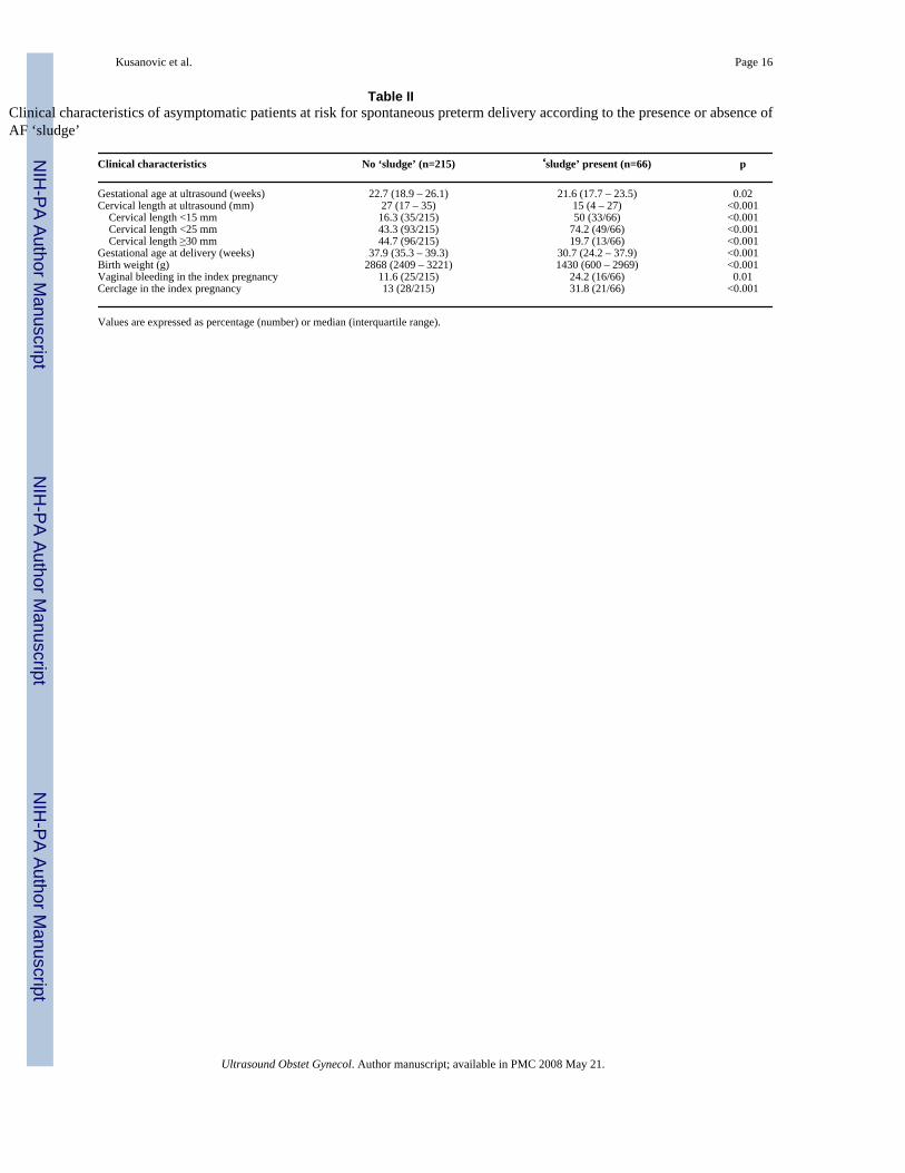
Demographic and clinical characteristics of the study populationThe demographic and clinical characteristics of the study population are described in Table Iand Table II, respectively. Patients with AF ‘sludge’ had significantly shorter cervical lengthat ultrasound, lower gestational age at delivery, and lower birth weight compared to patientswithout AF ‘sludge’ (Table II). A significantly higher proportion of patients with AF ‘sludge’had a cervical length <25 mm, as well as vaginal bleeding and cervical cerclage in the indexpregnancy than those without AF ‘sludge’ (Table II). In addition, the shorter the cervical length,the higher the frequency of AF ‘sludge’ (Table III).
AF ‘sludge’ and its association with spontaneous preterm deliveryA higher proportion of patients with AF ‘sludge’ had spontaneous preterm delivery at <28weeks [46.5% (20/43) vs. 5.8% (9/154), p<0.001], <32 weeks [55.6% (25/45) vs. 12.3%(21/171), p<0.001], and <35 weeks [62.2% (28/45) vs. 19.9% (34/171), p<0.001] than thosewithout AF ‘sludge’ (Table I in supplemental material). The frequency of spontaneous pretermdelivery at <28 weeks and <32 weeks was higher in patients with AF ‘sludge’ than in thosewithout it, regardless of cervical length (Figures 2 and 3).
Patients with ‘sludge’ had a shorter ultrasound-to-delivery interval than those without AF‘sludge’ [AF ‘sludge’ positive, median: 127 days (95% CI: 120-134) vs. AF ‘sludge’ negative,median: 161 days (95% CI: 153-169); log rank test, p<0.001] (Figure 4). These results remainedsignificant after adjusting for the presence of ‘sludge’, cervical length of <25 mm, prior pretermdelivery, gestational age at ultrasound, vaginal bleeding, and cervical cerclage (hazard ratio:2.96; 95% CI: 1.6-5.3).
Patients with AF ‘sludge’ also had a higher frequency of clinical chorioamnionitis [15.2%(10/66) vs. 5.1% (11/215), p=0.007], histologic chorioamnionitis [61.5% (40/65) vs. 28%(54/193), p<0.001], and funisitis [32.3% (21/65) vs. 19.2% (37/193), p=0.03] than thosewithout AF ‘sludge’ (Table I in supplemental material).
AF ‘sludge’ and its association with preterm PROMPatients with AF ‘sludge’ had a higher frequency of preterm PROM [(39.4% (26/66) vs. 13.5%(29/215), p<0.001], and a lower gestational age at preterm PROM [median: 24.7 weeks(22.3-28.1) vs. median: 32.3 weeks (27.7-34.8); p<0.001] than those without AF‘sludge’ (Table I in supplemental material). Patients with ‘sludge’ had a shorter ultrasound-to-preterm PROM interval than those without AF ‘sludge’ [AF ‘sludge’ positive, median: 23 days(95% CI: 7-39) vs. AF ‘sludge’ negative, median: 57 days (95% CI: 38-77); log rank test,p=0.003] (Figure 1 in supplemental material). These results remained significant after adjustingfor the presence of AF ‘sludge’ and other explanatory variables listed in the analysis of theultrasound-to-delivery interval in women with intact membranes (hazard ratio: 2.76; 95% CI:1.5-5.1).
A sub-analysis among patients with preterm PROM (n=55) indicated that those with AF‘sludge’ had a shorter cervical length at ultrasound and earlier gestational age at delivery thanthose without AF ‘sludge’. In addition, those with AF ‘sludge’ have a higher proportion ofhistologic chorioamnionitis, proven or suspected neonatal sepsis, composite severe neonatalmorbidity, and neonatal death than those without AF ‘sludge’ (Table II in supplementalmaterial).
Kusanovic et al. Page 5
Ultrasound Obstet Gynecol. Author manuscript; available in PMC 2008 May 21.
NIH
-PA Author Manuscript
NIH
-PA Author Manuscript
NIH
-PA Author Manuscript

AF ‘sludge’ and its association with intra-amniotic infection/inflammationAnalysis restricted to 51 asymptomatic patients who underwent transabdominal amniocentesiswithin 14 days of transvaginal ultrasound because of a short cervix indicated that a significantlyhigher proportion of patients with AF ‘sludge’ had MIAC [21.7 % (5/23) vs. 0% (0/28),p=0.01], and intra-amniotic inflammation [27.3% (6/22) vs. 3.6% (1/28), p=0.03] than thosewithout AF ‘sludge’. No significant differences were found in gestational age, cervical length,and cervical dilatation at the time of amniocentesis between patients with and without AF‘sludge’ (Table III in supplemental material). Microorganisms isolated from the AF of patientswith ‘sludge’ included Ureaplasma urealyticum (n=3), Staphylococcus aureus (n=1), andFusobacterium nucleatum (n=1).
AF ‘sludge’ and its association with neonatal outcomesThe prevalence of admission to NICU, composite severe neonatal morbidity, and neonataldeath in the study population was 20.7% (58/280), 22.8% (58/254), and 2.7% (7/255),respectively. A significantly higher proportion of neonates born to patients with AF ‘sludge’was admitted to NICU, had a composite severe neonatal morbidity, and died in the neonatalperiod than those born to women without AF ‘sludge’ (Table IV).
Subgroup of patients examined between 14-24 weeksThe prevalence of AF ‘sludge’ in this subgroup was 29.9% (52/174). A subanalysis performedat this gestational age interval showed similar results to the ones described for the entirepopulation (Table V). The diagnostic indices, predictive values, and likelihood ratios of thepresence of cervical length <25 mm or AF ‘sludge’ for the identification of patients withspontaneous preterm delivery at <28 weeks, <32 weeks, and <35 weeks are displayed in TableVI. Table VII displays the odds ratios for the occurrence of spontaneous preterm delivery at<28 weeks, <32 weeks, and <35 weeks in the presence of a cervical length of <25 mm, AF‘sludge’ positive, or both. Of note, the combination of a cervical length of <25 mm and AF‘sludge’ confers an odds ratio of 14.8 and 9.9 for spontaneous preterm delivery at <28 and <32weeks, respectively.
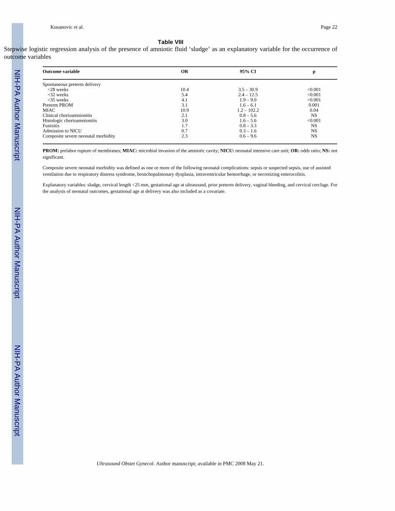
AF ‘sludge’ as an independent risk factor for the occurrence of outcome variablesMultivariable stepwise logistic regression analysis showed that the presence of AF ‘sludge’was an independent explanatory variable for the occurrence of spontaneous preterm deliveryat <28 weeks, <32 weeks, and <35 weeks of gestation, preterm PROM, MIAC, and histologicchorioamnionitis, but not for adverse neonatal outcomes. As expected, the most importantfactor associated with adverse neonatal outcomes was gestational age at delivery (Table VIII).Of note, cervical cerclage was not an explanatory variable for the occurrence of all outcomes.
DISCUSSIONPrincipal findings of this study
1) The prevalence of AF ‘sludge’ in this population of asymptomatic high-risk patients forpreterm delivery between 13 and 29 completed weeks of gestation is 23.5%; 2) AF ‘sludge’ isan independent risk factor for spontaneous preterm delivery at <28 weeks, <32 weeks, and <35weeks of gestation, preterm PROM, MIAC, and histologic chorioamnionitis; and 3)asymptomatic patients with AF ‘sludge’ had shorter ultrasound-to-delivery and ultrasound-to-preterm PROM intervals than those without AF ‘sludge’.
The prevalence of AF ‘sludge’ in asymptomatic high-risk patients for preterm deliveryOur group had recently reported that the prevalence of AF ‘sludge’ in normal pregnancies atterm is 1%, whereas in patients with spontaneous preterm labor and intact membranes is 22.6%.
Kusanovic et al. Page 6
Ultrasound Obstet Gynecol. Author manuscript; available in PMC 2008 May 21.
NIH
-PA Author Manuscript
NIH
-PA Author Manuscript
NIH
-PA Author Manuscript

1 Of note, this prevalence is similar to the one presented herein among asymptomatic patients(23.5%). We propose that this reflects the high risk nature of the population included in thepresent study. Indeed, the prevalence of spontaneous preterm delivery at <32 weeks and <37weeks of gestation was 21.3% (46/216) and 42.1% (91/216), respectively. The prevalence andclinical significance of AF ‘sludge’ in asymptomatic low-risk patients throughout gestation isunknown. Additional studies should be conducted to address this important clinical question.
AF ‘sludge’ and its association with intra-amniotic infection/inflammationPatients with AF ‘sludge’ had a significantly higher frequency of MIAC, intra-amnioticinflammation, clinical chorioamnionitis, and histologic chorioamnionitis than patients without‘sludge’. This is consistent with the previous report that AF ‘sludge’ is associated with positiveAF cultures and histologic chorioamnionitis in patients with preterm labor and intactmembranes.1 In addition, the frequency of funisitis, which is the histologic counterpart of thefetal inflammatory response syndrome (FIRS),12;21 was significantly higher among patientswith AF ‘sludge’.
Since the incidence of vaginal bleeding is higher in symptomatic and asymptomatic patientswith AF ‘sludge’ it is possible that, in some patients, the presence of AF ‘sludge’ may representblood breakdown products; however, the following observations make this possibility lesslikely: 1) we have obtained samples of AF ‘sludge’ under ultrasound guidance in patients withimpending preterm delivery, and the microbiologic examination of these samples indicate thatthey represent clusters of inflammatory cells and bacteria (Espinoza et al, unpublishedobservations); 2) a recent study22 reported that MIAC is present in 14% of patients with“idiopathic” vaginal bleeding between 18 to 35 weeks of gestation, and that “idiopathic”vaginal bleeding is associated with preterm delivery <32 weeks and subsequent pretermPROM, suggesting that vaginal bleeding may be the only clinical manifestation of intra-amniotic infection; and 3) vaginal bleeding was included in the logistic regression analysis asa covariate to determine its relationship with the presence of AF ‘sludge’ and other potentialexplanatory variables in the prediction of the study outcomes. The results of this analysisshowed that AF ‘sludge’ was an independent risk factor for the occurrence of the studyoutcomes.
It has been recently reported that the rate of MIAC in asymptomatic patients with a cervicallength of <25 mm in the mid-trimester is 9%.7 Of note, the prevalence of MIAC among patientswith AF ‘sludge’ who underwent amniocentesis within 2 weeks of the ultrasound was 21.7%;in contrast, no patients without AF ‘sludge’ had positive AF cultures. Collectively, these resultssupport the notion that AF ‘sludge’ may represent clusters of bacteria and inflammatory cells.
AF ‘sludge’ as a risk factor for adverse pregnancy outcomesThe observation that AF ‘sludge’ is associated with spontaneous preterm delivery at <28 weeks,<32 weeks, and <35 weeks of gestation, preterm PROM, MIAC, and histologicchorioamnionitis in asymptomatic patients at high-risk for spontaneous preterm delivery isnovel. In addition, those with AF ‘sludge’ have a higher frequency of short cervix (<25 mm),clinical chorioamnionitis, funisitis, admission to NICU, composite severe neonatal morbidityand neonatal death, earlier gestational age at the time of preterm PROM, as well as shorterultrasound-to-delivery and ultrasound-to-preterm PROM intervals than those without ‘sludge’.Thus, the sonographic finding of AF ‘sludge’ in asymptomatic patients at high-risk forspontaneous preterm delivery should be considered as a risk factor for adverse pregnancyoutcome. To the extent that AF ‘sludge’ may represent intra-amniotic infection/inflammation,these observations are consistent with a solid body of evidence indicating the associationbetween intra-amniotic infection and the aforementioned adverse pregnancy outcomes.3-5;7;23-28
Kusanovic et al. Page 7
Ultrasound Obstet Gynecol. Author manuscript; available in PMC 2008 May 21.
NIH
-PA Author Manuscript
NIH
-PA Author Manuscript
NIH
-PA Author Manuscript
Short cervix and AF ‘sludge’ in the prediction of spontaneous preterm delivery amongasymptomatic high-risk patients
Our results that a cervical length of <25 mm between 14-24 weeks is associated with anincreased risk for spontaneous preterm delivery at <28, <32, and <35 weeks are consistent witha solid body of evidence that a sonographic short cervix in the mid-trimester is a useful toolfor the assessment of risk for spontaneous preterm delivery in low-14;15;18;29 and high-riskpopulations.16;17;19;20;30-32 However, this study further demonstrates that the presence ofAF ‘sludge’ is an independent explanatory variable for the occurrence of spontaneous pretermdelivery, and that this sonographic finding confers an odds ratio of 9.1 and 6.2 for spontaneouspreterm delivery at <28 and <32 weeks, respectively. In addition, the combination of a cervicallength of <25 mm and AF ‘sludge’ confers an odds ratio of 14.8 and 9.9 for spontaneous pretermdelivery at <28 and <32 weeks, respectively. However, AF ‘sludge’ did not increase the oddsratios for spontaneous preterm delivery at < 35 weeks (Table VII).
Limitations of the studyThe limitations of this study include its retrospective nature, and that the study population mayrepresent a group of patients at very high-risk for preterm delivery, because 42% of themdelivered at <37 weeks of gestation. It could be argued that the identification of AF ‘sludge’is subjective; however, two experienced sonographers, blinded to the clinical information,ascertained the presence of AF ‘sludge’ using a strict definition previously described.1
ConclusionsOur results indicate that AF ‘sludge’ is an independent risk factor for spontaneous pretermdelivery, preterm PROM, microbial invasion of the amniotic cavity, and histologicchorioamnionitis in asymptomatic patients at high risk for spontaneous preterm delivery.Moreover, the combination of AF ‘sludge’ and a short cervix (<25 mm) confers a higher riskfor spontaneous preterm delivery at <28 and <32 weeks than that conferred by a short cervixalone.
Acknowledgements
The authors wish to acknowledge the contributions of the Registered Diagnostic Medical Sonographers staff of thePerinatology Research Branch: Ms Susan Stites, Ms Lisa Fisher, Ms Anna Peshkess, and Ms Catherine Ducharme, aswell as the Nursing staff of the Perinatology Research Branch and Detroit Medical Center: Ms Nancy Hauff, Ms SandyField, Ms Lorraine Nikita, Ms Vicky Ineson, Ms Evie Russell, Ms Mahbubeh Mahmoudieh, Ms Julie McKinley, MsSue Rehel, Ms Shannon Donegan, Ms Linda Bouey, Ms Carolyn Sudz, Ms Sylvia Warren, Ms Shelley Mullen, MsGail Bartley, Ms Denise Bayoneto, Ms Judy Kerman, Ms Barbara Steffy, Ms Milagros Kitchen, and Ms Leandra Ga-Pinlac.
This research was supported by the Intramural Research Program of the National Institute of Child Health and HumanDevelopment, NIH, DHHS.
Reference List1. Espinoza J, Goncalves LF, Romero R, Nien JK, Stites S, Kim YM, et al. The prevalence and clinical
significance of amniotic fluid ‘sludge’ in patients with preterm labor and intact membranes. UltrasoundObstet Gynecol 2005;25:346–52. [PubMed: 15789375]
2. Bujold E, Pasquier JC, Simoneau J, Arpin MH, Duperron L, Morency AM, et al. Intra-amniotic sludge,short cervix, and risk of preterm delivery. J Obstet Gynaecol Can 2006;28:198–202. [PubMed:16650357]
3. Cassell GH, Davis RO, Waites KB, Brown MB, Marriott PA, Stagno S, et al. Isolation of Mycoplasmahominis and Ureaplasma urealyticum from amniotic fluid at 16-20 weeks of gestation: potential effecton outcome of pregnancy. Sex Transm Dis 1983;10:294–302. [PubMed: 6665671]
4. Gray DJ, Robinson HB, Malone J, Thomson RB Jr. Adverse outcome in pregnancy following amnioticfluid isolation of Ureaplasma urealyticum. Prenat Diagn 1992;12:111–17. [PubMed: 1553356]
Kusanovic et al. Page 8
Ultrasound Obstet Gynecol. Author manuscript; available in PMC 2008 May 21.
NIH
-PA Author Manuscript
NIH
-PA Author Manuscript
NIH
-PA Author Manuscript
5. Horowitz S, Mazor M, Romero R, Horowitz J, Glezerman M. Infection of the amniotic cavity withUreaplasma urealyticum in the midtrimester of pregnancy. J Reprod Med 1995;40:375–79. [PubMed:7608879]
6. Guzman ER, Shen-Schwarz S, Benito C, Vintzileos AM, Lake M, Lai YL. The relationship betweenplacental histology and cervical ultrasonography in women at risk for pregnancy loss and spontaneouspreterm birth. Am J Obstet Gynecol 1999;181:793–97. [PubMed: 10521731]
7. Hassan S, Romero R, Hendler I, Gomez R, Khalek N, Espinoza J, et al. A sonographic short cervix asthe only clinical manifestation of intra-amniotic infection. J Perinat Med 2006;34:13–19. [PubMed:16489881]
8. Romero R, Emamian M, Quintero R, Wan M, Hobbins JC, Mazor M, et al. The value and limitationsof the Gram stain examination in the diagnosis of intraamniotic infection. Am J Obstet Gynecol1988;159:114–19. [PubMed: 2456013]
9. Romero R, Jimenez C, Lohda AK, Nores J, Hanaoka S, Avila C, et al. Amniotic fluid glucoseconcentration: a rapid and simple method for the detection of intraamniotic infection in preterm labor.Am J Obstet Gynecol 1990;163:968–74. [PubMed: 1698338]
10. Romero R, Quintero R, Nores J, Avila C, Mazor M, Hanaoka S, et al. Amniotic fluid white bloodcell count: a rapid and simple test to diagnose microbial invasion of the amniotic cavity and predictpreterm delivery. Am J Obstet Gynecol 1991;165:821–30. [PubMed: 1951538]
11. Gibbs RS, Blanco JD, St Clair PJ, Castaneda YS. Quantitative bacteriology of amniotic fluid fromwomen with clinical intraamniotic infection at term. J Infect Dis 1982;145:1–8. [PubMed: 7033397]
12. Pacora P, Chaiworapongsa T, Maymon E, Kim YM, Gomez R, Yoon BH, et al. Funisitis and chorionicvasculitis: the histological counterpart of the fetal inflammatory response syndrome. J Matern FetalNeonatal Med 2002;11:18–25. [PubMed: 12380603]
13. Gomez R, Romero R, Ghezzi F, Yoon BH, Mazor M, Berry SM. The fetal inflammatory responsesyndrome. Am J Obstet Gynecol 1998;179:194–202. [PubMed: 9704787]
14. Iams JD, Goldenberg RL, Meis PJ, Mercer BM, Moawad A, Das A, et al. The length of the cervixand the risk of spontaneous premature delivery. National Institute of Child Health and HumanDevelopment Maternal Fetal Medicine Unit Network. N Engl J Med 1996;334:567–72. [PubMed:8569824]
15. Heath VC, Southall TR, Souka AP, Elisseou A, Nicolaides KH. Cervical length at 23 weeks ofgestation: prediction of spontaneous preterm delivery. Ultrasound Obstet Gynecol 1998;12:312–17.[PubMed: 9819868]
16. Guzman ER, Mellon C, Vintzileos AM, Ananth CV, Walters C, Gipson K. Longitudinal assessmentof endocervical canal length between 15 and 24 weeks’ gestation in women at risk for pregnancyloss or preterm birth. Obstet Gynecol 1998;92:31–37. [PubMed: 9649088]
17. Berghella V, Daly SF, Tolosa JE, DiVito MM, Chalmers R, Garg N, et al. Prediction of pretermdelivery with transvaginal ultrasonography of the cervix in patients with high-risk pregnancies: doescerclage prevent prematurity? Am J Obstet Gynecol 1999;181:809–15. [PubMed: 10521734]
18. Hassan SS, Romero R, Berry SM, Dang K, Blackwell SC, Treadwell MC, et al. Patients with anultrasonographic cervical length < or =15 mm have nearly a 50% risk of early spontaneous pretermdelivery. Am J Obstet Gynecol 2000;182:1458–67. [PubMed: 10871466]
19. Guzman ER, Walters C, Ananth CV, O’Reilly-Green C, Benito CW, Palermo A, et al. A comparisonof sonographic cervical parameters in predicting spontaneous preterm birth in high-risk singletongestations. Ultrasound Obstet Gynecol 2001;18:204–10. [PubMed: 11555447]
20. Airoldi J, Berghella V, Sehdev H, Ludmir J. Transvaginal ultrasonography of the cervix to predictpreterm birth in women with uterine anomalies. Obstet Gynecol 2005;106:553–56. [PubMed:16135586]
21. Yoon BH, Romero R, Park JS, Kim M, Oh SY, Kim CJ, et al. The relationship among inflammatorylesions of the umbilical cord (funisitis), umbilical cord plasma interleukin 6 concentration, amnioticfluid infection, and neonatal sepsis. Am J Obstet Gynecol 2000;183:1124–29. [PubMed: 11084553]
22. Gomez R, Romero R, Nien JK, Medina L, Carstens M, Kim YM, et al. Idiopathic vaginal bleedingduring pregnancy as the only clinical manifestation of intrauterine infection. J Matern Fetal NeonatalMed 2005;18:31–37. [PubMed: 16105789]
Kusanovic et al. Page 9
Ultrasound Obstet Gynecol. Author manuscript; available in PMC 2008 May 21.
NIH
-PA Author Manuscript
NIH
-PA Author Manuscript
NIH
-PA Author Manuscript

23. Hillier SL, Krohn MA, Kiviat NB, Watts DH, Eschenbach DA. Microbiologic causes and neonataloutcomes associated with chorioamnion infection. Am J Obstet Gynecol 1991;165:955–61.[PubMed: 1951562]
24. Romero R, Salafia CM, Athanassiadis AP, Hanaoka S, Mazor M, Sepulveda W, et al. The relationshipbetween acute inflammatory lesions of the preterm placenta and amniotic fluid microbiology. Am JObstet Gynecol 1992;166:1382–88. [PubMed: 1595794]
25. Hillier SL, Witkin SS, Krohn MA, Watts DH, Kiviat NB, Eschenbach DA. The relationship ofamniotic fluid cytokines and preterm delivery, amniotic fluid infection, histologic chorioamnionitis,and chorioamnion infection. Obstet Gynecol 1993;81:941–48. [PubMed: 8497360]
26. Yoon BH, Chang JW, Romero R. Isolation of Ureaplasma urealyticum from the amniotic cavity andadverse outcome in preterm labor. Obstet Gynecol 1998;92:77–82. [PubMed: 9649098]
27. Yoon BH, Romero R, Moon JB, Shim SS, Kim M, Kim G, et al. Clinical significance of intra-amnioticinflammation in patients with preterm labor and intact membranes. Am J Obstet Gynecol2001;185:1130–36. [PubMed: 11717646]
28. Goncalves LF, Chaiworapongsa T, Romero R. Intrauterine infection and prematurity. Ment RetardDev Disabil Res Rev 2002;8:3–13. [PubMed: 11921380]
29. Taipale P, Hiilesmaa V. Sonographic measurement of uterine cervix at 18-22 weeks’ gestation andthe risk of preterm delivery. Obstet Gynecol 1998;92:902–07. [PubMed: 9840546]
30. Andersen HF. Transvaginal and transabdominal ultrasonography of the uterine cervix duringpregnancy. J Clin Ultrasound 1991;19:77–83. [PubMed: 1847952]
31. Cook CM, Ellwood DA. The cervix as a predictor of preterm delivery in ‘at-risk’ women. UltrasoundObstet Gynecol 2000;15:109–13. [PubMed: 10775991]
32. Owen J, Yost N, Berghella V, Thom E, Swain M, Dildy GA III, et al. Mid-trimester endovaginalsonography in women at high risk for spontaneous preterm birth. JAMA 2001;286:1340–48.[PubMed: 11560539]
Kusanovic et al. Page 10
Ultrasound Obstet Gynecol. Author manuscript; available in PMC 2008 May 21.
NIH
-PA Author Manuscript
NIH
-PA Author Manuscript
NIH
-PA Author Manuscript

Figure 1.A) Transvaginal ultrasound image showing the presence of dense aggregates of particulatematter (amniotic fluid ‘sludge’) in the proximity of the internal cervical os; B) Similar imageshowing a slight detachment of the fetal membranes (white arrows) at the level of the internalcervical os. The ‘sludge’ contour is well defined, confirming that ‘sludge’ is located in theamniotic cavity.
Kusanovic et al. Page 11
Ultrasound Obstet Gynecol. Author manuscript; available in PMC 2008 May 21.
NIH
-PA Author Manuscript
NIH
-PA Author Manuscript
NIH
-PA Author Manuscript

Figure 2.Frequency of spontaneous preterm delivery at <28 weeks of gestation according to cervicallength and the presence or absence of AF ‘sludge’: [<15 mm: ‘sludge’ (+) 53.8% vs. ‘sludge’ (-)12.1%; p=0.001]; [15-25 mm: ‘sludge’ (+) 27.3% vs. ‘sludge’ (-) 2.3%; p=0.02]; and [>25mm: ‘sludge’ (+) 37.5% vs. ‘sludge’ (-) 4.3%; p<0.001].
Kusanovic et al. Page 12
Ultrasound Obstet Gynecol. Author manuscript; available in PMC 2008 May 21.
NIH
-PA Author Manuscript
NIH
-PA Author Manuscript
NIH
-PA Author Manuscript

Figure 3.Frequency of spontaneous preterm delivery at <32 weeks of gestation according to cervicallength and the presence or absence of AF ‘sludge’: [<15 mm: ‘sludge’ (+) 69.2% vs. ‘sludge’ (-)24.2%; p<0.001]; [15-25 mm: ‘sludge’ (+) 36.4% vs. ‘sludge’ (-) 13.6%; p=0.1]; and [>25mm: ‘sludge’ (+) 37.5% vs. ‘sludge’ (-) 7.4%; p=0.006].
Kusanovic et al. Page 13
Ultrasound Obstet Gynecol. Author manuscript; available in PMC 2008 May 21.
NIH
-PA Author Manuscript
NIH
-PA Author Manuscript
NIH
-PA Author Manuscript

Figure 4.Kaplan-Meier survival analysis of the ultrasound-to-delivery interval (days) according to thepresence or absence of AF ‘sludge’ in asymptomatic high-risk patients for preterm delivery.Patients with ‘sludge’ (dotted line) had a shorter ultrasound-to-delivery interval than thosewithout AF ‘sludge’ (solid line). [AF ‘sludge’ positive, median: 127 (95% CI: 120-134) daysvs. AF ‘sludge’ negative, median: 161 (95% CI: 153-169) days; log rank test, p<0.001].
Kusanovic et al. Page 14
Ultrasound Obstet Gynecol. Author manuscript; available in PMC 2008 May 21.
NIH
-PA Author Manuscript
NIH
-PA Author Manuscript
NIH
-PA Author Manuscript

NIH
-PA Author Manuscript
NIH
-PA Author Manuscript
NIH
-PA Author Manuscript
Kusanovic et al. Page 15
Table IDemographic characteristics of asymptomatic patients at risk for spontaneous preterm delivery according to the presence or absenceof AF ‘sludge’
Demographic characteristics No ‘sludge’ (n=215) ‘sludge’ present (n=66) p
Maternal age (years) 26 (21 – 30) 26.5 (23 – 32) NSGravidity 4 (2 – 5) 4 (2 – 5) NSPrior preterm delivery 46.5 (100/215) 36.4 (24/66) NSRace African-American 89.8 (193/215) 90.9 (60/66) NS Caucasian 6 (13/215) 4.5 (3/66) NS Other 4.2 (9/215) 4.5 (3/66) NSRisk factors for preterm delivery History of preterm delivery 34.4 (74/215) 31.8 (21/66) NS History second trimester losses 10.2 (22/215) 9.1 (6/66) NS Short cervix 43.3 (93/215) 42.4 (28/66) NS More than one risk factor 12.1 (26/215) 16.7 (11/66) NSSmoking 22.4 (43/192) 17.5 (10/57) NS
Values are expressed as percentage (number) or median (interquartile range).
NS: not significant.
Ultrasound Obstet Gynecol. Author manuscript; available in PMC 2008 May 21.
NIH
-PA Author Manuscript
NIH
-PA Author Manuscript
NIH
-PA Author Manuscript
Kusanovic et al. Page 16
Table IIClinical characteristics of asymptomatic patients at risk for spontaneous preterm delivery according to the presence or absence ofAF ‘sludge’
Clinical characteristics No ‘sludge’ (n=215) ‘sludge’ present (n=66) p
Gestational age at ultrasound (weeks) 22.7 (18.9 – 26.1) 21.6 (17.7 – 23.5) 0.02Cervical length at ultrasound (mm) 27 (17 – 35) 15 (4 – 27) <0.001 Cervical length <15 mm 16.3 (35/215) 50 (33/66) <0.001 Cervical length <25 mm 43.3 (93/215) 74.2 (49/66) <0.001 Cervical length ≥30 mm 44.7 (96/215) 19.7 (13/66) <0.001Gestational age at delivery (weeks) 37.9 (35.3 – 39.3) 30.7 (24.2 – 37.9) <0.001Birth weight (g) 2868 (2409 – 3221) 1430 (600 – 2969) <0.001Vaginal bleeding in the index pregnancy 11.6 (25/215) 24.2 (16/66) 0.01Cerclage in the index pregnancy 13 (28/215) 31.8 (21/66) <0.001
Values are expressed as percentage (number) or median (interquartile range).
Ultrasound Obstet Gynecol. Author manuscript; available in PMC 2008 May 21.

NIH
-PA Author Manuscript
NIH
-PA Author Manuscript
NIH
-PA Author Manuscript
Kusanovic et al. Page 17
Table IIIFrequency of AF ‘sludge’ according to cervical length
AF ‘sludge’ present
Cervical length <5 mm 69 (20/29)Cervical length <15 mm 49 (33/68)Cervical length <25 mm 35 (49/142)Cervical length >30 mm 12 (12/99)
Values are expressed as percentage (number).
Ultrasound Obstet Gynecol. Author manuscript; available in PMC 2008 May 21.

NIH
-PA Author Manuscript
NIH
-PA Author Manuscript
NIH
-PA Author Manuscript
Kusanovic et al. Page 18
Table IVNeonatal outcomes according to the presence or absence of AF ‘sludge’ in asymptomatic patients at risk for spontaneous pretermdelivery
Outcome variables No ‘sludge’ (n=215) ‘sludge’ present (n=66) p
Admission to NICU 17.3 (37/214) 31.8 (21/66) 0.01Sepsis or suspected sepsis 5.8 (12/206) 27.1 (13/48) <0.001Mechanical ventilation 13.6 (28/206) 39.6 (19/48) <0.001Bronchopulmonary dysplasia 1.9 (4/215) 12.1 (8/66) 0.001Necrotizing enterocolitis 0 (0/206) 8.9 (4/45) 0.001Intraventricular hemorrhage 3.9 (8/206) 19.1 (9/47) 0.001Composite severe neonatal morbidity 17 (35/206) 47.9 (23/48) <0.001Neonatal death 0.5 (1/206) 12.2 (6/49) <0.001
Values are expressed as percentage (number).
NICU: neonatal intensive care unit.
Composite severe neonatal morbidity was defined as one or more of the following neonatal complications: sepsis or suspected sepsis, use of assistedventilation due to respiratory distress syndrome, bronchopulmonary dysplasia, intraventricular hemorrhage, or necrotizing enterocolitis.
Ultrasound Obstet Gynecol. Author manuscript; available in PMC 2008 May 21.

NIH
-PA Author Manuscript
NIH
-PA Author Manuscript
NIH
-PA Author Manuscript
Kusanovic et al. Page 19
Table VOutcome variables according to the presence or absence of AF ‘sludge’ in asymptomatic patients at risk for spontaneous pretermdelivery between 14-24 weeks of gestation
Outcome variables No ‘sludge’ (n=122) ‘sludge’ present (n=52) p
Cervical length <25 mm 36.9 (45/122) 73.1 (38/52) <0.001Cervical length <15 mm 17.2 (21/122) 48.1 (25/52) <0.001Gestational age at delivery (weeks) 37.9 (35.1 – 39.4) 26.9 (22.7 – 37.9) <0.001Spontaneous preterm delivery(*) <28 weeks 9.4 (9/96) 54.3 (19/35) <0.001 <32 weeks 14.6 (14/96) 60 (21/35) <0.001 <35 weeks 19.8 (19/96) 65.7 (23/35) <0.001Preterm PROM 13.9 (17/122) 42.3 (22/52) <0.001Gestational age at preterm PROM (weeks) 30 (27 – 33.6) 24 (21 – 27.1) 0.006Clinical chorioamnionitis 4.9 (6/122) 15.4 (8/52) 0.02Histologic chorioamnionitis 30.3 (33/109) 62.7 (32/51) <0.001Funisitis 17.4 (19/109) 29.4 (15/51) NSAdmission to NICU 15.7 (19/121) 36.5 (19/52) 0.002Sepsis or suspected sepsis 7 (8/115) 32.4 (12/37) <0.001Composite severe neonatal morbidity 17.4 (20/115) 51.4 (19/37) <0.001Neonatal death 0.9 (1/115) 13.2 (5/38) 0.004
Values are expressed as percentage (number) or median (interquartile range).
PROM: prelabor rupture of membranes; NICU: neonatal intensive care unit; NS: not significant.
(*)Patients with indicated preterm delivery were excluded from the analysis of the association between the presence of AF ‘sludge’ and spontaneous
preterm delivery.
Composite severe neonatal morbidity was defined as one or more of the following neonatal complications: sepsis or suspected sepsis, use of assistedventilation due to respiratory distress syndrome, bronchopulmonary dysplasia, intraventricular hemorrhage, or necrotizing enterocolitis.
Ultrasound Obstet Gynecol. Author manuscript; available in PMC 2008 May 21.

NIH
-PA Author Manuscript
NIH
-PA Author Manuscript
NIH
-PA Author Manuscript
Kusanovic et al. Page 20Ta
ble
VID
iagn
ostic
indi
ces,
pred
ictiv
e va
lues
, and
like
lihoo
d ra
tios
of th
e pr
esen
ce o
f cer
vica
l len
gth
of <
25 m
m o
r AF
‘slu
dge’
pos
itive
for t
he id
entif
icat
ion
ofpa
tient
s with
spon
tane
ous p
rete
rm de
liver
y at <
28 w
eeks
, <32
wee
ks, a
nd <
35 w
eeks
of ge
stat
ion i
n asy
mpt
omat
ic hi
gh-r
isk p
atie
nts a
t 14-
24 w
eeks
ultra
soun
d
CL
<25
mm
Prev
alen
ceSe
nsiti
vity
Spec
ifici
tyPP
VN
PVL
R (+
) (95
% C
I)L
R (-
) (95
% C
I)
Del
iver
y <2
8 w
eeks
21.4
%(2
8/13
1)75
%(2
1/28
)56
%(5
8/10
3)32
%(2
1/66
)89
%(5
8/65
)1.
7(1
.3-2
.3)
0.4
(0.3
-0.6
)D
eliv
ery
<32
wee
ks26
.7%
(35/
131)
74%
(26/
35)
58%
(56/
96)
39%
(26/
66)
86%
(56/
65)
1.8
(1.3
-2.4
)0.
4(0
.3-0
.6)
Del
iver
y <3
5 w
eeks
32.1
%(4
2/13
1)74
%(3
1/42
)61
%(5
4/89
)47
%(3
1/66
)83
%(5
4/65
)1.
9(1
.4-2
.6)
0.4
(0.3
-0.6
)
AF
‘slu
dge’
(+)
Prev
alen
ceSe
nsiti
vity
Spec
ifici
tyPP
VN
PVLR
(+) (
95%
CI)
LR (-
) (95
% C
I)
Del
iver
y <2
8 w
eeks
21.4
%(2
8/13
1)68
%(1
9/28
)85
%(8
7/10
3)54
%(1
9/35
)91
%(8
7/96
)4.
4(2
.6-7
.3)
0.4
(0.2
-0.6
)D
eliv
ery
<32
wee
ks26
.7%
(35/
131)
60%
(21/
35)
85%
(82/
96)
60%
(21/
35)
85%
(82/
96)
4.1
(2.4
-7.2
)0.
5(0
.3-0
.8)
Del
iver
y <3
5 w
eeks
32.1
%(4
2/13
1)55
%(2
3/42
)87
%(7
7/89
)66
%(2
3/35
)80
%(7
7/96
)4.
1(2
.2-7
.4)
0.5
(0.3
-0.9
)
CL
: cer
vica
l len
gth;
PPV
: pos
itive
pre
dict
ive
valu
e; N
PV: n
egat
ive
pred
ictiv
e va
lue;
LR
: lik
elih
ood
ratio
; CI:
con
fiden
ce in
terv
al.
Ultrasound Obstet Gynecol. Author manuscript; available in PMC 2008 May 21.

NIH
-PA Author Manuscript
NIH
-PA Author Manuscript
NIH
-PA Author Manuscript
Kusanovic et al. Page 21
Table VIIStepwise logistic regression analysis of the presence of cervical length <25 mm, amniotic fluid ‘sludge’ positive, or both asexplanatory variables for the occurrence of spontaneous preterm delivery at <28 weeks, <32 weeks, and <35 weeks of gestation inasymptomatic high-risk patients at 14-24 weeks ultrasound
OR (95% CI) OR (95% CI) OR (95% CI)CL <25 mm AF ‘sludge’ positive CL <25 mm + AF ‘sludge’
positive
Delivery <28 weeks 6.8 (1.2 – 39.5) 9.1 (2.9 – 28.6) 14.8 (3.9 – 56.5)Delivery <32 weeks 6.1 (1.4 – 27.5) 6.2 (2.2 – 17.3) 9.9 (3 – 32.4)Delivery <35 weeks 7.2 (1.8 – 28.3) 5.3 (2 – 14) 6.9 (2.3 – 20.5)
CL: cervical length; OR: odds ratio; CI: confidence interval.
Explanatory variables: sludge, cervical length <25 mm, gestational age at ultrasound, prior preterm delivery, vaginal bleeding, and cervical cerclage.
Ultrasound Obstet Gynecol. Author manuscript; available in PMC 2008 May 21.
NIH
-PA Author Manuscript
NIH
-PA Author Manuscript
NIH
-PA Author Manuscript
Kusanovic et al. Page 22
Table VIIIStepwise logistic regression analysis of the presence of amniotic fluid ‘sludge’ as an explanatory variable for the occurrence ofoutcome variables
Outcome variable OR 95% CI p
Spontaneous preterm delivery <28 weeks 10.4 3.5 – 30.9 <0.001 <32 weeks 5.4 2.4 – 12.5 <0.001 <35 weeks 4.1 1.9 – 9.0 <0.001Preterm PROM 3.1 1.6 – 6.1 0.001MIAC 10.9 1.2 – 102.2 0.04Clinical chorioamnionitis 2.1 0.8 – 5.6 NSHistologic chorioamnionitis 3.0 1.6 – 5.6 <0.001Funisitis 1.7 0.8 – 3.3 NSAdmission to NICU 0.7 0.3 – 1.6 NSComposite severe neonatal morbidity 2.3 0.6 – 9.6 NS
PROM: prelabor rupture of membranes; MIAC: microbial invasion of the amniotic cavity; NICU: neonatal intensive care unit; OR: odds ratio; NS: notsignificant.
Composite severe neonatal morbidity was defined as one or more of the following neonatal complications: sepsis or suspected sepsis, use of assistedventilation due to respiratory distress syndrome, bronchopulmonary dysplasia, intraventricular hemorrhage, or necrotizing enterocolitis.
Explanatory variables: sludge, cervical length <25 mm, gestational age at ultrasound, prior preterm delivery, vaginal bleeding, and cervical cerclage. Forthe analysis of neonatal outcomes, gestational age at delivery was also included as a covariate.
Ultrasound Obstet Gynecol. Author manuscript; available in PMC 2008 May 21.