
Brain Development in Infants Born Preterm: Looking Beyond Injury
10
Brain Development in Infants Born Preterm: Looking Beyond Injury Emma G. Duerden, PhD, *,‡ Margot J. Taylor, PhD, *,†,‡,¶ and Steven P. Miller, MDCM *,‡,¶ Infants born very preterm are high risk for acquired brain injury and disturbances in brain maturation. Although survival rates for preterm infants have increased in the last decades owing to improved neonatal intensive care, motor disabilities including cerebral palsy persist, and impairments in cognitive, language, social, and executive functions have not decreased. Evidence from neuroimaging studies exploring brain structure, function, and metabolism has indicated abnormalities in the brain development trajectory of very preterm- born infants that persist through to adulthood. In this chapter, we review neuroimaging approaches for the identification of brain injury in the preterm neonate. Advances in medical imaging and availability of specialized equipment necessary to scan infants have facilitated the feasibility of conducting longitudinal studies to provide greater understanding of early brain injury and atypical brain development and their effects on neurodevelop- mental outcome. Improved understanding of the risk factors for acquired brain injury and associated factors that affect brain development in this population is setting the stage for improving the brain health of children born preterm. Semin Pediatr Neurol 20:65-74 C 2013 Elsevier Inc. All rights reserved. Introduction Brain development in the third trimester and early neonatal period is rapid and dramatic. Those infants born very preterm (o32-weeks gestational age [GA]) are at high risk for primary brain injury and secondary maturational disturbances. 1 Pre- term birth is common and accounts for 8%-11% of live births, with very preterm births being 1%-2%; that is, more than 1 of every 100 babies is born very preterm. The incidence of very preterm birth is increasing worldwide, with a 25% increase in preterm births between 1990 and 2005 in high-income countries. 2 Survival rates in preterm infants have increased in the last decades because of improved neonatal intensive care; however, morbidity has not decreased. A number of distal, intermediate, and proximal factors may lead to impaired brain development in these infants. Distal factors include socioeconomic status (SES) or preconceptional health, whereas intermediate factors refer to the developmen- tal effect of preterm birth (ie, early birth) and proximal factors would be associated with postnatal risk factors. Significant brain injury, or more subtle impairments in brain develop- ment, may underlie the development of major disabilities, such as cerebral palsy, mental retardation, autism spectrum disorder, deafness, or cortical visual impairment 3-5 in the preterm neonate. Evidence from neuroimaging studies exploring brain structure, function, and metabolism (eg, Fig. 1) has indicated atypicalities in the developmental trajectory of very preterm-born infants that remain till adulthood 6-9 and are associated with motor disabilities including cerebral palsy as well as impairments in social and executive functions. 10,11 Crucial to the understanding of impaired brain development with very preterm birth is the need for longitudinal imaging data to relate to the behavioral and cognitive outcomes, and to ultimately identify the earliest brain abnormalities that could be targeted with interventions or pharmacologic agents or both. Advances in medical imaging and the availability of specialized equip- ment necessary to scan infants have facilitated the feasi- bility of conducting longitudinal studies to provide greater understanding of early brain injury and atypical brain 1071-9091/13/$-see front matter & 2013 Elsevier Inc. All rights reserved. 65 http://dx.doi.org/10.1016/j.spen.2013.06.007 This work was supported by operating grants from the Canadian Institutes of Health Research (M.J.T.: MOP 84399 and S.P.M.: MOP 79262). *Neurosciences & Mental Health, Research Institute, Hospital for Sick Children, Toronto, Ontario, Canada. †Department of Diagnostic Imaging, Hospital for Sick Children, Toronto, Ontario, Canada. ‡Department of Paediatrics, Hospital for Sick Children, Toronto, Ontario, Canada. ║ Department of Psychology, University of Toronto, Toronto, Ontario, Canada. ¶ Department Paediatrics, University of Toronto, Toronto, Ontario, Canada. Address reprint requests to Emma G. Duerden, PhD, Department of Neurology, Hospital for Sick Children, 555 University Ave, Toronto, Ontario, Canada M5G 1X8. E-mail: [email protected]
Transcript of Brain Development in Infants Born Preterm: Looking Beyond Injury
Brain Development in Infants Born Preterm: LookingBeyond InjuryEmma G. Duerden, PhD,*,‡ Margot J. Taylor, PhD,*,†,‡,¶ and Steven P. Miller, MDCM*,‡,¶
Infants born very preterm are high risk for acquired brain injury and disturbances in brainmaturation. Although survival rates for preterm infants have increased in the last decadesowing to improved neonatal intensive care, motor disabilities including cerebral palsypersist, and impairments in cognitive, language, social, and executive functions have notdecreased. Evidence from neuroimaging studies exploring brain structure, function, andmetabolism has indicated abnormalities in the brain development trajectory of very preterm-born infants that persist through to adulthood. In this chapter, we review neuroimagingapproaches for the identification of brain injury in the preterm neonate. Advances inmedical imaging and availability of specialized equipment necessary to scan infants havefacilitated the feasibility of conducting longitudinal studies to provide greater understandingof early brain injury and atypical brain development and their effects on neurodevelop-mental outcome. Improved understanding of the risk factors for acquired brain injury andassociated factors that affect brain development in this population is setting the stage forimproving the brain health of children born preterm.Semin Pediatr Neurol 20:65-74 C 2013 Elsevier Inc. All rights reserved.
IntroductionBrain development in the third trimester and early neonatalperiod is rapid and dramatic. Those infants born very preterm(o32-weeks gestational age [GA]) are at high risk for primarybrain injury and secondary maturational disturbances.1 Pre-term birth is common and accounts for 8%-11% of livebirths, with very preterm births being 1%-2%; that is, morethan 1 of every 100 babies is born very preterm. Theincidence of very preterm birth is increasing worldwide, witha 25% increase in preterm births between 1990 and 2005 inhigh-income countries.2 Survival rates in preterm infants haveincreased in the last decades because of improved neonatalintensive care; however, morbidity has not decreased. A
number of distal, intermediate, and proximal factors maylead to impaired brain development in these infants. Distalfactors include socioeconomic status (SES) or preconceptionalhealth, whereas intermediate factors refer to the developmen-tal effect of preterm birth (ie, early birth) and proximal factorswould be associated with postnatal risk factors. Significantbrain injury, or more subtle impairments in brain develop-ment, may underlie the development of major disabilities,such as cerebral palsy, mental retardation, autism spectrumdisorder, deafness, or cortical visual impairment3-5 in thepreterm neonate.Evidence from neuroimaging studies exploring brain
structure, function, and metabolism (eg, Fig. 1) hasindicated atypicalities in the developmental trajectory ofvery preterm-born infants that remain till adulthood6-9 andare associated with motor disabilities including cerebralpalsy as well as impairments in social and executivefunctions.10,11 Crucial to the understanding of impairedbrain development with very preterm birth is the need forlongitudinal imaging data to relate to the behavioraland cognitive outcomes, and to ultimately identify theearliest brain abnormalities that could be targeted withinterventions or pharmacologic agents or both. Advances inmedical imaging and the availability of specialized equip-ment necessary to scan infants have facilitated the feasi-bility of conducting longitudinal studies to provide greaterunderstanding of early brain injury and atypical brain
1071-9091/13/$-see front matter & 2013 Elsevier Inc. All rights reserved. 65http://dx.doi.org/10.1016/j.spen.2013.06.007
This work was supported by operating grants from the Canadian Institutesof Health Research (M.J.T.: MOP 84399 and S.P.M.: MOP 79262).
*Neurosciences & Mental Health, Research Institute, Hospital for SickChildren, Toronto, Ontario, Canada.
†Department of Diagnostic Imaging, Hospital for Sick Children, Toronto,Ontario, Canada.
‡Department of Paediatrics, Hospital for Sick Children, Toronto, Ontario,Canada.
║Department of Psychology, University of Toronto, Toronto, Ontario,Canada.
¶Department Paediatrics, University of Toronto, Toronto, Ontario, Canada.Address reprint requests to Emma G. Duerden, PhD, Department of
Neurology, Hospital for Sick Children, 555 University Ave, Toronto,Ontario, Canada M5G 1X8. E-mail: [email protected]
development, and their effects on neurodevelopmentaloutcome.This review will focus on neonatal neuroimaging techni-
ques and current knowledge gained from brain imaging onthe development of the preterm brain. Distal, intermediate,and proximal risk factors for adverse outcomes, includingcerebral palsy, in preterm neonates are outlined anddiscussed in the context of emerging brain protectionstrategies. We conclude with a discussion of the future ofneuroimaging protocols to study brain injury in preterm-born infants and identify new opportunities to optimizebrain development and prevent injury.
Brain Imaging Techniques Usedto Study the Development of thePremature BrainStructural Magnetic Resonance Imaging (MRI)Commonly used MRI sequences to study the preterm braininclude T1- and T2-weighted images and susceptibility-
weighted images. These images provide 3-dimensional viewsof gray and white matter and cerebrospinal fluid, and permitidentification of even small areas of ischemia or bleeding.Because of the high-resolution capabilities of structural MRI,it is possible to segment cortical and subcortical brainregions to assess their developmental trajectories.While MRI is increasingly used in clinical practice to
examine brain injury in preterm neonates, ultrasoundremains the most widely used imaging tool for routineclinical examinations. Serial ultrasound is used to identifyintraventricular hemorrhages (IVH), ventriculomegaly, andperiventricular leukomalacia (PVL), especially cystic abnor-malities of white matter seen in preterm infants. However,many infants who have no detectable brain injury on cranialultrasound still go on to develop neurodevelopmentaldeficits. Structural MRI has been shown to identify diffuseor small brain insults or both in preterm neonates with agreater sensitivity than ultrasound.12-17 Abnormalities onMRI, when performed early in life18 or at term-equivalentage19 are predictive of adverse neurodevelopmental out-come; thus clinical demand for MRI is increasing in thesehigh-risk infants.
Diffusion-Tensor Imaging (DTI)DTI is a technique that capitalizes on the 3-dimensionalspatial distribution of water diffusion in brain tissue. DTImeasures are sensitive to the shape and orientation ofcellular structures including axons, dendrites, and cellbodies. Commonly reported DTI indices are fractionalanisotropy (FA) and mean diffusivity (MD). FA describesthe magnitude of the asymmetry in the diffusion of water, oranisotropy, in brain tissue. FA values are higher in tissueswith oriented structures organized in a common direction,such as white-matter tracts. Subcomponents of FA includemeasures of axial and radial diffusivity (AD and RD) thatdescribe the diffusivity of water parallel and perpendicularto fiber bundles, respectively; MD is the average of AD andRD. Increasing AD reflects increases in axonal number orsize20 and a reduction in interaxonal space,21 whereasincreases in RD indicate changes in myelin leading toreduced permeability with development.22 Water diffusionchanges dramatically during early brain maturation, as watercontent of the brain decreases in the preterm period,becoming increasingly restricted to the longitudinal axes offiber tracts, as fiber density and myelination occur.23 Duringthe development of white-matter fiber pathways, MD, AD,and RD decrease and FA steadily increases,24 makingdiffusion imaging a powerful method for investigatingwhite-matter development.In addition, DTI can examine changes in developing cortical
structures when neurons are undergoing morphologic differ-entiation. In the preterm period, apical dendrites are alignedperpendicular to the pial surface restricting parallel waterdiffusion.25,26 Following the migration of pyramidal neuronsfrom germinal zones to the cortical plate, differentiation ofneuronal and glial processes begins.27 As these processesdevelop, their structure becomes more complex as they
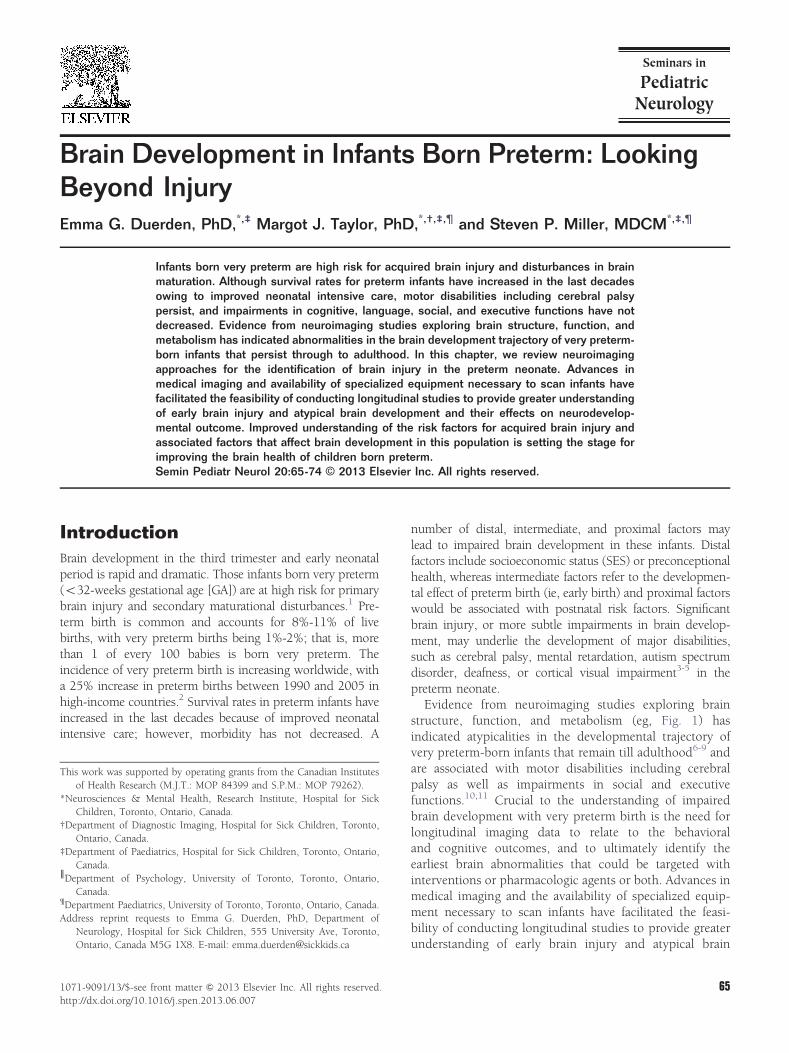
Figure 1 Brain structure, function, and metabolism measured inpreterm-born infants. Brain imaging data acquired in preterm-born infants (o32 weeks). Information about brain macrostruc-ture or microstructure can be obtained through anatomical MRI(T1- and T2-weighted images) and diffusion-tensor imaging (DTI).Top left: T1-weighted imaging permits the identification of white-matter injury as areas of abnormal hyperintensity in the whitematter (blue arrows). Data were acquired in a 31-week-oldneonate. Top middle: T2-weighted MRI of a 29-week-old infant.T2-weighted images may allow better assessment of corticalanatomy. Top right: DTI in a 28-week-old infant. Color mapsrepresent the diffusion of water along the principle eigenvectors inwhite-matter fiber pathways and cerebral cortex (red: left to right;green: anterior to posterior; blue: superior to inferior). MRIsequences can also be used to study brain function and metabo-lism. Bottom left: Functional MRI demonstrating activation in theprimary visual cortex in a preterm-born infant. Bottom right:Measures of brain metabolites that are indices of neuronalmaturation. Cho, choline; Cr, Creatine; La, lactate.
E.G. Duerden, M.J. Taylor and S.P. Miller66
arborize to form interconnected neural dependencies. Intypical and atypical development reductions in cortical FAmirror these developments of neuronal and glial processes.28,29
Magnetization Transfer Ratio (MTR)MTR is another MRI sequence that is used to assess white-matter integrity in the brain30 (Fig. 2). MTR permits theindirect observation of semisolid macromolecular compo-nents of tissue, such as protein matrices and cell mem-branes,31 and is particularly sensitive to myelin.32 MTRvalues are found to increase with age in the neonate,consistent with processes of myelination and matura-tion.33-36 Compared with DTI, the changes seen by MTRare more affected by myelination, whereas DTI is morestrongly affected by axonal development.35
MR Spectroscopy (MRS) ImagingMRS is an imaging technique that measures signals fromlow-concentration brain metabolites. The sequences can beperformed in 7-15 minutes. Metabolites in brain tissue thatcan be detected include lactate; choline and choline-containing compounds; creatine, phosphoryl-creatine, andN-acetylaspartate (NAA); glutamine; glutamate; gamma-aminobutyric acid; and myoinositol. Brain metabolite levelsmay be indicative of neuronal functioning. For instance,levels of NAA are thought to reflect neuronal integrity orfunction or both, creatine is involved in the creatine-kinasereaction, lactate is a measure of anaerobic metabolism, andcholine-containing compound is involved in the synthesisand breakdown of cellular membranes.MRS is a useful clinical and research tool that can monitor
brain metabolism after injury,37 and aid in the under-standing of the development of brain metabolism in term-born and preterm-born infants.38-40 For example, MRS hasbeen shown to be a useful predictor of poor neurodeve-lopmental outcome in asphyxiated term-born infants.41,42
These studies have consistently reported that increasedlactate and decreased NAA were associated with worseoutcomes. The elevated lactate levels are thought to indicatea disturbance in cerebral oxidative metabolism, whereasreduced NAA levels indicate delayed cellular maturation,
cellular dysfunction, or cell death. (For a review of this area,see Card et al.43)
Functional NeuroimagingBrain function in infants can be studied noninvasively usingelectroencephalography (EEG), magnetoencephalography(MEG), functional MRI (fMRI), and near-infrared spectro-scopy (NIRS).
EEGEEG is the oldest functional neuroimaging technique andmeasures electrical activity of neurons through the place-ment of electrodes on the scalp. EEG measures the currentflow emitted by the synaptic interchanges produced by thedendrites of pyramidal neurons from large populationsof neurons (�500,000-1,000,000). EEG is a techniquewith very high temporal resolution permitting the studyof neuronal events occurring at the millisecond level,although its spatial resolution is relatively poor (�1-2 cm).EEG has been used widely to study infant brain develop-
ment given its noninvasive nature and feasibility. EEGstudies have indicated that electrical activity parallels thatof structural development.44,45 EEG is commonly used tomonitor preterm-born infants such as those experiencingseizures or at risk of seizures,46-48 and studies havecorrelated EEG measures with neurodevelopmental outcomein preterm-term born infants.49,50
MEGMEG is similar to EEG in that it is a direct reflection ofon-going brain activity; it measures magnetic fields orthog-onal to the electrical activity evoked by populations ofneurons. MEG has excellent temporal and spatial resolution(�1-5 mm). Although it is more sensitive to movementthan EEG, it has proved useful for the study of infants.51,52
MEG data can also be used to understand brain connectivityand the development of coherent neural networks. MEGhas not been used widely to study the function of theinfant brain, which is due mainly to limited access tohardware, particularly infant-sized MEG helmets. However,
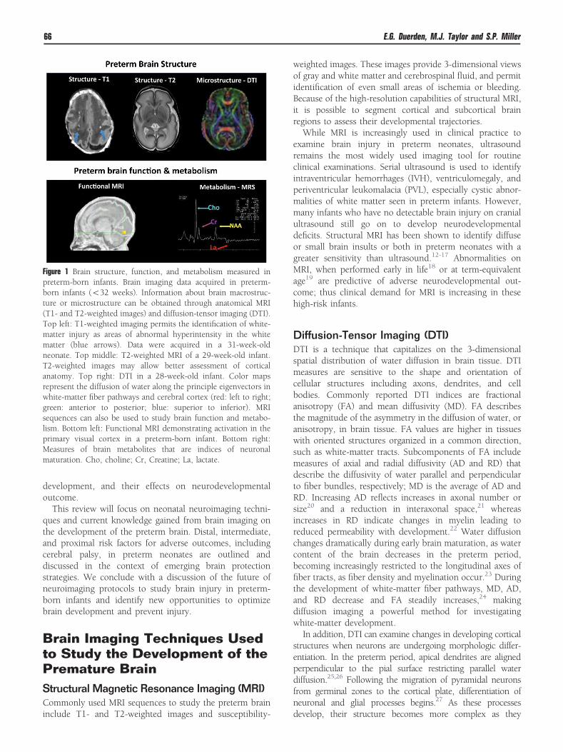
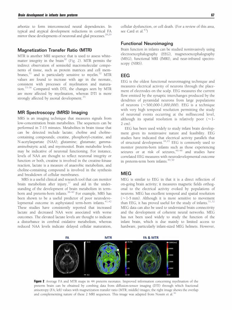
Figure 2 Average FA and MTR maps in 44 preterm neonates. Improved information concerning myelination of thepreterm brain can be obtained by combing data from diffusion-tensor imaging (DTI) through which fractionalanisotropy (FA; left) values with magnetization transfer ratio (MTR; middle) images; the right image shows the overlapand complementing nature of these 2 MRI sequences. This image was adapted from Nossin et al.36
Brain development in infants born preterm 67
somatosensory and auditory functions have been assessed inpreterm infants with MEG,53-56 and MEG has been used tostudy the longer-term effects of preterm birth on child andadolescent brain development.57-59
fMRIfMRI is an indirect measure of brain activity as it measureslocal blood flow occurring in response to increased neuronalactivity. fMRI measures the blood oxygen level–dependent(BOLD) signal that reflects the contrast between oxygenatedand deoxygenated hemoglobin. In response to neuralactivity an increase in oxygenated hemoglobin flows intocapillaries with a corresponding decrease in the amount ofdeoxygenated hemoglobin. This change in the ratio ofoxygenated and deoxygenated hemoglobin causes suscept-ibility in the magnetic field that is measurable using fMRIpulse sequences and can be visualized as an increase inimage intensity.60
fMRI activation in the visual cortex in response to visualstimuli has been reported in infants as young as 33-weeksGA.61,62 However, in very preterm infants (o32-weeks GA)reliable visual cortex activation is not usually evident,63
although BOLD activation in response to somatosensorystimuli is more reliably recorded.64
Another area of burgeoning interest in the preterm-borninfant is the study of resting state, measured by functionalconnectivity MRI.65,66 This technique identifies localizedlow frequency BOLD signals that are correlated when thebrain is at rest.67,68 The various patterns of activation inregions of the brain in the absence of stimulus-evokedactivity have been deemed resting-state networks.69 Brainregions that are functionally linked in resting-state networksare also anatomically connected.70
Resting-state networks have been established in typicallydeveloping children71,72 and atypical patterns in neuro-developmental disorders, such as attention-deficit hyper-activity disorder73 and autism spectrum disorder.74 In thepreterm infant brain, resting-state networks have also beenexplored to examine the development of cortical circuitsinvolved in basic neural processing and the inception ofhigher-order cognitive processes.66,75-82 These functionalconnectivity patterns mature in parallel with anatomicalconnectivity in the developing human brain, and may proveto be a valuable predictor of cognitive outcome.
NIRSNIRS is similar to fMRI in that it is sensitive to changes inoxygenated and deoxygenated hemoglobin and is thereforean indirect measure of neuronal activity. NIRS uses lightwavelengths (700-1000 nm) to measure tissue oxygenation,which reflects oxygen uptake in the tissue bed. NIRSinvolves putting probes on the head of the infants; theprobes emit light and also contain detectors to measure lightthat is reflected back from the superficial few centimetres ofbrain tissue. Advantages associated with the use of NIRS to
study brain development in infants are its ease of use, its lowcost, and its low sensitivity to head movement.83
Clinical applications for NIRS include monitoring peri-operative cerebral oxygenation in infants undergoing car-diopulmonary bypass surgery or during surgery to repairpatent ductus arteriosus. Additionally, NIRS also proved tobe a useful research tool in the preterm neonate to examinebaseline levels of regional cerebral blood oxygenation,84,85
and their relation to risk factors associated with adverseneurodevelopmental outcome,86 as well as normal develop-ment in infancy of cognitive processes.87
Early Brain Development inNeonates: Evidence FromNeuroimaging StudiesGray Matter DevelopmentThe burden of neurodevelopmental impairments, motor andcognitive, in preterm-born infants increases with earlierdegrees of prematurity.88 For example, infants born before25-weeks gestation show significantly more motor andcognitive deficits at 30 months than those born after 28-weeks gestation.89 By school age, many extremely preterm-born children continue to lag behind their peers in terms ofmotor90 and cognitive development, and these deficits aremore prevalent in boys than girls.91 The neurophysiologicalunderpinnings of these findings remain uncertain. Neuronalmigration into the cortical mantle is largely complete by 25weeks; however, glial migration, that is mainly responsiblefor the expansion in cortical surface area, continues throughthe third trimester.92-94 Cortical surface area is a reflection ofboth neuronal number and the folding of the corticalmantle. The folding process involves the migration ofneurons via radial glial cells to the cortical plate. Greaternumbers of radial glial cells would in turn allow for moreneurons to migrate and thus increase the overall surfacearea.95 This process may be stunted owing to the exposureto infection, hypoxia, and postnatal growth restriction96,97dueto under nutrition or epigenetic mechanisms.Evidence from structural MRI has indicated altered
cortical-folding patterns in extremely preterm-born chil-dren.98 In a series of 14 preterm-born infants cerebral tissuevolume, surface area and folding complexity were examinedin comparison with term-born infants. Volumes did notdiffer significantly between the preterm and term infants butcortical surface area and cortical-folding complexities wereless in the preterm infants. The authors concluded that thelack of volume change in the presence of reduced corticalcomplexity indicated either a disruption in the neuronalmigration process or a reflection of impaired migrationalong radial glia.99
A larger MRI study also examined the relation betweencortical surface area and total cerebral volume in a cohort ofpreterm-born infants (n ¼ 113, GA from 22-29 weeks100).They reported that in extremely preterm-born children thescaling component reflecting the relation between cortical
E.G. Duerden, M.J. Taylor and S.P. Miller68
surface area and cerebral volume was reduced comparedwith infants born later. These effects were more prominentwith males and were related to poorer neurodevelopmentmeasured at 2 years of age. The authors concluded that areduction in cortical growth was a reflection of reducedconnectivity rather than a paucity of neurons. Cortical scalingis dependent on the development of association fibers ratherthan an increase in neuronal number101 and disruptions inthe process may underlie neurocognitive deficits. In lateintrauterine development, the increase in association fibersand synapse formation plays a key role in the developmentof the cortex and alterations in these processes may result inatypical brain morphology in the preterm-born infants.Additional support for this hypothesis comes from animalmodels of preterm birth that have demonstrated thatalterations in the connectivity of the cortical subplate withthe thalamus result in disrupted cortical development.102,103
Subcortical DevelopmentSeveral MRI studies have also examined changes in sub-cortical structures in the preterm neonate. These studies havereported decreased volume in the thalami and lentiformnuclei104,105 in preterm infants imaged at term-equivalentage (37-44-weeks GA), indicating that development in theextrauterine environment may result in impaired growth ofthe subcortical nuclei. Furthermore, preterm-born infantswith supratentorial lesions (such as IVH or PVL) showedeven smaller subcortical volumes compared with preterm-born infants with no identifiable lesions.104 Subcorticallesions can be common in preterm-born infants and deter-mining their effect on brain and cognitive developmentaloutcome is essential.Another more recent MRI study extended their analyses
to examine the relation of thalamic volume with that ofother brain structures106 in 71 preterm-born infants (23-35-weeks GA), imaged at term-equivalent age. An associationwas apparent between increasing prematurity and bilateralreduced volume of the thalamus, hippocampi, orbitofrontal,and posterior cingulate cortices. Microstructural changes inthe thalamus and in thalamocortical tracts measured usingDTI, suggested disruptions in thalamocortical connectivityor reductions in neuronal or axonal numbers.
White-Matter DevelopmentAbnormalities in white matter, measured using DTI, havebeen reported in preterm infants at term age107 and on long-term follow-up108 that correlate with dysfunction. Inpreterm-born infants, white-matter tracts exhibit loweranisotropy and higher diffusion compared with full-terminfants.109 Preterm-born infants also demonstrate increasedRD and AD relative to term-born children,110 a finding thatmay reflect premyelination changes, such as increasedmembrane permeability and a reduced axonal caliber.Although alterations in white-matter integrity, such asreductions in FA, are often associated with evidence of earlywhite-matter injury in preterm infants, those infants without
neuroimaging evidence of injury also exhibit maturationaldifferences in white-matter architecture as they developfrom early in life to term-equivalent age.40,111
The importance of studying white-matter development inpreterm-born infants is crucial to understand impairedneurodevelopmental and motor outcome. White-matterinjury is common in infants born preterm. PVL wasclassically one of the most common injuries of the pretermbrain, although the incidence has decreased significantly inrecent years.112,113 PVL often precedes neurodevelopmentdisorders, such as cerebral palsy, cortical visual impairment,and impaired cognitive functions.113,114 Although the majorPVL lesions are less common now, white-matter injury isstill frequent and significantly related to morbidity. PVL andwhite-matter lesions are believed to arise at least in part fromischemia and inflammation owing to perinatal or postnatalinfection and are associated with disruptions in myelinationof the developing brain.1 Postmortem and experimental datahave suggested that PVL is related to the arrest of preoligo-dendrocyte development.115-117 A DTI study assessingpreterm-born infants with PVL noted decreased FA inseveral major white-matter fiber pathways and lower FAvalues were associated with greater cognitive impairment.118
These underlying neuropathologic changes in white matter,which affect oligodendroglial maturation and premyelina-tion processes, are critical to our understanding of the motordeficits seen in the preterm-born population.
Risk Factors for AdverseOutcomes in Preterm-BornChildrenPreterm birth is one of the leading causes of infant mortalityin the developed world.119-121 Although much research hasfocussed on the etiology of preterm birth, few preventivestrategies have emerged and the rates of preterm birthcontinue to rise in industrialized countries. A number ofdistal (socioeconomic), intermediate (maturational), andproximal (perinatal and neonatal) factors have emergedthat have been associated with poor neurodevelopmentaloutcome.
Distal FactorsFewer years of education,122 social disadvantage,123 andlower SES are significant risk factors for preterm birth.124
Furthermore, infants born into families with lower SES havehigher neonatal morbidity and mortality.125-127 Althoughthe relation between SES and preterm birth remainsuncertain, poor nutrition and adverse living conditions thatare more common in individuals with lower economicmeans may be contributory.Acute and chronic stress in mothers is believed to be a
significant risk factor for preterm birth and poor neuro-developmental outcome.128,129 The stress hormone pathway,the hypothalamic-pituitary-adrenal axis, is often altered inmothers who deliver early. Serum or plasma levels of stress
Brain development in infants born preterm 69

hormones including cortisol and corticotropin-releasinghormone measured in early pregnancy are associated withpreterm delivery130,131 and poor neurodevelopmental out-come.128 This may be owing to epigenetic mechanismsthat regulate gene expression for the receptors that bindcortisol.132
Intermediate Factors (MaturationalInfluences)Extremely Early BirthOwing to advances in perinatal care, survival rates ofextremely low-birth-weight infants born after o26 weeksgestation have increased,133,134 but they are at greater riskfor neonatal morbidity, neurodevelopmental impairments,and long-term adverse outcomes.134-138 The poor outcomesof these infants are often related to GA at birth, the lack ofplacental nutrients, inadequate postnatal nutrients, illness,and exposure to stress in the extrauterine environment. Yetrecent evidence suggests that the altered white-matterintegrity attributed to extreme preterm birth may relate tothe higher burden of systemic illness faced by these babiesrather than the degree of prematurity itself.139
Early Brain InjuryEarly brain injury and illness are common in the preterm-born neonate. Several imaging studies have examined white-matter integrity in infants with brain injury. For example, ina study of 55 preterm neonates studied serially withcorticospinal tract (CST) DT tractography (DTT), pretermneonates with evidence of moderate to severe white-matterabnormalities on MRI and those with postnatal infectionshad impaired development of the CST as they developed toterm-equivalent age.140 FA values increased at slower ratescompared with infants without evidence of injury or illness.These findings of very early white-matter damage, predictinglater development of primary motor pathways (ie, CST),underscores the possibilities for early interventions.In addition to alterations in white matter, the develop-
ment of the cerebellum has been shown to be impaired ininfants with early brain injury. Tam et al.141 collected DTIdata from 38 preterm newborns to investigate the relationbetween changes in the cerebellum and supratentorialmanifestations of injury (IVH or white-matter injury). SevereIVH was associated with higher MD and lower FA in themedial cerebellar peduncles and deep nuclear hila, andlower MD in the cerebellar cortex. White-matter injury wasnot significantly related to changes in diffusivity. Cerebellarvolume was reduced in infants with IVH, even in infantswho had mild IVH without significant supratentorial lesions,suggesting that blood products in the cerebrospinal fluiddisrupt cerebellar growth.142 Although PVL has previouslybeen associated with smaller cerebellar volumes,143 white-matter injury in this cohort was not associated withimpaired cerebellar growth, a finding the authors suggestedmay have been due to the relatively mild brain damage seenin their cohort.
Proximal VariablesProximal variables, such as postnatal infection, are detri-mental to brain development.40,140,144 Early severity ofillness, as measured using the clinical risk index for babies,predicted a reduction in cortical surface area.145 White-matter abnormalities were also associated with brain injury,postnatal infection,140 and illness severity in preterm-borninfants.146 Additionally, greater early illness severity (meas-ured using the Score for Neonatal Acute Physiology-II) werenegatively associated with FA values from the CST inpreterm-born infants.147
Related to the severity of illness is the repeated exposureto noxious stimuli in the neonatal intensive care unit.Critically ill infants are often intubated, receive numerousheel lances, and may experience postoperative pain. Stevenset al.148 reported an interaction between severity of illnessand behavioral pain responses, indicating that critically illinfants who receive noxious stimuli may have lower painthresholds. Using evoked potentials, preterm infants whoexperienced at least 40 days of intensive care were found tohave increased brain neuronal responses to noxious stimulicompared with healthy newborns at the same postmenstrualage.149 In turn, findings from a multimodal-imaging studyindicated that increased exposure to noxious stimuli wasassociated with reduced FA in white matter and reducedNAA in the subcortical gray matter.150 Together, thesestudies indicate that early painful procedures are associatedwith atypical brain development that may contribute toimpaired neurodevelopment in preterm-born infants, andsuggest new opportunities to improve outcomes throughneonatal intensive care strategies.
NeuroprotectionWith the steady rise in the number of infants being bornpreterm, the identification of neuroprotective agents toprevent brain injury is essential. Antenatal treatment withmagnesium sulfate can reduce the risk of infant death andcerebral palsy.151 Preliminary evidence in experimentalmodels also suggests that antenatal treatment with magne-sium sulfate is associated with fewer white-matter inju-ries.152 Future work in this area is needed to understand theimmediate and long-term effects of antenatal magnesiumsulfate on preterm neural and cognitive development. Theapplication of advanced brain imaging in the pretermneonate has also identified new potential avenues toimprove brain health, such as the prevention of infection,and the reduction in painful procedures. The increasedknowledge of the associated risk factors for poor outcome inpreterm-born children will aid in the development ofneuroimaging protocols, including combining multimodal-imaging techniques, which can be used to identify bio-markers that can be targeted therapeutically. Furthermore,neuroimaging will play an important role in monitoring theefficacy of interventions on brain development and newprotocols could serve as a bridge to identify brain atypical-ities in both infants and animal models of preterm birth.153
E.G. Duerden, M.J. Taylor and S.P. Miller70
Conclusions: Advances BeyondWhite-Matter VulnerabilityAdvances in medical imaging techniques have providedessential information on brain structure, function, andmetabolism in the preterm brain. It is increasingly clearthat the preterm brain is vulnerable to acquired injuries thatare not limited to the white matter, as well as abnormalitiesin the maturation of the white matter, cerebral cortex,subcortical nuclei (basal ganglia and thalamus), and cer-ebellum154 and their connectivity. Still questions remainregarding the optimal application of neuroimaging bio-markers to robustly predict neurodevelopmental outcome.Future research in this area is needed to combinemultimodal-imaging techniques to identify infants who areat risk and would benefit from emerging interventions toimprove brain health in the critical preterm period. Theapplication of advanced MR imaging techniques could befurther expanded to evaluate and monitor neuroprotectivestrategies. Additionally, long-term longitudinal imagingstudies are needed to assess preterm-born children tillschool age and adolescence to better understand opportu-nities to promote adaptability and resilience. Improvedknowledge concerning the broader distal factors, in additionto more intermediate and proximal factors, are also essentialto improve the long-term brain health of children bornpreterm.
AcknowledgmentsS.P.M. is currently supported by the Bloorview Children'sHospital Chair in Paediatric Neuroscience, with previoussupport from a Tier 2 Canadian Research Chair in NeonatalNeuroscience, and Michael Smith Foundation for HealthResearch Scholar Award.
References1. Volpe JJ: Brain injury in premature infants: A complex amalgam of
destructive and developmental disturbances. Lancet Neurol8:110-124, 2009
2. Behrman RE, Stith Butler A: Premature birth: Causes consequencesand prevention, Institute of Medicine, Committee on UnderstandingPremature Birth and Assuring Healthy Outcomes. Washington, D.C.,The National Academies Press, 2007
3. Hille ET, Weisglas-Kuperus N, van Goudoever JB, et al: Functionaloutcomes and participation in young adulthood for very preterm andvery low birth weight infants: The Dutch project on preterm andsmall for gestational age infants at 19 years of age. Pediatrics 120:e587-e595, 2007
4. Saigal S, Doyle LW: An overview of mortality and sequelae of pretermbirth from infancy to adulthood. Lancet 371:261-269, 2008
5. Synnes AR, Anson S, Arkesteijn A, et al: School entry age outcomesfor infants with birth weight o/¼ 800 grams. J Pediatr 157:989-994,2010. (e1)
6. Nagy Z, Lagercrantz H, Hutton C: Effects of preterm birth on corticalthickness measured in adolescence. Cereb Cortex 21:300-306, 2011
7. Allin MP, Kontis D, Walshe M, et al: White matter and cognition inadults who were born preterm. PLoS One 6:e24525, 2011
8. Ment LR, Kesler S, Vohr B, et al: Longitudinal brain volume changesin preterm and term control subjects during late childhood andadolescence. Pediatrics 123:503-511, 2009
9. Pyhala R, Lahti J, Heinonen K, et al: Neurocognitive abilities in youngadults with very low birth weight. Neurology 77:2052-2060, 2011
10. Lind A, Haataja L, Rautava L, et al: Relations between brain volumes,neuropsychological assessment and parental questionnaire in prema-turely born children. Eur Child Adolesc Psychiatry 19:407-417, 2010
11. Beauchamp MH, Thompson DK, Howard K, et al: Preterm infanthippocampal volumes correlate with later working memory deficits.Brain 131:2986-2994, 2008
12. Inder TE, Anderson NJ, Spencer C, Wells S, Volpe JJ: White matterinjury in the premature infant: A comparison between serial cranialsonographic and MR findings at term. Am J Neuroradiol 24:805-809,2003
13. Bigler ED, Tate DF, Neeley ES, et al: Temporal lobe, autism, andmacrocephaly. Am J Neuroradiol 24:2066-2076, 2003
14. Maalouf EF, Duggan PJ, Counsell SJ, et al: Comparison of findings oncranial ultrasound and magnetic resonance imaging in preterminfants. Pediatrics 107:719-727, 2001
15. Dyet LE, Kennea N, Counsell SJ, et al: Natural history of brain lesionsin extremely preterm infants studied with serial magnetic resonanceimaging from birth and neurodevelopmental assessment. Pediatrics118:536-548, 2006
16. Mirmiran M, Barnes PD, Keller K, et al: Neonatal brain magneticresonance imaging before discharge is better than serial cranialultrasound in predicting cerebral palsy in very low birth weightpreterm infants. Pediatrics 114:992-998, 2004
17. Miller SP, Cozzio CC, Goldstein RB, et al: Comparing the diagnosis ofwhite matter injury in premature newborns with serial MR imagingand transfontanel ultrasonography findings. Am J Neuroradiol24:1661-1669, 2003
18. Miller SP, Ferriero DM, Leonard C, et al: Early brain injury inpremature newborns detected with magnetic resonance imaging isassociated with adverse early neurodevelopmental outcome. J Pediatr147:609-616, 2005
19. Woodward LJ, Anderson PJ, Austin NC, et al: Neonatal MRI to predictneurodevelopmental outcomes in preterm infants. N Engl J Med355:685-694, 2006
20. Song SK, Sun SW, Ramsbottom MJ, et al: Dysmyelination revealedthrough MRI as increased radial (but unchanged axial) diffusion ofwater. NeuroImage 17:1429-1436, 2002
21. Takahashi M, Ono J, Harada K, et al: Diffusional anisotropy in cranialnerves with maturation: Quantitative evaluation with diffusion MRimaging in rats. Radiology 216:881-885, 2000
22. Suzuki Y, Matsuzawa H, Kwee IL, et al: Absolute eigenvalue diffusiontensor analysis for human brain maturation. NMR Biomed16:257-260, 2003
23. Partridge SC, Mukherjee P, Henry RG, et al: Diffusion tensor imaging:Serial quantitation of white matter tract maturity in prematurenewborns. Neuroimage 22:1302-1314, 2004
24. Salat DH, Tuch DS, Greve DN, et al: Age-related alterations in whitematter microstructure measured by diffusion tensor imaging. Neuro-biol Aging 26:1215-1227, 2005
25. Neil JJ, Shiran SI, McKinstry RC, et al: Normal brain in humannewborns: Apparent diffusion coefficient and diffusion anisotropymeasured by using diffusion tensor MR imaging. Radiology209:57-66, 1998
26. McKinstry RC, Mathur A, Miller JH, et al: Radial organization ofdeveloping preterm human cerebral cortex revealed by non-invasivewater diffusion anisotropy MRI. Cereb Cortex 12:1237-1243, 2002
27. Rakic P: A small step for the cell, a giant leap for mankind: Ahypothesis of neocortical expansion during evolution. Trends Neuro-sci 18:383-388, 1995
28. Mukherjee P, Miller JH, Shimony JS, et al: Diffusion-tensor MRimaging of gray and white matter development during normal humanbrain maturation. Am J Neuroradiol 23:1445-1456, 2002
29. Deipolyi AR, Mukherjee P, Gill K, et al: Comparing microstructuraland macrostructural development of the cerebral cortex in prematurenewborns: Diffusion tensor imaging versus cortical gyration. Neuro-image 27:579-586, 2005
Brain development in infants born preterm 71
30. Wolff SD, Balaban RS: Magnetization transfer contrast (MTC) andtissue water proton relaxation in vivo. Magn Reson Med 10:135-144,1989
31. Grad J, Bryant RG: Nuclear magnetic cross-relaxation spectroscopy. JMagn Reson 90:1-8, 1990
32. Rademacher J, Engelbrecht V, Burgel U, et al: Measuring in vivomyelination of human white matter fiber tracts with magnetizationtransfer MR. Neuroimage 9:393-406, 1999
33. Xydis V, Astrakas L, Drougia A, et al: Myelination process in pretermsubjects with periventricular leucomalacia assessed by magnetizationtransfer ratio. Pediatr Radiol 36:934-939, 2006
34. Xydis V, Astrakas L, Zikou A, et al: Magnetization transfer ratio in thebrain of preterm subjects: Age-related changes during the first 2 yearsof life. Eur Radiol 16:215-220, 2006
35. Nossin-Manor R, Chung AD, Whyte HE, et al: Deep gray mattermaturation in very preterm neonates: Regional variations and pathol-ogy-related age-dependent changes in magnetization transfer ratio.Radiology 263:510-517, 2012
36. Nossin-Manor R, Card D, Morris D, et al: Quantitative MRI in the verypreterm brain: Assessing tissue organization and myelination usingmagnetization transfer, diffusion tensor and T(1) imaging. Neuro-image 64:505-516, 2013
37. Robertson NJ, Kuint J, Counsell TJ, et al: Characterization of cerebralwhite matter damage in preterm infants using 1H and 31P magneticresonance spectroscopy. J Cereb Blood Flow Metab 20:1446-1456,2000
38. Vigneron DB: Magnetic resonance spectroscopic imaging of humanbrain development. Neuroimaging Clin N Am 16:75-85, 2006. (viii)
39. Xu D, Bonifacio SL, Charlton NN, et al: MR spectroscopy ofnormative premature newborns. J Magn Reson Imaging 33:306-311,2011
40. Chau V, Poskitt KJ, McFadden DE, et al: Effect of chorioamnionitis onbrain development and injury in premature newborns. Ann Neurol66:155-164, 2009
41. Miller SP, Newton N, Ferriero DM, et al: Predictors of 30-monthoutcome after perinatal depression: Role of proton MRS and socio-economic factors. Pediatr Res 52:71-77, 2002
42. Hanrahan JD, Cox IJ, Edwards AD, et al: Persistent increases incerebral lactate concentration after birth asphyxia. Pediatr Res44:304-311, 1998
43. Card D, Nossin-Manor R, Moore AM, et al: Brain metaboliteconcentrations associated with illness severity scores and white matterabnormalities in very preterm infants. Pediatric Res 74:75-81.doi:10.1038/pr.2013.62. Epub 2013 Apr 10.
44. Niedermeyer E: Ultra fast EEG activities and their significance. ClinEEG Neurosci 36:257-262, 2005
45. Vanhatalo S, Kaila K: Development of neonatal EEG activity: Fromphenomenology to physiology. Semin Fetal Neonatal Med11:471-478, 2006
46. Pisani F, Barilli AL, Sisti L, et al: Preterm infants with video-EEGconfirmed seizures: Outcome at 30 months of age. Brain Dev30:20-30, 2008
47. Rando T, Ricci D, Luciano R, et al: Prognostic value of EEG performedat term age in preterm infants. Childs Nerv Syst 22:263-269, 2006
48. Pisani F, Leali L, Parmigiani S, et al: Neonatal seizures in preterminfants: Clinical outcome and relationship with subsequent epilepsy. JMatern Fetal Neonatal Med 16:51-53, 2004 (suppl 2)
49. McAnulty GB, Duffy FH, Butler SC, et al: Effects of the NewbornIndividualized Developmental Care and Assessment Program (NID-CAP) at age 8 years: Preliminary data. Clin Pediatr (Phila) 49:258-270,2010
50. Okumura A, Hayakawa F, Kato T, et al: Developmental outcome andtypes of chronic-stage EEG abnormalities in preterm infants. Dev MedChild Neurol 44:729-734, 2002
51. Cheour M, Imada T, Taulu S, et al: Magnetoencephalography isfeasible for infant assessment of auditory discrimination. Exp Neurol190:S44-S51, 2004 (suppl 1)
52. Lutter WJ, Maier M, Wakai RT: Development of MEG sleep patternsand magnetic auditory evoked responses during early infancy. ClinNeurophysiol 117:522-530, 2006
53. Nevalainen P, Pihko E, Metsaranta M, et al: Does very premature birthaffect the functioning of the somatosensory cortex?—A magneto-encephalography study. Int J Psychophysiol 68:85-93, 2008
54. Pihko E, Lauronen L: Somatosensory processing in healthy newborns.Exp Neurol 190:S2-S7, 2004 (suppl 1)
55. Draganova R, Eswaran H, Murphy P, et al: Serial magnetoencephalo-graphic study of fetal and newborn auditory discriminative evokedresponses. Early Hum Dev 83:199-207, 2007
56. Leipala JA, Partanen E, Kushnerenko E, et al: Perinatal cerebral insultsalter auditory event-related potentials. Early Hum Dev 87:89-95, 2011
57. Doesburg SM, Ribary U, Herdman AT, et al: Altered long-range phasesynchronization and cortical activation in children born very preterm.IFMBE Proc 29:250-253, 2010
58. Doesburg SM, Ribary U, Herdman AT, et al: Magnetoencephalographyreveals slowing of resting peak oscillatory frequency in children bornvery preterm. Pediatr Res 70:171-175, 2011
59. Frye RE, Malmberg B, McLean J 3rd, et al: Increased left prefrontalactivation during an auditory language task in adolescents bornpreterm at high risk. Brain Res 1336:89-97, 2010
60. Heeger DJ, Ress D: What does fMRI tell us about neuronal activity?Nat Rev Neurosci 3:142-151, 2002
61. Lachaux JP, Rodriguez E, Martinerie J, et al: Measuring phasesynchrony in brain signals. Hum Brain Mapp 8:194-208, 1999
62. Yamada H, Sadato N, Konishi Y, et al: A milestone for normaldevelopment of the infantile brain detected by functional MRI.Neurology 55:218-223, 2000
63. Lee W, Donner EJ, Nossin-Manor R, et al: Visual functional magneticresonance imaging of preterm infants. Dev Med Child Neurol54:724-729, 2012
64. Arichi T, Fagiolo G, Varela M, et al: Development of BOLD signalhemodynamic responses in the human brain. Neuroimage63:663-673, 2012
65. Smyser CD, Snyder AZ, Neil JJ: Functional connectivity MRI ininfants: Exploration of the functional organization of the developingbrain. Neuroimage 56:1437-1452, 2011
66. Doria V, Beckmann CF, Arichi T, et al: Emergence of resting statenetworks in the preterm human brain. Proc Natl Acad Sci U S A107:20015-20020, 2010
67. Biswal B, Yetkin FZ, Haughton VM, Hyde JS: Functional connectivityin the motor cortex of resting human brain using echo-planar MRI.Magn Reson Med 34:537-541, 1995
68. Ballard CG, O'Brien J, Lowery K, et al: A prospective study ofdementia with Lewy bodies. Age Ageing 27:631-636, 1998
69. Cohen AL, Fair DA, Dosenbach NU, et al: Defining functional areas inindividual human brains using resting functional connectivity MRI.Neuroimage 41:45-57, 2008
70. Pinotsis DA, Hansen E, Friston KJ, et al: Anatomical connectivity andthe resting state activity of large cortical networks. NeuroImage65:127-138, 2013
71. Fair DA, Dosenbach NU, Church JA, et al: Development of distinctcontrol networks through segregation and integration. Proc Natl AcadSci U S A 104:13507-13512, 2007
72. Wiggins JL, Bedoyan JK, Peltier SJ, et al: The impact of serotonintransporter (5-HTTLPR) genotype on the development of resting-statefunctional connectivity in children and adolescents: A preliminaryreport. Neuroimage 59:2760-2770, 2012
73. Wang L, Zhu C, He Y, et al: Altered small-world brain functionalnetworks in children with attention-deficit/hyperactivity disorder.Hum Brain Mapp 30:638-649, 2009
74. Di Martino A, Kelly C, Grzadzinski R, et al: Aberrant striatal functionalconnectivity in children with autism. Biol Psychiatry 69:847-856, 2011
75. Fransson P, Skiold B, Horsch S, et al: Resting-state networks in theinfant brain. Proc Natl Acad Sci U S A 104:15531-15536, 2007
76. Lin W, Zhu Q, Gao W, et al: Functional connectivity MR imagingreveals cortical functional connectivity in the developing brain. Am JNeuroradiol 29:1883-1889, 2008
E.G. Duerden, M.J. Taylor and S.P. Miller72
77. Liu WC, Flax JF, Guise KG, et al: Functional connectivity of thesensorimotor area in naturally sleeping infants. Brain Res 1223:42-49,2008
78. Fransson P, Skiold B, Engstrom M, et al: Spontaneous brain activity inthe newborn brain during natural sleep—An fMRI study in infantsborn at full term. Pediatr Res 66:301-305, 2009
79. Gao W, Zhu H, Lin W: A unified optimization approach for diffusiontensor imaging technique. Neuroimage 44:729-741, 2009
80. Damaraju E, Phillips JR, Lowe JR, et al: Resting-state functionalconnectivity differences in premature children. Front Syst Neurosci 4.pii:23, 2010. doi:10.3389/fnsys.2010.00023.
81. Fransson P, Aden U, Blennow M, et al: The functional architecture ofthe infant brain as revealed by resting-state fMRI. Cereb Cortex21:145-154, 2011
82. Smyser CD, Inder TE, Shimony JS, et al: Longitudinal analysis ofneural network development in preterm infants. Cereb Cortex20:2852-2862, 2010
83. Cui X, Bray S, Reiss AL: Functional near infrared spectroscopy (NIRS)signal improvement based on negative correlation between oxy-genated and deoxygenated hemoglobin dynamics. Neuroimage49:3039-3046, 2010
84. Wijbenga RG, Lemmers PM, van Bel F: Cerebral oxygenation duringthe first days of life in preterm and term neonates: Differencesbetween different brain regions. Pediatr Res 70:389-394, 2011
85. Bokiniec R, Zbiec A, Seliga J, et al: Assessment of brain oxygenation interm and preterm neonates using near infrared spectroscopy. AdvMed Sci 16:1-8, 2012
86. Zhang Y, Chan GS, Tracy MB, et al: Spectral analysis of systemic andcerebral cardiovascular variabilities in preterm infants: Relationshipwith clinical risk index for babies (CRIB). Physiol Meas32:1913-1928, 2011
87. Nakato E, Otsuka Y, Kanazawa S, et al: Distinct differences in thepattern of hemodynamic response to happy and angry facialexpressions in infants—A near-infrared spectroscopic study. Neuro-image 54:1600-1606, 2011
88. Bhutta AT, Cleves MA, Casey PH, et al: Cognitive and behavioraloutcomes of school-aged children who were born preterm: A meta-analysis. J Am Med Assoc 288:728-737, 2002
89. Stephens BE, Vohr BR: Neurodevelopmental outcome of the prema-ture infant. Pediatr Clin North Am 56:631-646, 2009
90. Zwicker JG, Yoon SW, Mackay M, et al: Perinatal and neonatalpredictors of developmental coordination disorder in very low birth-weight children. Arch Dis Child 98:118-122, 2013
91. Marlow N, Wolke D, Bracewell MA, et al: Neurologic and devel-opmental disability at six years of age after extremely preterm birth. NEngl J Med 352:9-19, 2005
92. Rakic P: Neuronal migration and contact guidance in the primatetelencephalon. Postgrad Med J 54:25-40, 1978 (suppl 1)
93. Childs AM, Ramenghi LA, Cornette L, et al: Cerebral maturation inpremature infants: Quantitative assessment using MR imaging. Am JNeuroradiol 22:1577-1582, 2001
94. Barkovich AJ, Raybaud CA: Neuroimaging in disorders of corticaldevelopment. Neuroimaging Clin N Am 14:231-254, 2004
95. Rakic P, Caviness VS Jr.: Cortical development: View from neuro-logical mutants two decades later. Neuron 14:1101-1104, 1995
96. Dean JM, McClendon E, Hansen K, et al: Prenatal cerebral ischemiadisrupts MRI-defined cortical microstructure through disturbances inneuronal arborization. Sci Transl Med. 2013 Jan 16;5(168):168ra7.doi:10.1126/scitranslmed.3004669.
97. Vinall J, Grunau RE, Brant R, et al: Slower postnatal growth isassociated with delayed cerebral cortical maturation in pretermnewborns. Sci Transl Med. 2013 Jan 16;5(168):168ra8.doi:10.1126/scitranslmed.3004666.
98. Ajayi-Obe M, Saeed N, Cowan FM, et al: Reduced development ofcerebral cortex in extremely preterm infants. Lancet 356:1162-1163,2000
99. Marin O, Valiente M, Ge X, et al: Guiding neuronal cell migrations.Cold Spring Harb Perspect Biol. 2(2):a001834, 2010. doi:10.1101/cshperspect.a001834.
100. Kapellou O, Counsell SJ, Kennea N, et al: Abnormal corticaldevelopment after premature birth shown by altered allometric scalingof brain growth. PLoS Med 3:e265, 2006
101. Zhang K, Sejnowski TJ: A universal scaling law between gray matterand white matter of cerebral cortex. Proc Natl Acad Sci 97:5621-5626,2000
102. McQuillen PS, Sheldon RA, Shatz CJ, et al: Selective vulnerability ofsubplate neurons after early neonatal hypoxia-ischemia. J Neurosci23:3308-3315, 2003
103. Kanold PO, Kara P, Reid RC, et al: Role of subplate neurons infunctional maturation of visual cortical columns. Science301:521-525, 2003
104. Srinivasan L, Dutta R, Counsell SJ, et al: Quantification of deep graymatter in preterm infants at term-equivalent age using manualvolumetry of 3-tesla magnetic resonance images. Pediatrics119:759-765, 2007
105. Boardman JP, Counsell SJ, Rueckert D, et al: Abnormal deep greymatter development following preterm birth detected using deforma-tion-based morphometry. Neuroimage 32:70-78, 2006
106. Ball G, Boardman JP, Rueckert D, et al: The effect of preterm birth onthalamic and cortical development. Cerebral Cortex 22:1016-1024,2012
107. Bassi L, Ricci D, Volzone A, et al: Probabilistic diffusion tractographyof the optic radiations and visual function in preterm infants at termequivalent age. Brain 131:573-582, 2008
108. Nagy Z, Westerberg H, Skare S, et al: Preterm children havedisturbances of white matter at 111 years of age as shown bydiffusion tensor imaging. Pediatr Res 54:672-679, 2003
109. Counsell SJ, Allsop JM, Harrison MC, et al: Diffusion-weightedimaging of the brain in preterm infants with focal and diffuse whitematter abnormality. Pediatrics 112:1-7, 2003
110. Counsell SJ, Shen Y, Boardman JP, et al: Axial and radial diffusivity inpreterm infants who have diffuse white matter changes on magneticresonance imaging at term-equivalent age. Pediatrics 117:376-386, 2006
111. Miller SP, Vigneron DB, Henry RG, et al: Serial quantitativediffusion tensor MRI of the premature brain: Development innewborns with and without injury. J Magn Reson Imaging16:621-632, 2002
112. Hamrick SE, Miller SP, Leonard C, et al: Trends in severe brain injuryand neurodevelopmental outcome in premature newborn infants: Therole of cystic periventricular leukomalacia. J Pediatr 145:593-599, 2004
113. van Haastert IC, Groenendaal F, Uiterwaal CS, et al: Decreasingincidence and severity of cerebral palsy in prematurely born children.J Pediatr 159:86-91 e1, 2011
114. Follett PL, Rosenberg PA, Volpe JJ, et al: NBQX attenuates excitotoxicinjury in developing white matter. J Neurosci 20:9235-9241, 2000
115. Riddle A, Dean J, Buser JR, et al: Histopathological correlates ofmagnetic resonance imaging-defined chronic perinatal white matterinjury. Ann Neurol 70:493-507, 2011
116. Back SA, Luo NL, Borenstein NS, et al: Late oligodendrocyte progen-itors coincide with the developmental window of vulnerability forhuman perinatal white matter injury. J Neurosci 21:1302-1312, 2001
117. Buser JR, Maire J, Riddle A, et al: Arrested preoligodendrocytematuration contributes to myelination failure in premature infants.Ann Neurol 71:93-109, 2012
118. Wang S, Fan G, Xu K, et al: Potential of diffusion tensor MR imagingin the assessment of cognitive impairments in children with periven-tricular leukomalacia born preterm. Eur J Radiol 82:158-164, 2013
119. Goldenberg RL, Culhane JF: Low birth weight in the United States.Am J Clin Nutr 85:584S-590SS, 2007
120. McCormick MC: The contribution of low birth weight to infantmortality and childhood morbidity. N Engl J Med 312:82-90, 1985
121. Simhan HN, Caritis SN: Prevention of preterm delivery. N Engl J Med357:477-487, 2007
122. Gwin KM, Schrader R, Peters K, et al: An exploratory study of thevariables impacting preterm birth rates in New Mexico. BMCPregnancy Childbirth 12:53, 2012
123. Weightman AL, Morgan HE, Shepherd MA, et al: Social inequalityand infant health in the UK: Systematic review and meta-analyses.
Brain development in infants born preterm 73
BMJ Open. 2(3). pii: e000964, 2012. doi:10.1136/bmjopen-2012-000964
124. Peacock JL, Bland JM, Anderson HR: Preterm delivery: Effects ofsocioeconomic factors, psychological stress, smoking, alcohol, andcaffeine. BMJ 311:531-535, 1995
125. Martens PJ, Derksen S, Gupta S: Predictors of hospital readmission ofManitoba newborns within six weeks postbirth discharge: A popula-tion-based study. Pediatrics 114:708-713, 2004
126. Joseph KS, Marcoux S, Ohlsson A, et al: Changes in stillbirth andinfant mortality associated with increases in preterm birth amongtwins. Pediatrics 108:1055-1061, 2001
127. Calling S, Li X, Sundquist J, et al: Socioeconomic inequalities andinfant mortality of 46,470 preterm infants born in Sweden between1992 and 2006. Paediatr Perinat Epidemiol 25:357-365, 2011
128. Tu MT, Grunau RE, Petrie-Thomas J, et al: Maternal stress andbehavior modulate relationships between neonatal stress, attention,and basal cortisol at 8 months in preterm infants. Dev Psychobiol49:150-164, 2007
129. Coussons-Read ME, Lobel M, Carey JC, et al: The occurrence ofpreterm delivery is linked to pregnancy-specific distress and elevatedinflammatory markers across gestation. Brain Behav Immun26:650-659, 2012
130. Sandman CA, Wadhwa PD, Chicz-DeMet A, et al: Maternal stress,HPA activity, and fetal/infant outcome. Ann N Y Acad Sci814:266-275, 1997
131. Hobel CJ, Dunkel-Schetter C, Roesch SC, et al: Maternal plasmacorticotropin-releasing hormone associated with stress at 20 weeks'gestation in pregnancies ending in preterm delivery. Am J ObstetGynecol 180:S257-S263, 1999
132. Oberlander TF, Weinberg J, Papsdorf M, et al: Prenatal exposure tomaternal depression, neonatal methylation of human glucocorticoidreceptor gene (NR3C1) and infant cortisol stress responses. Epige-netics 3:97-106, 2008
133. Wilson-Costello D, Friedman H, Minich N, et al: Improved survivalrates with increased neurodevelopmental disability for extremely lowbirth weight infants in the 1990s. Pediatrics 115:997-1003, 2005
134. Eichenwald EC, Stark AR: Management and outcomes of very lowbirth weight. N Engl J Med 358:1700-1711, 2008
135. Hack M, Fanaroff AA: Outcomes of children of extremely lowbirthweight and gestational age in the 1990s. Semin Neonatol5:89-106, 2000
136. Wood NS, Marlow N, Costeloe K, et al: Neurologic and devel-opmental disability after extremely preterm birth. EPICure StudyGroup. N Engl J Med 343:378-384, 2000
137. Wood NS, Costeloe K, Gibson AT, et al: The EPICure study:Associations and antecedents of neurological and developmentaldisability at 30 months of age following extremely preterm birth.Arch Dis Child Fetal Neonatal Ed 90:F134-F140, 2005
138. Tyson JE, Parikh NA, Langer J, et al: Intensive care for extremeprematurity—Moving beyond gestational age. N Engl J Med358:1672-1681, 2008
139. Bonifacio SL, Glass HC, Chau V, et al: Extreme premature birth is notassociated with impaired development of brain microstructure. JPediatr 157:726-732, 2010. (e1)
140. Adams E, Chau V, Poskitt KJ, et al: Tractography-based quantitationof corticospinal tract development in premature newborns. J Pediatr156:882-888, 2010. (888 e1)
141. Tam EW, Ferriero DM, Xu D, et al: Cerebellar development in thepreterm neonate: Effect of supratentorial brain injury. Pediatr Res66:102-106, 2009
142. Tam EW, Miller SP, Studholme C, et al: Differential effects ofintraventricular hemorrhage and white matter injury on pretermcerebellar growth. J Pediatr 158:366-371, 2011
143. Limperopoulos C, Soul JS, Haidar H, et al: Impaired trophicinteractions between the cerebellum and the cerebrum among preterminfants. Pediatrics 116:844-850, 2005
144. Chau V, Brant R, Poskitt KJ, et al: Postnatal infection is associatedwith widespread abnormalities of brain development in prematurenewborns. Pediatr Res 71:274-279, 2012
145. Kaukola T, Kapellou O, Laroche S, et al: Severity of perinatal illnessand cerebral cortical growth in preterm infants. Acta Paediatr98:990-995, 2009
146. Horsch S, Hallberg B, Leifsdottir K, et al: Brain abnormalities inextremely low gestational age infants: A Swedish population basedMRI study. Acta Paediatr 96:979-984, 2007
147. Zwicker JG, Grunau R, Adams E, et al: Score for neonatal acutephysiology-II predicts corticospinal tract development in prematurenewborns. Pediatr Neurol 48:123-129.e1. doi:10.1016/j.pediatrneurol.2012.10.016
148. Stevens BJ, Johnston CC, Horton L: Factors that influence thebehavioral pain responses of premature infants. Pain 59:101-109, 1994
149. Slater R, Fabrizi L, Worley A, et al: Premature infants display increasednoxious-evoked neuronal activity in the brain compared to healthyage-matched term-born infants. Neuroimage 52:583-589, 2010
150. Brummelte S, Grunau RE, Chau V, et al: Procedural pain and braindevelopment in premature newborns. Ann Neurol 71:385-396, 2012
151. Magee L, Sawchuck D, Synnes A, et al: Magnesium sulphate for fetalneuroprotection. J Obstet Gynaecol Can 33:516-529, 2011
152. Burd I, Breen K, Friedman A, et al: Magnesium sulfate reducesinflammation-associated brain injury in fetal mice. Am J ObstetGynecol 202:292, 2010. (e1-e9)
153. Azzopardi D, Edwards AD: Magnetic resonance biomarkers of neuro-protective effects in infants with hypoxic ischemic encephalopathy.Semin Fetal Neonatal Med 15:261-269, 2010
154. Miller SP, Ferriero DM: From selective vulnerability to connectivity:Insights from newborn brain imaging. Trends Neurosci 32:496-505, 2009
E.G. Duerden, M.J. Taylor and S.P. Miller74