LUNG FUNCTION IN RELATION TO PRETERM BIRTH AND ...
78
From The Department of Clinical Science and Education, Södersjukhuset Karolinska Institutet, Stockholm, Sweden LUNG FUNCTION IN RELATION TO PRETERM BIRTH AND ASTHMA IN EARLY CHILDHOOD Per Thunqvist Stockholm 2016
-
Upload
khangminh22 -
Category
Documents
-
view
0 -
download
0
Transcript of LUNG FUNCTION IN RELATION TO PRETERM BIRTH AND ...

From The Department of Clinical Science and Education,
Södersjukhuset
Karolinska Institutet, Stockholm, Sweden
LUNG FUNCTION IN RELATION TO
PRETERM BIRTH AND ASTHMA IN EARLY
CHILDHOOD
Per Thunqvist
Stockholm 2016

All previously published papers were reproduced with permission from the publisher.
Published by Karolinska Institutet.
Printed by Eprint AB 2016
© Per Thunqvist, 2016
ISBN 978-91-7676-308-7
LUNG FUNCTION IN RELATION TO PRETERM BIRTH
AND ASTHMA IN EARLY CHILDHOOD
THESIS FOR DOCTORAL DEGREE (Ph.D.)
By
Per Thunqvist
Principal Supervisor:
Erik Melén
Karolinska Institutet
Institute of Environmental Medicine
Co-supervisor(s):
Jenny Hallberg
Karolinska Institutet
Institute of Environmental Medicine
Mikael Norman
Karolinska Institutet
Department of Clinical Science, Intervention
and Technology
Per M Gustafsson
University of Gothenburg
The Sahlgrenska Academy
Opponent:
Bill Hesselmar
University of Gothenburg
The Sahlgrenska Academy
Examination Board:
Kjell Alving
Uppsala University
Department of Women‘s and Children‘s
Health
Hans Hedenström
Uppsala University
Department of Medical Sciences, Clinical
Physiology
Baldvin Jonsson
Karolinska Institutet
Department of Women‘s and Children‘s
Health


A famous British professor, Andrew Bush, often emphasizes – Why? How? So what? – in
connection to research. The questions could be applied in wider perspectives and were central
to me when I decided to become a PhD-student.
Why should a middle-aged clinician, without the need for an additional career, double the
workload?
During the years as a clinician, I have often been concerned about the lack of knowledge on
how to care for and how to treat the respiratory symptoms seen in growing children born
preterm. In the evolving research friendly environment at Sachsska, I realized that I had the
possibility to in some way contribute to that ―black whole‖.
How?
The ―perfect storm‖ struck me. The hospital provided the infant lung function equipment. My
colleges Magnus and Gunnar got me started. Jenny and Erik were the perfect catalyst.
So what?
Personally, this has been a joyful experience and given me new insights, not only to research.
Hopefully, some of the results of the studies in the thesis actually put some new knowledge
into the ―black whole‖ I wanted to explore.
Per Thunqvist 2016


ABSTRACT
The prevalence of preterm birth (ie., before 37 weeks of gestation) is increasing and
estimated to be around 11 % worldwide. In high-income countries, survival is the most
probable outcome even after extreme preterm deliveries. Children born preterm exhibit
different stages of lung immaturity at birth and follow-up studies have shown lung function
impairment and respiratory morbidity, including asthma-like disease, during infancy and into
adulthood. Asthma or asthma-like disease is common in the general pediatric population.
Differentiation of early childhood asthma phenotypes demonstrates different underlying
pathophysiologic mechanisms and trajectories with age.
The general aims of the present thesis were:
- To investigate lung function from infancy to adolescence in relation to preterm birth and
asthma in early childhood.
- To investigate if similarities and differences in measured lung function after preterm birth
and among childhood asthma phenotypes may give information on mechanisms for the
obstructive patterns seen in these different conditions.
Longitudinal follow-up during the first 18 months of life of infants with bronchopulmonary
dysplasia (BPD) showed significant reductions of lung function in the cohort. Maximal
expiratory flows were on average below the 2.5th
percentile for both mild and
moderate/severe BPD at both 6 and 18 months, with the exception VmaxFRC at 6 months and
no improvement was seen between the two time-points. Compliance of the respiratory system
(COso) was the only lung function variable that differed statistically between mild and
moderate/severe BPD, with lower values for the more severe disease. Significant lower
values were consistently seen for Cso and all maximal expiratory variables in infants with
respiratory symptoms compared to infants without.
In a follow-up of children born extremely preterm (before gestational week 27), a subset of
the population based cohort EXPRESS, performed lung function at age 6½ years. The
extreme preterm children had lower forced expiratory volume in 1 sec (FEV1, z-score: -1.1,
95% CI: -1.4; -0.8) than the control group born at term. Impulse oscillometry (IOS)
measurements showed significantly higher peripheral airway resistance and reactance in
children born extremely preterm than in controls. In children born at 22-24 weeks of
gestation, 44% had FEV1 below the <5th
percentile.

Using the BAMSE cohort study, lung function after moderate-to-late preterm birth was
investigated at 8 and 16 years of age. At age 16 years, both genders in the preterm group
demonstrated lower FEV1 (female subjects: –116 mL [95 % CI: –212 to –20]; male subjects:
–177 mL [95 % CI: –329 to –25]) compared with the term group. IOS demonstrated higher
frequency dependence of resistance (R5-20) for male subjects (20.9 Pa・L–1・s−1 [95 % CI:
9.8 to 31.9]) compared with the term group. No catch up of lung function between ages 8 and
16 years was observed in either gender.
In the last study, children taking part in the BAMSE cohort study were categorized into
‗never asthma‘, ‗early transient asthma‘, ‗early persistent asthma‘, and ‗late onset asthma‘
and lung function data from the 8 and 16 year follow-ups was used to compare groups.
Compared with the never asthma group, all asthma groups were associated with lower FEV1
at 16 years of age (early transient—119 ml, 95% confidence interval 204 to 34; early
persistent—410 ml, 95 %CI 533; 287; and late onset—148 ml, 95%CI 237; 58). R5-20 was
significantly associated with active asthma at 16 years, but not transient asthma.
In conclusion:
- A significant proportion of children born very and extreme preterm have lung
function values below the normal range in infancy and at at early scool-age. Lung
function in adololescense is lower after moderate-to-late preterm birth than after term.
- Staging BPD severity did not predict lung function.
- Early measured lung function is associated with respiratory morbidity in children with
BPD.
- IOS identifies children with low lung function after preterm birth and those with
active pediatric asthma.
Clinical remark:
Measurements of lung function may identify children at risk for respiratory morbidity and
provide insights into long-term sequel of preterm birth. Regular assessments of lung function
from infancy, during childhood and possibly throughout life, are therefore suggested to be an
important tool when monitoring individuals born preterm.


LIST OF SCIENTIFIC PAPERS
I. Thunqvist Per, Gustafsson Per M, Norman Mikael, Wickman Magnus,
Hallberg Jenny.
Lung function at 6 and 18 months after preterm birth in relation to severity of
bronchopulmonary dysplasia. Pediatr Pulmonol. 2015 Oct;50(10):978-86.
II. Thunqvist Per, Tufvesson Ellen, Bjermer Leif, Fellman Vineta, Domellöf
Magnus, Melén Erik, Norman Mikael, Hallberg Jenny.
Lung function in 6-year-old children born extremely preterm -
a population-based cohort study (EXPRESS). Submitted.
III. Thunqvist Per, Gustafsson Per, Schultz Erica, Bellander Tom, Berggren-
Brostrom Eva, Norman Mikael, Wickman Magnus, Melén Erik, Hallberg
Jenny.
Lung Function at 8 and 16 years after Moderate-to-Late Preterm birth: A
Prospective Cohort Study. Pediatrics. 2016 Mar 23. pii: peds.2015-2056.
IV. Hallberg Jenny, Thunqvist Per, Schultz Erica S, Kull Inger, Bottai Matteo,
Merritt AS, Chiesa Flaminia, Gustafsson Per M, Melén Erik.
Asthma phenotypes and lung function up to 16 years of age-the BAMSE
cohort. Allergy. 2015 Jun;70(6):667-73.

CONTENTS
1 Introduction ..................................................................................................................... 1
2 Background ...................................................................................................................... 2
2.1 Normal lung development ..................................................................................... 2
2.2 Lung function after preterm birth .......................................................................... 4
2.3 Asthma phenotypes in early life in relation to lung function ............................... 6
2.4 Lung Function testing ............................................................................................ 7
2.4.1 Infant lung function testing (ILFT) ........................................................... 8
2.4.2 Dynamic spirometry ................................................................................ 10
2.4.3 Impulse oscillometry (IOS)..................................................................... 11
2.5 Treatment of obstructive disease ......................................................................... 13
3 Objectives ...................................................................................................................... 15
4 Subjects and methods .................................................................................................... 16
4.1 Study populations and design.............................................................................. 16
4.1.1 Preterm infant study population (Study 1) ............................................. 16
4.1.2 School-age preterm study population (Study 2) ..................................... 16
4.1.3 Moderate-to-late preterm (Study 3) and early childhood asthma
study populations (Study 4) .................................................................... 16
4.2 Definitions of study groups ................................................................................. 17
4.3 Definitions of asthma and respiratory outcomes ................................................ 19
4.4 Lung function tests .............................................................................................. 19
4.4.1 Infant lung function tests in Study 1 ....................................................... 19
4.4.2 Spirometry in Studies 2 – 4. .................................................................... 20
4.4.3 Impulse oscillometry (IOS) in Studies 2 – 4 .......................................... 20
4.4.4 Measures of bronchodilator response in Study 3 ................................... 21
4.5 Statistical methods ............................................................................................... 21
4.6 Ethical approvals ................................................................................................. 23
5 Results ............................................................................................................................ 24
5.1 Lung Function at 6 and 18 Months After Preterm Birth: Preterm Infant
Study (1) .............................................................................................................. 24
5.2 Lung function at 6½ years: School-age preterm study (2) ................................. 26
5.3 Lung function at 16 years after moderate-to-late preterm birth (3) ................... 28
5.4 Asthma phenotypes and lung function up to 16 years of ................................... 29
age: Early childhood asthma study (4) ......................................................................... 29
6 Discussion ...................................................................................................................... 32

6.1 Level of lung function after preterm birth .......................................................... 32
6.1.1 Maximal expiratory flows after preterm birth ........................................ 32
6.1.2 Lung volumes after preterm birth ........................................................... 33
6.2 BPD severity in relation to lung function ........................................................... 35
6.3 Longitudinal analysis of expiratory flows after preterm birth and early
bronchial obstructive symptoms ......................................................................... 36
6.4 Aspects of airway mechanics in relation to preterm birth and early
bronchial obstructive symptoms ......................................................................... 38
6.4.1 Compliance of the respiratory system (Crs) after preterm birth ............. 38
6.4.2 Impulse oscillometry after to preterm birth and in relation to
asthma phenotypes .................................................................................. 39
6.5 Clinical symptoms in relation to lung function after preterm birth ................... 40
6.6 Limitations and strengths .................................................................................... 42
7 Ethical considerations ................................................................................................... 43
8 Conclusions ................................................................................................................... 44
9 Clinical remarks ............................................................................................................ 45
10 Svensk sammanfattning ................................................................................................ 49
11 Acknowledgements ....................................................................................................... 55
12 References ..................................................................................................................... 57

LIST OF ABBREVIATIONS
BPD Bronchopulmonary Dysplasia
AX Area of reactance
BAMSE
CPAP
Swedish abbreviation for Children, Allergy, Milieu,
Stockholm, Epidemiology
Continuous Positive Airway Pressure
Cso Single occlusion method for compliance of the respiratory
system
EXPRESS Extremely Preterm Infants in Sweden Study
fdR Frequency dependence of resistance, R5-20
FEV0.5 Forced expiratory volume at 0.5 seconds
FEV1 Forced expiratory volume at 1 second
FOT Forced Oscillation Technique
FRC Functional Residual Capacity
FVC Forced Expiratory Volume
IFLT Infant Lung Function Test
IOS Impulse Oscillometry, a variant of FOT (see below)
MEF50 Maximal expiratory flow at 50 % of FVC
R20 Resistance at 20 Hz
R5 Resistance at 5 Hz
RVRTC Raised Volume Rapid Thoracic Compression, ―raised
squeeze‖
TVRTC Tidal Volume Rapid Thoracic Compression, ―tidal squeeze‖
V´maxFRC Maximal forced expiratory flow at FRC
X Reactance
Zrs Respiratory system impedance

1
1 INTRODUCTION
Early life exposures and insults to the respiratory system are recognized to have
consequences on respiratory health during childhood and in later life.(1, 2) Preterm birth is a
common event that has impact on lung function development and respiratory health during
childhood and into adulthood.(3, 4) The burden of health from preterm birth, defined as birth
before 37 (< 36+6) completed weeks of gestation, is substantial and increasing in most
regions of the world that have access to reliable data.(5) The prevalence of preterm birth is
estimated to be around 11 percent worldwide and ranging from 18 percent in some African
countries to five percent in high-income countries like Sweden. A further division of preterm
birth into extremely preterm (< 28 weeks), very preterm (28 to <32 weeks), and moderate-to-
late preterm (32 to <37 weeks) is important, since lower gestational age is associated with
increasing mortality and morbidity. The vast majority are moderate-to-late preterm births,
while 1 – 2 percent are born very or extremely preterm.(5, 6) In the Swedish national study
cohort EXPRESS (Extremely Preterm Infants in Sweden Study) the incidence of extremely
preterm birth (< 27 weeks of gestation) was 3/1 000 infants.(7) Further, infant survival is
nowadays the most probable outcome after extremely preterm birth (8) and the burden of
respiratory morbidity after preterm birth could therefore be assumed to increase in the future.
Very preterm birth (< 32 weeks of gestation) is associated with three times increased risk of
childhood wheezing disorder compared with term birth.(4) Respiratory disease after preterm
birth shares some clinical and pathophysiological features with asthma, such as airway
obstruction, airway hyper-responsiveness and susceptibility to respiratory wheezing.(9, 10)
Even though asthma is now described as a heterogeneous disease, it is usually characterized
by chronic airway inflammation (GINA guidelines 2015). Airway inflammation is not a
feature of the asthma-like disease seen after preterm birth and it has been suggested that the
word asthma should be avoided when describing the chronic pulmonary disease seen after
preterm birth.(11, 12)
Asthma or asthma-like syndrome after term birth is common in young children and
prevalence of current asthma is reported to be 8 – 9 % at school age.(13) Prevalence
estimations in younger children are often based on epidemiological studies that include data
on respiratory wheeze.(14) To aid in management and treatment of asthma or asthma-like
disease, a number of asthma phenotypes has been suggested.(15) Epidemiological studies
have identified asthma phenotypes with early onset of asthma-like symptoms and impairment

2
of lung function that track throughout life, not only in those with persisting symptoms, but
also after early transient symptoms.(16) The transient early wheeze phenotype is common
during the first years of life, with declining prevalence during the preschool period, and is not
associated with allergic inflammation.(17)
Whether or not asthma-like syndrome after preterm birth could be described as an asthma
phenotype is a matter of opinion. Anti-asthma drugs are commonly prescribed, though have
not yet been proven effective in treating the asthma-like symptoms in this growing group of
symptomatic children and adults. It is not known if the longstanding effects on lung function
development after preterm birth and in children with the transient early wheeze phenotype
share pathophysiological mechanisms, but the bronchial obstructive pattern and absence of
airway inflammation in both entities make comparisons reasonable.
2 BACKGROUND
2.1 NORMAL LUNG DEVELOPMENT
By volume, the lungs are the largest organs in the body, but consist of only half a liter of
tissue. The structures of the lungs separate approximately half a liter of blood from a certain
volume of air, which varies during the course of normal breathing. The configuration of the
lungs allows a minimum of tissue to create a fairly stable surface for gas exchange, at nearly
the size of a tennis court, inside a limited space (thorax cavity). The human airway tree
consists of on average 23 generations of branches. Generation 0 (trachea) to approximately
generation 15 are lined with bronchial epithelium and functionally named conducting
airways. In the conducting airways, which hold less than a tenth of the resting lung volume,
i.e.functional residual capacity, no gas exchange takes place. From airway generations 15 –
16, alveoli with gas-exchanging surfaces start to appear. As branching continues, the alveoli
increase in numbers, and in the last generation, alveolar sacs form blind endings.(18)
Generations 15 – 23 are called the acinar airways and hold the vast majority of the resting air
volume. Functionally, the acinar airways serve as the gas-exchanging zone. Bronchial
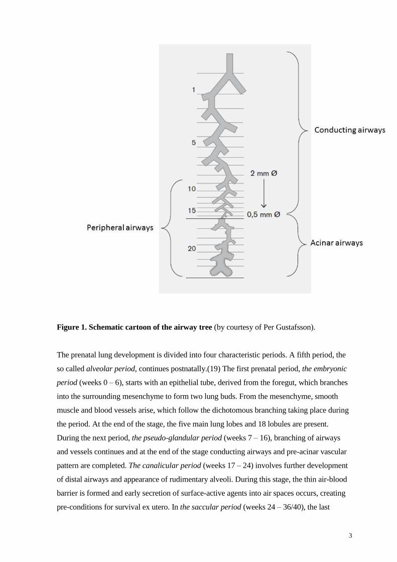
generations 8/9 to 23 are for practical reasons called peripheral or small airways.(Figure 1)
3
Figure 1. Schematic cartoon of the airway tree (by courtesy of Per Gustafsson).
The prenatal lung development is divided into four characteristic periods. A fifth period, the
so called alveolar period, continues postnatally.(19) The first prenatal period, the embryonic
period (weeks 0 – 6), starts with an epithelial tube, derived from the foregut, which branches
into the surrounding mesenchyme to form two lung buds. From the mesenchyme, smooth
muscle and blood vessels arise, which follow the dichotomous branching taking place during
the period. At the end of the stage, the five main lung lobes and 18 lobules are present.
During the next period, the pseudo-glandular period (weeks 7 – 16), branching of airways
and vessels continues and at the end of the stage conducting airways and pre-acinar vascular
pattern are completed. The canalicular period (weeks 17 – 24) involves further development
of distal airways and appearance of rudimentary alveoli. During this stage, the thin air-blood
barrier is formed and early secretion of surface-active agents into air spaces occurs, creating
pre-conditions for survival ex utero. In the saccular period (weeks 24 – 36/40), the last
4
generation of airways, the alveolar duct and, at the end of the period, the alveoli are formed.
True alveoli start to appear from week 32. The pulmonary surfactant system develops, starts
to mature and is increasingly active after the 32nd
gestational week. The last four to five
weeks before term, the alveolar period, is characterized by a large increase of gas-exchanging
surface. At birth alveolarization has barely started – numbers ranging from 50 to 150 million
alveoli have been suggested – and most of this further formation of alveoli takes place after
birth at term.(20) The length of the alveolar period has lately been debated. Previously,
alveoli have been thought to stop multiplying at between 2 – 7 years of age.(21) However,
recent human studies suggest that the process might continue into adulthood, which
completely changes the perspective on alveolar repair.(22, 23) The final numbers of alveoli in
adults are in the range of 300 to 800 million depending on lung size and sex.(24) From birth
and up to early adulthood (around 22 years of age in men and slightly earlier in females), the
lung size increases proportionally with body size and is affected by sex, age and
ethnicity.(25) This enormous development is described to represent a 30-fold increase in gas-
exchanging surface and at least a doubling of airway diameter and length.
2.2 LUNG FUNCTION AFTER PRETERM BIRTH
Understanding normal lung development is essential in order to comprehend the different
responses of the airways to insults during fetal life or after birth. The short version of this
section could be that ―the outcome of aberrant lung development depends on the type,
severity, and duration of insult, and the developmental stage at which it occurs.‖(20) Another
important cornerstone for the understanding is the concept of tracking lung function during
growth.(26, 27) This implies that infants born with impaired lung function, due to in utero or
extra-uterine insults, will usually remain at a low percentile for measured lung function from
childhood to adulthood.
Initially the effects of preterm birth were described in terms of airway epithelial metaplasia,
peribronchial fibrosis, vascular smooth muscle hyperplasia and radiologic findings in infants
suffering from severe respiratory distress syndrome. This description of the clinical picture
after preterm birth was named bronchopulmonary dysplasia (BPD) by Northway et al. in
1967.(28) After advances in peri- and neonatal care over the last decades, this description
only applies to a minority of patients today and is now referred to as ―old BPD‖. The ―new‖
BPD seen in children born today is characterized by disrupted alveolar and vascular

5
growth.(29) The purpose of diagnosing BPD in infants in the neonatal period could be
summarized as serving to enable prediction of length of neonatal care, requirement of oxygen
therapy, severity of future respiratory morbidity and lung function impairments, and to aid
develop of effective preventive and treatment strategies for respiratory syndrome following
preterm birth.(30, 31) To define BPD in the neonatal period, the need for prolonged
supplemental oxygen delivery during the first period of life was suggested as a surrogate
marker for the disease. Several options of this definition of BPD have been proposed over the
years. The most commonly accepted definition used in recent studies for infants born before
32 weeks of gestation is based on the need for supplementary O2 therapy at 28 days of age,
and severity is determined at 36 weeks of gestation as follows:
Mild BPD - breathing room air
Moderate BPD - breathing < 30 % oxygen
Severe BPD - need for ≥ 30 % supplementary oxygen and/or CPAP or ventilator use.(32)
Whether or not mild BPD should be included at all in the definition is debated. Guidelines
from the Swedish Neonatal Society, published by the National Board of Health and Welfare,
define BPD as the need for supplemental oxygen at gestational week 36, at least if an infant is
born very preterm.
The feasibility and criticism of use of BPD diagnosis, irrespective of severity levels, as a
prognostic tool for future respiratory morbidity will be discussed later. There is little evidence
that BPD relates to specific histopathology, whereas preterm birth seems to do so, as
presented below.
The pathogenesis of functional abnormalities seen after preterm birth is complex. The main
features in lung function at follow-up after preterm birth, not limited to BPD, are increased
airway resistance, reduced compliance of the respiratory system, hyperinflation, airway
obstruction, and bronchial hyper-responsiveness.(3, 9, 10, 33, 34) Infants born very or
extremely preterm are born in the canalicular and early saccular stages of lung development,
when the conducting airways are already developed, and thus vulnerable to insults. Airway
wall dimensions have been shown to be increased in infants who died from BPD, compared
with infants who died from SIDS (sudden infant death syndrome).(35) Structural and
histologic studies of airways in surviving infants after preterm birth are, for obvious reasons,
not common. In animal models, airway smooth muscle hyperplasia and airway remodeling
have been described after hyperoxia.(36) Being born before alveolar formation is thought to

6
lead to alveolar arrest, resulting in fewer and larger alveoli after preterm birth. This has been
shown in infants during the first years of life.(37) However, the arrest after preterm birth is
not thought to be permanent.(29) Recently, studies using 3Helium MRI have shown
intriguing results that suggest that alveolar development continues both in healthy children
and after preterm birth into adulthood.(22, 23) This is supported by Yammine et al., who
found normal alveolar compartment function at school age in former pre-terms using the
multiple-breath washout technique.(38) They also reported that impairments in convection-
dependent airways (peripheral conducting airways) could influence the obstructive pattern
seen after preterm birth. Further support for airway wall involvement in pathology after
preterm birth was presented by Henschen et al., who concluded that the physical ability of the
airways to carry flow differs in children born preterm compared with in those born at term,
and that the difference cannot be explained merely by the size of the airways.(39) In
conclusion, in spite of some encouraging findings in recent years, all preterm births are
associated with altered and reduced lung function. The complex relationship between lung
function and structural deficits is thus still only partially understood.
2.3 ASTHMA PHENOTYPES IN EARLY LIFE IN RELATION TO LUNG
FUNCTION
It is well recognized that asthma is not a single disease, but many, with large variations in
disease course and outcome. From this knowledge, the concept of asthma phenotypes has
evolved in the last decades. The purpose of phenotyping is to aid development of preventive
strategies and treatment. The term asthma phenotype describes observed properties or
developmental traits of the disease. It can encompass a number of biochemical, clinical and
physiological measures, which describe the characteristics of the phenotype, without any
reference to a particular pathophysiological process. It is likely that most of the airways, from
the central to the most peripheral structures, could be involved in asthma, suggesting different
pathophysiology behind a common clinical feature. Asthma endotypes describe the
underlying pathological mechanisms. An asthma phenotype is not the result of one single
endotype, but of several, and conversely a specific endotype could give rise to several
phenotypes.(40) This implies that ―one size fits all‖ concepts for asthma treatment and
prevention will fail and indicates the need for targeted approaches.
One approach of phenotyping is to use the relationship between asthma course and lung
function. Martinez et al. identified three latent asthma phenotypes based on the presence or
7
absence of respiratory wheeze during the first six years of life; transient early, persistent and
late-onset wheeze. They found that transient early wheeze was associated with lower lung
function, measured using spirometry soon after birth, and remained lower than that of never
wheezers at follow-up in adolescence.(14, 41) Persistent wheezers had maximal expiratory
flows, which were initially no different from those of non-wheezers, but lower at follow-up in
adolescence. Finally, the late-onset wheezers had lung function similar to that of non-
wheezers, both early after birth and at adolescence. When using more complex approaches
than a single presenting symptom, other asthma phenotypes have been identified.(17)
Nevertheless, latent asthma phenotypes, based on symptoms or biological measures, are
retrospective and provide important information on disease, but have limited utility for
clinicians as a prognostic tool. In the absence of evidence for symptom-defined asthma
phenotypes and discrete pathophysiological processes in children, studies using other lung
function techniques that are more sensitive to peripheral airways than spirometry could aid
decisions in the clinical setting. Both inert gas washout and the IOS technique – the latter
used in our studies – have been associated with asthma control during childhood. Singer et al.
showed abnormal acinar ventilation distribution in children with asthma despite normal
spirometry.(42) Similarly, Shi et al. found increased frequency dependence of resistance and
reactance in uncontrolled subjects as compared with subjects with controlled asthma, despite
spirometry findings were not different in asthmatics and healthy controls.(42, 43) These
techniques therefore have the potential to be clinical tools for identifying individuals who
could benefit from more intense treatment or interventions. However, it is fair to appreciate
that lung function only partly represents the features of a clinical asthma phenotype. It is
likely that asthma phenotypes in the future will be based on much more complex information
than symptoms and lung function, such as genetics, airway inflammation markers and
recorded environmental exposures.
2.4 LUNG FUNCTION TESTING
Objective assessment of lung function is an important component in the diagnosis,
management and understanding of respiratory disorders. Information on lung function in the
growing child yields information concerning:
- Lung growth and development
- Quantitiative measure of impairment (mild, moderate, severe disease)

8
- Physiological processes involved
- Effects of interventions
- Epidemiologic evaluations of risk factors for disease
There are several components of the airway walls and the lung parenchyma that could be
affected in the developing lung. Several methods have been developed over the past century
to assess abnormalities of these structures, with a boost of techniques in the last few decades
thanks to the rapid development of computer processing. Possible pathophysiological
findings in airway obstruction after preterm birth or in early childhood asthma are
abnormalities in airway smooth muscle, airway thickness and airway compliance. To assess
this in the present thesis, we have used a mixture of methods, applied from infancy to late
adolescence.
2.4.1 Infant lung function testing (ILFT)
The first attempts to measure lung function in infants were made in 1890. The first infant
plethysmograph was used in the 1960s in London. During the 1980s, techniques to measure
passive lung mechanics and maximal expiratory flows were developed. Generally, ILFT is
still mainly used in research, while routine clinical use is still considered limited and not yet
fully established. Among the limitations are the need for sedation, expensive equipment, a
time-consuming procedure requiring intensive training of staff and limited available reference
values. In brief, the IFLT protocol in Study 1 consisted of the single breath single occlusion
method for measuring respiratory system compliance (Cso) by passive lung mechanics,
whole body plethysmography for measuring functional residual capacity (FRC), and forced
expiratory flow volume loops using the tidal volume rapid thoracic compression (TVRTC)
and raised volume rapid thoracic compression (RVRTC) methods. The tests were performed
with the subject wearing a face mask over the nose and mouth and lying in the supine
position during quiet sleep after sedation with chloral hydrate (60 – 100 mg/kg body weight).
The single occlusion technique to measure compliance of the respiratory system, Cco, is based
on the ability to invoke the Hering Breuer inflation reflex. If the airway is occluded above
FRC, the reflex leads to a period of relaxation of respiratory muscles and a prolonged relaxed
expiration.(44) During this brief period, pressures can equilibrate across the respiratory
system so that alveolar pressure can be measured at airway opening. Cso is assumed to mainly

9
reflect the elastic recoil pressure of the lung, thus giving information on the elastic properties
of the lung. This is due to the highly compliant chest wall in first years of life.
FRC is determined through whole body plethysmography, which measures all gas in the
lungs at the end of expiration, including any trapped gas behind closed airways. The basic
principal is based on Boyle‘s law, which states that for a fixed mass, the product of pressure
and volume of a gas are constant.(45) Increased values of FRC indicate trapped gas due to
intrathoracic airway obstruction and decreased values could in infancy indicate congenital
problems or disrupted alveolar development.
Rapid thoracic compression (RTC) method, often referred to as the ―squeeze‖method,
measures flow limitations and is be performed during tidal breathing at the end of inspiration
(Tidal Volume RTC, TVRTC) or at the end of a maximal inspiration (Raised Volume RTC,
RVRTC).(46) The raised volume is produced by applying a pressure of 30 cm H2O to the
airway and is considered to be equal to a maximal voluntary inspiration. To obtain forced
expiratory flows, the infant is made to exhale at a maximal rate by compressing the thorax
using an inflatable jacket. Determinates of flow are the elastic recoil of the lung, the chest
wall, airway dimensions and airway stability. TVRTC measures flow at resting volume,
VmaxFRC, which means that values reflect either airway properties or lung volume or both.
Flow measures by the RVRTC method mimics conventional spirometry and are more closely
related to flows measured later during childhood and adulthood. Time flows, such as forced
expiratory volume at 0.5 sec, are commonly reported. RVRTC also produces the forced
expiratory volume, FVC, maximal expirable volume after a maximal inspiration. This
volume, in contrast to FRC, does not include trapped gas.
Figure 2. Infant lung function test (photo by P Thunqvist)

10
No single infant lung function test can on its own be used to fully characterize physiological
and mechanical properties; the use of several tests is often indicated.
2.4.2 Dynamic spirometry
Forced expiratory volume measurements in relation to time were first described by Robert
Tiffeneau in 1947. Since then, dynamic spirometry has become the most used and established
lung function method in both clinical settings and research. There is an extensive, well-
recognized body of tutorials and standardization literature on spirometry.(47)
Dynamic spirometry (Figure 3) measures the maximal volumes and flows that can be inhaled
and exhaled by an individual. Forced expiratory volume (FVC) is the volume change
between a full inspiration and a maximal forced expiration. FVC represents the size of the
lungs, but does not include the volume left in the lung after airway closure. Thus, a low value
indicates small lungs or air-trapping. To measure the volume left in the lungs after a maximal
exhalation either a plethysmographic or a gas-washout technique is required.
The maximal expiratory flow volumes curves are based on the assumption of reaching flow
limitation during the maximal expiration maneuver. If increasing expiratory effort is applied
to the airway and no further increase of flow is seen, flow limitation has been achieved. A
low flow, compared with reference values or healthy control subjects, indicates higher
resistance or smaller airway caliber, which is characteristic of airway obstruction. Dynamic
spirometry reports volumes at different time points during exhalation, typically at 1 second
(FEV1) in older children and adults and at 0.5 second in infants (FEV0.5). Flow is also
reported and referenced to a specific volume, typically flow at 50 % of FVC (MEF50).
Expired volumes at specific time points, particularly FEV1, have proven to be robust and less
variable than flows at specific volumes and are therefore preferred as a measure of lung
function. Finally, the ratio between FEV1 and FVC (FEV1/FVC) is reported as a measure of
proportionality between airway dimension and lung volumes. A low ratio is seen in airway
obstruction and a high ratio indicates a restrictive lung pattern in the presence of low lung
volume.

11
Figure 3. Dynamic spirometric flow volume loop (by courtesy of Per Gustafsson)
2.4.3 Impulse oscillometry (IOS)
IOS is a commercially available forced oscillation technique (FOT). FOT was first described
by Dubois in 1956 (interestingly in the same time span as dynamic spirometry).(48) The
principal of FOT is that respiratory mechanics can be measured using superimposition of
external pressure oscillations to the respiratory system during tidal breathing, which is a very
slow oscillatory process. The output, which is a complex relationship of the artificial pressure
waves from the loudspeaker and the test subject‘s tidal breathing waveform, is called
respiratory impedance (Zrs) and represents mechanical properties of the respiratory system
(rs).(49) The IOS system applies complex sound pulses 5 times per second to the airways.
Sophisticated signal processing by Fast Fourier Transform (FFT) analysis used by the
software resolves Zrs with respect to frequency of the sound waves, giving a spectrum of Zrs
from 5 to 35 Hz, and extrapolates data even down to 3 Hz. Zrs can be separated into its two
components resistance (R) and reactance (X). R represents energy loss due to ―friction‖,
while X represents energy captured by the respiratory system . X has two components:
capacitive reactance, also called capacitance (C), and inertive reactance, also called inertance

12
(I). Capacitance relates to the energy stored when the walls of the respiratory system,
including the airways and the chest wall, are forced to widen in response to the pressure force
put on them. Thus, it is related to the elements of the lungs determining compliance.
Inertance reflects the energy required to accelerate the air column in the airways, C and I
represent opposing forces. At lower frequencies of the sound wave C predominates, while I
predominates at higher frequencies. At an intermediate frequency, called the resonant
frequency (fres), C and I are balanced and X equals zero. The area under the curve of X from
5Hz to fres is called the area of reactance (AX). It was introduced by the American respiratory
physiologist Michael D Goldman and is sometimes referred to as Goldman´s triangle (Figure
4). The use of multiple oscillation frequencies allows a separation of large and smaller airway
resistance. Higher frequencies (> 20 Hz) travel shorter distances and can only reach large or
intermediate airways. Lower frequencies (< 15 Hz) are transmitted more distally in the lungs.
As a result, low frequencies represent small and large airways, and higher frequencies
represent only large airways. R, is in healthy adults nearly independent of oscillation
frequency (R is equal at low and high Hz). In young children and adults with peripheral
airway obstruction, R is higher at low frequencies, compared to high frequencies; this
phenomenon is called frequency-dependence of resistance (fdR). In practice, fdR is an index
of bronchial obstruction and is expressed as the resistance at 5 Hz less the resistance at 20 Hz
(R5-20). Similarly, low frequencies of the sound wave X therefore represents elastic properties
in the periphery.
AX correlates with fdR and is used as a sensitive index of peripheral airway obstruction.(50)
Forced oscillation techniques have the advantage over spirometry that they minimize
demands on the patient, requiring only passive cooperation and making the techniques
feasible to children too young to perform spirometry or for other reasons unable to perform a
expiratory maneuver.

13
Figure 4. Schematic illustration of components of impedance (Z) in a healthy subject
with absent frequency dependence of resistance ( by courtesy of Per Gustafsson)
2.5 TREATMENT OF OBSTRUCTIVE DISEASE
Treatment of pediatric asthma
Updated local and international guidelines for treatment of asthma in childhood have been
published (Global initiative for asthma, Läkemedelsverket 2015, Barnallergisektionen 2015).
However, treatment of early infant wheeze, most often trigged by viral respiratory infections
is less evidence based and still widely discussed.(51) As noted above, identifying asthma
phenotypes early in life with an increased risk of developing persistent obstructive disease is
challenging.(16) In the absence of sensitive and specific predictive algorithms to identify who
will and who will not respond to regular asthma treatment, it is likely that a significant
proportion of early asthma patients given treatment suggested by guidelines will not
respond.(15)
14
Treatment of asthma-like symptoms after preterm birth
There are no published guidelines available regarding treatment of respiratory symptoms,
often described as asthma-like, in children or adults born preterm. In clinical practice, it is
common that conventional asthma treatment is used, even though the scientific background is
almost non-existing.
Anti-inflammatory treatment for patients born preterm has been investigated in three studies.
A four-week crossover study with 400 microgram budesonide daily at 8 years of age in
preterm children (mean gestational age 28 weeks) had no effect on respiratory symptoms or
airway function during the active period.(52) Pelkonen et al. studied a similar preterm group
at 10 years of age. They demonstrated reduced lung function, increased responsiveness to
beta2-agonists and/or increased diurnal peak expiratory flow before the start of the study.
After 4 months of inhaled corticosteroid therapy (800 microgram budesonide daily) they
found no effect on clinical outcome or spirometric values, but some effect on peak expiratory
flow (PEF) variation.(53)
Several studies have addressed exercise-induced bronchoconstriction in preterm children and
reported significant bronchodilation of beta2-agonist after exercise.(54, 55) However, since
none of these studies used treatment before exercise, it remains unclear if asthma treatment
has any role in preventing exercise-induced bronchial obstruction.

15
3 OBJECTIVES
Overall aims
To determine lung function development from infancy to adolescence in relation to
preterm birth and asthma in early childhood.
To investigate if similarities and differences in measured lung function after preterm
birth and among childhood asthma phenotypes may provide information on
mechanisms for the obstructive patterns seen in these different conditions.
Specific research questions
What is the effect of preterm birth on lung function in infancy (Study 1), school age
(Study 2), and adolescence (Study 3)?
What is the relationship between early staging of BPD severity and subseqent
measured lung function (Study 1, Study 2)?
What are the long-term effects on lung function development after preterm birth
(Study 1, Study 2, Study 3)?
What is the association between different childhood asthma phenotypes and lung
function (Study 4)?
16
4 SUBJECTS AND METHODS
4.1 STUDY POPULATIONS AND DESIGN
The study subjects included in the present thesis are participants in one of three different
cohorts. The preterm infant study (Study 1) is based on subjects from Sachs‘ Children and
Youth Hospital and represent a longitudinal cohort study of patients recruited 2006 – 2008.
Subjects in the early school-age preterm study (Study 2) were recruited from a national birth
cohort (EXPRESS), a prospective cohort study of extremely premature children with case-
control design. The moderate-to-late preterm study (Study 3) and the early asthma study
(Study 4) are based on the prospective birth cohort BAMSE and include longitudinal follow-
up data.
4.1.1 Preterm infant study population (Study 1)
During 2006 – 2008, 58 prematurely born children (gestational weeks 23 – 30) diagnosed
with BPD and admitted to the Neonatal Units at Sachs‘ Children and Youth Hospital were
recruited to the study. They were consecutively invited to follow-ups, including lung
function tests, at 6 and 18 months postnatal age. The parents of 55 infants agreed to
participate in the study.
4.1.2 School-age preterm study population (Study 2)
EXPRESS (Extremely Preterm Infants in Sweden Study) included all infants in Sweden born
before 27 weeks of gestation between April 1 2004 and March 31 2007.(7, 56) In three out of
seven health care regions in Sweden, all participants in the EXPRESS study were invited to a
follow-up at 6½ years of age (± 3 months). In total, 250 children (51 % of the total EXPRESS
cohort alive at the time of inclusion) were eligible for inclusion. Thirty-four children were
lost to follow-up and 38 declined participation, leaving 178 children who were included in the
study.
4.1.3 Moderate-to-late preterm (Study 3) and early childhood asthma study
populations (Study 4)
The study populations in Studies 3 and 4 are both derived from the BAMSE (Swedish
abbreviation for Children, Allergy, Milieu, Stockholm, Epidemiology) cohort, which is a
17
prospective birth cohort including 4 089 Swedish children.(58) The plan was initially to
establish risk factors for allergy-related disease in childhood up to the age of 4 years.(57)
Since then, several follow-ups have been performed and a investigation at 24 years of age is
currently being planned. Parents of all infants born between 1994 and 1996 in predefined
areas of Stockholm, including inner city, urban and suburban districts were asked to
participate in the study. The original cohort consisted of 75 % of eligible children. Exclusion
criteria were: the family planned to move within one year of the study start; insufficient
knowledge of the Swedish language; the family had a seriously ill child; or an older sister or
brother was already included in the study. Data on detailed residential characteristics,
environmental factors, and allergic heredity were collected through parental questionnaires
when the children were approximately two months of age (time of inclusion). At 1, 2, 4, 8, 12
and 16 years of age, questionnaires focusing on symptoms and medications related to
respiratory and allergic diseases were administered, with response rates ranging from 82 – 96
% at each occasion. The participants have been invited to follow-ups that included lung
function assessments at three occasions: 4, 8 and 16 years of age. At 4 years of age, 2 599
children successfully took part in testing that included Peak Expiratory Flow (PEF)
measures.(59). Acceptable spirometry was performed in 1 832 children at 8 years of age.(60).
At 16 years of age, all subjects were invited to follow-ups including both spirometry and IOS.
The study samples in Studies 3 and 4 differ somewhat and are described below.
In Study 3, data from questionnaires at 2.5 months (baseline), 8 years and 16 years, and lung
function at 8 and 16 years of age were used. After exclusion of children born very and
extremely preterm (< gestational week 32, n = 24) and post-term (> gestational week 42, n =
373), 3 692 subjects (90 % of the original BAMSE cohort) were eligible for the study. Of
those, 2 782 participated in the 8- and/or 16-year follow-up and were included in the study.
In Study 4, data from questionnaires at 2.5 months (baseline), 1, 2, 4, 8 and 16 years of age
and lung function from 8 and 16 years were used. Of the 2 605 subjects participating in the
16-year follow-up, 2 355 (90 %) had sufficient data on wheeze/asthma to be classified into
the predefined asthma groups (see below), and were included in the study.
4.2 DEFINITIONS OF STUDY GROUPS
Preterm infant study group definitions (Study 1)
18
The participants were children born preterm between gestational weeks 23 – 30 and
diagnosed as having BPD in the neonatal period. Perinatal and neonatal data were obtained
from medical records. The diagnosis of BPD was based on the need for supplementary O2
therapy at 28 days of age, and severity was determined at 36 weeks of gestation as follows:
Mild BPD - breathing room air.
Moderate/severe BPD - need for supplementary oxygen, at least more than room air and/or
CPAP or ventilator use.(32)
School-age preterm group definitions (Study 2)
Children in the preterm group were all born extremely preterm, from week 22 and before
week 27. Perinatal and neonatal data were obtained from the EXPRESS study.(7)
Bronchopulmonary dysplasia (BPD) in the neonatal period was categorized as either
moderate BPD (breathing > 21 % but < 30 % oxygen) or severe BPD (breathing at least 30 %
oxygen and/or CPAP) at 36 weeks of postmenstrual age.(32) Small for gestational age (SGA)
was defined as a birth weight two standard deviations or more below the mean, and
appropriate for gestational age (AGA) was defined as birth weight within two standard
deviations of the mean, based on a Swedish sex and gestational age-specific reference for
normal fetal growth.(61)
Using the Swedish Medical Birth Register, each preterm child was matched to a randomly
selected healthy control born at term (gestational age 37 – 41 weeks). Matched factors were
mother‘s country of birth, date of delivery, hospital of birth and sex.
Moderate-to-late preterm study group definitions (Study 3)
The participants were divided into two groups categorized by gestational age (GA),
moderate-to-late preterm (32 – 36 weeks) and term (37 – 41 weeks). GA was obtained from
the Swedish Medical Birth Registry or, when registry data was not available, by parental
reporting in the baseline questionnaire.
Early childhood asthma study group definitions (Study 4)
The children were separated into symptom groups based on data about asthma and/or wheeze
onset and duration from the questionnaires at 1, 2, 4, 8 and 16 years of age. The following
definitions were used: ‘never asthma‘ (reference group, did not fulfill any asthma criteria
from birth to 16 years of age), ‗early transient asthma‘ (asthma between age 0 and 4 years,
no wheeze after), ‗early persistent‘ (asthma in the first 4 years and asthma at 16 years), and
‗late-onset asthma‘ (no asthma at ages 1, 2, or 4, but asthma at age 16).

19
4.3 DEFINITIONS OF ASTHMA AND RESPIRATORY OUTCOMES
Preterm infant study respiratory outcomes (Study 1)
In order to classify participants into symptom or no-symptom groups, details on ongoing
and previous respiratory symptoms, such as recurrent or chronic cough, wheeze, breathing
problems and medication for respiratory symptoms, were obtained from parents in follow-
ups at 6 and 18 months of age and from hospital-based medical records.
School-age preterm study respiratory outcomes (Study 2)
Information on asthma-like disease, defined as respiratory wheeze in the previous 12 months
and/or current use of asthma medication (beta2-agonists, inhaled corticosteroids,
antileukotrienes) in the previous 12 months, was obtained through questionnaires filled out at
the time of follow-up.
Moderate-to-late preterm study respiratory outcomes (Study 3)
Asthma was defined as fulfilling at least two of the following three criteria; 1) symptoms of
wheeze during the 12 months prior to the date of questionnaire, 2) doctor‘s diagnosed asthma
(from birth to the date of the questionnaire), 3) asthma medication taken occasionally or
regularly during the 12 months prior to the date of questionnaire.(62) Wheeze was defined as
at least one episode of wheeze during the 12 months prior to the date of the questionnaire.
Early childhood asthma study respiratory outcomes (Study 4)
Asthma at age 1 and 2 years was defined as at least 3 episodes of wheezing in the previous 12
months, combined with either inhaled steroid therapy or signs of airway hyper-reactivity
(wheezing or severe coughing at excitement or cold weather, or disturbing cough at night)
without ongoing cold. For subjects at 4, 8 and 16 years of age, the definition of asthma was at
least 3 episodes of wheezing in the previous 12 months, or 1 episode if the child had been
given inhaled steroid therapy.(63) Wheeze was defined as one or more episodes of
wheezing in the previous 12 months.
4.4 LUNG FUNCTION TESTS
4.4.1 Infant lung function tests in Study 1
Lung function tests were performed with the subject in the supine position during quiet sleep
after sedation with chloral hydrate (60 – 100 mg/kg administered orally or rectally) using the
Master Screen Baby BodyPlethysmograph (Erich Jaeger AG, Würtzburg, Germany). The
20
lung function protocol included four types of measurements: Passive lung mechanics
measuring compliance of the respiratory system by a single occlusion method (Cso),(44) lung
volume at functional respiratory capacity (FRC) by whole body plethysmography,(64)
maximal flow at FRC, VmaxFRC from a partial expiratory flow-volume loop (TVRTC) and
from raised volume technique (RVRTC) forced expiratory volume (FVC), forced expiratory
volume at 0.5 sec (FEV0.5) and mid-expiratory flow (MEF50) were obtained.(46) The
procedures for the measurements are described in detail elsewhere.(64-66) Briefly, Cso
results were reported as the highest value from three acceptable recordings and accepted
curves had to have a plateau during occlusion of 100 ms and linearity of the flow volume
curve during expiration of r2 > 0.98. FRC was measured during at least two end-inspiratory
occlusions by whole body plethysmography and calculated from at least two technically
acceptable measurements. VmaxFRC, was reported as the highest technically acceptable
recordings, where no further increments of air into the bladder under the non-elastic vest
resulted in higher flow. For RVRTC variables, the jacket pressure producing the highest
flow during TVRTC was used. Analysis was performed on the values for FVC, FEV0,5 and
MEF50 from the maneuver having the highest sum of FEV0,4 and FVC.
4.4.2 Spirometry in Studies 2 – 4.
When subjects were 6½ years (Study 2) and 16 years (Studies 3 and 4), spirometry was
performed using the Jaeger MasterScreen-IOS system (Carefusion Technologies, San Diego,
CA, USA).(67) When subjects were 8 years of age (Studies 3 and 4), lung function testing
was performed using the 2200 Pulmonary Function Laboratory (Sensormedics, Anaheim,
CA, USA). Spirometry was performed according to ATS/ERS criteria at all occasions.(47)
The participant performed at least three MEFV (Maximum Expiratory Flow Volume)
recordings in the sitting position, wearing a nose clip. The highest values of FVC and FEV1
were extracted and used for analysis, provided that the participant‘s effort was evaluated as
being maximal by the test leader, the MEFV curve passed visual quality inspection, and
provided that the two highest FVC and FEV1 and MEF50 (Studies 2 and 3) readings were
reproducible according to the ATS/ERS criteria. FEV1/FVC ratios were expressed as
percentages. The spirometry system was calibrated each day using a 3 L precision syringe.
4.4.3 Impulse oscillometry (IOS) in Studies 2 – 4
IOS measurements were performed using Jaeger MasterScreen-IOS system (Carefusion
Technologies, San Diego, CA, USA).(67) The IOS system has been described in detail
elsewhere.(49, 68) Briefly, the IOS system generates small pressure oscillations to the
airways through a loudspeaker and the software measures respiratory impedance, which

21
includes the respiratory resistance (Rrs) and respiratory reactance (Xrs). All IOS
measurements were performed during tidal breathing with the child sitting in an upright
position, with lips sealed around the mouthpiece and cheeks supported by the hands. For each
participant, a minimum of two recordings lasting at least 20 seconds, without artefacts, were
saved for later analysis. Quality control was performed at the time of the examination by
visual inspection of the waveforms. Recordings with sighs, swallows and coughs were not
accepted. A coherence between recordings of > 0.80 at 10 Hz was used as the criterion for an
appropriately performed test. The mean value of the resistance at 5 Hertz (R5), the resistance
at 20 Hertz (R20), the frequency dependence of resistance (fdR, i.e. R5-20) and the area under
the curve of negative reactance values (AX) were calculated off-line. AX was reported in
Study 2. In Studies 3 and 4, AX0.5
was used to report reactance instead. AX may boost the
assessed response inappropriately compared with the R5-20 response, because it multiplies two
partly independent reactions of the airways. We therefore ―linearized‖ AX in Studies 3 and 4
by reporting its square root as well (―AX0.5
‖). The IOS system was calibrated each day with a
reference resistance of 0.20 kPa·L-1
·s-1
.
4.4.4 Measures of bronchodilator response in Study 3
Pulmonary function after bronchodilation was measured using both methods (spirometry,
IOS as described above), approximately 20 minutes after an inhaled dose of salbutamol (4 x
100 microgram Airomir®, Teva) through a valved spacer device.
4.5 STATISTICAL METHODS
Analyses in all studies were performed with the Stata 12.1 software package (StataCorp LP,
College Station, TX, USA).
In all four presented studies, a p-value < 0.05 was considered to be statistically significant.
Infant preterm study statistics (Study 1)
Demographic, perinatal data and differences in symptoms were compared between groups
(BPD severity groups, symptom groups) of children using Student‘s t-tests or the Pearson
chi-square test. Each individual lung function test result was converted to a z-score using
published reference equations.(69-71) Abnormal lung function was defined as a z-score lower
than 1.96. For comparison with other studies, Cso and FRC were also reported as units per kg
body weight. Differences in lung function according to BPD severity or symptoms were
assessed using the Mann–Whitney rank sum test. Lung function change over time was

22
assessed by comparing z-scores between 6 and 18 months using the Wilcoxon matched-pairs
signed-rank test.
School-age preterm study statistics (Study 2)
Demographic and perinatal data are reported as means and standard deviations, ranges or
proportions and percentages. Comparisons between the preterm and control groups were
performed using the Student‘s t-test or the Pearson chi-square test. Lung function in the
different groups of children was assessed using multiple linear regression analyses, adjusting
group comparisons for age and height at the time of measurement, examination site and sex.
FVC, FEV1 and FEV0.75 were converted to z-scores using the Global Lung Initiative
reference values (GLI).(25) The lower limit of normal (LLN) was defined as z-scores for
FEV1 below -1.64 (< 5th
percentile) and -1.96 (< 2.5th
percentile),(25) respectively, and the
odds for children born preterm having a FEV1 below these LLN values were calculated using
logistic regression analysis.
Moderate-to-late preterm study and Early childhood asthma study statistics (Studies 3 – 4)
Demographic data, mean height and weight at time of examination were compared between
groups of children using Student‘s t-tests or the Pearson chi-square test. In Study 3, the
associations between term/preterm groups and spirometry variables were assessed through
linear regression analysis and IOS variables were assessed through quantile regression
analysis, for males and females separately, adjusting for age and height at the time of
measurement. IOS variables were also adjusted for maternal smoking. Since the distribution
of the values of IOS was skewed to the right we utilized quantile regression (72, 73), also
called regression on the median, to estimate associations between groups and IOS variables.
In Study 4, the associations between asthma groups and lung function (spirometry/IOS)
variables were assessed using linear regression analysis adjusted for age, height, weight and
sex. In Study 4, we also performed quantile regression on the median for IOS data.
In Study 3, associations between preterm birth and z-scores of FEV1 according to the Global
Lung Initiative reference values (GLI) were also evaluated.(25)
To make full use of the repeated data sampling, a longitudinal analysis with mixed models
was used in both studies. Time-dependent covariates included in the model were height and
age in Study 3, and in Study 4 weight was also included. Fixed covariates were, in Study 3,
term/preterm birth group, sex, and maternal smoking, and in Study 4 asthma group and sex.
To assess potential different effects for the change over time, a preterm/term group-by-time

23
interaction term was used in Study 3 and an interaction term between the time and asthma
group was included in the model in Study 4.
4.6 ETHICAL APPROVALS
Approvals were obtained from the Regional Ethical Review Board in Stockholm for all four
studies: Preterm infant study (Study 1); 457/03, 6½ years preterm study (Study 2); 2010/520-
31/2 and amendment 2011/376-32, and the two BAMSE cohort-based studies (Studies 3 and
4); 93:189 and amendments 02-420 and 210/1474-31/3.

24
5 RESULTS
5.1 LUNG FUNCTION AT 6 AND 18 MONTHS AFTER PRETERM BIRTH:
PRETERM INFANT STUDY (1)
An overview of the participants at 6 and 18 months of age is presented in figure 5.
Fig. 5. Overview of infants who met inclusion criteria and their
participation during the study.
Maximal forced expiratory flows were low, defined as below -1.96 z-scores, for the majority
of children at both 6 and 18 months, except for VmaxFRC at 6 months of age.
Cso was the only variable that differed statistically between BPD groups, where the
moderate/severe BPD group presented with lower values than the mild BPD group at both
time points, even though Cso was within normal limits for both BPD severity groups (except
for moderate/severe BPD at age 6 months). The symptomatic group had significantly lower
values for Cso, VmaxFRC and MEF50 at both 6 and 18 months compared to infants without
respiratory symptoms (Table 1).
Lung function development over time stratified by BPD group showed that z-scores for
VmaxFRC significantly decreased for both groups.

25
Table 1 – Comparison of lung function expressed as z-scores at age 6 and 18 months in infants without or with reported symptoms.
No reported symptoms Reported symptoms
n median range n median range P-value comparing
symptom groups
6 months
VmaxFRC (z-score) 34 -1.50 -3.38;0.51 12 -2.23 -3.37;-0.88 0.006
FVC (z-score) 26 -0.97 -3.33:1.47 8 -1.24 -3.50;-0.86 0.155
FEV0.5 (z-score) 26 -2.33 -4.17;-0.24 8 -2.61 -5.31;-1.31 0.239
MEF50 (z-score) 24 -2.15 -4.71;0.28 7 -3.87 -5.18;-2.87 0.006
FRC (z-score) 35 -1.01 -2.64;0.98 15 -0.80 -2.12;1.26 0.325
Cso (z-score) 27 -1.63 -3.32;0.10 16 -2.8 -4.41;-0.33 0.004
18 months
VmaxFRC (z-score) 22 -1.97 -3.34;-0.14 21 -2.69 -4.33;-1.45 0.001
FVC (z-score) 20 -1.92 -2.52;0.64 18 -2.11 -4.79;-0.53 0.152
FEV0.5 (z-score) 19 -2.14 -4.37;-1.32 17 -2.77 -5.02;-1.41 0.132
MEF50 (z-score) 21 -1.74 -5.33;-0.55 20 -2.97 -5.58;-0.93 0.048
FRC (z-score) 23 0.05 -1.74;2.16 18 -0.05 -1.45;2.75 0.431
Crso (z-score) 19 -0.44 -2.0;1.36 15 -1.23 -2.86;0.19 0.015
26
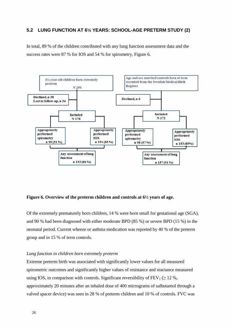
5.2 LUNG FUNCTION AT 6½ YEARS: SCHOOL-AGE PRETERM STUDY (2)
In total, 89 % of the children contributed with any lung function assessment data and the
success rates were 87 % for IOS and 54 % for spirometry, Figure 6.
Figure 6. Overview of the preterm children and controls at 6½ years of age.
Of the extremely prematurely born children, 14 % were born small for gestational age (SGA),
and 90 % had been diagnosed with either moderate BPD (85 %) or severe BPD (15 %) in the
neonatal period. Current wheeze or asthma medication was reported by 40 % of the preterm
group and in 15 % of term controls.
Lung function in children born extremely preterm
Extreme preterm birth was associated with significantly lower values for all measured
spirometric outcomes and significantly higher values of resistance and reactance measured
using IOS, in comparison with controls. Significant reversibility of FEV1 (≥ 12 %,
approximately 20 minutes after an inhaled dose of 400 micrograms of salbutamol through a
valved spacer device) was seen in 28 % of preterm children and 10 % of controls. FVC was

27
below the 5th
percentile (lower limit of normal) of the normal population in 14 % of the
preterm children and FEV1 was below the 5th
percentile in 23 %.
Gestational age and lung function in children born extremely preterm
Children born at 22 – 24 weeks of gestation had significantly lower values for all measured
spirometric outcomes than children born at 25 – 26 weeks of gestation. The proportion of
children born at 22 – 24 weeks with lung function below the lower limit of normal was 24 %
for FVC and 44 % for FEV1. IOS results did not differ significantly between the two
gestational age groups.
Lung function and asthma-like disease
Among the extremely preterm children, there was an association between reduced z-score for
FVC (-0.6, 95 % CI -1.0;-0.1), FEV1 (-0.8, 95 % CI -1.3;-0.3), increased AX (0.6, 95 % CI
0.1;1.6) and reported asthma-like disease in the previous 12 months (unpublished results).
Lung function in preterm-children born SGA and AGA
Lung function did not differ statistically between children born extremely preterm and SGA
and those born AGA, except for a higher frequency dependence of resistance (R5-20) among
children born SGA.
Lung function in preterm children and association to severity of BPD
There were no statistically significant lung function differences between the two levels of
BPD, except for z-scores for lower ratio FEV1/FVC in preterm children with severe BPD
compared with those with moderate BPD.

28
5.3 LUNG FUNCTION AT 16 YEARS AFTER MODERATE-TO-LATE PRETERM
BIRTH (3)
Of the 4 089 children in the original BAMSE cohort, 2 620 (64 %) and 2 605 (64 %)
participated in the 8- and 16-year follow-ups, respectively. The participants and number of
successful lung function tests in moderate-late-preterm and term groups are presented in
Figure 7.
Figure 7. Overview of the participants in the original BAMSE cohort and number of
performed and accepted lung function tests at 8 and 16 years used for analysis.
Lung function at 16 years
Among subjects aged 16 years, negative associations between preterm birth and all
spirometric indices, except FVC, were observed for both sexes (Table 2). In males, the
29
moderate-to-late preterm group demonstrated a FEV1 reduction of 4.0 % compared with term
controls, and in females the reduction was 3.4 %.
The IOS results in subjects aged 16 showed significantly higher estimated medians for R5, R5-
20, and AX0.5
in moderate-to-late preterm males compared with term males. Although a
similar trend was seen for females, a significant difference was only seen for R5 (Table 2).
To elucidate if the effect on lung function remained in those born closest to term, analysis
was restricted to those born at 35 to 36 weeks of gestation. Differences compared with
children born at term remained for spirometry results at age 16 (FEV1 -112 ml, 95 % CI -
217.6;-22.3) (unpublished results).
Lung function from age 8 to 16 years
There was an increasingly negative trend over time, between 8 and 16 years of age, for FEV1
in preterm males compared with term males.
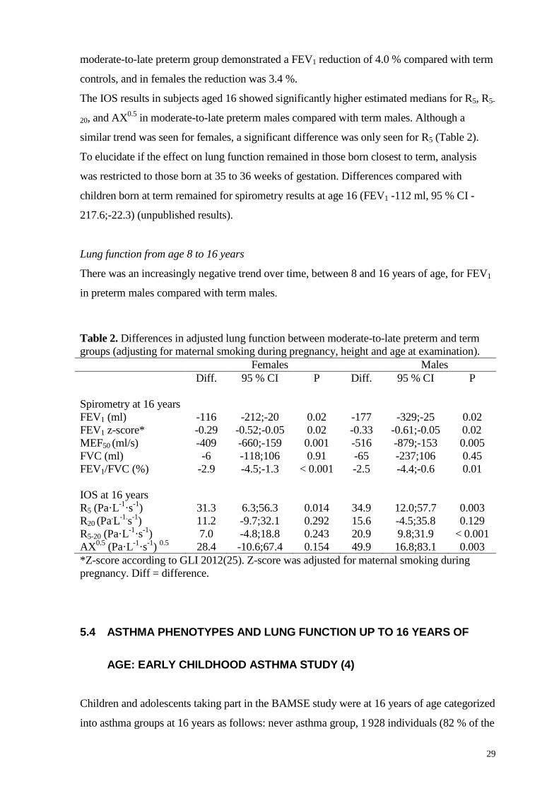
Table 2. Differences in adjusted lung function between moderate-to-late preterm and term
groups (adjusting for maternal smoking during pregnancy, height and age at examination).
Females Males
Diff. 95 % CI P Diff. 95 % CI P
Spirometry at 16 years
FEV1 (ml) -116 -212;-20 0.02 -177 -329;-25 0.02
FEV1 z-score* -0.29 -0.52;-0.05 0.02 -0.33 -0.61;-0.05 0.02
MEF50 (ml/s) -409 -660;-159 0.001 -516 -879;-153 0.005
FVC (ml) -6 -118;106 0.91 -65 -237;106 0.45
FEV1/FVC (%) -2.9 -4.5;-1.3 < 0.001 -2.5 -4.4;-0.6 0.01
IOS at 16 years
R5 (Pa·L-1
·s-1
) 31.3 6.3;56.3 0.014 34.9 12.0;57.7 0.003
R20 (Pa.L
-1.s
-1)
11.2 -9.7;32.1 0.292 15.6 -4.5;35.8 0.129
R5-20 (Pa·L-1
·s-1
) 7.0 -4.8;18.8 0.243 20.9 9.8;31.9 < 0.001
AX0.5
(Pa·L-1
·s-1
) 0.5
28.4 -10.6;67.4 0.154 49.9 16.8;83.1 0.003
*Z-score according to GLI 2012(25). Z-score was adjusted for maternal smoking during
pregnancy. Diff = difference.
5.4 ASTHMA PHENOTYPES AND LUNG FUNCTION UP TO 16 YEARS OF
AGE: EARLY CHILDHOOD ASTHMA STUDY (4)
Children and adolescents taking part in the BAMSE study were at 16 years of age categorized
into asthma groups at 16 years as follows: never asthma group, 1 928 individuals (82 % of the

30
eligible population); early transient asthma, 139 individuals (6 %); early persistent asthma, 61
individuals (3 %); and late-onset asthma, 109 individuals (5 %).
Asthma and spirometry at 16 years
All asthma groups were associated with lower FEV1, FEV1/FVC and FEF50 values in
comparison with the never asthma group at 16 years of age. The lowest values were seen for
the early persistent group.
Asthma and impulse oscillometry (IOS) at 16 years
All asthma groups were associated with higher estimated means for R5 and R20 than the never
asthma group. Figure 4 shows all percentiles for R5-20, an index indicating heterogeneity in
resistance distribution across the airway system. The distribution of R5-20 values was similar
in the never asthma and transient asthma groups. The persistent asthma group showed a
heterogeneous pattern including a wide range of R5-20 values and a skewed distribution with
extremely large R5-20 values.
Asthma and lung function change between 8 and 16 years
We observed significantly less increase in FEV1 between 8 and 16 years for the early
persistent and late-onset groups, but not for the early transient asthma group, as compared
with the reference group.

31
Figure 4. Distribution of R5-20 values for the never asthma group (A) and difference
between each asthma group and the “never asthma” group at all percentiles (B-D).
Estimates at each percentile are shown as solid black lines with 95 percent confidence
bands (shaded gray areas). The mean value (solid) and 95 percent confidence intervals
(dashed) are shown as parallel black lines.
32
6 DISCUSSION
6.1 LEVEL OF LUNG FUNCTION AFTER PRETERM BIRTH
6.1.1 Maximal expiratory flows after preterm birth
In Study 1, we found that patients at 6 and 18 months of age with either mild or
moderate/severe BPD demonstrated low (< -1.96 z-scores) mean values for VmaxFRC,
FEV0.5, and MEF50, with the exception of VmaxFRC at age 18 months for the mild BPD
group. The vast majority of preterm infants had clinically significant (< -1.96 z-scores)
reduced expiratory flows at 6 and 18 months of age.
Similar infant lung function techniques have been used in other previous studies. Tidal RTC
was used at approximately one year of age in a group of children born preterm (gestational
weeks 29 – 36) without BPD and a reduction of VmaxFRC of -2.0 z-score was found.(74) Two
other studies show similar reductions of VmaxFRC, -1.79 (40 % below -1.96 z-score) to -2.2 z-
score in moderate to severe BPD (75, 76) at 15 – 18 months of age. Filbrun et al. reported
that FEV0.5 was on average -1.94 z-score at a mean age of 18 months in children with
moderate to severe BPD.(77) Recently, infants with mild to severe BPD were shown to have
FEV0.5 with 2.1 lower z-score at corrected age of 18 months of age as compared with
published reference values.(78)
In Study 2, we examined children at 6½ years of age with a history of moderate-to-severe
BPD and found that 23 % of preterm children had FEV1 below the 5th
percentile (-1.64 z-
score). Among children born in gestational weeks 22 – 24, 44 % had values below the 5th
percentile. The EPICure study investigated a similar group as our study. They reported an
average reduction of -1.7 z-scores for FEV1 at 11 years of age in children earlier diagnosed
with moderate-to-severe BPD.(33)
In Study 3, we report on reduced maximal expiratory flows at 16 years of age in both females
and males born moderate-to-late preterm (gestational weeks 32 – 36). The proportion of
individuals with a FEV1 below the lower limit of normal (< -1.64 z-score) was, in males,
significantly larger in the moderate-to-late preterm group than in the term group. Reports on
lung function in adolescence in subjects born moderate-to-late preterm are sparse. Kotecha et
al. found lower mid-expiratory flows and FEV1/FVC ratio at 14 – 17 years of age in the
subgroup born in gestational weeks 33 – 34, but no difference compared with the term group
33
for the subgroup born in gestational weeks 35 – 36.(34) We also tested the effect on lung
function in those born closest to term by restricting analysis to those born at 35 through 36
weeks of gestation and found that differences compared with children born at term remained
for spirometry results.
In summary, in Study 1 and Study 2 we found clinically significant reductions of maximal
expiratory flows in infancy and early school age and the levels of reductions are similar to
findings in other studies. In Study 3, we found a 3 – 4 % reduction of FEV1 and more
individuals than expected having values below the lower limit of normal after moderate-to-
late preterm birth. To our knowledge, Study 2 is the first to report on lung function results
associated with the level of extreme preterm birth, showing that within the group of children
born extremely preterm, the length of pregnancy matters for lung function development in
childhood.
To clarify the possible clinical implication of the lung function reduction after preterm birth,
a comparison with natural age-related decline is presented. Natural age-related decline of
FEV1 in adulthood has been calculated to be on average 21 ml/year in females and 27 ml/year
in males.(79) The difference measured, for the whole preterm group at 6½ years (330 ml) in
Study 2, would correspond to approximately 12 – 15 years of preterm natural lung function
decline, depending on sex. Further, if tracking of lung function is assumed, the estimated
reduction difference of -1.2 z-score at 6½ years in Study 2 equals 650 ml in an adult male
(180 cm), suggesting approximately 24 years of expected preterm decline. Using the same
assumption after moderate-to-late preterm birth, the low FEV1 at 16 years (177 ml) in
moderate-to-late preterm males corresponds to a natural decline in adulthood of 7 years. If
these predictions have a corresponding predictive value on life expectancy, can only be
determined in long-term follow-up studies.
6.1.2 Lung volumes after preterm birth
In Study 1, plethysmographically measured FRC values were within normal range at both 6
and 18 months of age. FVC, measured using raised RTC, was on average lower or borderline
lower than expected at 18 months of age. Hofhuis et al. found FRCpleth to be within normal
range (-0.6 z-score) in moderate-to-severe BPD at approximately 15 months of age.(76)
Fakhoury et al. related FRC to ml per body weight (suggested normal range 16 – 26 ml/kg)
and reported normal values at similar ages.(75) However, lung volumes measured using the
gas dilution technique (Helium) showed lower FRCHe in children with BPD compared with
34
non-BPD or healthy preterm.(80, 81) Hilgendorf et al. showed that FRCpleth was reduced near
term in children with BPD and further reduced after administration of beta2-agonist,
suggesting over-inflation after preterm birth.(82) A study combining gas dilution techniques
and whole body plethysmography in the same infants reported higher FRCpleth than FRCgas in
follow-up of infants with a history of BPD, which also suggested over-inflation and gas
trapping in infants with BPD.(83) This notion is supported by results in a study that used
raised RTC and whole body plethysmography to calculate fractional lung volumes (TLC and
RV) at approximately 18 months of age.(77) They found FVC and TLC to be normal low, but
the RV/TLC ratio to be high.
Since FRC is dynamically maintained in infants with obstructed airways, as could be
assumed to be the case in infants with BPD, plethysmographically measured FRC may
remain normal-to-high. Hence, it is possible that FRC measured plethysmographically, as in
Study 1, cannot reflect trapped gas in the premature child, thus overestimating lung volume
due to an obstructive pattern.
In Study 2, the reduction of the measured volume among preterm children (gestational weeks
22 – 26) was modest but significant compared with controls (term). Follow-up at school age
has shown varying results for FVC in other studies. The EPICure study, which included a
cohort with similar level of preterm birth as that in Study 2, found similar significant
reduction of FVC, -0.8 z-score.(84) Studies that included a wider range of preterm birth
subjects showed no significant differences in FVC between the preterm and term groups.(85-
87) However, normal TLC and increased RV/TLC ratio was seen in those studies, indicating
an obstructive pattern.
In Study 3, the children born moderate-to-late preterm had normal FVC values, no different
to term controls at 16 years of age. This supports findings by other groups.(34, 88)
In summary, lung volumes after preterm birth are normal or normal low and reported
reduction in FVC is more likely to be a sign of airway collapsibility during exhalation than
pulmonary restrictivity in children born very or extremely preterm. A possible explanation for
this functional pattern could be loss of elastic recoil, altered airway wall mechanics and
altered alveolarization in early life seen after preterm birth.

35
6.2 BPD SEVERITY IN RELATION TO LUNG FUNCTION
The definition of BPD published in 2001 based on the NICHD/NHLBI workshop suggests
three levels of severity for BPD: mild, moderate/severe and severe disease.(32) In the
neonatal period, the grades of BPD severity have been found to be associated with neonatal
comorbidities, longer hospital stay and higher gestational age at discharge, showing the
usefulness of this definition.(89) A validation of the definition found an association to more
severe disease and the use of pulmonary medication at 18 – 22 months of age.(90) Most
studies on lung function after preterm birth compare the different definitions of BPD with
individuals born at term or non-BPD and do not investigate the effect of BPD severity.
Generally, those studies confirm significantly reduced lung function from infancy to
adulthood among those with BPD diagnosed in the neonatal period.(9, 84, 91, 92)
We used the consensus definition for BPD severity in Study 1 and Study 2. In Study 1, the
only significant difference in lung function at 6 and 18 months of age was lower Cso in the
moderate/severe group compared with the mild BPD group. Overall, maximal expiratory
flows measured using RTC-technique were low (< -1.96 z-scores) for both groups at both
time points, with the exception for VmaxFRC at age 18 months for the mild BPD group.
In Study 2, we compare lung function between moderate and severe BPD groups. Mild BPD
was not considered in Study 2, due to the low incidence of mild BPD. Except for lower
FEV1/FVC z-scores in children diagnosed in the neonatal period with severe BPD than in
those diagnosed with moderate BPD, lung function at 6½ years of age did not vary in relation
to severity of neonatal BPD. This was true for both spirometric and IOS variables. However,
we found significant associations between gestational age and reduced lung function
measured using spirometry, but not using IOS. Studies investigating lung function at follow-
up show conflicting results in relation to BPD severity. Hjalmarson et al. found lower FRC
and less efficient gas mixing in severe BPD compared with less severe forms at 9 – 10
months of age. A study from our hospital performed a follow-up at 6 – 8 years of age after
preterm birth, showed significantly lower lung function in severe BPD compared with
mild/moderate disease.(93) Large differences in percent of predicted FEV1 were reported
between mild and more severe BPD groups at follow-up at 11 – 17 years of age in a cohort of
children born in the 1980s and treated at a large tertiary center.(94) Results from a study
using RTC methods during the first year of life indicate a non-significant association to BPD
severity, in agreement with our findings.(95) A meta-analysis including follow-up studies of

36
subjects born between 1964 and 2000 at 24 – 36 weeks of gestation reported average FEV1
reductions of 16 % associated with mild BPD and 19 % for moderate to severe BPD.(3)
The lack of clear differences in lung function at follow-up in relation to BPD severity in
Study 1 and Study 2 could be due to the fact that all children in both studies were very or
extremely prematurely born, thus all having altered alveolarization due to prematurity. In
fact, the groups in Study 1 were similar in regards to most background variables. This could
indicate limited prognostic utility for BPD diagnose in this respect due to multifactorial
etiology and difficulties in early identification of the disease. A limitation of the diagnostic
criterion of BPD and BPD severity is the need of supplemental oxygen as a proxy for disease
severity. Walsh et al. have reported a physiologic definition of BPD by a standardized timed
room-air challenge (96, 97) that is recommended in Sweden since 2014 by the National board
of Health and Welfare. However, the length of time for supplemental oxygen therapy and
targeted oxygen saturation level could be a function of the immaturity of the child or of the
local policies of individual units as reflective of lung pathology during the time of our studies.
Further, mode of oxygen delivery, medications and possible upper airway obstruction may
also influence gas exchange. It could be questioned if the histopathology of BPD, which the
surrogate marker of oxygen need is thought to indicate, is not dichotomous but rather a
continuous graded condition. Long-term pulmonary outcomes and supplemental oxygen at
post menstrual age of 36 weeks have shown low values of sensitivity and specificity in some
studies and might therefore be falsely related.(90, 98) Finally, even the most conservative
definition of BPD, supplemental oxygen 28 days before gestational week 36 or at day 28, has
been shown to be insufficient as a predictor due to low sensitivity for long-term pulmonary
outcome.(93, 99)
In summary, our results suggest that early BPD classification provides limited information on
future lung function after preterm birth and other specific risk factors and mechanism for lung
function development should be sought. To what extent early measured lung function could
contribute needs further investigations.
6.3 LONGITUDINAL ANALYSIS OF EXPIRATORY FLOWS AFTER PRETERM
BIRTH AND EARLY BRONCHIAL OBSTRUCTIVE SYMPTOMS
In Study 1, measurements of maximal expiratory flows showed persistently reduced values
without catch-up from 6 to 18 months of age for both BPD and non-BPD groups. In fact,

37
VmaxFRC measured using tidal RTC decreased significantly at follow-up compared with
reference values, while FEV0.5 remained at the same level of reduction.
Serial measurements have been performed in a number of studies during the first years of life
after preterm birth. A decline of VmaxFRC was also seen during the first year in children with
moderate-to-severe BPD and in healthy children born preterm.(74, 76) Fakhoury et al.
performed measurements at 6, 12 and 24 months and reported no change over time in mean
z-score VmaxFRC in children with moderate-to-severe BPD.(75) The raised RTC technique
expiratory flows in healthy preterm and children with BPD were low compared with term
controls and no catch-up of lung function was seen in any of these three studies during
infancy or early childhood.(77, 78, 100)
Generally, these findings during infancy in our studies and others support the notion of
tracking of lung function seen in older children and adolescents after very and extreme
preterm birth.(92, 101, 102) The difference in development of variables from the tidal volume
and raised volume RTC seen in our studies and others could indicate that VmaxFRC and
FEV0.5 do not represent the same airway impairment. However, it is also possible that the
difference is attributable to the reference values used, as suggested by Lum et al.(103)
In Study 3, moderate-to-late preterm females and males showed no catch-up growth of lung
function from 8 to 16 years of age. Instead, the negative effects were augmented between age
8 and 16 years in males. Only two previous studies have longitudinally addressed lung
function development after moderate-to-late preterm birth into school age and adolescence.
Kotecha et al. found catch-up of FEV1 from 8 – 9 to 14 – 17 years of age in the subgroup of
children born in gestational weeks 33 – 34.(34) A follow-up by Narang et al. from mid-
childhood (7 – 9 years) into young adulthood stated that there was no evidence of airway
obstruction at 21 years of age in a cohort including participants born in gestational weeks 27
– 37 who, at 7 – 9 years of age, had demonstrated reduced FEV0.75.(88) In a meta-analysis
that included 24 birth cohorts, particularly late preterm, lung function was negatively
associated with preterm birth across the full range of prematurity (< 37 weeks).(104)
In Study 4, longitudinal lung function was compared from 8 to 16 years of age between
asthma groups and the never asthma group. The two active asthma groups (early persistent
and late-onset asthma) had significantly lower increases of FEV1. This was seen to a lesser
extent in those with early transient asthma and reached significance only for mid-expiratory
flows (MEF50). Similar findings were seen in a Norwegian study also identifying both active

38
asthma and asthma in remission to be associated with less increase of FEV1 during
adolescence.(105) The Melbourne study found a negative lung function development from 12
to 18 years for active asthma (early persistent and late-onset), but not for early transient
asthma, which was suggested to be a benign disorder.(106)
In summary, Study 1, Study 3 and Study 4 support the recognized concept of lung function
tracking from birth to adolescence.(26) There were no catch-up of lung function seen in the
first two years of life after very and extreme preterm birth, or from 8 to 16 years after
moderate-to-late preterm birth or in asthma phenotypes during that same age span. In fact, the
difference in lung function compared with controls was augmented after moderate-to-late
preterm birth and in active asthma suggesting a ―two-hit‖ model including an insult early in
life and ongoing processes. Decreasing FEV1/FVC ratios in young adults after very and
extreme preterm birth seen in two other studies (92, 102), particularly in BPD, indicate that
similar processes might continue after adolescence and need to be further investigated into
adulthood.
6.4 ASPECTS OF AIRWAY MECHANICS IN RELATION TO PRETERM BIRTH
AND EARLY BRONCHIAL OBSTRUCTIVE SYMPTOMS
6.4.1 Compliance of the respiratory system (Crs) after preterm birth
In Study 1, Cso was the only measured lung function variable that was associated with BPD
severity. It was also associated with respiratory symptoms. Except for infants with
moderate/severe BPD and infants reporting symptoms at 6 months, measured values were
within normal range and increased with age. Low compliance of the respiratory system in the
neonatal period after preterm birth has been shown to be associated with BPD severity in
other studies.(81, 107) Improvements during infancy and normalization compared with the
control group are shown in those studies. Hjalmarson et al. and Schmalisch et al. found lower
values in BPD, but after normalization for lung volume the significance disappeared.
However, both studies used FRCHe, which has been shown to be lower in BPD than in non-
BPD. In our study, we used FRCpleth, which also includes trapped gas, making this
comparison difficult. We compared with published reference values that take gestational age
and body length into account.(69) For completeness, we also normalized values for weight,
which did not change the conclusion (not published). A relationship between early measured
Cso and wheezing illness and asthma during childhood has been shown in studies after term
birth.(108, 109) The underlying pathophysiological mechanism for reduced Cso is complex,
39
involving the total respiratory system and is affected by alveoli as well as bronchi and chest
wall. A possible explanation for why early measured Cso is associated with both BPD severity
and respiratory symptoms is that hyperinflation, which stiffens the lungs, and the level of
alveolarization, both lower respiratory compliance.
In summary, compliance of the respiratory system measured in both sedated and un-sedated
infants early in life, has been shown to be related to later lung function and respiratory
symptoms and is therefore suggested to be tested as a marker of pulmonary outcome after
preterm birth.
6.4.2 Impulse oscillometry after to preterm birth and in relation to asthma
phenotypes
In Study 2, airway mechanics measured using the IOS technique showed higher values for all
indices (R5, R5-20, AX) among preterm children as compared with controls. IOS variables
were not different between the two preterm groups (gestational weeks 22 – 24 and 25 – 26).
In follow-up studies of children, with and without BPD, IOS results distinguished between
preterms and term controls as well as between preterm groups with and without BPD.(110)
(93) Both studies report some concordance for IOS variables and FEV1, however not for the
same indices. In our study, almost all participants had either moderate or severe BPD, making
comparison difficult. We can only speculate into the reasons for a lack of association between
the extreme preterm groups and IOS indices, in despite of significant spirometric differences.
Perhaps IOS has lower sensitivity to discriminate the affected lung region in the most preterm
individuals from the overall effect on airways seen after preterm birth. Or, since IOS is
performed during tidal breathing without effort, airway closure induced by deep breaths or
forced expirations, as during spirometry, will not interfere with the measurement.
In Study 3, IOS results at age 16 showed significantly higher estimated medians for R5, R5-20,
and AX0.5
in moderate-to-late preterm males compared with term males. Although a similar
trend was seen for females, a significant difference was only seen for R5, despite significant
spirometric differences also between moderate-to-late preterm and term females. To our
knowledge, this is the first study using IOS in follow-ups after moderate-to-late preterm birth.
In Study 4, the indices suggested to mirror peripheral airways R5-20 had a higher estimated
mean in both active asthma groups compared with the never asthma group. It was also shown
that the heterogeneity of resistance measured over the airway system was not different

40
between never asthma and early transient asthma. However, both active asthma groups,
particularly the early persistent group, showed skewed distributions with very high values in
the 90th
percentile. The notion that respiratory mechanics measured using IOS could give
additive information compared with spirometric values in active asthma has been shown in
studies addressing asthma treatment, asthma control and severity also including bronchial
hyper-reactivity without respiratory symptoms, thus supporting our findings.(111, 112)
In summary, Study 2, Study 3 and Study 4 show that for the active asthma phenotypes and
the asthma-like syndrome seen after preterm birth, all being bronchial obstructive entities
associated with increased smooth muscle airway tone and bronchial hyper-reactivity, the IOS
technique has the potential to identify reactive airways. Frequency dependence of resistance
(fdR, an index of bronchial obstruction) and AX (sensitive to peripheral bronchial
obstruction) both showed increased values after preterm birth and in active asthma
phenotypes groups, but not early transient asthma, despite reduced lung function in all groups
studied. Hence, the mechanism for bronchial obstruction is different in the early transient
asthma phenotype group compared to both active asthma and in the bronchial obstructive
disease seen after preterm birth.
Indeed, since the success rate of IOS measurements was also higher than conventional
spirometry, primarily because this technique only requires passive cooperation from
investigated subjects, it makes IOS a feasible technique particularly in children too young to
perform spirometry.
6.5 CLINICAL SYMPTOMS IN RELATION TO LUNG FUNCTION AFTER
PRETERM BIRTH
In Study 1, a third of infants reported respiratory symptoms at 6 months and the proportion
increased up to almost half at 18 months of age. In contrast to the modest association between
BPD severity and lung function, we found a clear association between reported respiratory
symptoms and reduced lung function. Both lung mechanics (Cso) and lung function measured
with TVRTC and RVRTC techniques showed significantly lower values among those
reporting symptoms at both 6 and 18 months of age. Associations between respiratory
symptoms and lung function after very and extreme preterm birth have only been investigated
in a few studies during early childhood. Fakhoury et al. that showed lower VmaxFRC at 6
(but not at 12 and 24) months of age among those who reported cough but not wheeze, and
41
Robin et al. found that recurrent wheezing was not associated with reduced lung function,
except increased RV among the extreme preterm children.(75, 91)
In Study 2, 40 % of children born preterm, compared with 15 % of controls, reported
respiratory wheeze or use of asthma medication (asthma-like disease) in the last 12 months
before follow-up at 6½ years. Within the preterm group, there was an association of
asthma-like disease to lower lung function measured using spirometry (lower z-scores for
FEV1 and FVC) and altered airway mechanics (higher AX). The EPIcure study reported
that 25 % of extreme preterm children at 11 years of age had a current diagnosis of
asthma.(84) They also studied the association between lung function and respiratory
morbidity (wheeze ever) using spirometry, inert-gas washout and plethymography. They
found associations to FEV1, FEV1/FVC ratio and specific resistance, but not to LCI or
RV/TLC. The latter three are thought to represent more peripheral airways.(33)
In Study 3, females born moderate-to-late preterm reported more respiratory symptoms at
both 8 and 16 years of age than females born at term, in spite of less reduced lung function
compared with term females than preterm males with term males. Respiratory symptoms
were, as expected, negatively associated with lung function in the whole cohort but not within
the moderate-to-late preterm group. Gender differences in respiratory health at 19 years of
age have been reported after preterm birth and are well recognized in the general term
population.(113, 114) It is suggested that this could be explained by different patterns of lung
growth between genders. Males have smaller airways in relation to lung size before puberty
and this change in puberty makes males less prone than females to respiratory symptoms as
adults. However, moderate-to-late preterm females reported more respiratory symptoms
already before puberty in our study, thus making this explanation less probable in the group
of moderate-to-late preterm children.
In Study 1 and Study 2, respiratory morbidity such as wheeze and asthma-like symptoms
during infancy and childhood were associated with lung function decrements seen after very
and extreme preterm birth. This was not seen after moderate-to-late preterm birth in late
adolescence. The full nature of altered airways and lung parenchyma is not completely
understood and needs to be further investigated. However, lung function tests in follow-up
after very and extreme preterm birth could be suggested to identify individuals at risk of
respiratory morbidity and possible treatment.

42
6.6 LIMITATIONS AND STRENGTHS
The major strength of this thesis is that the included studies are based on data collected
prospectively.
Study 1 and Study 2 included the vast majority of infants with BPD or the most extremely
preterm children and there was low loss to follow-up, both facts reducing the risk of selection
bias. In the two studies based on the BAMSE cohort, the size and limited loss to follow-up
minimizes random errors. In those two studies, a selection bias could have occurred at
recruitment (75 % of eligible children included) and at follow-up. To evaluate possible
selection bias at follow-up, study populations in Study 3 and Study 4 were compared with the
original BAMSE cohort. With respect to population characteristics, the group of children who
provided lung function data in Study 3 did not differ to any major extent from the original
cohort. In Study 4, individuals who participated in the clinical testing were more often
female, had more parental atopy and less maternal smoking during pregnancy than those who
did not participate, which might to some extent limit the generalizability.
Gestational age was obtained from the Swedish Medical Birth Registry and in more than 95
% of the pregnancies gestation was determined using ultrasound. The narrow border between
gestational age groups could introduce a risk of non-differential misclassification, however
that would likely result in an underestimation of the true differences in lung function between
the groups.
A limitation and a possible random error of Study 1 is that there are different numbers of
missing values for the measured variables, due to insufficient quality of measurement and
preterm awaking (despite sedation). It was unlikely that this differed between groups and
therefore we believe results were not affected. However, this reflects the challenges to
perform lung function tests in sedated infants. In Study 2, the success rate for spirometry was
low, but anticipated because of the young age at follow-up, which could introduce a selection
bias if children with more severe respiratory morbidity where less successful in performing
lung function tests. If so, we could have underestimated the differences between groups.
A major limitation of Study 1 is the absence of a control group, which for ethical reasons was
difficult to access.
Misinterpretation due to inappropriate reference data cannot be excluded in Study 1.

43
A limitation of Studies 2 – 4 is that at early school age, mean spirometric z-scores were not
centered at zero. Control groups had approximately 0.5 higher z-scores for spirometric
variables at 8 years than the published reference population.(25) This could introduce an
underestimation of the frequency of low lung function in our preterm groups in that age span.
The effect is likely to be non-differential and not affect comparisons between groups.
One of the major limitations in all but Study 2 is that examination after bronchodilation was
not performed. Post-bronchodilator measurements would have been desirable in order to
explore whether the observed impairments are due to static changes after remodeling of the
airways, impaired airway growth or reversible airway obstruction.
Finally, recall bias could have influenced the classification into symptom groups and
prevalence of asthma and asthma-like disease due to the use of questionnaires at the different
time points at follow-up.
7 ETHICAL CONSIDERATIONS
In Study 1, all parents were informed of the purpose of the procedures and informed consent
was obtained for all measurements. To perform ILFT, the child has to be sedated by orally
administrated choral hydrate, which has a bitter taste and could lead to some discomfort for
the child. The sedation method is recommended by consensus publications for ILFT. During
the time period of recruitment the infant lung function procedures used in the study were
implemented as standard follow-up of children with BPD at our center.
In Studies 2 – 4, lung function tests were performed in cooperation with the child/adolescent,
who was awake. Lung function tests do not involve large efforts or painful procedures. The
lung function methods used are well standardized and common in normal clinical setting
worldwide.

44
8 CONCLUSIONS
The majority of very and extreme preterm born children have expiratory flow values
below the normal range at infancy. At early school age, this is less pronounced but
still indicates a significantly reduced lung function. Lung function in adolescence
after moderate-to-late preterm birth is lower than after term birth.
Children born very and extreme preterm have impaired lung function compared with
published reference values and term controls, but early staging of BPD severity is
insufficent as a prognostic marker of lung function.
Early measured lung function after preterm birth is associated with respiratory
symptoms during infancy and childhood, and therefore suggested to identify infants at
risk of respiratory mobidity.
No catch-up of lung function was seen in the first two years of life after very and
extreme preterm birth, or from 8 to 16 years after moderate-to-late preterm birth. This
was also observed in early childhood asthma phenotypes between 8 and 16 years of
age.
Childhood asthma phenotypes including early transient disease, based on onset and
duration of symptoms, were all negatively associated with FEV1 at 16 years of age.
The bronchial obstructive pattern, measured by spirometry and IOS, seen in
adoloescense in the group with early transient asthma is different than the obstructive
patterns seen in active asthma phenotype groups and after preterm birth. However, the
bronchial obstructive pattern in the active asthma phenotypes at 16 years and after
preterm birth at 6 and 16 years of age show similarities.
IOS, which is suitable for lung function measurement in children too young to
perform spirometry, may identify children with low lung function after preterm birth
and those with active asthma.
45
9 CLINICAL REMARKS
The absence of catch-up growth of lung function after preterm birth or in early
asthma phenotypes indicates that low initial lung function after preterm and term
birth is associated with tracking of reduced lung function during childhood.
Children born moderate-to-late preterm are at risk of reaching adulthood with a
modest, but concerning, reduction of lung function.
Both time of onset and persistance of asthma symptoms are associated with lung
function impairment.
The combination of data from spirometry and IOS provides new insights about
asthma phenotypes and indicates similarities between active asthma and asthma-
like disease after preterm birth, which need to be further investigated.
Preterm birth interrupts normal lung development, which affects lung function
later in childhood. Measurements of lung function may identify children at risk
for respiratory morbidity and provide insights into long-term sequel of preterm
birth.
Regular assessments of lung function from infancy, during childhood and
possibly through life, is therefore suggested to be an important tool when
monitoring individuals born preterm, including moderate-to-late preterm birth.
Intervention studies on treatment of asthma-like symptoms after preterm birth are
highly warranted and the understanding of lung function in infancy and during
growth could improve the basis for such research.


47
Click to enter text


49
10 SVENSK SAMMANFATTNING
Bakgrund: Lungornas utveckling påverkas under fostertiden och barndomen av tidpunkten,
längden, typen och intensiteten av diverse händelser, sjukdomsprocesser och
omgivningsfaktorer. En vanlig sådan händelse är förtidig födsel. Ungefär 11 % av alla
graviditeter är kortare än 37 fullgångna graviditetsveckor, vilket är definitionen på förtidig
eller prematur födsel. Några få procent av alla födslar är mycket förtidiga (tidigare än 32
veckor). Incidensen för den mest extremt förtidigt födda gruppen, tidigare än 27 veckor, var
3/1 000 födda i den svenska multicenterstudien EXPRESS (Extremely Preterm Infants in
Sweden Study).
Bronkopulmonell dysplasi, eller också benämnt kronisk lungsjukdom efter förtidig födsel, är
det kanske vanligaste och allvarligaste kroniska resttillståndet efter extrem förtidig födsel. I
EXPRESS-studien hade nära alla deltagare diagnostiserats neonatalt med någon grad av
sjukdomen, varav 73 % med måttlig eller svår BPD. Uppföljningar från spädbarnsåldern till
ung vuxen ålder har visat kvarstående luftvägssjuklighet och nedsatt lungfunktion. Den
kliniska bilden av luftvägssjukdomen efter spädbarnstiden uppvisar en astmalik bild
inkluderande återkommande pip och väs i luftrören, ökad bronkiell hyperreaktivitet samt
obstruktiv lungfunktionsnedsättning. Man bör dock inte använda begreppet astma för detta
tillstånd eftersom astma i allmänhet definieras som en inflammatorisk luftvägssjukdom.
Lungsjukdomen efter förtidig födsel är emellertid inte associerad med luftvägsinflammation.
Astma och astmalika tillstånd är mycket vanliga under barndomen också efter födsel i
fullgången tid.
Över 30 % av alla barn i befolkningen har haft enstaka eller återkommande episoder av
astmalika symtom, främst vid luftvägsinfektioner, under sina första levnadsår. För
majoriteten är prognosen god och mindre än en tredjedel har kvar problemen efter sex års
ålder. I syfte att beskriva förlopp av astmasjukdom, välja och utveckla behandlings- och
preventionsstrategier, har flera astmafenotyper beskrivits. Dessa fenotyper är oftast
retrospektivt beskrivna och har alltså föga värde för klinkern som står med ett barn med
astmalika symtom framför sig. Att identifiera kliniska astmafenotyper med tillräckligt god
sensitivitet och specificitet i klinisk vardag för att avgöra prognos och välja behandling, har
visat sig vara svårt, men sannolikt kan information om lungfunktion, genetik och
omgivningsfaktorer vara en väg framåt. Den vanligaste fenotypen av småbarnsastma är den
med tidig debut och oftast symtomfrihet efter 4 – 5 års ålder; s.k. transient early

50
asthma/wheeze (ung. ‖tidig övergående astma‖) som vi på svenska ofta kallar ‖övergående
förkylningsastma‖. Övergående förkylningsastma har visats ge låg lungfunktion redan under
spädbarnstiden och kvarstående lägre lungfunktion som ung vuxen, trots att
luftvägssymtomen upphör redan under småbarnstiden. Huruvida det astmalika tillståndet som
ses efter förtidig födsel ska betraktas om en unik astmafenotyp kan ifrågasättas. Likheten
mellan övergående småbarnsastma och tillståndet efter förtidig födsel (tidigt nedsatt
lungfunktion, ej typisk allergisk inflammation) kan göra jämförelser intressanta.
Huvudfrågeställningar
Hur påverkas lungfunktionen från spädbarnstiden till adolescensen i relation till
förtidig födsel och tidigt utvecklad astma i barndomen?
Kan likheter och skillnader i lungfunktion hos förtidigt födda individer och personer
med olika astmafenotyper i barndomen ge information om mekanismerna bakom
luftvägsobstruktionen hos dessa tillstånd?
Delstudie 1: Lungfunktion vid 6 och 18 månader efter förtidig födsel i relation till
svårighetsgrad av bronkopulmonell dysplasi (BPD)
Femtiofem extremt förtidigt födda barn med olika grader av BPD undersöktes med flera olika
typer av lungfunktionsmetoder under inducerad sömn vid 6 och 18 månader efter födseln.
Majoriten av barnen hade vid både undersökningstillfällena definitionsmässigt nedsatta
expiratoriska utandningsflöden (< -1.96 standardavvikelser). Utom lägre compliance av
respiratoriska systemet (ung. elastiska egenskaper), som var lägre hos barn med måttlig/svår
BPD än hos lindrig BPD, fann vi inga skillnader i lungfunktion mellan svårighetsgraderna.
Emellertid fann vi statistiskt signifikant sämre lungfunktion i flertalet av de uppmätta
lungfunktionsmått hos de barn för vilka luftvägssymtom rapporterades vid 6 och/eller 18
månader, jämfört med hos dem som var symtomfria.
Slutsatsen av undersökningen var att extremt förtidigt födda barn hade låg lungfunktion vid 6
och 18 månader utan klar relation till BPD-grad och att luftvägssymtom var tydligt
associerade med sämre lungfunktion.
Delstudie 2: Lungfunktion hos 6 år gamla barn som fötts extremt tidigt - en
populationsbaserad kohortstudie (EXPRESS)

51
Drygt hälften av deltagarna i den nationella svenska multicenterstudien EXPRESS, som
inkluderat alla extremt förtidigt födda barn i Sverige (< 27 födelsevecka) inbjöds vid 6 års
ålder till studien. Vid uppföljningen gjordes lungfunktion med två metoder (spirometri och
impulsoscillometri). Lungfunktionsdata från 153 förtidigt födda barn och 157 kontrollbarn,
födda i fullgången tid, kunde analyseras.
Bägge metoderna visade statistiskt signifikant nedsatt lungfunktion för den prematurfödda
gruppen. Undergruppen av barn födda under födelsevecka 22 till och med 24 påvisades ha
sämst lungfunktion. Nära hälften av dessa hade ett forcerat expiratoriskt flöde vid 1 sekund
(FEV1) under den statistiska femte percentilen (förväntat 5 %) och en fjärdedel hade en
maximal utandad volym (FVC) under samma percentil. Signifikant förbättring av
lungfunktionen sågs hos 28 % av de prematurfödda efter inandning av luftrörsvidgande
läkemedel jämfört med 10 % av konrollerna. Om barnet var fött med låg födelsevikt (small
for gestational age, SGA) eller med svår BPD gav det bara marginellt sämre lungfunktion
jämfört med vid normal födelsevikt respektive lägre allvarlighetsgrad av BPD.
Slutsatserna var att ett stort antal förtidigt födda barn har kliniskt relevant nedsatt
lungfunktion vid 6 års ålder, att resultaten visade att luftvägsbehandling bör övervägas och att
fortsatta uppföljningar av lungfunktionen bör ske under uppväxten.
Delstudie 3: Lungfunktion vid 8 och 16 års ålder efter måttligt förtidig födsel: En
prospektiv kohortstudie.
Vi använde data från BAMSE-kohorten, som ursprungligen hade 4 089 deltagare med
prevalensen 4.8 % för gruppen måttligt till lindrigt förtidigt födda (födelsevecka 32 – 36).
Lungfunktion med spirometriundersökning vid 8 och 16 år samt impulsoscillometri (IOS) vid
16 års ålder användes vid analyserna. Totalt bidrog 2 621 deltagare, varav 149 prematurfödda
och 2 472 fullgångna (födelsevecka 37 – 41) med lungfunktionsdata.
Vid 16 års ålder hade båda könen statistiskt lägre lungfunktion mätt med spirometri (FEV1
3,4 % lägre för flickor och 4 % lägre för pojkar). Andelen pojkar med lågt FEV1 (under femte
percentilen) var vid 16 års ålder 15 % (kontroller 3,7 %). IOS-resultaten bekräftade lägre
lungfunktion för pojkar, men för flickor sågs en trend som inte nådde statistisk signifikans.
Longitudinell lungfunktion från 8 till 16 år visade att de prematurfödda barnen inte ökade sin
lungfunktion (FEV1, MEF50, FEV1/FVC) jämfört med dem som fötts i fullgången tid.
Skillnaden kvarstod för flickor, medan skillnaden ökade för pojkar.

52
Slutsatsen var att både flickor och pojkar födda bara några veckor före beräknad fullgången
tid även i tonåren hade lägre lungfunktion, 3,4 % respektive 4 %, jämfört med dem som fötts
i fullgången tid. Vidare sågs mellan 8 och 16 års ålder ingen relativ förbättring av
lungfunktionen för de förtidigt födda.
Delstudie 4: Astmafenotyper och lungfunktion upp till 16 års ålder – BAMSE-kohorten
Liksom i delstudie 3 användes data från BAMSE-kohorten och lungfunktionsresultaten från 8
och 16 års ålder. Baserat på enkätsvar (1-, 2-, 4-, 8- och 16-års-uppföljningarna)
kategoriserades deltagarna i astmafenotyperna: ‖aldrig astma‖ (82 %), ‖tidig övergående
astma‖ (6 %), ‖tidig kronisk astma‖ (3 %) samt ‖sent debuterande astma‖ (3 %).
Vid 8-års-uppföljningen bidrog 1 832 deltagare med spirometriresultat och vid 16-års-
uppföljningen bidrog 2 056 med spirometriresultat och 2 453 med IOS-resultat.
Spirometriresultaten visade att alla tre astmagrupperna hade statistiskt lägre lungfunktion vid
16 års ålder jämfört med individer utan astma. IOS-resultaten vid 16 års ålder visade att båda
grupperna med aktiv astmafenotyp hade statistiskt högre värden för R5-20 och AX (‖index‖
för perifer luftvägsobstruktivet) medan dessa värden inte skiljde sig mellan fenotypen ‖tidig
övergående astma‖ och gruppen ‖aldrig astma‖. Vidare sågs ingen relativ förbättring av
lungfunktion mellan 8 och 16 år för någon av astmafenotyperna. I stället sågs en ökning av
skillnaderna för FEV1 för de aktiva astmafenotyperna jämfört med fenotypen som aldrig haft
astma.
Slutsatsen blev att oavsett astmafenotyp hade individer med astma någon gång under
barndomen lägre spirometriskt mätt lungfunktion vid 16 års ålder. Ingen relativ förbättring av
lungfunktionen mellan 8 och 16 år sågs jämfört med barn som aldrig haft astma. IOS-
resultaten visade att grupperna som hade astmafenotyper med aktiv astma var associerade
med fynd som kan förklaras av obstruktivitet i små luftvägar.
Konklusioner
- Majoriteten av extremt förtidigt födda barn hade under spädbarnsperioden maximala
expiratoriska lungfunktionsvärden som var under nedre gränsen för normalvärden. I
tidig skolålder var detta mindre uttalat, men en påtaglig reduktion av lungfunktionen
kvarstod. Lungfunktionen var, vid 16 års ålder, lägre för dem som var födda bara
några veckor förtidigt jämfört med dem som fötts i fullgången tid.

53
- Tidig gradering av svårighetsgrad för BPD gav begränsad information om senare
lungfunktion och är en otillräcklig markör för lungfunktionsutveckling under
barndomen.
- Tidigt mätt lungfunktion efter förtidig födsel var associerad med luftvägssymtom och
kan därför identifiera individer med risk för luftvägssjukdom.
- Alla undersökta astmafenotyperna hade vid 16 års ålder lägre lungfunktion än
gruppen som aldrig haft astma.
- Longitudinellt såg man ingen relativ förbättring av lungfunktionen under
uppföljningsperioderna efter förtidig födsel eller efter tidig astmadebut.
- IOS-metoden, som är enkel att använda på yngre barn som ännu inte kan genomföra
spirometri, identifierar låg lungfunktion efter tidig födsel och individer som har
astmafenotyper med aktiv astmasjukdom.
Klinisk slutsats
- Lungfunktionsmätning kan identifiera barn med risk för symtom från luftvägarna och
kan öka kunskapen om framtida lungutveckling. Jag föreslår därför att förtidigt födda
barn ska genomföra undersökningar av lungfunktionen tidigt och regelbundet, upp till
vuxen ålder.


55
11 ACKNOWLEDGEMENTS
I would like to express my gratitude to all who helped me and made this thesis possible.
Particularly, all the children and families in the three cohorts studied: BPD at Sachsska,
BAMSE and CHARM.
The list of friends and colleges that in many ways are involved is long, but I would like to
highlight some of you:
My main supervisor Erik Melén, for having the enthusiasm and diplomatic skill to guide me
into research while at the same time being an excellent younger clinical college of mine.
My co-supervisor Jenny Hallberg, for providing daily mental and research support. For being
a patient friend when I had my numerous doubts to continue the work.
My co-supervisor Per Gustafsson, for attract my interest to pediatric lung function already 30
years ago. Since then, always ready to explain and to tutor me about respiratory physiology
and lung function techniques.
My co-supervisor Mikael Norman, for bringing the neonatologists perspective into the thesis
and for performing magic to my manuscript texts.
My mentor Gunnar Lilja, for bringing me to Sachsska. For providing the opportunity for my
clinical development in pediatric pulmonology. For being supportive when my stubborn
research hostile mind started to weaken.
My first main supervisor Magnus Wickman, for convincing me to start the formal research
education and for bringing Jenny to Sachsska and to my clinical and research world.
Gunilla Adenfelt, for being the link from the BPD-team to the IFLT venture that started in
2003. For also being a co-author in my first involvement in a BPD manuscript (not a part of
the thesis).
Eva Berggren-Broström, head of Sachsska and co-author, for taking the initiative to start the
BPD-team at Sachsska (the base for my interest in preterm lung development) and at your
return to Sachsska support my interest in lung function follow-up of the preterm infants.

56
Erica Schultz, PhD-student at IMM and co-author in several papers. For being the perfect
roommate and always willing to help me out when the statistics was too complicated for an
aging clinician.
―BAMSE-gänget‖ – Eva Hallner, André Lauber, Inger Kull (also a co-author), Sara Nilsson
and many more for guiding me in the BAMSE-project. My other co-authors at IMM, in
particular Göran Pershagen, who guided me in my first scientific attempts in the 1990th,
Matteo Bottai for giving statistics a friendly face.
My co-authors in the EXPRESS-group for starting an important national study of the preterm
infant, a literally growing group of children.
The staff at the Allergy and Lung department at Sachsska, in particular: Eva Östblom and
Natalia Ballardini, Caroline Nilsson for performing magic to the planning of the clinic
timetable (making my research possible). Catherine Fahlén for always being available for my,
often complicated, patient related issues, when I was away. Elsemarie Östlund, while still at
Sachsska, helped us organize and perform lung function in the Infant-study and in the
CHARM-study. Margareta Eriksson and Christina Ebersjö, who continued her work.
All other colleges at Sachsska for providing a friendly environment to work, in particular all
ski-buddies.
My mother, who told me already in ―mellanstadiet‖ that I should become a physician.
My kids – Anna, Kristina and Henrik – for almost never questioning (at least not in public)
my U-turn into research.
Anne, for initially unexpected joyful discussions in the fields of my research, but most of all
for being a guarantee against post-doctoral depression.

57
12 REFERENCES
1. Gluckman PD, Hanson MA, Cooper C, Thornburg KL. Effect of in utero and early-life
conditions on adult health and disease. The New England journal of medicine 2008;
359: 61-73.
2. Henderson AJ, Warner JO. Fetal origins of asthma. Seminars in fetal & neonatal medicine
2012; 17: 82-91.
3. Kotecha SJ, Edwards MO, Watkins WJ, Henderson AJ, Paranjothy S, Dunstan FD,
Kotecha S. Effect of preterm birth on later FEV1: a systematic review and meta-
analysis. Thorax 2013; 68: 760-766.
4. Been JV, Lugtenberg MJ, Smets E, van Schayck CP, Kramer BW, Mommers M, Sheikh
A. Preterm birth and childhood wheezing disorders: a systematic review and meta-
analysis. PLoS Med 2014; 11: e1001596.
5. Blencowe H, Cousens S, Oestergaard MZ, Chou D, Moller AB, Narwal R, Adler A, Vera
Garcia C, Rohde S, Say L, Lawn JE. National, regional, and worldwide estimates of
preterm birth rates in the year 2010 with time trends since 1990 for selected countries:
a systematic analysis and implications. Lancet 2012; 379: 2162-2172.
6. Buitendijk S, Zeitlin J, Cuttini M, Langhoff-Roos J, Bottu J. Indicators of fetal and infant
health outcomes. Eur J Obstet Gynecol Reprod Biol 2003; 111 Suppl 1: S66-77.
7. Fellman V, Hellstrom-Westas L, Norman M, Westgren M, Kallen K, Lagercrantz H,
Marsal K, Serenius F, Wennergren M. One-year survival of extremely preterm infants
after active perinatal care in Sweden. Jama 2009; 301: 2225-2233.
8. Rysavy MA, Li L, Bell EF, Das A, Hintz SR, Stoll BJ, Vohr BR, Carlo WA, Shankaran S,
Walsh MC, Tyson JE, Cotten CM, Smith PB, Murray JC, Colaizy TT, Brumbaugh
JE, Higgins RD. Between-hospital variation in treatment and outcomes in extremely
preterm infants. The New England journal of medicine 2015; 372: 1801-1811.
9. Doyle LW. Respiratory function at age 8-9 years in extremely low birthweight/very
preterm children born in Victoria in 1991-1992. Pediatric pulmonology 2006; 41:
570-576.
10. Halvorsen T, Skadberg BT, Eide GE, Roksund O, Aksnes L, Oymar K. Characteristics of
asthma and airway hyper-responsiveness after premature birth. Pediatric allergy and
immunology : official publication of the European Society of Pediatric Allergy and
Immunology 2005; 16: 487-494.
11. Baraldi E, Bonetto G, Zacchello F, Filippone M. Low exhaled nitric oxide in school-age
children with bronchopulmonary dysplasia and airflow limitation. American journal
of respiratory and critical care medicine 2005; 171: 68-72.
12. Filippone M, Carraro S, Baraldi E. The term "asthma" should be avoided in describing the
chronic pulmonary disease of prematurity. The European respiratory journal 2013;
42: 1430-1431.
13. Mallol J, Crane J, von Mutius E, Odhiambo J, Keil U, Stewart A. The International Study
of Asthma and Allergies in Childhood (ISAAC) Phase Three: a global synthesis.
Allergologia et immunopathologia 2013; 41: 73-85.
14. Martinez FD, Wright AL, Taussig LM, Holberg CJ, Halonen M, Morgan WJ. Asthma
and wheezing in the first six years of life. The Group Health Medical Associates. The
New England journal of medicine 1995; 332: 133-138.
15. Brand PL, Caudri D, Eber E, Gaillard EA, Garcia-Marcos L, Hedlin G, Henderson J,
Kuehni CE, Merkus PJ, Pedersen S, Valiulis A, Wennergren G, Bush A.

58
Classification and pharmacological treatment of preschool wheezing: changes since
2008. The European respiratory journal 2014; 43: 1172-1177.
16. Brand PL. Predicting the outcome of early childhood wheeze: mission impossible.
Primary care respiratory journal : journal of the General Practice Airways Group
2014; 23: 10-11.
17. Henderson J, Granell R, Heron J, Sherriff A, Simpson A, Woodcock A, Strachan DP,
Shaheen SO, Sterne JA. Associations of wheezing phenotypes in the first 6 years of
life with atopy, lung function and airway responsiveness in mid-childhood. Thorax
2008; 63: 974-980.
18. Weibel ER. What makes a good lung? Swiss medical weekly 2009; 139: 375-386.
19. Smith LJ, McKay KO, van Asperen PP, Selvadurai H, Fitzgerald DA. Normal
development of the lung and premature birth. Paediatric respiratory reviews 2010;
11: 135-142.
20. Stocks J, Hislop A, Sonnappa S. Early lung development: lifelong effect on respiratory
health and disease. Lancet Respir Med 2013; 1: 728-742.
21. Hislop AA. Airway and blood vessel interaction during lung development. Journal of
anatomy 2002; 201: 325-334.
22. Narayanan M, Owers-Bradley J, Beardsmore CS, Mada M, Ball I, Garipov R, Panesar
KS, Kuehni CE, Spycher BD, Williams SE, Silverman M. Alveolarization continues
during childhood and adolescence: new evidence from helium-3 magnetic resonance.
American journal of respiratory and critical care medicine 2012; 185: 186-191.
23. Narayanan M, Beardsmore CS, Owers-Bradley J, Dogaru CM, Mada M, Ball I, Garipov
RR, Kuehni CE, Spycher BD, Silverman M. Catch-up alveolarization in ex-preterm
children: evidence from (3)He magnetic resonance. American journal of respiratory
and critical care medicine 2013; 187: 1104-1109.
24. Ochs M, Nyengaard JR, Jung A, Knudsen L, Voigt M, Wahlers T, Richter J, Gundersen
HJ. The number of alveoli in the human lung. American journal of respiratory and
critical care medicine 2004; 169: 120-124.
25. Quanjer PH, Stanojevic S, Cole TJ, Baur X, Hall GL, Culver BH, Enright PL, Hankinson
JL, Ip MS, Zheng J, Stocks J. Multi-ethnic reference values for spirometry for the 3-
95-yr age range: the global lung function 2012 equations. The European respiratory
journal 2012; 40: 1324-1343.
26. Stern DA, Morgan WJ, Wright AL, Guerra S, Martinez FD. Poor airway function in early
infancy and lung function by age 22 years: a non-selective longitudinal cohort study.
Lancet 2007; 370: 758-764.
27. Sears MR, Greene JM, Willan AR, Wiecek EM, Taylor DR, Flannery EM, Cowan JO,
Herbison GP, Silva PA, Poulton R. A longitudinal, population-based, cohort study of
childhood asthma followed to adulthood. The New England journal of medicine 2003;
349: 1414-1422.
28. Northway WH, Jr., Rosan RC, Porter DY. Pulmonary disease following respirator
therapy of hyaline-membrane disease. Bronchopulmonary dysplasia. The New
England journal of medicine 1967; 276: 357-368.
29. Coalson JJ. Pathology of bronchopulmonary dysplasia. Seminars in perinatology 2006;
30: 179-184.
30. Laughon MM, Langer JC, Bose CL, Smith PB, Ambalavanan N, Kennedy KA, Stoll BJ,
Buchter S, Laptook AR, Ehrenkranz RA, Cotten CM, Wilson-Costello DE,
Shankaran S, Van Meurs KP, Davis AS, Gantz MG, Finer NN, Yoder BA, Faix RG,
Carlo WA, Schibler KR, Newman NS, Rich W, Das A, Higgins RD, Walsh MC.
Prediction of bronchopulmonary dysplasia by postnatal age in extremely premature
infants. American journal of respiratory and critical care medicine 2011; 183: 1715-
1722.

59
31. Rozycki HJ, Narla L. Early versus late identification of infants at high risk of developing
moderate to severe bronchopulmonary dysplasia. Pediatric pulmonology 1996; 21:
345-352.
32. Jobe AH, Bancalari E. Bronchopulmonary dysplasia. American journal of respiratory and
critical care medicine 2001; 163: 1723-1729.
33. Lum S, Kirkby J, Welsh L, Marlow N, Hennessy E, Stocks J. Nature and severity of lung
function abnormalities in extremely pre-term children at 11 years of age. The
European respiratory journal 2011; 37: 1199-1207.
34. Kotecha SJ, Watkins WJ, Paranjothy S, Dunstan FD, Henderson AJ, Kotecha S. Effect of
late preterm birth on longitudinal lung spirometry in school age children and
adolescents. Thorax 2012; 67: 54-61.
35. Tiddens HA, Hofhuis W, Casotti V, Hop WC, Hulsmann AR, de Jongste JC. Airway
dimensions in bronchopulmonary dysplasia: implications for airflow obstruction.
Pediatric pulmonology 2008; 43: 1206-1213.
36. Choi CW, Kim BI, Hong JS, Kim EK, Kim HS, Choi JH. Bronchopulmonary dysplasia in
a rat model induced by intra-amniotic inflammation and postnatal hyperoxia:
morphometric aspects. Pediatric research 2009; 65: 323-327.
37. Hislop AA, Haworth SG. Airway size and structure in the normal fetal and infant lung
and the effect of premature delivery and artificial ventilation. The American review of
respiratory disease 1989; 140: 1717-1726.
38. Yammine S, Schmidt A, Sutter O, Fouzas S, Singer F, Frey U, Latzin P. Functional
evidence for continued alveolarisation in former preterms at school age? The
European respiratory journal 2015.
39. Henschen M, Stocks J, Brookes I, Frey U. New aspects of airway mechanics in pre-term
infants. The European respiratory journal 2006; 27: 913-920.
40. Agache I, Akdis C, Jutel M, Virchow JC. Untangling asthma phenotypes and endotypes.
Allergy 2012; 67: 835-846.
41. Morgan WJ, Stern DA, Sherrill DL, Guerra S, Holberg CJ, Guilbert TW, Taussig LM,
Wright AL, Martinez FD. Outcome of asthma and wheezing in the first 6 years of
life: follow-up through adolescence. American journal of respiratory and critical care
medicine 2005; 172: 1253-1258.
42. Singer F, Abbas C, Yammine S, Casaulta C, Frey U, Latzin P. Abnormal small airways
function in children with mild asthma. Chest 2014; 145: 492-499.
43. Shi Y, Aledia AS, Tatavoosian AV, Vijayalakshmi S, Galant SP, George SC. Relating
small airways to asthma control by using impulse oscillometry in children. The
Journal of allergy and clinical immunology 2012; 129: 671-678.
44. Gappa M, Colin AA, Goetz I, Stocks J. Passive respiratory mechanics: the occlusion
techniques. The European respiratory journal 2001; 17: 141-148.
45. Stocks J, Godfrey S, Beardsmore C, Bar-Yishay E, Castile R. Plethysmographic
measurements of lung volume and airway resistance. ERS/ATS Task Force on
Standards for Infant Respiratory Function Testing. European Respiratory Society/
American Thoracic Society. The European respiratory journal 2001; 17: 302-312.
46. Lum S, Hulskamp G, Merkus P, Baraldi E, Hofhuis W, Stocks J. Lung function tests in
neonates and infants with chronic lung disease: forced expiratory maneuvers.
Pediatric pulmonology 2006; 41: 199-214.
47. Miller MR, Hankinson J, Brusasco V, Burgos F, Casaburi R, Coates A, Crapo R, Enright
P, van der Grinten CP, Gustafsson P, Jensen R, Johnson DC, MacIntyre N, McKay R,
Navajas D, Pedersen OF, Pellegrino R, Viegi G, Wanger J. Standardisation of
spirometry. The European respiratory journal 2005; 26: 319-338.
48. Dubois AB, Brody AW, Lewis DH, Burgess BF, Jr. Oscillation mechanics of lungs and
chest in man. Journal of applied physiology 1956; 8: 587-594.
60
49. Goldman MD. Clinical application of forced oscillation. Pulmonary pharmacology &
therapeutics 2001; 14: 341-350.
50. Skloot G, Goldman M, Fischler D, Goldman C, Schechter C, Levin S, Teirstein A.
Respiratory symptoms and physiologic assessment of ironworkers at the World Trade
Center disaster site. Chest 2004; 125: 1248-1255.
51. Paul SP, Bhatt JM. Preschool wheeze is not asthma: a clinical dilemma. Indian journal of
pediatrics 2014; 81: 1193-1195.
52. Chan KN, Silverman M. Increased airway responsiveness in children of low birth weight
at school age: effect of topical corticosteroids. Archives of disease in childhood 1993;
69: 120-124.
53. Pelkonen AS, Hakulinen AL, Hallman M, Turpeinen M. Effect of inhaled budesonide
therapy on lung function in schoolchildren born preterm. Respiratory medicine 2001;
95: 565-570.
54. Joshi S, Powell T, Watkins WJ, Drayton M, Williams EM, Kotecha S. Exercise-induced
bronchoconstriction in school-aged children who had chronic lung disease in infancy.
The Journal of pediatrics 2013; 162: 813-818.e811.
55. Hamon I, Varechova S, Vieux R, Ioan I, Bonabel C, Schweitzer C, Hascoet JM, Marchal
F. Exercise-induced bronchoconstriction in school-age children born extremely
preterm. Pediatric research 2013; 73: 464-468.
56. Incidence of and risk factors for neonatal morbidity after active perinatal care: extremely
preterm infants study in Sweden (EXPRESS). Acta paediatrica (Oslo, Norway :
1992) 2010; 99: 978-992.
57. Wickman M, Kull I, Pershagen G, Nordvall SL. The BAMSE project: presentation of a
prospective longitudinal birth cohort study. Pediatric allergy and immunology :
official publication of the European Society of Pediatric Allergy and Immunology
2002; 13 Suppl 15: 11-13.
58. Kull I, Melen E, Alm J, Hallberg J, Svartengren M, van Hage M, Pershagen G, Wickman
M, Bergstrom A. Breast-feeding in relation to asthma, lung function, and sensitization
in young schoolchildren. The Journal of allergy and clinical immunology 2010; 125:
1013-1019.
59. Hallberg J, Anderson M, Wickman M, Svartengren M. Sex influences on lung function
and medication in childhood asthma. Acta paediatrica (Oslo, Norway : 1992) 2006;
95: 1191-1196.
60. Hallberg J, Anderson M, Wickman M, Svartengren M. Factors in infancy and childhood
related to reduced lung function in asthmatic children: a birth cohort study (BAMSE).
Pediatric pulmonology 2010; 45: 341-348.
61. Marsal K, Persson PH, Larsen T, Lilja H, Selbing A, Sultan B. Intrauterine growth curves
based on ultrasonically estimated foetal weights. Acta paediatrica (Oslo, Norway :
1992) 1996; 85: 843-848.
62. Pinart M, Benet M, Annesi-Maesano I, von Berg A, Berdel D, Carlsen KC, Carlsen KH,
Bindslev-Jensen C, Eller E, Fantini MP, Lenzi J, Gehring U, Heinrich J, Hohmann C,
Just J, Keil T, Kerkhof M, Kogevinas M, Koletzko S, Koppelman GH, Kull I, Lau S,
Melen E, Momas I, Porta D, Postma DS, Ranciere F, Smit HA, Stein RT, Tischer CG,
Torrent M, Wickman M, Wijga AH, Bousquet J, Sunyer J, Basagana X, Guerra S,
Garcia-Aymerich J, Anto JM. Comorbidity of eczema, rhinitis, and asthma in IgE-
sensitised and non-IgE-sensitised children in MeDALL: a population-based cohort
study. Lancet Respir Med 2014; 2: 131-140.
63. Kull I, Bohme M, Wahlgren CF, Nordvall L, Pershagen G, Wickman M. Breast-feeding
reduces the risk for childhood eczema. The Journal of allergy and clinical
immunology 2005; 116: 657-661.

61
64. Hulskamp G, Pillow JJ, Dinger J, Stocks J. Lung function tests in neonates and infants
with chronic lung disease of infancy: functional residual capacity. Pediatric
pulmonology 2006; 41: 1-22.
65. Stocks J, Sly PD, Morris MG, Frey U. Standards for infant respiratory function testing:
what(ever) next? The European respiratory journal 2000; 16: 581-584.
66. Henschen M, Stocks J, Hoo AF, Dixon P. Analysis of forced expiratory maneuvers from
raised lung volumes in preterm infants. Journal of applied physiology 1998; 85: 1989-
1997.
67. Schultz ES, Hallberg J, Bellander T, Bergstrom A, Bottai M, Chiesa F, Gustafsson PM,
Gruzieva O, Thunqvist P, Pershagen G, Melen E. Early Life Exposure to Traffic-
related Air Pollution and Lung Function in Adolescence. American journal of
respiratory and critical care medicine 2015.
68. Malmberg LP, Pelkonen A, Poussa T, Pohianpalo A, Haahtela T, Turpeinen M.
Determinants of respiratory system input impedance and bronchodilator response in
healthy Finnish preschool children. Clin Physiol Funct Imaging 2002; 22: 64-71.
69. Nguyen TT, Hoo AF, Lum S, Wade A, Thia LP, Stocks J. New reference equations to
improve interpretation of infant lung function. Pediatric pulmonology 2012.
70. Hoo AF, Dezateux C, Hanrahan JP, Cole TJ, Tepper RS, Stocks J. Sex-specific prediction
equations for Vmax(FRC) in infancy: a multicenter collaborative study. American
journal of respiratory and critical care medicine 2002; 165: 1084-1092.
71. Jones MH, Davis SD, Kisling JA, Howard JM, Castile R, Tepper RS. Flow limitation in
infants assessed by negative expiratory pressure. American journal of respiratory and
critical care medicine 2000; 161: 713-717.
72. Bottai M, Frongillo EA, Sui X, O'Neill JR, McKeown RE, Burns TL, Liese AD, Blair
SN, Pate RR. Use of quantile regression to investigate the longitudinal association
between physical activity and body mass index. Obesity (Silver Spring) 2014; 22:
E149-156.
73. Bottai M, Pistelli F, Di Pede F, Baldacci S, Simoni M, Maio S, Carrozzi L, Viegi G.
Percentiles of inspiratory capacity in healthy nonsmokers: a pilot study. Respiration
2011; 82: 254-262.
74. Hoo AF, Dezateux C, Henschen M, Costeloe K, Stocks J. Development of airway
function in infancy after preterm delivery. The Journal of pediatrics 2002; 141: 652-
658.
75. Fakhoury KF, Sellers C, Smith EO, Rama JA, Fan LL. Serial measurements of lung
function in a cohort of young children with bronchopulmonary dysplasia. Pediatrics
2010; 125: e1441-1447.
76. Hofhuis W, Huysman MW, van der Wiel EC, Holland WP, Hop WC, Brinkhorst G, de
Jongste JC, Merkus PJ. Worsening of V'maxFRC in infants with chronic lung disease
in the first year of life: a more favorable outcome after high-frequency oscillation
ventilation. American journal of respiratory and critical care medicine 2002; 166:
1539-1543.
77. Filbrun AG, Popova AP, Linn MJ, McIntosh NA, Hershenson MB. Longitudinal
measures of lung function in infants with bronchopulmonary dysplasia. Pediatric
pulmonology 2011; 46: 369-375.
78. Sanchez-Solis M, Perez-Fernandez V, Bosch-Gimenez V, Quesada JJ, Garcia-Marcos L.
Lung function gain in preterm infants with and without bronchopulmonary dysplasia.
Pediatric pulmonology 2016.
79. Knudson RJ, Slatin RC, Lebowitz MD, Burrows B. The maximal expiratory flow-volume
curve. Normal standards, variability, and effects of age. The American review of
respiratory disease 1976; 113: 587-600.
80. Kavvadia V, Greenough A, Dimitriou G, Itakura Y. Lung volume measurements in
infants with and without chronic lung disease. Eur J Pediatr 1998; 157: 336-339.

62
81. May C, Kennedy C, Milner AD, Rafferty GF, Peacock JL, Greenough A. Lung function
abnormalities in infants developing bronchopulmonary dysplasia. Archives of disease
in childhood 2011; 96: 1014-1019.
82. Hilgendorff A, Reiss I, Gortner L, Schuler D, Weber K, Lindemann H. Impact of airway
obstruction on lung function in very preterm infants at term. Pediatric critical care
medicine : a journal of the Society of Critical Care Medicine and the World
Federation of Pediatric Intensive and Critical Care Societies 2008; 9: 629-635.
83. Schmalisch G, Wilitzki S, Roehr CC, Proquitte H, Buhrer C. Development of lung
function in very low birth weight infants with or without bronchopulmonary
dysplasia: Longitudinal assessment during the first 15 months of corrected age. BMC
Pediatr 2012; 12: 37.
84. Fawke J, Lum S, Kirkby J, Hennessy E, Marlow N, Rowell V, Thomas S, Stocks J. Lung
function and respiratory symptoms at 11 years in children born extremely preterm: the
EPICure study. American journal of respiratory and critical care medicine 2010; 182:
237-245.
85. Kaplan E, Bar-Yishay E, Prais D, Klinger G, Mei-Zahav M, Mussaffi H, Steuer G,
Hananya S, Matyashuk Y, Gabarra N, Sirota L, Blau H. Encouraging pulmonary
outcome for surviving, neurologically intact, extremely premature infants in the
postsurfactant era. Chest 2012; 142: 725-733.
86. Ronkainen E, Dunder T, Peltoniemi O, Kaukola T, Marttila R, Hallman M. New BPD
predicts lung function at school age: Follow-up study and meta-analysis. Pediatric
pulmonology 2015; 50: 1090-1098.
87. Korhonen P, Laitinen J, Hyodynmaa E, Tammela O. Respiratory outcome in school-aged,
very-low-birth-weight children in the surfactant era. Acta paediatrica (Oslo, Norway :
1992) 2004; 93: 316-321.
88. Narang I, Rosenthal M, Cremonesini D, Silverman M, Bush A. Longitudinal evaluation
of airway function 21 years after preterm birth. American journal of respiratory and
critical care medicine 2008; 178: 74-80.
89. Sahni R, Ammari A, Suri MS, Milisavljevic V, Ohira-Kist K, Wung JT, Polin RA. Is the
new definition of bronchopulmonary dysplasia more useful? Journal of perinatology :
official journal of the California Perinatal Association 2005; 25: 41-46.
90. Ehrenkranz RA, Walsh MC, Vohr BR, Jobe AH, Wright LL, Fanaroff AA, Wrage LA,
Poole K. Validation of the National Institutes of Health consensus definition of
bronchopulmonary dysplasia. Pediatrics 2005; 116: 1353-1360.
91. Robin B, Kim YJ, Huth J, Klocksieben J, Torres M, Tepper RS, Castile RG, Solway J,
Hershenson MB, Goldstein-Filbrun A. Pulmonary function in bronchopulmonary
dysplasia. Pediatric pulmonology 2004; 37: 236-242.
92. Vollsaeter M, Roksund OD, Eide GE, Markestad T, Halvorsen T. Lung function after
preterm birth: development from mid-childhood to adulthood. Thorax 2013; 68: 767-
776.
93. Brostrom EB, Thunqvist P, Adenfelt G, Borling E, Katz-Salamon M. Obstructive lung
disease in children with mild to severe BPD. Respiratory medicine 2010; 104: 362-
370.
94. Landry JS, Chan T, Lands L, Menzies D. Long-term impact of bronchopulmonary
dysplasia on pulmonary function. Canadian respiratory journal : journal of the
Canadian Thoracic Society 2011; 18: 265-270.
95. Sanchez-Solis M, Garcia-Marcos L, Bosch-Gimenez V, Perez-Fernandez V, Pastor-
Vivero MD, Mondejar-Lopez P. Lung function among infants born preterm, with or
without bronchopulmonary dysplasia. Pediatric pulmonology 2011.
96. Walsh MC, Wilson-Costello D, Zadell A, Newman N, Fanaroff A. Safety, reliability, and
validity of a physiologic definition of bronchopulmonary dysplasia. Journal of

63
perinatology : official journal of the California Perinatal Association 2003; 23: 451-
456.
97. Walsh MC, Yao Q, Gettner P, Hale E, Collins M, Hensman A, Everette R, Peters N,
Miller N, Muran G, Auten K, Newman N, Rowan G, Grisby C, Arnell K, Miller L,
Ball B, McDavid G. Impact of a physiologic definition on bronchopulmonary
dysplasia rates. Pediatrics 2004; 114: 1305-1311.
98. Lefkowitz W, Rosenberg SH. Bronchopulmonary dysplasia: pathway from disease to
long-term outcome. Journal of perinatology : official journal of the California
Perinatal Association 2008; 28: 837-840.
99. Davis PG, Thorpe K, Roberts R, Schmidt B, Doyle LW, Kirpalani H. Evaluating "old"
definitions for the "new" bronchopulmonary dysplasia. The Journal of pediatrics
2002; 140: 555-560.
100. Friedrich L, Pitrez PM, Stein RT, Goldani M, Tepper R, Jones MH. Growth rate of lung
function in healthy preterm infants. American journal of respiratory and critical care
medicine 2007; 176: 1269-1273.
101. Filippone M, Bonetto G, Cherubin E, Carraro S, Baraldi E. Childhood course of lung
function in survivors of bronchopulmonary dysplasia. Jama 2009; 302: 1418-1420.
102. Doyle LW, Faber B, Callanan C, Freezer N, Ford GW, Davis NM. Bronchopulmonary
dysplasia in very low birth weight subjects and lung function in late adolescence.
Pediatrics 2006; 118: 108-113.
103. Lum S, Hoo AF, Hulskamp G, Wade A, Stocks J. Potential misinterpretation of infant
lung function unless prospective healthy controls are studied. Pediatric pulmonology
2010; 45: 906-913.
104. den Dekker HT, Sonnenschein-van der Voort AM, de Jongste JC, Anessi-Maesano I,
Arshad SH, Barros H, Beardsmore CS, Bisgaard H, Phar SC, Craig L, Devereux G,
van der Ent CK, Esplugues A, Fantini MP, Flexeder C, Frey U, Forastiere F, Gehring
U, Gori D, van der Gugten AC, Henderson AJ, Heude B, Ibarluzea J, Inskip HM, Keil
T, Kogevinas M, Kreiner-Moller E, Kuehni CE, Lau S, Melen E, Mommers M,
Morales E, Penders J, Pike KC, Porta D, Reiss IK, Roberts G, Schmidt A, Schultz ES,
Schulz H, Sunyer J, Torrent M, Vassilaki M, Wijga AH, Zabaleta C, Jaddoe VW,
Duijts L. Early growth characteristics and the risk of reduced lung function and
asthma: A meta-analysis of 25,000 children. The Journal of allergy and clinical
immunology 2015.
105. Hovland V, Riiser A, Mowinckel P, Carlsen KH, Lodrup Carlsen KC. The significance
of early recurrent wheeze for asthma outcomes in late childhood. The European
respiratory journal 2013; 41: 838-845.
106. Lodge CJ, Lowe AJ, Allen KJ, Zaloumis S, Gurrin LC, Matheson MC, Axelrad C,
Welsh L, Bennett CM, Hopper J, Thomas PS, Hill DJ, Hosking CS, Svanes C,
Abramson MJ, Dharmage SC. Childhood wheeze phenotypes show less than expected
growth in FEV1 across adolescence. American journal of respiratory and critical
care medicine 2014; 189: 1351-1358.
107. Baraldi E, Filippone M, Trevisanuto D, Zanardo V, Zacchello F. Pulmonary function
until two years of life in infants with bronchopulmonary dysplasia. American journal
of respiratory and critical care medicine 1997; 155: 149-155.
108. van der Gugten AC, Uiterwaal CS, van Putte-Katier N, Koopman M, Verheij TJ, van der
Ent CK. Reduced neonatal lung function and wheezing illnesses during the first five
years of life. The European respiratory journal 2012.
109. Haland G, Carlsen KC, Sandvik L, Devulapalli CS, Munthe-Kaas MC, Pettersen M,
Carlsen KH, Oraacle. Reduced lung function at birth and the risk of asthma at 10
years of age. The New England journal of medicine 2006; 355: 1682-1689.

64
110. Malmberg LP, Mieskonen S, Pelkonen A, Kari A, Sovijarvi AR, Turpeinen M. Lung
function measured by the oscillometric method in prematurely born children with
chronic lung disease. The European respiratory journal 2000; 16: 598-603.
111. Larsen GL, Morgan W, Heldt GP, Mauger DT, Boehmer SJ, Chinchilli VM, Lemanske
RF, Jr., Martinez F, Strunk RC, Szefler SJ, Zeiger RS, Taussig LM, Bacharier LB,
Guilbert TW, Radford S, Sorkness CA. Impulse oscillometry versus spirometry in a
long-term study of controller therapy for pediatric asthma. The Journal of allergy and
clinical immunology 2009; 123: 861-867.e861.
112. Boudewijn IM, Telenga ED, van der Wiel E, van der Molen T, Schiphof L, Ten Hacken
NH, Postma DS, van den Berge M. Less small airway dysfunction in asymptomatic
bronchial hyperresponsiveness than in asthma. Allergy 2013; 68: 1419-1426.
113. Vrijlandt EJ, Gerritsen J, Boezen HM, Duiverman EJ. Gender differences in respiratory
symptoms in 19-year-old adults born preterm. Respiratory research 2005; 6: 117.
114. Sennhauser FH, Kuhni CE. Prevalence of respiratory symptoms in Swiss children: is
bronchial asthma really more prevalent in boys? Pediatric pulmonology 1995; 19:
161-166.