
heterochromia and other abnormalities of the iris - Nature
22
Don’t it make my blue eyes brown: heterochromia and other abnormalities of the iris IG Rennie Abstract Eye colour is one of the most important characteristics in determining facial appearance. In this paper I shall discuss the anatomy and genetics of normal eye colour, together with a wide and diverse range of conditions that may produce an alteration in normal iris pigmentation or form. Eye (2012) 26, 29–50; doi:10.1038/eye.2011.228; published online 7 October 2011 Keywords: eye colour; heterochromia; neoplasm; iris; melanoma; melanocyte Introduction Normal eye colour and pigmentation of the iris The anatomy of iris pigmentation. Anatomically the iris is composed of two layers of different embryological origin. The anterior layer is the iris stroma, which is derived from the mesoderm and consists of a loose collagenous network, which contains the sphincter pupillae muscle, blood vessels, nerves, and cellular elements, including fibroblasts, melanocytes, clump cells, and mast cells. The anterior border layer is a condensation of connective tissue of the anterior stroma and contains numerous pigment cells. Iris stromal, ciliary body, and choroidal melanocytes are all derived from the neural crest, a transient population of cells, unique to vertebrates. 1,2 Uveal melanocytes differ from their cutaneous counterparts in one important respect: cutaneous melanocytes ‘inoculate’ melanosomes into the surrounding epithelial cells; by contrast, uveal melanocytes remain continent and do not release melanosomes into the surrounding tissues. The posterior pigment epithelium forms the posterior layer and is neuroectodermal in origin. The posterior pigment epithelium is derived from the anterior portion of the optic cup. It consists of two layers of cuboidal pigment cells, which are tightly joined to each other by numerous intercellular junctions. 1,2 It is believed that there are four main factors, which determine iris colour: the pigment granules within the posterior pigment epithelium, the concentration of pigment within the iris stromal melanocytes, the nature of melanin pigment within the iris melanocytes, and the light-scattering and absorption properties of the extracellular stromal matrix. 3,4 It is generally considered that the concentration of melanosomes in the posterior pigment epithelium is relatively constant in normal individuals and as a result it has little impact differences in normal eye colour. The posterior pigment epithelium is only an important determinant of eye colour when it is deficient in normal melanosomes in conditions such as albinism. The density of the iris stroma is the main determinant of colour in blue irides. The blue appearance is a result of backscatter of incident light by stromal collagen fibres. Light of longer wavelength readily penetrates the iris and is absorbed, whereas light with a shorter wavelength is reflected back and scattered by the stromal matrix. 2,3 The pigment content of the iris stroma and anterior border layer is responsible for determining all of the shades of iris colour from green to dark brown. Increasing pigment within the iris stroma leads to greater light absorption and the resulting darker eye colour. 3 While increasing stromal pigment content undoubtedly influences iris colour, some controversy still remains as to whether this is due to an increased number of pigment cells, the density of pigment within the cells (melanosome density in size), or the type of melanin contained within the melanosome. Received: 19 July 2011 Accepted: 19 July 2011 Published online: 7 October 2011 Presented at the Oxford Ophthalmological Congress 2010. Academic Unit of Ophthalmology & Orthoptics, University of Sheffield, Sheffield, UK Correspondence: IG Rennie, Academic Unit of Ophthalmology & Orthoptics, University of Sheffield, Floor K, Royal Hallamshire Hospital, Glossop Road, Sheffield S10 2JF, UK Tel: þ 44 (0)114 271 2902; Fax: þ 44 (0)1298 872941. E-mail: i.g.rennie@ sheffield.ac.uk Eye (2012) 26, 29–50 & 2012 Macmillan Publishers Limited All rights reserved 0950-222X/12 www.nature.com/eye RCOphth EPONYMOUS LECTURE
-
Upload
khangminh22 -
Category
Documents
-
view
0 -
download
0
Transcript of heterochromia and other abnormalities of the iris - Nature

Don’t it make myblue eyes brown:heterochromia andother abnormalitiesof the iris
IG Rennie
Abstract
Eye colour is one of the most important
characteristics in determining facial
appearance. In this paper I shall discuss the
anatomy and genetics of normal eye colour,
together with a wide and diverse range of
conditions that may produce an alteration in
normal iris pigmentation or form.
Eye (2012) 26, 29–50; doi:10.1038/eye.2011.228;
published online 7 October 2011
Keywords: eye colour; heterochromia; neoplasm;
iris; melanoma; melanocyte
Introduction
Normal eye colour and pigmentation of the iris
The anatomy of iris pigmentation. Anatomically
the iris is composed of two layers of different
embryological origin. The anterior layer is the
iris stroma, which is derived from the
mesoderm and consists of a loose collagenous
network, which contains the sphincter pupillae
muscle, blood vessels, nerves, and cellular
elements, including fibroblasts, melanocytes,
clump cells, and mast cells. The anterior border
layer is a condensation of connective tissue of
the anterior stroma and contains numerous
pigment cells. Iris stromal, ciliary body, and
choroidal melanocytes are all derived from the
neural crest, a transient population of cells,
unique to vertebrates.1,2 Uveal melanocytes
differ from their cutaneous counterparts in one
important respect: cutaneous melanocytes
‘inoculate’ melanosomes into the surrounding
epithelial cells; by contrast, uveal melanocytes
remain continent and do not release
melanosomes into the surrounding tissues. The
posterior pigment epithelium forms the
posterior layer and is neuroectodermal in
origin. The posterior pigment epithelium is
derived from the anterior portion of the
optic cup. It consists of two layers of
cuboidal pigment cells, which are tightly
joined to each other by numerous intercellular
junctions.1,2
It is believed that there are four main factors,
which determine iris colour: the pigment
granules within the posterior pigment
epithelium, the concentration of pigment within
the iris stromal melanocytes, the nature of
melanin pigment within the iris melanocytes,
and the light-scattering and absorption
properties of the extracellular stromal matrix.3,4
It is generally considered that the concentration
of melanosomes in the posterior pigment
epithelium is relatively constant in normal
individuals and as a result it has little impact
differences in normal eye colour. The posterior
pigment epithelium is only an important
determinant of eye colour when it is deficient in
normal melanosomes in conditions such as
albinism.
The density of the iris stroma is the main
determinant of colour in blue irides. The blue
appearance is a result of backscatter of incident
light by stromal collagen fibres. Light of longer
wavelength readily penetrates the iris and is
absorbed, whereas light with a shorter
wavelength is reflected back and scattered by
the stromal matrix.2,3 The pigment content of
the iris stroma and anterior border layer is
responsible for determining all of the shades of
iris colour from green to dark brown. Increasing
pigment within the iris stroma leads to greater
light absorption and the resulting darker eye
colour.3 While increasing stromal pigment
content undoubtedly influences iris colour,
some controversy still remains as to whether
this is due to an increased number of pigment
cells, the density of pigment within the cells
(melanosome density in size), or the type of
melanin contained within the melanosome.
Received: 19 July 2011Accepted: 19 July 2011Published online: 7 October2011
Presented at the OxfordOphthalmological Congress2010.
Academic Unit ofOphthalmology &Orthoptics, University ofSheffield, Sheffield, UK
Correspondence: IG Rennie,Academic Unit ofOphthalmology &Orthoptics, University ofSheffield, Floor K, RoyalHallamshire Hospital,Glossop Road,Sheffield S10 2JF, UKTel: þ44 (0)114 271 2902;Fax: þ 44 (0)1298 872941.E-mail: [email protected]
Eye (2012) 26, 29–50& 2012 Macmillan Publishers Limited All rights reserved 0950-222X/12
www.nature.com/eyeRCOphth
EPONYMOUS
LECTURE

Although studies by Fuchs5 and Dietrich6 supported the
notion that the number of melanocytes in the anterior
border layer accounted for the differences in iris colour,
other studies have concluded that the number of
melanocytes within the anterior border layer are
relatively constant, irrespective of eye colour.7
Further studies using electron microscopy and
immunohistochemistry to examine the morphology and
quantity of stromal melanocytes have again concluded
that the relative numbers of these cells is not a major
determinant to iris colour.3,8,9 It would appear based on
the electron microscopy of the iris stroma that the
number and size of melanosomes contained within
melanocytes may have a significant role in determining
eye colour.8,9
Melanin is an inert biopolymer that exists in two
distinct forms: brown-black eumelanin and red-yellow
pheomelanin. Melanocytes have the capacity to produce
both forms of melanin; however, the ratio of the two
forms can vary widely in individuals, producing
different shades of hair and skin colour.10 In a recent
study Prota et al4 characterised the type of melanin,
which occurred in human irides. They concluded that the
melanocytes within the posterior pigment epithelium
contained essentially eumelanin, whereas those extracted
from iris tissue in which the pigment epithelium had
been removed by scraping (consisting of mainly stroma
and anterior iris pigment epithelium (IPE)) contained
both eumelanin and pheomelanin. Furthermore, they
noted that pheomelaninic-type pigmentation was
associated with green irides, whereas green blue mixed
colour irides contained mostly eumelanin. By contrast,
they were unable to categorise green-brown or brown
irides into either of the two forms and concluded that
they probably contained mixed pigment content.
The final adult iris colour is not present at birth, and in
Caucasians the neonatal iris is blue as a result of a
paucity of stromal melanocytes, which, presumably, have
yet to migrate from the neural crest or differentiate from
the primitive precursor cells. In non-White races the iris
appears slate grey at birth. The iris normally adopts its
true adult colour by the age of 3–5 months.1
The genetics of eye colour. In the first decade of the 20th
century two reports appeared in the literature, which
supported the notion that eye colour was inherited as a
simple Mendelian trait.11,12 Brown eye colour was
inherited as a dominant trait and blue eye colour as a
recessive one, and, as a result, two blue-eyed parents
were incapable of producing children with brown eyes.
Although this doctrine was widely taught, it soon
became apparent that occasionally blue-eyed parents
could produce brown-eyed offspring and that eye colour
was not inherited as a simple Mendelian trait.13 Indeed,
recent studies suggest that eye colour is inherited as a
polygenic trait, which, as yet, is incompletely
understood.
A number of pigment genes have been implicated in
determining eye colour: these include OCA2, TYRP1,
MAPT, and MYO5A.10 Of these, the OCA2 gene,
which is located in the long arm of chromosome-15
(15q11.2–15q-12) would appear to be the most
influential.14 Mutation of OCA2 is the underlying cause
of oculocutaneous albinism type-II. Moreover, deletion of
the region encompassing this gene on chromosome-15
has been associated with the hypo-pigmentation of hair,
skin, and eyes found in the Angelman and Prader–Willi
syndromes.15 Duffy et al15 have recently reported that a
Three-Single-Nucleotide polymorphism haplotype in
intron-1 of the OCA2 gene can explain most of the
variation in human eye colour.
Congenital anomalies and abnormalities of iris
pigmentation
Binocular and sectorial heterochromia. Although, as we
shall see later, both binocular and sectorial heterochromia
are frequently associated with pathological conditions
affecting the iris, they may, on occasion, arise as an
isolated congenital abnormality.16 Sectorial
heterochromia (heterochromia iridis) arises when areas
of the same iris are different in colour. This condition
may be unilateral or bilateral. Several reports in the early
literature suggests that this condition may arise as an
autosomal dominant trait.16 It remains uncertain as to
whether any of these early reports were in fact describing
patients with Waardenburg syndrome. Moreover,
sectorial heterochromia may be confused with an
extensive iris naevus. Recently, bilateral sectorial iris
heterochromia has been described in a case of
chromosome 13q deletion syndrome.17
Binocular heterochromia (heterochromia iridum) has
been recognised, in both humans and animals, from the
very early times and was referred to as ‘heteroglaucos’
by Aristotle.16 The Byzantine Emperor Anastasius I was
called Dicorous because, according to chroniclers, his
right eye was ‘glauci’ (bluish green or bluish grey) and
the left eye black. Both eyes were apparently normal in
all other respects. Anastasius I was over 90 years old
when he died and this would suggest that he was not
suffering from any systemic disease or syndrome, which
would have normally foreshortened his lifespan.18
According to Plutarch, Alexander the Great also suffered
from heterochromia iridum.16 Binocular heterochromia
may arise as an isolated congenital anomaly or as an
autosomal dominant trait.16 Heterochromia iridum has
also been reported in association with Sturge–Weber
syndrome,19 hypomelanosis of Ito,20 and linear
Heterochromia and other iris abnormalitiesIG Rennie
30
Eye

scleroderma.21 Recently, Quinlan and Shwayder22
reported a case of a large facial cafe au lait macule in
association with heterochromia iridum. The macule was
extensive and involved both the upper and the lower
eyelids. The patient had one blue and one brown iris.
Interestingly, the blue iris was on the same side as the
cafe au lait macule. Heterochromia has also been
described in association with iris colobomas. In a study
of 75 children in Scotland with iris colobomas, 13 (17.3%)
of these patients were noted to be suffering from iris
heterochromia. In cases where the coloboma was
unilateral, the affected iris was always darker in colour.23
Congenital Horner’s syndrome. In 1893 Angelucci noted a
de-pigmentation of the uveal tract of dogs and rabbits,
which had been subjected to surgical excision of the
superior cervical ganglion,16 and in 1904 Abelsdorff24
noted heterochromia in cats following similar
procedures. Subsequently, Calhoun in 191925 reported a
series of experiments on rabbits and rodents in which he
noted that hypo-pigmentation of the iris was related to
the length of survival after sympathectomy. Clinically,
the association of congenital Horner’s syndrome and iris
de-pigmentation, producing heterochromia, is well-
recognised. Weinstein et al26 found iris heterochromia in 9
of 11 patients with congenital Horner’s syndrome and in
all cases the site of the lesion was considered to be due to
disruption of the postganglionic neurone. In one of the
remaining patients, both irides were very light blue,
making the diagnosis of heterochromia impossible. The
final patient did not suffer from heterochromia and was
considered to have an interruption of the pre-ganglionic
pathway. They considered that disruption of the
postganglionic pathway could lead to a neurotropic
dysgenesis of iris melanocytes.26 In a further study of 23
children presenting with Horner’s syndrome in the first
year of life, 78% were found to have iris heterochromia.27
Although less common, it would appear that cases of
acquired Horner’s syndrome in childhood and adult life
may occasionally also give rise to iris heterochromia.
Laties28 reported a case of a 29-year-old female who had
developed Horner’s syndrome, with associated
heterochromia, following the removal of a
neurolemmoma when aged 14. Diesenhouse et al29
subsequently reported two cases of Horner’s syndrome
following sympathectomy, which were associated with
the development of iris hypo-pigmentation. A number of
reports in the literature have also indicated the
development of iris heterochromia in children who
develop Horner’s syndrome as a result of either a
cervical ganglioneuroma,30 neurolemmoma,31 or
neuroblastoma.32,33 Again, it is interesting to note that in
all cases of heterochromia-associated acquired Horner’s
syndrome, the causal lesion was postganglionic in origin.
The development of iris hypo-pigmentation following
disruption to the postganglionic sympathetic fibres to the
eye clearly suggests that adrenergic innervation is
important in the maintenance of iris pigmentation.
Laties28 in a study of the effect of sympathectomy in
rabbits, noted a rapid reduction in tyrosinase activity in
both the iris and the choroid following interruption to the
sympathetic innervation of the eye. Furthermore,
Mukuno and Witmer34 in an electron microscopic study
of the human iris, identified contacts between
melanocytes and nerve terminals in the stroma. They
described four distinct types of nerve terminals
apparently making synaptic contact with melanocytes
and concluded that at least two of these were adrenergic
in origin. Adrenergic innervation of melanocytes has also
been reported in the iris of monkeys and rabbits.35
McCartney et al36 undertook an electron microscopic
study of an iris in a case of congenital Horner’s
syndrome. They noted a significant reduction in
melanocytes in both the anterior border and stromal
layers when compared with the unaffected iris. The
number of melanosomes in the residual melanocytes did
not appear to be reduced. The authors concluded that the
reduction in melanocytes in the anterior border and
stromal layers may have been due to failure of migration
of neural crest-derived melanocytes in the early postnatal
period. Iris hypo-pigmentation in cases of Horner’s
syndrome in adult life could be due to an attrition of the
normal monocyte population following sympathetic
denervation.36 Interestingly, three naevi were present on
the surface of the affected iris and these showed no signs
of pigment loss: a finding also noted by Dryja and
Albert37 in a clinical study of an affected iris. These
findings suggest that naevus cells do not share the same
sympathetic innervation as stromal melanocytes.
Waardenburg syndrome. In 1947 Waardenburg, a Dutch
ophthalmologist and geneticist, described a deaf mute
man with medial canthal dystopia, blepharophimosis,
and partial iris atrophy to a meeting of the Dutch
Ophthalmological Society.38 He noted similarities in this
case and those previously described in a pair of
monozygotic twin girls.39 Waardenburg subsequently
undertook a systematic search among 1050 inmates of
five Dutch institutions for the deaf; he found 12
individuals with clinical manifestations of the
disease.38,40 The results of this study were published in a
seminal paper in the American Journal of Human Genetics
in 195141 and define the syndrome now known as
Waardenburg syndrome type-I, which had six main
features: lateral displacement of the medial canthi
combined with dystopia of the lacrimal punctum and
blepharophimosis; prominent broad nasal root;
hypertrichosis of the medial part of the eyebrows, white
Heterochromia and other iris abnormalitiesIG Rennie
31
Eye

forelock, heterochromia iridis; and deaf-mutism.40,41
There are now four recognised variants of Waardenburg
syndrome (types I–IV) all of which are inherited by an
autosomal dominant trait, with the exception of
Waardenburg syndrome type-IV, which appears to have
a mostly autosomal recessive mode of inheritance.38
Mutations of the PAX3 gene have been implicated in
Waardenburg syndrome types I and III, whereas
approximately 15% of type-II have mutations of the MITF
gene.40 Mutations of the endothelin-3 (EDN3), endothelin
receptor-B (EDNRB), and SOX10 genes have been found
in patients with Waardenburg syndrome type-IV.40,42
PAX3, MITF, and SOX10 are transcription factors and
EDNRB and EDN3 are signalling molecules, all of which
appear to have a role in the development of melanocytes
from primitive neural crest cells.43,44
Three types of pigmentary disturbance of the iris have
been observed in Waardenburg syndrome and include
complete heterochromia iridis, partial, or segmental
heterochromia (Figure 1a), which maybe unilateral or
bilateral, and bilateral isohypochromia iridis (pale blue
eyes).38,40 Iris heterochromia, either partial or complete, is
found in between 21 and 28% of individuals with
Waardenburg syndrome40 and appears to be most
common in the type-II variant, with a reported frequency
of 47%.45 Isohypochromia iridis has a reported incidence
of 14.9–42%.40
Histopathological studies of the irides of patients with
Waardenburg syndrome are limited. Mullaney et al 46
described the light and electron microscopic findings of
the irides in a patient with the type-II variant of
Waardenburg syndrome. They found a reduction in the
number of stromal melanocytes in deep blue iris when
compared with the fellow brown eye. Furthermore, the
melanosomes were smaller and fewer in number in the
blue iris when compared with the brown iris.46
Iris freckles. Iris freckles are flat, discrete areas of brown
pigmentation on the iris surface. They do not distort the
architecture of the iris stroma: an important clinical sign
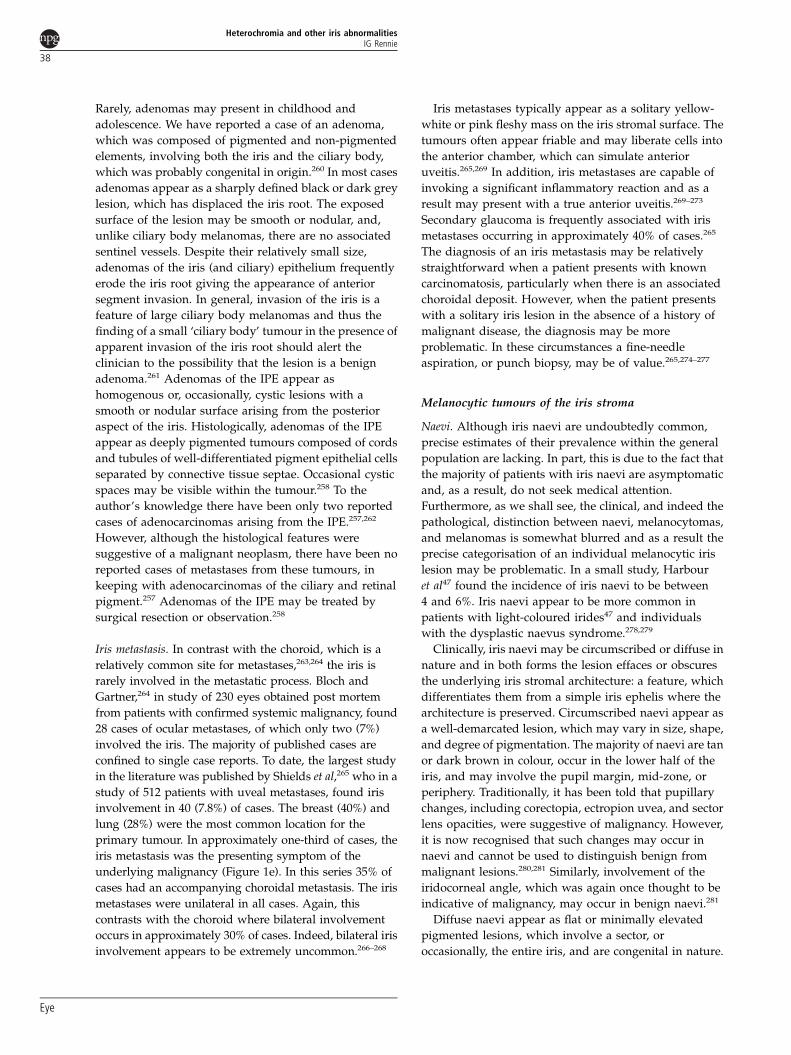
Figure 1 (a) Sectorial pigmentation in Waardenburg syndrome. (b) Lisch nodules in neurofibromatosis type-I. Note the predominantinferior location. (c) Anterior stromal cyst containing a turbid sediment. (d) Multiple posterior pigment epithelial cysts. This patientpresented with raised intraocular pressure. Note the plateaux iris configuration. (e) A large iris metastasis in a patient with an occultbronchiogenic carcinoma. (f) A peripheral iris melanocytoma.
Heterochromia and other iris abnormalitiesIG Rennie
32
Eye

when differentiating them from naevi and other
melanocytic tumours of the iris. Iris freckles are
extremely common and may be found in over 60%
of the general population.47 There is no evidence to
suggest that iris freckles are capable of malignant
transformation.
Lisch nodules. In 1937 Karl Lisch48 reported his
observations of brown nodules on the surface of the iris
in three patients with neurofibromatosis and as a result
such lesions now bear his name. However, as is the case
with many medical eponymous terms, Lisch was not first
to describe the presence of pigmented iris nodules in
neurofibromatosis. Snell and Treacher Collins,49
Goldstein and Wexler,50 Fuchs,51 and Sakurai52 all
describe similar patients prior to Lisch. Moreover, Van
der Hoeve53 in his 1932 Doyne Memorial lecture on the
eye symptoms in phakomatoses described a family with
neurofibromatosis and iris lesions originally observed by
Waardenburg. Although not pathognomonic, Lisch
nodules rarely occur in patients not suffering from
neurofibromatosis type-I. Lal et al54 reported a case of
unilateral Lisch nodules in a 14-year-old boy who
otherwise had no other signs of NF1. They commented
that this could represent a somatic mutation of the NF1
with limited mosaicism, perhaps limited to only that
sector of the iris. Lisch nodules have also been described
in Cushing’s disease,55 familial angiolipomatosis,56 and
neurofibromatosis type-II.57
Lisch nodules are not present at birth and usually
appear in the late childhood, after the appearance of cafe
au lait spots but before the development of overt
neurofibromas.58 Almost all patients with NF1 older than
20 years of age will have Lisch nodules.59 Clinically, Lisch
nodules are small (41 mm diameter) discrete yellow or
brown, dome-shaped nodules on the surface of the iris.
They usually have a smooth surface with a gelatinous
like interior. While the finding of two or more
Lisch nodules is one of the diagnostic criteria for NF1,
most affected irides contain many more and
occasionally in excess of a hundred are found to be
present in the adult eye.58
Early histopathological studies suggested that Lisch
nodules were melanocytic hamartomas. In 1982 Perry
and Font described the electron microscopic appearances
of Lisch nodules in an iridectomy specimen obtained
from a 75-year-old male suffering from NF1. They found
that the nodules were composed of spindle-shaped cells,
which were melanocytic in origin, and based on these
observations they suggested that they were indeed
melanocytic hamartomas.60 Williamson et al61 in a further
electron microscopic study also suggested that Lisch
nodules were melanocytic in origin. However, more
recent observations have cast doubt on the
hamartomatous nature of these lesions and suggest that
they should be considered as benign iris tumours.
Richetta et al62 studied the histopathological and
ultrastructural features of a Lisch nodule obtained from a
50-year-old woman with NF1 and found that it was
composed of three main cell types: pigmented cells,
fibroblast-like cells, and mast cells. They concluded
that Lisch nodules were histologically similar to
neurofibromas. Clinical studies have also challenged the
concept that Lisch nodules are true hamartomas. Nichols
et al63 in a study of 369 patients with NF1 noted that in
80% of affected eyes the Lisch nodules were located
inferiorly. Wood et al64 reported a single case of inferiorly
located Lisch nodules in a patient suffering from bilateral
ptosis. In a recent study Boley et al65 mapped and
quantified the distribution of Lisch nodules in 73 adults
with NF1 and found that the number of nodules in the
inferior hemifield was statistically greater when
compared with the superior hemifield (Figure 1b). They
also noted that the light irides harboured significantly
more Lisch nodules than the dark irides. These findings
suggest that Lisch nodules may arise secondary to
exposure to UV light from sunlight for it is thought that
the lower half of the iris receives a greater amount of
incident sunlight than the upper hemifield.66,67 Indeed, as
we shall see later, iris melanomas are also usually located
in the inferior hemifield. If UV exposure is a factor in the
development of Lisch nodules, then this presumably is due
to DNA damage, which would suggest that these lesions
are benign tumours rather than true hamartomas. Although
elements of this argument are persuasive, it does not
explain why these lesions are homogeneous in appearance,
fail to continue to grow throughout life, and apparently
do not undergo occasional malignant transformation as
would be expected with other benign tumours.
Ocular/oculodermal melanocytosis. In 1861 Hulke68 reported
a case of a uveal malignant melanoma arising in a female
patient who was noted to have pigmentation of the
eyelid, eyebrow sclera, and fundus. A number of
sporadic cases involving pigmentation of the peri-ocular
skin, sclera, and conjunctiva subsequently appeared in
the literature69 before, in 1938, Ota70 at a meeting of the
dermatological society in Japan, described the condition,
which now bears his name. The following year Tanino71,
Ota’s pupil, published a report of 26 cases of Naevus of
Ota. Fitzpatrick et al72 first proposed the term
oculodermal melanocytosis for Naevus of Ota and ocular
melanocytosis for pigmentation of the ocular tissues in
the absence of an associated peri-ocular naevus. It is now
apparent that these conditions represent a spectrum of
congenital pigmentation involving the skin and/or
ocular tissues. In a study by Teekhasaenee et al73 of 194
affected patients, they found that 67 (34.5%) had skin
Heterochromia and other iris abnormalitiesIG Rennie
33
Eye

involvement alone, 12 (6.2%) had only ocular
involvement, and the remaining 115 (59.3%) had both
ocular and dermal pigmentation.
Histologically, the cutaneous elements of oculodermal
melanocytosis contain spindle and dendritic cells in the
dermis resembling melanocytes migrating from the
neural crest to the epidermis.74 In this respect
oculodermal melanocytosis is similar to Mongolian spot,
naevus of Ito, and blue naevus. Histopathological studies
of affected eyes have shown the presence of pigmented
stellate melanocytes distributed throughout the sclera
and episcleral tissues together with increased numbers
of benign plump dendritic monocytes within the
choroid.75–77 An ultrastructural study of an affected iris
demonstrated the presence of numerous melanocytes in
the anterior border and iris stromal layers. Many of these
melanocytes, in addition to a normal-sized melanosomes,
contained large round macromelanosomes, which
probably reflects a basic defect in melanogenesis within
the cells.77
Naevus of Ota together with naevus of Ito and
Mongolian spot have been classified as hamartomatous
congenital dermal melanocytoses involving the
migration of neural crest-derived melanocytes during
embryogenesis.74 Indeed, it has been suggested that
Naevus of Ota arises as a result of an arrest in the
migration of neural crest melanocytes, which leads to the
aggregation of these cells in the dermis.78 While this
hypothesis provides a plausible explanation for the
presence of melanocytes within the affected dermis, it
does not explain the excessive numbers of melanocytes
found within the ocular tissues. Other explanations
include local changes in the embryonic environment,
which leads to the preferential differentiation of
migrating neural crest cells into a melanocytic
phenotype.79 Whatever the precise explanation, there can
be little doubt that ocular and oculodermal
melanocytosis arise as a result of a perturbation of
migrating neuro crest melanocytes.
Oculodermal melanocytosis is apparent at or soon
after birth; in over 50% of cases it is almost always
manifest by the end of the second decade of life.73,80
Ocular melanocytosis (Naevus of Ota) is more common
in Orientals than in Caucasian or Black individuals.
Studies in Japan have reported a prevalence ranging
from 0.4 to 0.84%.81 In a study to determine the
prevalence rate of ocular and oculodermal melanocytosis
Gonder et al81 found the prevalence of ocular
melanocytosis in White individuals to be 0.038% and
oculodermal melanocytosis in the Black population to be
0.014%. Oculodermal melanocytosis is generally
considered to occur more frequently in females. Hidano
et al80 reported that oculodermal melanocytosis occurred
five times more frequently in women than in men.
However, other investigators have suggested that this
apparent predilection for females may be distorted by the
fact that women may be more likely to present to
dermatology clinics as a result of their cosmetic
appearance than males.73 Oculodermal melanocytosis is
usually a unilateral, although sporadic cases of bilateral
involvement have been reported in the literature.82–88 The
vast majority of cases of ocular or oculodermal
melanocytosis are sporadic in nature and only rarely
have familial cases been reported in the literature.89–92
Clinically, oculodermal melanocytosis is characterised
by a bluish-grey pigmentation88 involving areas of skin
innervated by the first and the second divisions of the
trigeminal nerve. Occasionally, there may be
pigmentation of the eardrum, buccal mucosa, palate,
nasopharynx, and leptomeninges.78,93 A number of
ocular tissues may be involved, including the
conjunctiva, episclera, cornea, iris, lens, anterior chamber
angle, choroid, and optic disc.73 The episclera is almost
always involved and may range from relatively discreet
bluish spots to confluent dark mottled patches, which are
dispersed randomly on the globe, but usually do not
involve the limbus.73 Episcleral blood vessels crossing
the involved areas are frequently separated from the
pigmentation by a narrow, non-pigmented band. Hyper-
pigmentation of the choroid producing a dark fundus
when compared with the fellow eye is found in
approximately 80% of cases.73 Iris involvement, which
may be generalised or sectorial, is present in almost 90%
of cases. The degree of iris involvement is variable and
ranges from the presence of stellate granules on an
otherwise normal iris to a dense uniform pigmentation,
which obscures the underlying iris architecture.73 Iris
mammillations, which appear as regularly spaced, deep
brown, smooth, conical elevations that cover the iris
surface giving it a velvety appearance to the naked eye,
are found in cases of ocular and oculodermal
melanocytosis.94 These changes may be the initial
manifestation of ocular melanocytosis in the absence of
conjunctival or episcleral pigmentation.95 Iris
mammillations are not exclusive to ocular melanocytosis
and have been observed sporadically or in association
with systemic conditions, including phakomatosis
pigmentovascularis type-IIb and neurofibromatosis
type-I.94 Rarely, iris mammillations may be familial.96
Ocular and oculodermal melanocytosis have been
reported in association with a number of ocular
conditions, including; glaucoma,73,97–103 retinitis
pigmentosa,104 congenital cataract,105 and Duane’s
syndrome.106 However, there can be little doubt that the
most important association is the development of uveal
melanoma in the eyes involved by these conditions.
There have been numerous reports in the literature since
the first description by Hulke68 of the development of
Heterochromia and other iris abnormalitiesIG Rennie
34
Eye

uveal melanomas in cases of ocular and oculodermal
melanocytosis.75,76,86,95,107–135 Singh et al136 estimated the
lifetime risk of a patient with ocular or oculodermal
melanocytosis of developing a uveal melanoma to be
approximately 1 in 400 as compared with 1 in 13 000 for
the general population. This represents an approximately
30-fold increased risk. Ninety percent of the patients who
developed a melanoma did so between the ages of 31 and
80 years. There have been relatively few reports of iris
melanomas developing in the context of ocular or
oculodermal melanocytosis, which probably reflects a
relative rarity of these tumours when compared with
posterior uveal melanomas.113,132,134,135
Iris cysts
Primary iris stromal cysts. Primary iris stromal cysts are
rare, with the majority of reports in the literature limited
to single case studies.137–181 Clinically, primary iris
stromal cysts appear as dome-shaped translucent masses
arising from the mid or peripheral surface of the iris.
They may appear lobulated and usually contain a clear or
slightly turbid fluid, although occasionally they may
contain a sediment of white or yellowish material
resembling a hypopyon within the cyst155,171 (Figure 1c).
The majority of cases are diagnosed within the first year
of life, with occasional cases reported in adults.155 This
condition appears to be unilateral in that the author is
unaware of any reports of bilateral involvement. In most
cases a stromal cyst arises in the inferior or temporal
aspect of the iris.155 In general, primary iris stromal cysts,
particularly in children, undergo progressive
enlargement, which may lead to visual loss when they
encroach upon the visual axis.155 Rarely, spontaneous
collapse or regression of a stromal cyst has been
reported.156,176,177 Focal corneal oedema may arise as a
result of contact between the stromal cyst and the corneal
endothelium.137,142,161,163 Raised intraocular
pressure,159,163,172,173 hyphaema137 iritis, and subluxation
of the lens163,167,171,173–175 have been found in association
with primary iris stromal cysts.
Although primary iris stromal cysts may develop in
adults, they are generally considered to be congenital in
origin. Stromal cysts, which arise later in life, have
probably been dormant and become apparent as the
result of an accumulation of fluid within the cyst,
triggered by an unknown stimulus.170 The precise
aetiology of primary iris stromal cysts remains unclear
and proposed mechanisms include developmental
entrapment of surface ectoderm, neuroectoderm, or
surface ectodermal implantation as a result of occult
trauma.163 Histologically, primary iris stromal cysts
are lined with a multi-layered squamous or
cuboidal epithelium, which may or may not contain
mucin-secreting goblet cells.138,139,142,163,168–170,173,174,178,179
Occasionally, focal keratinisation of the epithelial lining
has been observed.170,180 Immunohistochemical studies
have demonstrated a positive reaction for epithelial
cytokeratin markers and a negative result for the S100
antigen.159,163,166,170 Electron microscopic studies have
shown the presence of microvilli on the luminal surface
of the epithelial cells together with desmosomes and
tonofilaments.163,166,170,178 These studies support the
notion that primary iris stromal cysts originate from the
surface ectoderm, displaced probably at the time of
formation of the lens vesicle.170 Rummelt et al182 reported
two cases of congenital epithelial iris cysts in children
who had a maternal history of diagnostic amniocentesis.
In one case a perforating limbal scar with a
corresponding break in a Descemet’s membrane was
observed. Lois et al155 in a series of 17 patients with
primary iris stromal cysts noted a history of diagnostic
amniocentesis in two patients. However, in neither case
was there any sign of a penetrating injury to the eye. It
would appear that while diagnostic amniocentesis is a
possible cause of congenital stromal iris cysts, the
majority of cases occur in the absence of any obvious
intrauterine trauma.
A wide range of modalities have been used to treat
symptomatic primary iris stromal cysts, including simple
aspiration,155,171 and injection with trichloroacetic acid,158
xenon photocoagulation,158 neodymium-YAG laser,164,171
argon laser photocoagulation,155,181 cryotherapy,155,181
iridectomy,158,142 and block excision.160,180 Unfortunately,
re-occurrence following treatment, particularly simple
puncture either surgical or laser, is common and
prognosis for vision, particularly in young children
where amblyopia may be a problem, is often poor.
Recently, Shen et al144 reported a promising surgical
technique by using a viscoelastic material to dissect the
cyst from the corneal endothelium, followed by
aspiration of the cyst, excision, and micro-diathermy.
Posterior cysts. Primary cysts of the IPE or ciliary body
have been recognised for over a century.183 In 1897
Zimmerman183 reported a case of bilateral pigmented
tumours, which he considered to be ciliary body cysts.
Following this report, a number of case reports appeared
in the literature noting the appearance of cysts of either
the ciliary body or IPE.184–187 Although initially
considered as separate entities, it would now appear that
the majority of cysts of the IPE and ciliary body arise
from the irido-ciliary sulcus or pars plicata, and should
best be considered as irido-ciliary cysts (vide infra).
Historically, irido-ciliary cysts were considered to be rare;
however, with the advent of the ultrasound
biomicroscope it is now recognised that small
asymptomatic cysts are extremely common and that only
Heterochromia and other iris abnormalitiesIG Rennie
35
Eye

a small proportion of them attain sufficient size or
number to become clinically apparent. Recently,
Kunimatsu et al188 in a prospective study of 232 eyes of
116 normal subjects by using an ultrasound
biomicroscope found evidence of irido-ciliary cysts in
54.3% of patients.
In 1981 Shields140 reported a study of 62 patients
with primary iris cysts, of which 59 were considered
to have arisen from the posterior pigment epithelium.
He classified the pigment epithelial cysts into five
groups: central, mid-zonal, peripheral, dislodged into the
anterior chamber, and dislodged into the posterior
chamber. In a subsequent study using the same
classification, Lois et al189 reported the clinical features
and the natural course of primary cysts of the IPE in 234
patients. They found central (confined to the pupillary
margin) in three patients (6%), mid-zonal cysts in 50
patients (21%), peripheral in 170 patients (73%), and
dislodged in eight patients (3%). However, less than
10% of these patients underwent an ultrasound
biomicroscope examination to confirm the extent and the
posterior origin of the cysts. Subsequent studies using
ultrasound biomicroscope would suggest that both
peripheral and mid-zonal cysts originate from the irido-
ciliary sulcus.190,191 Indeed, in the author’s personal
experience, all large cysts involving the mid-zone appear
to arise from the irido-ciliary sulcus. It is probable that
the majority, if not all, of pigment epithelial cysts (with
the exception of those that have become detached and
now float in either the vitreous or aqueous) arise either
from the pupil margin or the irido-ciliary sulcus.
Moreover, as we shall see, the aetiology of these two
groups appears to be quite distinct.
Although the majority of small peripheral pigment
epithelial (irido-ciliary) cysts are asymptomatic, large
cysts can cause focal abnormalities of the iris, which may,
on occasion, stimulate an ocular neoplasm. Sadly, prior to
the advent of the ultrasound biomicroscope, pigment
epithelial cysts had been mistaken for ciliary body
melanomas resulting in unnecessary enucleation.186
Clinically, isolated peripheral irido-ciliary cysts present
as a focal anterior bulging of the peripheral iris, with an
associated shallowing of the anterior chamber. Larger
cysts may be visible at the pupil margin, particularly
following pharmacological mydriasis. In these
circumstances the cyst appears as a smooth uniformly
pigmented mass lying between the posterior surface of
the iris and the lens. On careful inspection the cyst will
usually transilluminate when a fine slit-lamp beam is
shone upon its surface. The majority of solitary pigment
epithelial cysts appear to arise in the inferior temporal
quadrant.189 Bilateral pigment epithelial cysts are
common and indeed, when an individual presents with
an apparently unilateral lesion, careful inspection of the
fellow eye will frequently reveal the presence of an occult
lesion in that eye. Occasionally, patients may present
with multiple bilateral irido-ciliary cysts191 (Figure 1d)
and in extreme cases this may produce a plateau iris
leading to angle closure glaucoma.189,191–199 Although
irido-ciliary cysts are generally sporadic in nature, Vela
et al197 reported three families with pigment epithelial
cysts, which were multiple in 10 and bilateral in eight of
the 11 affected patients. Acute closed angle glaucoma
occurred in four of the affected patients.
Although the aetiology of irido-ciliary cysts remains
uncertain, numerous theories have been proposed,
including; persistence of the annular sinus of von
Szily;198 foetal iritis causing synechiae with resultant
separation of the two layers of the secondary optic
vesicle;200 and embryonic traction of the zonule of Zinn
on the posterior epithelial layer of the secondary optic
cup.186 It is tempting to speculate that, given peripheral
pigment epithelial cysts appear to arise from the irido-
ciliary sulcus where there is transition between the non-
pigmented epithelium of the ciliary body and the
pigment epithelium of the iris, their formation may be
due to a focal juxtaposition of these elements, leading to
the aberrant secretion of aqueous beneath the pigment
epithelium.
While the majority of peripheral pigment epithelial
cysts occur in isolation, it is well-recognised that on
occasion they may be associated with a ciliary body
tumour. Augsburger et al201 noted, in a study by using the
ultrasound biomicroscope, that in 39 patients with
pigment epithelial cysts, six occurred at the margin of a
ciliary body tumour. Fine and Pavlin190 in a further
ultrasound biomicroscopic study of 210 irido-ciliary cysts
found that 20% were associated with a tumour. While the
majority of these cysts are clinically undetectable and can
only be diagnosed with the aid of an ultrasound
biomicroscope, occasionally iris cysts may be the
presenting sign of a ciliary body tumour202 and for this
reason it is important to exclude an associated neoplasm
in any patient who presents with an apparently isolated
pigment epithelial cyst. Moreover, cystic ciliary body
melanomas may simulate a pigment epithelial cyst and
again this phenomena reinforces the need to undertake a
meticulous ultrasound biomicroscope examination in
any patient presenting with a solitary cyst.203
Central pigment epithelial cysts, arising from the
pupillary margin, are a well-recognised product of miotic
therapy. In 1923 Vogt204 described single or multiple cysts
at the pupillary margin in patients receiving miotic
(pilocarpine) therapy for chronic simple glaucoma.
Abraham205 in a study of 66 cases of accommodative
strabismus treated with di-isopropyl-fluoro-phosphate
noted that pupillary cysts developed in 42 (64%) patients
and that occasionally they occur as early as 2 weeks after
Heterochromia and other iris abnormalitiesIG Rennie
36
Eye

starting treatment. Chin et al206 reported the occurrence
of pupillary cysts in patients receiving phospholine
iodide for accommodative strabismus; they noted that
this effect could be inhibited by concomitant therapy
with the mydriatric phenylephrine. Histologically, the
cysts are found to contain finger-like projections
consisting of a double layer of pigment epithelial cells
enclosing cystic spaces.207
In 1936 Cowan208 described a case of familial cysts
and flocculi of the iris. Flocculi have been described as
tuft-like excrescences of the pigment epithelium
overlying the pupil margin and are often cystic in nature.
It is probable that they are aetiologically closely related to
simple pigment epithelial cysts and indeed in some
articles the term appears interchangeable. More recently,
Sallo and Hatvani209 reported four cases of primary
pupillary pigment epithelial cysts in a single family.
Although there was no mention of any systemic
abnormality in this pedigree, several articles have
suggested an association between iris flocculi/pupillary
pigment cysts and inherited ascending thoracic aortic
aneurysms and dissections (TAAD).210–214 In the past few
years several research groups have identified that TAAD
may be caused by mutations of the smooth muscle
a-actin (ACTA2)213,214 gene and moreover, that iris
flocculi may be specific to the p.R149C mutation.
Vascular tumours and malformations of the iris
Vascular tumours and malformations of the iris are
extremely rare. Ashton215 in a pathological study of 145
primary iris tumours found only three primary angiomas
of the iris. Indeed, Ferry216 in an archival study
concluded that the majority of tumours classified as
haemangiomas were in fact juvenile xanthogranulomas,
highly vascular melanomas or other non-vascular
tumours. However, true vascular tumours and
malformations of the iris have been reported in the
literature and include capillary,217–220 cavernous,221–226
racemose,227–231 microhaemangiomas,232–239 and iris
varix.240–243
Capillary haemangiomas are paediatric malformations
and appear to arise in association with either diffuse
neonatal haemangiomatosis or peri-orbital capillary
haemangiomas.217–220 In general, cavernous
haemangiomas arise in adulthood and are not associated
with systemic haemangiomas.222,241 However, sporadic
case reports have detailed their association with
haemangiomas in other non-ocular sites.241
Microhaemangiomas (iris vascular tufts) are probably
the most common vascular malformation to arise in the
iris, with a total of approximately 90 reported cases in the
literature.234 They typically occur bilaterally and appear
as solitary or multiple small (approximately 150mm in
diameter) vascular tufts on the pupil margin. Most cases
occur in patients over 50 years of age and indeed, iris
microhaemangiomas have not been reported in
childhood.234 Although microhaemangiomas have been
reported in association with a wide variety of systemic
conditions, these are probably, in the majority of cases,
merely coincidental, with the possible exceptions of
myotonic dystrophy244–246 and diabetes mellitus.232,246
Spontaneous hyphaema, which may be complicated
by secondary raised intraocular pressure, is a
well-recognised complication of iris vascular
tufts.237,239,247–256 Iris microhaemangiomas are thought to
be hamartomas arising from the stromal blood vessels.235
In 1983 Stur and Strasser227 described a sectorial
racemose arterio-venous malformation in a 32-year-old
male. Since this report approximately 36 further cases of
isolated iris arterial-venous malformation have been
reported in the literature.230,241 These lesions consist of
one or more abnormally large iris blood vessels that
originate in the iridocorneal angle, which pass through
the stroma (which in places may obscure them) towards
the pupil margin for a variable distance before forming
an abrupt loop and returning to the angle.229 Fluorescein
angiography readily identifies these vascular
malformations, which fill rapidly and demonstrate no or
only minimal leakage or staining of the vessel wall.229 A
dilated episcleral vessel in the same quadrant as the
arterio-venous malformation was noted in 50% of
cases.229 The presence of such a vessel may be confused
with sentinel vessels, which are associated with an
underlying ciliary body tumour. Iris arterio-venous
malformations are not associated with any systemic
condition, nor do they give rise to any symptoms.
Iris tumours
Adenomas and adenocarcinomas of the IPE. Adenomas and
adenocarcinomas of the IPE are rare. Spraul et al,257 in
1996, reported an adenocarcinoma of the IPE and cited 20
previously reported cases, which would appear to fulfil
the criteria to be classified as adenomas of the IPE.
Shields et al258 in 1999 reported their personal experience
of 20 cases of adenomas arising from the IPE. Since then,
only a further single report of an adenoma of the IPE has
appeared in the literature.259 The majority of adenomas
of the IPE appear to arise from the peripheral iris and
may involve the ciliary body. Moreover, adenomas may
arise from the pigment epithelium of the ciliary body
and, as a result, it is often difficult on clinical grounds to
determine which pigment epithelium the tumour has
arisen from. Clinically, adenomas of the IPE present as a
small asymptomatic mass in the peripheral iris. There
appears to be no sex or racial predilection, and typically
such lesions present in the fourth to sixth decade of life.
Heterochromia and other iris abnormalitiesIG Rennie
37
Eye
Rarely, adenomas may present in childhood and
adolescence. We have reported a case of an adenoma,
which was composed of pigmented and non-pigmented
elements, involving both the iris and the ciliary body,
which was probably congenital in origin.260 In most cases
adenomas appear as a sharply defined black or dark grey
lesion, which has displaced the iris root. The exposed
surface of the lesion may be smooth or nodular, and,
unlike ciliary body melanomas, there are no associated
sentinel vessels. Despite their relatively small size,
adenomas of the iris (and ciliary) epithelium frequently
erode the iris root giving the appearance of anterior
segment invasion. In general, invasion of the iris is a
feature of large ciliary body melanomas and thus the
finding of a small ‘ciliary body’ tumour in the presence of
apparent invasion of the iris root should alert the
clinician to the possibility that the lesion is a benign
adenoma.261 Adenomas of the IPE appear as
homogenous or, occasionally, cystic lesions with a
smooth or nodular surface arising from the posterior
aspect of the iris. Histologically, adenomas of the IPE
appear as deeply pigmented tumours composed of cords
and tubules of well-differentiated pigment epithelial cells
separated by connective tissue septae. Occasional cystic
spaces may be visible within the tumour.258 To the
author’s knowledge there have been only two reported
cases of adenocarcinomas arising from the IPE.257,262
However, although the histological features were
suggestive of a malignant neoplasm, there have been no
reported cases of metastases from these tumours, in
keeping with adenocarcinomas of the ciliary and retinal
pigment.257 Adenomas of the IPE may be treated by
surgical resection or observation.258
Iris metastasis. In contrast with the choroid, which is a
relatively common site for metastases,263,264 the iris is
rarely involved in the metastatic process. Bloch and
Gartner,264 in study of 230 eyes obtained post mortem
from patients with confirmed systemic malignancy, found
28 cases of ocular metastases, of which only two (7%)
involved the iris. The majority of published cases are
confined to single case reports. To date, the largest study
in the literature was published by Shields et al,265 who in a
study of 512 patients with uveal metastases, found iris
involvement in 40 (7.8%) of cases. The breast (40%) and
lung (28%) were the most common location for the
primary tumour. In approximately one-third of cases, the
iris metastasis was the presenting symptom of the
underlying malignancy (Figure 1e). In this series 35% of
cases had an accompanying choroidal metastasis. The iris
metastases were unilateral in all cases. Again, this
contrasts with the choroid where bilateral involvement
occurs in approximately 30% of cases. Indeed, bilateral iris
involvement appears to be extremely uncommon.266–268
Iris metastases typically appear as a solitary yellow-
white or pink fleshy mass on the iris stromal surface. The
tumours often appear friable and may liberate cells into
the anterior chamber, which can simulate anterior
uveitis.265,269 In addition, iris metastases are capable of
invoking a significant inflammatory reaction and as a
result may present with a true anterior uveitis.269–273
Secondary glaucoma is frequently associated with iris
metastases occurring in approximately 40% of cases.265
The diagnosis of an iris metastasis may be relatively
straightforward when a patient presents with known
carcinomatosis, particularly when there is an associated
choroidal deposit. However, when the patient presents
with a solitary iris lesion in the absence of a history of
malignant disease, the diagnosis may be more
problematic. In these circumstances a fine-needle
aspiration, or punch biopsy, may be of value.265,274–277
Melanocytic tumours of the iris stroma
Naevi. Although iris naevi are undoubtedly common,
precise estimates of their prevalence within the general
population are lacking. In part, this is due to the fact that
the majority of patients with iris naevi are asymptomatic
and, as a result, do not seek medical attention.
Furthermore, as we shall see, the clinical, and indeed the
pathological, distinction between naevi, melanocytomas,
and melanomas is somewhat blurred and as a result the
precise categorisation of an individual melanocytic iris
lesion may be problematic. In a small study, Harbour
et al47 found the incidence of iris naevi to be between
4 and 6%. Iris naevi appear to be more common in
patients with light-coloured irides47 and individuals
with the dysplastic naevus syndrome.278,279
Clinically, iris naevi may be circumscribed or diffuse in
nature and in both forms the lesion effaces or obscures
the underlying iris stromal architecture: a feature, which
differentiates them from a simple iris ephelis where the
architecture is preserved. Circumscribed naevi appear as
a well-demarcated lesion, which may vary in size, shape,
and degree of pigmentation. The majority of naevi are tan
or dark brown in colour, occur in the lower half of the
iris, and may involve the pupil margin, mid-zone, or
periphery. Traditionally, it has been told that pupillary
changes, including corectopia, ectropion uvea, and sector
lens opacities, were suggestive of malignancy. However,
it is now recognised that such changes may occur in
naevi and cannot be used to distinguish benign from
malignant lesions.280,281 Similarly, involvement of the
iridocorneal angle, which was again once thought to be
indicative of malignancy, may occur in benign naevi.281
Diffuse naevi appear as flat or minimally elevated
pigmented lesions, which involve a sector, or
occasionally, the entire iris, and are congenital in nature.
Heterochromia and other iris abnormalitiesIG Rennie
38
Eye

They are usually encountered in cases of ocular or
oculodermal melanocytosis.133 A diffuse iris naevus
may also be encountered in Cogan–Reese syndrome.
This condition first described in 1969,282 is a variant
of the iridocorneal endothelial syndrome, and is
characterised by unilateral glaucoma; abnormalities of
the corneal endothelium; and iris changes, including,
corectopia, ectropion uvea, pigmented nodules, and
iris atrophy.283
Iris naevi are generally indolent lesions, which remain
asymptomatic throughout life. Occasionally, secondary
glaucoma may arise as a result of pigment deposition
in the trabecular meshwork. Although rapid tumour
growth is considered to be indicative of malignancy,
iris naevi may slowly increase in size with time. In a
study of 175 melanocytic tumours of the iris, Territo
et al284 noted definite tumour growth in eight patients,
of which five were treated by iridocyclectomy. Two of
these lesions were subsequently found to be spindle
cell naevi.
Two reports have now appeared in the literature
describing an aggressive naevus of the iris in children.
Paridaens et al285 described an apparently unique familial
occurrence of an aggressive iris naevus arising in the
second decade of life in a mother and son. Carlson et al286
subsequently reported an aggressive iris naevus in a
16-year-old girl who presented with visual loss as a
result of uncontrolled secondary glaucoma.
While it is probable that iris naevi may occasionally
undergo malignant transformation, it is not possible to
estimate how frequently this change occurs. Many iris
melanocytic lesions, which histologically appear to be
malignant, may remain indolent for many years and as
result, should a lesion show a significant increase in size,
it may be impossible to determine whether this is due to
malignant transformation in a pre-existing naevus of
growth in a previously dormant melanoma.
Melanocytoma. A melanocytoma is a specific type of
naevus, which demonstrates characteristic histological
features. They are composed of deeply pigmented plump
or polyhedral naevus cells, which contain abundant
cytoplasm and demonstrate little nuclear
pleomorphism.287 Although melanocytomas typically
arise on or adjacent to the optic disc, they may
occasionally occur in other locations in the eye, including
the choroid,288–298 the ciliary body,299–312 and the
iris.299,301,313–327
In 1965 Zimmerman313 reported an iris melanocytoma
in a 34-year-old, which was treated by iridectomy and
subsequent enucleation, the initial specimen having been
reported to be a malignant melanoma. Since then a
number of reports have appeared in the literature
documenting the occurrence of melanocytomas within
the iris.297,299,311–325 These lesions are, however,
uncommon, and in a study of 200 patients referred to an
ocular oncology service with the suspected diagnosis of
an iris melanoma, 158 were found to have an alternative
diagnosis and of these only one proved to be a
melanocytoma.328 In a subsequent study of 47 cases of
iris melanocytoma, Demirci et al322 estimated that
melanocytomas represented only 3% of all iris naevi.
Clinically, iris melanocytomas appear as a darkly
pigmented nodule with an irregular or corrugated
surface322 (Figure 1f). In keeping with other melanocytic
iris lesions, melanocytomas usually involve the inferior
half of the iris. These lesions appear to be less cohesive
than ordinary naevi and may produce satellite lesions on
the iris surface or trabecular meshwork.287 This lack
of cohesion may be due to necrosis within the
melanocytoma.314,318,322,324 Involvement of the trabecular
meshwork may give rise to a raised intraocular
pressure.314,315,318,324 Indeed, Demirci et al322 in their
study noted that 11% of patients had developed a raised
intraocular pressure at 5 and 10 years, and that this had
risen to 55% at 15 years. Approximately 50% of iris
melanocytomas may demonstrate a gradual enlargement
with time,322 and although this is not indicative of
malignant transformation, there are sporadic reports of
melanomas arising from iris melanocytomas in the
literature.316,325,329
Melanoma. Iris melanomas are uncommon and account
for only between 2 to 5% of all uveal melanomas.330,331
Despite their relative rarity, iris melanomas remain an
enigma and controversy surrounds both their diagnosis
and management. Most studies indicate that the mean
age of presentation for iris melanomas is 40–45 years:
a decade earlier than their posterior uveal
counterparts.330–332 Iris melanomas may be either
circumscribed or diffuse, and in both forms the clinical
distinction between a melanoma and benign naevus may
be difficult.
Circumscribed melanomas have an affinity for the
inferior iris and approximately 80% of cases arise from
the inferior half of the iris.332 They appear as a raised
lesion with either a smooth or irregular surface. The
degree of pigmentation is variable and while the
majority of iris melanomas are brown in colour,
some are amelanotic: the latter often showing a
prominent intrinsic vasculature. Pupil distortion,
ectropion uveae, localised cataract, iridocorneal angle
involvement, pigment dispersion, raised intraocular
pressure, and spontaneous hyphaema may be found in
iris melanomas.284,330
Diffuse iris melanomas are extremely rare, accounting
for 7–10% of all iris melanomas.330,333 Clinically, diffuse
iris melanomas classically present as a patient with
Heterochromia and other iris abnormalitiesIG Rennie
39
Eye
unilateral hyperchromatic heterochromia and ipsilateral
glaucoma.333–335 In affected cases the iris has a diffuse or
multifocal pigmentation. Associated corectopia and
ectropion iridis occur in approximately 90% of cases and
involvement of the iridocorneal angle appears to be a
universal feature.333 Unfortunately, there is frequently a
significant delay in diagnosing diffuse iris melanomas.
Demirci et al333 in a report of 25 cases of diffuse iris
melanoma found that 14 (56%) of the cases referred to
their unit for further management had previously been
diagnosed elsewhere as suffering from glaucoma and
that as a result there was a mean delay of 30 months
before eventual diagnosis was made. A subsequent
review of the literature confirmed a similar delay in
diagnosing diffuse iris melanomas.333 This is in keeping
with the authors’ own experience where the condition
had initially been misdiagnosed as Cogan–Reese
syndrome. Histologically, diffuse iris melanomas, in
contrast to circumscribed tumours, frequently contain
epithelioid cells, which tend to be poorly cohesive and
may account for the diffuse nature of these lesions.333
In 1951 Stallard336 wrote that the prognosis for iris
melanomas treated by iridectomy was good. A few years
later Rones and Zimmerman332 in a retrospective study
of 125 cases of iris lesions, which they considered to be
either malignant or have malignant potential obtained
from the Armed Forces Institute of Pathology files, found
the incidence of metastases for patients with iris
melanoma to be 1% at 5 years and 6% at 10 years. They
observed that the iris melanomas appeared to have a
different natural history than their posterior uveal
counterparts. Subsequent studies have confirmed that
iris melanomas generally have a very favourable
prognosis and that the risk of death from metastasis-
related disease is small.337–343 The one exception to this
general rule appears to be diffuse iris melanomas where
the overall risk of metastases appears to be significantly
greater.333
The apparently favourable prognosis for iris
melanomas, together with the difficulty in clinically
differentiating many lesions from naevi, has prompted
many clinicians to adopt a conservative approach to the
treatment of such lesions; proposing treatment should
rapid growth or other significant complications occur.
Traditionally, circumscribed iris melanomas have been
treated by iridectomy or iridocyclectomy.331–345
Alternative treatment modalities include plaque
brachytherapy346,347 and proton beam irradiation.348–350
The management of diffuse iris melanomas is
problematic and most are probably best treated by
enucleation, although irradiation may be tried in cases
where the patient is reluctant to suffer loss of their eye.333
The apparently low risk of metastatic disease in cases
of iris melanoma contrasts with that of choroidal and
ciliary body tumours where the mortality rates are
significantly greater. In 1992, Diener-West et al351
published a meta-analysis of the 5-year mortality rates
among patients who had an eye enucleated for a
choroidal melanoma and found the combined weighted
estimates to be 16% for small, 32% for medium, and 53%
for large tumours. In a recent study of the very-long-term
prognosis of patients with choroidal and ciliary body
melanoma, Kujala et al352 found tumour-related mortality
to be 31% by 5 years, 45% by 15 years, 49% by 25 years,
and 52% by 35 years. This, of course, poses the intriguing
question as to why there should be such a disparity in
relative survivals between iris and posterior uveal
melanoma.
Jakobiec and Silbert280 in a retrospective clinical
pathological study of 189 iris and iris and ciliary body
lesions originally diagnosed as melanomas proposed a
new nine part histological classification for such
tumours. They subsequently reassigned 87% of the
tumours studied into one of six categories, all of which
they considered to be benign.280 In doing so they
suggested that most iris ‘melanomas’ were in fact benign
naevi. If this were indeed the case, then the apparently
favourable prognosis for such tumours in previously
reported series could be explained by the inclusion of
benign lesions into the study groups, which would have
the effect of diluting the true malignant neoplasms.
While this may the case, it does not entirely explain this
paradox. In 2001 Shields et al331 reported the results of a
study of 169 consecutive patients with microscopically
confirmed iris melanoma and found a metastatic rate of
3% at 5 years and 5% at 10 years, which is remarkably
similar to the results found by Rones and Zimmerman332
over 40 years earlier. However, although this excellent
study provides us with the incidence of metastatic
disease for patients with histologically proven iris
melanoma, it suffers from an inherent flaw when
considering the overall prognosis for patients with iris
melanomas. In this study they obtained the incidence of
metastases in 169 patients with histologically proven iris
melanoma from a total cohort of 1054 patients referred to
the ocular oncology service with suspicious iris
melanocytic tumours. The decision to treat or obtain
histological biopsy of the lesion was, of course, based on
their clinical criteria for possible malignancy. It is of
course conceivable that in the remaining 885 patients
with the presumptive diagnosis of an iris naevus
managed by observation, there may have been patients
where the diagnosis would have been that of a
melanoma had the tumour been removed and submitted
for histological examination.
It is an established fact that tumour size at the time of
diagnosis is an important factor in determining the
prognosis for posterior uveal melanomas.351 In general,
Heterochromia and other iris abnormalitiesIG Rennie
40
Eye

iris melanomas are significantly smaller than most
choroidal or ciliary body melanomas at the time of
diagnosis, and this has prompted the argument that the
apparent difference in biological behaviour can merely
be attributed to tumour size. Davidorf353 noted that the
mean volume of iris melanomas at the time of diagnosis
was 55 mm3 compared with a mean volume of 300 mm3
for choroidal melanomas and concluded that if the size of
the tumour at the time of diagnosis was taken into
account the metastatic rates would be comparable.
Again, the same problem arises: the major criterion for
intervention in the case of melanocytic lesion of the iris is
documented growth and it is possible that tumours,
which histologically would be considered malignant, are
left untreated because of the lack of any observable
change in size.
In 1990, Prescher et al354 reported apparently non-
random chromosome abnormalities in 14 cases of
posterior uveal melanoma. Shortly afterwards, my own
group reported similar findings in six cases of uveal
melanoma.355 Following these initial reports it has
now been established that not only do non-random
chromosome abnormalities occur in uveal melanomas,
but that certain abnormalities, namely loss of
chromosome-3 and additional copies of the long arm of
chromosome-8, arise predominantly in ciliary body
tumours and are highly sensitive predictors of patient
survival.356–359 Unfortunately, to date, there are only two
studies (a total of four cases), which have characterised
the cytogenetic changes in iris melanomas.360,361
However, based on this limited evidence it would appear
that, although iris melanomas experience relatively high
levels of chromosome alterations, they are different to
those that are typically found in posterior uveal tumours.
If the results of these preliminary results were confirmed
in a larger cohort of patients, it could implicate
differences in tumour karyotype as a possible cause for
the apparent disparity between the metastatic rates of the
iris and posterior uveal melanomas.
There is one further intriguing possibility that could
explain the apparently different biological nature of the
iris and posterior uveal melanomas: could differences
in the anterior chamber micro-environment have a role
in modulating the behaviour of iris melanomas?
Grossniklaus et al362 used a murine model to investigate a
difference in metastatic rate between anterior and
posterior ocular melanoma, and found that 33% of the
tumours inoculated into the anterior chamber
metastasised in comparison with 89% of those inoculated
into the choroid and/or vitreous. Recently, we have
investigated the possible role of the micro-environment
in tumour growth. In an in vitro study of the effect of
aqueous and vitreous humours on invasion and
proliferation, we found that although neither appeared to
influence tumour cell proliferation, vitreous promoted
tumour invasion whereas aqueous either had no effect or
was inhibitory.363 In a further study we identified six
enucleation specimens where there was clear invasion of
the tumour through the iris stroma and onto the surface.
We noted that the surface melanoma cells were smaller
when compared with those deeper within the lesion and
that fewer of these cells expressed cyclin-D1, a protein
that promotes the cell cycle. Moreover, expression of p27,
a factor, which inhibits the cell cycle, had a high level of
expression in these cells on the anterior tumour surface
than those located within the lesion. We also investigated
alterations of chromosome-3 and 8, and found them to be
less common among the iris surface melanoma cells than
those deeper within the body of the tumour.364 Although
these results to date must be considered as preliminary, it
is plausible that factors within the aqueous humour may
have a role in modulating the growth of iris melanomas.
If such factors could be identified then they may provide
the basis for developing alternative therapies to treat
uveal melanoma.
Conflict of interest
The authors declare no conflict of interest.
Acknowledgements
I am indebted to my colleagues who work closely in the
ocular oncology service, Paul Rundle, Rhona Jacques,
Lesley Hinchliffe, and Michele Evans, without whose
continuing support this lecture would not have been
possible. Finally, I dedicate this lecture to Sydney
Davidson, my friend and mentor for the past 30 years.
References
1 Bron AJ, Tripathi RC, Tripathi BJ. Wolff’s Anatomy of the Eyeand Orbit. 8th edn. Chapman & Hall Medical: London,1997.
2 Imesch PD, Wallow IH, Albert DM. The colour of thehuman eye: a review of morphologic correlates and ofsome conditions that affect iridial pigmentation. SurvOphthalmol 1997; 41(Suppl 2): S117–S123.
3 Wilkerson CL, Syed NA, Fisher MR, Robinson NL, WallowIH, Albert DM. Melanocytes and iris colour. Lightmicroscopic findings. Arch Ophthalmol 1996; 114(4): 437–442.
4 Prota G, Hu DN, Vincensi MR, McCormick SA, NapolitanoA. Characterization of melanins in human irides andcultured uveal melanocytes from eyes of different colours.Exp Eye Res 1998; 67(3): 293–299.
5 Fuchs E. Normal pigmentierte und albinotische iris. GraefesArch Clin Exp Ophthalmol 1913; 84/85: 521.
6 Dieterich CE. [The fine structure of melanocytes in thehuman iris]. Graefes Arch Clin Exp Ophthalmol 1972; 183(4):317–333.
7 Wolfrum P. Uber den Bau der Irisvorderflache desmenschlichen Auges mit vergleichend anatomischen
Heterochromia and other iris abnormalitiesIG Rennie
41
Eye
Bemerkungen. Graefes Arch Clin Exp Ophthalmol 1922; 109:
106–153.8 Eagle Jr RC. Iris pigmentation and pigmented lesions: an
ultra structural study. Trans Am Ophthalmol Soc 1988; 86:
581–687.9 Imesch PD, Bindley CD, Khademian Z, Ladd B, Gangnon R,
Albert DM et al. Melanocytes and iris colour. Electron
microscopic findings. Arch Ophthalmol 1996; 114(4): 443–447.10 Sturm RA, Frudakis TN. Eye colour: portals into
pigmentation genes and ancestry. Trends Genet 2004; 20(8):
327–332.11 Davenport GC, Davenport CB. Heredity of eye-colour in
man. Science 1907; 26: 590–592.12 Hurst CC. On the inheritance of eye-colour in man. Proc R
Soc Med Lond B Biol Sci 1908; 80: 81–86.13 Sturm RA, Larsson M. Genetics of human iris colour and
patterns. Pigment Cell Melanoma Res 2009; 22(5): 544–562.14 Sturm RA. Molecular genetics of human pigmentation
diversity. Hum Mol Genet 2009; 18: 9–17.15 Duffy DL, Montgomery GW, Chen W, Zhao ZZ, Le L,
James MR et al. A three-single-nucleotide polymorphism
haplotype in intron 1 of OCA2 explains most human eye-
colour variation. Am J Hum Genet 2007; 80(2): 241–252.16 Duke Elder S. System of Ophthalmology. Henry Kimpton:
London, 1964.17 Kutzbach B, Mendelsohn N, Rath P, Summers CG. Sectoral
iris heterochromia and retinal pigment variation in 13q-
syndrome. J Am Assoc Pediatr Ophthalmol Strabismus 2007;
11(5): 513–515.18 Lascaratos J. ‘Eyes’ on the thrones: imperial
ophthalmologic nicknames. Surv Ophthalmol 1999; 44(1):
73–78.19 Sullivan TJ, Clarke MP, Morin JD. The ocular
manifestations of the Sturge–Weber syndrome. J PediatrOphthalmol Strabismus 1992; 29(6): 349–356.
20 Pascual-Castroviejo I, Lopez-Rodriguez L, de la Cruz
Medina M, Salamanca-Maesso C, Roche Herrero C.
Hypomelanosis of Ito. Neurological complications in 34
cases. Can J Neurol Sci 1988; 15(2): 124–129.21 Stone RA, Scheie HG. Periorbital scleroderma associated
with heterochromia iridis. Am J Ophthalmol 1980; 90(6):
858–861.22 Quinlan K, Shwayder T. Cafe au lait macule associated
with heterochromia iridis. Pediatr Dermatol 2005; 22(2):
177–178.23 Morrison DA, FitzPatrick DR, Fleck BW. Iris coloboma
with iris heterochromia: a common association. ArchOphthalmol 2000; 118(11): 1590–1591.
24 Abelsdorff G. Uber Blauaugigkeit und Heterophthalmus
bei tauben albinotischen Tieren. Graefes Arch Clin ExpOphthalmol 1904; 59: 376–377.
25 Calhoun FP. Causes of heterochromia iridis with special
reference to paralysis of the cervical sympathetics. Am JOphthalmol 1919; 2: 255–269.
26 Weinstein JM, Zweifel TJ, Thompson S. Congenital
Horner’s syndrome. Arch Ophthalmol 1980; 98: 1074–1078.27 George ND, Gonzalez G, Hoyt CS. Does Horner’s
syndrome in infancy require investigation? Br J Ophthalmol1998; 82(1): 51–54.
28 Laties AM. Ocular melanin and the andrenergic innovation
of the eye. Trans Am Ophthalmol Soc 1974; 72: 560–605.29 Diesenhouse MC, Palay DA, Newman NJ, To K, Albert
DM. Acquired heterochromia with horner syndrome in
two adults. Ophthalmology 1992; 99(12): 1815–1817.
30 McRae Jr D, Shaw A. Ganglioneuroma, heterochromia
iridis, and Horner’s syndrome. J Pediatr Surg 1979; 14(5):
612–614.31 Sayed AK, Miller BA, Lack EE, Sallan SE, Levey RH.
Heterochromia iridis and Horner’s syndrome due to
paravertebral neurilemmoma. J Surg Oncol 1983; 22(1):
15–16.32 Jaffe N, Cassady R, Petersen R, Traggis D. Heterochromia
and Horner syndrome associated with cervical and
mediastinal neuroblastoma. J Pediatr 1975; 87(1): 75–77.33 Riggs Jr W, Benton C, Wood B. Cervical neurogenic
tumours presenting as thoracic apical masses in infants
and children. Pediatr Radiol 1977; 5(4): 201–203.34 Mukuno K, Witmer R. Innervation of melanocytes in
human iris. An electron microscopic study. Graefes ArchClin Exp Ophthalmol 1977; 203: 1–8.
35 Ringvold A. An electron microscopic study of iris stroma
in monkey and rabbit with particular reference to
intercellular contacts and sympathetic innervation of
anterior layer ceils. Exp Eye Res 1975; 20: 349–365.36 McCartney AC, Riordan-Eva P, Howes RC, Spalton DJ.
Horner’s syndrome: an electron microscopic study of a
human iris. Br J Ophthalmol 1992; 76(12): 746–749.37 Dryja TP, Albert DM. Lack of adrenergic influence on the
pigmentation of iris nevus cells. Arch Ophthalmol 1980;
98(11): 1996.38 Read AP, Newton VE. Waardenburg syndrome. J Med
Genet 1997; 34(8): 656–665.39 Van der Hoeve J. Augenanomalien bei kongenital-
familiarer Taubheit und bei Labyrintherkrankung. KlinMonbl Augenheilkd 1913; 51: 461–470.
40 Dourmishev AL, Dourmishev LA, Schwartz RA, Janniger
CK. Waardenburg syndrome. Int J Dermatol 1999; 38(9):
656–663.41 Waardenburg PJ. A new syndrome combining
developmental anomalies of the eyelids, eyebrows, and
nose root with pigmentary defects of the iris and head hair
and with congenital deafness. Dystopia canthi medialis et
punctorum lacrimalium lateroversa, hyperplasia supercilii
medialis et readicis nasi, heterochromia iridum totalis sive
partialis, albinismus circumscriptus (leucismus, poliosis),
et surditas congenital (surdimutitas). Am J Hum Genet 1951;
3: 195–253.42 Inoue K, Shilo K, Boerkoel CF, Crowe C, Sawady J, Lupski
JR et al. Congenital hypomyelinating neuropathy, central
demyelination and Waardenburg–Hirschsprung disease:
Phenotypes linked by SOX10 mutation. Ann Neurol 2002;
52: 836–842.43 Hou L, Pavan WJ. Transcriptional and signaling regulation
in neural crest stem cell-derived melanocyte development:
do all roads lead to Mitf? Cell Res 2008; 18(12): 1163–1176.44 Thomas AJ, Erickson CA. The making of a melanocyte: the
specification of melanoblasts from the neural crest. PigmentCell Melanoma Res 2008; 21(6): 598–610.
45 Liu XZ, Newton VE, Read AP. Waardenburg syndrome
type II: phenotypic findings and diagnostic criteria. Am JMed Genet 1995; 55(1): 95–100.
46 Mullaney PB, Parsons MA, Weatherhead RG, Karcioglu
ZA. Clinical and morphological features of Waardenburg
syndrome type II. Eye 1998; 12: 353–357.47 Harbour JW, Brantley Jr MA, Hollingsworth H, Gordon M.
Association between posterior uveal melanoma and iris
freckles, iris naevi, and choroidal naevi. Br J Ophthalmol2004; 88(1): 36–38.
Heterochromia and other iris abnormalitiesIG Rennie
42
Eye

48 Lisch K. Ueber Beteiligung der Augen, insbesondere das
Vorkommen von Irisknotchen bei der Neurofibromatose
(Recklinghausen). Augenheilkunde 1937; 93: 137–143.49 Snell S, Treacher Collins E. Plexiform neuroma
(elephantiasis neuromatosis) of temporal region, orbit,
eyelid and eyeball. Notes of three cases. Trans OphthalmolSoc UK 1903; 23: 157–177.
50 Goldstein I, Wexler D. Melanosis uveae and melanoma of
the iris in neurofibromatosis (Recklinghausen). ArchOphthalmol 1930; 3: 288–296.
51 Fuchs E. Naevus pigmentosus und Naevus vasculosus der Iris.
Albrecht Von Graefes Arch Klin Exp Ophthalmol 1913; 86: 155–169.52 Sakurai K. Multiple neurofibroma patient showing
multiple flecks on the anterior surface of the iris. Acta SocOphthalmologicae Japonicae 1935; 39: 87–89.
53 Van der Hoeve J. The Doyne Memorial Lecture. Eye
symptoms in phakomatoses. Trans Ophthalmol Soc UK 1932;
52: 380–401.54 Lal G, Leavitt JA, Lindor NM, Mahr MA. Unilateral Lisch
nodules in the absence of other features of
neurofibromatosis 1. Am J Ophthalmol 2003; 135(4): 567–568.55 Bouzas EA, Mastorakos G, Chrousos GP, Kaiser-Kupfer
MI. Lisch nodules in Cushing’s disease. Arch Ophthalmol1993; 111(4): 439–440.
56 Cina SJ, Radentz SS, Smialek JE. A case of familial
angiolipomatosis with Lisch nodules. Arch Pathol Lab Med1999; 123(10): 946–948.
57 Charles SJ, Moore AT, Yates JR, Ferguson-Smith MA. Lisch
nodules in neurofibromatosis type 2. Case report. ArchOphthalmol 1989; 107(11): 1571–1572.
58 Sippel KC. Ocular findings in neurofibromatosis type 1.
Int Ophthalmol Clin 2001; 41(1): 25–40.59 Ferner RE. Neurofibromatosis 1 and neurofibromatosis 2: a
twenty first century perspective. Lancet Neurol 2007; 6(4):
340–351.60 Perry HD, Font RL. Iris nodules in von Recklinghausen’s
Neurofibromatosis. Electron microscopic confirmation of
their melanocytic origin. Arch Ophthalmol 1982; 100(10):
1635–1640.61 Williamson TH, Garner A, Moore AT. Structure of Lisch
nodules in neurofibromatosis type 1. Ophthalmic PaediatrGenet 1991; 12(1): 11–17.
62 Richetta A, Giustini S, Recupero SM, Pezza M, Carlomagno
V, Amoruso G et al. Lisch nodules of the iris in
neurofibromatosis type 1. J Europ Acad Dermatol Venereol2004; 18(3): 342–344.
63 Nichols JC, Amato JE, Chung SM. Characteristics of Lisch
nodules in patients with neurofibromatosis type 1. J PediatrOphthalmol Strabismus 2003; 40(5): 293–296.
64 Wood TD, Egan RA, Portland OR. Inferior predilection of
Lisch nodules with ptosis. Neurology 2005; 64(8): 1370.65 Boley S, Sloan JL, Pemov A, Stewart DR. A quantitative
assessment of the burden and distribution of Lisch nodules
in adults with neurofibromatosis type 1. Invest OphthalmolVis Sci 2009; 50(11): 5035–5043.
66 Graziosi P, Rosmini F, Bonacini M, Ferrigno L, Sperduto
RD, Milton RC et al. Location and severity of cortical
opacities in different regions of the lens in age-related
cataract. Invest Ophthalmol Vis Sci 1996; 37: 1698–1703.67 Sydenham MM, Collins MJ, Hirst LW. Measurement of
ultraviolet radiation at the surface of the eye. InvestOphthalmol Vis Sci 1997; 38: 1485–1492.
68 Hulke JW. A series of cases of carcinoma of the eyeball.
Ophthalmic Hosp Rep 1860–61; 3: 279–286.
69 Mishima Y, Mevorah B. Nevus Ota and nevus
Ito in American Negroes. J Invest Dermatol 1961; 36:
133–154.70 Ota M. Nevus fusco-caeruleus ophthalmo-maxillaris. Jpn J
Dermatol 1939; 46: 369.71 Tanino H. Uber eine in Japan haufig vorkommende
Navusform: ‘‘Naevus fusco-caeruleus ophthalmo-
maxillaris Ota’’, I. Mitteilung: Beobachtung uber
Lokalisation, Verfarbung, Anordnung und histologische
Veranderung. Jpn J Dermatol Urol 1939; 46: 107–111.72 Fitzpatrick T, Zeller R, Kukita A, Kitamura H. Ocular
and oculodermal melanocytosis. Arch Ophthalmol 1956; 56:
830–832.73 Teekhasaenee C, Ritch R, Rutnin U, Leelawongs N. Ocular
findings in oculodermal melanocytosis. Arch Ophthalmol1990; 108(8): 1114–1120.
74 Zembowicz A, Mihm MC. Dermal dendritic melanocytic
proliferations: an update. Histopathology 2004; 45(5):
433–451.75 Honavar SG, Shields CL, Singh AD, Demirci H, Rutledge
BK, Shields JA et al. Two discrete choroidal melanomas in
an eye with ocular melanocytosis. Surv Ophthalmol 2002;
47(1): 36–41.76 Pomeranz GA, Bunt AH, Kalina RE. Multifocal choroidal
melanoma in ocular melanocytosis. Arch Ophthalmol 1981;
99(5): 857–863.77 Rennie IG, Bleehen SS. Melanosis oculi: an ultrastructural
study of an affected iris. Arch Ophthalmol 1983; 101: 1912–1916.78 Kopf AW, Weidman AL. Nevus of Ota. Arch Dermatol 1962;
85: 195–208.79 Benson MT, Rennie IG. Hemi-naevus of Ota: perturbation
of neural crest differentiation as a likely mechanism.
Graefes Arch Clin Exp Ophthalmol 1992; 230(3): 226–229.80 Hidano A, Kajima H, Ikeda S, Miyasato H, Niimura M.
Natural history of nevus of Ota. Arch Dermatol 1967; 95:
187–195.81 Gonder JR, Ezell PC, Shields JA, Augsburger JJ.
Ocular melanocytosis. A study to determine the
prevalence rate of ocular melanocytosis. Ophthalmology1982; 89(8): 950–952.
82 Kono T, Kurome H, Shibuya Y, Hayasaka S. Ocular
findings in Japanese women with nevus of Ota. GraefesArch Clin Exp Ophthalmol 1995; 233(11): 667–671.
83 Turnbull JR, Assaf C, Zouboulis C, Tebbe B. Bilateral
naevus of Ota: a rare manifestation in a Caucasian. J EurAcad Dermatol Venereol 2004; 18(3): 353–355.
84 Ruiz-Villaverde R, Blasco Melguizo J, Buendia Eisman A,
Serrano Ortega S. Bilateral Ota naevus. J Eur Acad DermatolVenereol 2003; 17(4): 437–439.
85 Ruiz-Villaverde R, Sanchez-Cano D, Villaverde-Gutierrez C.
Bilateral naevus of Ota in a pregnant White woman. ClinExp Dermatol 2009; 34(3): 422–424.
86 Gonder JR, Shields JA, Shakin JL, Albert DM. Bilateral
ocular melanocytosis with malignant melanoma of the
choroid. Br J Ophthalmol 1981; 65(12): 843–845.87 Hidano A, Kajima H, Endo Y. Bilateral nevus Ota
associated with nevus Ito. A case of pigmentation on the
lips. Arch Dermatol 1965; 91(4): 357–359.88 Skalka HW. Bilateral oculodermal melanocytosis.
Ann Ophthalmol 1976; 8(5): 565–567.89 Agero ALC, Lahmar JJ, Holzborn RM, Martin LK,
Freckmann ML, Murrell DF. Naevus of Ota presenting in
two generations: a mother and daughter. J Eur AcadDermatol Venereol 2009; 23(1): 102–104.
Heterochromia and other iris abnormalitiesIG Rennie
43
Eye

90 Kiewe P, Maneff I. Melanose bilaterale congenitale et
familiale de L’ceil et de la Face. Contribution au probleme
de la consanguinite. Ann Ocul 1934; 172: 723–728.91 Kumari R, Thappa DM. Familial nevus of Ota. Indian J
Dermatol 2006; 51: 198–199.92 Trese MT, Pettit TH, Foos RY, Hofbauer J. Familial nevus of
Ota. Ann Ophthalmol 1981; 13: 855–857.93 Gupta GP, Gangwar DN. Naevus of Ota. Br J Ophthalmol
1965; 49: 364–368.94 Ragge NK, Acheson J, Murphree AL. Iris mammillations:
significance and associations. Eye 1996; 10(Pt 1): 86–91.95 Gunduz K, Shields CL, Shields JA, Eagle Jr RC, Singh AD.
Iris mammillations as the only sign of ocular
melanocytosis in a child with choroidal melanoma. ArchOphthalmol 2000; 118(5): 716–717.
96 Joondeph BC, Goldberg MF. Familial iris melanosisFa
misnomer? Br J Ophthalmol 1989; 73(4): 289–293.97 Pal E, Chaine G, Hershkovitch D, Laroche L, Dallot A,
Amouroux J. [Nevus of Ota associated with chronic
glaucoma]. J Fr Ophtalmol 1997; 20(10): 771–774.98 Foulks GN, Shields MB. Glaucoma in oculodermal
melanocytosis. Ann Ophthalmol 1977; 9(10):
1299–1304.99 Kiratli H, Irkec M. Melanocytic glaucoma in a child
associated with ocular melanocytosis. J Pediatr OphthalmolStrabismus 1997; 34(6): 380–381.
100 Koca MR, Rummelt V, Fahlbusch R, Naumann GO.
[Orbital, osseous, meningeal and cerebral findings in
oculodermal melanocytosis (nevus of Ota). Clinico-
histopathologic correlation in 2 patients]. Klin MonblAugenheilkd 1992; 200(6): 665–670.
101 Teekhasaenee C, Ritch R, Rutnin U, Leelawongs N.
Glaucoma in oculodermal melanocytosis. Ophthalmology1990; 97(5): 562–570.
102 Sugar HS. Glaucoma with trabecular melanocytosis.
Ann Ophthalmol 1982; 14(4): 374–375.103 Weiss DI, Krohn DL. Benign melanocytic glaucoma
complicating oculodermal melanocytosis. Ann Ophthalmol1971; 3(9): 958–960.
104 Gold DH, Henkind P, Sturner WQ, Baden M. Oculodermal
melanocytosis and retinitis pigmentosa. Am J Ophthalmol1967; 63(2): 271–279.
105 Gewirtzman GB, Rasmussen JE. Nevus of Ota with
ipsilateral congenital cataract. Arch Dermatol 1976; 112(9):
1284–1285.106 Holtz SJ. Congenital ocular anomalies associated with
Duane’s syndrome: the naevus of Ota and axial
anisometropia. Am J Ophthalmol 1974; 77: 729–737.107 Shields JA, Shields CL, Eagle Jr RC, Santos C, Singh AD.
Malignant melanoma arising from a large uveal
melanocytoma in a patient with oculodermal
melanocytosis. Arch Ophthalmol 2000; 118(7): 990–993.108 Infante de German-Ribon R, Singh AD, Arevalo JF, Driebe
W, Eskin T. Choroidal melanoma with oculodermal
melanocytosis in Hispanic patients. Am J Ophthalmol 1999;
128(2): 251–253.109 Gunduz K, Shields JA, Shields CL, Eagle Jr RC.
Choroidal melanoma in a 14-year-old patient with
ocular melanocytosis. Arch Ophthalmol 1998; 116(8):
1112–1114.110 Biswas J, Shanmugam MP, Gopal L. Malignant melanoma
of the choroid in association with oculodermal
melanocytosis: a case report. Indian J Ophthalmol 1995;
43(3): 140–141.
111 Oguz V, Pazarli H, Ocakoglu O, Ustundag C, Celikkol L,
Oguz O. [Ocular site of pigmentation in oculodermal
melanocytosis]. J Fr Ophtalmol 1995; 18(1): 40–43.112 Bordon AF, Wray ML, Belfort R, McLean IW, Burnier M.
Choroidal malignant melanoma in association with
oculodermal melanocytosis in a black patient [letter]. Br JOphthalmol 1995; 79(2): 191–192.
113 Seregard S, Ladenvall G, Kock E. Multiple melanocytic
tumours in a case of ocular melanocytosis. Acta Ophthalmol1993; 71(4): 562–565.
114 Bercher L, Zografos L, Egger E, Chamot L, Uffer S,
Gailloud C. [Ocular melanocytosis, oculodermal
melanocytosis and choroid melanomas]. Klin MonblAugenheilkd 1991; 198(5): 361–364.
115 Gonder JR, Nichol J, Augsburger JJ, Shields JA. Ocular and
oculodermal melanocytosis. Can J Ophthalmol 1985; 20(5):
176–178.116 Brini A. A case of melanosis oculi with malignant
melanoma of the choroid. Int Ophthalmol 1985; 7(3–4):
169–174.117 Velazquez N, Jones IS. Ocular and oculodermal
melanocytosis associated with uveal melanoma.
Ophthalmology 1983; 90(12): 1472–1476.118 Gonder JR, Shields JA, Albert DM, Augsburger JJ, Lavin
PT. Uveal malignant melanoma associated with ocular and
oculodermal melanocytosis. Ophthalmology 1982; 89(8):
953–960.119 Gonder JR, Shields JA, Albert DM. Malignant melanoma of
the choroid associated with oculodermal melanocytosis.
Ophthalmology 1981; 88(4): 372–376.120 Seelenfreund MH, Freilich DB. Ocular melanocytosis and
malignant melanoma. N Y State J Med 1979; 79(6): 916–917.121 Laquis SJ, Freeman JM, Fleming JC, Wilson MW, Haik BG.
A rapidly growing choroidal melanoma. Am J Ophthalmol2002; 133(4): 580–581.
122 Shields JA, Shields CL, Naseripor M, Eagle RC, Miller J.
Choroidal melanoma in a black patient with oculodermal
melanocytosis. Retina 2002; 22(1): 126–128.123 Terheyden P, Rickert S, Kampgen E, Munnich S, Hofmann
UB, Brocker EB et al. [Nevus of Ota and choroid
melanoma]. Hautarzt 2001; 52(9): 803–806.124 Biswas J, Krishnakumar S. Choroidal melanoma in a Black
patient with oculodermal melanocytosis.[comment]. Retina2003; 23(1): 126; author reply.
125 Lopez-Caballero C, Saornil-Alvarez MA, Blanco-Mateos G,
Frutos-Baraja JM, Lopez-Lara F, Gonzalez-Sansegundo C.
[Choroidal melanoma in ocular melanosis]. Arch Soc EspOftalmol 2003; 78(2): 99–102.
126 Malik Rahman A, Augsburger JJ, Correa ZM. Iridociliary
melanoma associated with ocular melanocytosis in a
6-year-old boy. J AAPOS 2008; 12(3): 312–313.127 Shields CL, Eagle RC, Ip MS, Marr BP, Shields JA. Two
discrete uveal melanomas in a child with ocular
melanocytosis. Retina 2006; 26(6): 684–687.128 Szuscik I, Romanowska-Dixon B, Jakubowska B,
Orlowska-Heitzman J. [Uveal melanoma in patients with
ocular or oculodermal melanocytosis]. Klin Oczna 2008;
110(10–12): 380–383.129 Susskind D, Rohrbach JM. [Uveal melanoma in ocular
melanocytosis]. Klin Monbl Augenheilkd 2007; 224(3): 211–213.130 Cihelkova I, Soucek P, Sach J. Uveal melanoma in
congenital ocular melanocytosis mimicking malignant
transformation of the optic disc melanocytoma (a
controlled case). Neuroendocrinol Lett 2006; 27(6): 729–732.
Heterochromia and other iris abnormalitiesIG Rennie
44
Eye

131 Unal M, Gunalp I, Deery A, Durak I, Erekul S, Bulay O.
Malignant melanoma of the optic nerve head in a case of
oculodermal melanocytosis. Br J Ophthalmol 1992; 76(5):
313–315.132 Cu-Unjieng AB, Shields CL, Shields JA, Eagle Jr RC.
Iris melanoma in ocular melanocytosis. Cornea 1995; 14(2):
206–209.133 Ticho BH, Tso MO, Kishi S. Diffuse iris nevus in
oculodermal melanocytosis: a light and electron
microscopic study. J Pediatr Ophthalmol Strabismus 1989;
26(5): 244–250.134 Qian Y, Zakov ZN, Schoenfield L, Singh AD. Iris
melanoma arising in iris nevus in oculo(dermal)
melanocytosis. Surv Ophthalmol 2008; 53(4): 411–415.135 Yanoff M, Zimmerman LE. Histogenesis of malignant
melanomas of the uvea. 3. The relationship of congenital
ocular melanocytosis and neurofibromatosis in uveal
melanomas. Arch Ophthalmol 1967; 77(3): 331–336.136 Singh AD, De Potter P, Fijal BA, Shields CL, Shields JA,
Elston RC. Lifetime prevalence of uveal melanoma in
white patients with oculo(dermal) melanocytosis.
Ophthalmology 1998; 105(1): 195–198.137 Harrison Butler T. A case of cyst of the iris. Trans
Ophthalmol Soc UK 1921; 41: 452–455.138 Klien BA, Tanner GS. Congenital epithelial cyst of the iris
stroma. Am J Ophthalmol 1963; 55: 291–295.139 Roy FH, Hanna C. Spontaneous congenital iris cyst. Am J
Ophthalmol 1971; 72(1): 97–108.140 Shields JA. Primary cysts of the iris. Trans Am Ophthalmol
Soc 1981; 79: 771–809.141 Naumann G, Green WR. Spontaneous nonpigmented iris
cysts. Arch Ophthalmol 1967; 78(4): 496–500.142 Gogos K, Tyradellis C, Spaulding AG, Kranias G.
Iris cyst simulating melanoma. J AAPOS 2004; 8(5):
502–503.143 Hildreth T, Maino J, Hartong T. Primary and secondary iris
cysts. J Am Optom Assoc 1991; 62(8): 588–592.144 Shen CC, Netland PA, Wilson MW, Morris WR.
Management of congenital nonpigmented iris cyst.
Ophthalmology 2006; 113(9): 1639. e1-e7.145 Mghaieth F, Chaker N, Limeim R, El Matri L. [Cysts of the
iris in an infant. 2 case reports]. Bull Soc Belge Ophtalmol2005; 295: 23–27.
146 Chakraborty S, Chakrabarti A, Maity P, Ray B. Atypical
presentation of a primary iris cyst. J Indian Med Assoc 2004;
102(1): 36–37.147 Regis A, Roussat B, Ullern M, Dhalluin JF, Onfray B,
Mattar J et al. [Intrastromal epithelial cysts of the iris: two
case studies]. J Fr Opthalmol 2004; 27(4): 379–382.148 Behrouzi Z, Khodadoust A. Epithelial iris cyst treatment
with intracystic ethanol irrigation. Ophthalmology 2003;
110(8): 1601–1605.149 Haller JA, Stark WJ, Azab A, Thomsen RW, Gottsch JD.
Surgical management of anterior chamber epithelial cysts.
Am J Ophthalmol 2003; 135(3): 309–313.150 Haller JA, Stark WJ, Azab A, Thomsen RW, Gottsch JD.
Surgical approaches to the management of epithelial cysts.
Trans Am Ophthalmol Soc 2002; 100: 79–84.151 Casey M, Cohen KL, Wallace DK. Recurrence of iris
stromal cyst following aspiration and resection. J AAPOS2002; 6(4): 255–256.
152 Auw-Haedrich C, Schlunck G, Witschel H. [Primary iris
stromal cyst with unusual symptoms in an adult]. KlinMonbl Augenheilkd 2000; 216(6): 420–423.
153 Shields JA, Shields CL, Lois N, Mercado G. Iris cysts in
children: classification, incidence, and management. The
1998 Torrence A Makley Jr Lecture. Br J Ophthalmol 1999;
83(3): 334–338.154 Rosenthal G, Klemperer I, Zirkin H, Lifshitz T, Pe’er J.
Congenital cysts of the iris stroma. Arch Ophthalmol 1998;
116(12): 1696.155 Lois N, Shields CL, Shields JA, Mercado G, De Potter P.
Primary iris stromal cysts. A report of 17 cases.
Ophthalmology 1998; 105(7): 1317–1322.156 Brent GJ, Meisler DM, Krishna R, Baerveldt G.
Spontaneous collapse of primary acquired iris stromal
cysts. Am J Ophthalmol 1996; 122(6): 886–887.157 Rohrbach JM, Steuhl KP, Erb C. [Stromal iris cyst].
Klin Monbl Augenheilkd 1993; 203(2): 146–147.158 Capo H, Palmer E, Nicholson DH. Congenital cysts
of the iris stroma. Am J Ophthalmol 1993; 116(2):
228–232.159 Brooks SE, Baerveldt G, Rao NA, Smith RE. Primary iris
stromal cysts. J Pediatr Ophthalmol Strabismus 1993; 30(3):
194–198.160 Rummelt V, Naumann GO. Block excision of congenital
and infantile nonpigmented epithelial iris cysts. Report on
eight infants. Ger J Ophthalmol 1992; 1(5): 361–366.161 Yung R, Eiferman RA. Spontaneous iris stromal cyst: a case
report and review of literature. Ann Ophthalmol 1992; 24(4):
139–142.162 Paridaens AD, Deuble K, McCartney AC. Spontaneous
congenital non-pigmented epithelial cysts of the iris
stroma.[see comment]. Br J Ophthalmol 1992; 76(1): 39–42.163 Grutzmacher RD, Lindquist TD, Chittum ME, Bunt-Milam
AH, Kalina RE. Congenital iris cysts. Br J Ophthalmol 1987;
71(3): 227–234.164 Schrems W, Tomlinson GP, Belcher 3rd CD. Neodymium-
YAG laser therapy for iris cysts. Arch Ophthalmol 1986;
104(8): 1130.165 Kliman GH, Augsburger JJ, Shields JA. Lack of association
between iris colour and primary iris cysts. Am J Ophthalmol1986; 102(1): 95–96.
166 Coburn A, Messmer EP, Boniuk M, Font RL. Spontaneous
intrastromal iris cyst. A case report with
immunohistochemical and ultrastructural observations.
Ophthalmology 1985; 92(12): 1691–1695.167 Waeltermann JM, Hettinger ME, Cibis GW. Congenital
cysts of the iris stroma. Am J Ophthalmol 1985; 100(4):
549–554.168 Sugar HS, Nathan LE. Congenital epithelial cysts of the iris
stroma. Ann Ophthalmol 1982; 14(5): 483–485.169 Rush JA, Weinstein GW, Meriwether WA. Spontaneous
nonpigmented iris cyst. Arch Ophthalmol 1982; 100(2):
304–305.170 Paridaens AD, Deuble K, McCartney AC. Spontaneous
congenital non-pigmented epithelial cysts of the iris
stroma. Br J Ophthalmol 1992; 76(1): 39–42.171 Gupta M, Puri P, Rundle P, Rennie IG. Primary iris stromal
cyst with pseudohypopyon: an atypical presentation. Br JOphthalmol 2001; 85(7): 887.
172 Paul TO, Spencer WH, Webster R. Congenital intrastromal
epithelial cyst of the iris. Ann Ophthalmol 1994; 26(3): 94–96.173 Layden WE, Torczynski E, Font RL. Mucogenic
glaucoma and goblet cell cyst of the anterior chamber.
Arch Ophthalmol 1978; 96(12): 2259–2263.174 Mullaney J, Fitzpatrick C. Idiopathic cyst of the iris stroma.
Am J Ophthalmol 1973; 76(1): 64–68.
Heterochromia and other iris abnormalitiesIG Rennie
45
Eye
175 Naumann GO, Rummelt V. Block excision of cystic and
diffuse epithelial ingrowth of the anterior chamber. Report
on 32 consecutive patients. Arch Ophthalmol 1992; 110(2):
223–227.176 Winthrop SR, Smith RE. Spontaneous regression of an
anterior chamber cyst: a case report. Ann Ophthalmol 1981;
13(4): 431–432.177 Sneed SR, Vine AK. Spontaneous collapse of a primary iris
cyst associated with an iris nevus. Arch Ophthalmol 1991;
109(1): 21–22.178 Hvidberg-Hansen J, Larsen FE. Congenital iris cyst. A case
studied by light and electron microscopy. Acta Ophthalmol1972; 50(4): 501–514.
179 Laval J. Spontaneous cyst of the iris. Am J Ophthalmol 1947;
30: 55–57.180 Naumann GO, Rummelt V. Congenital nonpigmented
epithelial iris cyst removed by block-excision. Graefes ArchClin Exp Ophthalmol 1990; 228(5): 392–397.
181 Shin SY, Stark WJ, Haller J, Green WR. Surgical
management of recurrent iris stromal cyst. Am J Ophthalmol2000; 130(1): 122–123.
182 Rummelt V, Rummelt C, Naumann GO. Congenital
nonpigmented epithelial iris cyst after amniocentesis.
Clinicopathologic report on two children.[see comment].
Ophthalmology 1993; 100(5): 776–781.183 Zimmerman MW. A case of bilateral pigmented tumours,
probably cysts of the ciliary bodies. Ophthalmic Rev 1987;
19: 129–130.184 Pagenstecher AH. Multiple Cysten an der Irishinterflache
und am Corpus ciliare. Graefes Arch Clin Exp Ophthalmol1910; 74: 270–273.
185 Davidson SI. Spontaneous cysts of the ciliary body. Br JOphthalmol 1960; 44: 461–466.
186 Vail D, Merz EH. Embryonic intraepithelial cyst of the
ciliary processes. Trans Am Ophthalmol Soc 1951; 49:
167–183.187 Reese AB. Spontaneous cysts of the ciliary body simulating
neoplasms. Trans Am Ophthalmol Soc 1949; 47: 138–146.188 Kunimatsu S, Araie M, Ohara K, Hamada C. Ultrasound
biomicroscopy of ciliary body cysts. Am J Ophthalmol 1999;
127: 48–55.189 Lois N, Shields CL, Shields JA, Mercado G. Primary cysts
of the iris pigment epithelium. Clinical features and
natural course in 234 patients. Ophthalmology 1998; 105(10):
1879–1885.190 Fine N, Pavlin CJ. Primary cysts in the iridociliary sulcus:
ultrasound biomicroscopic features of 210 cases. Can JOphthalmol 1999; 34(6): 325–329.
191 McWhae JA, Rinke M, Crichton ACS, Van Wyngaarden C.
Multiple bilateral iridociliary cysts: ultrasound
biomicroscopy and clinical characteristics. Can JOphthalmol 2007; 42(2): 268–271.
192 Azuara-Blanco A, Spaeth GL, Araujo SV, Augsburger JJ,
Terebuh AK. Plateau iris syndrome associated with
multiple ciliary body cysts. Report of three cases. ArchOphthalmol 1996; 114(6): 666–668.
193 Baba T, Hirooka K, Takagishi M, Sato S, Shiraga F. Plateau
iris syndrome associated with cysts and nocturnal
elevation of intraocular pressure. Can J Ophthalmol 2008;
43(6): 725.194 Crowston JG, Medeiros FA, Mosaed S, Weinreb RN. Argon
laser iridoplasty in the treatment of plateau-like iris
configuration as result of numerous ciliary body cysts.
Am J Ophthalmol 2005; 139(2): 381–383.
195 Le Corre A, Dot C, Feraoun M, Burelle X, Grasswill C,
Perrenoud F et al. [Plateau iris-like configuration resulting
from numerous iridociliary cysts]. J Fr Opthalmol 2009;
32(7): 501–504.196 Tanihara H, Akita J, Honjo M, Honda Y. Angle closure
caused by multiple, bilateral iridociliary cysts. ActaOphthalmol 1997; 75(2): 216–217.
197 Vela A, Rieser JC, Campbell DG. The heredity and
treatment of angle-closure glaucoma secondary to iris and
ciliary body cysts. Ophthalmology 1984; 91(4): 332–337.198 Wintersteiner H. Ueber idiopthische Pigmentzysten der
iris. Klin Monbl Augenheilkd 1906; 44: 297–298.199 Chandler PA, Braconier HE. Spontaneous intraepithelial
cysts of iris and ciliary body with glaucoma. Am JOphthalmol 1958; 45: 64–74.
200 Loewenstein A, Foster J. Malignant melanoma of Linda’s
and spontaneous cyst of pigmented layer of iris in same
sector: contribution to histogenesis of tumours. ArchOphthalmol 1947; 37: 8–17.
201 Augsburger JJ, Affel LL, Benarosh DA. Ultrasound
biomicroscopy of cystic lesions of the iris and ciliary body.
Trans Am Ophthalmol Soc 1996; 94: 259–271.202 Krohn J, Arnes J, Rodahl E. Ciliary body melanoma
simulating multiple iris cysts. Arch Ophthalmol 2007; 125(3):
428–429.203 Zhang J, Demirci H, Shields CL, Leon JA, Shields JA, Eagle
Jr RC. Cavitary melanoma of ciliary body simulating a
cyst. Arch Ophthalmol 2005; 123: 569–571.204 Vogt A. Weitere Ergebnisse der Spaltlampenmikroskopie
des vorderen Bulbusabschnittes VIII. Abschnitt. Uber die
pathologisch veranderte Iris. Albrecht Von Graefes ArchOphthalmol 1923; 111: 91–128.
205 Abraham SV. Special reactions to the miotic, floropryl. Am JOphthalmol 1953; 36: 1122–1123.
206 Chin NB, Gold AA, Goodwin MB. Iris cysts and miotics.
Arch Ophthalmol 1964; 71: 611–616.207 Christensen L, Swan KC, Higgins HD. The histopathology
of iris pigment changes induced by miotics. ArchOphthalmol 1956; 55: 666–671.
208 Cowan A. Congenital and familial cysts and flocculi of the
iris. Am J Ophthalmol 1936; 19: 287–291.209 Sallo FB, Hatvani I. Recurring transitory blindness caused
by primary marginal pigment epithelial iris cysts. Am JOphthalmol 2002; 133(3): 407–409.
210 Lewis AL, Merin LM. Iris flocculi and familial aortic
dissection. Arch Ophthalmol 1995; 113: 1330–1331.211 Phowthongkum P, Burapasubkajorn P, Intarabeth P,
Buddhari W, Kowitoonkij W. Familial aortic dissection and
congenital iris flocculi with hypertension. Ophthalmic Genet2008; 29(3): 126–127.
212 Hashida N, Ohguro N, Morimoto Y, Oiki E,
Morisaki H, Morisaki T et al. Ultrastructural appearance
of iris flocculi associated with a thoracic aortic
aneurysm and dissections. Br J Ophthalmol 2009; 93(10):
1409–1410.213 Guo D-C, Pannu H, Tran-Fadulu V, Papke CL, Yu RK,
Avidan N et al. Mutations in smooth muscle alpha-actin
(ACTA2) lead to thoracic aortic aneurysms and dissections.
Nat Genet 2007; 39(12): 1488–1493.214 Morisaki H, Akutsu K, Ogino H, Kondo N, Yamanaka I,
Tsutsumi Y et al. Mutation of ACTA2 gene as an important
cause of familial and nonfamilial nonsyndromatic thoracic
aortic aneurysm and/or dissection (TAAD). Hum Mutat2009; 30(10): 1406–1411.
Heterochromia and other iris abnormalitiesIG Rennie
46
Eye

215 Ashton N. Primary tumours of the iris. Br J Ophthalmol1964; 48: 650–668.
216 Ferry AP. Haemangiomas of the iris and ciliary body. Do
they exist? A search for a histologically proved case. IntOphthalmol Clin 1972; 12(1): 177–194.
217 Ruttum MS, Mittelman D, Singh P. Iris haemangiomas in
infants with periorbital capillary haemangiomas. J PediatrOphthalmol Strabismus 1993; 30(5): 331–333.
218 Haik BG, Clancy P, Ellsworth RM, Perina A, Zimmerman
K. Ocular manifestations in diffuse neonatal
haemangiomatosis. J Pediatr Ophthalmol Strabismus 1983;
20(3): 101–105.219 Bryce IG, Pai V, Bradbury JA. Spontaneous resolution of
iris and cutaneous haemangiomata in diffuse neonatal
haemangiomatosis. Eye 1999; 13: 388–390.220 Chang CW, Rao NA, Stout JT. Histopathology of the eye in
diffuse neonatal haemangiomatosis. Am J Ophthalmol 1998;
125(6): 868–870.221 Prost M. Cavernous haemangioma of the iris.
Ophthalmologica 1987; 195(4): 183–187.222 Shields JA, Shields CL, Eagle Jr RC. Cavernous
haemangioma of the iris. Arch Ophthalmol 2008; 126(11):
1602–1603.223 Woo SJ, Kim CJ, Yu YS. Cavernous haemangioma of the iris
in an infant. J AAPOS 2004; 8(5): 499–501.224 Ebenezer GJ, Daniel E, Job CK. Cavernous haemangioma
of the iris in a leprosy patient. Br J Ophthalmol 1997; 81(7):
610–612.225 Lam S. Iris cavernous haemangioma in a patient with
recurrent hyphema. J AAPOS 1993; 28(1): 36–39.226 Thangappan A, Shields CL, Dinowitz M, Shields JA. Iris
cavernous haemangioma associated with multiple
cavernous haemangiomas in the kidney, brain, and
skin.[see comment]. Cornea 2007; 26(4): 481–483.227 Stur M, Strasser G. [Sectorial racemose vascular
malformation of the iris]. Klin Monbl Augenheilkd 1983;
183(1): 50–52.228 Prost M. Arteriovenous communication of the iris. Br J
Ophthalmol 1986; 70: 856–859.229 Shields JA, Streicher TFE, Spirkova JHJ, Stubna M, Shields
CL. Arteriovenous malformation of the iris in 14 cases.
Arch Ophthalmol 2006; 124(3): 370–375.230 Lee BJ, Jeng BH, Singh AD. OCT and ultrasound
biomicroscopic findings in iris arteriovenous malformation.
Ophthalm Surg Lasers Imaging 2008; 39(5): 426–428.231 Streicher T, Spirkova J, Stubna M. [Iris racemose vascular
anomalies]. Cesk Slov Oftalmol 2006; 62(2): 86–93.232 Cobb B. Vascular tufts of the pupillary margin: a
preliminary report on 44 patients. Trans Ophthalmol Soc UK1969; 88: 211–221.
233 Francis IC, Kappagoda MB. Iris microhaemangiomas. AustJ Ophthalmol 1982; 10(3): 167–171.
234 Bakke EF, Drolsum L. Iris microhaemangiomas and
idiopathic juxtafoveolar retinal telangiectasis. ActaOphthalmol Scand 2006; 84(6): 818–822.
235 Meades KV, Francis IC, Kappagoda MB, Filipic M. Light
microscopic and electron microscopic histopathology of an
iris microhaemangioma. Br J Ophthalmol 1986; 70(4): 290–294.236 Bandello F, Brancato R, Lattanzio R, Maestranzi G. Laser
treatment of iris vascular tufts. Ophthalmologica 1993;
206(4): 187–191.237 Robinson AJ, Izad AA, Noel L-P. Recurrent spontaneous
microhyphema from iris vascular tufts. Can J Ophthalmol2008; 43(1): 118–119.
238 Strauss EC, Aldave AJ, Spencer WH, Branco BC, Barsness
DA, Calman AF et al. Management of prominent iris
vascular tufts causing recurrent spontaneous hyphema.
Cornea 2005; 24(2): 224–226.239 Dahlmann AH, Benson MT. Spontaneous hyphema
secondary to iris vascular tufts. Arch Ophthalmol 2001;
119: 1728.240 Andersen SR, Other A. Varix of the iris. Arch Ophthalmol
1975; 93(1): 32–33.241 Broaddus E, Lystad LD, Schonfield L, Singh AD. Iris varix:
report of a case and review of iris vascular anomalies. SurvOphthalmol 2009; 54: 118–127.
242 Shields JA, Shields CL, Pulido J, Eagle Jr RC, Nothnagel
AF. Iris varix simulating an iris melanoma. ArchOphthalmol 2000; 118(5): 707–710.
243 Ang LP, Sim DH, Chiang GS, Yong VS. Iris varix. Eye 1997;
11(Pt 5): 733–735.244 Cobb B, Shilling JS, Chisholm IH. Vascular tufts at the
pupillary margin in myotonic dystrophy. Am J Ophthalmol1970; 69(4): 573–582.
245 Stern LZ, Cross HE, Crebo AR. Abnormal iris vasculature
in myotonic dystrophy. An anterior segment angiographic
study. Arch Neurol 1978; 35(4): 224–227.246 Mason GI. Iris neovascular tufts. Relationship to rubeosis,
insulin, and hypotony. Arch Ophthalmol 1979; 97(12):
2346–2352.247 Fechner PU. Spontaneous hyphaema with abnormal iris
vessels. Br J Ophthalmol 1958; 42(5): 311–313.248 Sellman A. Hyphaema from microhaemangiomas. Acta
Ophthalmol 1972; 50(1): 58–61.249 Perry HD, Mallen FJ, Sussman W. Microhaemangiomas of
the iris with spontaneous hyphaema and acute glaucoma.
Br J Ophthalmol 1977; 61(2): 114–116.250 Awan KJ. Microhyphema. Can J Ophthalmol 1977; 12(2):
153–154.251 Blanksma LJ, Hooijmans JM. Vascular tufts of the pupillary
border causing a spontaneous hyphaema. Ophthalmologica1979; 178(6): 297–302.
252 Podolsky MM, Srinivasan BD. Spontaneous hyphema
secondary to vascular tuft of pupillary margin of the iris.
Arch Ophthalmol 1979; 97(2): 301–302.253 Akram I, Reck AC, Sheldrick J. Iris microhaemangioma
presenting with total hyphaema and elevated intraocular
pressure. Eye 2003; 17(6): 784–785.254 Coleman SL, Green WR, Patz A. Vascular tufts of pupillary
margin of iris. Am J Ophthalmol 1977; 83(6): 881–883.255 Puri P, Chan J. Cobb’s tufts: a rare cause of spontaneous
hyphaema. Int Ophthalmol 2001; 24(6): 299–300.256 Elgohary MA, Sheldrick JH. Spontaneous hyphaema from
pupillary vascular tufts in a patient with branch retinal
vein occlusion. Eye 2005; 19(12): 1336–1338.257 Spraul CW, d’Heurle D, Grossniklaus HE.
Adenocarcinoma of the iris pigment epithelium. ArchOphthalmol 1996; 114(12): 1512–1517.
258 Shields JA, Shields CL, Mercado G, Gunduz K, Eagle Jr RC.
Adenoma of the iris pigment epithelium: a report of 20
cases: the 1998 Pan-American Lecture. Arch Ophthalmol1999; 117(6): 736–741.
259 Singh AD, Rundle PA, Longstaff S, Jacques R, Rennie IG.
Iris pigment epithelial adenoma: resection and repair. Eye2006; 20(3): 385–386.
260 Rennie IG, Parsons MA, Palmer CA. Congenital adenoma
of the iris and ciliary body: light and electron microscopic
observations. Br J Ophthalmol 1992; 76(9): 563–566.
Heterochromia and other iris abnormalitiesIG Rennie
47
Eye

261 Dinakaran S, Rundle PA, Parsons MA, Rennie IG.
Adenoma of ciliary pigment epithelium: a case series.
Br J Ophthalmol 2003; 87(4): 504–505.262 Vrabec F, Soukup F. Maligant epithelioma of the pigmented
epithelium of the human iris. Am J Ophthalmol 1963; 56:
403–409.263 Nelson CC, Hertzberg BS, Klintworth GK. A
histopathologic study of 716 unselected eyes in patients
with cancer at the time of death. Am J Ophthalmol 1983;
95(6): 788–793.264 Bloch RS, Gartner S. The incidence of ocular metastatic
carcinoma. Arch Ophthalmol 1971; 85(6): 673–675.265 Shields JA, Shields CL, Kiratli H, de Potter P. Metastatic
tumours to the iris in 40 patients. Am J Ophthalmol 1995;
119(4): 422–430.266 Mayama C, Ohashi M, Tomidokoro A, Kojima T. Bilateral
iris metastases from prostate cancer. Jpn J Ophthalmol 2003;
47(1): 69–71.267 Wyzinski P, Rootman J, Wood W. Simultaneous bilateral
iris metastases from renal cell carcinoma. Am J Ophthalmol1981; 92(2): 206–209.
268 O’Gorman CS, Awad ZT, Given HF. Bilateral choroidal
metastases as the initial presentation of breast carcinoma.
Ir Med J 2001; 94(3): 85–86.269 Woog JJ, Chess J, Albert DM, Dueker DK, Berson FG, Craft
J. Metastatic carcinoma of the iris simulating iridocyclitis.
Br J Ophthalmol 1984; 68(3): 167–173.270 Freeman TR, Friedman AH. Metastatic carcinoma of the
iris. Am J Ophthalmol 1975; 80(5): 947–952.271 Lieb WE, Shields JA, Shields CL, Spaeth GL.
Mucinous adenocarcinoma metastatic to the iris,
ciliary body, and choroid. Br J Ophthalmol 1990; 74(6):
373–376.272 Chua CH, Westlake W, Rich W. Iris metastasis: an unusual
cause of anterior uveitis. Postgrad Med J 1996; 72(845):
176–177.273 Michelson JB, Grossman KR, Lozier JR, Farber M,
Augsburger JJ, Crawford JB. Iridocyclitis masquerade
syndrome. Surv Ophthalmol 1986; 31(2): 125–130.274 Shields CL, Manquez ME, Ehya H, Mashayekhi A,
Danzig CJ, Shields JA. Fine-needle aspiration biopsy
of iris tumours in 100 consecutive cases: technique
and complications. Ophthalmology 2006; 113(11):
2080–2086.275 Grossniklaus HE. Fine-needle aspiration biopsy of the iris.
Arch Ophthalmol 1992; 110(7): 969–976.276 Gupta M, Puri P, Jacques R, Rennie IG. Fine needle
aspiration biopsy: an investigative tool for iris metastasis.
Eye 2001; 15(Pt 4): 541–542.277 Pe’er J, Blumenthal EZ, Frenkel S. Punch biopsy of iris
lesions: a novel technique for obtaining histology samples.
Br J Ophthalmol 2007; 91(5): 660–662.278 Rodriguez-Sains RS. Ocular findings in patients with
dysplastic nevus syndrome. An update. Dermatol Clin1991; 9(4): 723–728.
279 Toth-Molnar E, Olah J, Dobozy A, Hammer H. Ocular
pigmented findings in patients with dysplastic naevus
syndrome. Melanoma Res 2004; 14(1): 43–47.280 Jakobiec FA, Silbert G. Are most iris ‘melanomas’ really
naevi? A clinicopathologic study of 189 lesions. ArchOphthalmol 1981; 99(12): 2117–2132.
281 Harbour JW, Augsburger JJ, Eagle Jr RC. Initial
management and follow-up of melanocytic iris tumours.
Ophthalmology 1995; 102(12): 1987–1993.
282 Cogan DG, Reese AB. A syndrome of iris nodules, ectopic
Descemet’s membrane, and unilateral glaucoma. DocOphthalmol 1969; 26: 424–433.
283 Wilson MC, Shields MB. A comparison of the clinical
variations of the iridocorneal endothelial syndrome. ArchOphthalmol 1989; 107(10): 1465–1468.
284 Territo C, Shields CL, Shields JA, Augsburger JJ, Schroeder
RP. Natural course of melanocytic tumours of the iris.
Ophthalmology 1988; 95(9): 1251–1255.285 Paridaens D, Lyons CJ, McCartney A, Hungerford JL.
Familial aggressive naevi of the iris in childhood. ArchOphthalmol 1991; 109(11): 1552–1554.
286 Carlson DW, Alward WL, Folberg R. Aggressive nevus of
the iris with secondary glaucoma in a child. Am JOphthalmol 1995; 119(3): 367–368.
287 Shields JA, Shields CL, Eagle Jr RC. Melanocytoma
(hyperpigmented magnocellular nevus) of the uveal tract:
the 34th G Victor Simpson lecture. Retina 2007; 27(6):
730–739.288 Marquardt R. [On a benign melanocytoma of the choroid
in a 12-year-old girl]. Albrecht Von Graefes Arch Clin ExpOphthalmol 1967; 173(4): 318–322.
289 Shields JA, Font RL. Melanocytoma of the choroid
clinically simulating a malignant melanoma. ArchOphthalmol 1972; 87(4): 396–400.
290 Barker-Griffith AE, McDonald PR, Green WR.
Malignant melanoma arising in a choroidal magnacellular
nevus (melanocytoma). Can J Ophthalmol 1976; 11(2):
140–146.291 Haas BD, Jakobiec FA, Iwamoto T, Cox M, Bernacki EG,
Pokorny KL. Diffuse choroidal melanocytoma in a child. A
lesion extending the spectrum of melanocytic hamartomas.
Ophthalmology 1986; 93(12): 1632–1638.292 Heitman KF, Kincaid MC, Steahly L. Diffuse malignant
change in a ciliochoroidal melanocytoma in a patient of
mixed racial background. Retina 1988; 8(1): 67–72.293 Leidenix M, Mamalis N, Goodart R, Harrie R, Kjeldsberg
C. Malignant transformation of a necrotic melanocytoma of
the choroid in an amblyopic eye. Ann Ophthalmol 1994;
26(2): 42–46.294 Lehman LJ, Hohberger GG, Buettner H, Campbell RJ.
Necrotic melanocytoma of the choroid in a 2-year-old
child. J Pediatr Ophthalmol Strabismus 1997; 34(1): 40–43.295 Brownstein S, Dorey MW, Mathew B, Little JM, Lindley JI.
Melanocytoma of the choroid: atypical presentation and
review of the literature. Can J Ophthalmol 2002; 37(4):
247–252.296 Lafaut BA, Mietz H, Ortmann M, Bartz-Schmidt KU.
Melanocytoma of the choroid: angiographic and
histopathologic findings. Ophthalmic Surg Lasers 2002;
33(2): 158–162.297 Robertson DM, Campbell RJ, Salomao DR. Mushroom-
shaped choroidal melanocytoma mimicking malignant
melanoma. Arch Ophthalmol 2002; 120(1): 82–85.298 Kurli M, Finger PT, Manor T, McCormick SA, Grossniklaus
HE. Finding malignant change in a necrotic choroidal
melanocytoma: a clinical challenge. Br J Ophthalmol 2005;
89: 921–922.299 Shields JA, Augsburger JJ, Bernardino Jr V, Eller AW,
Kulczycki E. Melanocytoma of the ciliary body and iris.
Am J Ophthalmol 1980; 89(5): 632–635.300 Shammas HJ, Minckler DS, Hulquist R, Sherins RS.
Melanocytoma of the ciliary body. Ann Ophthalmol 1981;
13(12): 1381–1383.
Heterochromia and other iris abnormalitiesIG Rennie
48
Eye

301 Adenis JP, Loubet A, Leboutet MJ, Loubet R, Robin A.
[Melanocytoma of the iris and ciliary body and multiple
pigmented tumours ultrastructural study]. J Fr Ophtalmol1983; 6(3): 257–265.
302 Croxatto JO, Malbran ES, Lombardi AA. Cavitary
melanocytoma of the ciliary body. Ophthalmologica 1984;
189(3): 130–134.303 Frangieh GT, el Baba F, Traboulsi EI, Green WR.
Melanocytoma of the ciliary body: presentation of four
cases and review of nineteen reports. Surv Ophthalmol 1985;
29(5): 328–334.304 Stokes DW, O’Day DM, Glick AD. Melanocytoma of the
ciliary body with scleral extension. Ophthalmic Surg 1993;
24(3): 200–202.305 Rummelt V, Naumann GO, Folberg R, Weingeist TA.
Surgical management of melanocytoma of the ciliary body
with extrascleral extension. Am J Ophthalmol 1994; 117(2):
169–176.306 Biswas J, D’Souza C, Shanmugam MP. Diffuse melanotic
lesion of the iris as a presenting feature of ciliary body
melanocytoma: report of a case and review of the
literature. Surv Ophthalmol 1998; 42(4): 378–382.307 Bhorade AM, Edward DP, Goldstein DA. Ciliary body
melanocytoma with anterior segment pigment dispersion
and elevated intraocular pressure. J Glaucoma 1999; 8(2):
129–133.308 LoRusso FJ, Boniuk M, Font RL. Melanocytoma
(magnocellular nevus) of the ciliary body: report of 10
cases and review of the literature. Ophthalmology 2000;
107(4): 795–800.309 El-Harazi SM, Kellaway J, Font RL. Melanocytoma of the
ciliary body diagnosed by fine-needle aspiration biopsy.
Diagn Cytopathol 2000; 22(6): 394–397.310 Muinos Diaz Y, Saornil Alvarez MA, Mendez Diaz MC,
Blanco Mateos G, Becerra Aguiar E, Esteban Casado MR
et al. [Symptomatic ciliary body melanocytoma:
melanocytomalytic glaucoma]. Arch Soc Esp Oftalmol 2004;
79(10): 511–514.311 Gallego Y, Mendicute J, Ruiz M, Ruiz I, Ubeda M.
[Melanocytoma of the ciliary body]. Arch Soc Esp Oftalmol2005; 80(2): 109–112.
312 Mohamed MD, Gupta M, Parsons A, Rennie IG.
Ultrasound biomicroscopy in the management of
melanocytoma of the ciliary body with extrascleral
extension. Br J Ophthalmol 2005; 89: 14–16.313 Zimmerman LE. Melanocytes, melanocytic naevi, and
melanocytoma. Invest Ophthalmol Vis Sci 1965; 4: 11–41.314 Shields JA, Annesley Jr WH, Spaeth GL. Necrotic
melanocytoma of iris with secondary glaucoma. Am JOphthalmol 1977; 84(6): 826–829.
315 Nakazawa M, Tamai M. Iris melanocytoma with secondary
glaucoma. Am J Ophthalmol 1984; 97(6): 797–799.316 Cialdini AP, Sahel JA, Jalkh AE, Weiter JJ, Zakka K, Albert
DM. Malignant transformation of an iris melanocytoma. A
case report. Graefes Arch Clin Exp Ophthalmol 1989; 227(4):
348–354.317 Teichmann KD, Karcioglu ZA. Melanocytoma of the iris
with rapidly developing secondary glaucoma. SurvOphthalmol 1995; 40(2): 136–144.
318 Fineman MS, Eagle Jr RC, Shields JA, Shields CL, De Potter
P. Melanocytomalytic glaucoma in eyes with necrotic iris
melanocytoma. Ophthalmology 1998; 105(3): 492–496.319 Kiratli H, Bilgic S, Gedik S. Late normalization of
melanocytomalytic intraocular pressure elevation
following excision of iris melanocytoma. Graefes Arch ClinExp Ophthalmol 2001; 239(9): 712–715.
320 Shields JA, Eagle Jr RC, Shields CL, Nelson LB. Progressive
growth of an iris melanocytoma in a child. Am J Ophthalmol2002; 133(2): 287–289.
321 Zhao JC, Zacks DN, Gragoudas ES, Pasquale LR. Pupil
sparing excision of an atypical iris melanocytoma induces
remission of secondary glaucoma. Br J Ophthalmol 2004;
88(6): 842–843.322 Demirci H, Mashayekhi A, Shields CL, Eagle Jr RC, Shields
JA. Iris melanocytoma: clinical features and natural course
in 47 cases. Am J Ophthalmol 2005; 139(3): 468–475.323 Gutierrez-Ortiz C, Pareja J, Bolivar G, Cedazo MT, Busteros
JI, Teus MA. Iris melanocytoma mimicking the Cogan–
Reese syndrome with monocular pigment dissemination.
Eur J Ophthalmol 2006; 16(6): 873–875.324 Sagoo MS, Shields CL, Eagle Jr RC, Materin MA, Ehya H,
McQueen B et al. Brown trabeculectomy bleb from
necrotic iris melanocytoma. Acta Ophthalmol Scand 2007;
85(5): 571–572.325 Sagoo MS, Mruthyunjaya P, Cree I, Luthert PJ, Hungerford
JL. Malignant transformation of iris melanocytoma to iris
ring melanoma. Br J Ophthalmol 2007; 91(11): 1571–1572.326 Chen M-J, Liu J-L, Li W-Y, Lee F-L, Chou C-K, Hsu W-M.
Diode laser transscleral cyclophotocoagulation in the
treatment of refractory glaucoma with iris melanocytoma.
J Chin Med Assoc 2008; 71(10): 546–548.327 Malandrini A, Mittica V, Tosi GM, Paradiso A, Balestrazzi
A. Clinical and ultrasound biomicroscopic features in iris
melanocytoma. Ophthalmic Surg Lasers Imaging 2009; 40(1):
46–49.328 Shields JA, Sanborn GE, Augsburger JJ. The differential
diagnosis of malignant melanoma of the iris. A clinical
study of 200 patients. Ophthalmology 1983; 90(6): 716–720.329 Inoue R, Saishin Y, Shima C, Yoshikawa H, Ohguro N,
Tano Y. A case of iris melanocytoma transformed to
malignant melanoma. Jpn J Ophthalmol 2009; 53(3): 271–273.330 Conway RM, Chua WC, Qureshi C, Billson FA. Primary
iris melanoma: diagnostic features and outcome of
conservative surgical treatment. Br J Ophthalmol 2001;
85(7): 848–854.331 Shields CL, Shields JA, Materin M, Gershenbaum E, Singh
AD, Smith A. Iris melanoma: risk factors for metastasis
in 169 consecutive patients. Ophthalmology 2001; 108(1):
172–178.332 Rones B, Zimmerman LE. The prognosis of primary
tumours of the iris treated by iridectomy. Arch Ophthalmol1958; 60(2): 193–205.
333 Demirci H, Shields CL, Shields JA, Eagle Jr RC, Honavar
SG. Diffuse iris melanoma: a report of 25 cases.
Ophthalmology 2002; 109(8): 1553–1560.334 Richardson S. Diffuse malignant melanoma of iris. Report
or two cases. Trans American Acad Ophthalmol Otolaryngol1947; 45: 327–342.
335 Rones B, Zimmerman LE. The production of
heterochromia and glaucoma by diffuse malignant
melanoma of the iris. Trans Am Acad OphthalmolOtolaryngol 1957; 61: 447–463.
336 Stallard HB. Surgery of malignant melanoma of the Iris.
Br J Ophthalmol 1951; 35: 774–783.337 Duke JR, Dunn SN. Primary tumours of the iris. Arch
Ophthalmol 1958; 59(2): 204–214.338 Reese AB, Cleasby GW. The treatment of iris melanoma.
Trans Am Acad Ophthalmol Otolaryngol 1959; 63(3): 296–304.
Heterochromia and other iris abnormalitiesIG Rennie
49
Eye

339 Ashton N. Primary tumours of the iris. Br J Ophthalmol1964; 48: 650–668.
340 Heath P. Tumours of the iris: classification and clinicalfollow-up. Trans Am Ophthalmol Soc 1964; 62: 51–85.
341 Makley Jr TA. Management of melanomas of the anteriorsegment. Surv Ophthalmol 1974; 19(3): 135–153.
342 Arentsen JJ, Green WR. Melanoma of the iris: report of 72cases treated surgically. Ophthalmic Surg 1975; 6(2): 23–37.
343 Sunba MS, Rahi AH, Morgan G. Tumours of the anterioruvea. I. Metastasizing malignant melanoma of the iris.Arch Ophthalmol 1980; 98(1): 82–85.
344 Stallard HB. Surgery of malignant melanoma of the iris. BrJ Ophthalmol 1951; 35(12): 774–783.
345 Batioglu F, Gunalp I. Malignant melanomas of the iris. Jpn JOphthalmol 1998; 42(4): 281–285.
346 Shields CL, Naseripour M, Shields JA, Freire J, Cater J.Custom-designed plaque radiotherapy for nonresectableiris melanoma in 38 patients: tumour control and ocularcomplications. Am J Ophthalmol 2003; 135(5): 648–656.
347 Bianciotto C, Shields CL, Kang B, Shields JA. Treatment ofiris melanoma and secondary neovascular glaucoma usingbevacizumab and plaque radiotherapy. Arch Ophthalmol2008; 126(4): 578–579.
348 Damato B, Kacperek A, Chopra M, Sheen MA, Campbell IR,Errington RD. Proton beam radiotherapy of iris melanoma.Int J Radiat Oncol Biol Physics 2005; 63(1): 109–115.
349 Lumbroso-Le Rouic L, Delacroix S, Dendale R, Levy-Gabriel C, Feuvret L, Noel G et al. Proton beam therapy foriris melanomas. Eye 2006; 20(11): 1300–1305.
350 Rundle P, Singh AD, Rennie I. Proton beam therapy for irismelanoma: a review of 15 cases. Eye 2007; 21(1): 79–82.
351 Diener-West M, Hawkins BS, Markowitz JA, Schachat AP.A review of mortality from choroidal melanoma. II. Ameta-analysis of 5-year mortality rates followingenucleation, 1966 through 1988. Arch Ophthalmol 1992;110(2): 245–250.
352 Kujala E, Makitie T, Kivela T. Very long-term prognosis ofpatients with malignant uveal melanoma. InvestOphthalmol Vis Sci 2003; 44(11): 4651–4659.
353 Davidorf FH. The melanoma controversy. A comparison ofchoroidal, cutaneous, and iris melanomas. Surv Ophthalmol1981; 25(6): 373–377.
354 Prescher G, Bornfeld N, Becher R. Nonrandomchromosomal abnormalities in primary uveal melanoma.J Natl Cancer Inst 1990; 82(22): 1765–1769.
355 Sisley K, Rennie IG, Cottam DW, Potter AM, Potter CW,Rees RC. Cytogenetic findings in six posterior uvealmelanomas: involvement of chromosomes 3, 6, and 8.Genes Chromosomes Cancer 1990; 2(3): 205–209.
356 Prescher G, Bornfeld N, Horsthemke B, Becher R.Chromosomal aberrations defining uveal melanomaof poor prognosis [letter]. Lancet 1992; 339(8794):691–692.
357 Prescher G, Bornfeld N, Hirche H, Horsthemke B,Jockel KH, Becher R. Prognostic implications ofmonosomy 3 in uveal melanoma. Lancet 1996; 347(9010):1222–1225.
358 Sisley K, Rennie IG, Parsons MA, Jacques R, HammondDW, Bell SM et al. Abnormalities of chromosomes 3 and 8in posterior uveal melanoma correlate with prognosis.Genes Chromosomes Cancer 1997; 19(1): 22–28.
359 Damato B, Duke C, Coupland SE, Hiscott P, Smith PA,Campbell I et al. Cytogenetics of uveal melanoma: a 7-yearclinical experience. Ophthalmology 2007; 114(10): 1925–1931.
360 White VA, Horsman DE, Rootman J. Cytogeneticcharacterization of an iris melanoma. Cancer GenetCytogenet 1995; 82(1): 85–87.
361 Sisley K, Brand C, Parsons MA, Maltby E, Rees RC, RennieIG. Cytogenetics of iris melanomas: disparity with otheruveal tract melanomas. Cancer Genet Cytogenet 1998; 101(2):128–133.
362 Grossniklaus HE, Wilson MW, Barron BC, Lynn MJ.Anterior vs posterior intraocular melanoma. Metastaticdifferences in a murine model. Arch Ophthalmol 1996;114(9): 1116–1120.
363 Canovas D, Rennie IG, Nichols CE, Sisley K. Localenvironmental influences on uveal melanoma:vitreous humor promotes uveal melanoma invasion,whereas the aqueous can be inhibitory. Cancer 2008; 112(8):1787–1794.
364 Mudhar HS, Saunders E, Rundle P, Rennie IG, Sisley K.The in vivo modulatory effects of an anterior-chambermicroenvironment on uveal melanoma. Br J Ophthalmol2009; 93(4): 535–540.
Heterochromia and other iris abnormalitiesIG Rennie
50
Eye