
Structural Umbilical Cord and Placental Abnormalities
14
Donald School Journal of Ultrasound in Obstetrics and Gynecology, January-March 2016;10(1):23-36 23 DSJUOG Structural Umbilical Cord and Placental Abnormalities 1 Autumn J Broady, 2 Marguerite Lisa Bartholomew ABSTRACT The human placenta and umbilical cord are short lived organs that are indispensable for the growth and maturation of the developing fetus. When there is normal placental and cord function, maternal, fetal, childhood, and adult health is more common. Examination of the placenta and umbilical cord may be considered secondary to the fetal examination by sonographers. Ultrasound professionals must be cognizant of the importance of sonographic examination and documentation of the structure of the placenta and umbilical cord. This paper reviews several of the most common structure placental and umbilical cord abnormalities that are detectable with two dimensional ultrasound. Keywords: Abnormal umbilical cord insertions, Circumvallate placenta, Single umbilical artery, Umbilical cord coiling, Vasa previa. How to cite this article: Broady AJ, Bartholomew ML. Structural Umbilical Cord and Placental Abnormalities. Donald School J Ultrasound Obstet Gynecol 2016;10(1):23-36. Source of support: Nil Conflict of interest: None INTRODUCTION The human placenta and umbilical cord are short lived organs that are indispensable for the growth and maturation of the developing fetus. When there is normal placental and cord function, maternal, fetal, childhood, and adult health is more common. Abnormal insertion of the umbilical cord was recognized as early as the 1700’s and its association with fetal morbidity was recognized from the 1800’s. 1 Umbilical cord insertion sites are normally located centrally on the placenta. Cord insertion is easily visualized during the second trimester ultrasound, and can be seen in over 99% of cases. 1 Abnormal cord insertions, including velamentous and marginal insertions, occur in approximately 8% of pregnancies. 2,3 Marginal umbilical cord insertion, also known as battledore placenta, is generally defined as an exaggerated form of eccentric umbilical cord insertion; REVIEW ARTICLE 1 Instructor, 2 Assistant Professor 1,2 Department of Obstetrics and Gynecology and Women’s Health, John A Burns School of Medicine, University of Hawaii Honolulu, Hawaii, USA Corresponding Author: Autumn J Broady, Instructor, Department of Obstetrics and Gynecology and Women’s Health, John A Burns School of Medicine, University of Hawaii, Honolulu, Hawaii, USA Phone: 808-203-6503, e-mail: [email protected] 10.5005/jp-journals-10009-1439 it is typically diagnosed when the cord inserts within 1 to 2 cm from the placental edge. 2,4 Marginal umbilical cord insertions are more common than velamentous cord insertions, accounting for 88% of all abnormal cord insertions. Insertion of the cord within the placental membranes before reaching the placenta is known as velamentous type of cord insertion. 1 It occurs in 1 to 2% of singleton pregnancies, and can be found in up to 15% of monochorionic twin gestations. 4 Risk Factors for Marginal or Velamentous Cord Insertions Rates of abnormal cord insertions are generally higher for twin (both dichorionic and monochorionic) vs singleton gestations as well as in patients requiring assisted reproductive technologies, most specifically in vitro fertilization. 3,5-8 Other risk factors for the development of abnormal cord insertions include advanced maternal age, nulli- parity, maternal smoking, presence of a female fetus and prior cesarean section. 9-11 There is conflicting data on whether abnormal cord insertions are associated with increased risk of fetal malformations. 3 Maternal conditions including asthma, chronic hypertension, obesity, and type 1 and gestational diabetes have been associated with increased risk of velamentous cord insertion. 3,12 Asthma, chronic hypertension as well as pre- gestational and gestational diabetes have been associated with an increased risk of marginal cord insertion. 3 Visualization of Abnormal Cord Insertions by Ultrasound Multiple studies have evaluated the ability of ultrasound to detect abnormal umbilical cord insertions confirmed by pathological examination. The majority of studies were performed in the second and third trimesters. The current guidelines for obstetric ultrasound published by the American Institute of Ultrasound Medicine (AIUM) state that the placental cord insertion site should be documented when ‘technically feasible’ as part of the second and third trimester ultrasound examination, but placental examination is not included as part of the first trimester ultrasound. 13 There are case reports of the ability to detect velamentous cord insertions as early as 10 weeks gestation, particularly using transvaginal ultrasongraphy. 14 Hasegawa et al were able to identify the umbilical cord insertion site between 9 and 11 weeks
-
Upload
khangminh22 -
Category
Documents
-
view
4 -
download
0
Transcript of Structural Umbilical Cord and Placental Abnormalities
Structural Umbilical Cord and Placental Abnormalities
Donald School Journal of Ultrasound in Obstetrics and Gynecology, January-March 2016;10(1):23-36 23
DSJUOGDSJUOG
Structural Umbilical Cord and Placental Abnormalities1Autumn J Broady, 2Marguerite Lisa Bartholomew
ABSTRACTThe human placenta and umbilical cord are short lived organs that are indispensable for the growth and maturation of the developing fetus. When there is normal placental and cord function, maternal, fetal, childhood, and adult health is more common. Examination of the placenta and umbilical cord may be considered secondary to the fetal examination by sonographers. Ultrasound professionals must be cognizant of the importance of sonographic examination and documentation of the structure of the placenta and umbilical cord. This paper reviews several of the most common structure placental and umbilical cord abnormalities that are detectable with two dimensional ultrasound.
Keywords: Abnormal umbilical cord insertions, Circumvallate placenta, Single umbilical artery, Umbilical cord coiling, Vasa previa.
How to cite this article: Broady AJ, Bartholomew ML. Structural Umbilical Cord and Placental Abnormalities. Donald School J Ultra sound Obstet Gynecol 2016;10(1):23-36.
Source of support: Nil
Conflict of interest: None
INTRODUCTION
The human placenta and umbilical cord are short lived organs that are indispensable for the growth and maturation of the developing fetus. When there is normal placental and cord function, maternal, fetal, childhood, and adult health is more common. Abnormal insertion of the umbilical cord was recognized as early as the 1700’s and its association with fetal morbidity was recognized from the 1800’s.1 Umbilical cord insertion sites are normally located centrally on the placenta. Cord insertion is easily visualized during the second trimester ultrasound, and can be seen in over 99% of cases.1 Abnormal cord insertions, including velamentous and marginal insertions, occur in approximately 8% of pregnancies.2,3 Marginal umbilical cord insertion, also known as battledore placenta, is generally defined as an exaggerated form of eccentric umbilical cord insertion;
REVIEW ARTICLE
1Instructor, 2Assistant Professor1,2Department of Obstetrics and Gynecology and Women’s Health, John A Burns School of Medicine, University of Hawaii Honolulu, Hawaii, USA
Corresponding Author: Autumn J Broady, Instructor, Department of Obstetrics and Gynecology and Women’s Health, John A Burns School of Medicine, University of Hawaii, Honolulu, Hawaii, USA Phone: 808-203-6503, e-mail: [email protected]
10.5005/jp-journals-10009-1439
it is typically diagnosed when the cord inserts within 1 to 2 cm from the placental edge.2,4 Marginal umbilical cord insertions are more common than velamentous cord insertions, accounting for 88% of all abnormal cord insertions. Insertion of the cord within the placental membranes before reaching the placenta is known as velamentous type of cord insertion.1 It occurs in 1 to 2% of singleton pregnancies, and can be found in up to 15% of monochorionic twin gestations.4
Risk Factors for Marginal or Velamentous Cord Insertions
Rates of abnormal cord insertions are generally higher for twin (both dichorionic and monochorionic) vs singleton gestations as well as in patients requiring assisted repro ductive technologies, most specifically in vitro fertilization.3,5-8
Other risk factors for the development of abnormal cord insertions include advanced maternal age, nulli-parity, maternal smoking, presence of a female fetus and prior cesarean section.9-11 There is conflicting data on whether abnormal cord insertions are associated with increased risk of fetal malformations.3 Maternal conditions including asthma, chronic hypertension, obesity, and type 1 and gestational diabetes have been associated with increased risk of velamentous cord insertion.3,12 Asthma, chronic hypertension as well as pre-gestational and gestational diabetes have been associated with an increased risk of marginal cord insertion.3
Visualization of Abnormal Cord Insertions by Ultrasound
Multiple studies have evaluated the ability of ultrasound to detect abnormal umbilical cord insertions confirmed by pathological examination. The majority of studies were performed in the second and third trimesters. The current guidelines for obstetric ultrasound published by the American Institute of Ultrasound Medicine (AIUM) state that the placental cord insertion site should be documented when ‘technically feasible’ as part of the second and third trimester ultrasound examination, but placental examination is not included as part of the first trimester ultrasound.13 There are case reports of the ability to detect velamentous cord insertions as early as 10 weeks gestation, particularly using transvaginal ultrasongraphy.14 Hasegawa et al were able to identify the umbilical cord insertion site between 9 and 11 weeks
Autumn J Broady, Marguerite Lisa Bartholomew
24
gestation 94% of the time. They found that insertion sites identified as arising from in the lower 1/3 of the uterus were associated with increased risk of velamentous or marginal cord insertion found at the time of delivery.15
A small prospective study performed by DiSalvo et al evaluated 54 cord insertion sites in 46 pregnancies via ultrasound in the second and third trimester (mean gestational age was 27 weeks), and found a sensitivity of 69% (11/16), specificity of 100% (38/38) and accuracy of 91% for detection of marginal or velamentous cord insertions at a single tertiary care academic institution.5 All false negative ultrasound evaluations had confirmed marginal cord insertions on pathological follow-up. Another larger combined prospective and retrospective study by Pretorius et al found that the placental cord insertion site was seen in 99% of singletons and 87% of multiple gestations. Overall ultrasound had a sensitivity of 42% and specificity of 95% in detecting abnormal cord insertion sites during the second trimester in this study.16
In a prospective study by Sepulveda et al, ultrasound including use of color Doppler, the placental cord insertion site was identified in 99% of cases.17 Fetal positioning over posterior placentas was found in cases where placental cord insertion site could not visualized on serial ultrasound examinations. Eight cases of velamentous cord insertions were diagnosed in the second or third trimester by a single sonologist. Sensitivity was 88% (7/8). In the one misclassified case, prenatal diagnosis of velamentous cord insertion was found to be marginal at the time of delivery. Sensitivity could not be calculated because cord insertions identified as normal via ultrasound did not have a postpartum follow-up or pathological examination documented. The feasibility of three-dimensional (3D) ultrasound was piloted in a subgroup of this prospective cohort. It was found that additional 3D images did not add any significant information to those obtained by gray-scale or color Doppler; indeed, 3D imaging had a reduced detection rate of cord insertion site (10–40%) and was more time consuming than traditional ultrasound modes of evaluation.
Nomiayama et al also performed a prospective study on diagnosis of velamentous cord insertions in the second trimester by gray-scale and color Doppler.18 Sensitivity and specificity was found to be 100 and 99.8%, respectively using a single sonographer. One case of misclassified velamentous cord insertion was found in a twin pregnancy, which on pathology was found to be normally inserted; in two other cases the cord insertion was not seen prenatally but was found to be normal upon pathological examination after delivery. Improved detection as compared to those discussed
previously could be due to gestational age limitation of 18 to 20 weeks for study inclusion as compared to others which included both gestations in the late second and third trimesters. In a prospective study by Hasegawa et al, 97% of cases had cord insertion site identified.19 They found a sensitivity and specificity of velamentous insertion detection of 63 and 100%, respectively at 18 weeks gestation. Of the 40 cases of velamentous insertion found on post-delivery examination, 15 (38%) did not have a prenatal sonographic diagnosis. These were diagnosed as marginal (13%), normal (60%) or unknown (13%). The sensitivity and specificity was 72 and 99%, respectively for the diagnosis of marginal cord insertion at 18 weeks gestation. Twenty-eight percent of marginal cord insertions were not able to be identified prenatally (11/39); 64% were misclassified as normal and the remaining 36% were not able to be identified. Detection rates of abnormal cord insertion were significantly higher for those insertions located anteriorly vs posteriorly or at the fundus.
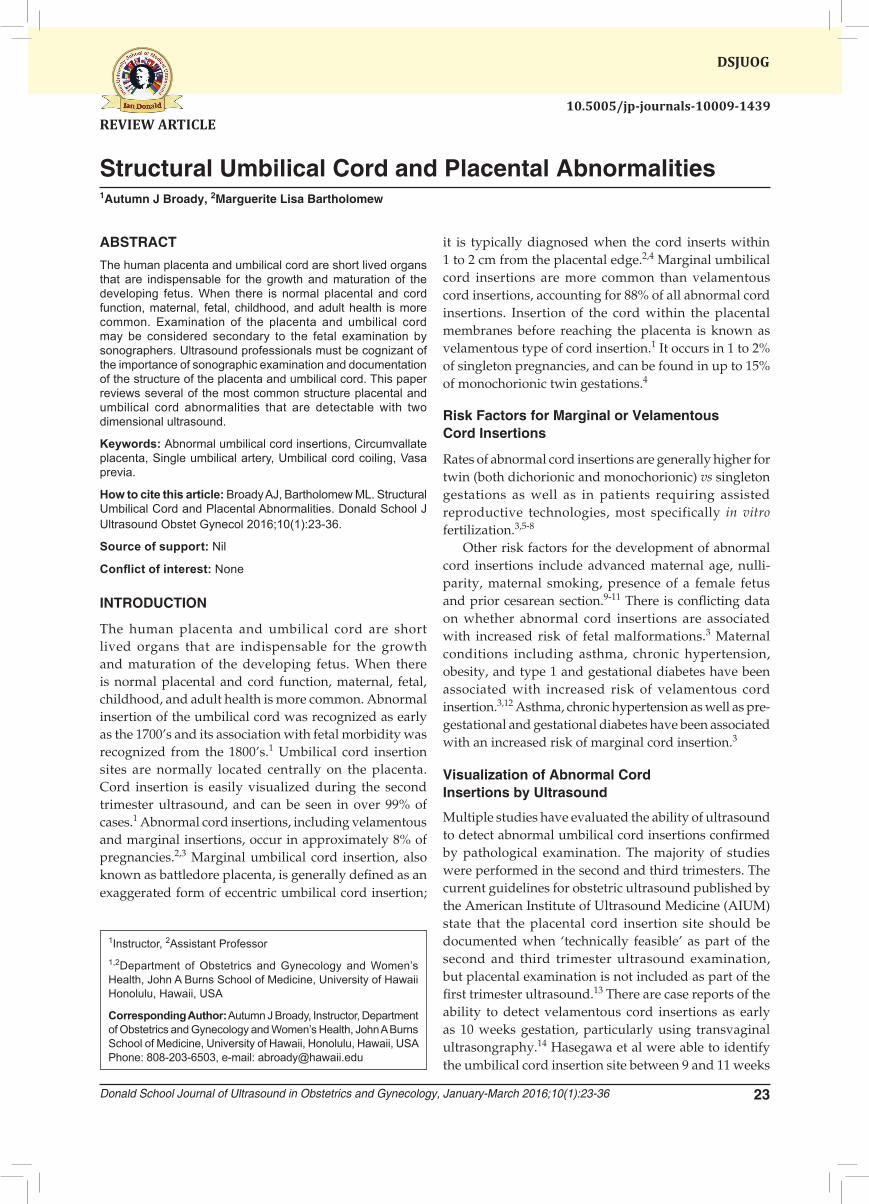
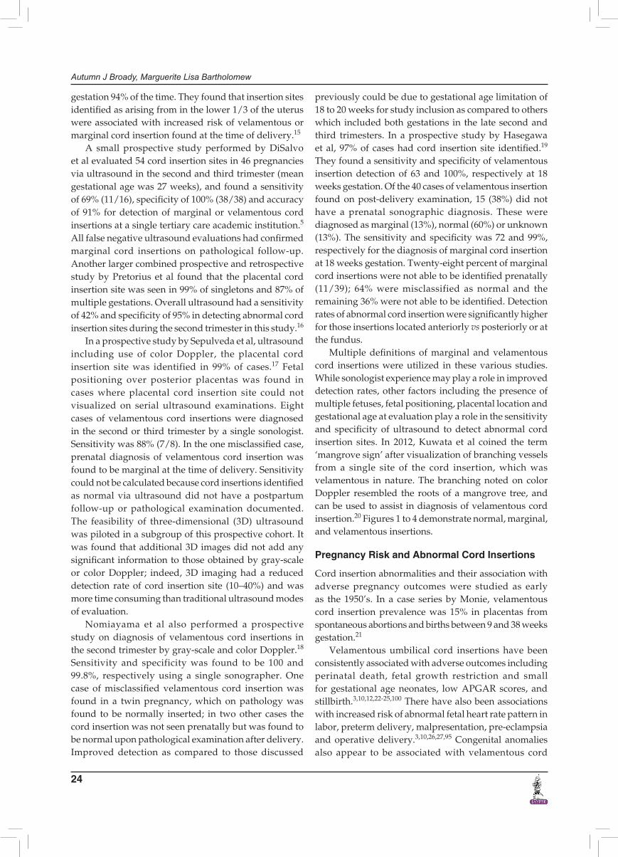
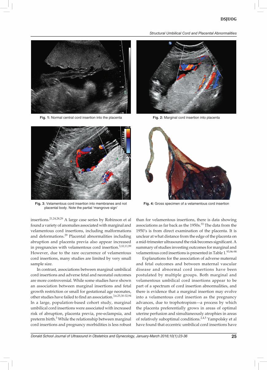
Multiple definitions of marginal and velamentous cord insertions were utilized in these various studies. While sonologist experience may play a role in improved detection rates, other factors including the presence of multiple fetuses, fetal positioning, placental location and gestational age at evaluation play a role in the sensitivity and specificity of ultrasound to detect abnormal cord insertion sites. In 2012, Kuwata et al coined the term ‘mangrove sign’ after visualization of branching vessels from a single site of the cord insertion, which was velamentous in nature. The branching noted on color Doppler resembled the roots of a mangrove tree, and can be used to assist in diagnosis of velamentous cord insertion.20 Figures 1 to 4 demonstrate normal, marginal, and velamentous insertions.
Pregnancy Risk and Abnormal Cord Insertions
Cord insertion abnormalities and their association with adverse pregnancy outcomes were studied as early as the 1950’s. In a case series by Monie, velamentous cord insertion prevalence was 15% in placentas from spontaneous abortions and births between 9 and 38 weeks gestation.21
Velamentous umbilical cord insertions have been consistently associated with adverse outcomes including perinatal death, fetal growth restriction and small for gestational age neonates, low APGAR scores, and stillbirth.3,10,12,22-25,100 There have also been associations with increased risk of abnormal fetal heart rate pattern in labor, preterm delivery, malpresentation, pre-eclampsia and operative delivery.3,10,26,27,95 Congenital anomalies also appear to be associated with velamentous cord
Structural Umbilical Cord and Placental Abnormalities
Donald School Journal of Ultrasound in Obstetrics and Gynecology, January-March 2016;10(1):23-36 25
DSJUOG
insertions.21,24,28,29 A large case series by Robinson et al found a variety of anomalies associated with marginal and velamentous cord insertions, including malformations and deformations.29 Placental abnormalities including abruption and placenta previa also appear increased in pregnancies with velamentous cord insertion.3,10,11,99 However, due to the rare occurrence of velamentous cord insertions, many studies are limited by very small sample size.
In contrast, associations between marginal umbilical cord insertions and adverse fetal and neonatal outcomes are more controversial. While some studies have shown an association between marginal insertions and fetal growth restriction or small for gestational age neonates, other studies have failed to find an association.3,6,25,30-32,94
In a large, population-based cohort study, marginal umbilical cord insertions were associated with increased risk of abruption, placenta previa, pre-eclampsia, and preterm birth.3 While the relationship between marginal cord insertions and pregnancy morbidities is less robust
than for velamentous insertions, there is data showing associations as far back as the 1950s.33 The data from the 1950’s is from direct examination of the placenta. It is unclear at what distance from the edge of the placenta on a mid-trimester ultrasound the risk becomes significant. A summary of studies investing outcomes for marginal and velamentous cord insertions is presented in Table 1.93,96-98
Explanations for the association of adverse maternal and fetal outcomes and between maternal vascular disease and abnormal cord insertions have been postu lated by multiple groups. Both marginal and velamentous umbilical cord insertions appear to be part of a spectrum of cord insertion abnormalities, and there is evidence that a marginal insertion may evolve into a velamentous cord insertion as the pregnancy advances, due to trophotropism—a process by which the placenta preferentially grows in areas of optimal uterine perfusion and simultaneously atrophies in areas of relatively suboptimal conditions.2,4,5 Yampolsky et al have found that eccentric umbilical cord insertions have
Fig. 1: Normal central cord insertion into the placenta Fig. 2: Marginal cord insertion into placenta
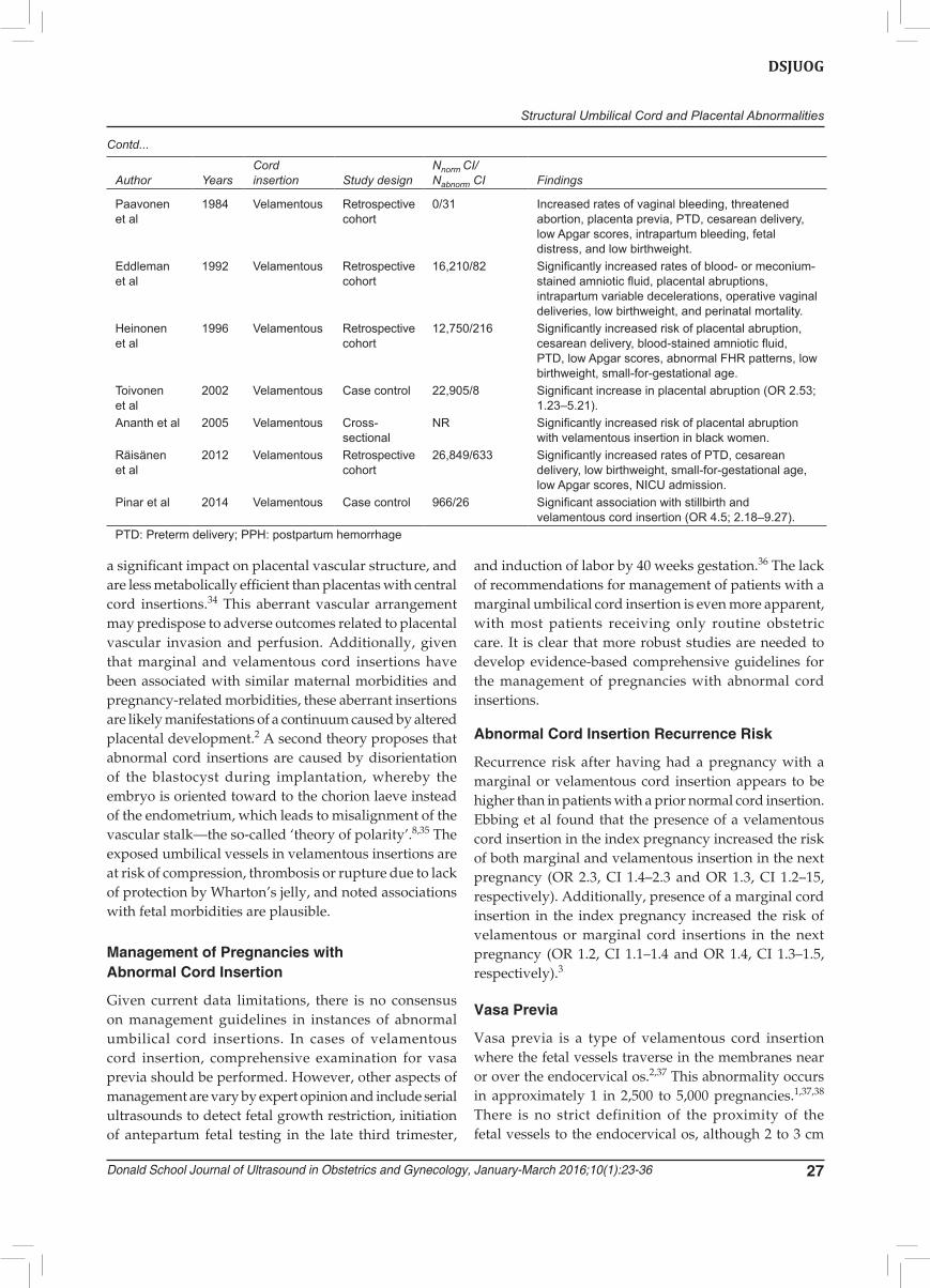
Fig. 3: Velamentous cord insertion into membranes and not placental body. Note the partial ‘mangrove sign’
Fig. 4: Gross specimen of a velamentous cord insertion

Autumn J Broady, Marguerite Lisa Bartholomew
26
Table 1: Summary table of outcomes for singleton pregnancies with velamentous and/or marginal cord insertions
Author YearsCord insertion Study design
Nnorm CI/ Nabnorm CI Findings
Brody et al 1953 Marginal Retrospective cohort
512/32 Significantly increased rates of PTD.
Woods et al 1978 Marginal Retrospective cohort
940/171 No association with birth weight and cord insertion location, including marginal cord insertions.
Davies et al 1984 Marginal Case control 35/35 Association with small-for-gestational age and marginal cord insertion.
Liu et al 2002 Marginal Retrospective cohort
0/100 No association with PTD or low birthweight.
Kramer et al 2011 Marginal Cross-sectional
NR Marginal cord insertions associated with significantly increased risk of maternal postpartum hemorrhage.
Luo et al 2013 Marginal Retrospective cohort
119/119 Significantly increased risk of PTD. No association with low birthweight, fetal death, congenital malformations or low Apgar scores.
Uyanwah-Akpom et al
1977 Marginal and velamentous
Retrospective cohort
1000/72 No association with intrauterine fetal hypoxia, fetal death, threatened abortion, PTD, fetal malformation; trend toward increase in low birthweight.
Rolschau 1978 Marginal and velamentous
Case control 447/19 No significant difference in birthweight, maternal infections, bleeding in pregnancy, labor complications or congenital anomalies with marginal insertions. Significantly increased rates of labor complications and congenital anomalies and decreased birthweights and gestational ages with velamentous insertions.
Hasegawa et al
2006 Marginal and velamenous
Prospective cohort
3367/79 No significant difference in birthweight with marginal or velamentous insertion. Increased risk of variable decelerations and non-reassuring FHR status with velamentous insertions.
Hasegawa et al
2009 Marginal and velamentous
Retrospective cohort
314/69 Significantly increased risk of low Apgar score and risk of FHR variable decelerations in velamentous insertion. Significantly increased risk of ‘abnormal delivery.’
Hasegawa et al
2009 Marginal and velamentous
Retrospective cohort
466/65 Significantly increased risk FHR variable decelerations in velamentous insertion. Significantly decreased rate of ‘normal delivery’ with marginal insertions.
Chan et al 2012 Marginal and velamentous
Retrospective cohort
858/275 Significantly decreased birthweight low Apgar score, nucleated red blood cells, and meconium in velamentous insertions. Significantly increased risk of IUFD and non-reassuring FHR in velamentous insertions. Significantly decreased birthweight in marginal insertions. Significantly increased risk of umbilical cord thrombosis in velamentous and marginal insertions.
Ebbing et al 2013 Marginal and velamentous
Retrospective cohort
623,478/48,903 Significantly increased rates of vaginal bleeding, placental abruption, placenta previa, pre-eclampsia, PTD, malpresentation, serious fetal malformations and low birthweight in velamentous and marginal insertions. Increased risk of low Apgar scores, perinatal mortality in velamentous cord insertion.
Nasiell et al 2015 Marginal and velamentous
Case control 100/41 Significantly increased risk of severe hypoxic-ischemic encephalopathy with marginal and velamentous cord insertions (OR 5.63; 1.64–18.82).
Ebbing et al 2015 Marginal and velamentous
Retrospective cohort
778,443/55,448 Significantly increased risk of third stage of labor complications, placental manual removal, PPH, Significantly increased risk of postpartum curettage for velamentous insertion.
Bjøro, Jr 1983 Velamentous Retrospective cohort
14,050/305 Increased rates of fetal malformations, intrauterine hypoxia, perinatal mortality.
Contd...
Structural Umbilical Cord and Placental Abnormalities
Donald School Journal of Ultrasound in Obstetrics and Gynecology, January-March 2016;10(1):23-36 27
DSJUOG
Paavonen et al
1984 Velamentous Retrospective cohort
0/31 Increased rates of vaginal bleeding, threatened abortion, placenta previa, PTD, cesarean delivery, low Apgar scores, intrapartum bleeding, fetal distress, and low birthweight.
Eddleman et al
1992 Velamentous Retrospective cohort
16,210/82 Significantly increased rates of blood- or meconium-stained amniotic fluid, placental abruptions, intrapartum variable decelerations, operative vaginal deliveries, low birthweight, and perinatal mortality.
Heinonen et al
1996 Velamentous Retrospective cohort
12,750/216 Significantly increased risk of placental abruption, cesarean delivery, blood-stained amniotic fluid, PTD, low Apgar scores, abnormal FHR patterns, low birthweight, small-for-gestational age.
Toivonen et al
2002 Velamentous Case control 22,905/8 Significant increase in placental abruption (OR 2.53; 1.23–5.21).
Ananth et al 2005 Velamentous Cross-sectional
NR Significantly increased risk of placental abruption with velamentous insertion in black women.
Räisänen et al
2012 Velamentous Retrospective cohort
26,849/633 Significantly increased rates of PTD, cesarean delivery, low birthweight, small-for-gestational age, low Apgar scores, NICU admission.
Pinar et al 2014 Velamentous Case control 966/26 Significant association with stillbirth and velamentous cord insertion (OR 4.5; 2.18–9.27).
PTD: Preterm delivery; PPH: postpartum hemorrhage
Author YearsCord insertion Study design
Nnorm CI/ Nabnorm CI Findings
Contd...
a significant impact on placental vascular structure, and are less metabolically efficient than placentas with central cord insertions.34 This aberrant vascular arrangement may predispose to adverse outcomes related to placental vascular invasion and perfusion. Additionally, given that marginal and velamentous cord insertions have been associated with similar maternal morbidities and pregnancy-related morbidities, these aberrant insertions are likely manifestations of a continuum caused by altered placental development.2 A second theory proposes that abnormal cord insertions are caused by disorientation of the blastocyst during implantation, whereby the embryo is oriented toward to the chorion laeve instead of the endometrium, which leads to misalignment of the vascular stalk—the so-called ‘theory of polarity’.8,35 The exposed umbilical vessels in velamentous insertions are at risk of compression, thrombosis or rupture due to lack of protection by Wharton’s jelly, and noted associations with fetal morbidities are plausible.
Management of Pregnancies with Abnormal Cord Insertion
Given current data limitations, there is no consensus on management guidelines in instances of abnormal umbilical cord insertions. In cases of velamentous cord insertion, comprehensive examination for vasa previa should be performed. However, other aspects of management are vary by expert opinion and include serial ultrasounds to detect fetal growth restriction, initiation of antepartum fetal testing in the late third trimester,
and induction of labor by 40 weeks gestation.36 The lack of recommendations for management of patients with a marginal umbilical cord insertion is even more apparent, with most patients receiving only routine obstetric care. It is clear that more robust studies are needed to develop evidence-based comprehensive guidelines for the management of pregnancies with abnormal cord insertions.
Abnormal Cord Insertion Recurrence Risk
Recurrence risk after having had a pregnancy with a marginal or velamentous cord insertion appears to be higher than in patients with a prior normal cord insertion. Ebbing et al found that the presence of a velamentous cord insertion in the index pregnancy increased the risk of both marginal and velamentous insertion in the next pregnancy (OR 2.3, CI 1.4–2.3 and OR 1.3, CI 1.2–15, respectively). Additionally, presence of a marginal cord insertion in the index pregnancy increased the risk of velamentous or marginal cord insertions in the next pregnancy (OR 1.2, CI 1.1–1.4 and OR 1.4, CI 1.3–1.5, respectively).3
Vasa Previa
Vasa previa is a type of velamentous cord insertion where the fetal vessels traverse in the membranes near or over the endocervical os.2,37 This abnormality occurs in approximately 1 in 2,500 to 5,000 pregnancies.1,37,38 There is no strict definition of the proximity of the fetal vessels to the endocervical os, although 2 to 3 cm

Autumn J Broady, Marguerite Lisa Bartholomew
28
has been previously used.39-41 Vasa previa was first reported in the 1800s and resultant fetal bleeding from the exposed vessels has been described as ‘Hemorrhage of Benckiser’ after one of the first clinicians to describe this phenomenon.1 Catanzarite et al classsifed vasa previa into two based on placental characteristics: Type I comprising vasa previa with a single placental lobe with velamentous cord insertion and Type II involving multilobular placentas with connecting vessels running over the cervical os.42
Risk Factors for Vasa Previa
Risk factors for vasa previa include placental abnormalities including succenturiate or bilobed placenta, previa or low-lying placenta, in vitro fertilization, and multiple gestations.37,43-45 The likelihood of vasa previa in the setting of velamentous cord insertion is approximately 1:50.46 Schachter et al reported a significantly increased odds ratio (OR 27.4; 7.4–917) for vasa previa in the setting of in vitro fertilization.47 Others have found an association with cord insertion into the lower uterine segment, particularly in the lower third of the uterus, and the presence of vasa previa.48-50
Visualization of Vasa Previa
Prior to routine ultrasonography, vasa previa was rarely diagnosed antepartum, which led to devastating antepartum consequences. The ultrasonographic diagnosis of vasa previa is first reported in the late 1980s to 1990s in multiple small case reports using gray-scale and Doppler sonographic imaging, initially with transabdominal approach, and then using transvaginal ultrasonography.51-61 Currently, targeted screening for vasa previa using transvaginal ultrasonography should be utilized in the presence of placental risk factors including placenta previa or low-lying placentation, multiple placental lobes or succenturiate lobe and velamentous cord insertion.37 Vasa previa can be identified on gray-scale transvaginal ultrasonography by visualization of an echolucent tubular structure adjacent to or overlying the internal cervical os.37 Diagnosis of vasa previa should confirmed with color or power Doppler demonstrating pulsatile flow with fetal vascular waveform pattern.37 It is important to exclude a false positive result from free loops of cord overlying the cervical os by visualizing the vessels’ immobility and persistence with maternal position changes.37 The use of three-dimensional ultrasonography and transperineal approach have also been reported in the diagnosis of vasa previa.44,62-65
In a case series by Lee et al, the diagnosis was made at a mean gestational age of 26 weeks, with the earliest recognition at 15 weeks gestation.44 The majority of cases
were identified prior to 31 weeks gestation. First trimester imaging of abnormal cord positioning in the lower uterus with increased risk of vasa previa has been reported as early as 9 to 13 weeks gestation.50,66 A case series by Bronsteen et al found that transabdominal approach was less likely to detect evidence of vasa previa as compared to transvaginal.39
Estimating the sensitivity and specificity of sonography in diagnosing vasa previa is difficult due to the difficulty in diagnosing cases after delivery.44 However, in Lee’s case series, there was only one case that was missed when records were reviewed.44 Catanzarite et al found a specificity of 91% in their prospective cohort study spanning 7 years and including 11 cases of vasa previa out of 33,000 women.42 A specificity of 97% was found in 32 cases of vasa previa that were identified out of 27,500 second trimester anatomic surveys performed by Rebarber et al.40 A systematic review of eight case series, including some discussed above, totaling 138 cases and over 442,600 women found a median prenatal detection rate of 93%, and specificity of 99 to 100%.67 Figures 5 to 8
Fig. 5: Left lateral posterior placenta and anterior succenturiate lobe in a transverse view
Fig. 6: Vessels in the fetal membranes connecting a posterior placenta and an anterior succenturiate lobe
Structural Umbilical Cord and Placental Abnormalities
Donald School Journal of Ultrasound in Obstetrics and Gynecology, January-March 2016;10(1):23-36 29
DSJUOG
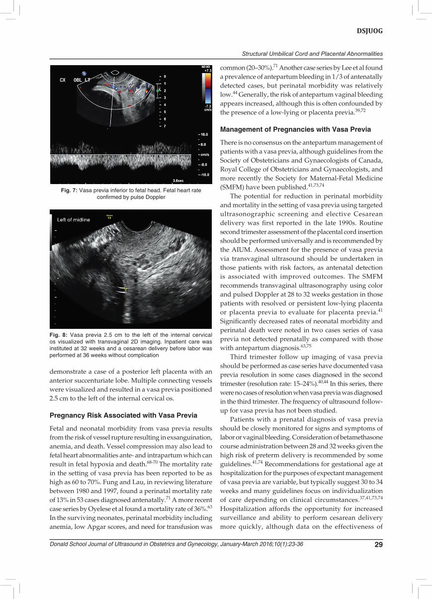
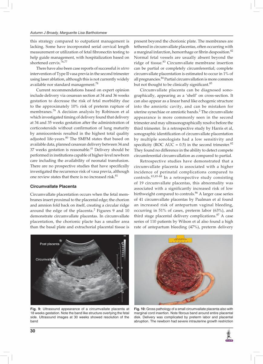
demonstrate a case of a posterior left placenta with an anterior succenturiate lobe. Multiple connecting vessels were visualized and resulted in a vasa previa positioned 2.5 cm to the left of the internal cervical os.
Pregnancy Risk Associated with Vasa Previa
Fetal and neonatal morbidity from vasa previa results from the risk of vessel rupture resulting in exsanguination, anemia, and death. Vessel compression may also lead to fetal heart abnormalities ante- and intrapartum which can result in fetal hypoxia and death.68-70 The mortality rate in the setting of vasa previa has been reported to be as high as 60 to 70%. Fung and Lau, in reviewing literature between 1980 and 1997, found a perinatal mortality rate of 13% in 53 cases diagnosed antenatally.71 A more recent case series by Oyelese et al found a mortality rate of 36%.63 In the surviving neonates, perinatal morbidity including anemia, low Apgar scores, and need for transfusion was
common (20–30%).71 Another case series by Lee et al found a prevalence of antepartum bleeding in 1/3 of antenatally detected cases, but perinatal morbidity was relatively low.44 Generally, the risk of antepartum vaginal bleeding appears increased, although this is often confounded by the presence of a low-lying or placenta previa.39,72
Management of Pregnancies with Vasa Previa
There is no consensus on the antepartum management of patients with a vasa previa, although guidelines from the Society of Obstetricians and Gynaecologists of Canada, Royal College of Obstetricians and Gynaecologists, and more recently the Society for Maternal-Fetal Medicine (SMFM) have been published.41,73,74
The potential for reduction in perinatal morbidity and mortality in the setting of vasa previa using targeted ultraso nographic screening and elective Cesarean delivery was first reported in the late 1990s. Routine second trimester assessment of the placental cord insertion should be performed universally and is recommended by the AIUM. Assessment for the presence of vasa previa via transvaginal ultrasound should be undertaken in those patients with risk factors, as antenatal detection is associated with improved outcomes. The SMFM recommends transvaginal ultrasonography using color and pulsed Doppler at 28 to 32 weeks gestation in those patients with resolved or persistent low-lying placenta or placenta previa to evaluate for placenta previa.41 Significantly decreased rates of neonatal morbidity and perinatal death were noted in two cases series of vasa previa not detected prenatally as compared with those with antepartum diagnosis.63,75
Third trimester follow up imaging of vasa previa should be performed as case series have documented vasa previa resolution in some cases diagnosed in the second trimester (resolution rate: 15–24%).40,44 In this series, there were no cases of resolution when vasa previa was diagnosed in the third trimester. The frequency of ultrasound follow-up for vasa previa has not been studied.
Patients with a prenatal diagnosis of vasa previa should be closely monitored for signs and symptoms of labor or vaginal bleeding. Consideration of betamethasone course administration between 28 and 32 weeks given the high risk of preterm delivery is recommended by some guidelines.41,74 Recommendations for gestational age at hospitalization for the purposes of expectant management of vasa previa are variable, but typically suggest 30 to 34 weeks and many guidelines focus on individualization of care depending on clinical circumstances.37,41,73,74 Hospitalization affords the opportunity for increased surveillance and ability to perform cesarean delivery more quickly, although data on the effectiveness of
Fig. 7: Vasa previa inferior to fetal head. Fetal heart rate confirmed by pulse Doppler
Fig. 8: Vasa previa 2.5 cm to the left of the internal cervical os visualized with transvaginal 2D imaging. Inpatient care was insti tuted at 32 weeks and a cesarean delivery before labor was performed at 36 weeks without complication
Autumn J Broady, Marguerite Lisa Bartholomew
30
this strategy compared to outpatient management is lacking. Some have incorporated serial cervical length measurement or utilization of fetal fibronectin testing to help guide management, with hospitalization based on shortened cervix.76,77
There have also been case reports of successful in utero intervention of Type II vasa previa in the second trimester using laser ablation, although this is not currently widely available nor standard management.78
Current recommendations based on expert opinion include delivery via cesarean section at 34 and 36 weeks gestation to decrease the risk of fetal morbidity due to the approximately 10% risk of preterm rupture of membranes.79 A decision analysis by Robinson et al which investigated timing of delivery found that delivery at 34 and 35 weeks gestation after the administration of corticosteroids without confirmation of lung maturity by amniocentesis resulted in the highest total quality adjusted life-years.80 The SMFM states that based on available data, planned cesarean delivery between 34 and 37 weeks gestation is reasonable.41 Delivery should be performed in institutions capable of higher-level newborn care including the availability of neonatal transfusion. There are no prospective studies that have specifically investigated the recurrence risk of vasa previa, although one review states that there is no increased risk.81
Circumvallate Placenta
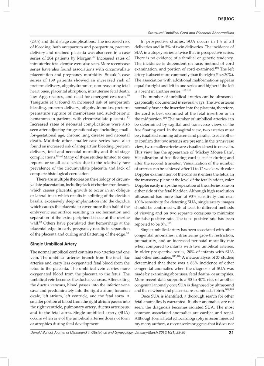
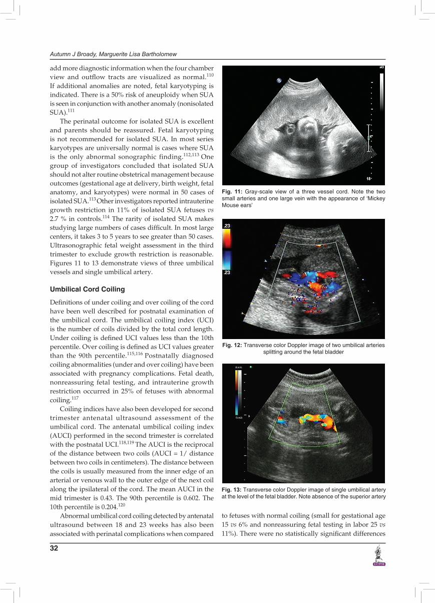
Circumvallate placentation occurs when the fetal mem-branes insert proximal to the placental edge; the chorion and amnion fold back on itself, creating a circular ridge around the edge of the placenta.2 Figures 9 and 10 demonstrate circumvallate placentas. In circumvallate placentation, the chorionic placte has a smaller area than the basal plate and extrachorial placental tissue is
present beyond the chorionic plate. The membranes are tethered in circumvallate placentas, often occurring with a marginal infarction, hemorrhage or fibrin desposition.82 Normal fetal vessels are usually absent beyond the ridge of tissue.83 Circumvallate membrane insertion can be partial or completely circumferential; complete circumvallate placentation is estimated to occur in 1% of all pregnancies.84 Partial circumvallation is more common but not thought to be clinically significant.85
Circumvallate placenta can be diagnosed sono-graphically, appearing as a ‘shelf’ on cross-section. It can also appear as a linear band like echogenic structure into the amniotic cavity, and can be mistaken for uterine synechiae or amniotic bands.2 The circumvallate appearance is more commonly seen in the second trimester and may ultrasonographically resolve before the third trimester. In a retrospective study by Harris et al, sonographic identification of circumvallate placentation by multiple sonologists had a low sensitivity and specificity (ROC AUC = 0.5) in the second trimester.82 They found no difference in the ability to detect compete circumferential circumvallation as compared to partial.
Retrospective studies have demonstrated that a circumvallate placenta is associated with a higher incidence of perinatal complications compared to controls.83,85-88 In a retrospective study consisting of 19 circumvallate placentas, this abnormality was associated with a significantly increased risk of low birthweight compared to controls.86 A larger case series of 41 circumvallate placentas by Paalman et al found an increased risk of antepartum vaginal bleeding, occurring in 51% of cases, preterm labor (63%), and third stage placental delivery complications.87 A case series of 110 patients by Wilson et al also found a high rate of antepartum bleeding (47%), preterm delivery
Fig. 9: Ultrasound appearance of a circumvallate placenta at 18 weeks gestation. Note the band like structure overlying the fetal side. Ultrasound images at 30 weeks showed resolution of the band
Fig. 10: Gross pathology of a small circumvallate placenta also with marginal cord insertion. Note fibrous band around entire placental disk. Delivery was complicated by preterm labor and placental abruption. The newborn had severe intrauterine growth restriction
Structural Umbilical Cord and Placental Abnormalities
Donald School Journal of Ultrasound in Obstetrics and Gynecology, January-March 2016;10(1):23-36 31
DSJUOG
(28%) and third stage complications. The increased risk of bleeding, both antepartum and postpartum, preterm delivery and retained placenta was also seen in a case series of 204 patients by Morgan.89 Increased rates of intrauterine fetal demise were also seen. More recent case series have also found associations with circumvallate placentation and pregnancy morbidity. Suzuki’s case series of 139 patients showed an increased risk of preterm delivery, oligohydramnios, non-reassuring fetal heart ones, placental abruption, intrauterine fetal death, low Apgar scores, and need for emergent cesarean.90 Taniguchi et al found an increased risk of antepartum bleeding, preterm delivery, oligohydramnios, preterm premature rupture of membranes and subchorionic hematoma in patients with circumvallate placenta.91 Increased rates of neonatal complications were also seen after adjusting for gestational age including small-for-gestational age, chronic lung disease and neonatal death. Multiple other smaller case series have also found an increased risk of antepartum bleeding, preterm delivery, fetal and neonatal mortality and third stage complications.85,92 Many of these studies limited to case reports or small case series due to the relatively rare prevalence of the circumvallate placenta and lack of complete histological correlation.
There are multiple theories on the etiology of circum-vallate placentation, including lack of chorion frondosum, which causes placental growth to occur in an oblique or lateral track which results in splitting of the decidua basalis, excessively deep implantation into the decidua which causes the placenta to cover more than half of the embryonic sac surface resulting in sac herniation and separation of the extra peripheral tissue at the uterine wall.82 Others have postulated that hemorrhage at the placental edge in early pregnancy results in separation of the placenta and curling and flattening of the edge.82
Single Umbilical Artery
The normal umbilical cord contains two arteries and one vein. The umbilical arteries branch from the fetal iliac arteries and carry less oxygenated fetal blood from the fetus to the placenta. The umbilical vein carries more oxygenated blood from the placenta to the fetus. The umbilical vein becomes the ductus venosus. After exiting the ductus venosus, blood passes into the inferior vena cava and predominately into the right atrium, foramen ovale, left atrium, left ventricle, and the fetal aorta. A smaller portion of blood from the right atrium passes into the right ventricle, pulmonary artery, ductus arteriosus, and to the fetal aorta. Single umbilical artery (SUA) occurs when one of the umbilical arteries does not form or atrophies during fetal development.
In prospective studies, SUA occurs in 1% of all deliveries and in 5% of twin deliveries. The incidence of SUA in autopsy series is twice that in prospective series. There is no evidence of a familial or genetic tendency. The incidence is dependent on race, method of cord examination, and portion of cord examined.101 The left artery is absent more commonly than the right (70 vs 30%). The association with additional malformations appears equal for right and left in one series and higher if the left is absent in another series.102,103
The number of umbilical arteries can be ultrasono-graphically documented in several ways. The two arteries normally fuse at the insertion into the placenta, therefore, the cord is best examined at the fetal insertion or in the midportion.104 The number of umbilical arteries can be determined by sagittal and transverse views of the free floating cord. In the sagittal view, two arteries must be visualized running adjacent and parallel to each other to confirm that two arteries are present. In the transverse view, two smaller arteries are visualized next to one vein. This view has the appearance of ‘Mickey Mouse Ears’. Visualization of free floating cord is easier during and after the second trimester. Visualization of the number of arteries can be achieved after 11 to 12 weeks with color Doppler examination of the cord as it enters the fetus. In the transverse plane at the level of the fetal bladder, color Doppler easily maps the separation of the arteries, one on either side of the fetal bladder. Although high resolution ultrasound has more than at 90% sensitivity and near 100% sensitivity for detecting SUA, single artery images should be confirmed with at least to different methods of viewing and on two separate occasions to minimize the false positive rate. The false positive rate has been reported to be 8%.105
Single umbilical artery has been associated with other congenital anomalies, intrauterine growth restriction, prematurity, and an increased perinatal mortality rate when com pared to infants with two umbilical arteries. In older prospective series, 20% of infants with SUA had other anomalies.106,107 A meta-analysis of 37 studies determined that there was a 66% incidence of other congenital anomalies when the diagnosis of SUA was made by examining abortuses, fetal deaths, or autopsies. More recent data supports a 30 to 40% risk of another congenital anomaly once SUA is diagnosed by ultrasound and the newborn and placenta are examined at birth.108,109
Once SUA is identified, a thorough search for other fetal anomalies is warranted. If other anomalies are not seen, the diagnosis becomes isolated SUA. The most common associated anomalies are cardiac and renal. Although formal fetal echocardiography is recommended my many authors, a recent series suggests that it does not
Autumn J Broady, Marguerite Lisa Bartholomew
32
add more diagnostic information when the four chamber view and outflow tracts are visualized as normal.110
If additional anomalies are noted, fetal karyotyping is indicated. There is a 50% risk of aneuploidy when SUA is seen in conjunction with another anomaly (nonisolated SUA).111
The perinatal outcome for isolated SUA is excellent and parents should be reassured. Fetal karyotyping is not recommended for isolated SUA. In most series karyotypes are universally normal is cases where SUA is the only abnormal sonographic finding.112,113 One group of investigators concluded that isolated SUA should not alter routine obstetrical management because outcomes (gestational age at delivery, birth weight, fetal anatomy, and karyotypes) were normal in 50 cases of isolated SUA.113 Other investigators reported intrauterine growth restriction in 11% of isolated SUA fetuses vs 2.7 % in controls.114 The rarity of isolated SUA makes studying large numbers of cases difficult. In most large centers, it takes 3 to 5 years to see greater than 50 cases. Ultrasonographic fetal weight assessment in the third trimester to exclude growth restriction is reasonable. Figures 11 to 13 demonstrate views of three umbilical vessels and single umbilical artery.
Umbilical Cord Coiling
Definitions of under coiling and over coiling of the cord have been well described for postnatal examination of the umbilical cord. The umbilical coiling index (UCI) is the number of coils divided by the total cord length. Under coiling is defined UCI values less than the 10th percentile. Over coiling is defined as UCI values greater than the 90th percentile.115,116 Postnatally diagnosed coiling abnormalities (under and over coiling) have been associated with pregnancy complications. Fetal death, nonreassuring fetal testing, and intrauterine growth restriction occurred in 25% of fetuses with abnormal coiling.117
Coiling indices have also been developed for second trimester antenatal ultrasound assessment of the umbilical cord. The antenatal umbilical coiling index (AUCI) performed in the second trimester is correlated with the postnatal UCI.118,119 The AUCI is the reciprocal of the distance between two coils (AUCI = 1/ distance between two coils in centimeters). The distance between the coils is usually measured from the inner edge of an arterial or venous wall to the outer edge of the next coil along the ipsilateral of the cord. The mean AUCI in the mid trimester is 0.43. The 90th percentile is 0.602. The 10th percentile is 0.204.120
Abnormal umbilical cord coiling detected by antenatal ultrasound between 18 and 23 weeks has also been associated with perinatal complications when compared
Fig. 11: Gray-scale view of a three vessel cord. Note the two small arteries and one large vein with the appearance of ‘Mickey Mouse ears’
Fig. 12: Transverse color Doppler image of two umbilical arteries splitting around the fetal bladder
Fig. 13: Transverse color Doppler image of single umbilical artery at the level of the fetal bladder. Note absence of the superior artery
to fetuses with normal coiling (small for gestational age 15 vs 6% and nonreassuring fetal testing in labor 25 vs 11%). There were no statistically significant differences
Structural Umbilical Cord and Placental Abnormalities
Donald School Journal of Ultrasound in Obstetrics and Gynecology, January-March 2016;10(1):23-36 33
DSJUOG
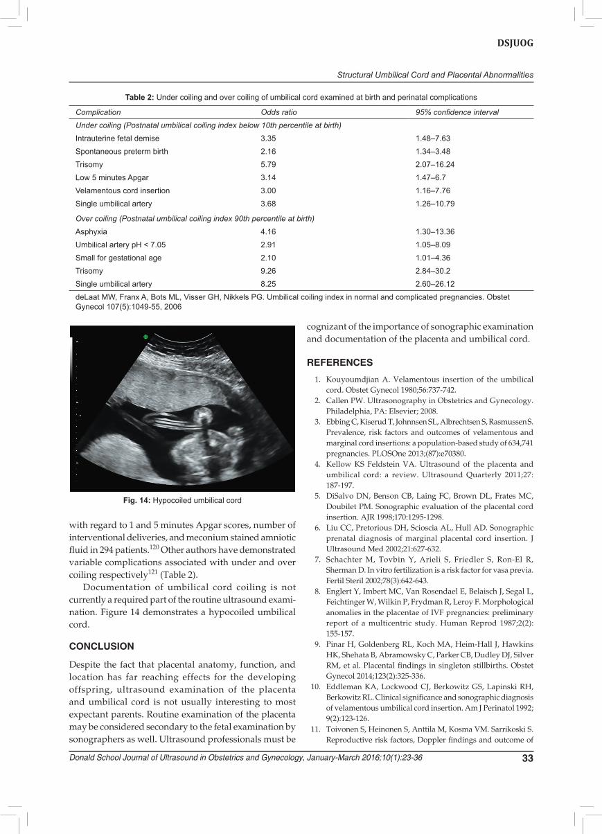
with regard to 1 and 5 minutes Apgar scores, number of interventional deliveries, and meconium stained amniotic fluid in 294 patients.120 Other authors have demonstrated variable complications associated with under and over coiling respectively121 (Table 2).
Documentation of umbilical cord coiling is not currently a required part of the routine ultrasound exami-nation. Figure 14 demonstrates a hypocoiled umbilical cord.
CONCLUSION
Despite the fact that placental anatomy, function, and location has far reaching effects for the developing offspring, ultrasound examination of the placenta and umbilical cord is not usually interesting to most expectant parents. Routine examination of the placenta may be considered secondary to the fetal examination by sonographers as well. Ultrasound professionals must be
cognizant of the importance of sonographic examination and documentation of the placenta and umbilical cord.
REFERENCES
1. Kouyoumdjian A. Velamentous insertion of the umbilical cord. Obstet Gynecol 1980;56:737-742.
2. Callen PW. Ultrasonography in Obstetrics and Gynecology. Philadelphia, PA: Elsevier; 2008.
3. Ebbing C, Kiserud T, Johnnsen SL, Albrechtsen S, Rasmussen S. Prevalence, risk factors and outcomes of velamentous and marginal cord insertions: a population-based study of 634,741 pregnancies. PLOSOne 2013;(87):e70380.
4. Kellow KS Feldstein VA. Ultrasound of the placenta and umbilical cord: a review. Ultrasound Quarterly 2011;27: 187-197.
5. DiSalvo DN, Benson CB, Laing FC, Brown DL, Frates MC, Doubilet PM. Sonographic evaluation of the placental cord insertion. AJR 1998;170:1295-1298.
6. Liu CC, Pretorious DH, Scioscia AL, Hull AD. Sonographic prenatal diagnosis of marginal placental cord insertion. J Ultrasound Med 2002;21:627-632.
7. Schachter M, Tovbin Y, Arieli S, Friedler S, Ron-El R, Sherman D. In vitro fertilization is a risk factor for vasa previa. Fertil Steril 2002;78(3):642-643.
8. Englert Y, Imbert MC, Van Rosendael E, Belaisch J, Segal L, Feichtinger W, Wilkin P, Frydman R, Leroy F. Morphological anomalies in the placentae of IVF pregnancies: preliminary report of a multicentric study. Human Reprod 1987;2(2): 155-157.
9. Pinar H, Goldenberg RL, Koch MA, Heim-Hall J, Hawkins HK, Shehata B, Abramowsky C, Parker CB, Dudley DJ, Silver RM, et al. Placental findings in singleton stillbirths. Obstet Gynecol 2014;123(2):325-336.
10. Eddleman KA, Lockwood CJ, Berkowitz GS, Lapinski RH, Berkowitz RL. Clinical significance and sonographic diagnosis of velamentous umbilical cord insertion. Am J Perinatol 1992; 9(2):123-126.
11. Toivonen S, Heinonen S, Anttila M, Kosma VM. Sarrikoski S. Reproductive risk factors, Doppler findings and outcome of
Table 2: Under coiling and over coiling of umbilical cord examined at birth and perinatal complications
Complication Odds ratio 95% confidence interval
Under coiling (Postnatal umbilical coiling index below 10th percentile at birth)
Intrauterine fetal demise 3.35 1.48–7.63
Spontaneous preterm birth 2.16 1.34–3.48
Trisomy 5.79 2.07–16.24
Low 5 minutes Apgar 3.14 1.47–6.7
Velamentous cord insertion 3.00 1.16–7.76
Single umbilical artery 3.68 1.26–10.79
Over coiling (Postnatal umbilical coiling index 90th percentile at birth)
Asphyxia 4.16 1.30–13.36
Umbilical artery pH < 7.05 2.91 1.05–8.09
Small for gestational age 2.10 1.01–4.36
Trisomy 9.26 2.84–30.2
Single umbilical artery 8.25 2.60–26.12
deLaat MW, Franx A, Bots ML, Visser GH, Nikkels PG. Umbilical coiling index in normal and complicated pregnancies. Obstet Gynecol 107(5):1049-55, 2006
Fig. 14: Hypocoiled umbilical cord

Autumn J Broady, Marguerite Lisa Bartholomew
34
affected births in placental abruption: a population-based analysis. Am J Perinatol 2002;19(8):451-459.
12. Räisänen S, Georgiadis L, Harju M, Keski-Nisula L, Heinonen S. Risk factors and adverse pregnancy outcomes among births affected by velamentous umbilical cord insertion: a retrospective population-based register study. Eur J Obstet Gynecol Reprod Biol 2012;165:231-234.
13. American Institute of Ultrasound Medicine. AIUM practice guideline for the performance of obstetric ultrasound examinations. J Ultrasound Med 2013;32:1083-1101.
14. Monteagudo A, Sfakianaki AK, Timor-Tritsch IE. Picture of the Month: velamentous insertion of the cord in the first trimester. Ultrasound Obstet Gynecol 2000;16:498-499.
15. Hasegawa J, Matsuoka R, Ichizuka K, Otsuki K, Sekizawa A, Farina A, Okai T. Cord insertion into the lower third of the uterus in the first trimester is associated with placental and umbilical cord abnormalities. Ultrasound Obstet Gynecol 2006;28:183-186.
16. Pretorius DH, Chau C, Poeltler DM, Mendoza A, Catanzarite VA, Hollenbach KA. Placental cord insertion visualization with pernal ultrasonography. J Ultrasound Med 1996;15: 585-593.
17. Sepulveda W, Rojas I, Robert A, Schnapp C, Alcalde JL. Prenatal detection of velamentous insertion of the umbilical cord: a prospective color Doppler ultrasound study. Ultra-sound Obstet Gynecol 2003;21:564-569.
18. Nomiayama M, Toyota Y, Kawano H. Antenatal diagnosis of velamentous umbilical cord insertion and vasa previa with color Doppler imaging. Ultrasound Obstet Gynecol 1998; 12:426-429.
19. Hasegawa J, Matsuoka R, Ichizuka K, Sekizawa A, Farina A, Okai T. Velamentous cord insertion into the lower third of the uterus is associated with intrapartum fetal heart rate abnormalities. Ultrasound Obstet Gynecol 2006;27:425-429.
20. Kuwata T, Suzuki H, Matsubara S. The ‘mangrove sign’ for velamentous cord insertion. Ultrasound Obstet Gynecol 2012; 40:241-242.
21. Monie IW. Velamentous insertion of the cord in early pregnancy. AJOG 1965;93:276-281.
22. Hasegawa J, Matsuoka R, Ichizuka K, Kotani M, Nakamura M, Mikoshiba T, Sekizawa A, Okai T. Atypical variable deceleration in the first stage of labor is a characteristic fetal heart-rate pattern for velamentous cord insertion and hypercoiled cord. J Obstet Gynaecol Res 2009;35(1):35-39.
23. Pinar H, Goldenberg RL, Koch MA, Heim-Hall J, Hawkins HK, Shehata B, Abramowsky C, Parker CB, Dudley DJ, Silver RM, Stoll B, Carpenter M, Saade G, Moore J, Conway D, Varner MW, Hogue CJR, Coustan DR, Sbrana E, Thorsten V, Willinger M, Reddy UM. Placental findings in singleton stillbirths. Obstet Gynecol 2014;123(2):325-336.
24. Rolschau J. The relationship between some disorders of the umbilical cord and intrauterine growth restriction. Acta Obstet Gynecol Scand Suppl 1978;72:15-21.
25. Chan JSY, Baergen RN. Umbilical cord complications are associated with placental lesions of circulatory stasis and fetal hypoxia. Pediatric and Developmental Pathology 2012; 15:487-494.
26. Heinonen S, Ryynänen M, Kirkinen P, Sarrikoski S. Perinatal diagnostic evaluation of velamentous umbilical cord insertion: clinical Doppler and ultrasonic findings. Obstet Gynecol 1996; 87:112-117.
27. Hasegawa J, Matsuoka R, Ichizuka K, Sekizawa A, Okai T. Velamentous cord insertion: significance of prenatal detection to predict perinatal complications. Taiwanese J Obstet Gynecol 2006;45(1):21-25.
28. Bjøro K Jr. Vascular anomalies of the umbilical cord. II. Perinatal and pediatric complications. Early Hum Dev 1983; 8:279-287.
29. Robinson LK, Jones KL, Benirschke K. The nature of structural defects associated with velamentous and marginal insertions of the umbilical cord. AJOG 1983;146(2):191-193.
30. Davies BR, Casanueva E, Arroyo P. Placentas of small-for-dates infants: A small controlled series from Mexico City, Mexico. AJOG 1984;149(7):731-736.
31. Woods DL, Malan AF. The site of umbilical cord insertion and birth weight. BJOG 1978;85:332-333.
32. Luo G, Redline R. Peripheral insertion of the umbilical cord. Ped Dev Pathol 2013;16:399:404.
33. Brody S, Frenkel DA. Marginal insertion of the cord and premature labor. AJOG 1953;65:1305-1312.
34. Yampolsky M, Salafia CM, Shlakhter O, Haas D, Eucker B, Thorp J. Centrality of the umbilical cord insertion in a human placenta influences the placental efficiency. Placenta 2009;30: 1058-1064.
35. Heifetz SA. The umbilical cord: obstetrically important lesions. Clin Obstet Gynecol 1996;39(3):571-587.
36. Lockwood CJ, Russo-Stieglitz K, Velamentous umbilical cord insertion and vasa previa. In: UpToDate, Post TW (Ed), UpToDate, Waltham, MA. (Accessed on August 24th, 2015.)
37. Silver RM. Abnormal placentation: placenta previa, vasa previa and placenta accreta. Obstet Gynecol 2015;126:654-668.
38. Oyelese KO, Turner M, Lees C, Campbell S. Vasa previa: an avoidable tragedy. Obstet Gynecol Surv 1999;54(2):138-145.
39. Bronsteen R, Whitten A, Balasubramanian M, Lee W, Lorenz R, Redman M, Goncalves L, Seubert D, Bauer S, Comstock C. Vasa previa: clinical presentations, outcomes and implications for management. Obstet Gynecol 2013;122:352-357.
40. Rebarber A, Dolin C, Fox NS, Klauser CK, Saltzman DH, Roman AS. Natural history of vasa previa across gestation using a screening protocol. J Ultrasound Med 2014;33:141-147.
41. Society for Maternal-Fetal Medicine Publications Committee, Sinkey RG, Odibo AO, Dashe J. SMFM Consult Series #37: Diagnosis and management of vasa previa. AJOG 2015 Aug 18. pii: S0002-9378(15)00897-2. doi: 10.1016/j.ajog.2015.08.031. [Epub ahead of print].
42. Catanzarite V, Maida C, Thomas W, Mendoza A, Stanco L, Piacquado KM. Sonographic diagnosis of vasa previa: ultrasound findings and obstetric outcome in ten cases. Ultrasound Obstet Gynecol 2001;18:109-115.
43. Oyelese KO, Schwärzler P, Coates S, Sanusi FA, Hamid R, Campbell S. Strategy for reducing the mortality rate from vasa previa using transvaginal sonography with color Doppler. Ultrasound Obstet Gynecol 1998;12:434-438.
44. Lee W, Kirk JS, Comstock CH, Romero R. Vasa previa: prenatal detection by three-dimensional ultrasonography. Ultrasound Obstet Gynecol 2000;16:384-387.
45. Oyelese Y, Spong C, Fernandez MA, McLaren RA. Second trimester low-lying placental and in-vitro fertilization? Exclude vasa previa. J Matern Fetal Med 2000;9(6):370-372.
46. Quek SP, Tan KL. Vasa Praevia. Aust. NZ J Obstet Gynaec 1972;12:206-209.
47. Schachter M, Tovbin Y, Arieli S, Friedler S, Ron-El R, Sherman D. Fertil Steril 2002;78(3):642-643.

Structural Umbilical Cord and Placental Abnormalities
Donald School Journal of Ultrasound in Obstetrics and Gynecology, January-March 2016;10(1):23-36 35
DSJUOG
48. Hasegawa J, Farina A, Nakamura M, Matsuoka R, Ichizuka K, Sekizawa A, Okai T. Analysis of the ultrasonographic findings predictive of vasa previa. Prenat Diagn 2010;30:1121-1125.
49. Kanda E, Matsuda Y, Kamitomo M, Maeda T, Mihara K, Hatae M. Prenatal diagnosis and management of vasa previa: A 6-year review. J Obstet Gynaecol Res 2011;37(10):1391-1396.
50. Hasegawa J, Nakamura M, Sekizawa A, Matsuoka R, Ichizuka K, Okai T. Prediction of risk for vasa previa at 9-13 weeks’ gestation. J Obstet Gynaecol Res 2011;37(10):1346-1351.
51. Gianopoulos J, Carver T, Tomich PG, Karlman R, Gadwood K. Diagnosis of vasa previa with ultrasonography. Obstet Gynecol 1987;69:488-491.
52. Reuter KL, Davidoff A, Hunter T. Vasa previa. J Clin Ultrasound 1988;16:346-348.
53. Harding JA, Lewis DF, Major CA, Crade M, Patel J, Nageotte MP. Color flow Doppler—a useful instrument in the diagnosis of vasa previa. AJOG 1990;163:1566-1568.
54. Nelson LH, Melone PJ, King M. Diagnosis of vasa previa with transvaginal and color flow Doppler ultrasound. Obstet Gynecol 1990;76:506-509.
55. Hsieh FJ, Chen HF, Ko TM, Hsieh CY, Chen HY. Antenatal diagnosis of vasa previa by color-flow mapping. J Ultrasound Med 1991;10:397-399.
56. Meyer WJ, Blumenthal L, Cadkin A, Gauthier DW, Rotmensch S. Vasa previa: prenatal diagnosis with transvaginal color Doppler flow imaging. AJOG 1993;169:1627-1629.
57. Arts H, van Eyck J. Antenatal diagnosis of vasa previa by transvaginal color Doppler sonography. Ultrasound Obstet Gynecol 1993;3:276-278.
58. Hata K, Hata T, Fujiwaki R, Ariyuki Y, Manabe A, Kitao M. An accurate antenatal diagnosis of vasa previa with transvaginal color Doppler ultrasonography. AJOG 1994;171:265-267.
59. Raga F, Ballester MJ, Osborne NG, Bonilla-Musoles F. Role of color flow Doppler ultrasonography in diagnosing velamentous insertion of the umbilical cord and vasa previa. J Reprod Med 1995;40(11):804-808.
60. Devesa R, Muñoz A, Torrents M, Carrera JM. Prenatal diagnosis of vasa previa with transvaginal color Doppler ultra sound. Ultrasound Obstet Gynecol 1996;8(2):139-141.
61. Fleming AD, Johnson C, Targy M. Diagnosis of vasa previa with ultrasound and color flow Doppler: a case report. Nebr Med J 1996;81(7):191-193.
62. Hertzberg BS, Kliewer MA. Vasa previa: prenatal diagnosis by transperineal sonography with Doppler evaluation. J Clin Ultrasound 1998;26:405-408.
63. Oyelese Y, Catanzarite V, Prefumo F, Lashley S, Schachter M, Tovbin Y, Goldstein V, Smulian JC. Vasa previa: the impact of prenatal diagnosis on outcomes. 2004; Obstet Gynecol 2004; 193:937-942.
64. Canterino JC, Mondestin-Sorrentino M, Muench MV, Feld S, Baum JD, Fernandez CO. Vasa previa: prenatal diagnosis and evaluation with 3-dimensional sonography and power angiography. J Ultrasound Med 2005;24:721-724.
65. Mabuchi Y, Yamoto M, Minami S, Boshi E, Yagi S, Oba N, Tanaka K, Umesaki N. Two cases of vasa previa diagnosed prenatally using three-dimensional ultrasonography. J Clin Ultrasound 2010;38:389-392.
66. Hasegawa J, Nakamura M, Ichizuka K, Matsuoka R, Sekizawa A, Okai T. Vasa previa is not infrequent. J Matern Fetal Neonatal Med 2012;25(12):2795-2796.
67. Ruiter L, Kok N, Limpens J, Derks JB, De Graaf IM, Mol BW, Pajkrt E. Systematic review of accuracy of ultrasound in the
diagnosis of vasa previa. Ultrasound Obstet Gynecol 2015; 45:516-522.
68. Naftolin F, Mishell DR Jr. Vasa previa: report of 3 cases. Obstet Gynecol 1965;26:561-565.
69. Cordero DR, Helfgott AW, Landy HJ, Reik RF, Medina C, O’Sullivan MJ. A non-hemorrhagic manifestation of vasa previa: a clinicopathologic case report. Obstet Gyneol 1993; 82(4 Pt 2 Suppl):698-700.
70. Dougall A, Baird CH. Vasa praevia—report of three cases and review of literature. Br J Obstet Gynaecol 1987;94(7):712-715.
71. Fung TY, Lau TK. Poor perinatal outcome associated with vasa previa: is it preventable? A report of three cases and review of the literature. Ultrasound Obstet Gynecol 1998;12:430-433.
72. Francois K, Mayer S, Harris C, Perlow JH. Association of vasa previa at delivery with a history of second-trimester placenta previa. J Reprod Med 2003;48:771-774.
73. Gagnon R, Morin L, Bly S, Butt K, Cargil YM, Denis N, Hietala-Coyle MA, Lim KI, Ouellet A, Racicot MH, et al. Diagnostic Imaging Committee; Maternal Fetal Medicine Committee. SOGC Clinical Practice Guideline: guidelines for the management of vasa previa. Int J Gynaecol Obstet 2010;108:85-89.
74. Royal College of Obstetricans and Gynaecologists. Green-top Guideline No 27: Placenta praevia, placenta praevia accreta, and vasa praevia: diagnosis and management. 2011.
75. Smorgick N, Tovbin Y, Ushakov F, Vaknin Z, Barzilay B, Herman A, Maymon R. Is neonatal risk from vasa previa preventable? The 20-year experience from a single medical center. J Clin Ultrasound 2010;38:118-122.
76. Creasy RK, Resnik R, Iams JD, Lockwood CJ. Moore TR, Greene MF. Maternal-Fetal Medicine. Philadelphia, PA: Elsevier, 2014.
77. Gibson S, Hezelgrave NL, Shennan AH. Management of vasa previa: a potential role for cervical length and quantitative fetal fibronectin measurement. J Obstet Gynecol 2013;33(8): 905-906.
78. Rao KP, Belogolovkin V, Yankowitz J, Spinnato JA. Abnormal placentation: evidence-based diagnosis and management of placenta previa, placenta accreta, and vasa previa. Obstet Gynecol Surv 2012;67(8):503-519.
79. Oyelese Y, Smulian JC. Placenta previa, placenta accreta and vasa previa. Obstet Gynecol 2006;107:927-941.
80. Robinson BK, Grobman WA. Effectiveness of timing strategies for delivery of individuals with vasa previa. Obstet Gynecol 2011;117:542-549.
81. Derbala Y, Grochal F, Jeanty P. Vasa previa. J Prenat Med 2007;1(1):2-13.
82. Harris RD, Wells WA, Black WC, Chertoff JD, Poplack SP, Sargent SK, Crow HC. Accuracy of prenatal sonography for detecting circumvallate placenta. AJR 1997;168:1603-1608.
83. Sistrom CL, Ferguson JE. Abnormal membranes in obstetrical ultrasound: incidence and significant of amniotic sheets and circumvallate placenta. 1993;3:249-255.
84. Middleton RJ, Taylor WH. Circumvallate placenta: its clinical significance. Nebr State Med J 1956;41(3):85-87.
85. Ziel HA. Circumvallate placenta, a cause of antepartum bleeding, premature delivery and perinatal mortality. Obstet Gynecol 1963;22(6):798-802.
86. Rolschau J. Circumvallate placenta and intrauterine growth restriction. Acta Obstet Gynecol Scand Suppl 1978;72:11-14.
87. Paalman RJ, Vander Veer CG. Circumvallate placenta. AJOG 1953;65(3):491-497.

Autumn J Broady, Marguerite Lisa Bartholomew
36
88. Wilson D, Paalman RJ. Clinical significance of circumvallate placenta. Obstet Gynecol 1967;29(6):774-778.
89. Morgan J. Circumvallate placenta. J Obstet Gynaecol Br Emp 1955;62(6):899-900.
90. Suzuki S. Incidence of pregnancies with circumvallate placenta. J Obstet Gynaecol Res 2008;34(1):51-54.
91. Taniguchi H, Aoki S, Sakamaki K, Kurasawa K, Okuda M, Takahasi T, Hirahara F. Circumvallate placenta: associated clinical manifestations and complications—a retrospective study. Obstet Gynecol Int 2014. doi: 10.1155/2014/986230. Epub 2014 Nov 13.
92. Naftolin F, Khudr G, Benirschke K, Hutchison DL. The syndrome of chronic abruption placentae, hydrorrhea and circumvallate placenta. AJOG 1973;116(3):347-350.
93. Kramer MS, Dahhou M, Vallerand D, Liston R, Joseph KS. Risk factors for postpartum hemorrhage: can we explain the recent temporal increase? J Obstet Gynaecol Can 2011;33(8): 810-819.
94. Uyanwah-Akpom P, Fox H. The clinical significance of marginal and velamentous insertion of the cord. Br J Obstet Gynaecol 1977;84(12):941-943.
95. Hasegawa J, Matsuoka R, Ichizuka K, Sekizawa A, Okai T. Do fetal heart rate deceleration patterns during labor differ between various umbilical cord abnormalities? J Perinat Med 2009;37:276-280.
96. Nasiell J, Papadogiannakis N, Löf E, Elofsson F, Hallberg B. Hypoxic ischemic encephalopathy in newborns linked to placental and umbilical cord abnormalities. J Matern Fetal Neonatal Med 2015 Jul 26:1-6. [Epub ahead of print].
97. Ebbing C, Kiserud T, Johnsen SL, Abrechtsen S, Rasmussen S. Third stage of labor risks in velamentous and marginal cord insertion: a population-based study. Acta Obstet Gynecol Scand 2015;94:878-883.
98. Paavonen J, Jouttunpää K, Kangasluoma P, Aro P, Heinonen PK. Velamentous insertion of the umbilical cord and vasa previa. Int J Gynaecol Obstet 1984;22:207-211.
99. Ananth CV, Oyelese Y, Yeo L, Pradhan A, Vintzileos AM. Placental abruption in the United States, 1979 through 2001: temporal trends and potential determinants. AJOG 2005;192:191-198.
100. Räisänen S Georgiadis L, Harju M, Keski-Nisula L, Heinonen S. Risk factors and adverse pregnancy outcomes among births affected by velamentous umbilical cord insertion: a retrospective population-based study. Eur J Obstet Gynecol Reprod Biol 2012;165(2):231-234.
101. Heifetz SA. Single umbilical artery. A statistical analysis of 237 autopsy cases and review of the literature. Perspect Pediatr Pathol 1984;8(4):345-378.
102. Geipel A, Germer U, Welp T, Schwinger E, Gembruch U. Prenatal diagnosis of single umbilical artery: determination of the absent side, associated anomalies, Doppler findings, and perinatal outcome. Ultrasound Obstet Gynecol 2000;15 (2):114-117.
103. Abuhamad AZ, Shaffer W, Mari G, Copel JA, Hobbins JC, Evans AT. Single Umbilical artery: does it matter which artery is missing? Am J Obstet Gynecol 1995;173 (3 part 1):728-732.
104. Benirschke K Driscoll SG. The Pathology of the Human Placenta. Berlin: Springer-Verlag; 1974.
105. Gornall AS, et al. Antenatal detection of a single umbilical artery: does it matter: Prenat Diagn 2003;23(2):117-123.
106. Bryan EM, Kohler HG. The missing umbilical artery. I. Prospective study based on a maternity unit. Arch Dis Child 1974;49:844.
107. Froehlich LA, Fujikura T. Follow-up of infants with single umbilical artery. Pediatrics 1973;52:6.
108. Thummala MR, Raju TN, Langenberg P. Isolated single umbilical artery anomaly and the risk for congenital malformations: a meta-analysis. J Pediatr Surg 1998;33(4): 580-585.
109. Pierce BT, Dance VD, Wagner RK, Apodaca CC, Nielsen PE, Calhoun BC. Perinatal outcome following single umbilical artery diagnosis. J Matern Fetal Med 2001;10(1):59-63.
110. Gossett DR, Lantz ME, Chisholm CA. Antenatal diagnosis of single umbilical artery: is fetal echocardiography warranted? Obstet Gynecol 2002;100 (5 part 1):903-908.
111. Khong TY, George K. Chromosomal abnormalities associated with a single umbilical artery. Prenat Diag 1992;12(11):965-968.
112. Nyberg DA, Mahony BS, Luthy D, Kapur R. Single umbilical artery. Prenatal detection of concurrent anomalies. J Ultrasound Med 1991;10(5):247-253.
113. Parilla BV, Tamura RK, MacGregor SN, Geilbel LJ, Sabbagha RE. The clinical significance of a single umbilical artery as an isolated finding on prenatal ultrasound. Obstet Gynecol 1995; 85(4):570-572.
114. Battarbee AN, Palatnik A, Ernst LM, Grobman WA. Asso ciation of isolated single umbilical artery with small for gestational age and preterm birth Obstet Gyencol 2015;126(4):760-764.
115. Rana J, Ebert GA, Kappy KA. Adverse perinatal outcome in patients with an abnormal umbilical coiling index. Obstet Gynecol 1995;85:573-577.
116. Strong TH Jr, Jarles DL, Vega JS, Feldman DB. The umbilical coiling index. Am J Obstet Gynecol 1994;170:29-32.
117. deLaat MW, Franx A, Bots ML, Visser GH, Nikkels PG. Umbilical coiling index in normal and complicated preg-nancies. Obstet Gynecol 2006;107(5):1049-1055.
118. Qin Y, Lau TK, Rogers MS. Second-trimester Ultrasonographic assess ment of the umbilical coiling index. Ultrasound Obstet Gynecol 2002;20:458-463.
119. Predanic M, Perni SC, Chasen ST, Baergen R, Chervenak FA. An assessment of the umbilical cord coiling during the routine fetal sonographic anatomic survey in the second trimester. J Ultrasound Med 2005;2324:185-191.
120. Predanic M, Perni SC, Chasen ST, Baergen RN, Chervenak FA. Ultrasound evaluation of abnormal umbilical cord coiling in second trimester of gestation in association with adverse pregnancy outcome. Am J Obstet Gynecol 2005;193(2):387-394.
121. deLaat MW, Franx A, Bots ML, Visser GH, Nikkels PG. Umbilical coiling index in normal and complicated preg-nancies. Obstet Gynecol 2006;107(5):1049-1055.