Rare TERT Promoter Mutations Present in Benign and ... - MDPI
8
Article Rare TERT Promoter Mutations Present in Benign and Malignant Cutaneous Vascular Tumors Philipp Jansen 1, * , Georg Christian Lodde 1 , Anne Zaremba 1 , Carl Maximilian Thielmann 1 , Johanna Matull 1 , Hansgeorg Müller 2 , Inga Möller 1 , Antje Sucker 1 , Stefan Esser 1 , Jörg Schaller 3 , Dirk Schadendorf 1 , Thomas Mentzel 4 , Eva Hadaschik 1 and Klaus Georg Griewank 1,5, * Citation: Jansen, P.; Lodde, G.C.; Zaremba, A.; Thielmann, C.M.; Matull, J.; Müller, H.; Möller, I.; Sucker, A.; Esser, S.; Schaller, J.; et al. Rare TERT Promoter Mutations Present in Benign and Malignant Cutaneous Vascular Tumors. Dermato 2021, 1, 18–25. https://doi.org/ 10.3390/dermato1010003 Academic Editors: Armand Bensussan and Jose Manuel Lopes Received: 10 May 2021 Accepted: 21 June 2021 Published: 25 June 2021 Publisher’s Note: MDPI stays neutral with regard to jurisdictional claims in published maps and institutional affil- iations. Copyright: © 2021 by the authors. Licensee MDPI, Basel, Switzerland. This article is an open access article distributed under the terms and conditions of the Creative Commons Attribution (CC BY) license (https:// creativecommons.org/licenses/by/ 4.0/). 1 Department of Dermatology, University Hospital Essen, University Duisburg-Essen and German Cancer Consortium (DKTK), 45147 Essen, Germany; [email protected] (G.C.L.); [email protected] (A.Z.); [email protected] (C.M.T.); [email protected] (J.M.); [email protected] (I.M.); [email protected] (A.S.); [email protected] (S.E.); [email protected] (D.S.); [email protected] (E.H.) 2 Dermatohistologie am Stachus, 80331 München, Germany; [email protected] 3 Dermatopathologie Duisburg, 47166 Duisburg, Germany; [email protected] 4 MVZ Dermatopathologie Friedrichshafen, Bodensee, 88048 Friedrichshafen, Germany; [email protected] 5 Dermatopathologie bei Mainz, 55268 Nieder-Olm, Germany * Correspondence: [email protected] (P.J.); [email protected] (K.G.G.); Tel.: +49-201-723-4700 (P.J.); +49-201-723-4700 (K.G.G.) Abstract: Mutations in the promoter of the telomerase reverse transcriptase (TERT) gene have been described as the most common hot-spot mutations in different solid tumors. High frequencies of TERT promoter mutations have been reported to occur in tumors arising in tissues with low rates of self- renewal. For cutaneous vascular tumors, the prevalence of TERT promoter mutations has not yet been investigated in larger mixed cohorts. With targeted next-generation sequencing (NGS), we screened for different known recurrent TERT promoter mutations in various cutaneous vascular proliferations. In our cohort of 104 representative cutaneous vascular proliferations, we identified 7 TERT promoter mutations. We could show that 4 of 64 (6.3%) hemangiomas and vascular malformations harbored TERT promoter mutations (1 Chr.5:1295228 C > T mutations, 1 Chr.5:1295228_9 CC > TT mutation, and 2 Chr.5:1295250 C > T mutations), 1 of 19 (5.3%) angiosarcomas harbored a Chr.5:1295250 C > T TERT promoter mutation, and 2 of 21 (9.5%) Kaposi’s sarcomas harbored TERT promoter mutations (2 Chr.5:1295250 C > T mutations). To our knowledge, this is the first general description of the distribution of TERT promoter mutations in a mixed cohort of cutaneous vascular tumors, revealing that TERT promoter mutations seem to occur with low prevalence in both benign and malignant cutaneous vascular proliferations. Keywords: TERT promoter mutation; vascular tumors; genetics; dermatology; next-generation sequencing 1. Introduction Mutations in the promoter of the telomerase reverse transcriptase (TERT) gene have been described as the most common hot-spot mutations in various solid tumors [1]. These mutations induce TERT promoter activity and subsequent TERT gene transcription [2,3]. The recurrent hotspot point mutations, primarily affecting two sites, were first identified in melanomas [2,3] and consequently described in more than 50 distinct cancer types [1]. Based on their hg 19 genomic coordinates, the mutations are referred to as Chr.5:1295228 C > T (228 C > T) and Chr.5:1295250 C > T (250 C > T), being located 124 and 146 bp upstream of the translation start. For different solid tumors, both the sole occurrence of TERT promoter mutations and the frequent occurrence with other activating oncogenes, e.g., BRAF in thyroid cancer [4,5] or FGFR3 in bladder cancer [6], have been described to promote aggressiveness [7–9] and to be associated with poorer prognosis [10,11]. TERT promoter mutation status has been described as an independent prognostic marker in Dermato 2021, 1, 18–25. https://doi.org/10.3390/dermato1010003 https://www.mdpi.com/journal/dermato
-
Upload
khangminh22 -
Category
Documents
-
view
4 -
download
0
Transcript of Rare TERT Promoter Mutations Present in Benign and ... - MDPI
Article
Rare TERT Promoter Mutations Present in Benign andMalignant Cutaneous Vascular Tumors
Philipp Jansen 1,* , Georg Christian Lodde 1, Anne Zaremba 1, Carl Maximilian Thielmann 1 , Johanna Matull 1,Hansgeorg Müller 2, Inga Möller 1, Antje Sucker 1, Stefan Esser 1, Jörg Schaller 3, Dirk Schadendorf 1,Thomas Mentzel 4, Eva Hadaschik 1 and Klaus Georg Griewank 1,5,*
�����������������
Citation: Jansen, P.; Lodde, G.C.;
Zaremba, A.; Thielmann, C.M.;
Matull, J.; Müller, H.; Möller, I.;
Sucker, A.; Esser, S.; Schaller, J.; et al.
Rare TERT Promoter Mutations
Present in Benign and Malignant
Cutaneous Vascular Tumors. Dermato
2021, 1, 18–25. https://doi.org/
10.3390/dermato1010003
Academic Editors:
Armand Bensussan and Jose
Manuel Lopes
Received: 10 May 2021
Accepted: 21 June 2021
Published: 25 June 2021
Publisher’s Note: MDPI stays neutral
with regard to jurisdictional claims in
published maps and institutional affil-
iations.
Copyright: © 2021 by the authors.
Licensee MDPI, Basel, Switzerland.
This article is an open access article
distributed under the terms and
conditions of the Creative Commons
Attribution (CC BY) license (https://
creativecommons.org/licenses/by/
4.0/).
1 Department of Dermatology, University Hospital Essen, University Duisburg-Essen and German CancerConsortium (DKTK), 45147 Essen, Germany; [email protected] (G.C.L.);[email protected] (A.Z.); [email protected] (C.M.T.);[email protected] (J.M.); [email protected] (I.M.); [email protected] (A.S.);[email protected] (S.E.); [email protected] (D.S.); [email protected] (E.H.)
2 Dermatohistologie am Stachus, 80331 München, Germany; [email protected] Dermatopathologie Duisburg, 47166 Duisburg, Germany; [email protected] MVZ Dermatopathologie Friedrichshafen, Bodensee, 88048 Friedrichshafen, Germany; [email protected] Dermatopathologie bei Mainz, 55268 Nieder-Olm, Germany* Correspondence: [email protected] (P.J.); [email protected] (K.G.G.);
Tel.: +49-201-723-4700 (P.J.); +49-201-723-4700 (K.G.G.)
Abstract: Mutations in the promoter of the telomerase reverse transcriptase (TERT) gene have beendescribed as the most common hot-spot mutations in different solid tumors. High frequencies of TERTpromoter mutations have been reported to occur in tumors arising in tissues with low rates of self-renewal. For cutaneous vascular tumors, the prevalence of TERT promoter mutations has not yet beeninvestigated in larger mixed cohorts. With targeted next-generation sequencing (NGS), we screenedfor different known recurrent TERT promoter mutations in various cutaneous vascular proliferations.In our cohort of 104 representative cutaneous vascular proliferations, we identified 7 TERT promotermutations. We could show that 4 of 64 (6.3%) hemangiomas and vascular malformations harboredTERT promoter mutations (1 Chr.5:1295228 C > T mutations, 1 Chr.5:1295228_9 CC > TT mutation,and 2 Chr.5:1295250 C > T mutations), 1 of 19 (5.3%) angiosarcomas harbored a Chr.5:1295250 C > TTERT promoter mutation, and 2 of 21 (9.5%) Kaposi’s sarcomas harbored TERT promoter mutations(2 Chr.5:1295250 C > T mutations). To our knowledge, this is the first general description of thedistribution of TERT promoter mutations in a mixed cohort of cutaneous vascular tumors, revealingthat TERT promoter mutations seem to occur with low prevalence in both benign and malignantcutaneous vascular proliferations.
Keywords: TERT promoter mutation; vascular tumors; genetics; dermatology; next-generation sequencing
1. Introduction
Mutations in the promoter of the telomerase reverse transcriptase (TERT) gene havebeen described as the most common hot-spot mutations in various solid tumors [1]. Thesemutations induce TERT promoter activity and subsequent TERT gene transcription [2,3].The recurrent hotspot point mutations, primarily affecting two sites, were first identifiedin melanomas [2,3] and consequently described in more than 50 distinct cancer types [1].Based on their hg 19 genomic coordinates, the mutations are referred to as Chr.5:1295228C > T (228 C > T) and Chr.5:1295250 C > T (250 C > T), being located 124 and 146 bpupstream of the translation start. For different solid tumors, both the sole occurrence ofTERT promoter mutations and the frequent occurrence with other activating oncogenes,e.g., BRAF in thyroid cancer [4,5] or FGFR3 in bladder cancer [6], have been described topromote aggressiveness [7–9] and to be associated with poorer prognosis [10,11]. TERTpromoter mutation status has been described as an independent prognostic marker in
Dermato 2021, 1, 18–25. https://doi.org/10.3390/dermato1010003 https://www.mdpi.com/journal/dermato
Dermato 2021, 1 19
melanoma [12,13] and head and neck cancer [14]. For many entities, it remains unclearwhether TERT promoter mutations are mandatory at an early stage of tumorigenesis orfor sustained tumor growth [1,15,16]. High frequencies of TERT promoter mutations havebeen reported to occur in tumors arising in tissues with low rates of self-renewal [17].
Comparative genetic analysis of TERT promoter mutation status may aid to under-stand the role of these mutations in tumorigenesis. In melanocytic tumors, TERT promotermutations are rarely found in benign common nevi; however, they are more frequent indysplastic or atypical nevi and most frequently present in melanomas [18]. TERT promotermutations with a UV mutation signature are also frequently present in the most common cu-taneous neoplasm (basal cell carcinomas and cutaneous squamous cell carcinomas) [19,20].
We hypothesized that TERT promoter mutations may also be present in cutaneousvascular neoplasms. To determine the presence and frequency of TERT promoter mutationsin cutaneous vascular entities and observe if the mutation frequency is enhanced in malig-nant tumor entities, we analyzed a larger cohort of hemangiomas, vascular malformations,Kaposi’s sarcomas, and angiosarcomas by next-generation sequencing (NGS).
2. Materials and Methods2.1. Sample Selection
Vascular tumor samples were obtained from the Department of Dermatology Univer-sity Hospital Essen (n = 38), Dermatopathologie bei Mainz (n = 43), DermatopathologieDuisburg (n = 8), and Dermatopathologie Friedrichshafen (n = 15), Germany. All tumorsamples included in the study were primary cutaneous proliferations (no metastases).All cases were screened by at least one experienced board-certified dermatopathologist(KGG, EH, JS, TM, HM). The study was performed in accordance with the approval ofthe ethics committee of the University of Duisburg-Essen (IRB-number 20–9688-BO). Thecohort has been described previously [21,22]. Hemangiomas with characteristic morphol-ogy were sub-classified as cherry hemangioma, a term not applied in the current ISSVAclassification [23].
2.2. DNA Isolation
DNA was isolated from 10-µm-thick sections, cut from formalin-fixed, paraffin-embedded tumor tissues. The sections were de-paraffinized and the whole tissue wasmanually macrodissected. DNA isolation was performed with the QIAamp DNA Mini Kit(Qiagen, Hilden, Germany) according to the manufacturer’s instructions.
2.3. Targeted Sequencing
PCR amplification of the TERT promoter region was performed using primers: hTERT_FACGAACGTGGCCAGCGGCAG and hTERT_R CTGGCGTCCCTGCACCCTGG (474 bpproduct), or primers hTERT_short_F CAGCGCTGCCTGAAACTC and hTERT_short_RGTCCTGCCCCTTCACCTT (163 bp product) as previously described [2]. PCR productswere used as templates for sequencing after purification with the QIAquick PCR Purifi-cation Kit (Qiagen (Hilden, Germany)). Adapter ligation and barcoding of individualsamples were done by applying the NEBNext Ultra DNA Library Prep Mastermix Set andNEBNext Multiplex Oligos for Illumina from New England Biolabs. Up to 100 sampleswere sequenced in parallel on an Illumina MiSeq next-generation sequencer. Sequencinganalysis was performed applying the CLC Cancer Research Workbench from QIAGEN®. Inbrief, the following steps were applied: The workflow in CLC included adapter trimmingand read pair merging before mapping to the human reference genome (hg19). Inser-tions and deletions as well as single nucleotide variant detection, local realignment, andprimer trimming followed. Additional information was then obtained regarding poten-tial mutation type, known single nucleotide polymorphisms, and conservation scores bycross-referencing varying databases (COSMIC, ClinVar, dbSNP, 1000 Genomes Project,HAPMAP, and PhastCons-Conservation_scores_hg19). The resulting csv files were ana-lyzed manually and in all cases, the location of the recurrent TERT promoter mutations
Dermato 2021, 1 20
were assessed (Chr.5:1295–228, −242, and −250). Protein coding gene mutations wereidentified as previously described [2]. The mean coverage achieved of the TERT promoterregion in all samples was 865 reads. Mutations were reported if the overall coverage of themutation site was ≥30 reads, ≥5 reads reported the mutated variant, and the frequency ofmutated reads was ≥2%.
3. Results3.1. Mutation Analysis for TERT Promoter Mutations
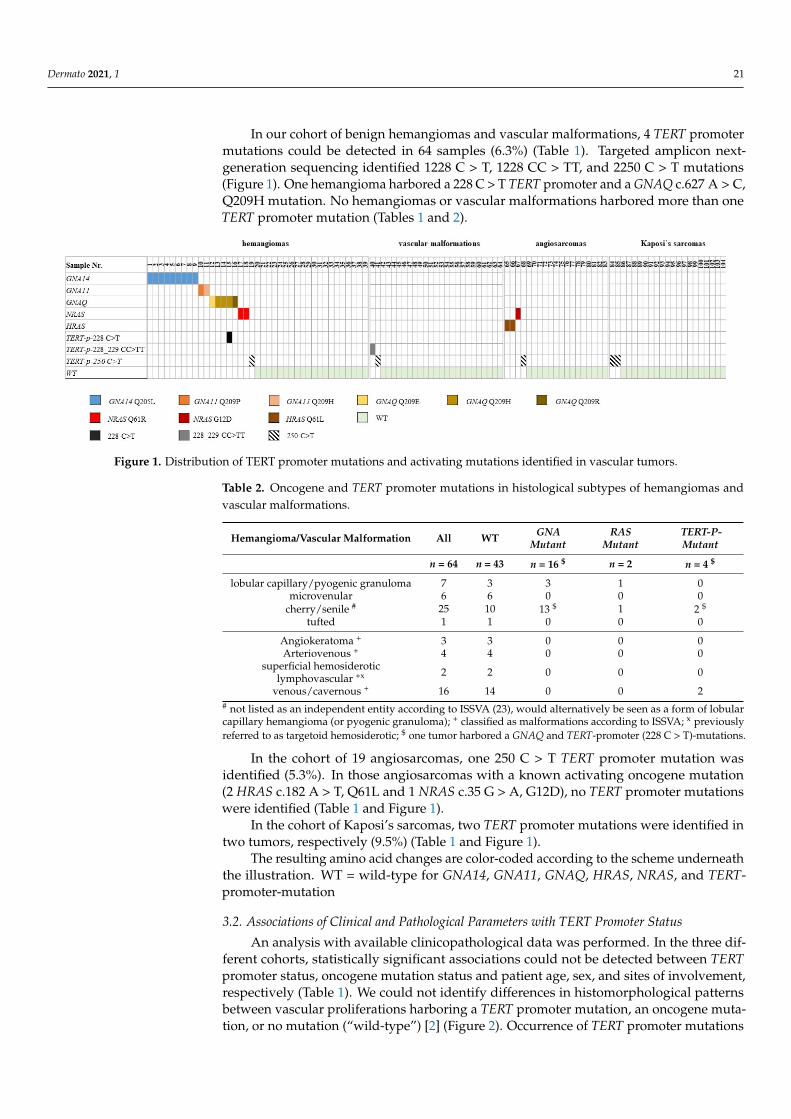
In our cohort of 104 vascular tumors from 102 patients, the TERT promoter regionshowed wild-type reads in 97 tumors (93.3%) and harbored one mutation in 7 cases(6.7%) (Table 1). Mutations identified were located at the previously described hotspots:Chr.5:1295228 C > T, Chr.5:1295228_1295229 CC > TT, or Chr.5:1295250 C > T. All anno-tations are reported according to human genome assembly 19 (hg19). For simplicity, themutations will further be referred to using solely the last three digits of the chromosomelocation nomenclature representing the first nucleotide altered as 228 C > T, 228 CC > TT,and 250 C > T, respectively.
Table 1. Clinical variables of vascular proliferations with oncogene and TERT promoter mutations.
Hemangioma/Vascular Malformation All WT GNA Mutant RAS Mutant TERT-P-Mutant p-Value *
n = 64 n = 43 n = 16 $ n = 2 n = 4 $
Mean age (years) 53 54 48 60 57 0.4
SexFemale 28 20 5 $ 0 3 $
0.36Male 35 # 22 10 # 2 1
Sites of involvement
head/neck 21 16 3 $ 1 2 $
0.06ventral trunk 16 8 7 1 0dorsal trunk 13 7 6 0 0
upper extremity 5 4 0 0 1lower extremity 7 7 0 0 0
LND 2 1 0 0 1
Angiosarcoma All WT GNA Mutant RAS Mutant TERT-P-Mutant p-Value *
n = 19 n = 15 n = 0 n = 3 n = 1
Mean age (years) 67 65 - 75 74 0.4
SexFemale 15 11 - 3 1
1.0Male 4 4 0 0
Sites of involvement
head/neck 8 6 - 2 0
1.0ventral trunk 7 5 - 1 1dorsal trunk 0 0 - 0 0
upper extremity 0 0 - 0 0lower extremity 1 1 - 0 0
LND 3 3 - 0 0
Kaposi’s sarcoma All WT GNA Mutant RAS Mutant TERT-P-Mutant p-Value *
n = 21 n = 19 n = 0 n = 0 n = 2
Mean age (years) 56 58 - - 34 0.1
SexFemale 4 4 - - 0
1.0Male 16 ! 14 ! 2
Sites of involvement
head/neck 2 2 - - 0
0.13ventral trunk 2 1 - - 1dorsal trunk 0 0 - - 0
upper extremity 2 1 - - 1lower extremity 13 13 - - 0
LND 2 2 - - 0
LND—localization not determined, TERT-P-mutant = TERT-promoter mutation (228 C > T, 228 CC > TT or 250 C > T), WT = wild-typefor GNA14, GNA11, GNAQ, HRAS, NRAS and TERT-promoter-mutation, # one male had two hemangiomas both showing a GNA14mutation, $ one female patient showed a GNAQ and a TERT-promoter (228 C > T)-mutation, ! one male had two Kaposi’s sarcomas,* Age—Kruskal–Wallis test; all others—Fisher exact test.
Dermato 2021, 1 21
In our cohort of benign hemangiomas and vascular malformations, 4 TERT promotermutations could be detected in 64 samples (6.3%) (Table 1). Targeted amplicon next-generation sequencing identified 1228 C > T, 1228 CC > TT, and 2250 C > T mutations(Figure 1). One hemangioma harbored a 228 C > T TERT promoter and a GNAQ c.627 A > C,Q209H mutation. No hemangiomas or vascular malformations harbored more than oneTERT promoter mutation (Tables 1 and 2).
Dermato 2021, 1, FOR PEER REVIEW 5
Figure 1. Distribution of TERT promoter mutations and activating mutations identified in vascular tumors.
The resulting amino acid changes are color-coded according to the scheme under-neath the illustration. WT = wild-type for GNA14, GNA11, GNAQ, HRAS, NRAS, and TERT-promoter-mutation
3.2. Associations of Clinical and Pathological Parameters with TERT Promoter Status An analysis with available clinicopathological data was performed. In the three dif-
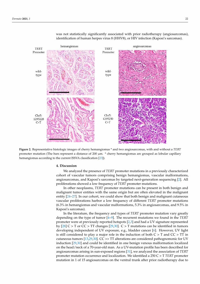
ferent cohorts, statistically significant associations could not be detected between TERT promoter status, oncogene mutation status and patient age, sex, and sites of involvement, respectively (Table 1). We could not identify differences in histomorphological patterns between vascular proliferations harboring a TERT promoter mutation, an oncogene mu-tation, or no mutation (“wild-type”) [2] (Figure 2). Occurrence of TERT promoter muta-tions was not statistically significantly associated with prior radiotherapy (angiosarco-mas), identification of human herpes virus 8 (HHV8), or HIV infection (Kaposi’s sarco-mas).
Figure 1. Distribution of TERT promoter mutations and activating mutations identified in vascular tumors.
Table 2. Oncogene and TERT promoter mutations in histological subtypes of hemangiomas andvascular malformations.
Hemangioma/Vascular Malformation All WT GNAMutant
RASMutant
TERT-P-Mutant
n = 64 n = 43 n = 16 $ n = 2 n = 4 $
lobular capillary/pyogenic granuloma 7 3 3 1 0microvenular 6 6 0 0 0
cherry/senile # 25 10 13 $ 1 2 $
tufted 1 1 0 0 0
Angiokeratoma + 3 3 0 0 0Arteriovenous + 4 4 0 0 0
superficial hemosideroticlymphovascular +x 2 2 0 0 0
venous/cavernous + 16 14 0 0 2# not listed as an independent entity according to ISSVA (23), would alternatively be seen as a form of lobularcapillary hemangioma (or pyogenic granuloma); + classified as malformations according to ISSVA; x previouslyreferred to as targetoid hemosiderotic; $ one tumor harbored a GNAQ and TERT-promoter (228 C > T)-mutations.
In the cohort of 19 angiosarcomas, one 250 C > T TERT promoter mutation wasidentified (5.3%). In those angiosarcomas with a known activating oncogene mutation(2 HRAS c.182 A > T, Q61L and 1 NRAS c.35 G > A, G12D), no TERT promoter mutationswere identified (Table 1 and Figure 1).
In the cohort of Kaposi’s sarcomas, two TERT promoter mutations were identified intwo tumors, respectively (9.5%) (Table 1 and Figure 1).
The resulting amino acid changes are color-coded according to the scheme underneaththe illustration. WT = wild-type for GNA14, GNA11, GNAQ, HRAS, NRAS, and TERT-promoter-mutation
3.2. Associations of Clinical and Pathological Parameters with TERT Promoter Status
An analysis with available clinicopathological data was performed. In the three dif-ferent cohorts, statistically significant associations could not be detected between TERTpromoter status, oncogene mutation status and patient age, sex, and sites of involvement,respectively (Table 1). We could not identify differences in histomorphological patternsbetween vascular proliferations harboring a TERT promoter mutation, an oncogene muta-tion, or no mutation (“wild-type”) [2] (Figure 2). Occurrence of TERT promoter mutations
Dermato 2021, 1 22
was not statistically significantly associated with prior radiotherapy (angiosarcomas),identification of human herpes virus 8 (HHV8), or HIV infection (Kaposi’s sarcomas).
Dermato 2021, 1, FOR PEER REVIEW 5
Figure 1. Distribution of TERT promoter mutations and activating mutations identified in vascular tumors.
The resulting amino acid changes are color-coded according to the scheme under-neath the illustration. WT = wild-type for GNA14, GNA11, GNAQ, HRAS, NRAS, and TERT-promoter-mutation
3.2. Associations of Clinical and Pathological Parameters with TERT Promoter Status An analysis with available clinicopathological data was performed. In the three dif-
ferent cohorts, statistically significant associations could not be detected between TERT promoter status, oncogene mutation status and patient age, sex, and sites of involvement, respectively (Table 1). We could not identify differences in histomorphological patterns between vascular proliferations harboring a TERT promoter mutation, an oncogene mu-tation, or no mutation (“wild-type”) [2] (Figure 2). Occurrence of TERT promoter muta-tions was not statistically significantly associated with prior radiotherapy (angiosarco-mas), identification of human herpes virus 8 (HHV8), or HIV infection (Kaposi’s sarco-mas).
Figure 2. Representative histologic images of cherry hemangiomas * and two angiosarcomas, with and without a TERTpromoter mutation (The bars represent a distance of 200 µm. * cherry hemangiomas are grouped as lobular capillaryhemangiomas according to the current ISSVA classification [23]).
4. Discussion
We analyzed the presence of TERT promoter mutations in a previously characterizedcohort of vascular tumors comprising benign hemangiomas, vascular malformations,angiosarcomas, and Kaposi’s sarcomas by targeted next-generation sequencing [2]. Allproliferations showed a low frequency of TERT promoter mutations.
In other neoplasms, TERT promoter mutations can be present in both benign andmalignant tumor entities with the same origin but are often elevated in the malignantentity [24–27]. In our cohort, we could show that both benign and malignant cutaneousvascular proliferations harbor a low frequency of different TERT promoter mutations(6.3% in hemangiomas and vascular malformations, 5.3% in angiosarcomas, and 9.5% inKaposi’s sarcomas).
In the literature, the frequency and types of TERT promoter mutation vary greatlydepending on the type of tumor [4–9]. The recurrent mutations we found in the TERTpromoter were at previously reported hotspots [2,3] and had a UV signature representedby [28] C > T or CC > TT changes [29,30]. C > T mutations can be identified in tumorsdeveloping independent of UV exposure, e.g., bladder cancer [6]. However, UV lightis still considered to play a major role in the induction of both C > T and CC > TT incutaneous tumors [13,29,30]. CC >> TT alterations are considered pathognomonic for UVinduction [29,30] and could be identified in one benign venous malformation localizedon the head/neck of a 70-year-old man. As a UV-mutation profile has been described forangiosarcomas arising in sun-exposed regions [31], we analyzed the association of TERTpromoter mutation occurrence and localization. We identified a 250 C > T TERT promotermutation in 1 of 15 angiosarcomas on the ventral trunk after prior radiotherapy due to
Dermato 2021, 1 23
breast cancer. In the other six angiosarcomas known to have arisen following radiotherapydue to breast cancer, no TERT promoter mutation could be identified.
Of the seven cutaneous vascular proliferations harboring one TERT promoter muta-tion, two were located in the head/neck region, two on the ventral trunk, and two on thelower extremity (one with unknown localization). Although proliferations with TERT pro-moter mutations were identified in areas favoring sun exposure, a statistically significantassociation between the occurrence of a TERT promoter mutation and localization couldnot be calculated in any of our cohorts.
The prevalence of TERT promoter mutations detected (hemangiomas/vascular mal-formations (6.3%), angiosarcomas (5.3%), and Kaposi’s sarcomas (9.5%)) was low in bothbenign and malignant cutaneous vascular proliferations. Rare TERT promoter mutationsin angiosarcomas fit previous studies [28,32]. GNA14, GNA11, and GNAQ mutations arefrequent in congenital and cherry hemangiomas [33–35] (cherry or senile hemangiomasis still a widely used term, although not recognized as an individual entity according tothe ISSVA classification 2018. This classification would group these lesions as a form ofpyogenic granuloma or lobular capillary hemangioma). A single hemangioma harboreda 228 C > T TERT promotor and a GNAQ c.627 A > C, Q209H mutation. Otherwise, noco-occurrence with known GNA or RAS mutations was observed. In melanoma, TERTpromoter mutations frequently co-occur with activating BRAF mutations [36,37]. In ourstudy, a similar co-occurrence of mutations could not be observed.
The role for the TERT (telomerase reverse transcriptase) protein in cutaneous vasculartumors remains intriguing. Our finding suggests that it promotes tumor proliferation ina few cutaneous vascular proliferations. However, based on the frequency of mutations,a mutation of the promoter to increase protein activity appears to be non-mandatory inmost tumors. Potentially, gains of the region do occur in vascular tumors lacking TERT pro-moter mutations. Additionally, alternative lengthening of telomeres (ALT), e.g., mutationsin ATRX or DAXX, may contribute to the progression of cutaneous vascular prolifer-ations. These mechanisms, both generally occurring much rarer than TERT promotermutations [38], could not be assessed in the assay we applied and will need to be examinedin future studies.
The overall frequency (<10%) and its variation between tumor groups was low. Thissuggests determining TERT promoter mutation status is of no diagnostic aid in terms ofclassifying tumor entities or predicting prognosis.
A limitation of our study is the lack of detailed clinical data, including therapy andfollow-up information. Strengths of our study are the considerable number of vasculartumors included in the analysis (n = 104), making it the largest cohort of vascular tumorsanalyzed for the presence of TERT promoter mutations.
5. Conclusions
In conclusion, we could show that TERT promoter mutations are present in bothbenign and malignant cutaneous vascular tumors and malformations. The prevalencewas 6.3% in hemangiomas and vascular malformations, 5.3% in angiosarcomas, and 9.5%in Kaposi’s sarcomas. In our comparative analysis, we could not confirm the tendencyobserved in other cutaneous tumors of TERT promoter mutations being more prevalentin malignant entities. While TERT promoter mutations appear to be relevant in a smallpercentage of tumors, the majority of these tumors arise independent of these mutations.
Author Contributions: Conceptualization, methodology and software, K.G.G. and P.J.; validation,P.J., D.S., E.H., K.G.G.; histological analysis, H.M., J.S., T.M., E.H., K.G.G.; investigation all au-thors.; data curation, P.J. and K.G.G.; writing—original draft preparation, P.J., H.M., T.M., E.H.,K.G.G.; writing—review and editing, all authors; visualization and supervision, P.J. and K.G.G.;project administration, P.J. and K.G.G. All authors have read and agreed to the published version ofthe manuscript.
Funding: This research received no external funding.
Dermato 2021, 1 24
Institutional Review Board Statement: The study was conducted according to the guidelines of theDeclaration of Helsinki, and approved by the Ethics Committee of the University of Duisburg-Essen(IRB-number 20–9688-BO, date of approval: 26 November 2020).
Informed Consent Statement: Informed consent was obtained from all subjects involved in the study.
Data Availability Statement: The datasets presented in this study can be found in online repositories.The names of the repository/repositories and accession number(s) can be found below: https://www.ncbi.nlm.nih.gov/genbank/, PRJNA717731 (accessed on 27 March 2021).
Conflicts of Interest: The authors declare no conflict of interest.
References1. Bell, R.J.; Rube, H.T.; Xavier-Magalhaes, A.; Costa, B.M.; Mancini, A.; Song, J.S.; Costello, J.F. Understanding TERT Promoter
Mutations: A Common Path to Immortality. Mol. Cancer Res. 2016, 14, 315–323. [CrossRef]2. Horn, S.; Figl, A.; Rachakonda, P.S.; Fischer, C.; Sucker, A.; Gast, A.; Kadel, S.; Moll, I.; Nagore, E.; Hemminki, K.; et al. TERT
promoter mutations in familial and sporadic melanoma. Science 2013, 339, 959–961. [CrossRef]3. Huang, F.W.; Hodis, E.; Xu, M.J.; Kryukov, G.V.; Chin, L.; Garraway, L.A. Highly recurrent TERT promoter mutations in human
melanoma. Science 2013, 339, 957–959. [CrossRef] [PubMed]4. Liu, X.; Qu, S.; Liu, R.; Sheng, C.; Shi, X.; Zhu, G.; Murugan, A.K.; Guan, H.; Yu, H.; Wang, Y.; et al. TERT promoter mutations
and their association with BRAF V600E mutation and aggressive clinicopathological characteristics of thyroid cancer. J. Clin.Endocrinol. Metab. 2014, 99, E1130–E1136. [CrossRef]
5. Rusinek, D.; Pfeifer, A.; Krajewska, J.; Oczko-Wojciechowska, M.; Handkiewicz-Junak, D.; Pawlaczek, A.; Zebracka-Gala, J.;Kowalska, M.; Cyplinska, R.; Zembala-Nozynska, E.; et al. Coexistence of TERT Promoter Mutations and the BRAF V600EAlteration and Its Impact on Histopathological Features of Papillary Thyroid Carcinoma in a Selected Series of Polish Patients.Int. J. Mol. Sci. 2018, 19, 2647. [CrossRef]
6. Hosen, I.; Rachakonda, P.S.; Heidenreich, B.; de Verdier, P.J.; Ryk, C.; Steineck, G.; Hemminki, K.; Kumar, R. Mutations in TERTpromoter and FGFR3 and telomere length in bladder cancer. Int. J. Cancer 2015, 137, 1621–1629. [CrossRef] [PubMed]
7. Spiegl-Kreinecker, S.; Lotsch, D.; Neumayer, K.; Kastler, L.; Gojo, J.; Pirker, C.; Pichler, J.; Weis, S.; Kumar, R.; Webersinke, G.; et al.TERT promoter mutations are associated with poor prognosis and cell immortalization in meningioma. Neuro. Oncol. 2018, 20,1584–1593. [CrossRef] [PubMed]
8. Ren, H.; Shen, Y.; Hu, D.; He, W.; Zhou, J.; Cao, Y.; Mao, Y.; Dou, Y.; Xiong, W.; Xiao, Q.; et al. Co-existence of BRAF(V600E) andTERT promoter mutations in papillary thyroid carcinoma is associated with tumor aggressiveness, but not with lymph nodemetastasis. Cancer Manag. Res. 2018, 10, 1005–1013. [CrossRef]
9. Jeong, D.E.; Woo, S.R.; Nam, H.; Nam, D.H.; Lee, J.H.; Joo, K.M. Preclinical and clinical implications of TERT promoter mutationin glioblastoma multiforme. Oncol. Lett. 2017, 14, 8213–8219. [CrossRef]
10. Campos, M.A.; Macedo, S.; Fernandes, M.; Pestana, A.; Pardal, J.; Batista, R.; Vinagre, J.; Sanches, A.; Baptista, A.; Lopes, J.M.;et al. TERT promoter mutations are associated with poor prognosis in cutaneous squamous cell carcinoma. J. Am. Acad. Dermatol.2019, 80, 660–669 e666. [CrossRef] [PubMed]
11. Malkki, H. Neuro-oncology: TERT promoter mutations could indicate poor prognosis in glioblastoma. Nat. Rev. Neurol. 2014,10, 546. [CrossRef] [PubMed]
12. Blateau, P.; Coyaud, E.; Laurent, E.; Beganton, B.; Ducros, V.; Chauchard, G.; Vendrell, J.A.; Solassol, J. TERT Promoter Mutationas an Independent Prognostic Marker for Poor Prognosis MAPK Inhibitors-Treated Melanoma. Cancers 2020, 12, 2224. [CrossRef][PubMed]
13. Griewank, K.G.; Murali, R.; Puig-Butille, J.A.; Schilling, B.; Livingstone, E.; Potrony, M.; Carrera, C.; Schimming, T.; Moller, I.;Schwamborn, M.; et al. TERT promoter mutation status as an independent prognostic factor in cutaneous melanoma. J. Natl.Cancer Inst. 2014, 106. [CrossRef] [PubMed]
14. Arantes, L.; Cruvinel-Carloni, A.; de Carvalho, A.C.; Sorroche, B.P.; Carvalho, A.L.; Scapulatempo-Neto, C.; Reis, R.M. TERTPromoter Mutation C228T Increases Risk for Tumor Recurrence and Death in Head and Neck Cancer Patients. Front. Oncol. 2020,10, 1275. [CrossRef] [PubMed]
15. Borah, S.; Xi, L.; Zaug, A.J.; Powell, N.M.; Dancik, G.M.; Cohen, S.B.; Costello, J.C.; Theodorescu, D.; Cech, T.R. Cancer. TERTpromoter mutations and telomerase reactivation in urothelial cancer. Science 2015, 347, 1006–1010. [CrossRef] [PubMed]
16. Chiba, K.; Lorbeer, F.K.; Shain, A.H.; McSwiggen, D.T.; Schruf, E.; Oh, A.; Ryu, J.; Darzacq, X.; Bastian, B.C.; Hockemeyer, D.Mutations in the promoter of the telomerase gene TERT contribute to tumorigenesis by a two-step mechanism. Science 2017, 357,1416–1420. [CrossRef]
17. Killela, P.J.; Reitman, Z.J.; Jiao, Y.; Bettegowda, C.; Agrawal, N.; Diaz, L.A., Jr.; Friedman, A.H.; Friedman, H.; Gallia, G.L.;Giovanella, B.C.; et al. TERT promoter mutations occur frequently in gliomas and a subset of tumors derived from cells with lowrates of self-renewal. Proc. Natl. Acad. Sci. USA 2013, 110, 6021–6026. [CrossRef]
18. Shain, A.H.; Yeh, I.; Kovalyshyn, I.; Sriharan, A.; Talevich, E.; Gagnon, A.; Dummer, R.; North, J.; Pincus, L.; Ruben, B.; et al. TheGenetic Evolution of Melanoma from Precursor Lesions. N. Engl. J. Med. 2015, 373, 1926–1936. [CrossRef]
Dermato 2021, 1 25
19. Griewank, K.G.; Murali, R.; Schilling, B.; Schimming, T.; Moller, I.; Moll, I.; Schwamborn, M.; Sucker, A.; Zimmer, L.; Schadendorf,D.; et al. TERT promoter mutations are frequent in cutaneous basal cell carcinoma and squamous cell carcinoma. PLoS ONE 2013,8, e80354. [CrossRef]
20. Scott, G.A.; Laughlin, T.S.; Rothberg, P.G. Mutations of the TERT promoter are common in basal cell carcinoma and squamouscell carcinoma. Mod. Pathol. 2014, 27, 516–523. [CrossRef] [PubMed]
21. Murali, R.; Chandramohan, R.; Moller, I.; Scholz, S.L.; Berger, M.; Huberman, K.; Viale, A.; Pirun, M.; Socci, N.D.; Bouvier, N.;et al. Targeted massively parallel sequencing of angiosarcomas reveals frequent activation of the mitogen activated protein kinasepathway. Oncotarget 2015, 6, 36041–36052. [CrossRef]
22. Jansen, P.; Müller, H.; Lodde, G.C.; Zaremba, A.; Möller, I.; Sucker, A.; Paschen, A.; Esser, S.; Schaller, J.; Gunzer, M.; et al. GNA14,GNA11, and GNAQ Mutations Are Frequent in Benign but Not Malignant Cutaneous Vascular Tumors. Front. Genet. 2021, 12,656. [CrossRef] [PubMed]
23. ISSVA Classification for Vascular Anomalies. 2018. Available online: https://www.issva.org/UserFiles/file/ISSVA-Classification-2018.pdf (accessed on 23 June 2021).
24. Koopmans, A.E.; Ober, K.; Dubbink, H.J.; Paridaens, D.; Naus, N.C.; Belunek, S.; Krist, B.; Post, E.; Zwarthoff, E.C.; de Klein,A.; et al. Prevalence and implications of TERT promoter mutation in uveal and conjunctival melanoma and in benign andpremalignant conjunctival melanocytic lesions. Investig. Ophthalmol. Visual Sci. 2014, 55, 6024–6030. [CrossRef] [PubMed]
25. Munoz-Jimenez, M.T.; Blanco, L.; Ruano, Y.; Carrillo, R.; Santos-Briz, A.; Riveiro-Falkenbach, E.; Requena, L.; Kutzner, H.;Garrido, M.C.; Rodriguez-Peralto, J.L. TERT promoter mutation in sebaceous neoplasms. Virchows Arch. 2021. [CrossRef]
26. Jansen, P.; Cosgarea, I.; Murali, R.; Moller, I.; Sucker, A.; Franklin, C.; Paschen, A.; Zaremba, A.; Brinker, T.J.; Stoffels, I.; et al.Frequent Occurrence of NRAS and BRAF Mutations in Human Acral Naevi. Cancers 2019, 11, 546. [CrossRef]
27. Zaremba, A.; Murali, R.; Jansen, P.; Moller, I.; Sucker, A.; Paschen, A.; Zimmer, L.; Livingstone, E.; Brinker, T.J.; Hadaschik, E.;et al. Clinical and genetic analysis of melanomas arising in acral sites. Eur. J. Cancer 2019, 119, 66–76. [CrossRef] [PubMed]
28. Painter, C.A.; Jain, E.; Tomson, B.N.; Dunphy, M.; Stoddard, R.E.; Thomas, B.S.; Damon, A.L.; Shah, S.; Kim, D.;Gomez Tejeda Zanudo, J.; et al. The Angiosarcoma Project: Enabling genomic and clinical discoveries in a rare cancerthrough patient-partnered research. Nat. Med. 2020, 26, 181–187. [CrossRef]
29. Pleasance, E.D.; Cheetham, R.K.; Stephens, P.J.; McBride, D.J.; Humphray, S.J.; Greenman, C.D.; Varela, I.; Lin, M.L.; Ordonez, G.R.;Bignell, G.R.; et al. A comprehensive catalogue of somatic mutations from a human cancer genome. Nature 2010, 463, 191–196.[CrossRef] [PubMed]
30. Brash, D.E.; Rudolph, J.A.; Simon, J.A.; Lin, A.; McKenna, G.J.; Baden, H.P.; Halperin, A.J.; Ponten, J. A role for sunlight in skincancer: UV-induced p53 mutations in squamous cell carcinoma. Proc. Natl. Acad. Sci. USA 1991, 88, 10124–10128. [CrossRef]
31. Chan, J.Y.; Lim, J.Q.; Yeong, J.; Ravi, V.; Guan, P.; Boot, A.; Tay, T.K.Y.; Selvarajan, S.; Md Nasir, N.D.; Loh, J.H.; et al. Multiomicanalysis and immunoprofiling reveal distinct subtypes of human angiosarcoma. J. Clin. Investig. 2020, 130, 5833–5846. [CrossRef]
32. Koelsche, C.; Renner, M.; Hartmann, W.; Brandt, R.; Lehner, B.; Waldburger, N.; Alldinger, I.; Schmitt, T.; Egerer, G.; Penzel, R.;et al. TERT promoter hotspot mutations are recurrent in myxoid liposarcomas but rare in other soft tissue sarcoma entities. J. Exp.Clin. Cancer Res. 2014, 33, 33. [CrossRef] [PubMed]
33. Liau, J.Y.; Lee, J.C.; Tsai, J.H.; Chen, C.C.; Chung, Y.C.; Wang, Y.H. High frequency of GNA14, GNAQ, and GNA11 mutations incherry hemangioma: A histopathological and molecular study of 85 cases indicating GNA14 as the most commonly mutatedgene in vascular neoplasms. Mod. Pathol. 2019, 32, 1657–1665. [CrossRef] [PubMed]
34. Liau, J.Y.; Lee, J.C.; Tsai, J.H.; Chen, C.C.; Wang, Y.H.; Chung, Y.C. Thrombotic Hemangioma With Organizing/AnastomosingFeatures: Expanding the Spectrum of GNA-mutated Hemangiomas With a Predilection for the Skin of the Lower AbdominalRegions. Am. J. Surg. Pathol. 2020, 44, 255–262. [CrossRef]
35. Ayturk, U.M.; Couto, J.A.; Hann, S.; Mulliken, J.B.; Williams, K.L.; Huang, A.Y.; Fishman, S.J.; Boyd, T.K.; Kozakewich, H.P.;Bischoff, J.; et al. Somatic Activating Mutations in GNAQ and GNA11 Are Associated with Congenital Hemangioma. Am. J.Hum. Genet. 2016, 98, 789–795. [CrossRef] [PubMed]
36. Zehir, A.; Benayed, R.; Shah, R.H.; Syed, A.; Middha, S.; Kim, H.R.; Srinivasan, P.; Gao, J.; Chakravarty, D.; Devlin, S.M.; et al.Mutational landscape of metastatic cancer revealed from prospective clinical sequencing of 10,000 patients. Nat. Med. 2017, 23,703–713. [CrossRef] [PubMed]
37. Macerola, E.; Loggini, B.; Giannini, R.; Garavello, G.; Giordano, M.; Proietti, A.; Niccoli, C.; Basolo, F.; Fontanini, G. Coexis-tence of TERT promoter and BRAF mutations in cutaneous melanoma is associated with more clinicopathological features ofaggressiveness. Virchows Arch. 2015, 467, 177–184. [CrossRef]
38. Shay, J.W.; Wright, W.E. Telomeres and telomerase: Three decades of progress. Nat. Rev. Genet. 2019, 20, 299–309. [CrossRef]


![Lower Rim Substituted p-tert -Butyl-Calix[4]arene. Part 15. Pb(II)-Ion-Selective Electrodes Based on p-tert -Butyl-calix[4]arene Thioamides](https://static.fdokumen.com/doc/165x107/6342a72ff9c0d1681b0ad302/lower-rim-substituted-p-tert-butyl-calix4arene-part-15-pbii-ion-selective.jpg)

![Ethene/norbornene copolymerization by [Me 2 Si(3- tert BuCp)(N tert Bu)]TiCl 2 /MAO-catalyst](https://static.fdokumen.com/doc/165x107/6312231a48b4e11f7d08cd0e/ethenenorbornene-copolymerization-by-me-2-si3-tert-bucpn-tert-buticl-2-mao-catalyst.jpg)