BENIGN TUMORS OF THE BREAST. ENCAPSULATED ...
26
BENIGN TUMORS OF THE BREAST. ENCAPSULATED ADENOMA A BRIEF SUMMARY OF THEIR CLINICAL AND PATHOLOGICAL FEATURES BY JOSEPH COLT BLOODGOOD, M.D. OF BALTIMORE, MD. SYNopsIs.-The more common tumors of the breast. Multiple tumors. Multiple encapsulated adenoma. Single encapsulated adenoma. Single encapsulated adenoma clinically malignant. Palpation of encapsulated adenoma. Palpation of larger encapsu- lated adenomas. Aberrant adenoma. Sarcoma in intracanalicular myxoma. Differential diagnosis between small encapsulated adenoma and sarcoma. Encapsulated adenoma microscopically suspicious of cancer. Ultimate results in patients operated on for encapsulated adenoma, without the removal of the breast. Adenoma in pregnancy and lactation. Operations upon the breast during pregnancy and lactation. Exploratory incision and excision of benign tumors of the breast. First method of exploratory incision. Second method of exploratory incision. Third method of exploratory incision. Plastic closure. Conclusions. The more common tumors of the breast are the encapsulated adenoma, some types of chronic cystic viastitis (the most frequent of which is the blue- domed cyst), and fully developed carcinoma. The scirrhus carcinoma largely leads the soft medullary carcinoma. Other lesions of the breast, both benign and malignant, are relatively inf requent. In teaching students, both before and after graduation, and in reviewing one s own experience for the purpose of improving methods for the recog- nition of the benign from the malignant, both clinically (based on history and palpation) and pathologically (based on the gross and microscopic appearance at the exploratory incision), one should bear in mind the common occurrence of these three great groups and first master them. Mfultiple Tumors.- (Figs. I and 2.) I employ the designation A to mean a single tumor in one breast, B a single tumor in both breasts, C multiple tumors in one breast, D multiple tumors in both breasts. When the letter E is placed after A, B, C, or D, it indicates that the single, or the multiple, tumors are indefinite. Multiple definite tumors in the groups, B, C, and D, are rarely malignant, and, if so, in my experience, always incurable. I have recently checked this most carefully. Therefore, if one can palpate a definite tumor in each breast, or multiple definite tumors in one or both breasts, a mistake in diagnosing benign will never do any harm. To completely excise one or both breasts, or even to perform the complete operation for cancer on both sides, would be an unnecessary mutilating operation and should not be done without exploring one of the tumors in each breast and ascertaining their pathological nature. Personally, I have never observed a definite single tumor in each breast, 172
-
Upload
khangminh22 -
Category
Documents
-
view
1 -
download
0
Transcript of BENIGN TUMORS OF THE BREAST. ENCAPSULATED ...

BENIGN TUMORS OF THE BREAST. ENCAPSULATED ADENOMAA BRIEF SUMMARY OF THEIR CLINICAL AND PATHOLOGICAL FEATURES
BY JOSEPH COLT BLOODGOOD, M.D.OF BALTIMORE, MD.
SYNopsIs.-The more common tumors of the breast. Multiple tumors. Multipleencapsulated adenoma. Single encapsulated adenoma. Single encapsulated adenomaclinically malignant. Palpation of encapsulated adenoma. Palpation of larger encapsu-lated adenomas. Aberrant adenoma. Sarcoma in intracanalicular myxoma. Differentialdiagnosis between small encapsulated adenoma and sarcoma. Encapsulated adenomamicroscopically suspicious of cancer. Ultimate results in patients operated on forencapsulated adenoma, without the removal of the breast. Adenoma in pregnancy andlactation. Operations upon the breast during pregnancy and lactation. Exploratoryincision and excision of benign tumors of the breast. First method of exploratoryincision. Second method of exploratory incision. Third method of exploratory incision.Plastic closure. Conclusions.
The more common tumors of the breast are the encapsulated adenoma,some types of chronic cystic viastitis (the most frequent of which is the blue-domed cyst), and fully developed carcinoma. The scirrhus carcinoma largelyleads the soft medullary carcinoma.
Other lesions of the breast, both benign and malignant, are relativelyinfrequent.
In teaching students, both before and after graduation, and in reviewingone s own experience for the purpose of improving methods for the recog-nition of the benign from the malignant, both clinically (based on historyand palpation) and pathologically (based on the gross and microscopicappearance at the exploratory incision), one should bear in mind the commonoccurrence of these three great groups and first master them.
Mfultiple Tumors.- (Figs. I and 2.) I employ the designation A to meana single tumor in one breast, B a single tumor in both breasts, C multipletumors in one breast, D multiple tumors in both breasts.
When the letter E is placed after A, B, C, or D, it indicates that thesingle, or the multiple, tumors are indefinite.
Multiple definite tumors in the groups, B, C, and D, are rarely malignant,and, if so, in my experience, always incurable. I have recently checked thismost carefully. Therefore, if one can palpate a definite tumor in each breast,or multiple definite tumors in one or both breasts, a mistake in diagnosingbenign will never do any harm. To completely excise one or both breasts,or even to perform the complete operation for cancer on both sides, would bean unnecessary mutilating operation and should not be done without exploringone of the tumors in each breast and ascertaining their pathological nature.
Personally, I have never observed a definite single tumor in each breast,172

BENIGN TUMORS OF THE BREAST
FIG. i.-Pathol. No. 9627. Multiple adenoma removed from one breast; all encapsulated; thelarger tumor is of the fibroadenoma type, the smaller represents an example of each type, fibroadenoma,intracanalicular and cystic adenoma. From original painting.
~~~~~~~~~~~~~~~~~~~~~~~~~~~~~~~~~.. .... w..:'.
FIG. 2.-Pathol. No. 9627. On inspection unilateral hypertrophy of left breast in a girl agedthirteen. On palpation one large and many small movable tumors in left breast (see Fig. I) and manysmall, movable tumors in right breast. Operation: Removal of the larger and some of the smaller tumorsin the left breast. Result: Ten years later the remaining tumors have disappeared.
173

JOSEPH COLT BLOODGOOD
or definite multiple tumors in one or both breasts to be malignant, unless therewas definite clinical evidence of malignancy.
The more common multiple tumors are the encapsulated adenoma and theblue-domed cyst, and for such lesions, complete removal of the breast isnot necessary. The larger, or the growing, or the painful tumors may beremoved. My records show that too many breasts have been sacrificed formultiple tumors in one or both breasts. These women run no more risk ofcancer, and perhaps less risk, than women in whose breasts no lumps canbe palpated.
Multiple Encapsulated Adenomna.-(Fig. 3.) There are three types ofadenoma of the breast-fibroadeniomna [82 cases] (sometimes called periductaladenom,a), intracanalucilar myxoadenorna [I98 cases], and cystic adenoma
a b
. . ~ ~ ~~~~~~~~~~~~~~~~~~~~~~~~~~~~~~~~~~~~....._~.p
FIG. 3.-a. and b. Pathol. No. 211I93. Typical encapsulated adenoma projecting like a dome above thesurrounding breast-the appearance one gets at the exploratory incision. The dome is gray, not blue. Forappearance of bisection see Fig. 3-b. Pathol. No. 2II93. The tumor in Fig. 3-a bisected. Note the dis-tinct capsule and the zone of breast removed with the tumor. The tumor is ofthe fibroadenoma type andresembles somewhat normal breast at puberty. It has less stroma and more parenchyma. See Fig. 4formicroscopic picture.
(22 cases). The first two varieties are far more frequent than the cysticadenoma. One will observe aboUt 22 cystic adenomas to 280 Of the other twotypes. The cystic adenoma is rarely multiple, the other two varieties are quitefrequently multiple.
Multiple adenomas have been observed at any age, but are distinctly morecommon at puberty and before twenty-five years of age.
In the majority of cases the multiple tumors belong to one type, but notinfrequently two, or even three, types may be present. There is no objectionto the removal of the larger, the growing or the extremely painful tumor.It is never necessary to remove the breast or the breasts, and in many casesin which 'all the t'umors have not been removed, their disappearance hasbeen noted.
Single Encapsulated Adenosna.-When palpation finds but one definitetumor, and the patient is over twenty-five years of age, cancer must alwaysbe considered. A small encapsulated adenoma buried in breast tissue orsituated in a large fatty breast may palpate like a small infiltrating scirrhus.
174

BENIGN TUMORS OF THE BREAST
Then, again, circumscribed carcinoma, especially when buried in breast tissue,or of deep position in a fatty breast, cannot be distinguished from an encapsu-lated adenoma. The earlier a patient seeks advice after first feeling thetumor, the more frequently one palpates benign tumors suggesting malignancy,and malignant tumors suggesting benignancy. Therefore, every surgeonshould be on his guard. The most important points are: first, never toconsent to any delay; second, always to operate in a hospital and prepare thepatient for the complete operation for cancer; third, whether you explorethe tumor, or excise it for diagnosis, always be prepared for the immediatechemical or ther-mal cauterizationof -the wound. I
alcohol and a fiftper cent.solution .~~~~~~~~~~~~~~~~~~~~~~~~~~~~~~~~~~~.~~~. .
of zinc chloride to B 'i9Yft..
geon, one of my .....
own students a __-
17
teacher of sur- 1t_gery andsurgicalpathology in afft
pecen. solutio
medical s c h o o 1, -was so certain _ >that the palpable _ | rsingle tumor wasbenign that heallowed the pa- FIG. 4.-Pathol. No. 2II93. Microscopic se-ction (low power) of fibro-:
toprud adenoma shown in Fig. 3. The breast beyond the capsule is very fatty. Thietient eparenchyma in the tumor has no normal lobules with developed acini.him to remove itunder local anasthesia in his office. After its removal and gross bisection,he was horrified to find that it suggested malignancy, but he was not in aposition to proceed with the complete operation. Fortunately microscopicstudy demonstrated the tumor to be a non-encapsulated beniign adenoma-.andnot a carcinoma.
In my own clinic I have the greatest difficulty in getting older and moreexperienced internes to remember to prepare for immediate chemicalcauterization when a breast tumor is explored for diagnosis.
As a matter of fact, every single tumor of the breast in a woman overtwenty-five years of age, in which the operator concludes that it is not of a
175

JOSEPH COLT BLOODGOOD
character to justify the complete operation without exploration, must belooked upon as potential cancer and be explored.
Single Entcapsulated Adentoina, Clinically Malignant.-(Fig. 5.) My rec-ords show that encapsulated adenomas of all types are so rarely associated withretraction and fixation of the nipple, with atrophy of the subcutaneous fat,dimpling and fixation of the skin, that if the complete operation were per-
FIG. 5.-Pathol. No. 15983. Section through encapsulated adenoma (clinically malignant)'and thesurrounding breast. This patient, an adult woman at the menopause, had a known movable tumor ofmany years' duration. After a trauma, it grew from the size of a twenty-five-cent piece to that of a fifty-cent plece. There was atrophy of the subcutaneous fat and slight fixation of the skin. The completeoperation for cancer was performed. Exploring the tumor after operation demonstrated cedema outsidethe capsule. Microscopic section of the tumor (Fig. 6) shows no evidence of carcinoma or sarcoma. Theglands show no metastasis, and the patient is well more than five years since operation.
formed in all such cases, very few women would be mutilated. Neverthelessthese clinical signs of malignancy now and then do occur with encapsulatedadenoma. It has been my rule, that when the tumor has the palpation of anencapsulated adenoma or a cyst, and the free mobility of a benign tumor,I am apt to explore the tumor, even when there is a suggestion of dimpling ofthe skin or atrophy of the fat, or slight fixation or retraction of the nipple.This is especially true if the little tumtor is in thec niipple zonie.
More frequently have the clinical signs of malignancy been observed whenthe encapsulated adenomas have been multiple and the malignant signs have
176

BENIGN TUMORS OF THE BREAST
been present over one of the tumors. This is a very important thing toremember, because this knowledge will save an unnecessary mutilation of thepatient. I have just noted under the heading Multiple Tumors, that theyare usually benign, and if malignant never curable. Therefore, if there aremultiple tumors and one of them clinically suggests malignancy, it is quitejustifiable to explore this tumor.
Palpation of Entcapsulated Adenoma.-These tumors (of the size of apea, or a bean, or from the size of a ten-cent piece to that of a silver dollar)are usually recog-nized as benignby the majority....of surgeons. Themost characteris-tic feature is theirfree mobility, nomatter whetherthey are situatedoutside the breast,in the periphery,in the midzone, orin the nipple cen-tral area. Nowand then they arefelt in the axilla.They can bemoved about likea marble beneaththe skin. A ma-lignant t u m o rrarely, if ever, has FiG. 6.-Pathol. No. I5983. Microscopicpicture (high power) os tumorthis free mobility, shown in Fig. 5. A papillary cystadenomatous area. The majority of the
andetumor resembled Fig. 4.
and, of course,encapsulated adenomas, do not always show it. The shape of the adenomavaries. But if one can palpate the tumor and it is freely movable, andspherical, it cannot be distinguished from a cyst. More often, however, theadenoma is not spherical, but somewhat lobulated, now and then like amulberry. When we come to consistency, some of them are hard andfirm, but never of the hardness of scirrhus cancer. Many are elasticand not infrequently, especially the intracanalicular myxoma, are doughyand semi-fluctuating.
It is very important, right here, to record the fact that a small, buriedscirrhus cancer surrounded by cedematous breast tissue may give to thepalpating finger a sensation of fluctuation and suggest a cyst or a softerencapsulated adenoma. The same is true of a certain type of medullary cancer.
1" -177

JOSEPH COLT BLOODGOOD
Here, again, we have evidence of the difficulty of always distinguishing bypalpation the benign from the malignant tumor. But if one will carefullyrecord and try to memorize the sensations during the palpation of breasttumors, it; is quickly found that the majority of encapsulated adenomas aswell as blue-domed cysts are recognized by their mobility and their peculiarconsistency and shape on palpation.
It is only in a relatively small number of cases that the palpation of thebenign and malignant overlap.
Palpation of Larger Encapsulated Adenomntas.-The moment a tumorreaches the size of
;.;...... a quadrant of the.; ......... ......breast, or larger,
....j;~~~~~~~~and is still freely~~~~~~movable, and has
F.4 the palpation ofthe encapsulatedadenoma, onemust think ofsarcoma.
There are twotypes of the largerencapsulated ade-noma. One morefrequently situ-
A" ated outside thebreast (aberrantadenoma) is of thefibroadenomat o u stype and has notendency to sarco-
FIG. 7.-Pathol. No. 7135. Encapsulated aberrant fibroadenoma. Photo- matous change.graph of patient. The freely movable, large tumor is upwards and to theright; the breast to the medial and lower side. The patient was sixteen The larger en-years of age. Operation: Excision of tumor only, breast preserved. It thispatient had been over twenty-five, sarcoma in intracanalicular myxoma capsulated adeno-could not have been excluded. For gross appearance see Fig. 8. mas of the aber-rant adenoma type can be distinguished from the larger adenoma of theintracanalicular myxoma type only by the age of the patient and the situationof the tumor. The adenoma of the aberrant breast type is observed chieflyat puberty and in young women under twenty-five years of age; it is usuallysituated outside the breast. I have never observed sarcoma in the intra-canalicular myxoma type at an age under twenty-five, or to be situatedoutside the breast.
Therefore, a large tumor which on palpation suggests encapsulation in awoman under twenty-five years of age is as yet without an exception abenign fibroadenoma and can be removed and the breast saved, but a similar
178

BENIGN TUMORS OF THE BREAST
large tumor in a woman over twenty-five years of age should be looked uponas suspicious of sarcoma, and either removed without exploration with thebreast and major pectoral muscle or exposed for diagnosis.
In going over a large number of these cases one is impressed with howfrequently in the past the breast of a voung woman under twenty-five hasbeen sacrificed for the benignaberrant adenoma because ofthe clinical diagnosis of sar-coma, and how frequently localrecurrence has taken placeafter the removal by enuclea-tion, or removal of the breast A
only for these larger encapsu-lated adenomas of the intra-canalicular type in olderwomen.
The Differential DiagnosisBetweeni Smlaller EncapsulatedAdenoma and Sarcoma.-Nowand then sarcoma may be asmaller tumor and palpate likean encapsulated adenoma. AsI have reiterated in this paper,one should always bear inmind the possibility of malig:nancy, so that when one makesthe incision for the removal ofan apparently benign tumor ofthe breast the divided tissuesshould be carefully inspectedas the incision is made downupon the tumor. Should onein dividing fat or breast findany evidence of cedema thetumor should be treated as, FIG. 8.-Pathol. No. 2I406. Photograph of encapsu-
ty
lated aberrant adenoma, removed by ioctor Royster ofmalignant and in the majority Raleigh, N. C. Note the distinct capsule in the lower left
quadrant. The remainder of the tumor had been removedof cases it will prove to be sar- with a thin zone of breast tissue. Onlytheyoungageofthecoma. The cedematous condi- patient in this case allowed sarcoma to be excluded.
tion is easy to recognize. If on removal and bisection of the tumor the cedemais observed outside the capsule or within the capsule, sarcoma should beconsidered, and a frozen section made.
Sarcoma is never associated with fibroadeitosna, only with intracanalicularmyxoadenoma. One, however, must be familiar with the usual very cellularstroma of the benign intracanalicular type. A few sarcoma of the breastarise independently.
179

JOSEPH COLT BLOODGOOD
My experience teaches me that the cedcm1a outside the tumor is almostpositive of sarcoma, and one will quickly learn to recognize by inspection andfrom the frozen section the sarcoma in the smaller intracanalicular myxoma..As previously stated, every apparently encapsulated adenoma of the intra-canalicular myxoma type which is larger than a quadrant of the breast, shouldbe considered suspicious of sarcoma.ENCAPSULATED ADENOMA MICROSCOPICALLY SUSPICIOUS OF CARCINOMA
Cystic Adenoma.-This is relatively infrequent. I have studied about22 cases. The majority have been received in the laboratory from outside
sources with thishistory: The sur-
....... geon has palpatedwhat he consid-ered a benign en-capsulated tumor;
M TN in removing thetumor he found itto be distinctlyencapsulated andcontaining a num-
_ w sS _ ~ ~~ ~~ befofamfoznuteccysts; either at thetime of operationfrom a frozen sec-
tion, or later, the_. _ SSiwpathological re-
port has been"adenocarcinoma"or "suspicious ofmalignancy." Thesurgeon has beenin a dilemma, andinstead of per-forming the com-
_ f ; ,! plete excision ofthe breast or the
FIG. g.-Pathol. No. 7105. Unilateral hypertrophy of left breast, in agirl aged sixteen. Large palpable tumor in inner hemisphere. Unnecessary complete o p e r a-complete removal of breast on diagnosis of sarcoma. For gross see Fig. IO.
tion for cancer,he has submitted the section to other pathologists. In all of these casesthere has been a divided opinion. In none has the operation been morethan the removal of the tumor. -I have followed every patient, some morethan ten years-there is yet to be a recurrence, or any evidence of local or
general malignancy.This is still happening with about the same frequency. The gross and
180
BENIGN TUMORS OF THE BREAST
FIG. io.-Pathol. No. 7I05. Encapsulated aberrant fibroadenoma to right. Nipple, areola andbreast to left. The large tumor to the right could have been removed and the breast saved. The age ofthe patient excludes sarcoma.
FIG. ii.-Pathol. No. Io8S. Gross section through large tumor of the breast in woman over thirtyyears of age. Chiefly an encapsulated cystic and solid intracanalicular myxoadenoma. with sarcomatousareas. Enucleation of tumor only. Local recurrence in pectoral muscle. Death due to infiltration of chestwall and lung. The only distinguishing feature of this patient in Fig. i I from the patients in Figs. 7 and 9is the age. If the large tumors are explored, the aberrant fibroadenoma (Fig. Io) is easily distinguishedfrom the intracanalicular myxoma (Fig. II). Malignancy (sarcoma) has never been observed in thefibroadenoma no matter what its size, while sarcoma is the rule in large intracanalicular myxoadenomas.
181

JOSEPH COLT BLOODGOOD
microscopic appearances of these encapsulated cystic adenomas micro-scopically suspicious of carcinoma are identical with i8 cases of non-encapsulated cystic adenoma, which I have reported in the Archives ofSurgery for November, I921, vol. iii, pp. 446 and 513. They are classedunder BB-I3-7.. The chief difference was that,the majority of the non-encapsulated cystic adenomas microscopically suspicious of malignancy weresubjected to the complete operation for cancer. These patients have alsobeen followed and without evidence of local or general metastasis up to date.
FIG. 12.Pathol. No. 29383. Small, almost encapsulated sarcoma in intracanalicular myxoma. Thistumor palpated like a benign tumor. At the exploration cedema was encountered, and the nicked tumorwas juicy like a sarcoma. For this reason the complete operation for cancer was done. The right photo-graph shows the tumor to right and the cauterized fat of the exploratory wound and the skin to left. Thepartial obliteration of the capsule is quite distinct in the right photograph. The left photograph showsa tumor almost encapsulated. Compare with Fig. 3-b. The cedema outside the tumor and the juiciness ofthe tumor does not show. For microscopic pictures see Figs. 13 and I4.
The practical question is what to do. My experience teaches me thatif the tumor is distinctly encapsulated, treat it as' a benign tumor, unlessit is an intracanalicular myxoma with microscopic appearance of sarcoma.Then treat it as a sarcoma. If the explore'd tumor is not encapsulated,treat it as a malignant tumor unless you are positive from the frozen sectionthat it is benign. This is a safe working rule. It means that you will never
do an incomplete operation for cancer, but now and then you will do a
complete operation for a benign lesion.Fibroadenoma.-Much less frequently have the encapsulated fibro-
a'denomas shown microscopic areas suspicious of cancer. But in one group182

BENIGN TUMORS OF THE BREAST
areas suspicious of cancer are quite frequent-almost cQnstant, and that is inthe calcified old fibroadenomcas. Here in zones of fibrous tissue we find nestsof epithelial cells, remains of the old parenchyma, duct and acini arrange-ment is lost, basement membrane is not to be seen. You know it cannot becancer, at least in a biological sense. These calcified tumors have been presentfor years and have remained quiescent. It is very important to rememberthis histological picture, because one now and then meets it in encapsulatedf i broaden omasthat are not calci-fied arid in non-
encapsulatedrtumors of the 4Lbreast.
Intracapnaliu-c_;lar Myxoade--
infrequently have .44 'such tumors beenreferred to thelaboratory as sus-
picious of carci-noma,_ but everynow and then,. asmentioned before,the very cellular
haet etroma of ithecne ftebes.Asal e et aertre
benign encapsu-lated tumor hasbeen looked upon FIG. I3.-Pathol. No. 29383 Phtmcorp (low power) of intra-canalicular myxoadenoma witheturned of sarcoma. For gross appear-as sarcoma. ance see Fig. I2. For high power see Fig. I4.
THE ULTIMATE RESULTS IN PATIENTS OPERATED ON FOR ENCAPSULATED
ADENOMA WITHOUT REMOVAL OF THE BREAST
It is now more than thirty years since the first.operation of this kind inHalsted's clinic, and there is a record of almost 400 cases. In a largeniumber the age 'of the patient at the time of operation has been over twenty-five years. Remarkable is the observation that up to date, none of themhave returned with cancer of the breast. A small per cent. have returnedwith a-tumor in the operated breast, or,in the other breast, and these tumorswhen explored have proved to be encapsulated adenomas. It seems strangethat in such a large number- of women, -many of whom were at the cancerage at the time of operation, not one has returned with cancer of the breast.Another remarkable observation is that we rarely find encapsulated adenomaand cancer in the same breast. Billr'oth pictures -an example of multipleencapsulated adenomas and cancer in the same breast. I have two such
183

JOSEPH COLT BLOODGOOD
examples, in which there was no evidence that the cancer developed in theadenoma but in the breast between.
When we come to carcinoma, the same is true-practically all patients oper-ated upon for cancer of one breast, when they return with a tumor in theother breast, this tumor with the rarest exceptions is also cancer. Thesimultaneous development of cancer in both breasts is very, very infrequent,but this is not so as regards adenoma.
How shall we explain cancer in tumors of five, ten, fifteen and twentyyears' duration? In-the first place, it is very rare. Many of them are areas
of chronic lacta-Aw. .... t_; .....tion mastitis leav-
ing a residual scar
tumor which hasnever disappeared.My observationsprove this possi-
No _bility, but as yet Ihave been unableto positively provethe developmentof carcinoma inpreexisting ade-noma. The re-moval therefore,of a distinctlyencapsulated smallquiescent adenomais not based uponthe theory that itsremoval will pro-
FIG. 14.-Pathol. No. 29383. High power photomicrograph. Sarcoma in tect the patientintracanalicular myxoma. For low power see Fig. 13, for gross see Fig. I2.
from future can-cer. The object of such an operation is based upon the evidence that in theearly stage cancer and adenoma cannot be differentiated. One operatesbecause of the possibility of cancer and to- give the patient the benefit of theearlier radical removal. If the tumor proves to be benign there is nodifficulty in removing it without injury to the breast.
Adenoma in Pregnancy and Lactation.-Most of the encapsulated ade-nomas removed from the breast .during pregnancy or lactation, which havebeen received in the laboratory from outside sources, came with the clinicaldiagnosis by the operator of a benign encapsulated tumor, and the micro-scopic diagnosis of " adenocarcinoma " or " suspicious of malignancy." Yearsago, Billroth observed that adenoma of the breast like aberrant breast under-went the same histological changes as the breast during pregnancy and
184

BENIGN TUMORS OF THE BREAST
lactation, even to the secretion of milk, and if the pathologist is not familiarwith the microscopic picture of lactation hypertrophy, the mistake of callingsuch an adenoma " adenocarcinoma " will be frequently made.A good reason for removing benign tumors in women under twenty-five
and under twenty is that during a later pregnancy and lactation the tumormay enlarge with the breast, give pain, and more frequently great anxiety,and may make anoperation necessaryat a more incon-venient time. Theyalso run a greaterrisk of a completeoperation for can-cer based upon anincorrect pathologi-cal diagnosis.
0 p e r a t i o n s
Upon the BreastDuring Pregnancyand Lactation.-My records show 'that the removal of?a benign tumorrf rom the breast _,during pregnancy A '> I*.or lactation is de- A ivoid of complica- j%i Ms.
tions on the part ;4iof the mother, herbreast or the child.IJam inclined tofeel, however, thatit requires greaterskill. The breastin pre'gnancy isvery vascular, and
FIG. I5.-Pathol. No. 13599. Encapsulated cystic adenoma diagnosedgreater care must microscopically by many pathologists as suspicious of cancer or adenocarc-
inoma. Patient aged twenty-two. Capsule as distinct as in Fig. 3-a.be exercised in con- Tumor and zone of breast only removed. No recurrence after eight years.
trolling hemorrhageand the closure of the wound. Otherwise there will be haematoma, infectionand mastitis. In operations during lactation one must expect a discharge ofmilk from the wound for a few days. It is my rule to have the child nurseand empty both breasts. Then immediately remove the tumor under localanaesthesia, if possible, and the child can nurse again at the regular time.
185
JOSEPH COLT BLOODGOOD
Exploratory Inicision and Excision of Bentignz 7Tumiors of the -Breast.-As repeatedly emphasized in this article, the object of the operation, if thepatient is over twenty-five, is not so much to find and remove a benign tumor,as to recognize quickly the possible malignant tumor and then to cauterizethe wound chemically or thermally and to perform at once the completeoperation for cancer.
One only has to explore a few lumps in the breast to realizewhy the majority of surgeons prefer to remove the breast. This
is due to thedifficulty of recog-
5 s*:jjf<* nizing the benign' ~~~from the malig-
nant by the grossappearances, and
_;0 even in the im-mediate f r o z e nsection. There isalways the fear
. l Xa | - 111* of~~~cancer, the
fear of overlook-ing cancer. Re-
cety Ihavelearned fromsome of my mostexperienced col-leagues that theydislike to removea lump from thebreast, becausethey had difficulty
FIG. i6.-Pathol. No. 26574. Section (low power) from a small, dis- with the healingtinctly encapsulated fibroadenoma, which might be interpreted as suspicious o f t h e w o u n dof malignancy. No recurrence after excision of the tumor only.
after closure.In my first ten years of experience in the diagnosis, and surgery of breast
tumors, my records show that if Doctor Halsted and his associates hadperformed the complete operation for cancer of the breast upon everywoman who presented herself to the clinic, it would have been unnecessaryin about twenty-five per cent. of the cases. But during those ten yearsthere was a sufficient number of benign tumors and doubtful tumors, to leadDoctor Halsted to develop his method of exploratory incision, recognition bythe gross appearance only, and chemical disinfection, if the tumor provedto be malignant. During these ten years in only two instances was an incom-plete operation for cancer performed, and in about ten per cent. of the casesbenign tumors were incorrectly diagnosed malignant clinically, or in the gross,or in the microscopic section.
186

BENIGN TUMORS OF THE BREAST l
Since ig20, in many clinics, due to the education of the public, there hasalmost been a reversal in the relative frequency of benign and malignantlesions. In my own clinic it is now running about seventy-five per cent.benign and twenty-five per cent. mvalignant. It is important to note thatof the seventy-five per cent. benign two-thirds are not subjected to operation.
Exploratory incision, therefore, is becoming more and more frequentand necessarily so.
From a diagnostic standpoint the explored tumor may be divided intothree groups, thedistinctly benign, towhich belong theencapsulated a d e-n o m a, the blue-domed cyst -ofchronic cystic mas-titis and a few dis-tinctly benignintracystic papillo-mas and practicallvall single galacto-celes. T o the___ _
second group be-longs the distinctcarcinoma. chieflyscirrhus and medul-lary, and most ofthe cancer cysts V
which are very rare.I also feel that themajority of sur-geons should easily FIG. 17.-Pathol. No. 265I7. Low power photomicrograph of typicalrecognizec*ollo d fibrous, old intracanalicular myxoma. Tumor the size ot a bean. The
cellular area (see Fig. I8) is suspicious of malignancy.cancer as a non-encapsulated circumscribed tumor which looks somewhat like colloid thvroidadenoma, and from which often gelatinous material can be expressed, and thenon-encapsulated, circumscribed comedo, or duct cancer, from which worm-like masses of comedoes can be expressed on pressure.
These two great groups-the distinctly benign and the distinctly malignant-should be easily recognized in the gross, and the cancer cases are just asdistinct in the frozen section. It is, important to recollect, as already broughtout, that immediate and even permanent sections of the distinctly benign andencapsulated adenomas may reveal microscopic pictures suspicious of cancer.The same is true of the breast-tissue wall of a blue-domed cyst.
The third group is composed of non-encapsulated areas of breast tissue187
a

JOSEPH COLT BLOODGOOD
which differ somewhat from the surrounding breast or of iniore definitetumor areas which differ from the encapsulated adenomas in the absenceof a capsule and the inability to enucleate them or shell them out. To thisgroup also belong some doubtful cysts,. some with and some without intra-cystic papillomas. The areas, whatever their nature, are never encapsulated,although they may be circumscribed, and there may also be diffuse infiltrationof the breast.
At the operation of exploration, I gather from the evidence, the fearof cancer has moreto do with their
_ "M.- apparent r e s e m-blance to cancerthan the real ap-pearance of thetissues themselves.
difficul~~~~Wen thesen
_!l .fi.........../. cases are studiedin cold blood afteroperation t h e i rbenign n a t u r e isusually thoroughlyunderstood.
This g r oup,however, even tothe most experi-enced, presentsdifficulties, andthere must be aworking rule, thatis, when in doubt
FIo . i8.-Pathol NO 26s 7 High power photomicrograph of cellular perform the com-area shown in Fig 7 Suspicious of malignancy Such areas are fre l operatooquently seen in the very fibrous parts or calcified parts of old adenomas of pee tio forall types, whether encapsulated or not In this case the tumor only was cancer.removed and there is no recurrence.
I have evidenceto show that in many clinics throughout this country, this group is beingcorrectly recognized as a benign lesion by miany operators from the grossappearance at the exploratory incision and by many surgical pathologistsfrom the frozen section.
It is very important to record here that many of the cases in this thirdgroup are pathological processes which tend to spontaneous recovery, or toassume, in the later stages, a gross and microscopic picture typical ofbenignancy. For example, tubercular mastitis before the stage of abscess andcaseation closely resembles infiltrating scirrhus; in the stage of abscess andsinus it presents no difficulties.
188
BENIGN TUMORS OF THE BREAST
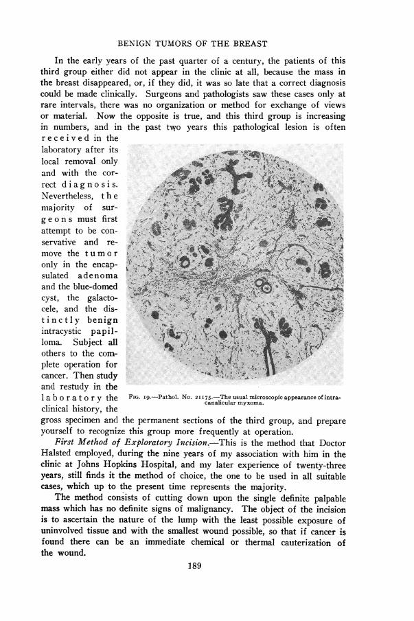
In the early years of the past quarter of a century, the patients of thisthird group either did not appear in the clinic at all, because the mass inthe breast disappeared, or, if they did, it was so late that a correct diagnosiscould be made clinically. Surgeons and pathologists saw these cases only atrare intervals, there was no organization or method for exchange of viewsor material. Now the opposite is true, and this third group is increasingin numbers, and in the past two years this pathological lesion is oftenreceived in thelaboratory after itslocal removal onlyand with the cor-rect diagnosis.Nevertheless, t h e
majority of sur-
ge o must first F ipiattempt to be conservative and re- p smove the tumor t go m fqn ao tn
only in the encap- E I isulated adenomad tiand the blue-domed mtacyst, the galacto- t prsn t r t mcele, and the dis-outinctly benign - Iintracystic papil-4loma. Subject allothers to the comn -plete operation for E
cancer. Then study 1'.and restudy in the1lab or at o ry the FIG. i9.-Pathol. No. 21175.-The usual microscopic appearance of intra-
clinical history, thealiurmyoa
gross specimen and the permanent sections of the third group, and prepareyourself to recognize this group more frequently at operation.
First Method of Exploratory Inicision.-This is the method that DoctorHalsted employ'ed, during the nine years of my association with him in theclinic at Johns Hopkins Hospital, and my later experience of twenty-threeyears, still finds it the method of choice, the one to be used in all suitablecases, which up to the present time represents the majority.
The method consists of cutting down upon the single definite palpablemass which has no definite signs of malignancy. The object of the incisionis to ascertain the nature of the lump with the least possible exposure ofuninvolved tissue and with the smallest wound possible, so that if cancer isfound there can be an immediate chemical or thermal cauterization ofthe wound.
189

JOSEPH COLT BLOODGOOD
Palpable tumors have three definite positions: One, tumor tissue is atthe surface of the breast, so that one cuts through skin and subcutaneousfat only. Two, the tumor is imbedded in breast tissue and one must cutthrough breast tissue before the tumor is seen. Three, the tumor is betweenthe breast and the pectoral fascia. This is a very rare position, and in myexperience only benign tumors-encapsulated adenoma and the blue-domedcysts are found in this locality. I have had now, more than thirty years'experience in making this form of exploratory incision. In every case in
which I assistedji gg ~~~~~Doctor Halsted or
.,11.M.OR,,!., -:::N Doctor Finney in.-..~~~~~~~~~~.. the early yea~rs
and in every casein which I wast he operator, Ihave on record inthe laboratory awritten note of
k and felt at thisexploratoryincision.
SubfcutaneousTuinco rs-WNhen
the breast tumorsur-
..ore...riface of the breast...... ..... it has no covering
importt dl f . Ibut the subcuta-neous fat. When
FIG. 20.-Pathol. No 26493 Lactation hypertrophy in an encapsulated a scirrhus carci-adenoma removed during lactation.noma has reached
this point there is usually dimpling of the skin or evidence of atrophy of thesubcutaneous fat or adherent skin but when the tumor is a circumscribedscirrhus or a medullary carcinoma, or a cancer cyst, or a small sarcoma, itmay palpate like a benign tumor and not be associated with changes in theskin or subcutaneous fat.
My. recorded experiences and my memory ot them emphasize one veryimportant easily demonstrable feature. In cutting through the skin andsubcutaneous fat down upon the palpable tumor and moving the knife fromside to side, the benign tumor is exposed at once, readily, because the tissueis easily moved from the capsule of a benign cyst or encapsulated adenoma,while the tissue is more fixed to the malignant cyst or to the circumscribedor infiltrating area of cancer, and, as mentioned before, usually outside themore circumscribed sarcoma there is cdema. When the exploration is made
190

BENIGN TUMORS OF THE BREAST
slowly, one, so to speak, feels that the tumor is malignant before it is seen.If it is benign, a blue or gray dome of a cyst comes into view, or the distinctbrownish-gray capsule of an adenoma. When one nicks the benign cyst thematerial is clear or cloudy; the galactocele contains milk; the cancer cyst hasthick, grumous material. If the cyst contains blood it should be treatedas malignant unless one can quickly demonstrate a distinctly benign intra-cystic papilloma. The contents of the cyst, therefore, is characteristic of thebenign and of the malignant cyst.
The most characteristic feature about the benign encapsulated adenomawhen it is exposedbeneath the sub-cutaneous fat, isits distinct cap- -sule, the fact thatit can be shelledout, that in sepa-rating it from the-surrounding t i s-sue one can seef ine cobwebconnective-t is suebetween the cap- Xsule and the sur-rounding fat or 4breast. I h a v en e v e r observedthis in the malig-nant tumor. In afew instances Ih a v e observedcedema about anencapsulated ade- FIG. 2I.-Pathol. No. I55I8. Lactation hypertrophy suspicious of malig-nancy in an encapsulated adenoma removed at the end of lactation.noma, but in allof these cases there had been recent trauma, otherwise oedema has beenassociated with malignancy. It is therefore easier to distinguish thebenign encapsulated adenoma from its surroundings, its enucleabilitv andits capsule. When the tumor is cut into, it cannot be so readily distin-guished from a sarcoma of the fibrosarcoma type, nor from a circumscribedscirrhus. Some small medullary carcinomas closely resemble the smallintracanalicular myxoma.
The cancer cyst, whether arising in a papilloma or not, may have ablue dome. Its wall may resemble that of the benign cyst, but it is neveras readily separated from the surrounding tissue, the cobweb tissue is usuallyabsent, there is frequently cedema. (The blue dome over a cancer cyst hasbeen seen only once in ten cases.) Fortunately, as stated before, the differ-
191

JOSEPH COLT BLOODGOOD
ential diagnosis is quickly made when we nick the cyst and examineits contents.
I am beginning to feel that as our experience grows, we will be ableto recognize the malignant cyst without cutting into it.
When the tumor is buried in the breast tissue, one cannot recognizethe benign from the malignant by the surface of the breast, exposed after
dividing the sub-cutaneous fat, nor
is there anythingcharacteristic i nthe breast tissueitself when it isdivided, until onecomes to the im-mediate surround-ings of the tumor.Then what I havejust describedagain holds true.
S e c o n dMethod of Ex-ploratory In c i -sion.-When t h epalpable tumor isvery small, or theb r e a s t is large,f atty, I havef oound it moreconvenient to ex-cise the palpablearea with a goodmargin of sur-rounding breastand, af ter re-
FIG. 22.-Pathol. No. I4430. The correct appearance of the breast moval to place anafter the excision of a benign tumor. The position and radiation of the in-cision varies. In this case the tumor was aberrant in the periphery towards alcohol s p o n g ethe axilla. Note the symmetry of the breast is preserved.
into the wound.Then take the tumor, place it on a towel, hold it in the left hand, and with theknife in the right hand explore and bisect the tumor. This makes the differen-tial diagnosis easier, it is better for a rapid frozen section, but I rarely employit, except for the above indications.- Perhaps for these surgeons whoseexperience is still limited, this might be the better method to employ.
Third Method of Exploratory Incision.-The idea comes from Warren,of Boston, who made a circular incision around the periphery of the breast,turned the breast up and explored the palpable lump or lumps from the
192

BENIGN TUMORS OF THE BREAST
posterior surface. I have known of this method since its introduction andpublication, employed it on a few occasions many years ago, but abandonedit and found it a few days ago very satisfactory in a very difficult case.
The patient was a young woman about twenty-eight years of age, unmar-ried; both breasts were large and of the diffuse virginal hypertrophy type.That is, their size was due to parenchyma and not fat. Both nipples werecongenitally retracted. There was a definite palpable mass in the midzone of
Fics...-Pathoi. No. 2..3-Wound corn.icaton after excision of a benin b t t. N.... ta............ .$..:..; .............,
the' symmetry.. of the'breast;ispreserved. In this case a matoma formedbecameinfected, ad te.in..
ciin wer neesr for dringe. But th ratwssvdNt h orsi cr
E t E~~~~~~~~~~~~~~~~~~~~~~~~~~~~~~~~~~~~~~~~~~~~~~~~~~..- ..... ... . .decrbe as msis.I Thi=spalpabe tye is mor frqenl mainn than. .
in further. All of us in the clinic concluded that it was malignant .n view
of its situationdeep in the hypertrophied breast I decided upon thesecond~~~~~IMMR-; i ;E iil~ ~ AlMt
methdGoex-Pahl. or 9.Wonoation,bthn nhbreast paexinchymaweinrastexposed Iotfoundith couldtrbftee rasil iprseprated.fro thlcae fatheandoitfoccured,tcameinetdadtorfollow
wa rren'lmt and loo andufeel a tes involveareea from the posterior
surface.ceOn doring solalpation.TemssuggesedaurcystI thenrsecedsth
in 13 ll l c l t it.;g ...i
Warren's~~~meho an loo adfel at th inole area from th postrosurf~~~~~~~~~~~~~~~laceOn don so, papto suggested,','..,.'' cyst'. I then rescte the''................:'193

JOSEPH COLT BLOODGOOD
involved area, cutting through normal breast tissue, and then, on bisection,revealed a gray-domed, thick-walled benign cyst, and excluded malignancy.
Plastic Closure.-The method still employed is the one taught me byHalsted in I893, and during these thirty years it has been employed withsuccess with increasing frequency. One change has been made after thefirst three cases. Silk is never employed, either for ligature or suture, in theplastic closure.
In removing the tumor, plan the excision as nearly as possible to a wedge-
FIG. 24.-Photograph of patient showing the roper result after the excision of a benign tumor in theright breast and a faulty result in the left breast. The latter is due to failure to suture the breast defect-a definite plastic procedure (see text page 24).
shaped piece with the apex towards the areola or nipple. The skin incisionshould radiate from the areola. It is better to excise down to the pectoralfascia, except in very small tumors in large breasts. Every bleeding pointshould be ligated. The defect is closed with chromic catgut suture of oo size;heavy catgut is not necessary. According to the thickness of the breast thesutures are in two, three or four layers. The first layer is posterior fromnipple zone to periphery and the other layers follow, approximating thedefect from the nipple outwards and from within outwards. The subcu-taneous fat is approximated with catgut, and the skin is closed with inter-rupted fine silk passed with straight intestinal needles. If there is anypuckering, remove the stitches and resuture. Even with a large defect, an
194

BENIGN TUMORS OF THE BREAST
irregular defect, this plastic suture is possible and the symmetry of the breastis restored to normal. The reduction in size of the breast varies with theamount of breast tissue removed. A perfect and an imperfect plastic opera-tion are shown in Fig. 24 on the two breasts of the same patient.
The only complication has been a haeniatoma. In two of my cases whichI dressed myself, the wounds healed by granulation, and the cosmetic resultwas as good as in wounds healing per primam. In a third the hematomabecame infected, and multiple incisions were necessary to save the breast(Fig. 23). In one casethe p a t i e n t unfortu-nately was dischargedwith an unrecognizedhaematoma which be-came infected, and theb r e a s t was removedelsewhere. 'fhere areabout 400 operationsof this kind with thesefew haematomas, twodefinite infections andthe loss of one breast.
Therefore, hsemo-stasis is essential. Thenext is the proper fixa-tion of the breast withpadding and bandage,important in all casesand essential in largependulous breasts. Itis our rule to re-dress daily in order FIG. 25.-Pathol. No. 9808. Definitely encapsulated fibroadenoma
buried in breast tissue. Removal of breast unnecessary.to evacuate any serumby pressure, or immediately to recognize any haematoma and evacuate it.
The skin stitches are never removed until the fourth day, and then everyalternate stitch only. All stitches are out on the sixth or seventh day.When tumor, incision and the breast are small, the patient often leaves thehospital the day after the operation. The larger the wound and the breastthe longer the patient is kept quietly in the hospital, up to the seventh ortenth day.
The object of this operation, if the tumor is benign, is to save the patientfrom the mutilation of a disfigured or removed breast. Therefore, every careis essential in the plastic closure and in the subsequent proper dressing of thewound and fixation of the parts.
195

JOSEPH COLT BLOODGOOD
FIG. 26.-Pathol. No. 702. Encapsulated intracanalicular myxoadenoma. To theright notice the capsule being pealed from the tumor. To the left the surface of the capsuleand the cut surface of the tumor.
FIG. 27.-a. and b. Pathol. No. 31I94. Encapsulated intracanalicular myxoadenoma, removed withzone of breast. The tumor is shown bisected with the surrounding breast. b. Pathol. No. 31I94. Encap-sulated adenoma. Tumor with capsule above; breast tissue below. The white lines represent the cob-webconnective-tissue between the capsule of the benign tumor and the breast. I have never seen this inmalignancy. It may also be obliterated by the inflammatory reaction of trauma (see Fig. s).
196

BENIGN TUMORS OF THE BREAST
Conclusions.-A colleague and a pathologist has just written me: " Whyremove benign encapsulated adenomas?" They are only isolated areas ofnormal breast tissue and as such have no more tendency to become malignantthan the breast left behind after their removal." I trust this paper willanswer his question. I have tried to emphasize that the object of explorationis not to remove a benign tumor, but to expose and recognize a possiblemalignant tumor in the most favorable stage for a cure by the radical opera-tion. Having exposed a benign tumor, it requires very little more time tocompletely remove it, because the differential diagnosis often goes to a pointwhere the benign tumor must be partially removed and in some cases com-pletely removed before the differentiation from cancer is possible andmade certain.- There is a second reason for the removal of a benign encapsulatedadenoma. The evidence is that sarcoma usually develops in the intra-canalicular myxoma type of adenoma. It is quite possible that the malignanttumor giving a long history may have developed in preexisting adenoma.
197