Odontogenic Cysts and Tumors
87
Odontogenic Cysts and Tumors
-
Upload
khangminh22 -
Category
Documents
-
view
3 -
download
0
Transcript of Odontogenic Cysts and Tumors

Odontogenic Cysts and Tumors

Introduction Variety of cysts and tumors Uniquely derived from tissues of
developing teeth May present to otolaryngologist

Odontogenesis Projections of dental lamina into
ectomesenchyme Layered cap (inner/outer enamel
epithelium, stratum intermedium, stellate reticulum)
Odontoblasts secrete dentin ameloblasts (from IEE) enamel
Cementoblasts cementum Fibroblasts periodontal membrane
Odontogenesis
Diagnosis Complete history
Pain, loose teeth, occlusion, swellings, dysthesias, delayed tooth eruption
Thorough physical examination Inspection, palpation, percussion,
auscultation Plain radiographs
Panorex, dental radiographs CT for larger, aggressive lesions
Diagnosis Differential diagnosis Obtain tissue
FNA – r/o vascular lesions, inflammatory Excisional biopsy – smaller cysts,
unilocular tumors Incisional biopsy – larger lesions prior to
definitive therapy

Odontogenic Cysts Inflammatory
Radicular Paradental
Developmental Dentigerous Developmental
lateral periodontal Odontogenic
keratocyst Glandular
odontogenic

Radicular (Periapical) Cyst Most common (65%) Epithelial cell rests of Malassez Response to inflammation Radiographic findings
Pulpless, nonvital tooth Small well-defined periapical radiolucency
Histology Treatment – extraction, root canal

Radicular Cyst

Radicular Cyst

Residual Cyst
Paradental Cyst Associated with partially impacted 3rd
molars Result of inflammation of the gingiva
over an erupting molar 0.5 to 4% of cysts Radiology – radiolucency in apical
portion of the root Treatment – enucleation

Paradental Cyst

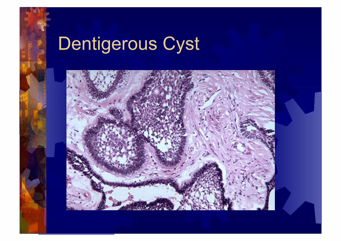
Dentigerous (follicular) Cyst Most common developmental cyst (24%) Fluid between reduced enamel epithelium
and tooth crown Radiographic findings
Unilocular radiolucency with well-defined sclerotic margins
Histology Nonkeratinizing squamous epithelium
Treatment – enucleation, decompression

Dentigerous Cyst
Dentigerous Cyst
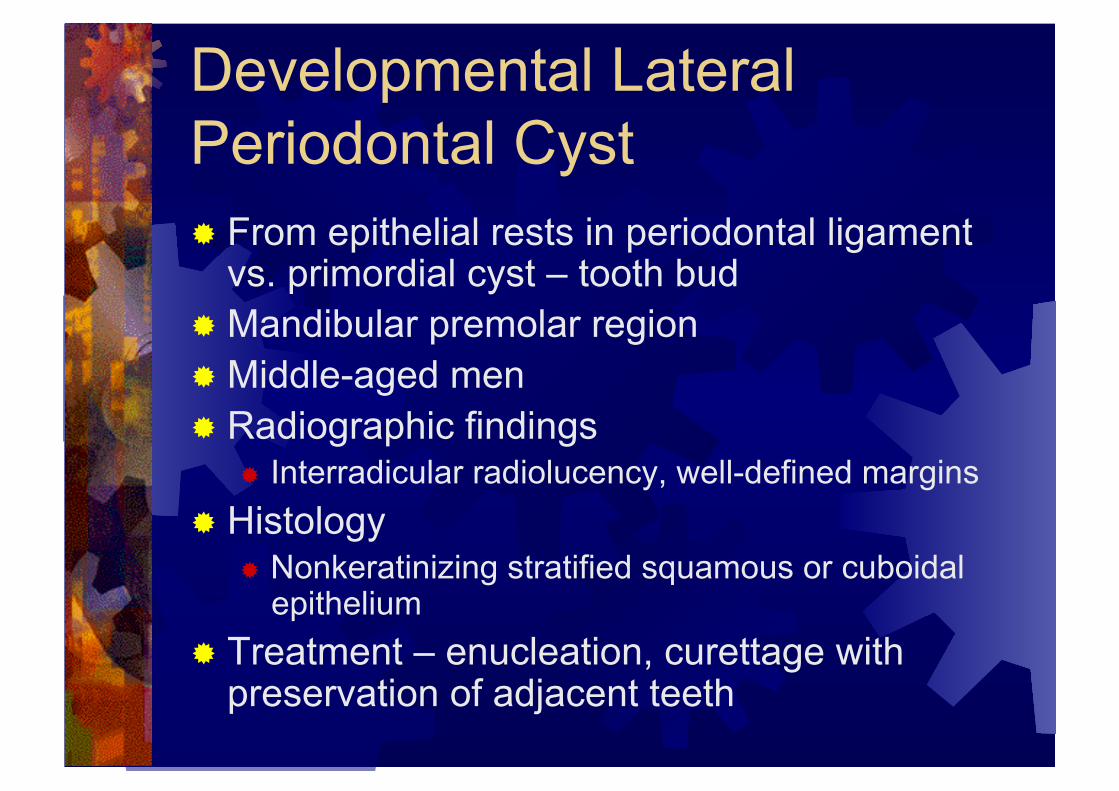
Developmental Lateral Periodontal Cyst From epithelial rests in periodontal ligament
vs. primordial cyst – tooth bud Mandibular premolar region Middle-aged men Radiographic findings
Interradicular radiolucency, well-defined margins Histology
Nonkeratinizing stratified squamous or cuboidal epithelium
Treatment – enucleation, curettage with preservation of adjacent teeth

Developmental Lateral Periodontal Cyst
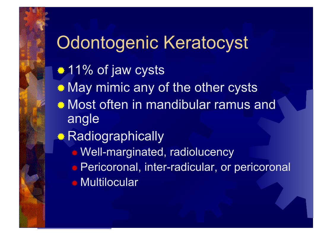
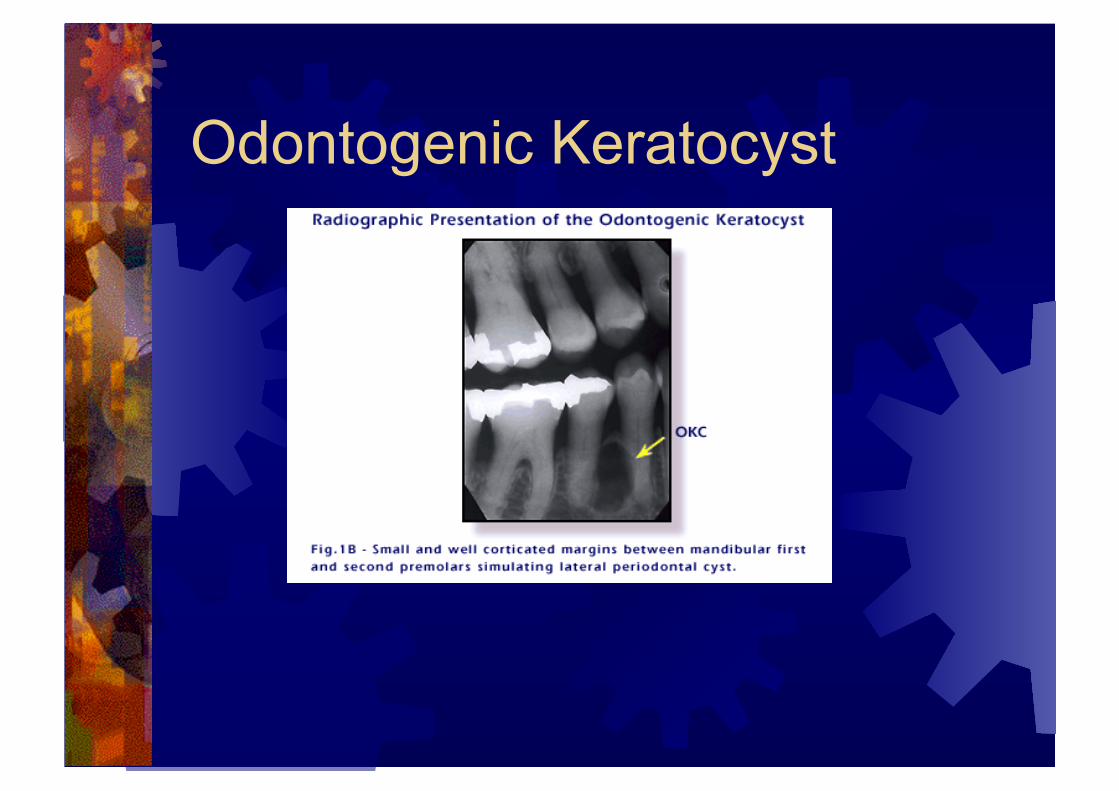
Odontogenic Keratocyst 11% of jaw cysts May mimic any of the other cysts Most often in mandibular ramus and
angle Radiographically
Well-marginated, radiolucency Pericoronal, inter-radicular, or pericoronal Multilocular

Odontogenic Keratocyst
Odontogenic Keratocyst

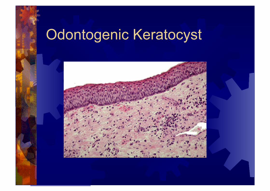
Odontogenic Keratocyst Histology
Thin epithelial lining with underlying connective tissue (collagen and epithelial nests)
Secondary inflammation may mask features High frequency of recurrence (up to 62%) Complete removal difficult and satellite
cysts can be left behind
Odontogenic Keratocyst
Treatment of OKC Depends on extent of lesion Small – simple enucleation, complete removal
of cyst wall Larger – enucleation with/without peripheral
ostectomy Bataineh,et al, promote complete resection
with 1 cm bony margins (if extension through cortex, overlying soft tissues excised)
Long term follow-up required (5-10 years)

Glandular Odontogenic Cyst More recently described (45 cases) Gardner, 1988 Mandible (87%), usually anterior Very slow progressive growth (CC:
swelling, pain [40%]) Radiographic findings
Unilocular or multilocular radiolucency

Glandular Odontogenic Cyst
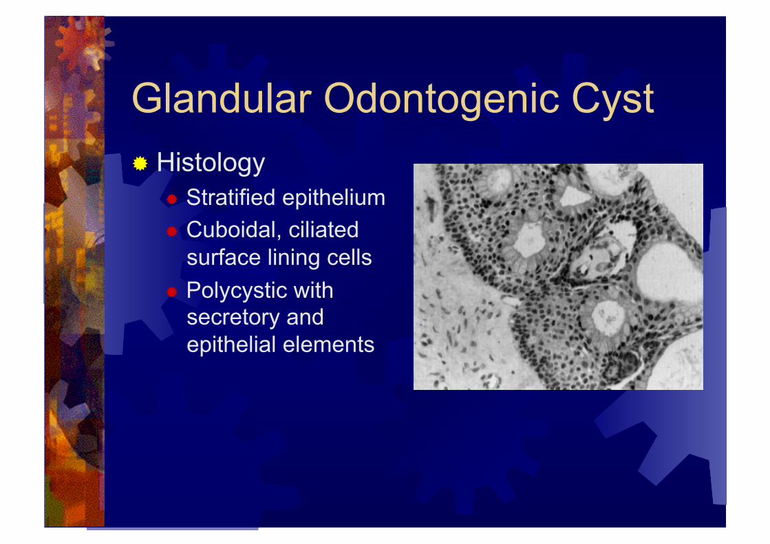
Glandular Odontogenic Cyst Histology
Stratified epithelium Cuboidal, ciliated
surface lining cells Polycystic with
secretory and epithelial elements

Treatment of GOC Considerable recurrence potential 25% after enucleation or curettage Marginal resection suggested for larger
lesions or involvement of posterior maxilla Warrants close follow-up

Nonodontogenic Cysts Incisive Canal Cyst Stafne Bone Cyst Traumatic Bone Cyst Surgical Ciliated Cyst (of Maxilla)

Incisive Canal Cyst Derived from epithelial remnants of the
nasopalatine duct (incisive canal) 4th to 6th decades Palatal swelling common, asymptomatic Radiographic findings
Well-delineated oval radiolucency between maxillary incisors, root resorption occasional
Histology Cyst lined by stratified squamous or
respiratory epithelium or both

Incisive Canal Cyst

Incisive Canal Cyst Treatment consists of surgical
enucleation or periodic radiographs Progressive enlargement requires
surgical intervention

Stafne Bone Cyst Submandibular salivary gland depression Incidental finding, not a true cyst Radiographs – small, circular, corticated
radiolucency below mandibular canal Histology – normal salivary tissue Treatment – routine follow up

Stafne Bone Cyst

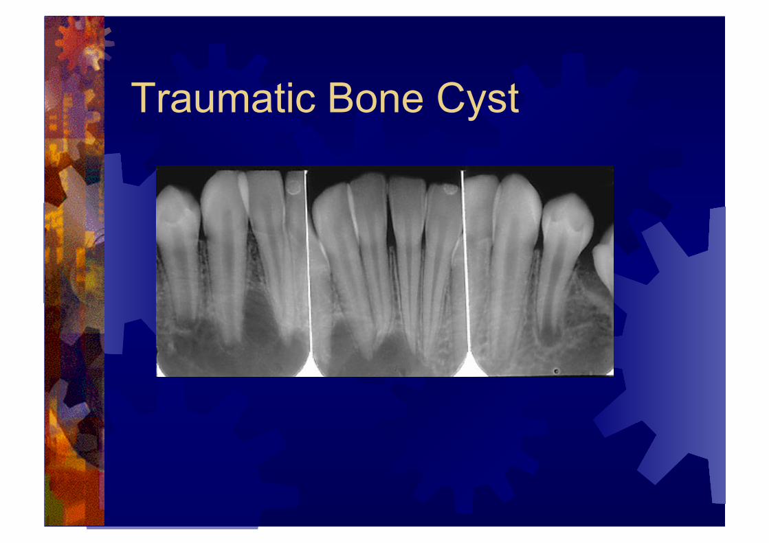
Traumatic Bone Cyst Empty or fluid filled cavity associated
with jaw trauma (50%) Radiographic findings
Radiolucency, most commonly in body or anterior portion of mandible
Histology – thin membrane of fibrous granulation
Treatment – exploratory surgery may expedite healing
Traumatic Bone Cyst

Surgical Ciliated Cyst May occur following Caldwell-Luc Trapped fragments of sinus epithelium
that undergo benign proliferation Radiographic findings
Unilocular radiolucency in maxilla Histology
Lining of pseudostratified columnar ciliated Treatment - enucleation

Surgical Ciliated Cyst

Odontogenic Tumors Ameloblastoma Calcifying Epithelial
Odontogenic Tumor Adenomatoid
Odontogenic Tumor
Squamous Odontogenic Tumor
Calcifying Odontogenic Cyst

Ameloblastoma Most common odontogenic tumor Benign, but locally invasive Clinically and histologically similar to BCCa 4th and 5th decades Occasionally arise from dentigerous cysts Subtypes – multicystic (86%), unicystic
(13%), and peripheral (extraosseous – 1%)

Ameloblastoma Radiographic findings
Classic – multilocular radiolucency of posterior mandible
Well-circumscribed, soap-bubble Unilocular – often confused with
odontogenic cysts Root resorption – associated with
malignancy

Ameloblastoma

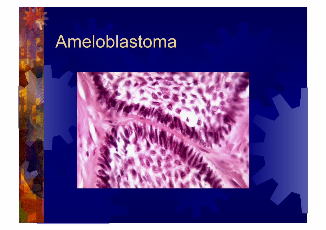
Ameloblastoma Histology
Two patterns – plexiform and follicular (no bearing on prognosis)
Classic – sheets and islands of tumor cells, outer rim of ameloblasts is polarized away from basement membrane
Center looks like stellate reticulum Squamous differentiation (1%) – Diagnosed
as ameloblastic carcinoma
Ameloblastoma

Treatment of Ameloblastoma According to growth characteristics and type Unicystic
Complete removal Peripheral ostectomies if extension through cyst
wall Classic infiltrative (aggressive)
Mandibular – adequate normal bone around margins of resection
Maxillary – more aggressive surgery, 1.5 cm margins
Ameloblastic carcinoma Radical surgical resection (like SCCa) Neck dissection for LAN

Calcifying Epithelial Odontogenic Tumor a.k.a. Pindborg tumor Aggressive tumor of epithelial derivation Impacted tooth, mandible body/ramus Chief sign – cortical expansion Pain not normally a complaint

Calcifying Epithelial Odontogenic Tumor Radiographic findings
Expanded cortices in all dimensions Radiolucent; poorly defined, noncorticated
borders Unilocular, multilocular, or “moth-eaten” “Driven-snow” appearance from multiple
radiopaque foci Root divergence/resorption; impacted tooth

Calcifying Epithelial Odontogenic Tumor

Calcifying Epithelial Odontogenic Tumor Histology
Islands of eosinophilic epithelial cells Cells infiltrate bony trabeculae Nuclear hyperchromatism and
pleomorphism Psammoma-like calcifications (Liesegang
rings)

Calcifying Epithelial Odontogenic Tumor

Treatment of CEOT Behaves like ameloblastoma Smaller recurrence rates En bloc resection, hemimandibulectomy
partial maxillectomy suggested

Adenomatoid Odontogenic Tumor Associated with the crown of an impacted
anterior tooth Painless expansion Radiographic findings
Well-defined expansile radiolucency Root divergence, calcified flecks (“target”)
Histology Thick fibrous capsule, clusters of spindle cells,
columnar cells (rosettes, ductal) throughout Treatment – enucleation, recurrence is rare

Adenomatoid Odontogenic Tumor

Squamous Odontogenic Tumor Hamartomatous proliferation Maxillary incisor-canine and mandibular molar Tooth mobility common complaint Radiology – triangular, localized radiolucency
between contiguous teeth Histology – oval nest of squamous epithelium
in mature collagen stroma Treatment – extraction of involved tooth and
thorough curettage; maxillary – more extensive resection; recurrences – treat with aggressive resection

Squamous Odontogenic Tumor

Calcifying Odontogenic Cyst Tumor-like cyst of mandibular premolar
region ¼ are peripheral – gingival swelling Osseous lesions – expansion, vital teeth Radiographic findings
Radiolucency with progressive calcification Target lesion (lucent halo); root divergence
Histology Stratified squamous epithelial lining Polarized basal layer, lumen contains ghost cells
Treatment – enucleation with curettage; rarely recur

Mesenchymal Odontogenic Tumors Odontogenic Myxoma Cementoblastoma
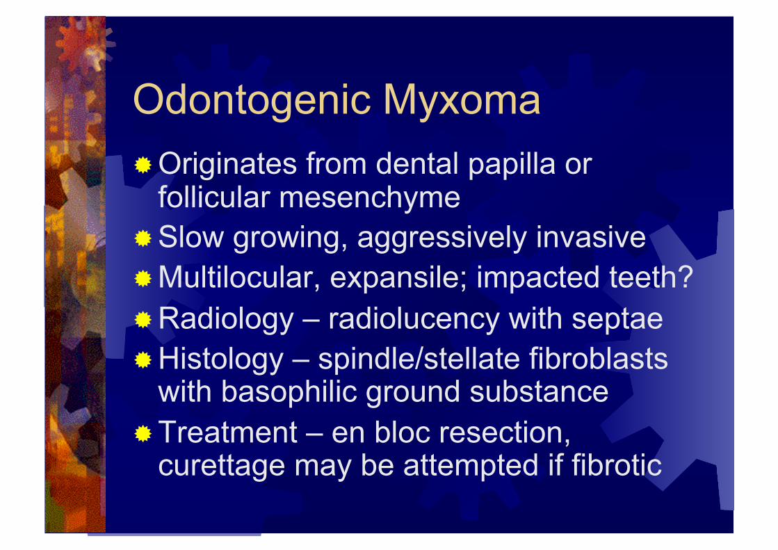
Odontogenic Myxoma Originates from dental papilla or
follicular mesenchyme Slow growing, aggressively invasive Multilocular, expansile; impacted teeth? Radiology – radiolucency with septae Histology – spindle/stellate fibroblasts
with basophilic ground substance Treatment – en bloc resection,
curettage may be attempted if fibrotic

Cementoblastoma True neoplasm of cementoblasts First mandibular molars Cortex expanded without pain Involved tooth ankylosed, percussion Radiology – apical mass; lucent or solid,
radiolucent halo with dense lesions Histology – radially oriented trabeculae from
cementum, rim of osteoblasts Treatment – complete excision and tooth
sacrifice

Cementoblastoma

Mixed Odontogenic Tumors Ameloblastic fibroma, ameloblastic
fibrodentinoma, ameloblastic fibro-odontoma, odontoma
Both epithelial and mesenchymal cells Mimic differentiation of developing tooth Treatment – enucleation, thorough
curettage with extraction of impacted tooth Ameloblastic fibrosarcomas – malignant,
treat with aggressive en bloc resection
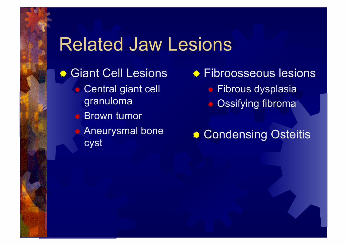
Related Jaw Lesions Giant Cell Lesions
Central giant cell granuloma
Brown tumor Aneurysmal bone
cyst
Fibroosseous lesions Fibrous dysplasia Ossifying fibroma
Condensing Osteitis

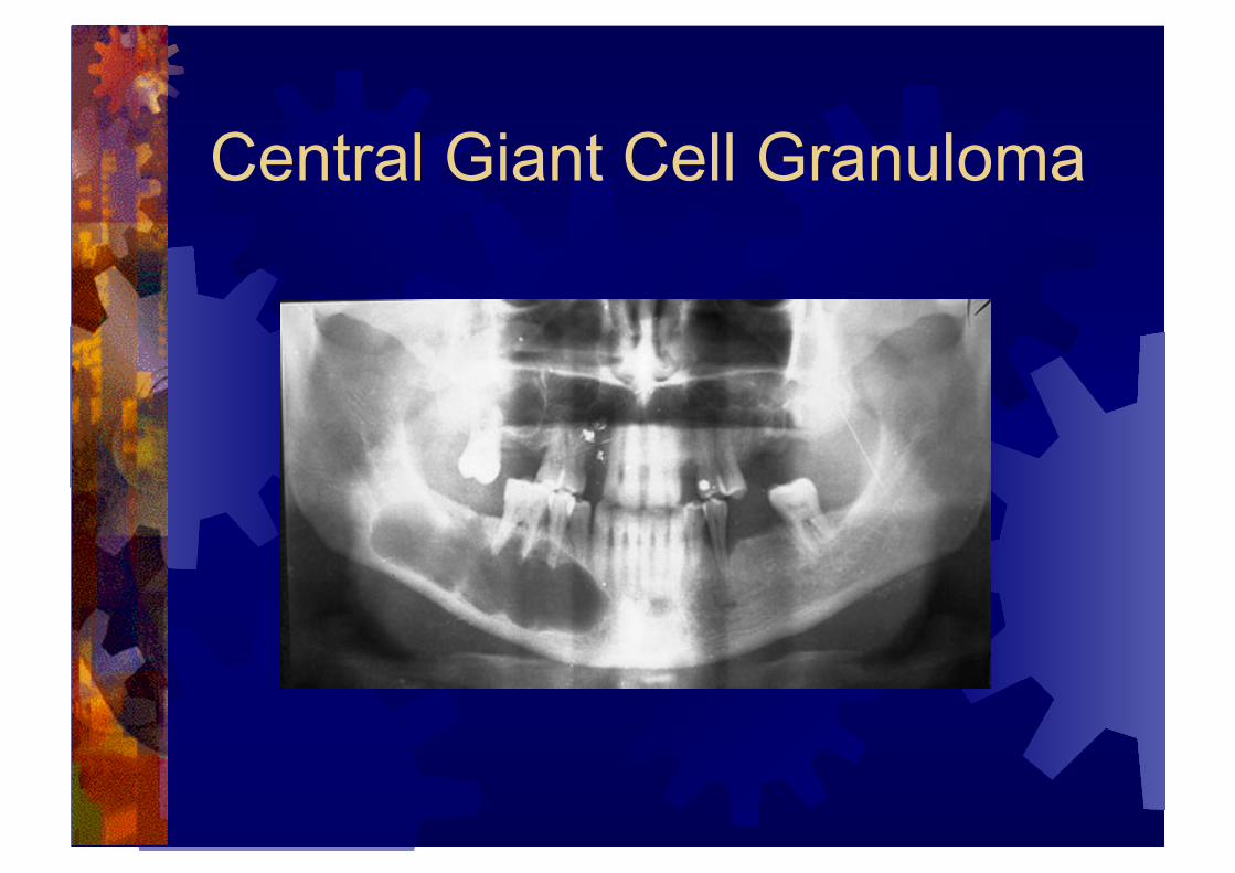
Central Giant Cell Granuloma Neoplastic-like reactive proliferation Common in children and young adults Females > males (hormonal?) Mandible > maxilla Expansile lesions – root resorption Slow-growing – asymptomatic swelling Rapid-growing – pain, loose dentition
(high rate of recurrence)

Central Giant Cell Granuloma Radiographic findings
Unilocular, multilocular radiolucencies Well-defined or irregular borders
Histology Multinucleated giant cells, dispersed
throughout a fibrovascular stroma
Central Giant Cell Granuloma

Central Giant Cell Granuloma
Central Giant Cell Granuloma Treatment
Curettage, segmental resection Radiation – out of favor (risk of sarcoma) Intralesional steroids – younger patients,
very large lesions Individualized treatment depending on
characteristics and location of tumor

Brown Tumor Local manifestation of hyperparathyroid Histologically identical to CGCG Serum calcium and phosphorus More likely in older patients

Aneurysmal Bone Cyst Large vascular sinusoids (no bruit) Not a true cyst; aggressive, reactive Great potential for growth, deformity Multilocular radiolucency with cortical
expansion Mandible body Simple enucleation, rare recurrence

Fibrous Dysplasia Monostotic vs. polystotic Monostotic
More common in jaws and cranium Polystotic
McCune-Albright’s syndrome Cutaneous pigmentation, hyper-functioning
endocrine glands, precocious puberty

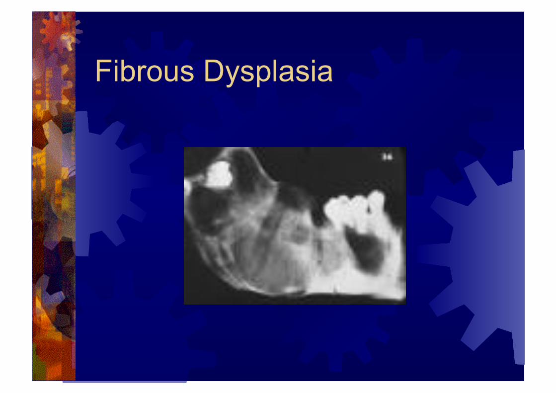
Fibrous Dysplasia Painless expansile dysplastic process of
osteoprogenitor connective tissue Maxilla most common Does not typically cross midline (one bone) Antrum obliterated, orbital floor
involvement (globe displacement) Radiology – ground-glass appearance
Fibrous Dysplasia

Fibrous Dysplasia
Fibrous Dysplasia Histology – irregular osseous trabeculae in
hypercellular fibrous stroma Treatment
Deferred, if possible until skeletal maturity Quarterly clinical and radiographic f/u If quiescent – contour excision (cosmesis or
function) Accelerated growth or disabling functional
impairment - surgical intervention (en bloc resection, reconstruction)

Ossifying Fibroma True neoplasm of medullary jaws Elements of periodontal ligament Younger patients, premolar – mandible Frequently grow to expand jaw bone Radiology
radiolucent lesion early, well-demarcated Progressive calcification (radiopaque – 6 yrs)

Ossifying Fibroma
Ossifying Fibroma Histologically similar to fibrous dysplasia Treatment
Surgical excision – shells out Recurrence is uncommon
Condensing Osteitis 4% to 8% of population Focal areas of radiodense sclerotic bone Mandible, apices of first molar Reactive bony sclerosis to pulp
inflammation Irregular, radiopaque Stable, no treatment required

Condensing Osteitis

Conclusion

Case Presentation 20 year-old hispanic female with several
month history of lesion in right maxilla, treated initially by oral surgeon with multiple curettage.
Has experienced recent onset of rapid expansion, after pregnancy, with complaints of loose dentition and pain.
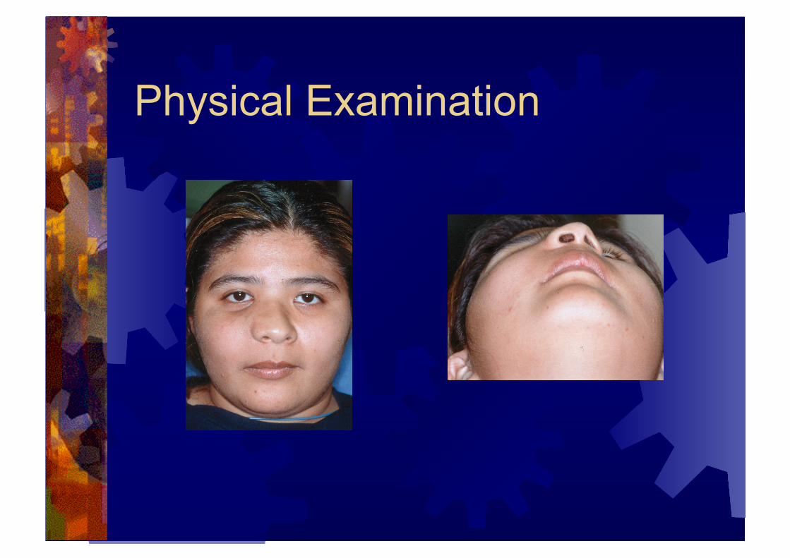
Physical Examination

Physical Examination

Radiographs Plain films – facial series Computerized Tomography of facial
series

Pathology

Treatment
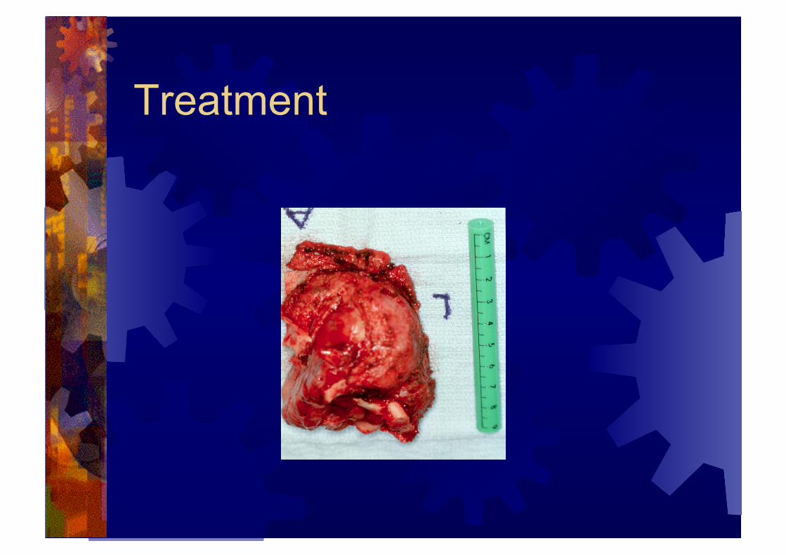
Treatment