EVOLVING CLASSIFICATION OF SOFT TISSUE TUMORS
143
EVOLVING CLASSIFICATION OF SOFT TISSUE TUMORS: AN UPDATE BASED ON THE NEW 2013 WHO CLASSIFICATION Christopher D.M. Fletcher, M.D., FRCPath Brigham and Women’s Hospital and Harvard Medical School, Boston MA
-
Upload
khangminh22 -
Category
Documents
-
view
3 -
download
0
Transcript of EVOLVING CLASSIFICATION OF SOFT TISSUE TUMORS

EVOLVING CLASSIFICATION OFSOFT TISSUE TUMORS:
AN UPDATE BASED ON THE NEW2013 WHO CLASSIFICATION
Christopher D.M. Fletcher, M.D., FRCPathBrigham and Women’s Hospital and Harvard Medical School, Boston MA

MAJOR STRENGTHS
Broad concensus viewNew approach to authorship
More extensive text / illustrationsInclusion of genetics
Timeliness

SIGNIFICANT CHANGES FROM1994 CLASSIFICATION
Approach to biologic potentialApproach to fibrohistiocytic tumoursApproach to haemangiopericytomaRecategorisation of ‘intermediate’
vascular tumoursNumerous ‘new’ entities
More tumours of ‘uncertain differentiation’
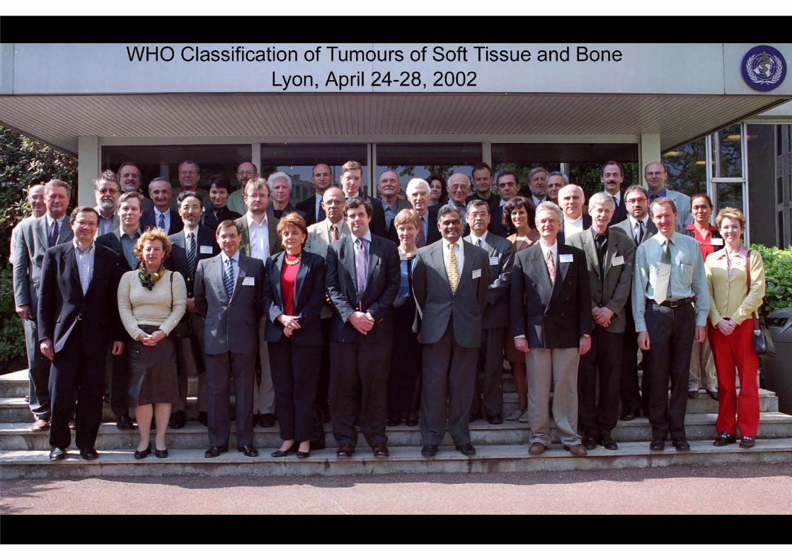
World Health Organization (2002) Classification of Tumours of Soft Tissue

BENIGN CATEGORYMost benign soft tissue tumours do not recur locally. Those that do recur do so in a non-destructive fashion and are almost always readily cured by complete local excision. Exceedingly rarely (almost certainly <1/50,000 cases, and probably even less than that), a morphologically benign lesion may give rise to distant metastases. This is entirely unpredictable on the basis of conventional histological examination and, to date, has been best documented in cutaneous benign fibrous histiocytoma.
World Health Organization (2002) Classification of Tumours of Soft Tissue

INTERMEDIATE CATEGORY(Locally aggressive)
Soft tissue tumours in this category often recur locally and are associated with an infiltrative and locally destructive growth pattern. Lesions in this category do not have any evident potential to metastasise but typically require wide excision with a margin of normal tissue in order to ensure local control. The prototypical lesion in this category is desmoid fibromatosis.
World Health Organization (2002) Classification of Tumours of Soft Tissue

INTERMEDIATE CATEGORY(Rarely metastasising)
Soft tissue tumours in this category are often locally aggressive but, in addition, show the well documented ability to give rise to distant metastases in occasional cases. The risk of such metastases appears to be <2% and is not reliably predictable on the basis of histomorphology. Metastasis in such lesions is usually to lymph node or lung. Prototypical examples in this category include plexiform fibrohistiocytic tumour and so-called angiomatoid fibrous histiocytoma.
World Health Organization (2002) Classification of Tumours of Soft Tissue

MALIGNANT CATEGORYIn addition to the potential for locally destructive growth and recurrence, malignant soft tissue tumours (known as soft tissue sarcomas) have significant risk of distant metastasis, ranging in most instances from 20% to almost 100%, depending upon histological type and grade. Some (but not all) histologically low grade sarcomas have a metastatic risk of only 2-10%, but such lesions may advance in grade in a local recurrence, and thereby acquire a higher risk of distant spread (e.g., myxofibrosarcoma and leiomyosarcoma).
World Health Organization (2002) Classification of Tumours of Soft Tissue


CONTEMPORARY CONCENSUS CLASSIFICATION
- NOT A TEXTBOOK

• Reproducible diagnostic criteria• Better determine biologic potential• Better understand intrinsic biology• Reflect conceptual evolution
REASONS TO HAVE CLASSIFICATIONS


• ‘MFH’ is gone• ‘Haemangiopericytoma’ is gone• Pericytic (perivascular) tumours
better defined• GIST and nerve sheath tumours
now allocated to this volume• Category of undifferentiated
sarcomas introduced• Some “new entities” added• More genetic data added
SIGNIFICANT CHANGES FROM 2002 CLASSIFICATION

“NEW ENTITIES”
World Health Organization (2013) Classification of Tumours of Soft Tissue
Pseudomyogenic (ES-like) haemangioendothelioma
Hybrid nerve sheath tumours
Acral (digital) fibromyxoma
Haemosiderotic fibrolipomatous tumour
Phosphaturic mesenchymal tumour

Lipoma MyolipomaLipomatosis Chondroid lipomaLipomatosis of nerve Extrarenal angiomyolipomaLipoblastoma(tosis) Extra-adrenal myelolipomaAngiolipoma Spindle cell/pleomorphic lipoma
Hibernoma
ADIPOCYTIC TUMOURSBenign
World Health Organization (2013) Classification of Tumours of Soft Tissue

Intermediate Malignant(locally aggressive)Atypical lipomatous tumour / Dedifferentiated
well differentiated liposarcoma liposarcomaMyxoid liposarcomaPleomorphic liposarcomaLiposarcoma, nototherwise specified
ADIPOCYTIC TUMOURS
World Health Organization (2013) Classification of Tumours of Soft Tissue

FIBROBLASTIC / MYOFIBROBLASTIC TUMOURSBenign (1)
Nodular fasciitis ElastofibromaProliferative fasciitis (?) Fibrous hamartoma of Proliferative myositis (?) infancyMyositis ossificans and fibro- Fibromatosis colliosseous pseudotumour of digits Juvenile hyaline fibromatosis
Ischaemic fasciitis Inclusion body fibromatosis
World Health Organization (2013) Classification of Tumours of Soft Tissue

t(17;22)(p13;q12.3)(cryptic)
USP6-MYH9
ICD-0: 8828/0Never had acode before !
NODULAR FASCIITIS

Fibroma of tendon sheath Cellular angiofibromaDesmoplastic fibroblastoma Nuchal-type fibromaMammary-type myofibroblastoma Gardner fibromaCalcifying aponeurotic fibroma Calcifying fibrous tumourAngiomyofibroblastoma
FIBROBLASTIC / MYOFIBROBLASTIC TUMOURSBenign (2)
World Health Organization (2013) Classification of Tumours of Soft Tissue

Locally aggressive Rarely metastasisingSuperficial fibromatoses Dermatofibrosarcoma protuberansDesmoid-type fibromatoses Solitary fibrous tumourLipofibromatosis Inflammatory myofibroblastic tumourGiant cell fibroblastoma Low grade myofibroblastic sarcoma
Myxoinflammatory fibroblastic sarcoma/atypical myxoinflammatory fibroblastic tumour
Infantile fibrosarcoma
FIBROBLASTIC / MYOFIBROBLASTIC TUMOURSIntermediate category
World Health Organization (2013) Classification of Tumours of Soft Tissue

FIBROBLASTIC / MYOFIBROBLASTIC TUMOURS
Malignant
Adult fibrosarcomaMyxofibrosarcomaLow grade fibromyxoid sarcomaSclerosing epithelioid fibrosarcoma
World Health Organization (2013) Classification of Tumours of Soft Tissue


LGFMS/SEF MOLECULAR GENETICS
PURE LGFMS Most have t(11;16)(q33;p11) FUS-CREB3L2Few have t(11;16)(p11;p11) FUS-CREB3L1 Basically all are MUC4 immunopositive
PURE SEFIf MUC4 +ve (70%) – 40% have FUS rearrangement
(some with CREB3L1 or CREB3L2)MUC4 -ve – FUS negative, otherwise unknown
HYBRID LGFMS/SEFAll are MUC4 +ve – Most have either FUS or EWSR1
rearrangement (usually with CREB3L1)
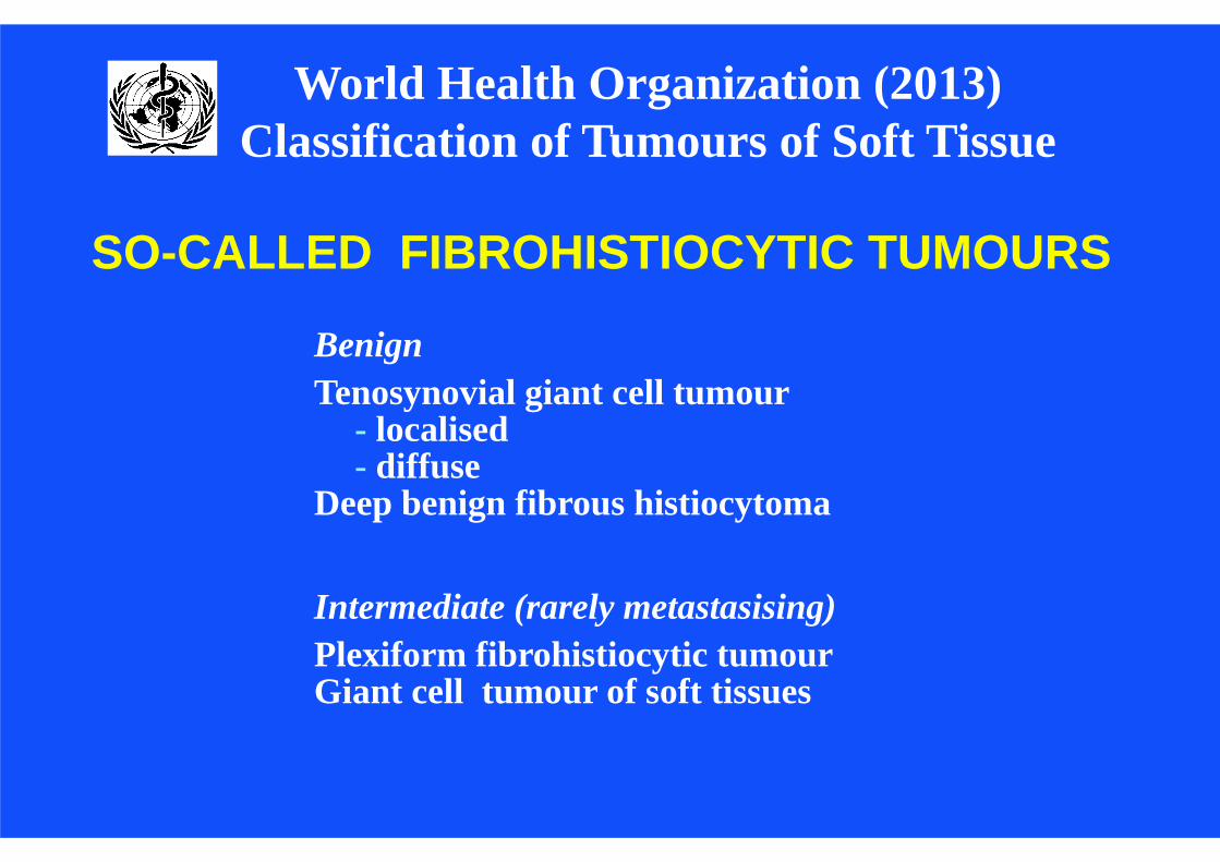
SO-CALLED FIBROHISTIOCYTIC TUMOURS
BenignTenosynovial giant cell tumour
- localised- diffuse
Deep benign fibrous histiocytoma
Intermediate (rarely metastasising)Plexiform fibrohistiocytic tumourGiant cell tumour of soft tissues
World Health Organization (2013) Classification of Tumours of Soft Tissue

SMOOTH MUSCLE TUMOURS
Deep leiomyomaLeiomyosarcoma (excluding skin)
World Health Organization (2013) Classification of Tumours of Soft Tissue
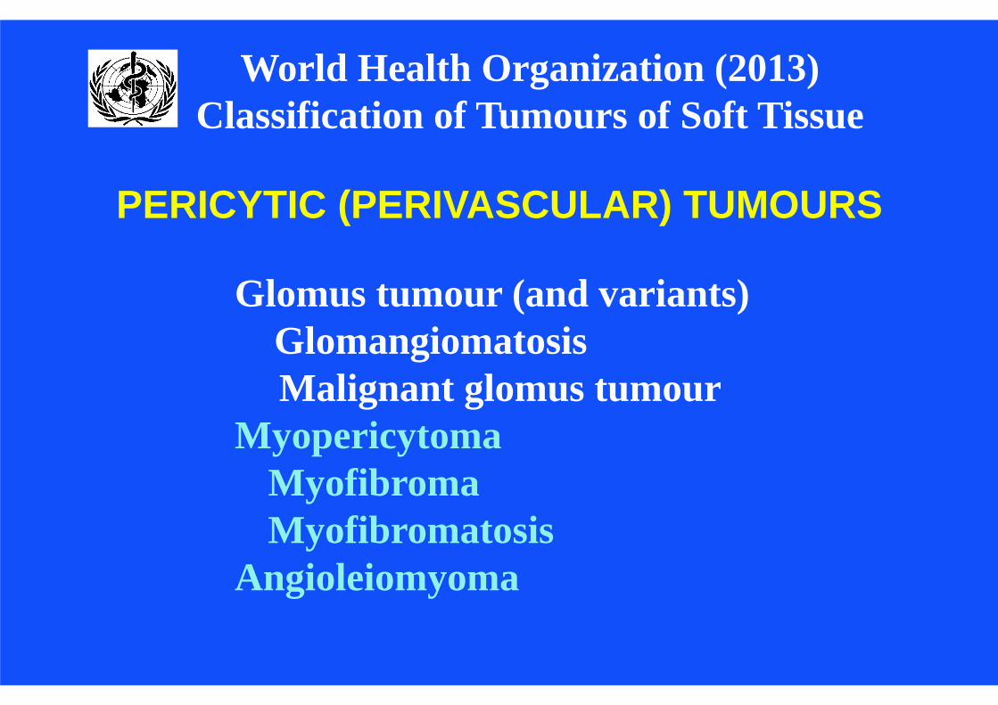
PERICYTIC (PERIVASCULAR) TUMOURS
Glomus tumour (and variants)GlomangiomatosisMalignant glomus tumour
MyopericytomaMyofibromaMyofibromatosis
Angioleiomyoma
World Health Organization (2013) Classification of Tumours of Soft Tissue


• Multiple familial glomus tumours result from germline GLMN mutation
• NF-1 may be associated with multiple digital glomus tumours (which show NF-1 inactivation)
• NOTCH2 or NOTCH3 rearrangement identified in 60% of sporadic soft tissue glomus tumours
• BRAF (10%) and KRAS (rare) mutations identified in sporadic glomus tumours
PERICYTIC (PERIVASCULAR)TUMOURS
NEW GENETIC DATA

• Morphologic continuum, includes myofibroma
• Angioleiomyoma fits better here• Could ultimately be renamed as
haemangiopericytoma !
PERICYTIC (PERIVASCULAR)TUMOURS
KEY POINTS

SKELETAL MUSCLE TUMOURS
BenignRhabdomyoma
adult typefetal typegenital type
MalignantEmbryonal rhabdomyosarcoma
(incl. botryoid, anaplastic)Alveolar rhabdomyosarcoma
(incl. solid, anaplastic)Pleomorphic rhabdomyosarcomaSpindle cell/sclerosing
rhabdomyosarcoma
World Health Organization (2013) Classification of Tumours of Soft Tissue

SPINDLE CELLRHABDOMYOSARCOMA
• 1st described 1992 / 1993Long regarded as variant of embryonal
• Commonest in children / adolescents• Mainly paratesticular• 5 year survival > 90% in children• Easily confused with other spindle cell
sarcomas


DESMIN

MYF-4

SPINDLE CELLRHABDOMYOSARCOMA
IN ADULTS• Mainly 3rd / 4th decades• Males > females• Head and neck commonest (50%)
- but wide range of sites• More aggressive than in children
? Relationship to sclerosing rhabdoMorphologic overlap in 15-20% of cases


SCLEROSINGRHABDOMYOSARCOMA
• 1st described 2000 / 2002(Mentzel et al; Folpe et al)
• Affects adults & children / adolescents• Head & neck = Limbs• Prognosis still somewhat uncertain• No clear genetic data yet• Easily confused with angiosarcoma or
perhaps even osteosarcoma


DESMIN

MYF-4

• No clear relationship to embryonal• Form a morphologic continuum• Affect both children and adults• Worse prognosis in adults• More genetic data may be informative…
SPINDLE CELL / SCLEROSINGRHABDOMYOSARCOMA
KEY POINTS

• NCOA2 gene rearrangements in congenital/ infantile spindle cell rhabdo (Mosquera et al)
• MYOD1 mutations in spindle cell rhabdo in adults (Szuhai et al)
• MYOD1 (+PI3K) mutations in BOTH spindle cell and sclerosing rhabdos in children and adults (distinct from NCOA2 group) (Agaram et al)
• MYOD1 (+ PI3K) mutations in “aggressive embryonal” rhabdos (Kohsaka et al)
SPINDLE CELL / SCLEROSINGRHABDOMYOSARCOMA
POST-WHO GENETIC FINDINGS

VASCULAR TUMOURS
BenignHaemangiomas
synovial venousarteriovenous (malformation)intramuscular
Epithelioid haemangiomaAngiomatosisLymphangioma
World Health Organization (2013) Classification of Tumours of Soft Tissue
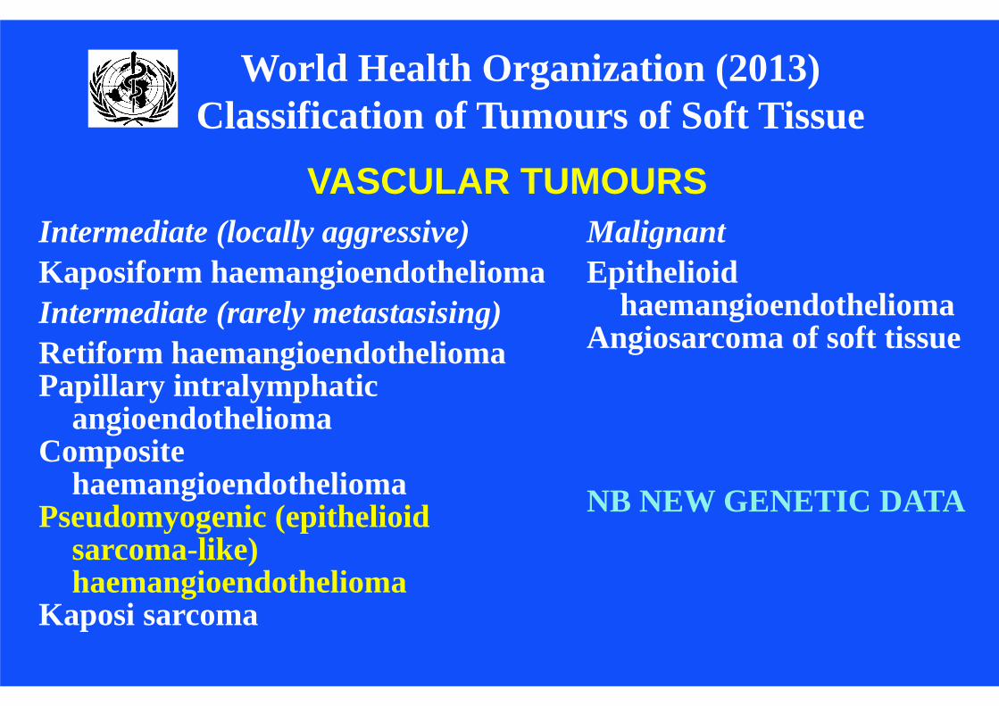
VASCULAR TUMOURSMalignantEpithelioid
haemangioendotheliomaAngiosarcoma of soft tissue
NB NEW GENETIC DATA
Intermediate (locally aggressive)Kaposiform haemangioendotheliomaIntermediate (rarely metastasising)Retiform haemangioendotheliomaPapillary intralymphatic
angioendotheliomaComposite
haemangioendotheliomaPseudomyogenic (epithelioid
sarcoma-like) haemangioendothelioma
Kaposi sarcoma
World Health Organization (2013) Classification of Tumours of Soft Tissue

Young adultsMales > females ++
80% limbs – leg > armMultiple nodules – usually < 3 cm
Multiple planes – skin > subcutis > muscle > boneOften painful
Metastasis infrequent - ? Indolent / delayed
PSEUDOMYOGENIC (‘EPITHELIOID SARCOMA-LIKE’)
HAEMANGIOENDOTHELIOMACLINICAL FEATURES


AE1/AE3 EMA
CD 31ERG

CHONDRO-OSSEOUS TUMOURS
Soft tissue chondromaExtraskeletal mesenchymal chondrosarcomaExtraskeletal osteosarcoma
World Health Organization (2013) Classification of Tumours of Soft Tissue

GASTROINTESTINAL STROMAL TUMOURS
GIST, benignGIST, uncertain malignant potential
GIST, malignant
World Health Organization (2013) Classification of Tumours of Soft Tissue

PROPOSED GUIDELINES FOR DEFINING RISK OF AGGRESSIVE BEHAVIOUR IN GISTs (NCI 2002)
Size Mitotic Count
Very low risk < 2 cm < 5 per 50 HPF
Low risk 2-5 cm 5 per 50 HPF
Intermediate risk 5 cm 5-10 cm
6-10 per 50 HPF
5 per 50 HPF
High risk
> 5 cm > 10 cm > Any size
> 5 per 50 HPF Any mitotic rate > 10 per 50 HPF


WHO 2013

• Approx. 5-7% of gastric GISTs• Wild-type KIT and PDGFRA • Distinct subsets of GIST – pediatric-type,
Carney-Stratakis syndrome, Carney triad• Epithelioid, multinodular, show LVI• Frequent nodal mets – but v. indolent• All SDHB-neg by IHC – but few mutations• 25% also SDHA-neg – predicts mutation
SDH-DEFICIENT GISTSKEY POINTS REGARDING A NEW GROUP


NERVE SHEATH TUMOURS
Benign (1)
Schwannoma (including variants) Melanotic schwannoma
Neurofibroma (including variants)Plexiform neurofibroma
PerineuriomaGranular cell tumour
World Health Organization (2013) Classification of Tumours of Soft Tissue

NERVE SHEATH TUMOURS
Benign (2)
Dermal nerve sheath myxomaSolitary circumscribed neuroma
Ectopic meningiomaNasal glial heterotopiaBenign Triton tumour
Hybrid nerve sheath tumours
World Health Organization (2013) Classification of Tumours of Soft Tissue
HYBRID NERVE SHEATH TUMOURS
Hybrid schwannoma / perineuriomaHybrid neurofibroma / schwannoma
Hybrid granular cell tumour / perineuriomaHybrid neurofibroma / perineurioma (?)


EMAS-100


NERVE SHEATH TUMOURS
Malignant
Malignant peripheral nerve sheath tumourEpithelioid malignant peripheral nerve sheath tumour
Malignant Triton tumourMalignant granular cell tumour
Ectomesenchymoma
World Health Organization (2013) Classification of Tumours of Soft Tissue

TUMOURS OF UNCERTAIN DIFFERENTIATION
BenignAcral fibromyxomaIntramuscular myxoma
(incl. cellular variant)Juxta-articular myxomaDeep ‘aggressive’ angiomyxomaPleomorphic hyalinizing angiectatic tumourEctopic hamartomatous thymoma
World Health Organization (2013) Classification of Tumours of Soft Tissue

Adults; M > FToes / fingers ++
Often adjacent to nail bedDermal / subcutaneous nodule
Most < 2 cmLocal recurrence 10%
ACRAL (DIGITAL) FIBROMYXOMACLINICAL FEATURES


CD34

TUMOURS OF UNCERTAIN DIFFERENTIATIONIntermediate (locally aggressive)Haemosiderotic fibrolipomatous tumourIntermediate (rarely metastasising)Atypical fibroxanthomaAngiomatoid fibrous histiocytomaOssifying fibromyxoid tumour
(incl. atypical / malignant)Mixed tumour / Myoepithelioma / Myoep. CarcinomaPhosphaturic mesenchymal tumour
World Health Organization (2013) Classification of Tumours of Soft Tissue

HAEMOSIDEROTIC FIBROLIPOMATOUS TUMOUR
(aka “haemosiderotic fibrohistiocyticlipomatous lesion”)
Adults > childrenFemales slightly > males
Ankle / foot ++Subcutaneous / poorly marginated
Usually < 5 cmLocal recurrence 30%
Possible potential to progress (?)
CLINICAL FEATURES

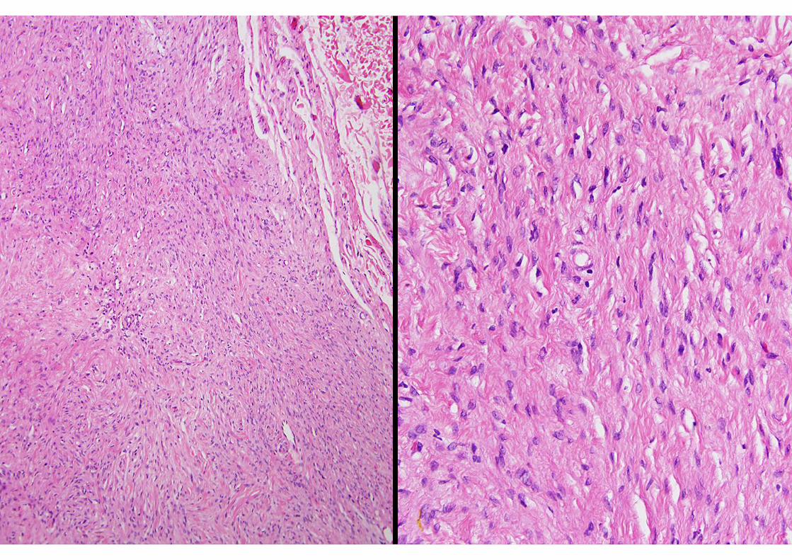
HAEMOSIDEROTIC FIBROLIPOMATOUS TUMOUR
(aka “haemosiderotic fibrohistiocyticlipomatous lesion”)
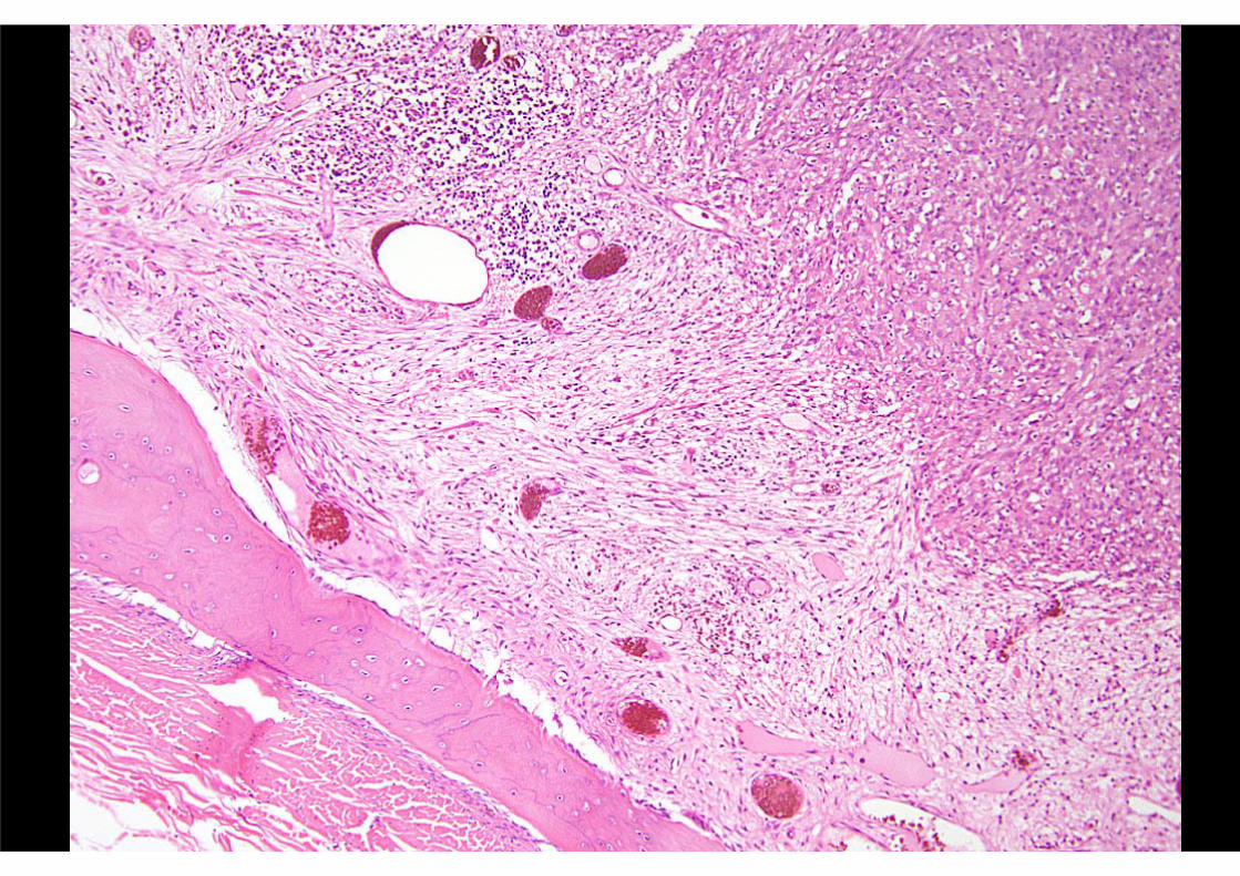
Admixture, in variable proportions, of:- Fibroblastic spindle cells
- Mature adipocytesFascicular or whorled pattern
Usually prominent haemosiderinScattered osteoclastic giant cells
Mitoses scarceOccasional atypia / pleomorphism
PATHOLOGIC FEATURES


CD34
SMA

HAEMOSIDEROTIC FIBROLIPOMATOUS TUMOUR
(aka “haemosiderotic fibrohistiocyticlipomatous lesion”)
Reactive (??) vs. neoplasticSignificance of atypia
Biologic potentialRelationship to other entities ??
ISSUES OF INTEREST


1st Recurrence 2nd Recurrence
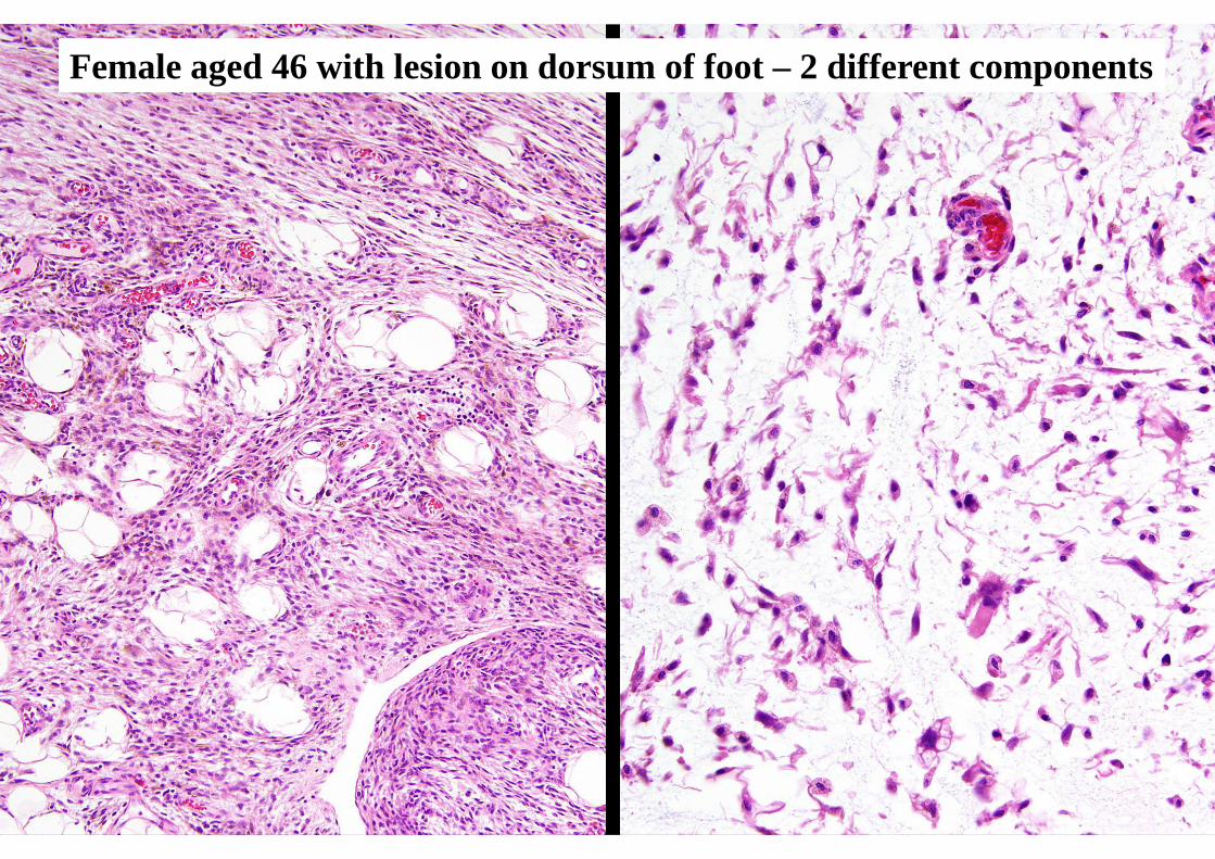
Female aged 46 with lesion on dorsum of foot – 2 different components

MYXOINFLAMMATORY FIBROBLASTIC SARCOMA
CLINICAL FEATURESAdults, wide age rangeEqual sex distribution
Slowly growing, ill-defined massDistal extremities
- especially hands and feetSubcutaneous / tenosynovial
Usually < 5 cmLocal recurrence common
Metastasis very rare


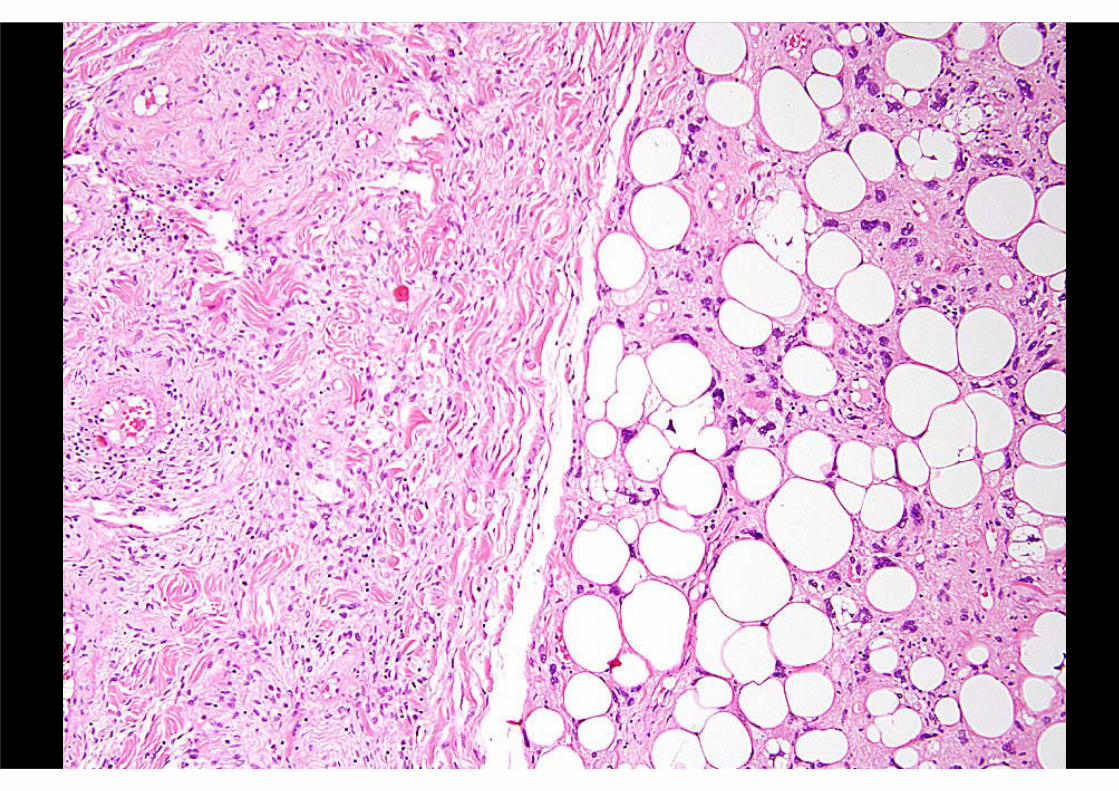
MYXOINFLAMMATORY FIBROBLASTIC SARCOMAAND
HEMOSIDEROTIC FIBROLIPOMATOUS TUMORSHARED CLINICOPATHOLOGIC & GENETIC FEATURES
Predilection for distal extremities, esp. feet Recur ++ - but ? almost never metastasise
Isolated cases show hybrid morphologic featuresBoth show reciprocal t(1;10)(p22;q24)
Gene fusion TGFBR3 – MGEA5Leads to up-regulation of FGF8
Also amplified 3p in ring chromosomesLambert et al, Virchows Arch 2001; 438:509-512
Wettach et al, Cancer Genet Cytogenet 2008; 182:140-143Hallor et al, J Pathol 2009; 217:716-727
Antonescu et al, Genes Chromosomes & Cancer 2011; 50:757-764

ATYPICAL FIBROXANTHOMACLINICAL FEATURES
Mainly elderly patientsMales > females
Head and neck++ / limbs rareRapidly enlarging exophytic
Sometimes multiple / asynchronousRecurrence infrequent
No metastasis if carefully diagnosed


PAN-K

• Mostly middle-aged adults – almost any site• Most assoc. with tumour-induced osteomalacia
due to FGF23 production• Varied morphology but most are myoid or
myopericytoma-like with blueish matrix and granular calcification
• Vast majority are benign
PHOSPHATURIC MESENCHYMAL TUMOUR
KEY POINTS


TUMOURS OF UNCERTAIN DIFFERENTIATION
Malignant
Synovial sarcoma Desmoplastic small roundEpithelioid sarcoma cell tumourAlveolar soft part sarcoma Extra-renal rhabdoid tumourClear cell sarcoma of soft tissue PEComaExtraskeletal myxoid chondrosarcoma Intimal sarcomaExtraskeletal Ewing sarcoma
World Health Organization (2013) Classification of Tumours of Soft Tissue

UNDIFFERENTIATED / UNCLASSIFIED SARCOMAS
Undifferentiated spindle cell sarcomaUndifferentiated pleomorphic sarcoma
Undifferentiated round cell sarcomaUndifferentiated epithelioid sarcoma
Undifferentiated sarcoma NOS
World Health Organization (2013) Classification of Tumours of Soft Tissue


CIC-DUX4

NEW GENETICS (1)Fibroblastic / Myofibroblastic tumours
Nodular fasciitis t(17;22)(p13;q12.3) USP6-MYH9Myxoinflammatory t(1;10)(p22;q24) TGFBR3-MGEA5
fibroblastic sarcoma- also seen in HFLT and hybrid lesions
Low grade fibromyxoid t(7;16)(q33;p11) FUS-CREB3L2sarcoma t(11;16)(p13;p11) FUS-CREB3L1- also seen in a subset of SEF and hybrid lesions
(Post-WHO – Solitary fibrous tumor inv12(q13q13) NAB2-STAT6)
World Health Organization (2013) Classification of Tumours of Soft Tissue

NEW GENETICS (2)Vascular tumours
Pseudomyogenic t(7;19)(q22;q13) SERPINE1-FOSBhaemangioendothelioma
Epithelioid t(1;13)(p36.3;q25) WWTR1-CAMTA1haemangioendothelioma
Angiosarcoma (breast) KDR mutation (25%)Angiosarcoma (secondary) MYC amplification (100%)
FLT4 coamplification (25%)
World Health Organization (2013) Classification of Tumours of Soft Tissue
(Post-WHO – EHE – new subset with YAP1-TFE3 gene fusion)

EHE Subset with TFE3 gene overexpression
TFE3CD31

NEW GENETICS (3)Tumours of Uncertain Differentiation
Angiomatoid ‘FH’ t(2;22)(q33;q12) EWSR1-CREB1 > 90%t(12;22)(q13;q12) EWSR1-ATF1 (rare)t(12;16)(q13;p11) FUS-ATF1 (rare)
OFMT PHF1 rearrangement (on 6p21) (50%)Myoepithelial tumours EWSR1 rearrangement (45% of cases)
(mainly malignant) with various partnersPEComa TSC2 deletion /rearrangement
TFE3 rearrangement (rare, distinct subset)Undifferentiated round t(4;19)(q35;q13.1) CIC-DUX4
cell sarcoma t(10;19)(q35;q26) CIC-DUX4
World Health Organization (2013) Classification of Tumours of Soft Tissue

• Tumors with similar morphology• Tumors that may show hybrid morphology• Seemingly totally unrelated tumors• Tumors of different lineages
SOFT TISSUE TUMORSEXAMPLES OF GENETIC OVERLAP

• Tumors with similar morphology e.g. spindle cell lipoma, cellular angiofibroma, mammary-type myofibroblastoma
• Tumors that may show hybrid morphologye.g. DFSP and giant cell fibroblastoma
• Seemingly totally unrelated tumorse.g. clear cell sarcoma and angiomatoid “MFH”
• Tumors of different lineagese.g. infantile fibrosarcoma and secretory carcinoma
EXAMPLES OF GENETIC OVERLAP


• Generally different anatomic sites -does this influence the phenotype ?
• Morphologic overlap with subtle differences
• Immunophenotypic differences• Same rearrangement/loss of 13q14 (Rb)• All benign/rarely recur• Cellular angiofibroma may perhaps have
potential for progression
RELATIONSHIP BETWEEN SPINDLE CELL LIPOMA,
MAMMARY-TYPE MYOFIBROBLASTOMA & CELLULAR ANGIOFIBROMA

• Frequently involved genes in multiple different tumor types, e.g. EWSR1, HMGA2
• Interchangeable genes in multiple distinct tumor types, e.g. EWSR1 and FUS
• Shared fusion genes in tumors thought to be distinct entities, e.g. TGFBR3-MGEA5
• Shared fusion genes in tumors which appear totally unrelated, e.g. EWSR1-ATF1
SOFT TISSUE TUMORSTYPES OF GENETIC OVERLAP

IMPACT OF GENETICSSHARED GENE REARRANGEMENTS
• EWSR1• FUS• CREB1• ATF1• HMGA-2
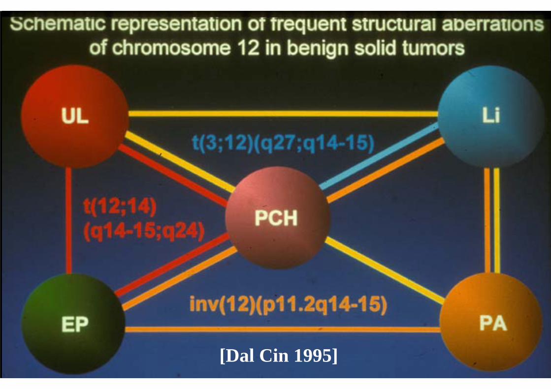
[Dal Cin 1995]

[2001]

Courtesy of Dr. Alex Lazar, MDACC (2008)

SHARED FUSION GENES IN SOFT TISSUE SARCOMAS
Szuhai & Bovee, 2012
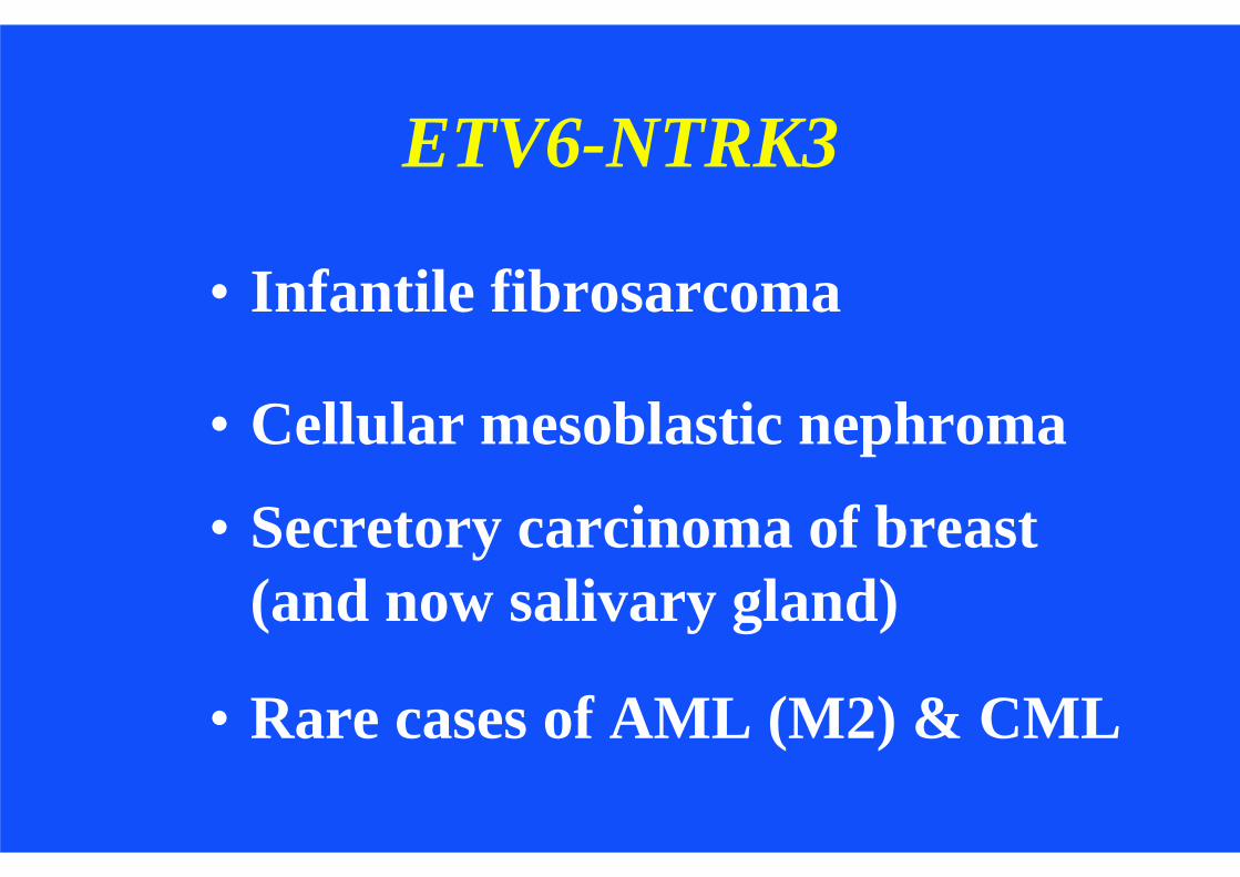
ETV6-NTRK3
• Infantile fibrosarcoma
• Cellular mesoblastic nephroma
• Secretory carcinoma of breast (and now salivary gland)
• Rare cases of AML (M2) & CML


EWSR1-ATF1EWSR1-CREB1
• Clear cell sarcoma
• Melanocytic• Deep soft tissue/GI• Adults (mainly
young)• > 50% metastasise
• Angiomatoid “MFH”
• Lineage unknown ?? dendritic cell
• Mostly subcutaneous• Commonest < 20 years• < 2% metastasise

• Some ‘newer’ entities or genetic findings left out
• Continued nomenclatural anomalies• Rigidity of ICD-0 coding system• Increasing problem/challenge of
genetic overlap
WHO CLASSIFICATION OFSOFT TISSUE TUMOURS 2013
WEAKNESSES

FUTURE IMPROVEMENTS• Hard to predict future !• Greater refinement
- more genetics / biology- more ‘entities’
• Nomenclatural advances• Better prognostication
Need to maintain open-mindedness to allow classification to evolve