Low Intensity Permanent Magnets in the Treatment of Chronic Lumbar Radicular Pain
Upload
kimsuniversityCategory
view
3download
0
2015 January Edition |www.jbino.com | Innovative Association
J.Bio.Innov4(1),pp:01-11,2015|ISSN2277-8330 (Electronic)
Kumar Nilesh et al.
UNUSUALLY LARGE RADICULAR CYSTS OF MAXILLA : STEPS IN DIAGNOSIS &
REVIEW OF MANAGEMENT
Dr. Kumar Nilesh*, Dr. Anuj S Dadhich1, & Dr. Pramod R Chandrappa
Department of Oral & Maxillofacial Surgery, School of Dental Sciences, KIMSDU, Karad 1 Department of Oral & Maxillofacial Surgery, Rural Dental College & Hospital, Loni
Email:[email protected]
(Received on Date: 13th November 2014 Date of Acceptance: 2nd December 2014)
ABSTRACT
Radicular cystsare common odontogenic cyst of jaw bone which invariably presents as well-
defined radiolucency around the apex of the involved tooth. It is diagnosed on routine
radiography or causes a slow growing swelling, limited to apical region of a tooth. Radicular cyst
presenting as unusually large maxillary lesions is uncommon. This paper reports two cases of large
radicular cyst involving maxilla. The first case presented as a single lesion occupying almost the
entire anterior maxilla, while the second case presented a large cyst occupying the entire left half
of the maxilla and the maxillary sinus. Unusually large radicular cyst of maxilla require stepwise
clinical, radiological and laboratory evaluation to reach a definitive diagnosis. Although no
consensusexist regarding the precise treatment modality for large radicular cyst of maxilla, both
cases were successfully treated by surgical removal of the cyst following endodontic therapy.
Keywords: Large, Radicular cyst, Maxilla, Enucleation
No. of Figures: 8 No. of References : 28
2015 January Edition |www.jbino.com | Innovative Association
J.Bio.Innov4(1),pp:01-11,2015|ISSN2277-8330 (Electronic)
Kumar Nilesh et al.
INTRODUCTION
Radicular Cysts are believed to be formed
from epithelial cell rests of Malassez (ERM),
which are remnants of Hertwig’s epithelial
root sheath, present within the periodontal
ligament. Proliferation of these epithelial
cell rests is frequently associated with
stimuli from periapical periodontal
inflammation secondary to pulpitis (1).
During periapical inflammation, host cells in
the periapical tissues release many
inflammatory mediators, proinflammatory
cytokines, and growth factors which
induce proliferation of the ERM in all
directions to form a three-dimensional ball
mass(2,3). As the epithelial mass grows, the
central cells move further away from their
source of nutrition and undergo necrosis
and liquefaction degeneration, forming
central cystic cavity lined by epithelial wall.
Following its formation, radicular cysts
grows by periapical bone resorption
mediated by prostagladins and cytokines.
It is interesting to note that most
inflammatory mediators which induce
proliferation of epithelial cell rests also
mediate bone resorption in inflammatory
periapical lesion (4-9). However there is no
time-lapse study showing that the size of
apical cysts gradually increases as
periapical bone destruction increases.
Clinically radicular cyst presents as
periapical lesion, ranging from 0.5 to 1.5
cm in size. Smaller periapical cysts can
often be treated conservatively by non-
surgical endodontic therapy. Proper
endodontic therapy of the involved teeth
removes irritants in the canals by chemo-
mechanical instrumentation. As the root
canal is completely sealed, all cell
components participating in inflammatory
reaction gradually resolve (10).
Subsequently majority of inflammatory
cells, endothelial cells and fibroblasts in the
paeriapical lesion are deleted by
apoptosis or programmed cell death (11).
However surgical removal following
endodontic treatment is required for cases
not amenable to conservative therapy
(12). Presence of unusually large
periapical lesion may often lead to
diagnostic dilemma. Such cases need to
be evaluated in a step wise manner to
reach a definite diagnosis. This article
reports two cases of large radicular cyst of
maxilla thepresentation, steps in diagnosis
and management of these lesions are also
discussed.
Case Report:
Case 1:A 34 year old female presented
with complaint of painless swelling over
roof of the mouth. The patient first noticed
the swelling about 6 months back, which
had grown gradually to its present size. No
history of trauma or tooth pain was
reported. Extraoral examination revealed
no facial swelling or obvious facial
asymmetry. Intraoral examination showed
a palatal swelling starting distal to right 1st
premolar and extending anteriorly, across
the midline to the contra laterallateral
incisor, measuring about 4 x 3.5 cm in size
(Figure 1a). Swelling was non-tender and
firm on palpation. The overlying palatal
mucosa appeared normal. Maxillary
anterior teeth were grade one mobile, with
normal depth of gingival sulcus on
diagnostic probing. All the maxillary
2015 January Edition |www.jbino.com | Innovative Association
J.Bio.Innov4(1),pp:01-11,2015|ISSN2277-8330 (Electronic)
Kumar Nilesh et al.
anteriors were non-vital on electric pulp
testing. The clinical presentation was
suggestive of a benign intraosseous tumor
or an odontogenic cyst. Clinical differential
diagnosis included central giant cell lesion,
radicular cyst, odontogenic Keratocyst,
globulomaxillary cyst and ameloblastoma.
Aspiration of the lesion yielded yellow color
fluid which was subjected to cytochemical
evaluation (Figure 1b). The fluid consisted
of cholesterol crystal and numerous
inflammatory cells, suggestive of an
inflammatory cystic lesion.
A cone beam computed tomography
(sironaorthophos X3GD, Germany) was
advised, to study the size and extent of the
lesion. The 3D reconstruction showed a
large osteolytic lesion extending form
maxillary right 1st premolar to maxillary left
canine and superiorly till the floor of the
nasal fossa. The roots of the associated
teeth were displaced and showed no
resorption (Figure 2a). Axial section showed
5 x 3.5 cm intraosseous radiolucent lesion
with defined sclerotic borders involving
almost entire anterior maxilla (Figure 2b).
Diagnosis of inflammatory odontogenic
cyst was established based on the clinical,
radiological and aspirate evaluation.
Surgical enucleation of the cyst was
planned under general anesthesia after
root canal therapy of the involved
maxillary teeth. Biomechanical
perpetration and irrigation (with saline &
1% sodium hypochlorite alternatively) of
root canals of all the maxillary anterior was
done under antibiotic cover. Calcium
hydroxide intracanal medication was
placed for period of 1 week. Subsequently
the root canals were obturated with gutta-
percha. The cyst was approached through
labial gingiva mucoperiosteal flap and
removed by enucleation. Apicoectomy
and retrograde root end filling for maxillary
anterior teeth was done with MTA to obtain
apical seal. The labial and interdental
bone support around the maxillary anterior
teeth appeared to be compromised due
to bone resorption caused by the
periapical lesion. After through irrigation of
the bone cavity & hemostasis, the bone
defect was grafted with platelet rich
concentrate to facilitate periapical wound
healing. Autologous platelet rich
concentrate was prepared from 10 ml of
patient’s own blood centrifuged at 2888
rpm for 12 minutes (Remi Lab centrifuge)
(Figure 3). The defect was closed primarily
and cyst lining was sent for histopathology
examination, which confirmed the
diagnosis of radicular cyst [Figure 4].
Splinting of maxillary anterior teeth was
done for 3 weeks, using 22-gauge stainless
steel wire stabilized with light cured
composite in immediate postoperative
period. At 1 year follow up the patient was
asymptomatic and showed no sign of
recurrence. There was no mobility of the
anterior teeth and imaging of the surgical
site showed normal healing of the bone
defect (Figure 5).Case 2:A thirty six year old
female, presented with complain of
painless swelling of eight months duration
over left side of midface. The patient gave
history of fall from bicycle during her
childhood. On examination a large extra-
oral swelling was evident, extending from
left infraorbital region to level of corner of
mouth, measuring roughly 4 x 5 cm in size.
The nasolabial fold was obliterated along

2015 January Edition |www.jbino.com | Innovative Association
J.Bio.Innov4(1),pp:01-11,2015|ISSN2277-8330 (Electronic)
Kumar Nilesh et al.
with elevation of ala of left nose (Figure 6).
The swelling was non-tender and firm on
palpation. Intraoral examination showed
soft fluctuant swelling localized over left
maxillary vestibule in premolar-molar
region. Maxillary left central and later
incisors were non-responsive to electric
pulp vitality test. On radiological
evaluation, a large radiolucent lesion was
evident involving the entire left maxilla.
Orthopantamogram showed a well-
defined lesion with sclerotic border,
extending from maxillary midline till the
maxillary tuberosity. The entire left maxillary
sinus appeared hazy on paranasal sinus
view (Figure 7). The presenting features
were suggestive of cyst of odontogenic or
maxillary sinus origin. Content of the lesion
was aspirated intra-orally under local
anaesthesia using 18 gauge needle. Cyto-
chemical examination of the straw color
aspirate showed numerous inflammatory
cells and cholestrol crystals. Based on the
history, clinical presentation, vitality test,
radiographic features and evaluation of
aspirate, diagnosis of inflammatory
odontogenic cyst following pulp necrosis of
left maxillary incisors was made. However,
looking into the unusually large size of the
lesion, incision biopsy was planned to
conform the diagnosis. The lesion was
accessed intraorally through Caldwell-Luc
approach (by creating bony window in
region of canine fossa). A portion of the
cyst lining was removed and sent for sent
for histopathological examination.
Diagnosis of radicular cyst was confirmed
based on incision biopsy report. Following
chemo-mechanical preparation of pulp
chambers of the involved left maxillary
incisors and placement of calcium
hydroxide intracanal dressing for one
week, the patient was prepared for
surgical removal of the cyst under general
anesthesia. After the cyst was enucleated
in-toto, apicoectomy of the left maxillary
incisors and retrograde root end filling with
MTA was done. The cyst lining was
submitted for microscopic evaluation
which confirmed diagnosis of radicular cyst
(Figure 8). The postoperative healing was
uneventful. The patient was symptom free
and showed healing intraosseous surgical
defect, at 6 months follow-up.Radicular
cysts are the most common odontogenic
cyst affecting the human jaw (13). It is
believed to be formed from proliferation of
epithelial residues in the periodontal
ligament as a result of periapical
inflammation secondary to pulpitis.
Although they are direct sequel to apical
periodontitis, not every case of apical
periodontitis causes formation of radicular
cyst (14). Since radicular cyst form at the
apex of tooth root(s) it is also called
periapical cyst. They may be associated
with any tooth. However it commonly
involves maxillary anterior teeth and
seldom involves primary dentition. Their
prevalence is highest in the third decade
of life and are more commonly seen in
males than females (15). Position of the
maxillary anterior teeth makes them more
prone to injury and subsequent pulp
necrosis. This may be the possible
explanation for their prevalence in males
and in maxillary anterior region. One of our
reported cases (case 2) gave history of
trauma to maxillary anterior teeth, which
could have been the possible cause of
2015 January Edition |www.jbino.com | Innovative Association
J.Bio.Innov4(1),pp:01-11,2015|ISSN2277-8330 (Electronic)
Kumar Nilesh et al.
initiation of cyst formation. Periapical
inflammation and pulpitis secondary to
dental caries is another cause of formation
of radicular cyst. Most of the radicular cysts
are symptomless and discovered when
periapical radiographs are taken for non-
vital teeth. They present as round to oval
radiolucency associated with root apices,
varying in size from 0.5 to 1.5 cm. As the
cyst grows in size by resorption of the
surrounding periapical bone, it causes
localized jaw swelling. Pain and pus
discharge are presenting feature of an
infected radicular cyst. Only few cases of
radicular cysts involving large part of
maxilla have been previously reported in
literature (16-19). Interestingly the reported
cases of large radicular cyst involve maxilla
and very often extended to occupy large
portions of the maxillary sinus. A logical
reasoning to explain this association can
be the relatively thin and cancellous
nature of the maxillary bone as compared
to dense mandible, which would be more
resistant to expansion and resorption. Also
extension of these periapical lesions into
the maxillary sinus would allow its
unrestricted growth, leading to formation
of giant radicular cyst.
Although intra-oral radiographs are
sufficient for studying a periapical cysts,
larger lesions often require more extensive
imaging to evaluate the location, size,
proximity to vital structures and extent of
the lesion (20). Both extra-oral radiographs
(including orthopantamogram¶nasal
sinus view) and three dimensional imaging
using cone beam computed tomography
were used in our cases for radiological
assessment. Aspiration of the cyst fluid is
another important diagnostic test for cystic
lesion. It helps to rule out any vascular
lesion and provides fluid specimen for
cytochemical evaluation. In our reported
cases the cyst content was aspirated
under local anaesthesia, using wide gauge
needle. The fluid was yellowish-white in
color and contained cholesterol crystal
and numerous inflammatory cells. The
treatment of radicular cyst, as a disease of
root canal infection or inflammation,
consists of eradicating or substantially
reducing the microbial load and
inflammatory mediators from the canals
and periapical tissue. Numerous measures
have been described to reduce the
number of microorganisms in the root
canal system, including the use of various
irrigation regimens and intracanal
medicaments. Various root canal irrigants
used include Sodium hypochlorite,
ethylenediaminetetraacetic acid (EDTA),
citric acid and chlorhexidine. Sodium
hypochloride is the most commonly used
endodontic irrigant (21). In our cases 1%
Sodium hypochloride and saline was used
alternatively as root canal irrigant. Intra-
canal medicaments eliminate or reduce
number of microorganisms, rendering
canal contents inert. It helps in prevention
of post-treatmentpain, and enhances
anesthesia (22). Calcium hydroxide is most
widely used intracanal medicament. It also
plays a major role as an inter-visit dressing
in the disinfection of the root canal system.
Reduction of inflammatory cells and
mediators from roots of the involve tooth
may often lead to resolution of smaller
periapical lesion. However cases which do
not respond to conservative management
2015 January Edition |www.jbino.com | Innovative Association
J.Bio.Innov4(1),pp:01-11,2015|ISSN2277-8330 (Electronic)
Kumar Nilesh et al.
require surgical intervention. Surgical
treatment is preferred approach to treat
large radicular cyst. For apical
radiolucency larger than 20 mm in
diameter or having cross-sectional area
greater than 200 mm2, surgical removal
may be the best option (23). Surgical
treatment is also recommended when the
canal appears calcified or obstructed and
cannot be negotiated with endodontic
instruments. The surgical treatments for
radicular cyst include total enucleation,
marsupialization or a combination of these
techniques (24, 25). Removal of entire cyst
allows histopathological evaluation of cyst
lining, thereby helping in reaching a
definitive diagnosis and ruling out any
malignant or dysplastic changes in large
cyst. After the enucleation of the cyst,
apicoectomy of the involve teeth is done.
Apicoectomy involves sectioning of the
apex of the root and reterograde filling to
attain a sound periapical seal. Historically
amalgam was the most frequently used
material in apicoectomy procedure. IRM,
super EBA and MTA are newer and more
suitable materials, and give better results in
apicoectomy procedures than Amalgam
(26). Advantages of MTA include high
biocompatibility and excellent sealing
ability with low marginal leakage. However
high cost of MTA is a major disadvantage
to its use in every clinical situation. All our
cases were treated with surgical
enucleation of the periapical cyst along
with apicoectomy and retrograde fillings of
involved roots with MTA.
The bony defect left after the removal of
the cyst lining heal uneventfully after a
proper apical seal is attained and the
intraosseous bony cavity is irrigated
generously to remove all periapical
irritants. In our first case report we had
grafted the bony cavity with autologous
platelet rich concentrate to facilitate
healing of the large defect of anterior
maxilla, which had caused significant
bone loss around the maxillary anterior
teeth. The teeth were subsequently
splinted in immediate postoperative
period. Platelet rich concentrate is known
to initiate osteoinduction process which is
mediated by growth factors present in
platelets. High number of platelets in the
concentrate produces large amount of
growth factors, initiating bone formation
(27). Beside bone regeneration, it also aid
in healing of surrounding soft tissues (28).
Both the reported cases were followed up
for 6- 12 months and showed uneventful
healing of the lesion with no reoccurrence
or complication.Periappical pathology
secondary to pulp inflammation is a
common finding. However usually large
periapical lesion require stepwise clinical,
radiological and laboratory evaluation to
reach a definitive diagnosis. Such large
lesion often warrants detailed radiological
assessment to study the location, size and
extent of the lesion. Cytochemical
evaluation of the aspirate provides
valuable input regarding nature of the
cyst. Although no consensusexist regarding
the precise treatment modality for large
radicular cyst of maxilla, both cases were
successfully treated by surgical removal of
the cyst following endodontic therapy of
the involved teeth. Enucletion of the cyst
provided the lining for complete
histological evaluation of the periapical

2015 January Edition |www.jbino.com | Innovative Association
J.Bio.Innov4(1),pp:01-11,2015|ISSN2277-8330 (Electronic)
Kumar Nilesh et al.
pathology and to rule out any malignant
or dysplastic changes in these unusually
large cysts.
Figure 1: (Case 1) Swelling over anterior palate (block arrows)
Figure 2: (Case 1) (a) Cone beam 3D reconstruction showing large radiolucent lesion involving almost entire
anterior maxilla (b) Axial section, at the level of Apical third of maxillary anterior teeth showing the dimensions
of the cyst.
Figure 3: (Case 1) Platelet rich concentrate prepared from patients’ blood (centrifuged 10 ml blood at 2888
rpm for 12 minutes; Remi Lab centrifuge)
2015 January Edition |www.jbino.com | Innovative Association
J.Bio.Innov4(1),pp:01-11,2015|ISSN2277-8330 (Electronic)
Kumar Nilesh et al.
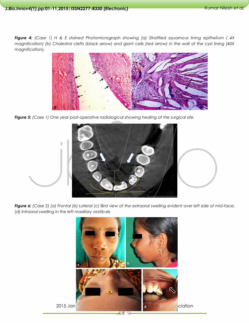
Figure 4: (Case 1) H & E stained Photomicrograph showing (a) Stratified squamous lining epithelium ( 4X
magnification) (b) Cholestrol clefts (black arrow) and giant cells (red arrow) in the wall of the cyst lining (40X
magnification).
Figure 5: (Case 1) One year post-operative radiological showing healing at the surgical site.
Figure 6: (Case 2) (a) Frontal (b) Lateral (c) Bird view of the extraoral swelling evident over left side of mid-face;
(d) Intraoral swelling in the left maxillary vestibule
2015 January Edition |www.jbino.com | Innovative Association
J.Bio.Innov4(1),pp:01-11,2015|ISSN2277-8330 (Electronic)
Kumar Nilesh et al.
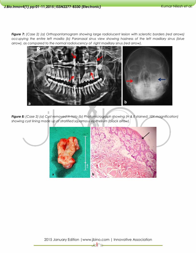
Figure 7: (Case 2) (a) Orthopantamogram showing large radiolucent lesion with sclerotic borders (red arrows)
occupying the entire left maxilla (b) Paranasal sinus view showing haziness of the left maxillary sinus (blue
arrow), as compared to the normal radiolucency of right maxillary sinus (red arrow).
Figure 8: (Case 2) (a) Cyst removed in-toto (b) Photomicrograph showing (H & E stained; 10X magnification)
showing cyst lining made up of stratified squamous epithelium (black arrow).
2015 January Edition |www.jbino.com | Innovative Association
J.Bio.Innov4(1),pp:01-11,2015|ISSN2277-8330 (Electronic)
Kumar Nilesh et al.
REFERENCES
Ahmed C, Wafae El, Bouchra T. Massive Radicular
Cyst Involving the Maxillary Sinus: A Case Report.
International Journal of Oral & Maxillofacial
Pathology. 2013;4(1):68-71
Chaudhary CP, Ravikant, Ravishankar MN, Anurag
Y, Gunjan Y. Healing of bone defects by
autogenous platelet rich Plasma in pediatric
patients. Journal of recent advances in applied
sciences. 2012; 27:11-13.
Freedland JB. Conservative reduction of large
periapical lesions. Oral Surg Oral Med Oral Pathol.
1970;29(3):455-64.
Gervasio AM, Silva DAO, Taketomi EA, Souza CJA,
Sung SSJ, Loyola AM. Levels of GM-CSF, IL-3, IL-6 in
fluid and tissue from human radicular cysts. J Dent
Res 2002;81:64–8.
Gibson GM, Pandolfi PJ, Luzader JO. Case report: a
large radicular cyst involving the entire maxillary
sinus. General dentistry. 2002; 50(1): 80-81.
Grossman I, Abu NA, Peled M. Root-end filling
materials in apicoectomy-a review.
RefuatHapehVehashinayim. 2003 Apr; 20(2):49-54,
80.
Hamachi T, Anan H, Akamine A, Fujise O, Maeda K.
Detection of interleukin-mRNA in rat periapical
lesions. J Endod 1995;1:118 –21.
Majno G, Joris I. Apoptosis, oncosis, and necrosis:
an overview of cell death. Am J Pathol1995;146:3–
15.
Matsumoto A, Anan H, Maeda K. An
immunohistochemical study of the behavior of cells
expressing interleukin-1 and interleukin-1 within
experimentally induced periapical lesions in rats. J
Endod 1998;24:811– 6.
McNicholas S, Torabinejad M, Blankenship J,
Bakland L. The concentration of prostaglandin E2 in
human periapical lesions. J Endod 1991;17:97–100.
Nair PNR. Non-microbial etiology: periapical cysts
sustain post-treatment apical Periodontitis.
Endodontic Topics 2003;6 : 96-113
Nair PNR. Pathogenesis of Apical Periodontitis and
the Causes of Endodontic Failures. Crit Rev Oral Biol
Med (2004);15(6):348-81
Neaverth EJ, Burg HA. Decompression of large
periapical cystic lesions. J Endod. 1982;8(4):175-82.
Pekiner FN, Borahan MO, Uğurlu F, Horasan S, Sener
BC, Olgaç V. Clinical and radiological features of
large radicular cyst involving the entire maxillary
sinus. MÜSBED. 2012; 2(1): 31-36
Ramachandran Nair PN, Pajarola G, Schroeder HE.
Types and incidence of human periapical lesions
obtained with extracted teeth. Oral Surg Oral Med
Oral Pathol Oral RadiolEndod. 1996;81(1):93-102.
Sagit M, Guler S, Tasdemir A, Somdas M. Large
radicular cyst in the maxillary sinus. J Craniofac
Surg. 2011 Nov; 22(6):e64-5.
Seltzer S, Soltanoff W, Bender IB. Epithelial
proliferation in periapical lesions. Oral Surg Oral
Med Oral Pathol 1969;27:111–21.
Seltzer S. Endodontology. Biologic Considerations in
Endodontic Procedures. 2nd ed. Philadephia, PA:
Lea &Febiger, 1988.
Shear M. Cysts of the Oral Regions. 3rd ed. Boston,
Wright, 1992, pp. 136-70.
Simon JH, Enciso R, Malfaz JM, Roges R, Perry M,
Patel A. Differential diagnosis of large periapical
lesions using cone-beam computed tomography
measurements and biopsy. J Endod. 2006 Sep;
32(9):833-7.
Stern MH, Dreizen S, Mackler BF, Levy BM. Antibody-
producing cells in human periapical granulomas
and cysts. J Endod 1981;7:447–52.

2015 January Edition |www.jbino.com | Innovative Association
J.Bio.Innov4(1),pp:01-11,2015|ISSN2277-8330 (Electronic)
Kumar Nilesh et al.
Sushma J, Prashant J, Newer Root Canal Irrigants in
Horizon: A Review; International Journal of Dentistry.
2011,851359, 1-9.
Tani-Ishii N, Wang CY, Stashenko P.
Immunolocalization of bone resortive cytokines in
rat pulp and periapical lesions following surgical
pulp exposure. Oral MicrobiolImmunol1995;10:213–
9.
Taschieri S , Fabbro MD, Testori T, Weinstein R.
Efficacy of Xenogeneic Bone Grafting With Guided
Tissue Regeneration in the Management of Bone
Defects After Surgical Endodontics. Journal of Oral
and Maxillofacial Surgery.2011 June
Tolasaria S, Das UK. Surgical and Nonsurgical
Management of Bilateral Periapical Lesions in the
Maxillary Anterior Region. Journal of Surgical
Technique and Case Report.2011; 3(1).
Torabinejad M, Bakland L. Prostaglandins: their
possible role in the pathogenesis of pulpal and
periapical disease. J Endod 1980;733–9, 769 –76.
Wang CY, Tani-Ishii N, Stashenko P. Bone resorptive
cytokine gene expression in developing rat
periapical lesions. Oral MicrobiolImmunol
1997;12:65–71.
Wergedal JE, Mohan S, Lundy M, Baylink DJ. Skeletal
growth factor and other factors known to be
present in bone matrix stimulate proliferation and
protein synthesis in human bone cells. J Bone Miner
Res 1990; 5:179-186.
Copyright © 2022 FDOKUMEN