PROBIT analysis predicts hatching rates of brine shrimp Artemia sp. cysts
Upload
independentCategory
view
2download
0
Tumorigenesis and Neoplastic Progression
USP6 and CDH11 Oncogenes Identify theNeoplastic Cell in Primary Aneurysmal Bone Cystsand Are Absent in So-Called Secondary AneurysmalBone Cysts
Andre M. Oliveira,*† Antonio R. Perez-Atayde,‡
Carrie Y. Inwards,† Fabiola Medeiros,*Victoria Derr,* Bae-Li Hsi,* Mark C. Gebhardt,§
Andrew E. Rosenberg,¶ andJonathan A. Fletcher*�
From the Department of Pathology,* Brigham and Women’s
Hospital, Boston; the Department of Laboratory Medicine and
Pathology,† Mayo Clinic, Rochester, Minnesota; the Department of
Pathology,‡ Children’s Hospital Medical Center, Boston; the
Departments of Orthopedic Surgery,§ Beth Israel Deaconess
Medical Center and Children’s Hospital Medical Center, Boston;
the Department of Pathology,¶ Massachusetts General Hospital,
Boston; and the Department of Pediatric Oncology,� Dana-Farber
Cancer Institute, Boston, Massachusetts
Aneurysmal bone cyst (ABC) is a locally recurrentbone lesion that has been regarded as a reactive pro-cess. Recently, a neoplastic basis in primary ABC wasevidenced by demonstration of clonal chromosomeband 17p13 translocations that place the USP6 (TRE2or TRE17) oncogene under the regulatory influenceof the highly active CDH11 promoter. Herein, wereport CDH11 and/or USP6 rearrangements in 36 of52 primary ABCs (69%), of which 10 had CDH11-USP6fusion, 23 had variant USP6 rearrangements withoutCDH11 rearrangement, and three had variant CDH11rearrangements without USP6 rearrangement. USP6and CDH11 rearrangements were restricted to spindlecells in the ABC and were not found in multinucleatedgiant cells, inflammatory cells, endothelial cells, orosteoblasts. CDH11 and USP6 rearrangements did notcorrelate with recurrence-free survival, or with otherclinicopathological features. CDH11 and USP6 rear-rangements were not found in any of 17 secondaryABC associated with giant cell tumor, chondroblas-toma, osteoblastoma, and fibrous dysplasia. Thesefindings demonstrate that primary ABC are mesen-chymal neoplasms exhibiting USP6 and/or CDH11oncogenic rearrangements. By contrast, secondary
ABC lack CDH11 and USP6 rearrangements, and al-though morphological mimics of primary ABC, ap-pear to represent a non-specific morphological pat-tern of a diverse group of non-ABC neoplasms. (AmJ Pathol 2004, 165:1773–1780)
Aneurysmal bone cyst (ABC) is an intriguing bone lesionwith the potential for local recurrence that has been re-garded as a reactive process since its initial descriptionin 1942 by Jaffe and Lichtenstein.1 Different theorieshave been proposed for the pathogenesis of ABC, andamong the most widely accepted has been that a localcirculatory abnormality leads to an increased venouspressure and resultant dilation of the vascular network.2–5
The reactive nature of ABC has also been suggested bythe fact that a variety of benign and malignant boneneoplasms,5–8 including giant cell tumor of bone, chon-droblastoma, osteoblastoma, fibrous dysplasia, and os-teosarcoma may contain areas within the lesion thatclosely mimic ABC histologically. For these cases, theterm “secondary ABC” has been coined.3,7 However, it isunclear whether primary and secondary ABC have asimilar pathogenesis, or whether secondary ABC mightrather be a common morphological pattern of growth thatoccurs as a non-specific phenomenon in a variety ofprimary bone tumors.
The purported reactive nature of ABC was refuted bythe work of Panoutsakopoulos et al,9 which demonstratedchromosomal translocation t(16;17)(q22;p13) as a recur-rent cytogenetic abnormality in primary ABC. Subse-quently, other groups confirmed 17p13 rearrangement asa frequent cytogenetic aberration in primary ABC.10–15
Recently, we extended these studies by showing that the
Supported by Mayo Clinic and Mayo Clinic Foundation.
Accepted for publication August 3, 2004.
Address reprint requests to Jonathan A. Fletcher, Department of Pa-thology, Brigham and Women’s Hospital, 75 Francis Street, Boston, MA02115. E-mail: [email protected] or [email protected].
American Journal of Pathology, Vol. 165, No. 5, November 2004
Copyright © American Society for Investigative Pathology
1773

t(16;17)(q22;p13) fuses the promoter region of the osteo-blast cadherin 11 gene (CDH11) on chromosome 16q22to the entire coding sequence of the ubiquitin proteaseUSP6 gene (also known as TRE2 or TRE17) on chromosome17p13, suggesting that the pathogenesis of many primaryABC involves up-regulation of USP6 transcription.16
In this study we determined the frequency and theclinicopathologic consequences of CDH11 and USP6 ge-netic abnormalities in 52 primary ABC, and we also usedthese molecular markers to evaluate the pathogeneticrelationship or lack thereof between primary and second-ary ABC. Further, we identify the neoplastic cell in pri-mary ABC by determining which cell components con-tained the genetic rearrangements.
Materials and Methods
Tumor Samples
Fifty-two primary ABC and 17 secondary ABC were his-tologically characterized according to established crite-ria.8 Metaphase preparations were available from eight ofthese cases, including two for which the cytogeneticfindings have been reported previously (cases 19 and29).16 Frozen tissue was available from 16 cases.
Fluorescence in Situ Hybridization (FISH)
BAC clones were obtained from Children’s Hospital Oak-land Research Institute (Oakland, CA) and Research Ge-netics (Huntsville, AL). BAC minicontigs were assembledbased on genomic mapping and sequence data from theHuman Genome Working Draft. Minicontigs telomericand centromeric to the USP6 locus were USP6. T (BACsRP11–124C16, RP11–111I16, and RP11–177H5) andUSP6. C (CTD-2367F23 and RP11–457I18), respectively.Those telomeric and centromeric to CDH11 were CDH11.T (RP11–137A18, RP11–631H23, and RP11–351A20)and CDH11. C (RP11–615M9, RP11–730A21, and RP11–76J1), respectively. BAC DNA isolations and labelingwere performed as described previously.16
FISH was performed using 4-�m paraffin-embeddedtissue sections which were deparaffinized in xylene (3 �10 minutes), dehydrated twice in 100% ethanol for 2minutes, and treated with 100 mmol/L Tris and 50 mmol/LEDTA (pH 7.0) for 15 minutes at 93°C. Tissue sectionswere then rinsed once in 1X PBS and protein digestedwith Digest All-3 (Zymed, San Francisco, CA). Afterbriefly washing in 1X PBS, the slides were sequentiallydehydrated in alcohol (70%, 85%, 95%, and 100%), andair-dried for 1 hour at room temperature. Tissue sectionswere denaturated at 75°C for 2 minutes and BAC probehybridization was carried out overnight in a humidifiedchamber at 37°C. Tissue sections were then washed in0.5X SSC for 5 minutes at 73°C and treated with CASblock (Zymed) for 10 minutes. Probe detection was per-formed using FITC-anti-digoxigenin (1:500) and AlexaFluor 594-streptavidin (1:500) (Molecular Probe, Eugene,OR)17 for 30 minutes. Slides were then mounted inVECTASHIELD mounting medium with 1.5 �g/ml of 4�,6-
diamidino-2-phenylindole (DAPI). ABC were scored aspositive for gene rearrangement if more than 5% of cellsshowed splitting apart of the flanking FISH probes. Iffewer than 20% of cells showed rearrangement, the find-ings were corroborated by repeating the FISH assay inanother section from the same paraffin block.
RNA Isolation and RT-PCR
RNA was isolated from frozen tumors after mechanicalhomogenization and overnight incubation in Trizol (In-vitrogen, Carlsbad, CA) at 4°C. RNA reverse transcriptioninto cDNA was performed using the GeneAmp RNA PCRKit (Applied Biosystems, Foster City, CA) for 2 hours at42°C using random hexamers. RNA isolation from paraf-fin sections were performed according to a previouslydescribed protocol.18
PCR reactions were performed using the Takara ExTaq kit with the following parameters for 35 cycles: de-naturation at 94°C for 30 seconds, annealing at 65°C for30 seconds, and extension at 72°C for 1 minute. PCRprimers for RT-PCR evaluation of the CDH11-USP6 fusiononcogene were CDH11�71F (5�-CGCCGCTGACTTGT-GAAT-3�) and USP6�1781R (5�-CTCGGTGTCCCTTGT-CATACTT-3�). Evaluation of low-abundance fusion tran-scripts in tumors where the first round PCR was negativewas performed by nested PCR using primersCDH11�83F (5�-GTGAATGGGACCGGGACT-3�) andUSP6�1736R (5�-CAGGAGCGGAAGGACATACTTA-3�)at the same cycling parameters as above for 25 cycles.
Statistical Analysis
Statistical analyses were performed with the S-PLUS 6.0software package (Insightful Corp., 2001). The Fisherexact test was used to evaluate associations among cat-egorical variables. The Mann-Whitney U-test was used toevaluate associations between categorical and continu-ous variables. Recurrence-free survival was calculatedusing the Kaplan-Meier product-limit method, and univar-iate survival analyses were calculated using the log-ranktest. Multivariate survival analyses were performed usingthe Cox multivariate regression model. Proportionality as-sumption was evaluated using Kaplan-Meier and log-minus-log survival curves. Reported P values were two-sided and statistical significance was set at P � 0.05.
Results
Overall Clinical and Morphological Features ofPrimary ABC
Clinical features are summarized in Table 1. Median pa-tient age at diagnosis was 14 years (range, 2 to 42 years),and the genders were equally represented (27 femalesand 25 males). The most frequent primary sites were tibia(n � 11), femur (n � 8), fibula (n � 7), vertebra (n � 5),and humerus (n � 4). Thirty-nine tumors arose in periph-eral locations, whereas 13 were central. Median tumor
1774 Oliveira et alAJP November 2004, Vol. 165, No. 5

size was 4 cm (range, 1 to 8 cm). Treatment informationwas available for 49 patients, of whom 42 (86%) weretreated by curettage and seven by local excision. Nopatient received radiotherapy. Clinical follow-up was avail-able for 44 patients, of whom 17 (39%) had a local recur-rence after a median interval of 35 months (range, 2 to 35months). Two patients had more than one recurrence.
All ABC were reviewed histologically and were classi-fied according to contemporary criteria.8 Forty-five cases
exhibited classic histology, featuring cavernous or slit-like hemorrhagic spaces surrounded by fibrous septacontaining spindle cells and occasional osteoclast-likemultinucleated giant cells. Osteoid formation with osteo-blastic rimming was observed in all cases, and matrixcalcification was observed in 17 cases (33%). Seven caseswere solid variants of ABC, featuring a prominent solidgrowth and minimal or no cystic formation but otherwisehistologically indistinguishable from the classic ABC.
Table 1. Clinical Features of Primary ABC
CaseAge
(years) Sex Location*Size(cm) Treatment
Follow-up(mo) Recurrence
Time forrecurrence
(mo)
1 2 M Femur 4 Curettage 73 Y 302 2 F Femur N/A Curettage 4 N3 5 M Pubis 5 Curettage 9 N4 7 M Femur 4 Curettage 9 Y 55 7 F Tibia 5 Curettage 1 N6 8 F Tibia 5 Curettage 54 N7 8 F Fibula 2 Curettage N/A N/A8 10 M Calcaneous 4 Curettage 25 N9 10 M Tibia N/A Curettage 108 N
10 10 M Femur N/A N/A N/A N/A11 11 F Mandible 1 Excision N/A N/A12 11 M Vertebra 5 Curettage 18 Y 213 11 M Phalanx 1 Curettage 9 Y 614 11 M Mandible 3 Curettage 21 Y 1115 11 F Humerus 3 Curettage 94 N16 11 M Femur 6 Curettage 25 Y 617 12 M Tibia 4 Curettage 30 N18 12 M Fibula 4 Curettage N/A N/A19† 13 F Tibia 4 Curettage 2 N20 13 M Femur 4 Curettage 18 N21 13 M Femur 7 Curettage 70 N22 13 F Fibula 3 Curettage 18 N23 13 F Phalanx 1 Curettage 30 N24 14 M Tibia N/A N/A N/A N/A25 14 F Clavicle 2 Curettage 21 Y 2126 14 M Fibula 4 Curettage 8 N27 14 F Tibia 2 Curettage 51 Y 1228 14 F Radius 1 Curettage 24 Y 729† 15 F Pubis 4 Curettage 28 Y 630 15 F Fibula 5 Curettage 49 Y 631 15 F Vertebra 4 Excision 60 N32 15 F Tibia N/A N/A N/A N/A33 16 F Vertebra 6 Excision 12 N34 16 F Tibia 2 Curettage 6 N35 17 M Vertebra 5 Excision 12 Y 1036 17 F Mandible 4 Curettage 22 N37 17 F Tibia 5 Curettage 46 Y 538 20 F Tibia 4 Curettage 7 N39 26 F Phalanx 4 Excision 33 N40 27 M Humerus 3 Curettage 5 N41 27 F Clavicle 2 Curettage 6 Y 642 29 F Calcaneous N/A Curettage 44 Y 3543 32 M Talus 3 Curettage 12 N44 42 M Femur 3 Curettage 12 N45 6 M Humeruss 5 Curettage 31 N46 7 F Ulnas 2 Curettage N/A N/A47 8 M Clavicles 2 Curettage 78 Y 548 9 F Vertebras 3 Excision 18 N49 9 M Calcaneouss 4 Curettage 18 Y 650 15 F Fibulas 4 Curettage 4 N51 18 M Fibulas 3 Curettage 1 N52 8 M Shoulderst 8 Excision N/A N/A
*, s, denotes solid variant; st, denotes soft tissue tumor; N/A, not available.†, previously reported cases.16
Identification of Neoplastic Cell in ABC 1775AJP November 2004, Vol. 165, No. 5

Molecular Cytogenetics and Molecular Geneticsof Primary and Secondary ABC
FISH analyses in 52 primary ABC showed that 36 (69%)had CDH11 and/or USP6 locus rearrangement (Table 2).Of these, 10 cases (28%) exhibited rearrangement ofboth loci, and a fusion transcript CDH11-USP6 was con-firmed by RT-PCR in each of these cases. Notably, 23ABC featured USP6 rearrangement without associated
CDH11 rearrangement, consistent with variant USP6 ac-tivation mechanisms (Table 2). Only three ABC hadCDH11 rearrangement without associated USP6 rear-rangement (Table 2). CDH11-USP6 RT-PCR validationswere performed in a representative group of the ABC withFISH rearrangements of USP6 only, CDH11 only, or nei-ther USP6 or CDH11, and each of these tumors lackedthe CDH11-USP6 fusion transcript (Table 2). Generally,the CDH11 and USP6 rearrangements appeared to begenomically balanced, although two ABCs had an unbal-anced USP6 rearrangement with loss of chromosomalmaterial telomeric to USP6, as evidenced by deletion ofthe FISH probe in that region. Another ABC had an un-balanced rearrangement of the CDH11 locus with loss ofchromosome material centromeric to CDH11. Amongseven solid variants of ABC, six cases had CDH11 orUSP6 loci rearrangements (Table 2).
FISH and RT-PCR analysis were also performed in 17secondary ABC, of which eight were associated withgiant cell tumors, five with chondroblastomas, three withosteoblastoma, and one with fibrous dysplasia. In theselesions, both the primary tumor component as well as thesecondary ABC lacked CDH11 and USP6 rearrange-ments by FISH (Figure 1, D and E), and lacked CDH11-USP6 fusion transcripts by RT-PCR.
Identification of Cell Types with USP6Rearrangement
In primary ABC (Figure 1A) with USP6 or CDH11 rear-rangement, the percentage of abnormal cells varied from7% to 82% (median, 27%). These cells were usuallyspindled, wavy, or oval (Figure 1B), and were indistin-guishable morphologically from surrounding spindle cellsthat lacked USP6 rearrangement. The cytogenetically ab-normal cells were generally scattered diffusely through-out the lesion and were often grouped in small or largeclusters adjacent to multinucleated giant cells (Figure1C). USP6 and CDH11 rearrangement were never seen inthe multinucleated giant cells, inflammatory cells, endo-thelial cells, and in the metaplastic bone-associated os-teoblasts, corroborating their non-neoplastic nature. Inaddition, ABC cyst wall lining cells did not have USP6 orCDH11 locus rearrangement. Ki-67 antigen (MIB-1) me-dian labeling index in ABC was 7% (range, 1 to 30%),and there was no association between the presence ofUSP6 locus rearrangement and MIB1 labeling index (P �0.47).
Clinicopathologic Correlations of USP6 andCDH11 Rearrangements in ABC
Clinical correlates were compared in primary ABC withmolecular aberrations (USP6 or CDH11 rearrangement)versus those lacking demonstrable USP6 or CDH11 al-terations. Patients with ABC with and without the molec-ular aberrations did not differ in age at diagnosis (P �0.27), gender (P � 0.38), tumor size (P � 0.99), orlocation (central versus peripheral; P � 0.30). In addition,
Table 2. Molecular Genetic Features of Primary ABC
Case*
FISH rearrangement RT-PCR
USP6 CDH11 %CDH11-USP6
1 � � 0 �2 � � 183 � � 58 �4 � � 32 �5 � � 40 �6 � � 07 � � 60 �8 � � 0 �9 � � 52
10 � � 10 �11 � � 44 �12 � � 2813 � � 82 �14 � � 015 � � 016 � � 60 �17 � � 5218 � � 019† � � 36 �20 � � 27 �21 � � 1 �22 � � 023 � � 2024 � � 33 �25 � � 35 �26 � � 80 �27 � � 30 �28 � � 029† � � 56 �30 � � 81 �31 � � 68 �32 � � 033 � � 32 �34 � � 6035 � � 34 �36 � � 3237 � � 0 �38 � � 039 � � 16 �40 � � 0 �41 � � 042 � � 043 � � 844 � � 5045s � � 3946s � � 3547s � � 5848s � � 3049s � � 5450s � � 1 �51s � � 7 �52st � � 38
*, s, denotes solid variant; st, denotes soft tissue tumor.†, previously reported cases.16
1776 Oliveira et alAJP November 2004, Vol. 165, No. 5

Figure 1. Histological and molecular cytogenetic features in ABC. A: Histology (H&E) of primary ABC shows an inflammatory cell (large arrow), endothelialcells (small arrows), and a cluster of ovoid-to-spindled neoplastic cells (arrowheads) adjacent to a multinucleated giant cell. B–E: Dual-color “split-apart” USP6fluorescence in situ hybridization (FISH) was performed in paraffin-embedded tissues using probes on the centromeric (green) and telomeric (red) sides of theUSP6 locus. Splitting apart of a green-red probe signal indicates USP6 rearrangement. USP6 rearrangement is seen in spindle-shaped cells in a primary ABC (B)and in spindle cells clustered around a multinucleated giant cell (C), whereas the multinucleated cell lacks USP6 rearrangement. A secondary ABC occurring inassociation with a chondroblastoma lacks USP6 rearrangement in the ABC (D) and chondroblastoma (E) components.
Identification of Neoplastic Cell in ABC 1777AJP November 2004, Vol. 165, No. 5
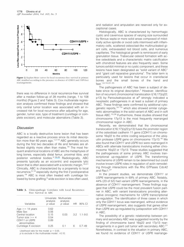
there was no difference in local recurrence-free survivalafter a median follow-up of 28 months (range, 1 to 108months) (Figure 2 and Table 3). Cox multivariate regres-sion analysis confirmed these findings and showed thatonly central tumor location was associated with an in-creased risk for local recurrence after adjusting for age,gender, tumor size, type of treatment (curettage or com-plete excision), and molecular aberrations (Table 3).
Discussion
ABC is a locally destructive bone lesion that has beenregarded as a reactive process since its initial descrip-tion more than 60 years ago.1,2,19 ABC generally occursduring the first two decades of life and females are af-fected slightly more often than males.19 The most fre-quent anatomical locations of ABC are the metaphyses oflong bones, especially distal femur, proximal tibia, andposterior vertebral bodies.3,19,20 Radiologically, ABCpresents typically as an eccentric and expansile lyticlesion that is often associated with a sclerotic margin.19,21
Clinically, ABC is characterized by a propensity to localrecurrence,8,19 especially during the first 2 postoperativeyears.19 ABC is most often treated with curettage fol-lowed by bone grafting;3 wide excision is less often used,
and radiation and amputation are reserved only for ex-ceptional cases.
Histologically, ABC is characterized by hemorrhagiccystic and cavernous spaces of varying size surroundedby fibrous septa or more solid areas composed of mitot-ically active spindle or ovoid cells intermixed with inflam-matory cells, scattered osteoclast-like multinucleated gi-ant cells, extravasated red blood cells, and numerouscapillaries. The histological growth is reminiscent of earlygranulation tissue. Trabecular osteoid formation with ac-tive osteoblasts and a characteristic matrix calcificationwith chondroid features are also frequently seen. Sometumors exhibit minimal or no cystic component, and theselesions have been designated as “solid variant of ABC”and “giant cell reparative granuloma”. The latter term isparticularly used for lesions that occur in craniofacialbones and the small bones of the hand andfeet.1,8,19,22–25
The pathogenesis of ABC has been a subject of de-bate since its original description.1 However, identifica-tion of recurrent chromosomal translocation t(16;17)(q22;p13) by Panoutsakopoulos et al9 strongly indicated aneoplastic pathogenesis in at least a subset of primaryABC. These findings were confirmed by additional cyto-genetic reports,10–15 which also showed similar cytoge-netic abnormalities in the solid variant of ABC and in softtissue ABC.10,26 Furthermore, these studies showed thatchromosome 17p13 is the most frequently rearrangedchromosomal region in ABC.
Recently, we demonstrated that the chromosomaltranslocation t(16;17)(q22;p13) fuses the promoter regionof the osteoblast cadherin 11 gene (CDH11) on chromo-some 16q22 to the entire coding sequence of the ubiq-uitin protease USP6 gene on chromosome 17p13.16 Wealso found that CDH11 and USP6 loci were rearranged inABCs with alternate translocations involving either chro-mosome 16q22 or 17p13. These studies suggested thatthe pathogenesis of some primary ABC involves tran-scriptional up-regulation of USP6. The transformingmechanisms of USP6 remain to be determined but couldinvolve known USP6 roles in regulation of actin remodel-ing through interactions with the Rho GTPases Cdc42and Rac1.27
In the present studies, we demonstrate CDH11 orUSP6 rearrangements in 69% of primary ABC. Notably,44% (23 of 52) had variant USP6 rearrangements, in theabsence of CDH11 rearrangement. These findings sug-gest that USP6 could be the most prevalent fusion part-ner in ABC, with variant translocations providing alter-native oncogenic mechanisms for USP6 transcriptionalup-regulation. The identification of three cases in whichonly the CDH11 locus was rearranged, without evidenceof USP6 rearrangement, also suggests that genes otherthan USP6 are up-regulated by juxtaposition with CDH11in ABC.
The possibility of a genetic relationship between pri-mary and secondary ABC was suggested recently by thefinding of chromosome band 16q22 and 17p13 rear-rangements in a giant cell tumor with secondary ABC.21
Nonetheless, in contrast to the situation in primary ABC,we found no evidence of CDH11 or USP6 rearrange-
Figure 2. Kaplan-Meier curves for local recurrence-free survival in primaryABC stratified according to the presence or absence of CDH11 and USP6 locirearrangements.
Table 3. Clinicopathogic Correlates with Local Recurrence-Free Survival in ABC
Variables
Univariateanalysisp value
Multivariateanalysisp value HR 95% CI
Age �� 14 years 0.62Male Sex 0.56Central location 0.15 0.03 3.2 1.1–9.0Tumor size �� 4 0.54CDH11 or USP6
abnormalities0.89
Curretage X excision 0.25 0.1 6 0.7–49
Likelihood ratio for the model: p � 0.05.p to exclude in the multivariate model: �0.10.
1778 Oliveira et alAJP November 2004, Vol. 165, No. 5

ments in secondary ABC associated with giant cell tumor,chondroblastoma, osteoblastoma, and fibrous dysplasia.Although chromosome 16 and 17 abnormalities havebeen found infrequently in giant cell tumors,21,28,29 it isunclear whether any of these involve CDH11 or USP6 loci.Based on the present evidence, we conclude that sec-ondary ABC and primary ABC have a different pathogen-esis. Secondary ABC, presumably, is a common end-point of differentiation or a non-specific morphologicalpattern in various non-ABC tumors. Despite being a mor-phological mimic of primary ABC, secondary ABC likelyhas varied genetic features, corresponding to the spe-cific bone tumors with which it is associated.
Presence of USP6 and CDH11 rearrangements did notcorrelate with ABC clinicopathological variables, nor, inunivariate and multivariate analyses, with recurrence-freesurvival. The only variable that correlated with recur-rence-free survival was tumor location, with central ABCbeing at greater risk for recurrence. However, we cannotexclude the possibility that specific USP6 or CDH11 re-arrangement mechanisms are predictive of recurrence.Therefore, it would be worthwhile to evaluate relation-ships between molecular aberrations and ABC recur-rence further, ideally in the context of a larger-scale andprospective clinical study.
The paraffin-section FISH analyses in this study en-abled evaluation of ABC cell types which had the USP6and CDH11 rearrangements. All cells with USP6 orCDH11 rearrangements were spindle or ovoid, and werediffusely admixed within the other ABC cell components,as well as being clustered around osteoclast-like multinu-cleated giant cells. The lack of cytogenetic aberrations inthe inflammatory cells, endothelial cells, metaplasticbone-associated osteoblasts, and the multinucleated os-teoclast-like giant cells supports their reactive nature.Conceivably, they are involved in a host response to theneoplastic ABC cells. It is also possible that one or moreof the non-neoplastic cellular constituents release factorsthat stimulate division of the neoplastic ABC cells. Thefrequent clustering of neoplastic cells in the vicinity ofmultinucleated giant cells suggests a role for these cellsin the neoplastic proliferation. Notably, the ABC cells withUSP6 or CDH11 rearrangements were generally admixedwith morphologically indistinguishable spindle cells lack-ing such rearrangements. We hypothesize that the lattergroup is composed by reactive fibroblasts/myofibro-blasts, but it is also possible that some of these arecommitted ABC neoplastic cells at an earlier point intransformation, before acquisition of the USP6 or CDH11rearrangements. Although the majority of cells in someABC had USP6 or CDH11 rearrangements, there werealso tumors in which fewer than 10% of cells had rear-rangements (Table 2). The small numbers of apparentlyrearranged cells were not false-positive findings, as theywere reproducibly present in these tumors when the FISHwas repeated, and when scored by independent observ-ers. Furthermore, RT-PCR reactions corroborated thesemolecular cytogenetic results showing the presence ofCDH11-USP6 fusion transcripts in these tumors. In all,these findings suggest that the neoplastic ABC compo-nent induces a vigorous, reactive, host response mimick-
ing young granulation tissue, including inflammatory, myo-fibroblastic, osteoclast-like giant cells, osteoblastic cells,and numeous capillaries, therefore accounting for the his-torical perception that these are largely reactive lesions.
In conclusion, primary ABC is a mesenchymal neo-plastic disease characterized by a spindle cell prolifera-tion exhibiting USP6 or CDH11 rearrangements in ap-proximately two thirds of the cases. Secondary ABC is amorphological mimic of primary ABC, but lacks the hall-mark USP6 or CDH11 rearrangements of primary ABC,and likely represents a common endpoint of differentia-tion in various non-ABC bone tumors.
References
1. Jaffe H, Lichtenstein L: Solitary unicameral bone cyst: with emphasison the roentgen picture, the pathologic appearance, and the patho-genesis. Arch Surg 1942, 44:1004–1025
2. Fechner R, Mills S: Non-neoplastic lesions that mimic neoplasms:aneurysmal bone cyts. Edited by J Rosai. Washington, Armed ForcesInstitute of Pathology, 1993, pp 253–258
3. Dorfman H, Czerniak B: Cystic lesions. Bone Tumors. St. Louis,Mosby, 1998
4. Clough JR, Price CH: Aneurysmal bone cyst: pathogenesis and longterm results of treatment. Clin Orthop 1973, 97:52–63
5. Kransdorf MJ, Sweet DE: Aneurysmal bone cyst: concept, contro-versy, clinical presentation, and imaging. AJR Am J Roentgenol 1995,164:573–580
6. Levy WM, Miller AS, Bonakdarpour A, Aegerter E: Aneurysmal bonecyst secondary to other osseous lesions: report of 57 cases. Am J ClinPathol 1975, 63:1–8
7. Martinez V, Sissons HA: Aneurysmal bone cyst: a review of 123 casesincluding primary lesions and those secondary to other bone pathol-ogy. Cancer 1988, 61:2291–2304
8. Rosenberg AE, Nielsen GP, and Fletcher JA: Aneurysmal bone cyst.World Health Organization Classification of Tumours: Pathology andGenetics of Tumours of Soft Tissue and Bone. Edited by FletcherCDM, Unni KK, Mertens F. Lyon, IARC Press, 2002, pp 338–339
9. Panoutsakopoulos G, Pandis N, Kyriazoglou I, Gustafson P, MertensF, Mandahl N: Recurrent t(16;17)(q22;p13) in aneurysmal bone cysts.Genes Chromosomes Cancer 1999, 26:265–266
10. Dal Cin P, Kozakewich HP, Goumnerova L, Mankin HJ, RosenbergAE, Fletcher JA: Variant translocations involving 16q22 and 17p13 insolid variant and extraosseous forms of aneurysmal bone cyst. GenesChromosomes Cancer 2000, 28:233–234
11. Althof PA, Ohmori K, Zhou M, Bailey JM, Bridge RS, Nelson M, NeffJR, Bridge JA: Cytogenetic and molecular cytogenetic findings in 43aneurysmal bone cysts: aberrations of 17p mapped to 17p13.2 byfluorescence in situ hybridization. Mod Pathol 2004, 17:518–525
12. Baruffi MR, Neto JB, Barbieri CH, Casartelli C: Aneurysmal bone cystwith chromosomal changes involving 7q and 16p. Cancer GenetCytogenet 2001, 129:177–180
13. Herens C, Thiry A, Dresse MF, Born J, Flagothier C, Vanstraelen G,Allington N, Bex V: Translocation (16;17)(q22;p13) is a recurrentanomaly of aneurysmal bone cysts. Cancer Genet Cytogenet 2001,127:83–84
14. Winnepenninckx V, Debiec-Rychter M, Jorissen M, Bogaerts S, SciotR: Aneurysmal bone cyst of the nose with 17p13 involvement. Vir-chows Arch 2001, 439:636–639
15. Wyatt-Ashmead J, Bao L, Eilert RE, Gibbs P, Glancy G, McGavran L:Primary aneurysmal bone cysts: 16q22 and/or 17p13 chromosomeabnormalities. Pediatr Dev Pathol 2001, 4:418–419
16. Oliveira AM, Hsi BL, Weremowicz S, Rosenberg AE, Dal Cin P,Joseph N, Bridge JA, Perez-Atayde AR, Fletcher JA: USP6 (Tre2)fusion oncogenes in aneurysmal bone cyst. Cancer Res 2004, 64:1920–1923
17. Hibbard MK, Kozakewich HP, Dal Cin P, Sciot R, Tan X, Xiao S,Fletcher JA: PLAG1 fusion oncogenes in lipoblastoma. Cancer Res2000, 60:4869–4872
18. Argani P, Zakowski MF, Klimstra DS, Rosai J, Ladanyi M: Detection of
Identification of Neoplastic Cell in ABC 1779AJP November 2004, Vol. 165, No. 5

the SYT-SSX chimeric RNA of synovial sarcoma in paraffin-embed-ded tissue and its application in problematic cases. Mod Pathol 1998,11:65–71
19. Vergel De Dios AM, Bond JR, Shives TC, McLeod RA, Unni KK:Aneurysmal bone cyst: a clinicopathologic study of 238 cases. Can-cer 1992, 69:2921–2931
20. Biesecker JL, Marcove RC, Huvos AG, Mike V: Aneurysmal bone cysts:a clinicopathologic study of 66 cases. Cancer 1970, 26:615–625
21. Sciot R, Dorfman H, Brys P, Dal Cin P, De W, I, Fletcher CD, JonsonK, Mandahl N, Mertens F, Mitelman F, Rosai J, Rydholm A, Samson I,Tallini G, Van den BH, Vanni R, Willen H: Cytogenetic-morphologiccorrelations in aneurysmal bone cyst, giant cell tumor of bone andcombined lesions: a report from the CHAMP study group. Mod Pathol2000, 13:1206–1210
22. Sanerkin NG, Mott MG, Roylance J: An unusual intraosseous lesionwith fibroblastic, osteoclastic, osteoblastic, aneurysmal, and fibro-myxoid elements: “solid” variant of aneurysmal bone cyst. Cancer1983, 51:2278–2286
23. Wold LE, Dobyns JH, Swee RG, Dahlin DC: Giant cell reaction (giant
cell reparative granuloma) of the small bones of the hands and feet.Am J Surg Pathol 1986, 10:491–496
24. Waldron CA, Shafer WG: The central giant cell reparative granuloma ofthe jaws: an analysis of 38 cases. Am J Clin Pathol 1966, 45:437–447
25. Gipple JR, Pritchard DJ, Unni KK: Solid aneurysmal bone cyst. Or-thopedics 1992, 15:1433–1436
26. Nielsen GP, Fletcher CD, Smith MA, Rybak L, Rosenberg AE: Softtissue aneurysmal bone cyst: a clinicopathologic study of five cases.Am J Surg Pathol 2002, 26:64–69
27. Masuda-Robens JM, Kutney SN, Qi H, Chou MM: The TRE17 onco-gene encodes a component of a novel effector pathway for RhoGTPases Cdc42 and Rac1 and stimulates actin remodeling. Mol CellBiol 2003, 23:2151–2161
28. Bridge JA, Neff JR, Mouron BJ: Giant cell tumor of bone: chromo-somal analysis of 48 specimens and review of the literature. CancerGenet Cytogenet 1992, 58:2–13
29. Molenaar WM, van den BE, Dolfin AC, Zorgdrager H, Hoekstra HJ:Cytogenetics of fine needle aspiration biopsies of sarcomas. CancerGenet Cytogenet 1995, 84:27–31
1780 Oliveira et alAJP November 2004, Vol. 165, No. 5
Copyright © 2022 FDOKUMEN