Brain Tumors
337
Transcript of Brain Tumors


C o n s u l t a n t Pa t h o l o g y
brain tumors

Consultant Pathology Series
David E. Elder, MB, ChBSeries Editor
Tumorigenic Melanocytic Proliferations
David E. Elder
Brain Tumors
Richard Prayson, Bette Kleinschmidt-DeMasters, and Mark L. Cohen
Forthcoming Volumes in the Series
Thyroid Papillary Lesions
Virginia A. LiVolsi and Jennifer L. Hunt
Urinary Bladder Diagnosis
Robert O. Petersen
Head and Neck Pathology
Leon Barnes, Raja Seethala, and Simion Chiosea
Liver Pathology
Linda Ferrell and Sanjay Kakar

Richard Prayson, MD
Section Head of Neuropathology
Cleveland Clinic Main Campus
Professor of Pathology
Lerner College of Medicine
Cleveland, Ohio
Bette K. Kleinschmidt-DeMasters, MD
Head of Neuropathology
University of Colorado
Professor of Pathology
University of Colorado School of Medicine
Denver, Colorado
Mark L. Cohen, MD
Director, Division of Neuropathology
University Hospital of Cleveland
Professor of Pathology
Case Western Reserve University School of Medicine
Cleveland, Ohio
C o n s u l t a n t Pa t h o l o g y
brain tumors
New York

Acquisitions Editor: Richard Winters
Cover Design: Joe Tenerelli
Compositor: Publication Services, Inc.
Printer: Sheridan Press
Visit our website at www.demosmedpub.com
© 2010 Demos Medical Publishing, LLC. All rights reserved. This book is protected by copyright. No part of it may be reproduced,stored in a retrieval system, or transmitted in any form or by any means, electronic, mechanical, photocopying, recording, or otherwise,without the prior written permission of the publisher.
Medicine is an ever-changing science. Research and clinical experience are continually expanding our knowledge, in particular ourunderstanding of proper treatment and drug therapy. The authors, editors, and publisher have made every effort to ensure that allinformation in this book is in accordance with the state of knowledge at the time of production of the book. Nevertheless, the authors,editors, and publisher are not responsible for errors or omissions or for any consequences from application of the information in this book and make no warranty, express or implied, with respect to the contents of the publication. Every reader should examine care-fully the package inserts accompanying each drug and should carefully check whether the dosage schedules mentioned therein or thecontraindications stated by the manufacturer differ from the statements made in this book. Such examination is particularly importantwith drugs that are either rarely used or have been newly released on the market.
Library of Congress Cataloging-in-Publication Data
Prayson, Richard A. Brain tumors / Richard Prayson, Mark L. Cohen, Bette Kleinschmidt-DeMasters. p. ; cm. — (Consultant pathology) Includes bibliographical references and index.
ISBN 978-1-933864-69-3 (hardcover) 1. Brain — Tumors — Case studies. I. Cohen, Mark L., 1957- II. Kleinschmidt-DeMasters, Bette. III. Title. IV. Series: Consultantpathology. [DNLM: 1. Brain Neoplasms — diagnosis — Case Reports. 2. Brain Neoplasms — pathology — Case Reports. WL 358 P921b 2010] RC280.B7P725 2010 616.99'481 — dc22 2009040379
Made in the United States of America
09 10 11 12 13 5 4 3 2 1
Special discounts on bulk quantities of Demos Medical Publishing books are available to corporations, professional associations,pharmaceutical companies, health care organizations, and other qualifying groups. For details, please contact:
Special Sales Department
Demos Medical Publishing
11 W. 42nd Street, 15th Floor
New York, NY 10036
Phone: 800–532–8663 or 212–683–0072
Fax: 212–941–7842
E-mail: [email protected]

To Beth, Brigid, and Nick for their unwavering support.
To my family—husband Bob, sons Tim and Tor, and daughter Julie— without whom nothing in my career would have been possible.
To my parents, Beverly and Murray Cohen, without whom my contribution would not have been possible.
To our patients, who have taught us so that we may better help others.

This page intentionally left blank

C o n t e n t s
Series Foreword xiPreface xiiiAcknowledgments xv
1. Normal Tissue 1 2. Gliosis 4 3. Recurrent High-Grade Glioma with Radiation Changes 6 4. Low-Grade Astrocytoma 10 5. Anaplastic Astrocytoma 13 6. Glioneronal Tumor with Neuropil-Like Islands 16 7. Glioblastoma Multiforme 19 8. Gemistocytic Astrocytoma 22 9. Granular Cell Glioblastoma 25 10. Giant Cell Glioblastoma 28 11. Pleomorphic Xanthoastrocytoma 31 12. Gliosarcoma 34 13. Small Cell Glioblastoma 37 14. Epithelioid Glioblastoma 40 15. Gliomatosis Cerebri 43 16. Pilocytic Astrocytoma 45 17. Pilomyxoid Astrocytoma 49 18. Subependymal Giant Cell Astrocytoma 52 19. Low-Grade Oligodendroglioma 55 20. Anaplastic Oligodendroglioma 59 21. Low-Grade Oligoastrocytoma (Low-Grade Mixed Glioma) 62 22. Anaplastic Oligoastrocytoma (Anaplastic Mixed Glioma) 65 23. Glioblastoma with Oligodendroglioma Component 67 24. Subependymoma 70 25. Myxopapillary Ependymoma 73 26. Ependymoma 76 27. Anaplastic Ependymoma 80 28. Tanycytic Ependymoma 84
vii

29. Clear Cell Ependymoma 87 30. Choroid Plexus Papilloma 91 31. Atypical Choroid Plexus Papilloma 94 32. Choroid Plexus Carcinoma 97 33. Chordoid Glioma 100 34. Angiocentric glioma 103 35. Astroblastoma 106 36. Dysplastic Cerebellar Gangliocytoma (Lhermitte-Duclos Disease) 108 37. Desmoplastic Infantile Astrocytoma/Ganglioglioma 110 38. Dysembryoplastic Neuroepithelial Tumor 114 39. Ganglioglioma 117 40. Anaplastic Ganglioglioma 120 41. Papillary Glioneuronal Tumor 123 42. Rosette-Forming Glioneuronal Tumor of the Fourth Ventricle 125 43. Central Neurocytoma 128 44. Atypical Neurocytoma 131 45. Extraventricular Neurocytoma 134 46. Paraganglioma 137 47. Pineocytoma 140 48. Pineal Parenchymal Tumor of Intermediate Differentiation 143 49. Pineoblastoma 146 50. Yolk Sac Tumor of the Pineal Gland 148 51. Supratentorial Primitive Neuroectodermal Tumor 150 52. Classic Medulloblastoma 153 53. Desmoplastic Medulloblastoma 156 54. Medulloblastoma with Extensive Nodularity 159 55. Anaplastic Medulloblastoma 161 56. Atypical Teratoid/Rhabdoid Tumor 164 57. Embryonal Tumor with Abundant Neuropil and True Rosettes 168 58. Schwannoma with Ancient Change 171 59. Neurofi broma 174 60. Perineurioma 177 61. Malignant Peripheral Nerve Sheath Tumor 180 62. Cellular Schwannoma 183 63. Melanotic Schwannoma 186 64. Fibrous Meningioma 190 65. Ectopic Meningioma 193 66. Clear Cell Meningioma 195 67. Chordoid Meningioma 198
vii i
C O N T E N T S

68. Papillary Meningioma 201 69. Rhabdoid Meningioma 204 70. Brain-Invasive Meningioma 207 71. Atypical Meningioma 209 72. Anaplastic Meningioma 211 73. Angiomatous Meningioma 214 74. Hemangiopericytoma 217 75. Solitary Fibrous Tumor 221 76. Primary Sarcoma of the Central Nervous System 224 77. Meningioangiomatosis 228 78. Hemangioblastoma 231 79. Meningeal Melanocytoma 234 80. Malignant Melanoma 237 81. Lymphoma with First Presentation as Spinal Cord Compression 240 82. Marginal Zone B-Cell Lymphoma 244 83. Post-Transplant Lymphoproliferative Disorder 247 84. Plasmacytoma 251 85. Langerhans Cell Histiocytosis 254 86. Intravascular Lymphomatosis (Angiotropic Large Cell Lymphoma) 257 87. Germinoma 260 88. Pineal Teratoma 262 89. Cystic Craniopharyngioma 265 90. Papillary Craniopharyngioma 268 91. Granular Cell Tumor of the Pituitary Gland 271 92. Pituicytoma 273 93. Pituitary Adenoma with Apoplexy 276 94. Pituitary Adenoma in an Ectopic Site 280 95. Metastatic Small Cell Carcinoma of Lung 282 96. Leukemic Involvement of the Central Nervous System 285 97. Chordoma 288 98. Metastatic Papillary Carcinoma of the Thyroid 291 99. Leptomeningeal Carcinomatosis 294100. Dural Carcinomatosis 296101. Mesenchymal Chondrosarcoma 299Index 303
ix
C O N T E N T S

This page intentionally left blank

xi
S e r i e s F o r e w o r d
Diagnostic surgical pathology remains the gold standard for diagnosis of most tumors and many
infl ammatory conditions in most, if not all, organ sys-tems. The power of the morphologic method is such that, in many instances, a glance at a thin section of tissue stained with two vegetable dyes is suffi cient to determine with absolute certainty whether a patient should undergo a major procedure or not, or whether a patient is likely to live a healthy life or die of an inoperable tumor. In such cases, the diagnostic pro-cess is one of “gestalt,” a form of almost instantaneous pattern recognition that is similar to the recognition of faces, different brands of automobiles, or breeds of dogs. In other “diffi cult” cases, the diagnosis is not so obvious. In many of these cases, a diagnosis may be possible, but may be outside of the experience of the routine practitioner. In such a circumstance, it may be possible for a practitioner with more experience—a consultant—to make a diagnosis rather readily. In other cases, the problem may really not be suited to the histologic method. In these cases as well, a consul-tant may be invaluable in determining that it is sim-ply not possible to make a reliable diagnosis with the materials available. In yet other cases, the diagnosis may be ambiguous, and again a consultant’s opinion can be important in establishing a differential diagno-sis that may guide clinical investigation.
There are many fi ne consultants available to the practicing surgical pathology community. Many of them have authored textbooks, and many of them give presentations at national meetings. However, these materials can offer only a superfi cial insight
into the vast amount of knowledge that is embed-ded in these individuals’ cerebral cortices—and in their fi ling cabinets. This series represents an effort to enable the dissemination of this hitherto-inaccessible knowledge to the wider community. Our authors are individuals who have accumulated large collections of diffi cult cases and are willing to share their material and their knowledge. The cases are based on actual consultations, and the indications for the consulta-tion, when available, are presented, because these are the records of the manner in which these cases presented themselves as being problematic. We have asked the consultants, when possible, to present their consultation letters in much the same form (albeit edited to some degree) as that in which they were fi rst presented, because these represent the true records of the clinical encounter. In addition, we asked the authors to amplify upon these descriptions, with brief reference to the literature, and to richly illustrate the case reports with high-quality digital images. The images from the book, as well as additional images to amplify the presentation of the case, are available on a website for downloading, study, and use in educa-tion. These images, in some cases, have been derived from virtual slides, which also may be made available in the future from a digital repository for their addi-tional educational value.
David E. Elder, MB, ChB, FRCPAProfessor of Pathology and Laboratory Medicine
Hospital of the University of PennsylvaniaPhiladelphia, Pennsylvania

This page intentionally left blank

xii i
P r e f a c e
The practice of surgical neuropathology is challenging. In part, this is related to the relative
lack of experience most pathologists have in this arena as compared with other areas of surgical pathology. Thus, a selection of neuropathology cases fi ts well into the Consultant Pathology series—a series of texts that will cover the spectrum of surgical pathology and will examine topics in a case-based format, similar to the real practice of pathology.
The focus of this text is on brain tumors. Examples of over 100 brain tumors, running the gamut from the very common to the rare, are pre-sented in a case-based format. The cases were taken from our surgical neuropathology practices. We attempt to share with the reader our approaches and thought processes in evaluating the spectrum of central nervous system neoplasms. The wide variety of cases presented covers the entire scope of brain
tumors and offers both an opportunity to review the basics for the beginner or relatively inexperienced pathologist, and a chance to see some of the rare entities. When relevant, current practical appli-cations of immunohistochemistry and molecular pathology are discussed.
Each case is formatted as if it were a consult case and includes a brief clinical history, a description of the pathologic fi ndings with numerous illustrations, the line diagnosis, a discussion of the entity and the diagnostic thought process, and a few pertinent refer-ences for further reading.
Our hope is that by sharing a bit of our experi-ence, we can add to the reader’s experience.
Richard Prayson, MDBette Kleinschmidt-DeMasters, MD
Mark Cohen, MD

This page intentionally left blank

xv
A c k n o w l e d g m e n t s
Special thanks to Ms. Denise Egleton, who provided secretarial assistance for this project.

This page intentionally left blank

C o n s u l t a n t Pa t h o l o g y
brain tumors

This page intentionally left blank

1
Case 1: Normal Tissue
C L I N I C A L I N F O R M A T I O N
The patient is a 28-year-old female who presents with headaches and complaints of dizziness. On imaging, subtle abnormalities are noted in the right cerebel-lar hemisphere. Because of persistent symptoms, a decision is made to biopsy the patient, and histologic sections are reviewed.
O P I N I O N
Biopsies show normocellular cerebellar parenchyma. Because of the unusual orientation of the specimen, a grouping of small cells appears to be positioned in the middle of the gray matter.
We consider the biopsy as representing normal tissue and characterize it as follows: Right Cerebel-lum, Biopsy—Cerebellar Tissue with No Signifi cant Pathologic Changes.
C O M M E N T
There is no defi nite evidence of neoplasm on the biopsy. There is no evidence of infl ammation.
D I S C U S S I O N
The usual target of a brain biopsy does not include normal tissue. Occasionally, however, the surgeon may not be on target, and the normal tissue may be inadvertently biopsied. In most instances, recognition of the tissue as normal is not problem-atic. On occasion, however, as a result of either the
FIGURE 1.1 Section of cerebellum from this biopsy, show-ing a group of small, round cells from the granular cell layer, surrounded by molecular layer cortex. If one does not appreciate the unusual orientation of the section and the location of the biopsy, an erroneous diagnosis of chronic infl ammation or encephalitis may be made.
FIGURE 1.2 Normal, well-oriented cerebellum, showing the superfi cial molecular layer, Purkinje cell layer, and granular cell layer.

2
C A S E 1 : N O R M A L T I S S U E
orientation of the specimen or lack of information regarding the location of the biopsy, normal tissue may mimic a tumor. This case illustrates one such example, in which failure to recognize the tissue as being from the cerebellum may result in a misinter-pretation of the granular cells as lymphocytes; the result would be an erroneous diagnosis of chronic infl ammation or encephalitis. Other circumstances in which normal tissue may be confused with a pathologic process include the following: (1) when
one is unsure of the exact location of the biopsy, a biopsy from the pineal gland may be misinter-preted as representing either a pineal gland tumor, such as pineocytoma or a glioma; (2) a biopsy from the subependymal zone may be interpreted as a low-grade glioma; this region frequently is mildly hypercellular in the normal state; (3) a biopsy from the pituitary neurohypophysis may resemble a low-grade astrocytoma, such as pilocytic astrocytoma, or
FIGURE 1.3 Normal pineal gland can mimic an anaplastic glioma.
FIGURE 1.4 The subependymal zone may appear mildly hypercellular and cause confusion with a low-grade glioma.
FIGURE 1.5 Pituitary neurohypophysis may resemble a low-grade astrocytoma or schwannoma.
FIGURE 1.6 Corpora amylacea may resemble rounded cells at low magnifi cation and can be confused with an oligoden-droglioma.

3
C A S E 1 : N O R M A L T I S S U E
a schwannoma; (4) the presence of numerous cor-pora amylacea (which may be a normal fi nding in a biopsy) can be misinterpreted as representing a low-grade tumor, particularly an oligodendroglioma, given the generally rounded nature of the corpora amylacea bodies.
To help avoid some of these confusions, com-municating with the surgeon and knowing exactly where the biopsy is from are important.
References 1. Brat DJ. Overview of central nervous system anatomy
and histology. In: Prayson RA, editor. Neuropathology. Philadelphia: Elsevier Churchill Livingstone; 2005. p. 1–36.
2. Kleinschmidt-DeMasters BK, Prayson RA. An algorithmic approach to the brain biopsy—Part 1. Arch Pathol Lab Med 2006;130:1630–8.

4
Case 2: Gliosis
C L I N I C A L I N F O R M A T I O N
The patient is a 46-year-old female who presents with seizures starting seven months ago. On imaging, a low signal intensity area in the right frontal lobe, which is suspicious for a low-grade glioma, is identifi ed. An open biopsy is performed, and diagnostic material is not obtained. The procedure is aborted early because of hemorrhage. A subsequent attempt, four months later, to reassess yields histologic sections that are reviewed.
O P I N I O N
The second resection shows mild hypercellular-ity. The increased cellularity is a result of cells marked by abundant eosinophilic cytoplasm and an eccentrically placed, round-to-oval nucleus.
Occasional nuclei show small nucleoli. Mitotic fi g-ures are not observed. Malignant appearing astrocytic cells are not noted. There is no evidence of necrosis.
We consider the fi ndings in this lesion to be consistent with a benign reactive process and char-acterize it as follows: Right Frontal Lobe, Excision—Reactive Astrocytosis.
C O M M E N T
There is no defi nite evidence of neoplasm. The changes seen in this biopsy may be related to the patient’s prior surgery.
D I S C U S S I O N
One of the most diffi cult differential diagnostic situations in surgical neuropathology involves the
FIGURE 2.1 Low magnifi cation view of the biopsy, showing a mild increase in cellularity. The increased cellularity appears to be relatively evenly distributed in this microscopic fi eld, which is more consistent with reactive astrocytosis.
FIGURE 2.2 High magnifi cation appearance, showing scat-tered larger cells with abundant eosinophilic cytoplasm, corresponding to reactive astrocytes.
5
C A S E 2 : G L I O S I S
differentiation of reactive astrocytosis from low-grade glioma. It is particularly problematic because most gliomas elicit some degree of reactive astrocytosis. Reactive astrocytes are usually marked by increased eosinophilic cytoplasm and nuclear enlargement. The nuclear-to-cytoplasmic ratio is relatively low. In contrast, low-grade astrocytomas are marked by cells
with a higher nuclear-to-cytoplasmic ratio. Nuclear contour irregularities and nuclear hyperchromasia are more typically noted. An occasional mitotic fi gure may also be present. Additionally, reactive astrocytes are fairly evenly distributed in a microscopic fi eld—in contrast to malignant astrocytic cells, which may show some uneven distribution of cellularity. Calcifi -cations, microcystic degenerative changes, and satel-litosis, in the form of secondary structures of Scherer, are more typically observed in gliomas than in glio-sis.
References 1. Burger PC, Vogel FS. Frozen section interpretation in surgi-
cal neuropathology. I. Intracranial lesions. Am J Surg Pathol 1977;1:323–47.
2. Prayson RA, Cohen ML. Gliosis. In: Practical Differential Diagnosis in Surgical Neuropathology. Totowa, NJ: Humana Press; 2000. p. 5–7.
3. Taratuto AL, Sevlever G, Piccardo P. Clues and pitfalls in ste-reotactic biopsy of the central nervous system. Arch Pathol Lab Med 1991;115:596–602.
FIGURE 2.5 More dense gliosis can develop over time, adja-cent to a previous surgical site or secondary to radiation therapy.
FIGURE 2.4 Prominent perivascular gliosis is present.
FIGURE 2.3 High magnifi cation, showing the characteris-tic features of reactive astrocytes, including low nuclear-to-cytoplasmic ratio, nuclear enlargement with mild nuclear contour irregularities, and lack of nuclear hyperchromasia.

6
Case 3: Recurrent High-Grade Glioma with Radiation Changes
C L I N I C A L I N F O R M A T I O N
The patient is a 58-year-old male who presents with a right parietal-temporal, focally enhancing mass. The patient is biopsied, and a diagnosis of glioblastoma is made. He subsequently undergoes a course of radiother-apy. On recent follow-up imaging studies, the lesion appears to be expanding in size, and a decision to place an intraoperative chemotherapeutic agent is made. The patient subsequently undergoes partial resection of the region, and histologic sections are reviewed.
O P I N I O N
Histologic sections show focally prominent num-bers of reactive astrocytes, accompanied by perivas-cular chronic infl ammation consisting primarily of
benign-appearing lymphocytes. Several vessels show sclerotic changes, with homogeneous thickening of ves-sel walls. Areas of geographic necrosis, not rimmed by a pseudopalisade of atypical-appearing cells, are identifi ed. Focal microcalcifi cations are observed. In areas includ-ing the frozen section slide, more prominent cellularity, marked by atypical-appearing cells and fi brosis, is seen. Occasional mitotic fi gures are identifi ed in the more cellular areas. Foci of vascular proliferative changes, in some instances focally altered by fi brosis, are observed.
We consider the pathology to be that of a recurrent or residual high-grade astrocytoma (glioblastoma) with superimposed therapy effect, and we characterize it as follows: Right Parietal-Temporal Lobe, Excision—Recurrent/Residual Glioblastoma, WHO Grade IV. Changes Consis-tent with Radiation Therapy Effect.
FIGURE 3.1 Reactive astrocytes, characterized by abundant eosinophilic cytoplasm and eccentrically placed, round-to-oval nuclei, are commonly seen in the setting of radio-therapy effect.
FIGURE 3.2 Focal perivascular chronic infl ammation, marked by benign-appearing lymphocytes is frequently seen.

7
C A S E 3 : R E C U R R E N T H I G H - G R A D E G L I O M A W I T H R A D I A T I O N C H A N G E S
C O M M E N T
Although many of the changes in the resection are secondary to radiation therapy effect, there is evidence of defi nitive recurrent or residual glioblastoma.
D I S C U S S I O N
Standard treatment for high-grade astrocytoma includes radiotherapy. Radiation can cause changes in tissue that are important to recognize so that
they are not confused with tumor. It is important to remember that the morphologic changes sec-ondary to radiotherapy are typically delayed and do not manifest themselves until weeks or months after the radiation has been administered. The ear-liest changes include the development of edema and perivascular chronic infl ammation. Reactive astrocytosis and gliosis may be focally prominent. Areas of white matter demyelination can develop. Vascular sclerotic changes can become prominent, and eventually necrosis may develop. It is com-mon for dystrophic mineralization, often associated with zones of necrosis, to be evident. Frequently, areas of necrosis may contain macrophages. In some instances, particularly in the meninges, a promi-nent fi broblastic reaction may ensue. In contrast to necrosis associated with glioblastoma, a palisading of reactive astrocytes around necrotic zones is not seen.
In order to distinguish recurrent or residual glioma from radiation therapy effect, useful features to assess include the following: (1) Look for cytologic atypia. Cells with high nuclear-to-cytoplasmic ratio, irregular nuclear contours and nuclear hyperchromasia
FIGURE 3.3 Vascular sclerotic changes, similar to those observed in other organ systems, can be seen in the irradi-ated brain.
FIGURE 3.4 Sclerotic vascular changes are accompanied by a zone of geographic necrosis.
FIGURE 3.5 Focal dystrophic mineralization may develop in the irradiated brain.

8
C A S E 3 : R E C U R R E N T H I G H - G R A D E G L I O M A W I T H R A D I A T I O N C H A N G E S
FIGURE 3.6 Fibrosis, with intervening atypical-appearing cells, is present. An occasional cell shows marked cytologic atypia, which may represent radiation-related atypia in a neoplastic astrocyte.
are generally tumor cells. Radiation may also induce cytologic atypia. In this instance, the atypical cells are often very bizarre and may be multinucleated. (2) Mitotic fi gures are indicative of tumor, not reactive astrocytosis or gliosis associated with radiotherapy. (3) True vascular proliferative changes are indicative of recurrent or residual tumor. The vascular changes
associated with radiotherapy are primarily those of sclerosis and fi brosis of vessel walls. (4) Palisaded ne-crosis is a feature of glioblastoma, not radionecrosis.
In this particular case, recognition of the pres-ence of recurrent or residual tumor at the time of intraoperative consultation is important in dictating
FIGURE 3.7 Scattered, bizarre-appearing astrocytic cells, in the background of dense gliosis, are indicative of irradiated, recurrent tumor.
FIGURE 3.9 Vascular proliferative changes (also indicative of recurrent or residual glioblastoma) are focally altered by radiation-associated vascular sclerosis. Several adjacent smaller vessels also show vascular sclerotic changes second-ary to radiation.
FIGURE 3.8 Mitotic fi gures, in association with atypical-appearing astrocytic cells, indicate recurrent or residual glioblastoma.

9
C A S E 3 : R E C U R R E N T H I G H - G R A D E G L I O M A W I T H R A D I A T I O N C H A N G E S
placement of the chemotherapeutic agent into the surgical bed. Sometimes, in the setting of radiated gliomas, patients are biopsied to assess or evaluate the nature of changes observed on imaging studies, i.e., whether changes are secondary to radiation therapy effect or the tumor is resistant to radiation therapy and another treatment modality should be considered.
References 1. Burger PC, Mahaley Jr MS, Dudka L, et al. The morpho-
logic effects of radiation administered therapeutically for intracranial gliomas: a postmortem study of 25 cases. Cancer 1979;44:1256–72.
2. Prayson RA, Cohen ML. Radiation change. In: Practical Differential Diagnosis in Surgical Neuropathology. Totowa, NJ: Humana Press; 2000. p. 27–31.

10
Case 4: Low-Grade Astrocytoma
C L I N I C A L I N F O R M A T I O N
The patient is a 38-year-old female who presents with seizures. On computed tomography (CT) imaging, an ill-defi ned, homogeneous-appearing mass of low signal intensity is noted in the right temporal lobe. Stereotactic biopsies of the tumor are taken, and his-tologic sections are reviewed.
O P I N I O N
On the frozen section slide, mildly hypercellular parenchyma is noted. Occasional atypical-appearing astrocytic cells, marked by nuclear enlargement and irregularities of nuclear contour, are identifi ed, sugges-tive of an infi ltrating astrocytoma. On permanent sec-tions, hypercellularity is evident. Atypical- appearing
astrocytic cells are clearly present. In focal areas, subpial aggregation of atypical astrocytic cells is noted. Mitotic fi gures, vascular proliferative changes, and necrosis are not seen. There is no evidence of Rosenthal fi bers or granular bodies. A Ki-67 immunostain shows a label-ing index of 0.4.
We consider the lesion an astrocytoma and characterize it as follows: Right Temporal Lobe, Biopsies—Low-Grade Fibrillary Astrocytoma, WHO Grade II.
C O M M E N T
The tumor has a low rate of cell proliferation (Ki-67 labeling index 0.4), consistent with a low-grade tumor. Features of higher-grade astrocytoma, including
FIGURE 4.1 Low magnifi cation appearance of the frozen section, showing a mildly hypercellular parenchyma, raising the question of a glioma versus gliosis differential diagnosis.
FIGURE 4.2 Hypercellular parenchyma shows scattered, atypical-appearing astrocytic cells suggestive of a low-grade astrocytoma.

11
C A S E 4 : L O W - G R A D E A S T R O C Y T O M A
mitotic activity, vascular proliferative changes, and necrosis, are not seen.
D I S C U S S I O N
The most common primary tumor of the central ner-vous system is the astrocytoma; in most instances, the tumors are high-grade (glioblastoma multiforme). Astrocytomas are infi ltrative lesions. Low-grade tumors usually present as low signal intensity, some-what ill-defi ned lesions on imaging. Because of the widely infi ltrative nature of these tumors, surgical cure is seldom realized. Microscopically, the tumors are marked by mildly hypercellular parenchyma. The increased cellularity is often not evenly distrib-uted. Increased cellularity is a result of two compo-nents. Atypical-appearing astrocytic cells, marked by nuclear enlargement, nuclear hyperchromasia, and nuclear pleomorphism, are evident. The tumor cells may be accompanied by reactive astrocytes, which are generally characterized by increased eosinophilic cytoplasm and enlarged nuclei, with round-to-oval nuclear contours. With infi ltration, the malignant tumor cells have a propensity to satellite around pre-existing structures, including neurons and blood
vessels. Subpial aggregation of tumor cells, as they infi ltrate toward the surface of the cortex, may also be observed. True microcystic change is a variable but useful diagnostic feature of tumor, as opposed to gliosis. Microcalcifi cations may be evident in approx-imately 15% of low-grade astrocytomas. Mitotic fi g-ures are generally diffi cult to identify or are evident in low numbers.
Cell proliferation markers can be helpful in dif-ferentiating between low-grade tumors and tumors that are more likely to behave in an aggressive fashion. Ki-67 is probably the most widely utilized of these im-munohistochemical markers. Low-grade astrocytomas typically have low labeling indices, usually less than 3–4%. Higher labeling indices are often associated with more cellular lesions and may be indicative of a more aggressive-behaving tumor, akin to anaplastic astrocytoma or WHO grade III neoplasms. In trying to differentiate gliosis from low-grade glioma, a high labeling index is more indicative of a glioma. A low labeling index is not helpful in this differential diag-nosis, because low rates of cell proliferation have been described in association with reactive astrocytosis.
FIGURE 4.3 Low magnifi cation appearance on permanent section is marked by increased cellularity.
FIGURE 4.4 Atypical-appearing astrocytes, marked by nuclear enlargement, irregularities to nuclear contour, and nuclear hyperchromasia, are present.

12
C A S E 4 : L O W - G R A D E A S T R O C Y T O M A
FIGURE 4.5 Subpial aggregation is present in this infi ltrat-ing, low-grade astrocytoma.
FIGURE 4.6 Ki-67 immunostaining in this tumor shows a labeling index of 0.4.
Astrocytomes are also heterogeneous lesions; different regions of the tumor may have different rates of cell proliferation. Consequently, a low labeling index may also be attributable to sampling.
References 1. Adelman LS. Grading astrocytomas. Neurosurg Clin N Am
1994;5:35–41. 2. Burger PC, Scheithauer BW. Tumors of neuroglia and
choroid plexus. In: Tumours of the Central Nervous System.
AFIP Atlas of Tumour Pathology Series 4. Washington DC: American Registry of Pariology 2007. p. 33–50.
3. Montine TJ, Vandersteenhoven JJ, Aguzzi A, et al. Prog-nostic signifi cance of Ki-67 proliferation index in supra-tentorial fi brillary astrocytic neoplasms. Neurosurgery 1994;34:674–9.
4. von Deimling A, Burger PC, Nakazato Y, et al. Diffuse astro-cytoma. In: Louis DN, Ohgaki H, Wiestler OD, Cavenee WK, editors. WHO Classifi cation of Tumours of the Central Nervous System. Lyon, FR: IARC Press;2007. p. 25–9.

13
Case 5: Anaplastic Astrocytoma
C L I N I C A L I N F O R M A T I O N
A 51-year-old female presents with seizures and left-sided weakness. On imaging, a right frontal-parietal, ill-defi ned mass is noted. The patient undergoes sub-total resection of the mass, and histologic sections are reviewed.
O P I N I O N
Histologic sections show a variably cellular lesion. The center of the lesion shows moderate hypercellularity, which trails off at the infi ltrating edge of the tumor. In areas where the tumor is observed to infi ltrate into the cortex, satellitosis of atypical cells around pre-existing structures, including blood vessels and neurons, is observed. Tumor cells are marked by nuclear hyperchromasia, nuclear enlargement, and
irregular nuclear contours. Mitotic fi gures are readily identifi able. Although an increased number of blood vessels is observed in the tumor, there is no evidence of vascular proliferative changes or necrosis. Focally, the tumor appears to infi ltrate into the leptomeninges. A Ki-67 immunostain was performed and a labeling index of 5.2% noted.
We consider the lesion to be an astrocytoma of intermediate grade and characterize it as follows: Right Frontal-Parietal Lobe, Excision—Anaplastic Astrocytoma, WHO Grade III.
C O M M E N T
There is no evidence of vascular proliferative changes or necrosis to indicate that the lesion represents a glioblastoma multiforme.
FIGURE 5.1 Low magnifi cation appearance shows a moder-ately hypercellular tumor.
FIGURE 5.2 Higher magnifi cation appearance shows mod-erate hypercellularity and clear evidence of nuclear atypia.
14
C A S E 5 : A N A P L A S T I C A S T R O C Y T O M A
D I S C U S S I O N
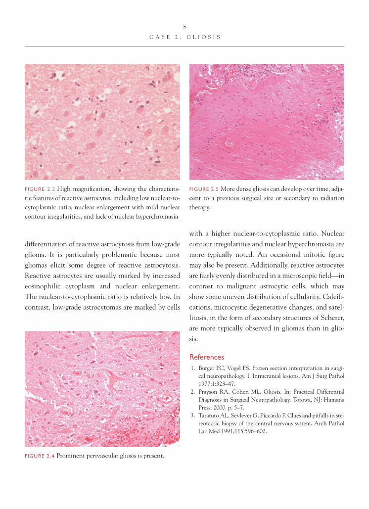
Anaplastic astrocytoma represents an intermedi-ate grade lesion between the low-grade fi brillary astrocytoma (WHO grade II) and the glioblastoma multiforme (WHO grade IV). Distinction of these tumors from low-grade astrocytomas is predicated on the presence of increased cellularity, more promi-nent nuclear atypia, and readily identifi able mitotic
activity. Distinction of this entity from glioblastoma multiforme is marked by the absence of vascular pro-liferative changes and/or necrosis in the anaplastic astrocytoma.
Cell proliferation markers, such as Ki-67 or MIB-1, can be useful in corroborating the diagno-sis. Labeling indices typically range between 5 and 10; this is generally higher than the labeling indices
FIGURE 5.3 The infi ltrating edge of the tumor has the appearance of a low-grade astrocytoma. This underscores the heterogeneity that is commonly encountered in gliomas.
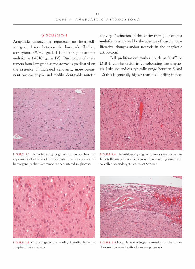
FIGURE 5.4 The infi ltrating edge of tumor shows perivascu-lar satellitosis of tumor cells around pre-existing structures, so-called secondary structures of Scherer.
FIGURE 5.5 Mitotic fi gures are readily identifi able in an anaplastic astrocytoma.
FIGURE 5.6 Focal leptomeningeal extension of the tumor does not necessarily afford a worse prognosis.

15
C A S E 5 : A N A P L A S T I C A S T R O C Y T O M A
observed in low-grade astrocytomas. It is important to remember, however, that these tumors are hetero-geneous in nature, and labeling indices and rates of cell proliferation may vary from region to region in the tumor. Consequently, a low labeling index in an anaplastic astrocytoma may be a result of sampling.
From prognostic and treatment standpoints, distinction of anaplastic astrocytoma from low-grade astrocytoma is signifi cant. Anaplastic astrocytomas are more often treated like high-grade astrocytomas
and generally undergo a course of radiation therapy. The prognosis is intermediate between low-grade as-trocytoma and the poor prognosis glioblastoma.
References 1. Burger PC, Vogel FS, Green SB, et al. Glioblastoma multi-
forme and anaplastic astrocytoma: pathologic criteria and prognostic implications. Cancer 1985;56:1106–11.
2. Coons SW, Johnson PC. Regional heterogeneity in the pro-liferative activity of human gliomas as measured by the Ki-67 labeling index. J Neuropath Exp Neurol 1993;52:609–18.
3. Kleihues P, Burger PC, Rosenblum MK, et al. Anaplastic astrocytoma. In: Louis DN, Ohgaki H, Wiestler OD, Cavenee WK, editors. WHO Classifi cation of Tumours of the Central Nervous System. Lyon, FR: IARC Press; 2007. p. 30–2.
FIGURE 5.7 Although an increased number of small cali-ber blood vessels may be encountered in the many gliomas, this particular tumor shows no evidence of vascular pro-liferative changes or necrosis to warrant a WHO grade IV designation.
FIGURE 5.8 A Ki-67 labeling index of 5.2 is observed in this tumor.

16
Case 6: Glioneuronal Tumor with Neuropil-Like Islands
C L I N I C A L I N F O R M A T I O N
The patient is a 38-year-old female who presents with seizures and headaches. On magnetic reso-nance imaging (MRI) studies, a mass involving the left frontal and temporal lobes and measuring 4.2 cm in diameter is noted; there is no evidence of enhancement. The tumor is associated with promi-nent vasogenic edema. Histologic sections from a subtotal resection are reviewed.
O P I N I O N
Histologic sections show an infi ltrative neoplasm, marked by a proliferation of atypical-appearing astrocytic cells characterized by nuclear enlarge-ment, nuclear hyperchromasia, and irregular nuclear contours. Rare mitotic fi gures are observed. There is no
evidence of vascular proliferative changes or necrosis. An atypical ganglion cell component is not identifi ed in the lesion. Scattered focally in the neoplasm are neuropil-like islands. These islands appear somewhat sharply demarcated from the background tumor and demonstrate diffuse positive staining with antibody to synaptophysin. In contrast, GFAP immunoreactivity stains the background tumor and does not stain the neuropil-like islands. The overlying cortex shows sat-ellitosis of tumor cells around pre-existing structures (secondary structures of Scherer).
We consider the lesion to be an astrocytoma and characterize it as follows: Left Frontal and Temporal Lobes, Subtotal Resection—Glioneu-ronal Tumor with Neuropil-Like Islands, WHO Grade III.
FIGURE 6.1 Low magnifi cation appearance of the tumor shows less cellular, neuropil-like islands, arranged against the background of an infi ltrating astrocytoma.
FIGURE 6.2 Higher magnifi cation shows well- circumscribed neuropil island in the background of a fi brillary astrocytoma.

17
C A S E 6 : G L I O N E U R O N A L T U M O R W I T H N E U R O P I L - L I K E I S L A N D S
C O M M E N T
In the most recent WHO Classifi cation of Tumours of the Central Nervous System, the glioneuronal tumor with neuropil-like islands is considered a variant of diffuse or fi brillary astrocytoma. The cellularity of the tumor and presence of mitotic fi gures in this case sup-port a diagnosis of grade III astrocytoma.
D I S C U S S I O N
The glioneuronal tumor with neuropil-like islands or rosetted glioneuronal tumor is a recent addition to the WHO classifi cation of central nervous system tumors and represents a pattern of anaplastic astro-cytoma. The clinical presentation overlaps with that of ordinary diffuse or fi brillary astrocytomas. The
FIGURE 6.3 The background tumor resembles an ordinary anaplastic astrocytoma.
FIGURE 6.4 Occasional mitotic fi gures are present in this tumor.
FIGURE 6.5 Satellitosis or secondary structures of Scherer are present at the infi ltrating margin of the neoplasm.
FIGURE 6.6 GFAP immunostaining shows a relative absence of staining in the neuropil-like island.

18
C A S E 6 : G L I O N E U R O N A L T U M O R W I T H N E U R O P I L - L I K E I S L A N D S
majority of cases seem to affect adults and involve the cerebral hemispheres as nonenhancing lesions on imaging. The background of these tumors resembles an ordinary anaplastic astrocytoma, and the tumor is marked by focally prominent cellularity, read-ily identifi able nuclear atypia, and scattered mitotic activity. Vascular proliferative changes and necrosis are generally not observed in this tumor. A distinct morphologic feature of this lesion is the presence of fairly circumscribed neuropil-like or rosetted islands. By immunohistochemistry, these islands demonstrate immunoreactivity with neural antibodies, such as synaptophysin, and generally demonstrate a paucity of GFAP immunostaining. This is in sharp contrast to the background tumor, which is GFAP-positive and demonstrates little in the way of synaptophysin immunostaining.
These tumors are graded according to the back-ground astrocytic component and usually represent grade II and, more commonly, grade III tumors. Their prognosis seems to correspond to the WHO grade of the tumor.
References 1. Edgar MA, Rosenblum MK. Mixed glioneuronal tumors,
recently described entities. Arch Pathol Lab Med 2007;131:228–33.
2. Prayson RA, Abramovich CM. Glioneuronal tumor with neuropil-like islands. Hum Pathol 2000;31:14:35–8.
3. Teo JGC, Gultekin H, Bilsky M, Gutin P, et al. A distinctive glioneuronal tumor of the adult cerebrum with neuropil-like (including “rosetted”) islands. Report of 4 cases. Am J Surg Pathol 1999;23:502–10.
FIGURE 6.7 Prominent synaptophysin immunoreactivity is observed in the neuropil-like island.
19
Case 7: Glioblastoma Multiforme
C L I N I C A L I N F O R M A T I O N
The patient is a 72-year-old male who presents with a focally enhancing mass in the right frontal lobe. Biop-sies are taken, and histologic sections are reviewed.
O P I N I O N
Histologic sections show a markedly cellular neo-plasm with a fi brillary background, consistent with a high-grade glioma. Focal dystrophic calcifi cation is observed. Tumor cells are marked by a high nuclear-to-cytoplasmic ratio, nuclear hyperchromasia, and irregular nuclear contour. Focally, cells assume a some-what spindled confi guration. Mitotic fi gures are read-ily observable. Focal vascular proliferative change is also noted. There is no defi nite evidence of necrosis in this tumor. A Ki-67 labeling index of 22.6 is noted.
We consider the lesion to be a high-grade astro-cytoma and characterize it as follows: Right Frontal Lobe, Biopsies—Glioblastoma Multiforme (or Glio-blastoma), WHO Grade IV.
C O M M E N T
The presence of vascular proliferative changes, even in the absence of necrosis, is suffi cient for a diagnosis of glioblastoma multiforme.
D I S C U S S I O N
Glioblastoma multiforme represents the highest grade of a diffuse fi brillary astrocytoma. These tumors are typically marked by prominent cellular-ity, readily discernible atypia, and prominent mitotic activity. The WHO requires either the presence of vascular proliferative changes, as observed in this
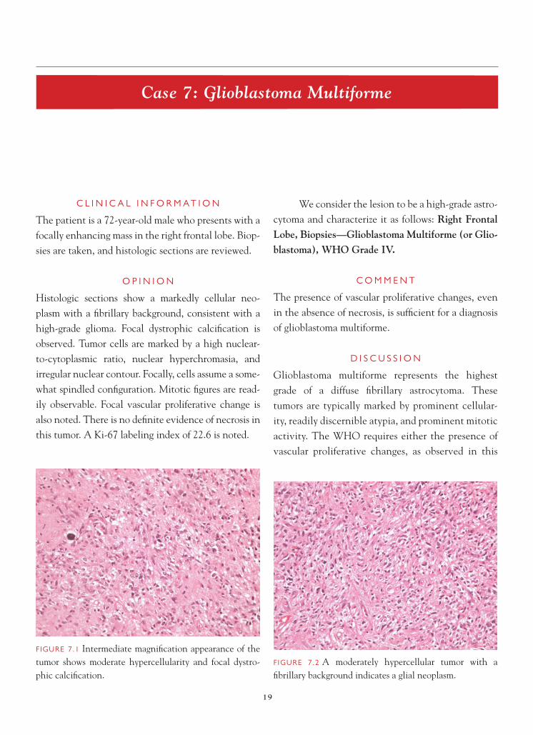
FIGURE 7.1 Intermediate magnifi cation appearance of the tumor shows moderate hypercellularity and focal dystro-phic calcifi cation.
FIGURE 7.2 A moderately hypercellular tumor with a fi brillary background indicates a glial neoplasm.

20
C A S E 7 : G L I O B L A S T O M A M U L T I F O R M E
tumor, or necrosis in order to make a diagnosis of glioblastoma multiforme. Older grading systems required the presence of necrosis for a diagnosis of glioblastoma. Necrosis, when present, may or may not have a pseudopalisade of tumor cells around it. Care needs to be taken in interpreting biopsies from a patient who has been previously treated with radio-therapy, because radiation can induce necrosis. The presence of nonpalisaded necrosis in the setting of
prior radiation therapy should not necessarily war-rant an upgrading of the tumor. Vascular prolifera-tive changes refer to a proliferation of cells around vascular lumina. These are not tumor cells, but nor-mal blood vessel wall constituents (i.e., endothelial cells, smooth muscle cells, fi broblasts, pericytes). An increased number of blood vessels, which is common in many gliomas, does not warrant an upgrading of the tumor.
FIGURE 7.4 Focal spindled cell region in a glioblastoma is seen.
FIGURE 7.3 A focal area of the tumor with a low-grade astro-cytoma appearance is seen here. This underscores the regional heterogeneity that is a characteristic feature of gliomas.
FIGURE 7.5 Readily identifi able mitotic activity is present in the tumor.
FIGURE 7.6 Focal vascular proliferative changes are present in the tumor.

21
C A S E 7 : G L I O B L A S T O M A M U L T I F O R M E
Particularly in the setting of biopsies, sam-pling of the tumor is important in arriving at the correct grade designation. Glioblastoma multiforme is a heterogeneous lesion, which may have areas resembling low-grade astrocytoma. Communica-tion with the neurosurgeon at the time of surgery to ensure that appropriate tissue has been acquired is important.
Ki-67 labeling indices are typically high in glio-blastoma and frequently exceed 10–15. Occasional tumors may have focal areas with labeling indices in excess of 50. There does not appear to be any prog-nostic implication of determining a Ki-67 labeling
index in a glioblastoma. Prognosis is poor, even with standard treatment, which is usually radiotherapy. Most patients have a survival of 1–2 years.
References 1. Burger PC, Vogel FS, Green SB, et al. Glioblastoma
multiforme and anaplastic astrocytoma: pathologic criteria and prognostic implications. Cancer 1985;56:1106–11.
2. Kleihues P, Burger PC, Aldape KD, et al. Glioblastoma. In: Louis DN, Ohgaki H, Wiestler OD, Cavenee WK, editors. WHO Classifi cation of Tumours of the Central Nervous System. Lyon, FR: IARC Press; 2007. p. 33–47.
3. Prayson RA. Histologic classifi cation of high-grade gliomas. In: Barnett GH, editor. High-Grade Glioma. Diagnosis and Treatment. Totowa, NJ: Humana Press;2007. p. 3–35.
FIGURE 7.7 Perinecrotic pseudopalisading of tumor cells (not observed in this particular case) is a common feature of glioblastoma.
FIGURE 7.8 Ki-67 immunostaining demonstrates a labeling index of 22.6 in this tumor.

22
Case 8: Gemistocytic Astrocytoma
C L I N I C A L I N F O R M A T I O N
The patient is a 42-year-old male who presents with right-sided weakness. On MRI imaging studies, a 3.2-cm mass involving the left frontal and parietal lobes is noted. The tumor shows no defi nite evidence of enhancement. Histologic sections from a subtotal resection are reviewed.
O P I N I O N
Histologic sections show a hypercellular neoplasm, marked by a predominant population of large cells with abundant eosinophilic cytoplasm and eccen-trically placed nuclei. The nuclei appear slightly irregular in shape. Intermixed with these cells are focal areas, marked by a proliferation of atypical-ap-pearing astrocytic cells, characterized by high nucle-
ar-to-cytoplasmic ratio, nuclear hyperchromasia, and irregular nuclear contour. Focal microcystic degen-erative changes are observed in the tumor. A careful search reveals scattered mitotic fi gures. There is no defi nite evidence of vascular proliferative changes or necrosis. GFAP immunostaining highlights promi-nent immunoreactivity of both the large and small cells. Ki-67 immunostaining shows mild positivity, confi ned primarily to the small, atypical-appearing astrocytic cells; a labeling index of 5.6 is noted in this tumor.
We consider the lesion to be an astrocytoma with prominent numbers of gemistocytes and charac-terize it as follows: Left Frontal and Parietal Lobes, Subtotal Resection—Gemistocytic Astrocytoma, WHO Grade III.
FIGURE 8.1 Low magnifi cation appearance of the neoplasm shows hypercellularity, with clusters of larger cells.
FIGURE 8.2 Microcystic degenerative changes are observed in the tumor.

23
C A S E 8 : G E M I S T O C Y T I C A S T R O C Y T O M A
C O M M E N T
The presence of mitotic fi gures in this tumor, in the absence of vascular proliferation and necrosis, warrants a diagnosis of a WHO grade III tumor.
D I S C U S S I O N
A well-recognized variant of astrocytoma is marked by prominent numbers of gemistocytes. Gemistocytes are characterized by abundant eosinophilic cytoplasm and eccentrically placed, slightly irregularly shaped nuclei. Perivascular chronic infl ammatory cells, in particular lymphocytes, may be focally present in these tumors.
It is well recognized that astrocytic tumors with a signifi cant gemistocytic component tend to behave in a more aggressive fashion, although the WHO still recognizes these lesions as grade II neoplasms. Designation of a tumor as gemistocytic astrocytoma, versus an ordinary diffuse or fi brillary astrocytoma with occasional gemistocytes, is somewhat arbitrary. One study suggested a minimum 20% gemistocytic com-ponent as being clinically signifi cant. Grading param-eters that are used in evaluating diffuse fi brillary astro-cytomas are also employed in evaluating gemistocytic astrocytomas. In the current case, the presence of read-ily identifi able mitotic activity warrants the designa-tion of a WHO grade III neoplasm. The presence of vascular proliferative changes or necrosis in the tumor warrants the designation of a grade IV neoplasm.
FIGURE 8.3 High magnifi cation appearance shows a focus of confl uent gemistocytes, marked by abundant eosino-philic cytoplasm and eccentrically placed, irregularly shaped nuclei.
FIGURE 8.4 A focal gemistocyte-poor area, marked by a proliferation of more conventional-appearing, atypical astrocytic cells, is seen.
FIGURE 8.5 Mitotic activity is readily observable in this neoplasm.

24
C A S E 8 : G E M I S T O C Y T I C A S T R O C Y T O M A
FIGURE 8.6 GFAP immunoreactivity highlights positive staining gemistocytes, underscoring their astrocytic lineage.
Differential diagnostic considerations include reactive astrocytosis, oligodendroglioma with minige-mistocytes, and ganglioglioma. In contrast to reactive astrocytes, a gemistocytic astrocytoma should have a second population of cells that appear conventionally malignant and indicate the true nature of the lesion. Occasional oligodendrogliomas can have a prominent gemistocytic-like component. In these instances, the background of the tumor appears to be very much like an oligodendroglioma with rounded nuclei and scant cytoplasm. GFAP immunoreactivity in the gemisto-cytic cells in the current tumor indicates the astrocytic lineage of the cells. Occasionally, in a ganglioglioma,
some cells may assume a gemistocytic-like appearance. Immunohistochemistry can be helpful in making the distinction, in that ganglion cells stain with markers of neural differentiation, including synaptophysin.
References 1. Krouwer HGJ, Davis RL, Silver P, et al. Gemistocytic astrocy-
tomas: a reappraisal. J Neurosurg 1991;74:399–406. 2. Tihan T, Vohra P, Berger MS, et al. Defi nition and diagnostic
implications of gemistocytic astrocytomas: a pathological per-spective. J Neurooncol 2006;76:175–83.
3. Watanabe K, Tachibana O, Yonekawa Y, et al. Role of gemisto-cytes in astrocytoma progression. Lab Invest 1997;76:277–84.
4. Yang HJ, Kim JE, Paek SH, et al. The signifi cance of gemisto-cytes in astrocytoma. Acta Neurochir 2003;145:1097–1103.
FIGURE 8.7 A Ki-67 labeling index of 5.6 is observed in this tumor. Ki-67 staining is typically confi ned to the nongemistocytic component of the tumor.

25
Case 9: Granular Cell Glioblastoma
C L I N I C A L I N F O R M A T I O N
The patient is a 59-year-old male who presents with seizures and a right temporal lobe mass on imaging studies. On imaging, the lesion shows focal enhance-ment. Subtotal resection of the tumor is performed, and histologic sections are reviewed.
O P I N I O N
Sections show a moderately hypercellular neoplasm, comprised primarily of plump, eosinophilic cells. These cells are marked by abundant granular cytoplasm. Nuclei are generally eccentrically placed and round-to-oval in confi guration. Occasional small nucleoli are observed. Perivascular chronic infl ammation, consisting primarily of benign-appearing lymphocytes, is noted. Mitotic activity is not observed in the large granular cells. Focal areas of the tumor demonstrate
necrosis. Vascular proliferative changes are noted at the parameter of the lesion. In areas of vascular prolif-eration, atypical-appearing astrocytic cells, with high nuclear-to-cytoplasmic ratio and an appearance more typical of a diffuse or fi brillary type astrocytoma, are identifi ed. Several mitotic fi gures are observed in this region of the tumor.
We consider this lesion to be a malignant astro-cytoma and characterize it as follows: Right Temporal Lobe, Excision—Granular Cell Glioblastoma, WHO Grade IV.
C O M M E N T
This neoplasm is best considered a variant of glioblas-toma multiforme. The presence of granular cell differ-entiation does not affect the prognosis of this tumor.
FIGURE 9.1 Low magnifi cation, showing a granular cell predominant area of the tumor.
FIGURE 9.2 Higher magnifi cation highlights the eosino-philic, granular quality to the cytoplasm of the granular cell component.

26
C A S E 9 : G R A N U L A R C E L L G L I O B L A S T O M A
D I S C U S S I O N
Rarely, glioblastoma may show evidence of granu-lar cell differentiation. The cells in this variant are marked by abundant, eosinophilic, granular cytoplasm and resemble granular cells seen elsewhere in the body in granular cell tumors. If one searches long enough, this granular cell component is often accompanied by areas of the tumor that resemble a conventional-appearing, high-grade fi brillary or diffuse astrocytoma.
Occasionally, on intraoperative consultation, only the granular cell component of the tumor may be sampled. In such cases, the suggestion that the tumor represents part of a high-grade astrocytoma should be made. One exception to this are tumors arising in the supra-sellar region; pure granular cell tumors are known to arise from the neurohypophysis of the pituitary gland and generally represent benign lesions. Ultrastruc-turally, the granular cells are fi lled with abundant, large lysosomes. Interestingly, a Ki-67 immunostain
FIGURE 9.3 Perivascular chronic infl ammation, consisting of benign-appearing lymphocytes, is seen in the granular cell area of the tumor.
FIGURE 9.4 Areas of the tumor have the appearance of a conventional fi brillary or diffuse astrocytoma.
FIGURE 9.5 Mitotic fi gures are readily identifi able in the fi brillary astrocytoma-appearing area of the tumor.
FIGURE 9.6 Vascular proliferative changes are noted in the fi brillary astrocytoma-appearing area of the tumor.

27
C A S E 9 : G R A N U L A R C E L L G L I O B L A S T O M A
generally does not label the granular cells but high-lights increased cell proliferation in the fi brillary astrocytoma-appearing component of the neoplasm. Granular cell differentiation in a glioblastoma does not appear to affect the prognosis of these lesions.
References 1. Brat DJ, Scheithauer BW, Medina-Flores R, et al. Infi ltra-
tive astrocytomas with granular cell features (granular cell astrocytomas). A study of histopathologic features, grading, and outcome. Am J Surg Pathol 2002;26:750–7.
2. Chorny JA, Evans LC, Kleinschmidt-DeMasters BK. Cerebral granular cell astrocytomas: a MIB-1, bcl-2, and telomerase study. Clin Neuropathol 2000;19:170–9.
3. Geddes JF, Thom M, Robinson SFD, et al. Granular cell change in astrocytic tumors. Am J Surg Pathol 1996;20:55–63.
4. Melaragno MJ, Prayson RA, Murphy MA, et al. Anaplastic astrocytoma with granular cell differentiation: case report and review of the literature. Hum Pathol 1993;24:805–8.
FIGURE 9.7 Focal necrosis is present in the tumor.

28
Case 10: Giant Cell Glioblastoma
C L I N I C A L I N F O R M A T I O N
The patient is a 67-year-old male who presents with a left parietal lobe mass. On imaging, the lesion measures approximately 5.2 cm in greatest extent, with focal areas of enhancement. Subtotal resection of the tumor is performed, and histologic sections are reviewed.
O P I N I O N
The histologic sections show a markedly cellular neoplasm, characterized by a large number of giant tumor cells. Many of these tumor cells are marked by promi-nent eosinophilic cytoplasm and large pleomorphic nuclei. Many of the cells are multinucleated, and intranuclear pseudoinclusions are frequently evident. Several of the cells show evidence of mitotic activity. Scattered cells demonstrate nuclear hyperchromasia. Occasional cells show somewhat cleared or lipidized
cytoplasm. Occasional blood vessels show perivascular chronic infl ammation, consisting primarily of benign-appearing lymphoid cells. Focal necrosis is observed. In many areas, the giant tumor cells are intermixed with a population of smaller cells resembling those seen in a typical diffuse or fi brillary astrocytoma. Mild vascular proliferative changes are also observed.
We consider this lesion to be a malignant astrocytoma, and we characterize it as follows: LeftParietal Lobe, Excision—Giant Cell Glioblastoma, WHO Grade IV.
C O M M E N T
This neoplasm is best considered a variant of glioblas-toma. The presence of prominent numbers of giant cells in this tumor may portend a slightly better prog-nosis, compared with an ordinary type glioblastoma.
FIGURE 10.1 Low magnifi cation appearance of the tumor shows a neoplasm marked by many giant cells.
FIGURE 10.2 Many of the giant cells show nuclear pseu-doinclusions (cytoplasmic invaginations).
29
C A S E 1 0 : G I A N T C E L L G L I O B L A S T O M A
D I S C U S S I O N
One of the histologic variants of glioblastoma is marked by the presence of giant, bizarre-appearing cells. Frequently, these cells are multinucleated. This particular variant is relatively uncommon and repre-sents up to 5% of glioblastoma. The age distribution of this variant covers a wider range than many of the other diffuse astrocytomas and includes children. Because of an often prominent stromal reticulin net-work in this tumor, the lesion was historically referred
to as a monstrocellular sarcoma; however, the GFAP immunoreactivity of the giant cells indicates its astro-cytic lineage. p53 mutations are frequently observed in these tumors. In contrast to the small cell variant of a glioblastoma, this variant typically lacks evidence of epidermal growth factor receptor (EGFR) amplifi -cation or overexpression. The presence of prominent mitotic activity and necrosis in this tumor allow for its ready distinction from the pleomorphic xanthoastro-cytoma. Ki-67 labeling indices are typically high, of the order that is usually observed in other glioblastoma
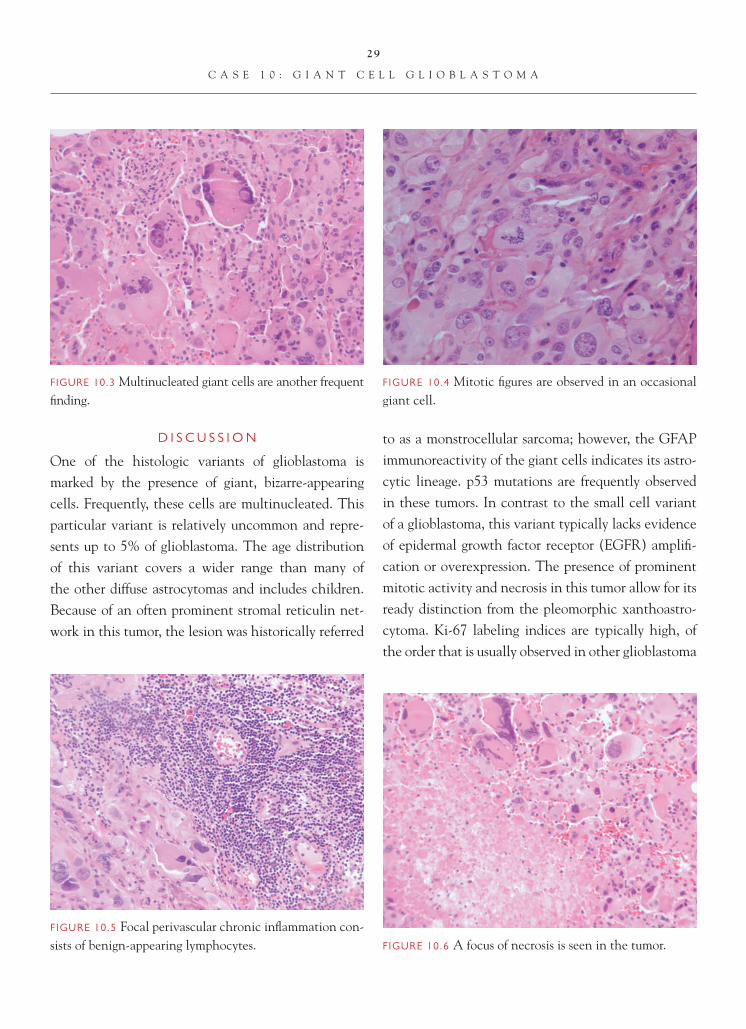
FIGURE 10.3 Multinucleated giant cells are another frequent fi nding.
FIGURE 10.4 Mitotic fi gures are observed in an occasional giant cell.
FIGURE 10.5 Focal perivascular chronic infl ammation con-sists of benign-appearing lymphocytes. FIGURE 10.6 A focus of necrosis is seen in the tumor.

30
C A S E 1 0 : G I A N T C E L L G L I O B L A S T O M A
lesions. Although most of the giant cell glioblastomas have a poor prognosis, similar to ordinary glioblas-toma, there is some literature to suggest that a subset of these patients may have a somewhat better prognosis, perhaps related to the tumor’s less infi ltrative nature or the younger age of presentation of some patients.
References 1. Margetts JC, Kalyan R. Giant-celled glioblastoma of brain.
A clinicopathological and radiological study of ten cases
(including immunohistochemistry and ultrastructure). Cancer 1989;63:524–31.
2. Meyer-Puttlitz B, Hayashi Y, Waha A, et al. Molecular genetic analysis of giant cell glioblastoma. Am J Pathol 1997;151:853–7.
3. Peraud A, Watanabe K, Schwechheiner K, et al. Genetic profi le of the giant cell glioblastoma. Lab Invest 1999;79:123–9.
4. Queiroz LS, Faria AV, Zanardi VA, et al. Lipidized giant cell glioblastoma of cerebellum. Clin Neuropathol 2005;24:262–6.
FIGURE 10.7 Vascular proliferative changes are present in the tumor.
FIGURE 10.8 Part of the tumor is relatively devoid of giant cells and has an appearance similar to that of a conven-tional, high-grade astrocytoma.
31
Case 11: Pleomorphic Xanthoastrocytoma
C L I N I C A L I N F O R M A T I O N
The patient is a 20-year-old female who presents with a four-year history of seizures. On imaging, she has a partially cystic and enhancing left tem-poral lobe mass. The patient undergoes gross total resection of the tumor, and histologic sections are reviewed.
O P I N I O N
Histologic sections show a tumor marked by promi-nent cellularity and readily apparent nuclear pleo-morphism. Scattered giant cells are marked by frequent multinucleation. Intranuclear pseudoin-clusions are occasionally seen in these giant cells. The amount of cytoplasm in these cells is variable and ranges from scant to prominent. Some of the
cells have a light, homogeneous, eosinophilic cyto-plasm, whereas others show lipidization. Dystrophic calcifi cation is present. Perivascular chronic infl am-mation, consisting primarily of benign-appearing lymphocytes, is also saliently noted. Mitotic fi gures are absent. There is no evidence of necrosis. Focal microcystic degenerative changes are observed in the tumor. Also of note is the sharp interface between the tumor and adjacent cortex. Reticulin staining highlights increased reticulin deposition between cells and small groups of cells. A Ki-67 labeling index of 3.2 is noted.
We consider this lesion to be a low-grade astrocytic neoplasm and characterize it as follows: Left Temporal Lobe, Excision—Pleomorphic Xan-thoastrocytoma, WHO Grade II.
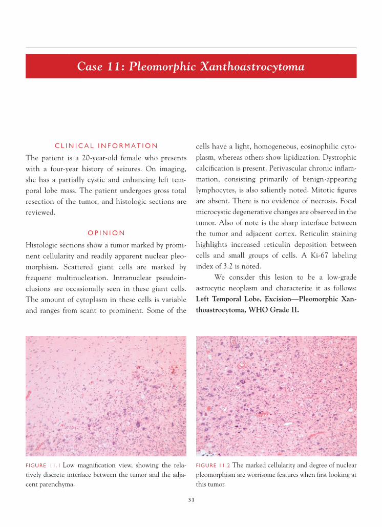
FIGURE 11.1 Low magnifi cation view, showing the rela-tively discrete interface between the tumor and the adja-cent parenchyma.
FIGURE 11.2 The marked cellularity and degree of nuclear pleomorphism are worrisome features when fi rst looking at this tumor.

32
C A S E 1 1 : P L E O M O R P H I C X A N T H O A S T R O C Y T O M A
C O M M E N T
The absence of prominent mitotic activity and necrosis supports a diagnosis of pleomorphic xan-thoastrocytoma.
D I S C U S S I O N
The pleomorphic xanthoastrocytoma is an important tumor to distinguish from a glioblastoma because of its signifi cantly better prognosis. These tumors typically present in children and young adults, often with a
history of intractable seizures. The vast majority of these tumors arise either in the temporal or parietal lobes and are superfi cially based. On imaging stud-ies, they frequently are cystic, with enhancing mural nodules. The superfi cial location also contrasts with typical glioblastoma, which is a white matter-based tumor in many cases. On fi rst inspection, these tumors appear to be worrisome, given the degree of cellularity and pleomorphism of the cells. Many of the cells show features similar to that of the giant cell glioblastoma. However, in contrast to the glioblastoma, the pleo-morphic xanthoastrocytoma generally demonstrates little or any mitotic fi gures and no necrosis. Reti-culin staining may be prominently observed in this tumor. Perivascular chronic infl ammation is a fairly frequent fi nding. A subset of tumors may demonstrate calcifi cation. Many of the large, bizarre-appearing cells demonstrate GFAP immunoreactivity. It is not unusual to fi nd occasional cells in these tumors demonstrating evidence of neural differentiation by immunohistochemistry (synaptophysin-positive). These tumors are generally considered low-grade (WHO grade II), although a subset recurs and these tumors require follow-up. Rare cases of more aggres-sive behavior in these lesions have been described;
FIGURE 11.3 Scattered multinucleated giant cells, some with nuclear pseudoinclusions, are present.
FIGURE 11.4 Large astrocytic cells with lipidized cytoplasm are seen. FIGURE 11.5 Focal dystrophic calcifi cation is present.

33
C A S E 1 1 : P L E O M O R P H I C X A N T H O A S T R O C Y T O M A
FIGURE 11.6 Perivascular chronic infl ammation is a fre-quent fi nding.
this is often associated with worrisome histologic features, including the presence of increased mitotic activity and/or necrosis. These later tumors are desig-nated as anaplastic pleomorphic xanthoastrocytoma and represent WHO grade III neoplasms.
References 1. Giannini C, Scheihauer BW, Burger PC, et al. Pleomorphic
xanthoastrocytoma. What do we really know about it? Cancer 1999;85:2033–45.
2. Kepes JJ. Pleomorphic xanthoastrocytoma: the birth of a diagnosis and a concept. Brain Pathol 1993;3:269–74.
3. Kepes JJ, Rubinstein LJ, Eng LW. Pleomorphic xanthoastro-cytoma: a distinctive meningocerebral glioma of young sub-jects with relatively favorable prognosis. A study of 12 cases. Cancer 1979;44:1839–52.
4. Prayson RA, Morris III, HH. Anaplastic pleomorphic xanthoastrocytoma. Arch Pathol Lab Med 1998;122:1082–6.
FIGURE 11.7 A focal area of microcystic degeneration is evident.
FIGURE 11.8 Increased reticulin deposition between tumor cells is evident.
FIGURE 11.9 A low Ki-67 labeling index of 3.2 is present in this tumor.

34
Case 12: Gliosarcoma
C L I N I C A L I N F O R M A T I O N
The patient is a 58-year-old male who presents with seizures. On MRI imaging studies, a 5.6-cm mass, involving the right frontal and parietal lobes and focally crossing the corpus callosum to involve the contralateral side, is present. The tumor demonstrates focal areas of enhancement. The patient undergoes a subtotal resection, and histologic sections are reviewed.
O P I N I O N
Histologic sections show a markedly cellular neoplasm, characterized by a proliferation of atypical-appearing cells resembling malignant astrocytes. Areas of the tumor resemble an ordinary glioblastoma multiforme; the tumor shows focal necrosis, vascular proliferative
changes, and readily identifi able mitotic activity. Intermixed with the glioblastomatous-appearing areas are foci of tumor with a malignant spindle-cell appearance. The spindle cells contain atypical nuclei and demonstrate evidence of mitotic activity. Focally, malignant bone formation (osteosarcoma) is observed in the tumor. GFAP immunostaining shows positive staining, confi ned to the glioblastomatous-appearing areas of the tumor. Specifi cally, the spindle-cell areas do not stain with GFAP. Reticulin staining shows increased reticulin deposition between cells and small groups of cells in the spindle-cell regions of the tumor. Increased reticulin staining is only noted in vascular proliferative foci in the glioblastomatous component of the lesion.
We consider the lesion to be a variant of high-grade astrocytoma and characterize it as follows: Right
FIGURE 12.1 Low magnifi cation appearance, highlight-ing a glioblastomatous-appearing region of the tumor with necrosis.
FIGURE 12.2 The glioblastomatous component is juxta-posed to a spindle-cell component in the gliosarcoma.

35
C A S E 1 2 : G L I O S A R C O M A
Frontal and Parietal Lobes, Subtotal Resection—Gliosarcoma, WHO Grade IV.
C O M M E N T
The sarcomatous component of this tumor consists of a nondescript spindle-cell proliferation, as well as an osteosarcomatous component. Increased reticulin staining highlights the spindle-cell sarcomatous com-ponent tumor. GFAP immunoreactivity highlights the glioblastomatous component of the neoplasm.
D I S C U S S I O N
Gliosarcoma is a variant of glioblastoma, marked by a glioblastomatous component juxtaposed with a sarcomatous component. Approximately 2% of all glioblastoma multiforme are gliosarcoma type. The age of presentation, clinical presentation, and imag-ing fi ndings are quite similar to those of the glioblas-toma. The key to the diagnosis is recognition of the sarcomatous component. Most commonly, the sar-comatous component resembles either fi brosarcoma or malignant fi brous histiocytoma. A combination of GFAP and reticulin staining can be useful in delin-eating this component. The sarcomatous component is generally GFAP-negative and reticulin-rich. The only increased reticulin staining usually observed in a glioblastoma resides in areas of vascular proliferative change. In occasional glioblastoma, particularly those growing up near the surface of the brain, the tumor cells can assume a spindle confi guration and resemble the gliosarcoma lesion. The spindle-cell glioblastoma shows a paucity of reticulin and evidence of GFAP immunoreactivity in the spindle-cell component. Sarcoma represents the other main differential diag-nostic consideration. Sarcomas are GFAP-negative tumors.
FIGURE 12.3 High magnifi cation, showing glioblastoma-tous component with necrosis.
FIGURE 12.4 Vascular proliferative changes are present in the gliosarcoma.
FIGURE 12.5 The sarcomatous component is seen here, with readily identifi able mitotic activity in a gliosarcoma.

36
C A S E 1 2 : G L I O S A R C O M A
FIGURE 12.6 Focal osteosarcomatous differentiation is pres-ent in this tumor.
FIGURE 12.7 Reticulin staining highlights the reticulin-rich, sarcomatous component of the gliosarcoma.
The current thinking suggests that the sarcoma-tous component represents a form of mesenchymal dif-ferentiation in a glioblastoma. A number of studies have shown similar genetic alterations in both the gliobla-stomatous and sarcomatous components of the tumor. Recognition of the gliosarcoma pattern does not appear to have any signifi cant clinical or prognostic signifi -cance and, unfortunately, shares the same dismal prog-nosis that marks ordinary glioblastoma multiforme.
References 1. Beirnat W, Aguzzi A, Sure U, et al. Identical mutations of the
p53 tumor suppressor gene in the gliomatous and sarcomatous components of gliosarcomas suggest a common origin from glial cells. J Neuropathol Exp Neurol 1995;54:651–6.
2. Meis JM, Ho KL, Nelson JS. Gliosarcoma: a histologic and immunohistochemical reaffi rmation. Mod Pathol 1990;3:19–24.
3. Sreenan JJ, Prayson RA. Gliosarcoma. A study of 13 tumors, including p53 and CD34 immunohistochemistry. Arch Pathol Lab Med 1997;121:129–33.

37
Case 13: Small Cell Glioblastoma
C L I N I C A L I N F O R M A T I O N
The patient is a 62-year-old male with a 20-pack-year history of smoking who presents with an enhancing mass in the right frontal lobe. A biopsy of the tumor is performed, and histologic sections are reviewed.
O P I N I O N
Sections show a markedly cellular neoplasm, char-acterized by a monomorphous proliferation of small cells with a high nuclear-to-cytoplasm ratio. Promi-nent mitotic activity and focal vascular proliferative changes are observed. Individual cell necrosis and a focus of geographic necrosis are also present. Immu-nohistochemical staining of the tumor shows focal positive immunoreactivity, with antibodies to GFAP and EGFR. The tumor did not stain with antibodies to CD45RB (CLA) or cytokeratin CAM5.2.
We consider the lesion to be a malignant astrocytoma and characterize it as follows: RightFrontal Lobe, Biopsy—Small Cell Glioblastoma, WHO Grade IV.
C O M M E N T
This neoplasm represents a variant of glioblastoma multiforme. The small cell change in glioblastoma does not affect the prognosis.
D I S C U S S I O N
In glioblastoma predominantly composed of small cells, the designation of small cell glioblastoma is appropriate. These tumors appear monomorphic, with slightly elongated nuclei and prominent mitotic activity. The presence of vascular proliferation and/or
FIGURE 13.1 Low magnifi cation appearance of the tumor, showing a markedly hypercellularity neoplasm.
FIGURE 13.2 Higher magnifi cation highlights the relative monotonous appearance of the cells and the high nuclear-to-cytoplasmic ratio.

38
C A S E 1 3 : S M A L L C E L L G L I O B L A S T O M A
necrosis is defi nitional of glioblastoma. Biopsies are particularly challenging to evaluate because of the sometimes homogeneous appearance of this tumor. The differential diagnosis may include other small cell neoplasms that may be found in older adults in the brain, including diffuse large cell lymphoma, metastatic small cell carcinoma, and anaplastic oligo-dendroglioma. Immunohistochemistry may be help-ful in sorting out this differential diagnosis in cases where morphologic clues are absent. A diagnosis of
lymphoma is excluded with CD45RB immunostain-ing. The lack of cytokeratin CAM5.2 immunoreac-tivity and the focal GFAP staining (which, at times, may be sparse in this variant) argues against a meta-static small cell carcinoma. Increased overexpression or amplifi cation of EGFR is a frequent fi nding in the small cell glioblastoma and uncommon in an anaplas-tic oligodendroglioma.
FIGURE 13.3 Focal vascular proliferative change is present. FIGURE 13.4 Mitotic fi gures are readily identifi ed in the tumor.
FIGURE 13.5 Focal necrosis is present.FIGURE 13.6 GFAP immunoreactivity is present and expected with a glioblastoma.

39
C A S E 1 3 : S M A L L C E L L G L I O B L A S T O M A
References 1. Burger PC, Pearl DK, Aldape K, et al. Small cell architecture—
a histological equivalent of EGFR amplifi cation in glio-blastoma multiforme? J Neuropathol Exp Neurol 2001;60:1099–1104.
2. Perry A, Aldape KD, George DH, et al. Small cell astrocy-toma: an aggressive variant that is clinicopathologically and genetically distinct from anaplastic oligodendroglioma. Cancer 2004;101:2318–26.
FIGURE 13.7 EGFR overexpression, as evidenced byimmunohistochemistry.
40
Case 14: Epithelioid Glioblastoma
C L I N I C A L I N F O R M A T I O N
The patient is a 72-year-old female who presents with headaches and persistent nausea. On MRI studies, she is noted to have a gadolinium-enhancing, right frontal-temporal mass with marked peritumoral edema. Biopsies of the mass are taken, and histologic sections reviewed.
O P I N I O N
Histologic sections show a markedly cellular neo-plasm, characterized by a proliferation of generally rounded cells. The cells show a moderate amount of cytoplasm, nuclear irregularities, and prominent nucleolation. Readily identifi able mitotic activity is observed in the tumor. Focal necrosis, accompanied by vascular proliferative changes, is also seen. Given the differential diagnosis of this lesion with a metastatic
non-small cell carcinoma, a combination of cytok-eratin markers and GFAP are evaluated. The tumor demonstrates diffuse positive staining, with antibody to GFAP. Scattered cytokeratin AE1/3 immunoreac-tivity is observed. The tumor did not stain with anti-body to cytokeratin CAM5.2. The tumor also fails to demonstrate immunoreactivity with antibodies to Melan-A and HMB 45.
We consider the lesion to be a high-grade astrocytoma and characterize it as follows: RightFrontal-Temporal Lobes, Biopsies—Epithelioid Glioblastoma, WHO Grade IV.
C O M M E N T
The tumor demonstrates diffuse positive staining with antibody to GFAP and an absence of staining with melanoma markers and cytokeratin CAM5.2. Cytokeratin AE1/3 staining may represent cross
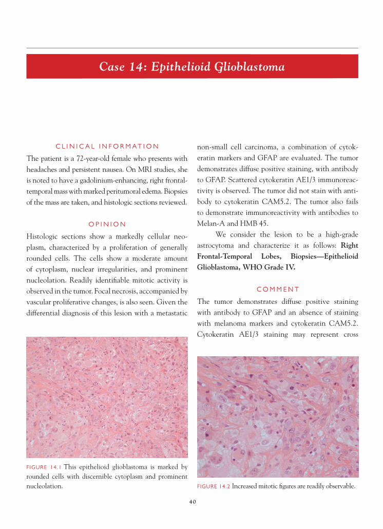
FIGURE 14.1 This epithelioid glioblastoma is marked by rounded cells with discernible cytoplasm and prominent nucleolation. FIGURE 14.2 Increased mitotic fi gures are readily observable.

41
C A S E 1 4 : E P I T H E L I O I D G L I O B L A S T O M A
immunoreactivity. The presence of vascular prolif-erative changes and necrosis warrant a diagnosis of a grade IV astrocytoma.
D I S C U S S I O N
Glioblastoma multiforme can assume an epithe-lioid appearance. In some instances, this takes the form of cells with lipidized cytoplasm. In these cases, differentiating the tumor from metastatic clear cell carcinoma (such as renal cell carcinoma
or adrenocortical carcinoma) or pleomorphic xan-thoastrocytoma is of consideration. In contrast to the pleomorphic xanthoastrocytoma, the epithelioid glioblastoma demonstrates features typical of glioblas-toma, including vascular proliferative changes, necro-sis, and prominent mitotic activity.
In other instances, the cells have more eosino-philic cytoplasm and, with the presence of prominent nucleolation, can resemble either metastatic non-small
FIGURE 14.3 Focal necrosis is seen in this tumor. FIGURE 14.4 Vascular proliferative changes are present.
FIGURE 14.5 Strong diffuse positive immunostaining with antibody to GFAP is observed in this epithelioid glioblastoma.
FIGURE 14.6 Scattered cytokeratin AE1/3 immunoreactiv-ity can be observed in glioblastoma multiforme and repre-sents cross immunoreactivity. The tumor did not stain with cytokeratin CAM5.2 antibody.

42
C A S E 1 4 : E P I T H E L I O I D G L I O B L A S T O M A
cell carcinoma or, occasionally, malignant melanoma. In such cases, immunohistochemistry can be helpful in uncovering the true lineage of the tumor, if there are no morphologic clues in the neoplasm; i.e., an area of lower-grade tumor resembling astrocytoma is not evident. Glioblastoma stains with antibodies to GFAP and S-100 protein. In differentiating glioblastoma from melanoma, which can also stain with S-100 protein, additional melanoma markers, such as Melan-A and HMB 45, can be useful. In differentiating carcinoma from glioblastoma, a combination of GFAP with cy-tokeratin markers is helpful. One needs to be care-ful which cytokeratin marker one employs. It is well known that cytokeratins AE1/3 can cross immunoreact
with the intermediate molecular weight glial fi laments of a glioblastoma (as in the current case) and result in positive staining. Use of other keratin markers, such as CAM5.2, may be more helpful in this situation.
This variant of glioblastoma has a similar prog-nosis to an ordinary glioblastoma multiforme.
References 1. Kepes JJ. Astrocytomas: old and newly recognized variants,
their spectrum of morphology and antigen expression. Can J Neurol Sci 1987;14:109–21.
2. Mueller W, Lass U, Herms J, Kuchelmeister, et al. Clinical analysis in glioblastoma with epithelial differentiation. Brain Pathol 2001;11:39–43.
3. Rosenblum MK, Erlandson RA, Budzilovich GN. The lipid-rich epithelioid glioblastoma. Am J Surg Pathol 1991;15:925–34.
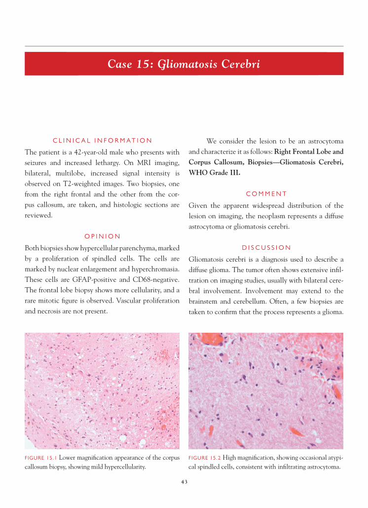
43
Case 15: Gliomatosis Cerebri
C L I N I C A L I N F O R M A T I O N
The patient is a 42-year-old male who presents with seizures and increased lethargy. On MRI imaging, bilateral, multilobe, increased signal intensity is observed on T2-weighted images. Two biopsies, one from the right frontal and the other from the cor-pus callosum, are taken, and histologic sections are reviewed.
O P I N I O N
Both biopsies show hypercellular parenchyma, marked by a proliferation of spindled cells. The cells are marked by nuclear enlargement and hyperchromasia. These cells are GFAP-positive and CD68-negative. The frontal lobe biopsy shows more cellularity, and a rare mitotic fi gure is observed. Vascular proliferation and necrosis are not present.
We consider the lesion to be an astrocytoma and characterize it as follows: Right Frontal Lobe and Corpus Callosum, Biopsies—Gliomatosis Cerebri, WHO Grade III.
C O M M E N T
Given the apparent widespread distribution of the lesion on imaging, the neoplasm represents a diffuse astrocytoma or gliomatosis cerebri.
D I S C U S S I O N
Gliomatosis cerebri is a diagnosis used to describe a diffuse glioma. The tumor often shows extensive infi l-tration on imaging studies, usually with bilateral cere-bral involvement. Involvement may extend to the brainstem and cerebellum. Often, a few biopsies are taken to confi rm that the process represents a glioma.
FIGURE 15.1 Lower magnifi cation appearance of the corpus callosum biopsy, showing mild hypercellularity.
FIGURE 15.2 High magnifi cation, showing occasional atypi-cal spindled cells, consistent with infi ltrating astrocytoma.

44
C A S E 1 5 : G L I O M A T O S I S C E R E B R I
The diagnosis is usually made by correlating the biopsy results with the imaging fi ndings. Morphologically, the glioma component may resemble an astrocytoma, oli-godendroglioma, or mixed glioma. Although much of the tumor may appear low-grade, foci of higher-grade tumor may be present. Because tumor cells may be spindled, distinction of this entity from a pathologic process marked by prominent microglial cell prolifera-tion may be challenging. Immunohistochemistry can be helpful in these circumstances, with a combina-tion of GFAP and CD68 staining; this allows for the assessment of the lineage of the spindled cells. Progno-sis corresponds to a WHO grade III neoplasm.
References 1. Jennings MT, Frenchman M, Shahab T, et al. Gliomatosis
cerebri presenting as intractable epilepsy during early child-hood. J Child Neurol 1995;10:37–45.
2. Kros JM, Zheng P, Dinjens WNM, et al. Genetic aberrations in gliomatosis cerebri support monoclonal tumorigenesis. J Neuropathol Exp Neurol 2002;61:806–14.
3. Mawrin C. Molecular genetic alterations in gliomatosis cerebri: what can we learn about the origin and course of the disease? Acta Neuropathol 2005;110:527–36.
FIGURE 15.4 The frontal lobe biopsy shows more in the way of cellularity, and a rare mitotic fi gure is present.
FIGURE 15.5 Subpial aggregation of spindled tumor cells is present in the frontal lobe biopsy.
FIGURE 15.3 Another area from the corpus callosum biopsy, showing an increased number of spindled cells.

45
Case 16: Pilocytic Astrocytoma
C L I N I C A L I N F O R M A T I O N
The patient is a 3-year-old male who presents with a history of dysconjugate gaze since infancy. A minor accident with a toy prompts an emergency depart-ment visit and, after physical examination, imaging studies are obtained. A mass is identifi ed in the mid-line of the cerebellum. A decision is made to resect the lesion, and histologic sections are reviewed.
O P I N I O N
Biopsies show a moderately hypercellular glial tumor that, in most areas, forms a solid mass but, at the edges, shows individual cell infi ltration of cerebellar parenchyma. Adjacent cerebellar folia demonstrate neuronal loss and gliosis as a result of compression from the long-standing, low-grade tumor. The tumor
is biphasic and composed of zones of compact, highly fi brillated astrocytes, alternating with loose-textured microcystic areas containing cells with short, mul-tipolar processes. The tumor is focally calcifi ed and extends into leptomeninges.
We consider the pathology to be that of a low- grade astrocytoma and characterize it as follows: Midline Cerebellum, Excision—Pilocytic Astrocy-toma, WHO Grade I.
C O M M E N T
There are no high-grade features in this neoplasm. Extension into the leptomeninges is a common fi nding in pilocytic astrocytoma, especially in cases involving cerebellum or optic nerve, and does not
FIGURE 16.1 Section showing a biphasic tumor, composed of compact, eosinophilic areas of highly fi brillary astrocytes,alternating with looser microcystic areas.
FIGURE 16.2 Non-neoplastic cerebellar folia, adjacent to the tumor, show atrophy from compression by this long-standing, low-grade tumor; there has been loss of both Pur-kinje cell and granule cell neurons.

46
C A S E 1 6 : P I L O C Y T I C A S T R O C Y T O M A
indicate aggressive or malignant behavior, nor does it predict eventual cerebrospinal fl uid spread. Similarly, although most of the tumor is discrete and cohesive, infi ltrating cells at the edge of a pilocytic astrocytoma do not predict recurrence or affect prognosis.
D I S C U S S I O N
Pilocytic astrocytoma is an important variant of astrocytoma, to distinguish from diffuse infi ltrating high-
er-grade astrocytomas, because pilocytic astrocytoma has a much better prognosis. Pilocytic astrocytoma can be seen in both adults and children, but it is the most common glioma in children. Characteristic anatomic locations for this tumor include cerebellum, optic nerve, thalamus, basal ganglia, cerebellum, brainstem (dorsally as an exophytic lesion), and spinal cord. Tumor location infl uences prognosis; hypothalamic and brainstem sites usually preclude total removal, and
FIGURE 16.5 Dense, compact areas contain variable num-bers of eosinophilic, sausage-shaped Rosenthal fi bers.
FIGURE 16.3 High magnifi cation illustrates the coarse, “hair-like” piloid, bipolar cell processes in the denser areas of tumor.
FIGURE 16.4 Dense, pilocytic areas of tumor manifest strong GFAP immunoreactivity.
FIGURE 16.6 Rosenthal fi bers should be distinguished from erythrocytes in tissue and can be highlighted by trichrome stain.

47
C A S E 1 6 : P I L O C Y T I C A S T R O C Y T O M A
tumor can eventually, after years or decades, cause patient demise because of compromise of critical brain functions. Nevertheless, in many instances, even with known residual tumor, growth can be absent, and tumor may even regress. Progression to higher grades (i.e., malignant transformation) almost never occurs unless the patient has received radiotherapy.
Histologically, diagnosis rests on identify-ing a biphasic tumor, with the dense compact areas containing coarsely fi brillar astrocytes with bipolar, “hair-like” processes that are strongly immunoreac-tive for GFAP. Rosenthal fi bers (amorphous, sausage-shaped, eosinophilic structures that are easily seen on hematoxylin and eosin stain) are more frequent in compact areas, but they may be very irregular in distribution and are not absolutely required for the
FIGURE 16.7 Looser, microcystic areas often contain pools of pale, eosinophilic mucin; nearby astrocytes manifest short, multipolar processes rather than coarse pilocytic features.
FIGURE 16.8 Calcifi cations are an infrequent fi nding in pilocytic astrocytomas.
FIGURE 16.9 Extension of pilocytic astrocytoma to lep-tomeninges is common and does not indicate aggressive tumor or foreshadow cerebrospinal fl uid metastases.
FIGURE 16.10 Pilocytic astrocytomas grow as relatively solid tumor masses and thus envelope few neurofi lament-positive axons except near the edge of the tumor.

48
C A S E 1 6 : P I L O C Y T I C A S T R O C Y T O M A
astrocytoma displaces normal brain tissue, leading to few anti- neurofi lament-positive axons within the tumor, compared to diffuse astrocytomas, which tend to infi ltrate between axons and preserve them deep within the infi ltrating tumor mass. Mitoses are infre-quent in pilocytic astrocytomas but can occasionally be seen. MIB-1 cell cycle labeling index is usually less than 4%.
Abundant vasculature is characteristic of pilo-cytic astrocytoma. Both hyalinized and “glomeruloid” vessels can be seen, with the latter accounting for the enhancement seen preoperatively on neuroimag-ing studies. Glomeruloid vessels often are arranged in chains near the edge of macroscopic cysts, or in clusters, and should not prompt the pathologist to overgrade the tumor. Calcifi cations are relatively in-frequent in pilocytic astrocytoma but are thought to be a “regressive” change, as is the occasional fi nding of necrosis. Degenerative features include nuclear hyperchromatism, pleomorphism, and nuclear pseu-doinclusions and also do not affect prognosis.
References 1. Kleinschmidt-DeMasters BK, Prayson RA. An algorithmic
approach to the brain biopsy—Part 1. Arch Pathol Lab Med 2006;130:1630–8.
2. Perry, A. Glial and glioneuronal tumors. In: Prayson RA, edi-tor. Neuropathology. Philadelphia, PA: Elsevier Churchill Livingstone;2005. p. 434–8.
3. Scheithauer BW, Hawkins C, Tihan T, et al. Pilocytic astro-cytoma. In: Louis DN, Ohgaki H, Wiestler OD, Cavenee WK, editors. WHO Classifi cation of Tumours of the Central Nervous System. Lyon, FR: IARC Press;2007. p. 14–21.
diagnosis. Eosinophilic granular bodies (EGBs), which are composed of proteinaceous material, can also be found in some pilocytic astrocytomas and are highlighted by periodic acid Schiff stain. Neither Rosenthal fi bers nor EGBs are specifi c to pilocytic astrocytoma, or even to neoplastic conditions, but both tend to be seen in low-grade tumors (i.e., WHO grades I and II), such as ganglioglioma, pilocytic astro-cytoma, pleomorphic xanthoastrocytoma, and subep-endymoma, when they do occur in glial neoplasms.
Looser microcystic areas in pilocytic astrocyto-mas may show small cysts, fi lled with pools of pale, eosinophilic mucin. Astrocytes in these areas have short, blunted cell processes and stain considerably less well with GFAP. More solid growth of pilocytic
FIGURE 16.11 Pilocytic astrocytoma may show an infi ltra-tive margin; this also does not have prognostic signifi cance, but tumor in this area cannot be distinguished from diffuse astrocytoma.

49
Case 17: Pilomyxoid Astrocytoma
C L I N I C A L I N F O R M A T I O N
The patient is a 6-month-old male who presents with failure to thrive and abnormal head shape. On imag-ing, a heterogeneously enhancing, hypothalamic-chiasmatic mass is identifi ed. Subtotal resection is performed, and histologic sections are reviewed.
O P I N I O N
The tumor is a mildly hypercellular, monomorphic lesion, with clustering of tumor cells around blood ves-sels (“angiocentric” tumor cell arrangement). Unlike pilocytic astrocytomas, no biphasic pattern, coarsely fi brillated “piloid” astrocytes, Rosenthal fi bers, or eosinophilic granular bodies are identifi ed in this relatively monotonous neoplasm. Tumor cells have
small, bland, round-to-oval nuclei and wispy, delicate, eosinophilic cytoplasm. Mitotic fi gures are variably observed but are few in this case. There is no necrosis.
We consider the fi ndings in this lesion to be those of a low-grade glioma and characterize it as follows: Hypothalamic/Optic Chiasm, Excision— Pilomyxoid Astrocytoma, WHO Grade II.
C O M M E N T
The bland cytological appearance of the lesion, cou-pled with mild hypercellularity and absence of mito-ses or necrosis, belies the often aggressive nature of this tumor.
D I S C U S S I O N
Pilomyxoid astrocytoma is a relatively newly described tumor, which is a variant of a pilocytic
FIGURE 17.1 Low magnifi cation view of the biopsy, show-ing a mildly hypercellular neoplasm with a monomorphic population of delicate wispy cells; note absence of Rosen-thal fi bers or eosinophilic granular bodies.
FIGURE 17.2 Some regions of tumor manifest prominent angiocentricity of tumor cells.
50
C A S E 1 7 : P I L O M Y X O I D A S T R O C Y T O M A
astrocytoma but has been assigned a WHO grade of II (rather than a grade of I, as for pilocytic astrocy-toma), to refl ect the more aggressive biological course of pilomyxoid compared to pilocytic astrocytoma. Pilomyxoid astrocytomas may manifest local recur-rence, as well as cerebrospinal fl uid metastases, and patients can succumb to locally destructive effects
of tumor on the hypothalamus. The typical patient is less than 2 years of age, with a median age of 10 months. The hypothalamic-chiasmatic region is the most common location, although bona fi de examples have been described in sites frequented by pilocytic astrocytoma, including cerebellum, brainstem, and spinal cord. Occasional pilomyxoid astrocytomas evolve into typical pilocytic astrocytomas over time,
FIGURE 17.3 High magnifi cation, showing the charac-teristic features of tumor cells, including low nuclear-to-cytoplasmic ratio, bland nuclear features, delicate wispy eosinophilic cytoplasm, and absence of mitoses.
FIGURE 17.4 Mucin background is variably present in pilomyxoid astrocytoma but may be prominent in some examples.
FIGURE 17.5 MIB-1 is variable and cannot be used to distinguish most cases of pilomyxoid from pilocytic astrocytoma.
FIGURE 17.6 GFAP highlights delicate wispy cytoplasm in tumor cells, contrasting with the more coarsely pilocytic cytoplasm in pilocytic astrocytoma.

51
C A S E 1 7 : P I L O M Y X O I D A S T R O C Y T O M A
suggesting a common origin for these two tumors. Unfortunately, this does not happen for all patientswith pilomyxoid astrocytoma and, indeed, this child succumbed 6 months after his diagnosis.
Histologically, the tumor is only moderately hypercellular and usually does not show necrosis. Often, pilomyxoid astrocytoma does not demonstrate any higher mitotic rate than ordinary pilocytic astro-cytoma. The diagnosis of pilomyxoid astrocytoma is made easier if the MIB-1 rate is high, but the range of labeling index is quite broad, from 2–20%, and overlaps at the lower end with pilocytic astrocytoma. Hence, correct diagnosis requires recognition of the architectural pattern, including angiocentricity of tumor cells, monomorphic appearance, mucin pools, and absence of Rosenthal fi bers. Immunostaining of
pilocytic astrocytoma with GFAP often shows thin-ner and more delicate, wispy tumor cell processes in tumor cells than those seen in coarsely fi brillated pilo-cytic astrocytoma. Very focal areas, with pilomyxoid-like histology within an otherwise ordinary pilocytic astrocytoma, should not prompt this diagnosis.
References 1. Komakula ST, Fenton LZ, Kleinschmidt-DeMasters BK, et al.
Pilomyxoid astrocytoma: neuroimaging with clinicopatho-logic correlates in 4 cases followed over time. J Pediatr Hema-tol Oncol 2007;29:465–70.
2. Scheithauer BW, Hawkins C, Tihan T, et al. Pilocytic astro-cytoma. In: Louis DN, Ohgaki H, Wiestler OD, Cavenee WK, editors. WHO Classifi cation of Tumours of the Central Nervous System. Lyon, FR: IARC Press; 2007. p. 20–21.
3. Tihan T, Fisher PG, Kepner JL, et al. Pediatric astrocytomas with monomorphous pilomyxoid features and a less favorable outcome. J Neuropathol Exp Neurol 1999;58:1061–8.

52
Case 18: Subependymal Giant Cell Astrocytoma
C L I N I C A L I N F O R M A T I O N
The patient is a 12-year-old male who presents with history of seizures and skin lesions in the nasolabial fold region. On physical exam, he is also noted to have periungual fi broma on the third digit of the right hand and pitting in the dental enamel of several of his teeth. On imaging, a large, contrast-enhancing, lateral ventricular mass is identifi ed. The lesion is resected, and histologic sections are reviewed.
O P I N I O N
Histologic sections show a mass marked by a pro-liferation of large cells with abundant eosinophilic cytoplasm and eccentrically placed nuclei with promi-nent nucleoli. Occasionally, these cells appear to be arranged around blood vessels. Focally, perivascular sclerosis is observed. Vascular proliferative changes,
prominent mitotic activity, and necrosis are not observed. Microcalcifi cations are present. In areas, the tumor cells assume a more spindled appearance. A Ki-67 labeling index of 0.8 is observed in the tumor.
We consider the lesion to be a low-grade astrocytic neoplasm and characterize it as follows: Lateral Ventricle, Excision—Subpendymal Giant Cell Astrocytoma, WHO Grade I.
C O M M E N T
The clinical fi ndings, in this patient, along with the diagnosis of subependymal giant cell astrocytoma, suggest that the patient may have tuberous sclerosis.
D I S C U S S I O N
Subependymal giant cell astrocytomas (SEGA) repre-sent low-grade, well-demarcated, predominantly intra-ventricular tumors. There is a well-known association
FIGURE 18.1 Low magnifi cation, appearance marked by a sheetlike proliferation of large cells. FIGURE 18.2 Foci of perivascular sclerosis are present.

53
C A S E 1 8 : S U B E P E N D Y M A L G I A N T C E L L A S T R O C Y T O M A
of this tumor with tuberous sclerosis, although the tumor may be encountered outside the setting of tuber-ous sclerosis. It is thought that SEGAs evolve from enlargement of hamartomatous subependymal nodules, which are commonly encountered in the lateral ven-tricular walls in patients with tuberous sclerosis. On imaging, they are contrast-enhancing and may show variable evidence of calcifi cation.
Histologically, the SEGA, as its name sug-gests, is marked by a proliferation of large astro-cytic cells, with abundant eosinophilic cytoplasm
and eccentrically placed nuclei with prominent nucleoli. GFAP immunoreactivity confi rms the astrocytic nature of these cells. This component is usually admixed with a spindle-cell component. Evidence of higher-grade tumor, such as necrosis, vascular proliferative changes, and mitotic activity, is unusual in these tumors. Cell proliferation mark-ers indicate a very low rate of cell proliferation, as in the current case.
FIGURE 18.3 Mild perivascular chronic infl ammation, asso-ciated with perivascular sclerosis.
FIGURE 18.4 Focal microcalcifi cations are present in the tumor.
FIGURE 18.5 The spindle-cell region of the tumor.
FIGURE 18.6 Higher magnifi cation appearance of the large, rounded astrocytic cells, with abundant eosinophilic cyto-plasm and large nuclei with prominent nucleoli.
54
C A S E 1 8 : S U B E P E N D Y M A L G I A N T C E L L A S T R O C Y T O M A
The importance of the diagnosis lies in recog-nizing the association with tuberous sclerosis. The tumor is considered benign and potentially curable with surgical excision. Many people have viewed the tumor more as a hamartoma than as a true malignancy. Differential diagnostic considerations include distin-guishing this tumor from a gemistocytic astrocytoma, neuronal tumor, such as gangliocytoma or ganglio-glioma, and glioblastoma. Gemistocytic astrocytomas are generally not intraventricular lesions and are more infi ltrative by nature. Mitotic activity and increased rates of cell proliferation are more commonly encoun-tered in the gemistocytic astrocytoma. The large cells, with prominently nucleolated nuclei, may ostensibly resemble neuronal cells and suggest ganglioglioma in the differential diagnosis. Immunohistochemistry, in such cases, can be useful in delineating the glial nature of the large cells. Glioblastoma multiforme involving the ventricle demonstrate evidence of necrosis and/or
vascular proliferative changes, usually accompanied by readily identifi able mitotic activity. Although the intraventricular location of the SEGA may also raise ependymomas as a differential diagnostic possibil-ity, the astrocytic cells in SEGA are generally much larger than the cells observed in an ependymoma; prominent nucleolation is also an unusual fi nding in ependymoma.
References 1. Gyure KA, Prayson RA. Subependymal giant cell astrocytoma:
a clinicopathologic study with HMB45 and MIB-1 immuno-histochemical analysis. Mod Pathol 1997;10:313–17.
2. Kim SK, Wang KC, Cho BK, et al. Biological behavior and tumorigenesis of subependymal giant cell astrocytomas. J Neurooncol 2001;52:217–25.
3. Lopes MB, Altermatt HJ, Scheithauer BW, et al. Immunohis-tochemical characterization of subependymal giant cell astro-cytomas. Acta Neuropathol (Berl) 1996;91:368–75.
4. Shepherd CW, Scheithauer BW, Gomez MR, et al. Subependymal giant cell astrocytoma: a clinical, pathologi-cal, and fl ow cytometric study. Neurosurgery 1991;28:864–8.

55
Case 19: Low-Grade Oligodendroglioma
C L I N I C A L I N F O R M A T I O N
The patient is a 32-year-old male who presents with focal left-sided arm weakness and a right frontal- parietal lobe mass. On MRI studies, a hypointense lesion on T1-weighted images, measuring approxi-mately 3.2 cm, is observed. T2-weighted images show a well-demarcated lesion with little edema. Histologic sections from a subtotal resection are reviewed.
O P I N I O N
Histologic sections show an infi ltrative neoplasm, marked by mild hypercellularity, most pronounced in the white matter. The tumor cells are characterized by a generally round nuclear shape, a vesicular chro-matin pattern, an absence of prominent nucleolation, and scant cytoplasm. Focally, pericellular clearing is
observed in a subpopulation of these cells. An arcu-ate capillary vascular pattern is observed in the back-ground of the tumor. Focal microcystic changes are also present. Tumor cells show a propensity to satel-lite around neurons and blood vessels as they infi ltrate the cortex. Focal aggregation of tumor cells under-neath the pial surface is also present. Focal dystro-phic calcifi cation is observed in the neoplasm. Only rare mitotic fi gures are noted. Vascular proliferative changes and necrosis are not seen. A Ki-67 labeling index of 3.2 is noted. Evidence of allelic loss on chro-mosomes 1p and 19q is observed using fl uorescent in situ hybridization (FISH) analysis.
We consider the lesion to be an oligodendro-glioma and characterize it as follows: Right Frontal-Parietal Lobes, Subtotal Resection—Low-Grade Oligodendroglioma, WHO Grade II.
FIGURE 19.1 Low magnifi cation appearance of the tumor, showing a mildly hypercellular white matter.
FIGURE 19.2 Pericellular clearing or “fried egg” appearance of tumor cells is present in this low-grade oligodendroglioma.

56
C A S E 1 9 : L O W - G R A D E O L I G O D E N D R O G L I O M A
C O M M E N T
The tumor demonstrates mild nuclear pleomorphism and rare mitotic fi gures. Vascular proliferative changes and necrosis are not observed. These fi ndings, in con-junction with a relatively low Ki-67 labeling index, are consistent with a low-grade oligodendroglioma.
D I S C U S S I O N
Low-grade oligodendrogliomas are much less common than their astrocytoma counterparts, com-prising approximately 5–6% of all gliomas. The clini-cal presentation of these tumors overlaps with that of their fi brillary astrocytoma counterparts. Similar to astrocytomas, these tumors typically arise in the white matter of the cerebral hemispheres, with the frontal lobe being the most common location. Imag-ing studies often show a fairly well-circumscribed tumor with minimal edema. Evidence of calcifi ca-tions and hemorrhage are common in these neo-plasms. Despite their apparent circumscription on imaging studies, these tumors microscopically are infi ltrative by nature, and it is not unusual for tumor cells to extend into the cortex and aggregate under the subpial surface. Morphologically, these tumors can be distinguished from astrocytomas because of their generally round nuclear contour and their overall monotony. They typically lack the hyper-chromasia that is more commonly encountered in astrocytic tumors. The presence of calcifi cations, although quite common in oligodendrogliomas, can be observed in astrocytomas. Pericellular clearing, or
FIGURE 19.3 High magnifi cation appearance of the tumor, showing a monomorphic population of cells with rounded nuclei and a background arcuate capillary vascular pattern.
FIGURE 19.4 Oligodendroglioma infi ltrating cortex, show-ing satellitosis of tumor cells around pre-existing neurons and blood vessels.
FIGURE 19.5 Focal microcystic degenerative change is present in this tumor.

57
C A S E 1 9 : L O W - G R A D E O L I G O D E N D R O G L I O M A
FIGURE 19.6 A rare mitotic fi gure is observed in this tumor. Rare mitotic fi gures are not suffi cient to warrant upgrading the tumor.
the “fried egg” appearance, is also a somewhat char-acteristic feature of these tumors, but it represents an artifact of delayed formalin fi xation. Therefore, the “fried egg” change is not evident at frozen sec-tion or in tissue that has been promptly fi xed. The prominent arcuate capillary network is also a salient feature of these process-poor tumors.
Unfortunately, there is a lack of specifi c im-munomarkers for oligodendroglial tumors. Prolifera-
tion markers, such as Ki-67, demonstrate indices in the low-grade tumors that are comparable to those observed with the grade II astrocytomas. The majority of low-grade oligodendrogliomas demonstrate large deletions on chromosomes 1p and 19q. The presence of these fi ndings has been correlated, particularly in the higher-grade oligodendrogliomas, with better
FIGURE 19.7 Focal dystrophic mineralization is seen.
FIGURE 19.8 Subpial aggregation of tumor cells is noted. FIGURE 19.9 In situ hybridization study (FISH), show-ing evidence of loss on chromosome 1p in a low-grade oligodendroglioma.
58
C A S E 1 9 : L O W - G R A D E O L I G O D E N D R O G L I O M A
References 1. Burger PC, Rawlings CE, Cox EB, et al. Clinicopathologic cor-
relations in the oligodendroglioma. Cancer 1987;59:1345–52. 2. Coons SW, Johnson PC, Pearl DK. The prognostic sig-
nifi cance of Ki-67 labeling indices for oligodendrogliomas. Neurosurgery 1997;41:878–84; discussion 884–5.
3. Mork SJ, Halvorsen TB, Lindegaard KF, et al. Oligodendro-glioma. Histologic evaluation and prognosis. J Neuropathol Exp Neurol 1986; 45:65–78.
4. Reifenberger G, Kros JM, Louis DN, et al. Oligodendro-glioma. In: Louis DN, Oghaki H, Wiestler OD, Cavenec WK, editors. WHO classifi cation of tumours of the central nervous system. Lyon, FR: IARC Press, 2007.
5. Wiestler OD, Cavenee W, eds. WHO classifi cation of tumours of the central nervous system. Lyon, FR: IARC Press; 2007. pp 54–9.
prognosis and chemoresponsiveness. Other tumors that morphologically may consist of rounded cells with pericellular clearing, such as central neurocy-toma and dysembryoplastic neuroepithelial tumors, generally lack the large chromosomal deletions that mark oligodendrogliomas.
The importance of distinguishing this tumor from an astrocytoma lies in the treatment approach and, ultimately, in the prognosis. The majority of oli-godendrogliomas are chemoresponsive, and a trial of chemotherapy is warranted in the majority of cases. Grade for grade, oligodendrogliomas have a signifi -cantly better prognosis than astrocytomas.
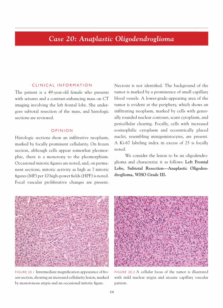
59
Case 20: Anaplastic Oligodendroglioma
C L I N I C A L I N F O R M A T I O N
The patient is a 49-year-old female who presents with seizures and a contrast-enhancing mass on CT imaging involving the left frontal lobe. She under-goes subtotal resection of the mass, and histologic sections are reviewed.
O P I N I O N
Histologic sections show an infi ltrative neoplasm, marked by focally prominent cellularity. On frozen section, although cells appear somewhat pleomor-phic, there is a monotony to the pleomorphism. Occasional mitotic fi gures are noted, and, on perma-nent sections, mitotic activity as high as 7 mitotic fi gures (MF) per 10 high-power fi elds (HPF) is noted. Focal vascular proliferative changes are present.
Necrosis is not identifi ed. The background of the tumor is marked by a prominence of small capillary blood vessels. A lower-grade-appearing area of the tumor is evident at the periphery, which shows an infi ltrating neoplasm, marked by cells with gener-ally rounded nuclear contours, scant cytoplasm, and pericellular clearing. Focally, cells with increased eosinophilic cytoplasm and eccentrically placed nuclei, resembling minigemistocytes, are present. A Ki-67 labeling index in excess of 25 is focally noted.
We consider the lesion to be an oligodendro-glioma and characterize it as follows: Left Frontal Lobe, Subtotal Resection—Anaplastic Oligoden-droglioma, WHO Grade III.
FIGURE 20.1 Intermediate magnifi cation appearance of fro-zen section, showing an increased cellularity lesion, marked by monotonous atypia and an occasional mitotic fi gure.
F IGURE 20.2 A cellular focus of the tumor is illustrated with mild nuclear atypia and arcuate capillary vascular pattern.

60
C A S E 2 0 : A N A P L A S T I C O L I G O D E N D R O G L I O M A
C O M M E N T
The degree of cellularity, presence of prominent mitotic activity, and vascular proliferative changes warrant a diagnosis of anaplastic oligodendroglioma. The Ki-67 labeling index is consistent with this diagnosis.
D I S C U S S I O N
The monotony of cells on frozen section is sugges-tive of the diagnosis. In the vast majority of cases,
differentiating a high-grade astrocytoma from a high-grade oligodendroglioma, in the context of intraop-erative consultation, is not critical. Because of the differences in prognosis and treatment approach, however, accurate classifi cation of the tumor for pur-poses of fi nal diagnosis is signifi cant.
Morphologically, distinction of high-grade oli-godendroglioma from low-grade oligodendroglioma is
FIGURE 20.3 High magnifi cation appearance, showing moderate nuclear pleomorphism and readily identifi able mitotic activity.
FIGURE 20.4 A focal area around the edge of the tumor shows an appearance resembling a low-grade oligodendro-glioma, with pericellular clearing, or “fried-egg” change, which is an artifact of delayed formalin fi xation.
FIGURE 20.5 Vascular proliferative changes are focally present.
FIGURE 20.6 Minigemistocytic cells are marked by increased cytoplasm and eccentrically placed nuclei.

61
C A S E 2 0 : A N A P L A S T I C O L I G O D E N D R O G L I O M A
somewhat subjective. In general, the parameters that are used to grade astrocytomas (cellularity and aty-pia, increased mitotic activity, vascular proliferative changes, and necrosis) are also evaluated in grading oligodendroglial neoplasms. Oligodendrogliomas have only two grade designations, compared with the three-tiered grading system for fi brillary astrocytomas. In gen-eral, anaplastic oligodendroglioma are more cellular and pleomorphic than low-grade tumors, with increased mi-totic activity (generally in excess of 5–6 MF per 10 HPF) and may variably demonstrate other worrisome morpho-logic features, including vascular proliferation and/or necrosis. The presence of minigemistocytes may cause confusion with an astrocytoma; these cells are accept-able within the context of oligodendroglioma and do not afford any particular prognosis when present. Oc-casional tumors may have small hypercellular nodules or foci within an otherwise low-grade-appearing tumor. The signifi cance of these small foci is uncertain. Simi-lar to astrocytomas, the Ki-67 labeling indices roughly correlate with tumor grade, with higher-grade tumors generally having higher labeling indices.
The major differential diagnostic consideration is high-grade astrocytoma. On purely morphologic
grounds, this may, at times, be diffi cult. As in the current case, a lower-grade-appearing area of the tu-mor at the perimeter presents a clue as to the prop-er diagnosis. Immunohistochemistry is generally of little avail in distinguishing these two tumor types. Molecular genetic alterations can be helpful in this instance. Large deletions on chromosomes 1p and 19q have been well established to be associated with the oligodendroglioma phenotype. Smaller deletions on one or both chromosomes may be occasionally encountered in glioblastoma. However, it is impor-tant to remember that these chromosomal alterations may not always be present. Genetic alterations more commonly encountered in glioblastomas, including losses on chromosome 10q, p53 mutations, and EGFR amplifi cation or overexpression, are not commonly observed in anaplastic oligodendrogliomas.
Despite the high-grade designation, many of these tumors are remarkably responsive to chemo-therapy and radiotherapy. Assessment of chromosome 1p/19q status in these tumors can be helpful in pre-dicting which tumors are more likely to respond to treatment and, ultimately, allow for a longer survival.
References 1. Cairncross JG, Ueki K, Zlatescu MC, et al. Specifi c genetic
predictors of chemotherapeutic response and survival in patients with anaplastic oligodendrogliomas. J Natl Cancer Inst 1998;90:1473–9.
2. Coons SW, Johnson PC, Pearl DK. The prognostic sig-nifi cance of Ki-67 labeling indices for oligodendrogliomas. Neurosurgery 1997;41:878-84; discussion 884–5.
3. Giannini C, Scheithauer BW, Weaver AL, et al. Oligo-dendrogliomas: reproducibility and prognostic value of his-tologic diagnosis and grading. J Neuropathol Exp Neurol 2001;60:248–62.
4. Ino Y, Betensky RA, Zlatescu MC, et al. Molecular subtypes of anaplastic oligodendroglioma: implications for patient management at diagnosis. Clin Cancer Res 2001;7:839–45.
5. Miller CR, Dunham CP, Scheithauer BW, et al. Signifi cance of necrosis in grading of anaplastic oligodendroglial tumors: a clinicopathological and genetic study of 916 high-grade gliomas. J Clin Oncol 2006;24:5419–26.
FIGURE 20.7 The interface is present between a lower-grade-appearing area of the tumor on the left and a more cellular, higher-grade-appearing area of the tumor on the right.

62
Case 21: Low-Grade Oligoastrocytoma (Low-Grade Mixed Glioma)
C L I N I C A L I N F O R M A T I O N
The patient is a 38-year-old male who presents with seizures and a right frontal-temporal lobe mass on imaging. A subtotal resection of the lesion is per-formed, and histologic sections are reviewed.
O P I N I O N
Approximately 60% of the lesion is marked by a mild hypercellularity and a proliferation of cells with rounded nuclear contour and pericellular clearing, resembling a low-grade oligodendroglioma. Mitotic fi g-ures, vascular proliferative changes, necrosis, and cal-cifi cations are not observed. Approximately 20–25% of the tumor shows areas resembling low-grade fi bril-lary astrocytoma, marked by a proliferation of more elongated nuclei and high nuclear-to- cytoplasmic ratio. Again, mitotic fi gures, vascular proliferative
changes, and necrosis are not identifi ed in these later areas. The remainder of the tumor consists of a mix-ture of the two aforementioned cell types. A Ki-67 labeling index of 2.8 is noted.
We consider the lesion to be a low-grade gli-oma and characterize it is as follows: Right Frontal-Temporal Lobes, Subtotal Resection—Low-Grade Oligoastrocytoma (Low-Grade Mixed Glioma), WHO Grade II.
C O M M E N T
The tumor consists of geographically distinct areas resembling low-grade oligodendroglioma and low-grade astrocytoma. Mitotic activity, vascular prolifer-ative changes, and necrosis are not observed. The low Ki-67 labeling index is consistent with a low-grade neoplasm.
FIGURE 21.1 Intermediate magnifi cation, showing an area of the tumor resembling a low-grade oligodendroglioma.
FIGURE 21.2 High magnifi cation area of tumor resembling oligodendroglioma with generally rounded nuclei.

63
C A S E 2 1 : L O W - G R A D E O L I G O A S T R O C Y T O M A ( L O W - G R A D E M I X E D G L I O M A )
D I S C U S S I O N
The clinical and imaging features of so-called oli-goastrocytoma, or mixed glioma, overlap with those of pure oligodendroglioma and pure fi brillary astro-cytoma. The pathologic criteria for the diagnosis of oligoastrocytoma remain somewhat nebulous. Most pathologists require geographically distinct areas of astrocytoma and oligodendroglioma in order to render the diagnosis. The percentages of the minor compo-
nent of tumor are not clearly delineated; percent-ages of the minor component in the literature range from 20–35%. Particularly challenging are tumors in which there appears to be an admixture of cell types in the same area. It is well recognized that, within tumors that are generally considered pure oligoden-drogliomas or pure fi brillary astrocytomas, there may be a small population of cells that phenotypically resemble the other tumor type intermixed in the neo-plasm. There is some literature to suggest that evalu-ation of mixed gliomas for deletions on chromosomes 1p and 19q can provide some guidance in terms of eventual prognosis and treatment; those tumors that demonstrate large deletions tend to behave more like oligodendrogliomas.
The histologic parameters used to grade oligo-dendrogliomas and astrocytomas, including cellular-ity, mitotic activity, vascular proliferative changes, and necrosis, are also used to assign grade in mixed gliomas. Cell proliferation indices generally show lower labeling indices in low-grade tumors, com-pared with higher-grade neoplasms. Because of the issues of problems with the defi nition of entity and the lack of uniform approach in terms of diagnosis
FIGURE 21.3 Small areas of the tumor show an admixture of oligodendroglioma and astrocytoma type cells.
FIGURE 21.4 Intermediate magnifi cation of an area of the tumor resembling a low-grade fi brillary astrocytoma.
FIGURE 21.5 High magnifi cation of an astrocytoma-like area of the tumor is marked by cells with more elongated nuclei.

64
C A S E 2 1 : L O W - G R A D E O L I G O A S T R O C Y T O M A ( L O W - G R A D E M I X E D G L I O M A )
of these tumors, it is diffi cult to ascertain from the literature what the true natural behavior of these tu-mors is. Their prognosis has generally been thought to be intermediate between pure astrocytomas and oligodendrogliomas.
References 1. Beckmann MJ, Prayson RA. A clinicopathologic study of
30 cases of oligoastrocytoma including p53 immunohisto-chemistry. Pathology 1997;29:159–64.
2. Fuller CE, Schmidt RE, Roth KA, et al. Clinical utility of fl uorescence in situ hybridization (FISH) in morphologically ambiguous gliomas with hybrid oligodendroglial/astrocytic features. J Neuropathol Exp Neurol 2003;62:1118–28.
3. Hart MN, Petito CK, Earle KM. Mixed gliomas. Cancer 1974;33:134–40.
4. Jaskolsky D, Zawieski M, Papierz W, et al. Mixed glio-mas. Their clinical course and results of surgery. Zentralbl Neurochir 1987;48:120–3.
5. Kraus JA, Koopmann J, Kaskel P, et al. Shared allelic losses on chromosomes 1p and 19q suggest a common origin of oligodendroglioma and oligoastrocytoma. J Neuropathol Exp Neurol 1995;54:91–5.

65
Case 22: Anaplastic Oligoastrocytoma (Anaplastic Mixed Glioma)
C L I N I C A L I N F O R M A T I O N
A 52-year-old female presents with right-sided weak-ness. On imaging studies, a focally enhancing tumor in the left frontal-parietal lobes is noted on MRI stud-ies. Subtotal resection of the lesion is performed, and histologic sections are reviewed.
O P I N I O N
Histologic sections show a glioma, marked by geographically distinct areas resembling oligoden-droglioma and astrocytoma. The astrocytoma areas are marked by mild nuclear pleomorphism, focal vascular proliferative changes, and identifi able mitotic activity. There is no evidence of necro-sis. Areas around the periphery of the lesion show
low-grade tumor resembling both oligodendroglioma and astrocytoma.
We consider the lesion to be a high-grade glioma and characterize it as follows: Left Frontal-Parietal Region, Subtotal Resection— AnaplasticOligoastrocytoma (Anaplastic Mixed Glioma), WHO Grade III.
C O M M E N T
The tumor is marked by geographically distinct areas resembling astrocytoma and oligodendroglioma. Moderate nuclear atypia, readily identifi able mitotic activity, and vascular proliferative changes are con-sistent with the diagnosis of anaplastic oligoastrocy-toma. There is no evidence of necrosis.
FIGURE 22.1 Focal areas of the tumor, particularly at the periphery, resemble a low-grade astrocytoma.
FIGURE 22.2 Other areas of the neoplasm look like low-grade oligodendroglioma.

66
C A S E 2 2 : A N A P L A S T I C O L I G O A S T R O C Y T O M A ( A N A P L A S T I C M I X E D G L I O M A )
D I S C U S S I O N
The diagnosis of anaplastic oligoastrocytoma is par-ticularly challenging. In addition to the defi nitional issues for mixed glioma discussed in Case 21, the degree of atypia and pleomorphism in these tumors make distinction from high-grade oligodendroglioma and glioblastoma multiforme, at times, challenging. The key to the diagnosis lies in the recognition of distinct areas of oligodendroglioma and astrocytoma.
Particularly helpful in this case is the presence of lower-grade, more readily recognizable phenotypes of both glioma types in the tumor. Similar to low-grade oligoastrocytomas, the status of chromosomes 1p and 19q serves as an important prognosticating factor.
Historically, a subset of these tumors demon-strated areas of necrosis. In the most recent update in the WHO classifi cation of brain tumors, anaplas-tic oligoastrocytomas with necrosis are classifi ed as glioblastoma with oligodendroglioma component, despite the fact that this particular lesion carries a better prognosis than ordinary glioblastoma.
References 1. Eoli M, Bissola L, Bruzzone MG, et al. Reclassifi cation of oli-
goastrocytomas by loss of heterozygosity studies. Int J Cancer 2006;119:84–90.
2. Mueller W, Hartmann C, Hoffmann A, et al. Genetic sig-nature of oligoastrocytomas correlates with tumor loca-tion and denotes distinct molecular subsets. Am J Pathol 2002;161:313–19.
3. Shaw EG, Scheithauer BW, O’Fallon JR, et al. Mixed oligoastrocytomas: a survival and prognostic factor analysis. Neurosurgery 1994;34:577–82.
FIGURE 22.3 Focal vascular proliferative changes are pres-ent in the tumor.
FIGURE 22.4 A cellular focus of tumor is situated in the cortex.
FIGURE 22.5 Mitotic fi gures are readily identifi able in the tumor.

67
Case 23: Glioblastoma with Oligodendroglioma Component
C L I N I C A L I N F O R M A T I O N
The patient is a 64-year-old male who presents with headaches and seizures. On imaging, a ring- enhancing mass is located in the right temporal lobe. Biopsies of the mass are taken, and histologic sections are reviewed.
O P I N I O N
Biopsies show a high-grade neoplasm, marked by prominent cellularity, moderate nuclear atypia, and focally prominent arcuate vascular pattern. Mitotic activity is readily observable in the tumor. Focal vascular proliferative changes are noted. Areas of the tumor resemble an oligodendroglioma with cells marked by rounded nuclear contours and pericellular
clearing. Other areas of the tumor clearly resemble an astrocytoma with more elongated-appearing nuclei. Foci of necrosis, at times, rimmed by a pseudopalisade of cells and intravascular thrombi, are also present.
We consider the lesion to be a high-grade glio-ma and characterize it as follows: Right Temporal Lobe, Biopsies—Glioblastoma with Oligodendro-glioma Component, WHO Grade IV.
C O M M E N T
The tumor is marked by geographic areas resem-bling high-grade oligodendroglioma and high-grade astrocytoma. In addition, focal palisaded necrosis is observed. The tumor’s morphologic appearance is most consistent with a glioblastoma with oligoden-droglioma component.
FIGURE 23.1 The tumor is markedly cellular, with moder-ate nuclear atypia and a focally prominent arcuate capillary vascular pattern.
FIGURE 23.2 Mitotic fi gures are readily identifi able in the neoplasm.

68
C A S E 2 3 : G L I O B L A S T O M A W I T H O L I G O D E N D R O G L I O M A C O M P O N E N T
D I S C U S S I O N
Glioblastoma with oligodendroglioma component is a new designation in the WHO. Historically, these tumors were classifi ed as anaplastic oligoastrocy-tomas (anaplastic mixed gliomas). Recent studies have demonstrated that anaplastic mixed gliomas that demonstrate necrosis tended to behave more aggressively than anaplastic mixed gliomas without
necrosis. Although this variant of glioblastoma is not felt to be quite as aggressive as the ordinary glio-blastoma, the behavior of this lesion is thought to be more akin to glioblastoma than to anaplastic mixed glioma.
The challenges from a pathology standpoint are similar to those previously discussed in the prior case (Anaplastic Oligoastrocytoma).
FIGURE 23.3 Vascular proliferative changes are also present in the tumor.
FIGURE 23.4 Areas resembling oligodendroglioma are noted in the tumor.
FIGURE 23.5 Vascular proliferative changes are observable in the oligodendroglioma-like areas of the tumor.
FIGURE 23.6 Palisaded necrosis is present in the neoplasm, consistent with a high-grade glioma.

69
C A S E 2 3 : G L I O B L A S T O M A W I T H O L I G O D E N D R O G L I O M A C O M P O N E N T
References 1. He J, Mokhtari K, Sanson M, et al. Glioblastomas with an
oligodendroglial component: a pathological and molecular study. J Neuropathol Exp Neurol 2001;60:863–71.
2. Homme T, Fukushina T, Vaccarella S, et al. Correlation among pathology, genotype and patient outcomes in glioblas-toma. J Neuropathol Exp Neurol 2006;65:846–54.
3. Kraus JA, Lamszus K, Glesmann N, et al. Molecular genetic alterations in glioblastomas with oligodendroglial compo-nent. Acta Neuropathol 2001;101:311–20.
FIGURE 23.7 More elongated nuclear features, suggestive of an astrocytoma phenotype, are seen, associated with focal geographic necrosis and intravascular thrombi adjacent to the necrosis.

70
Case 24: Subependymoma
C L I N I C A L I N F O R M A T I O N
The patient is a 48-year-old male who presents with headaches. Work-up includes a MRI scan, which reveals a sharply demarcated, non-contrast-enhancing mass in the left lateral ventricle, measuring 2.5 cm in greatest dimension. A decision is made to excise the lesion, and histological sections are reviewed.
O P I N I O N
Biopsies show a hypocellular neoplasm, with a nodu-lar or lobular overall architectural pattern and multi-focal calcifi cations. Extensive microcystic change is present, but no necrosis is seen. The tumor is com-posed of clusters of small, cytologically bland, and monotonous cells, embedded in a densely eosinophilic
fi brillar background. Cellular areas alternate with anuclear eosinophilic zones. Vessels are focally hya-linized, but perivascular pseudorosettes are poorly developed, and mitotic activity is absent.
We consider the tumor to be a low-grade neoplasm and characterize it as follows: Left Lateral Ventricle, Excision—Subependymoma, WHO Grade I.
C O M M E N T
Microcystic change is often most prominent in lateral ventricle examples of subependymoma.
D I S C U S S I O N
Subependymomas occur most frequently in the fourth ventricle, followed by the lateral ventricle. Less common supratentorial sites include the third
FIGURE 24.1 Low-power magnifi cation illustrates the nod-ular or lobular architectural pattern of this tumor, which was attached to the wall of the lateral ventricle, but sharply demarcated from the adjacent subependymal brain tissue.
FIGURE 24.2 Clusters of bland monotonous cells alternate with anuclear zones composed of eosinophilic matrix.

71
C A S E 2 4 : S U B E P E N D Y M O M A
ventricle and septum pellucidum. In the spinal cord, subependymomas are most frequent in the cervical and cervico-thoracic levels.
Subependymomas are usually sharply demar-cated, lobular masses, with varying amounts of cal-cifi cation, vascular hyalinization, and sometimes even vascular proliferation and thrombosis. They may show recent or remote hemorrhage. Necrosis
and mitoses are usually absent. The striking pat-tern on low-power magnifi cation is that of clusters of monotonous cells, alternating with anuclear areas composed of dense eosinophilic fi brillar matrix. Mi-crocysts are usually most conspicuous in lateral ven-tricular examples and may be absent at other sites. True perivascular pseudorosettes can be discerned in some examples, but, in others, the observer must uti-lize their imagination.
Cytologically, the tumor cells are monotonous, with bland, round nuclei containing small nucleoli. In some examples, tumor cells show considerable nuclear pleomorphism, but this does not infl uence prognosis. Cytoplasm is coarsely fi brillar and strongly immunoreactive for GFAP. The microcysts contain faintly basophilic mucin. Mitoses are infrequent, and MIB-1 labeling indices are less than 1–2.
It is important to distinguish subependymo-mas from ependymomas because of their considerably better outcome. Surgical removal is curative. Neo-plasms with mixed ependymoma and subependymoma features also occur, but they should be graded accord-ing to the ependymoma component, which is usually WHO grade II.
FIGURE 24.3 Microcysts contain basophilic mucin and are particularly conspicuous in this lateral ventricle example of subependymoma.
FIGURE 24.4 Vessels are focally hyalinized, and tumor cells occasionally orient around the vessels, but perpendicularly oriented cell processes and fully developed perivascular pseudorosettes may be infrequent to absent.
FIGURE 24.5 High power magnifi cation shows the round nuclei, with small nucleoli and minimal hyperchromatism. Note the absence of mitotic activity.

72
C A S E 2 4 : S U B E P E N D Y M O M A
FIGURE 24.6 Strong immunoreactivity for GFAP is consis-tently identifi ed, although the GFAP may fail to disclose perpendicularly oriented fi brillary cell processes around blood vessels.
References 1. McLendon RE, Schiffer D, Rosenblum MK, et al.
Subependymoma. In: Louis DN, Ohgaki H, Wiestler OD, Cavenee WK, editors. WHO Classifi cation of Tumours of the Central Nervous System. Lyon, FR: IARC Press; 2007. p. 70–71.
2. Perry, A. Glial and glioneuronal tumors. In: Prayson RA, editor. Neuropathology. Philadelphia, PA: Elsevier Churchill Livingstone; 2005. p. 476–7.
FIGURE 24.7 The MIB-1 rate is low, usually less than 1.
3. Prayson RA, Suh JH. Subependymomas: clinicopathologic study of 14 tumors, including comparative MIB-1 immuno-histochemical analysis with other ependymal neoplasms. Arch Pathol Lab Med 1999;123:306–9.
73
Case 25: Myxopapillary Ependymoma
C L I N I C A L I N F O R M A T I O N
The patient is a 16-year-old female who presents with leg pain. Work-up includes an MRI scan, which reveals a sharply demarcated, intradural mass, 3.4 × 1.6 cm, lying just below the level of the conus medullaris and attached to the fi lum terminale. The decision is made to resect the lesion, and histological sections are reviewed.
O P I N I O N
Biopsies show a papillary neoplasm, composed of elon-gate cells that are radially arrayed around fi brovascular cores. There is abundant interspersed myxoid matrix, both between individual tumor cells and around blood vessels, as well as pooled in microcysts. Mitotic activity is very low, and necrosis is absent.
We consider the tumor to be a low-grade gliol neoplasm and characterize it as follows: Filum Ter-minale, Excision—Myxopapillary Ependymoma, WHO Grade I.
C O M M E N T
Myxopapillary ependymoma is almost confi ned to the conus medullaris-cauda equina-fi lum terminale region.
D I S C U S S I O N
Myxopapillary ependymomas display several different patterns, depending on how intact the perivascular pseudorosettes are in the tumor. In this example, per-pendicularly oriented delicate glial processes are still easily identifi ed around blood vessels. However, in
FIGURE 25.1 Intraoperative touch preparation shows the monotonous tumor cells and a well-developed perivascular pseudorosette.
FIGURE 25.2 Low power magnifi cation illustrates the radial arrangement of perivascular cells, as well as mucin lying between perpendicular cellular processes, seen here as rela-tively clear, vacuolated spaces.

74
C A S E 2 5 : M Y X O P A P I L L A R Y E P E N D Y M O M A
other examples of myxopapillary ependymoma, the glial cell processes are obliterated by the massive accu-mulation of mucin, leaving few visible blood vessels in the center of an epithelial-like circle of cells. Tumors with more mucin and fewer visible perivascular cell processes are easily misinterpreted by the unwary as metastatic adenocarcinoma. Extremely large amounts of mucin in tumors may also raise consideration of chordoma or myxoid chrondrosarcoma. Neither met-
astatic papillary adenocarcinoma nor most chordo-mas have strong diffuse immunoreactivity for GFAP. However, myxopapillary ependymomas, chordomas, and myxoid chondrosarcomas share immunoreactivity for S-100 protein, and both chordoma and metastatic papillary adenocarcinoma usually express cytokera-tins. Myxopapillary ependymomas, like almost all ependymomas, show immunostaining for GFAP, S-100, and vimentin, but not for cytokeratin.
FIGURE 25.3 Higher-power view shows the absence of mitotic activity, as well as the elongate, tapering, eosino-philic fi brillary glial processes.
FIGURE 25.4 Note the bland oval nuclei, with delicately stippled chromatin pattern.
FIGURE 25.5 Alcian-blue at pH 2.5 highlights the mucin between cell processes, as well as in microcyst pools.
FIGURE 25.6 Reticulin is seen only around blood vessels and not between individual tumor cells, as would be seen with schwannoma.
75
C A S E 2 5 : M Y X O P A P I L L A R Y E P E N D Y M O M A
The mucin is typically positive on staining with Alcian-blue. Mitoses are rare, and MIB-1 labeling is very low. Myxopapillary ependymomas are WHO grade I, with high-grade variants virtually unknown. Progno-sis is most favorable with complete surgical resection, which is usually achievable, with well- demarcated examples confi ned to the intradural space.
References 1. McLendon RE, Rosenblum MK, Schiffer D, et al.
Myxopapillary ependymoma. In: Louis DN, Ohgaki H, Wiestler OD, Cavenee WK, editors. WHO Classifi cation of Tumours of the Central Nervous System. Lyon, FR: IARC Press; 2007. p. 72–3.
2. Perry A. Glial and glioneuronal tumors. In: Prayson RA, editor. Neuropathology. Philadelphia, PA: Elsevier Churchill Livingstone; 2005. p. 476–7.
FIGURE 25.7 Strong immunoreactivity for GFAP high-lights the perpendicularly oriented fi brillary cell processes around blood vessels.
FIGURE 25.8 The MIB-1 rate is low, usually less than 1.

76
Case 26: Ependymoma
C L I N I C A L I N F O R M A T I O N
The patient is a 63-year-old male who presents with headache, nausea, and dizziness. Work-up includes an MRI scan, which reveals a large, well-circumscribed mass with heterogeneous contrast enhancement, fi ll-ing the fourth ventricle. The decision is made to resect the lesion, and histological sections are reviewed.
O P I N I O N
Biopsies show a moderately cellular neoplasm, with multifocal calcifi cations, but no necrosis. At low-power magnifi cation, perivascular pseudorosettes (defi ned as an eosinophilic, cell-process-rich collar of perpendicularly oriented glial cells surrounding a central blood vessel) are easily identifi ed. Vessels
are hyalinized, and mitotic activity is present only at low levels.
We consider the tumor to be an ependymoma and characterize it as follows: Fourth Ventricle, Excision—Ependymoma, WHO Grade II.
C O M M E N T
Ependymomas occur anywhere along the ventricu-lar system, as well as occasionally in extraventricular sites. The fourth ventricle and cervico-thoracic spinal cord are the most common locations for ependymo-mas, and these two sites are equally affected in adults. Most pediatric patients develop infratentorial tumors.
FIGURE 26.1 Low-power inspection shows a moderately cellular tumor, with a “pattern” consisting of prominent eosinophilic collars surrounding blood vessels.
FIGURE 26.2 Higher power confi rms that these eosinophilic collars are composed of perpendicularly oriented, coarsely fi brillar cell processes of the tumor cells, not hyalinized blood vessels. Note the nuclear monotony and absence of mitotic activity.

77
C A S E 2 6 : E P E N D Y M O M A
D I S C U S S I O N
Ependymomas are usually well-demarcated tumors that, in some instances, are quite bulky. Posterior fossa examples can fi ll the fourth ventricle, exuding out of the foramina of Luschka or Magendie. Obstruction of cerebrospinal fl uid pathways causes hydrocephalus, with resultant headache, nausea, vomiting, and dizziness. Examples in the spinal cord present with motor and sen-sory defi cits and may involve multiple intramedullary spinal cord segments. Some spinal cord ependymomas
are further associated with a cystic syrinx that increases the length of cord involvement. Uncommon supraten-torial ependymomas may manifest with seizures.
The diagnosis of ependymoma rests on recogniz-ing a specifi c pattern at low-power magnifi cation—a feature that usually differentiates an ependymoma from a diffuse astrocytoma that tends to be patternless. The most common pattern in ependymomas is the formation within the tumor of perivascular pseudorosettes, dis-tinguishable on low power as eosinophilic collars
FIGURE 26.3 Focal hemosiderin pigment, caused by intra-tumoral hemorrhage and occasional enlarged nuclei, can be seen in some cases but does not affect prognosis or grading.
FIGURE 26.4 Calcifi cations are variable and considered “regressive” features.
FIGURE 26.5 Vessels may show marked hyalinization.
FIGURE 26.6 High-power magnifi cation illustrates the stip-pled or speckled chromatin pattern of the round-to-oval tumor cell nuclei, as well as the delicate nucleoli.

78
C A S E 2 6 : E P E N D Y M O M A
surrounding blood vessels. Usually, these are present in multiple areas of a true ependymoma. Closer inspection at high power reveals these eosinophilic collars to be composed of perpendicularly oriented coarse glial processes. Perivascular pseudorosettes should not be confused with the eosinophilic, extensively hyalinized vasculature seen in other types of gliomas.
Less commonly, the ependymoma contains true ependymal canals with a central lumen; these
are seldom isolated features and usually occur in con-junction with pseudorosettes elsewhere in the tu-mor. Usually, true rosettes are also best appreciated at low-power magnifi cation but, rarely, the lumen of the central canal can be quite small and recognizable only on higher-power magnifi cation.
The last pattern to recognize at both low- and high-power magnifi cation that should prompt consideration of ependymoma is that of monoto-ny in nuclear size, shape, and chromatin pattern.
FIGURE 26.7 The coarse nature of the fi brillar tumor cell processes in perivascular pseudorosettes is typical of epen-dymomas and contrasts with the delicate, indistinct nature of cell processes in neurocytomas.
FIGURE 26.8 GFAP immunostaining is usually positive in perivascular pseudorosettes and can highlight the perpen-dicular arrangement of the glial processes.
FIGURE 26.9 Ring-like and dot-like immunoreactivity for EMA can be very helpful features.
FIGURE 26.10 Mitoses are few in number in grade II epen-dymomas, and the MIB-1 index is typically less than 4.

79
C A S E 2 6 : E P E N D Y M O M A
Ependymomas are composed of cells with round-to-oval nuclei, “salt and pepper” speckled chromatin, and small nucleoli. Appreciation of these features can be particularly critical in small tissue samples (such as from spinal cord) or in ependymomas with relatively few or poorly developed pseudorosettes.
In some instances, immunohistochemistry or electron microscopy is necessary to resolve the diag-nosis of ependymoma. Almost all ependymomas are positive for GFAP, but the strongest reaction is in the “glial” portions of the tumor, i.e., especially in the peri-vascular pseudorosettes. True ependymal canals often are GFAP-negative. Ependymomas are also immuno-reactive for S-100 protein and vimentin, but neither of these helps distinguish them from various types of gliomas. Dot-like and ring-like immunoreactivity for epithelial membrane antigen (EMA) is slightly more specifi c but has been seen in other types of gliomas. Ultimately, in diffi cult cases, electron microscopy may be necessary to establish ependymal features.
Once the categorization of the tumor as ependymoma is established, grading becomes the next issue. Ependymomas do not generally show as
tight a correlation between histological features and prognosis as do diffuse astrocytomas. In one study, “the conclusions are as follows: (1) histologic ap-pearance and MIB-1 indices were not reliably pre-dictive of tumor behavior, probably due in part to tumor heterogeneity; (2) tumors with two or more of the following features: identifi able mitotic fi gures, hypercellularity, vascular proliferation, and necrosis were more likely to behave in an aggressive manner; and (3) elevated MIB-1 labeling indices (≥4.0 in this study) were encountered in a higher percentage of fatal and recurrent tumors than in nonfatal or non-recurrent tumors.”
References 1. McLendon RE, Wiestler OD, Kros JM, et al. Ependymoma.
In: Louis DN, Ohgaki H, Wiestler OD, Cavenee WK, editors. WHO Classifi cation of Tumours of the Central Nervous System. Lyon, FR: IARC Press; 2007. p. 74–8.
2. Perry A. Glial and glioneuronal tumors. In: Prayson RA, editor. Neuropathology. Philadelphia, PA: Elsevier Churchill Livingstone; 2005. 468–76.
3. Prayson RA. Clinicopathologic study of 61 patients with ependymoma including MIB-1 immunohistochemistry. Annals Diagn Pathol 1999;3:11–18.

80
Case 27: Anaplastic Ependymoma
C L I N I C A L I N F O R M A T I O N
The patient is a 12-year-old male who presents with headaches, nausea, and dizziness that has developed over a relatively short time. Work-up includes an MRI scan, which reveals a large, well-circumscribed mass with heterogeneous contrast enhancement, fi lling the fourth ventricle. The decision is made to resect the lesion, and histological sections are reviewed.
O P I N I O N
Biopsies show a hypercellular neoplasm with micro-vascular proliferation, but no necrosis. At low-power magnifi cation, much of the tumor is composed of patternless sheets of small tumor cells. However,
perivascular pseudorosettes (defi ned as an eosino-philic, cell-process-rich collar of perpendicularly oriented glial cells surrounding a central blood ves-sel) can be focally identifi ed. Where they are present, the pseudorosettes tend to be narrow and are less con-spicuous than in lower-grade ependymomas. Mitotic activity is brisk.
We consider the tumor to be a high-grade ep-endymoma and characterize it as follows: FourthVentricle, Excision—Anaplastic Ependymoma, WHO Grade III.
C O M M E N T
Although ependymomas of lower grade can involve supratentorial, infratentorial, and spinal cord sites, most anaplastic ependymomas are found in either the
FIGURE 27.1 Intraoperative touch preparation shows a monotonous population of small cells with oval hyperchro-matic nuclei and small amounts of eosinophilic cytoplasm; a small true ependymal rosette establishes the diagnosis of an ependymal tumor.
FIGURE 27.2 This patternless, anaplastic ependymoma is composed of small monotonous cells and shows sharp demarcation from adjacent brain tissue.

81
C A S E 2 7 : A N A P L A S T I C E P E N D Y M O M A
fourth ventricle or supratentorial areas. Most ana-plastic ependymomas occur in children. Spinal cord ependymomas of adults are rarely anaplastic.
D I S C U S S I O N
Anaplastic ependymomas are defi ned in the 2007 World Health Organization classifi cation system as “a malignant glioma of ependymal differentiation with accelerated growth and unfavorable clinical out-come, particularly in children; histologically charac-terized by high mitotic activity, often accompanied by microvascular proliferation and pseudopalisading necrosis.” Anaplastic ependymoma is a WHO grade III neoplasm. The exact number of mitoses necessary to qualify as “brisk” is not specifi ed, but the accompa-nying high cell density is an equally helpful feature in making the diagnosis.
Like lower-grade ependymomas, anaplastic ependymomas tend to be well demarcated from the surrounding brain, rather than diffusely infi ltrative. In some anaplastic ependymomas, the patternless, sheetlike architecture places primitive neuroecto-dermal tumor or medulloblastoma in the differential
diagnosis. Hence, the diagnosis of anaplastic ependymoma rests on recognition of “ependymal differentiation” in the tumor.
Usually, ependymal differentiation in anaplas-tic ependymomas is evidenced by perivascular pseu-dorosettes, although these are often narrower than in lower-grade ependymomas. They may also be less frequent or more irregular in distribution. Small, true ependymal rosettes can sometimes also be seen, but conspicuous large ependymal canals are uncommon.
Almost all anaplastic ependymomas are positive for GFAP, but the strongest reaction is in the more “glial” and less “epithelial” portions of the tumor, i.e., in the processes forming the perivascular pseudoro-settes. GFAP expression is usually less widespread in anaplastic ependymomas than in lower-grade ependymomas. Ultimately, in diffi cult cases, electron microscopy may be necessary to establish ependymal features.
Grading of ependymomas has always been prob-lematic. Ependymomas do not show as tight a cor-relation between histological features and prognosis as do other types of gliomas. Foci of necrosis without
FIGURE 27.3 Hypercellularity and mitotic activity are present; necrosis and microvascular proliferation may be seen in anaplastic ependymoma but are not required for diagnosis.
FIGURE 27.4 At least three mitotic fi gures are recognizable in this one high-power microscopic fi eld alone; note the oval nuclei, small nucleoli, and less homogeneous and hyperchro-matic features than usually seen in medulloblastoma.

82
C A S E 2 7 : A N A P L A S T I C E P E N D Y M O M A
FIGURE 27.5 This example of anaplastic ependymoma had only vaguely developed perivascular pseudorosettes; the presence of tiny true ependymal rosettes was recognized on touch preparation (see Figure 27.1) and on immunostain-ing for epithelial membrane antigen (see Figure 27.6).
FIGURE 27.6 Immunostaining for epithelial membrane antigen highlights the microlumens of small, true ependy-mal rosettes in this tumor.
pseudopalisading can be seen in many grade II and III ependymomas and do not seem to infl uence progno-sis in most studies. Sample size, differences in grading criteria used by different authors, and inclusion of adults in some studies likely confounds correlation. The role of anatomic location and its effect on the
ability to achieve gross total surgical resection also play a role in determining prognosis.
In most studies of pediatric ependymomas, patient age less than 3 years, anaplastic histopatho-logic features, incomplete tumor resection, and evi-dence of cerebrospinal fl uid dissemination are adverse prognostic factors. In a recent retrospective study of prognostic factors in 96 cases of ependymoma—with the study confi ned to posterior fossa examples in
FIGURE 27.7 A second example of anaplastic ependymoma in a different patient also shows sharp demarcation from adjacent brain but, in this case, perivascular pseudorosettes make the tumor easy to categorize as ependymal.
FIGURE 27.8 Microvascular proliferation, but not necrosis, was present in this second example of anaplastic ependymoma.

83
C A S E 2 7 : A N A P L A S T I C E P E N D Y M O M A
FIGURE 27.9 GFAP immunostaining highlights the cell processes forming the pseudorosettes; note the paucity of GFAP immunoreactivity in the other cells in this anaplas-tic ependymoma.
children—“overall survival signifi cantly correlated with extent of resection and age, but not with his-tological grade” (Tihan et al.). This poor correla-tion with outcome was evident, despite the fact that strict histological criteria were utilized in this study
to defi ne anaplastic ependymoma (i.e., at least two of the four following features: true microvascular prolif-eration, palisading necrosis, mitotic rate greater than 10 per 10 HPFs, and marked hypercellularity with nuclear pleomorphism or hyperchromasia) versus grade II ependymoma (i.e., all four of the following features: perivascular pseudorosettes, uniform nuclear morphology [with focal nuclear pleomorphism al-lowed], mitoses less than 5 per 10 HPFs [focal areas with 5–10 per 10 HPFs allowed], and no vascular en-dothelial proliferation).
References 1. McLendon RE, Wiestler OD, Kros JM, et al. Anaplastic
ependymoma. In: Louis DN, Ohgaki H, Wiestler OD, Cave-nee WK, editors. WHO Classifi cation of Tumours of the Cen-tral Nervous System. Lyon, FR: IARC Press; 2007, p. 79–80.
2. Prayson RA. Clinicopathologic study of 61 patients with ependymoma including MIB-1 immunohistochemistry. Ann Diag Pathol 1999;3:11–18.
3. Tihan T, Zhou T, Holmes E, et al. The prognostic value of histological grading of posterior fossa ependymomas in chil-dren: a Children’s Oncology Group study and a review of prognostic factors. Mod Pathol 2008;21:165–77.

84
Case 28: Tanycytic Ependymoma
C L I N I C A L I N F O R M A T I O N
The patient is a 71-year-old female who presents with initial onset of symptoms approximately 2 years prior. She describes pain in the right axilla, radiating to the anterior chest and right arm, associated with right lower extremity numbness and left buttock and leg pain. She was initially treated for presumed left sciatica. Symptoms progressed, and she ultimately develops a T4 sensory level, which prompts MRI of the thoracic spine. This demonstrates an expansile, intradural intramedullary spinal cord tumor, extend-ing from C7 to T5, with contrast enhancement. The decision is made to resect the mass, and histological sections are reviewed.
O P I N I O N
The specimen shows a moderately cellular tumor, composed of fascicles of cytologically uniform cells. Perivascular pseudorosettes are poorly developed, and ependymal rosettes are absent. The tumor also lacks Antoni A and B alternating dense and loose areas, as might be seen with schwannoma. Calcifi ca-tions, microcysts, necrosis, and mitotic activity are all absent. Tumor cell nuclei exhibit salt-and-pepper speckling, relative nuclear monotony, and spindly eosinophilic cytoplasm.
We consider the tumor to be an ependymoma variant and characterize it as follows: Spinal Cord C7-T5, Excision—Tanycytic Ependymoma, WHO Grade II.
FIGURE 28.1 Low-power magnifi cation illustrates a tumor composed of cells loosely arranged in fascicles. Note the absence of biphasic pattern, perivascular pseudorosettes, calcifi cations, microcysts, or necrosis in this tumor.
FIGURE 28.2 The tumor is composed of relatively mono-morphic cells with wispy eosinophilic bipolar cytoplasm.
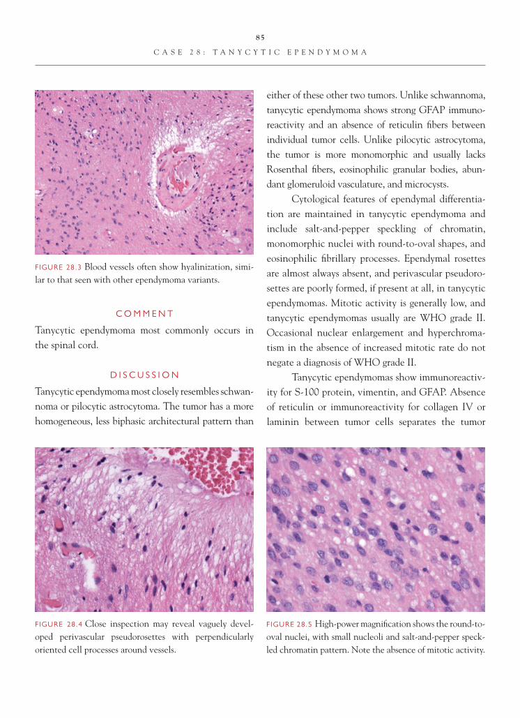
85
C A S E 2 8 : T A N Y C Y T I C E P E N D Y M O M A
C O M M E N T
Tanycytic ependymoma most commonly occurs in the spinal cord.
D I S C U S S I O N
Tanycytic ependymoma most closely resembles schwan-noma or pilocytic astrocytoma. The tumor has a more homogeneous, less biphasic architectural pattern than
either of these other two tumors. Unlike schwannoma, tanycytic ependymoma shows strong GFAP immuno-reactivity and an absence of reticulin fi bers between individual tumor cells. Unlike pilocytic astrocytoma, the tumor is more monomorphic and usually lacks Rosenthal fi bers, eosinophilic granular bodies, abun-dant glomeruloid vasculature, and microcysts.
Cytological features of ependymal differentia-tion are maintained in tanycytic ependymoma and include salt-and-pepper speckling of chromatin, monomorphic nuclei with round-to-oval shapes, and eosinophilic fi brillary processes. Ependymal rosettes are almost always absent, and perivascular pseudoro-settes are poorly formed, if present at all, in tanycytic ependymomas. Mitotic activity is generally low, and tanycytic ependymomas usually are WHO grade II. Occasional nuclear enlargement and hyperchroma-tism in the absence of increased mitotic rate do not negate a diagnosis of WHO grade II.
Tanycytic ependymomas show immunoreactiv-ity for S-100 protein, vimentin, and GFAP. Absence of reticulin or immunoreactivity for collagen IV or laminin between tumor cells separates the tumor
FIGURE 28.3 Blood vessels often show hyalinization, simi-lar to that seen with other ependymoma variants.
FIGURE 28.5 High-power magnifi cation shows the round-to-oval nuclei, with small nucleoli and salt-and-pepper speck-led chromatin pattern. Note the absence of mitotic activity.
FIGURE 28.4 Close inspection may reveal vaguely devel-oped perivascular pseudorosettes with perpendicularly oriented cell processes around vessels.

86
C A S E 2 8 : T A N Y C Y T I C E P E N D Y M O M A
from a schwannoma. Another consideration in some cases is diffuse astrocytoma of the spinal cord. GFAP does not discriminate between diffuse astrocytoma and tanycytic ependymoma, making the nuclear mo-notony and cytology particularly important to recog-nize in this ependymoma variant. Ependymomas are often suspected intraoperatively by the neurosurgeon because they tend to be more sharply demarcated from the adjacent spinal cord tissue than diffuse astro-cytomas. Thus, clinicopathologic correlation should be sought by the pathologist.
On very small biopsies, it may be impossible to distinguish tanycytic ependymoma from astrocytoma
without additional electron microscopic (EM) ex-amination. Thus, in small biopsies taken from spinal cord intramedullary tumors, it is prudent to set aside a small portion of the biopsy in glutaraldehyde for elecron microscopic studies.
References 1. McLendon RE, Wiestler OD, Kros JM, et al. Anaplastic
ependymoma. In: Louis DN, Ohgaki H, Wiestler OD, Cavenee WK, editors. WHO Classifi cation of Tumours of the Central Nervous System. Lyon, FR: IARC Press; 2007, p. 74–8.
2. Perry A. Glial and glioneuronal tumors. In: Prayson RA, editor. Neuropathology. Philadelphia, PA: Elsevier Churchill Livingstone; 2005. p. 468–76.

87
Case 29: Clear Cell Ependymoma
C L I N I C A L I N F O R M A T I O N
The patient is a 13-year-old male who presents with a seizure. Neuroimaging studies are performed and demonstrate a large, contrast-enhancing, well- demarcated mass located deep within the left cerebral hemisphere. Calcifi cations are noted in the mass. The decision is made to resect the lesion, and histological sections are reviewed.
O P I N I O N
The specimen shows a moderately cellular tumor, punctuated by arborizing, delicate blood vessels devoid of microvascular proliferation. Perivascular pseudorosettes are manifest as eosinophilic anuclear areas around these blood vessels. Tumor cells are
cytologically monotonous and contain round-to-oval nuclei and clear cytoplasm. Calcifi cations are pres-ent, but necrosis is absent. Scattered mitotic fi gures are seen, but mitotic activity is not brisk.
After immunostaining to exclude other clear cell tumor types, we diagnose this lesion as an ependymoma variant and characterize it as follows: Left Cerebral Hemisphere, Excision—Clear Cell Ependymoma, WHO Grade II.
C O M M E N T
Clear cell ependymomas most often occur in supratentorial regions of children or young adults and histologically closely simulate oligodendroglioma, neurocytoma, or hemangioblastoma.
FIGURE 29.1 Low-power magnifi cation illustrates a tumor composed of oligodendroglial-like clear cells, separated by delicate vascular septae.
FIGURE 29.2 The tumor is composed of relatively monomor-phic cells with optically clear cytoplasm. Perivascular pseudo-rosettes are more subtle than in other ependymoma variants.

88
C A S E 2 9 : C L E A R C E L L E P E N D Y M O M A
D I S C U S S I O N
Clear cell areas can occasionally be focally identifi ed in otherwise typical ependymomas, but the occur-rence of a pure clear cell ependymoma is uncommon. The diagnosis of clear cell ependymoma can be dif-fi cult, and immunohistochemistry or electron micros-copy is often more necessary for a correct diagnosis of the clear cell variant than it is for other ependymoma subtypes.
Cytological nuclear features of ependymal differentiation are maintained in clear cell epen-dymoma and include salt-and-pepper speckling of chromatin and monomorphic nuclei with round-to-oval shapes. The clear cell quality of the cytoplasm mimics perinuclear halos of oligodendroglioma quite closely. Adding to the diffi culty is the presence of arborizing vasculature and calcifi cations in both clear cell ependymoma and oligodendroglioma. The
FIGURE 29.3 Microcalcifi cations are prominent in this example.
FIGURE 29.4 Close inspection on high power reveals the fi brillar cell processes.
FIGURE 29.5 High-power magnifi cation shows round-to-oval nuclei, with small nucleoli and salt-and-pepper speckled chromatin pattern.
FIGURE 29.6 Note the presence of scattered mitotic activ-ity in this example; many clear cell ependymomas meet cri-teria for WHO grade III because of brisk mitotic activity.

89
C A S E 2 9 : C L E A R C E L L E P E N D Y M O M A
eosinophilic fi brillary processes of ependymoma are variable in number, and perivascular pseudorosettes may be poorly formed but are usually recognizable af-ter careful scrutiny.
A feature that further aids in distinction between clear cell ependymoma and oligodendroglioma is that the former tends to be more sharply demarcated from surrounding brain tissue than oligodendrogliomas and lacks the deletions in chromosomes 1p or 19q that are present in many oligodendrogliomas.
Clear cell ependymomas show immunoreactivity for S-100 protein, vimentin, and GFAP, but the latter can be quite patchy and isolated to the few perivascular pseudorosettes that are present. Epithelial membrane antigen (EMA) immunostaining reveals the ring-like and dot-like features seen in other ependymomas but, again, the changes may be less prominent in clear cell ependymoma than in other ependymoma subtypes. Two other tumor types that enter into the differential diagnosis in clear cell ependymoma are neurocytoma and hemangioblastoma. Synaptophysin immunoreac-tivity is either absent entirely or very focal in clear cell ependymoma compared to neurocytoma. Immunore-activity for GFAP and the identifi cation of perivas-
cular pseudorosettes, even if focal, excludes consider-ation of hemangioblastoma.
Electron microscopic features of clear cell epen-dymoma are those of other ependymomas, with forma-tion of microlumens containing microvilli and cilia. Well-developed intercellular junctions are present.
FIGURE 29.7 GFAP highlights the perivascular fi brillar cell processes and excludes consideration of hemangioblastoma.
FIGURE 29.8 EMA immunoreactivity is focally present in this example and shows a ring-like signal that highlights the surface portion of the cells forming a microlumen.
FIGURE 29.9 Electron microscopic examination of clear cell ependymoma often shows features seen in any ependymoma, with microvilli and lumen formation (large arrows), as well as cilia (small arrow). Note also the numerous intercellular junctions. (Courtesy of Gary Mierau, Ph.D.)

90
C A S E 2 9 : C L E A R C E L L E P E N D Y M O M A
Clear cell ependymomas may be WHO grade II tumors but often meet criteria for WHO grade III because of signifi cant mitotic activity. Some studies have suggested that clear cell ependymomas show a more aggressive course than other ependymoma subtypes.
References 1. Fouladi M, Helton K, Dalton J, et al. Clear cell ependymoma:
a clinicopathologic analysis of 10 patients. Cancer 2003;98: 2232–44.
2. McLendon RE, Wiestler OD, Kros JM, et al. Anaplastic ependymoma. In: Louis DN, Ohgaki H, Wiestler OD, Cavenee WK, editors. WHO Classifi cation of Tumours of the Central Nervous System. Lyon, FR: IARC Press; 2007, p. 74–8.
3. Min KW, Scheithauer BW. Clear cell ependymoma: a mimic of oligodendroglioma: clinicopathologic and ultrastructural considerations. Am J Surg Pathol 1997;21:820–6.
4. Perry A. Glial and glioneuronal tumors. In: Prayson RA, editor. Neuropathology. Philadelphia, PA: Elsevier Churchill Livingstone; 2005. p. 468–76.

91
Case 30: Choroid Plexus Papilloma
C L I N I C A L I N F O R M A T I O N
The patient is a 12-year-old girl who presents with a 6- to 9-month history of “diffi culty seeing the black-board.” She also reports intermittent double vision and headaches. She has recently failed a school vision screening exam. Referral to an ophthalmologist discloses 20/50 vision bilaterally and mild bilateral papilledema. MRI studies are obtained, which reveal a large, lobulated, homogeneously enhancing, fourth ventricular mass that extends through the left foramen of Luschka. Resection is undertaken, and sections are available for review.
O P I N I O N
Sections show a highly papillary neoplasm, devoid of necrosis or brain invasion. Delicate fi brovascular
connective tissue cores are covered by a single layer of monotonous epithelial cells. The columnar cells have abundant cytoplasm and generally maintain good polarity of nuclei, with nuclei located at the cell base. Mitotic activity is not identifi ed.
We consider the pathology to be that of a choroid plexus papilloma, without atypical or malig-nant features, and characterize it as follows: Fourth Ventricle, Excision—Choroid Plexus Papilloma, WHO Grade I.
C O M M E N T
Choroid plexus papillomas of the fourth ventricle are found in equal proportions in children and adults. In contrast, 80% of lateral ventricular tumors occur in patients less than 20 years of age.
FIGURE 30.1 Gross surgical pathology specimen of a cho-roid plexus papilloma showing a tan-yellow tumor with a caulifl ower-like appearance and bosselated surface.
FIGURE 30.2 Low-power view of the choroid plexus pap-illoma shows a hyperplastic-appearing, complex papillary tumor with fi brovascular cores and no necrosis.
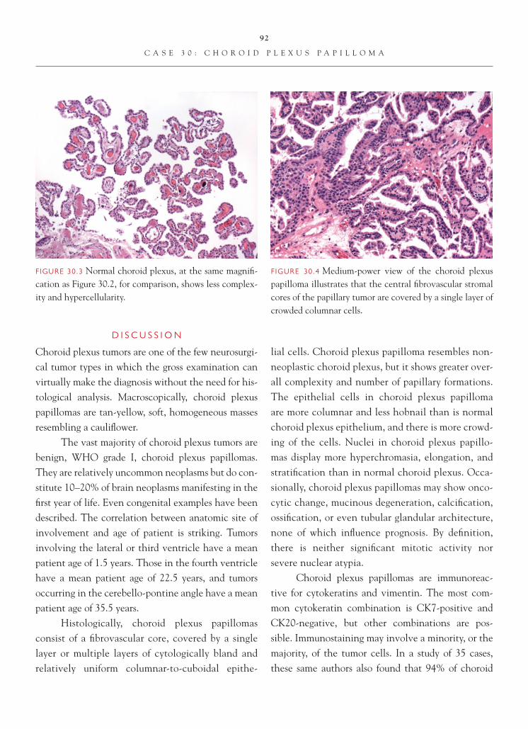
92
C A S E 3 0 : C H O R O I D P L E X U S P A P I L L O M A
D I S C U S S I O N
Choroid plexus tumors are one of the few neurosurgi-cal tumor types in which the gross examination can virtually make the diagnosis without the need for his-tological analysis. Macroscopically, choroid plexus papillomas are tan-yellow, soft, homogeneous masses resembling a caulifl ower.
The vast majority of choroid plexus tumors are benign, WHO grade I, choroid plexus papillomas. They are relatively uncommon neoplasms but do con-stitute 10–20% of brain neoplasms manifesting in the fi rst year of life. Even congenital examples have been described. The correlation between anatomic site of involvement and age of patient is striking. Tumors involving the lateral or third ventricle have a mean patient age of 1.5 years. Those in the fourth ventricle have a mean patient age of 22.5 years, and tumors occurring in the cerebello-pontine angle have a mean patient age of 35.5 years.
Histologically, choroid plexus papillomas consist of a fi brovascular core, covered by a single layer or multiple layers of cytologically bland and relatively uniform columnar-to-cuboidal epithe-
lial cells. Choroid plexus papilloma resembles non- neoplastic choroid plexus, but it shows greater over-all complexity and number of papillary formations. The epithelial cells in choroid plexus papilloma are more columnar and less hobnail than is normal choroid plexus epithelium, and there is more crowd-ing of the cells. Nuclei in choroid plexus papillo-mas display more hyperchromasia, elongation, and stratifi cation than in normal choroid plexus. Occa-sionally, choroid plexus papillomas may show onco-cytic change, mucinous degeneration, calcifi cation, ossifi cation, or even tubular glandular architecture, none of which infl uence prognosis. By defi nition, there is neither signifi cant mitotic activity nor severe nuclear atypia.
Choroid plexus papillomas are immunoreac-tive for cytokeratins and vimentin. The most com-mon cytokeratin combination is CK7-positive and CK20-negative, but other combinations are pos-sible. Immunostaining may involve a minority, or the majority, of the tumor cells. In a study of 35 cases, these same authors also found that 94% of choroid
FIGURE 30.3 Normal choroid plexus, at the same magnifi -cation as Figure 30.2, for comparison, shows less complex-ity and hypercellularity.
FIGURE 30.4 Medium-power view of the choroid plexus papilloma illustrates that the central fi brovascular stromal cores of the papillary tumor are covered by a single layer of crowded columnar cells.

93
C A S E 3 0 : C H O R O I D P L E X U S P A P I L L O M A
FIGURE 30.5 High-power view of the choroid plexus papil-loma illustrates the mild hyperchromasia and elongation of nuclei within the columnar cells, but maintenance of basi-lar polarity of nuclei within the epithelium and absence of signifi cant mitotic activity.
FIGURE 30.6 Normal choroid plexus, at the same magni-fi cation as Figure 30.5 for comparison, shows the hobnail, cuboidal epithelial cells of non-neoplastic choroid plexus. Note the basophilic calcifi cation that typically occurs in normal choroid plexus with aging.
plexus papillomas are positive for CAM 5.2 and 69% are positive, at least focally, for GFAP.
References 1. Gyure KA, Morrison AL. Cytokeratin 7 and 20 expres-
sion in choroid plexus tumors; utility in differentiating these neoplasms from metastatic carcinomas. Mod Pathol 2000;13:638–43.
2. Paulus W, Brandner S. Choroid plexus tumors. In: Louis DN, Ohgaki H, Wiestler OD, Cavenee WK, editors. WHO Classifi cation of Tumours of the Central Nervous System. Lyon, FR: IARC Press; 2007. p. 82–5.
3. Wolff JE, Sajedi M, Brant R, et al. Choroid plexus tumors. Br J Cancer 2002;87:1086–91.

94
Case 31: Atypical Choroid Plexus Papilloma
C L I N I C A L I N F O R M A T I O N
The patient is a 6-year-old boy who presents with 5–6 months of abnormal eye movements and visual defi cits. There is also a history of diffi culty walking and progressive deterioration in gross motor function. He is brought to this country from his native Ukraine for diagnosis and treatment. Neuroimaging studies reveal a large, lobulated mass in the left lateral ven-tricle with surrounding edema and midline shift.
O P I N I O N
Sections show a choroid plexus papilloma with maintenance of papillary architecture, but increased mitotic activity and multifocal necrosis. Cells retain reasonable nuclear polarity, with nuclei at the base of the columnar epithelium, but crowding and
elongation of the cells are noted. Mitoses number more than 2 per 10 high-power fi elds.
We consider this tumor to be a choroid plexus neoplasm and characterize it as follows: Left LateralVentricle, Excision—Atypical Choroid Plexus Papilloma, WHO Grade II.
C O M M E N T
Criteria for WHO grade II choroid plexus papilloma were recently adopted for the fi rst time in the World Health Organization 2007 publication, WHO Classi-fi cation of Tumours of the Central Nervous System.
D I S C U S S I O N
Choroid plexus tumors are suffi ciently infrequent that there had been considerable debate over the impact
FIGURE 31.1 Low-power view shows a papillary neoplasm with zonal necrosis; this alone, however, is insuffi cient to diag-nose WHO grade II aCPP if it occurs as an isolated feature.
FIGURE 31.2 Areas with complex tubular and solid, less papillary, pattern can be seen, but again, this feature in iso-lation is insuffi cient to diagnose WHO grade II aCPP.
95
C A S E 3 1 : A T Y P I C A L C H O R O I D P L E X U S P A P I L L O M A
of various histological features on prognosis until the publication of a large study in 2006 (Jeibmann et al.). These authors collected 164 choroid plexus tumors, of which 24 showed frank signs of malignancy and were diagnosable as choroid plexus carcinoma. Of the remaining 124 assessable cases of papillomas, a high percentage [46 cases (37%)] displayed at least one of the following features: increased mitotic activity, increased cellularity, nuclear pleomorphism, blur-
ring of papillary growth pattern, or necrosis. On uni-variate analysis, however, only incomplete surgical resection and mitotic activity correlated with tumor recurrence. These authors proposed that the fi nding of increased mitotic activity (2 or more mitoses per 10 randomly selected HPFs) should allow designation as a WHO grade II tumor. This proposal was sub-sequently ratifi ed in the 2007 WHO Classifi cation System. WHO grade II “atypical” choroid plexus papilloma (aCPP) now occupies a position interme-diate between the far more common choroid plexus
FIGURE 31.3 Necrosis in the tumor is better appreciated at higher magnifi cation.
FIGURE 31.4 At least three mitoses can be seen in this single high-power microscopic fi eld.
FIGURE 31.5 Mitotic activity is accompanied by a greater degree of nuclear pleomorphism, with more conspicuous nucleoli. FIGURE 31.6 The MIB-1 labeling is increased in this aCPP.
96
C A S E 3 1 : A T Y P I C A L C H O R O I D P L E X U S P A P I L L O M A
papilloma, WHO grade I and the rare choroid plexus carcinoma, WHO grade III.
Indeed, it is interesting to note that after Jeibmann et al. utilized their own criteria for reclas-sifi cation of choroid plexus papillomas, only 15%, not 37%, could be truly categorized as “atypical.” Thus, the diagnoses of “atypical” choroid plexus papilloma or even choroid plexus “carcinoma” were likely “overcalled” in the past by some investigators, and WHO grades II and III choroid plexus tumors may be even rarer than was formerly thought. Indeed, using these newer criteria, atypical choroid plexus papillomas in the large study by Jeibmann et al. were found to actually be only about one-seventh as frequent as WHO grade I choroid plexus papillomas.
An important additional point from this study of 124 cases was the fact that, although increased
mitotic activity alone correlated with a 4.9-fold higher risk of recurrence after 5 years of follow-up, “increased mitotic activity was only rarely encountered as an isolated atypical feature” in aCPP ( Jeibmann et al.). Thus, the pathologist can expect to see other worrisome features in addition to ≥2 mitoses per 10 HPFs in most aCPPs. Mean MIB-1 labeling indices are often elevated for aCPP compared to CPP, but a precise discriminatory level was not established in the 2007 WHO Classifi cation system.
References 1. Jeibmann A, Hasselblatt M, Gerss J, et al. Prognostic impli-
cations of atypical histologic features in choroid plexus papilloma. J Neuropathol Exp Neurol 2006; 65:1069–73.
2. Paulus W, Brandner S. Choroid plexus tumours. In: Louis DN, Ohgaki H, Wiestler OD, Cavenee WK, Editors. WHO Classifi cation of Tumours of the Central Nervous System. Lyon, FR: IARC Press; 2007. p. 82–5.

97
Case 32: Choroid Plexus Carcinoma
C L I N I C A L I N F O R M A T I O N
The patient is a 2-year-old child who presents with increased head circumference. Work-up reveals hydrocephalus and a large fourth ventricular mass. The decision is made to resect the lesion, and slides are available for review.
O P I N I O N
Sections show a hypercellular neoplasm, with focal maintenance of papillary architecture, but other areas show a more patternless, sheetlike arrangement of cells with solid growth. There is increased mitotic activity, exceeding 2 per 10 high-power microscopic fi elds, and multifocal necrosis. Cells manifest high nuclear-to-cytoplasmic ratios, considerable nuclear
pleomorphism, and only small amounts of eosinophilic cytoplasm with rounded profi les. Tumor invades the nearby cerebellum.
We consider this tumor to be a choroid plexus neoplasm and characterize it as follows: FourthVentricle/Cerebellum, Excision—Choroid Plexus Carcinoma, WHO Grade III.
C O M M E N T
Choroid plexus carcinomas (CPC) are rare neoplasms, and the vast majority of cases occur in very young children (median age 2–3 years), not in older children or adults. In adults with obviously malignant papillary neoplasms, metastatic carcinoma should always be the fi rst diagnostic consideration.
FIGURE 32.1 Low-power magnifi cation demonstrates a hypercellular, papillary neoplasm with several small foci of necrosis.
FIGURE 32.2 Tumor invades into the nearby normal cer-ebellum.

98
C A S E 3 2 : C H O R O I D P L E X U S C A R C I N O M A
D I S C U S S I O N
The differential diagnosis for CPC (WHO grade III) in most very young children involves separat-ing it from lower-grade choroid plexus papillomas, especially WHO grade II, atypical choroid plexus papillomas.
Choroid plexus tumors are suffi ciently infre-quent that there had been considerable debate over the impact of various histological features on prog-nosis until the publication of a large study in 2006 (Jeibmann et al.). These authors collected 164 cho-roid plexus tumors, of which 24 showed frank signs of malignancy and were diagnosable as CPC, WHO grade III. Criteria utilized by these authors to diag-nose CPC were the presence of four of the fi ve follow-ing features: high cellularity, solid growth, necrosis, nuclear pleomorphism, and at least 2 mitoses per 10 randomly selected high-power microscopic fi elds.
It should be noted that 37% of the choroid plex-us papillomas in the same series were also identifi ed to possess at least one of these same histological features. Thus, it is important to emphasize that fi nding necro-sis or high cellularity or nuclear pleomorphism in a
choroid plexus papilloma should not prompt diagno-sis of CPC. It is likely that some workers in the past overcalled choroid plexus papillomas that possessed one to three of these features as (incorrectly) a CPC.
An unresolved question is that of the impor-tance of brain invasion in distinguishing between choroid plexus papilloma versus CPC. Too few cases with brain invasion were identifi ed in the large series by Jeibmann et al. to assess the prognostic importance of this feature.
In some examples of CPC, the sheetlike, patternless architecture prompts diagnostic consider-ation of atypical teratoid/rhabdoid tumor (AT/RT), another tumor usually found in very young children less than 3 years of age. CPCs show reaction for cytokeratins, with positivity for S-100 protein and transthyretin being found less frequently than in choroid plexus papillomas. Vimentin, epithelial mem-brane antigen (EMA), and GFAP immunoreactivity has also been reported in some CPCs. Immunostain-ing for all these antibodies can also be found in AT/RT; thus, the immunohistochemistry (IHC) profi le of CPC overlaps with that for AT/RT. An exception is
FIGURE 32.3 High-power magnifi cation reveals mitotic activity, as well as cells with nuclear pleomorphism, high nuclear-to-cytoplasmic ratios, and prominent nucleoli.
FIGURE 32.4 In other areas of the tumor, a patternless, sheetlike architecture can make distinction from atypical teratoid/rhabdoid tumor that is nearly impossible on hema-toxylin and eosin light microscopy alone.

99
C A S E 3 2 : C H O R O I D P L E X U S C A R C I N O M A
that immunostaining for smooth muscle actin (SMA) is identifi ed in most AT/RTs, but not in CPCs.
Further IHC distinction between CPC and AT/RT is necessary in some cases. The advent of an antibody directed toward the nuclear protein product of INI1 was reported by Judkins et al. to be valuable in this regard. Nearly all CPCs in that study showed retention of nuclear expression of INI1 protein, serv-ing to distinguish them from histologically similar AT/RTs, which usually demonstrated nuclear loss of INI1 protein.
In the rare adult patient in whom CPC enters the differential diagnosis, immunostaining for meta-static epithelial neoplasms should be undertaken. An algorithmic approach for the workup of metastatic disease to the central nervous system has been out-lined by Becher et al. This involves a “fi rst round” of antibodies for adenocarcinoma, using CAM5.2, CK7,
CK20, and thyroid transcription factor-1 (TTF-1). Depending on the outcome of these stains, metastatic epithelial tumors can be further differentiated using immunostains for GCDFP, CA125, CDX2, CD10, and estrogen receptor.
References 1. Becher MW, Abel T, Thompson RC, et al. Immunohisto-
chemical analysis of metastatic neoplasms of the central nervous system. J Neuropathol Exp Neurol 2006;65:935–44.
2. Jeibmann A, Hasselblatt M, Gerss J, et al. Prognostic implications of atypical histologic features in choroid plexus papilloma. J Neuropathol Exp Neurol 2006;65:1069–73.
3. Judkins AR, Burger PC, Hamilton RL, et al. INI1 protein expression distinguishes atypical teratoid/rhabdoid tumor from choroid plexus carcinoma. J Neuropathol Exp Neurol 2005;64:391–7.
4. Paulus W, Brandner S. Choroid plexus tumours. In: Louis DN, Ohgaki H, Wiestler OD, Cavenee WK, Editors. WHO Classifi cation of Tumours of the Central Nervous System. Lyon, FR: IARC Press; 2007. p. 82–5.

100
Case 33: Chordoid Glioma
C L I N I C A L I N F O R M A T I O N
The patient is a 32-year-old female who presents with headaches. On imaging, she is noted to have a dis-crete mass lying within the third ventricle. The tumor is homogeneously contrast enhancing. The mass is excised, and histologic sections are reviewed.
O P I N I O N
Histologic sections show a neoplasm, marked by a pro-liferation of epithelial cells arranged in clusters and cords. The clusters of cells are separated by a mucino-us-like stroma. Focal perivascular chronic infl amma-tion, consisting of benign-appearing lymphocytes and plasma cells, is observed. Individual tumor cells are marked by abundant eosinophilic cytoplasm. Occa-sional Rosenthal fi bers are noted at the periphery
of the lesion. Scattered eosinophilic globular bodies resembling Russell bodies are also observed. Mitotic fi gures are not seen in this lesion. Vascular prolifera-tive changes and necrosis are also not observed. The tumor demonstrates diffuse positive staining with antibody to GFAP.
We consider the lesion to represent a low-grade glioma and characterize it as follows: ThirdVentricular Region, Excision—Chordoid Glioma, WHO Grade II.
C O M M E N T
Chordoid gliomas are relatively rare lesions that arise in the third ventricular region. Cases that have been reported appear to behave like low-grade neoplasms.
FIGURE 33.1 Low magnifi cation appearance, marked by spindled cells arranged against a mucinous background.
FIGURE 33.2 A mucinous background is a salient feature of the tumor.
101
C A S E 3 3 : C H O R D O I D G L I O M A
D I S C U S S I O N
Chordoid gliomas are rare, slow-growing, nonin-vasive glial tumors that typically are situated in the suprasellar and third ventricular regions. They typi-cally arise in adults and show a female predominance. Because of the tumor’s location in the anterior por-tion of the third ventricle, patients with chordoid gliomas often present with symptoms related to obstructive hydrocephalus. Occasional tumors may
cause endocrine hypofunctioning because of their proximity to the optic chiasm and pituitary region. On imaging, they are usually well circumscribed and uniformly contrast-enhancing.
Microscopically, the tumor is marked by clusters and cords of epithelioid cells, arranged against a mucinous background. Perivascular chronic infl amma-tion and Russell body formations are quite common. Mitotic fi gures are only rarely observable. Vascular
FIGURE 33.3 A more cellular region of the tumor, with a fi brillary background.
FIGURE 33.4 Focal, perivascular, chronic infl ammation is present.
FIGURE 33.5 An occasional eosinophilic Russell body is evident.
FIGURE 33.6 The tumor cells demonstrate widespread cytoplasmic staining with GFAP antibody.

102
C A S E 3 3 : C H O R D O I D G L I O M A
proliferative changes and necrosis are not present. On immunohistochemical staining, the tumor cells stain positively with antibodies to GFAP and vimentin. Occasional positive staining with antibody to epithe-lial membrane antigen (EMA) has also been variably reported. Low rates of cell proliferation are evidenced by low Ki-67 and MIB-1 cell proliferative indices.
Differential diagnostic considerations, given the choroid appearance, include chordoid men-ingioma and chordoma. The lack of a dural or bony involvement, GFAP immunoreactivity, and physaliphorous cells (in chordoma) exclude those possibilities.
Chordoid gliomas are generally treated by surgi-cal excision. If incompletely excised, they may recur. Postoperative complication may result by damaging adjacent structures, including the hypothalamus.
References 1. Brat DJ, Scheithauer BW, Staugaitis SM, et al. Third ven-
tricular chordoid glioma: a distinct clinicopathologic entity. J Neuropathol Exp Neurol 2001;15:147–50.
2. Leeds NE, Lang FF, Ribalta T, et al. Origin of chordoid glioma of the third ventricle. Arch Pathol Lab Med 2006;130:625–36.
3. Reifenberger G, Weber T, Weber RG, et al. Chordoid glioma of the third ventricle: immunohistochemical and molecular genetic characterization of a novel tumor entity. Brain Pathol 1999;9:617–27.

103
Case 34: Angiocentric Glioma
C L I N I C A L I N F O R M A T I O N
The patient is a 32-year-old male with a long history of drug-resistant epilepsy. An MRI scan shows a cor-tically based, circumscribed mass, 13 × 13 × 15 mm, in the right parietal lobe, without signifi cant sur-rounding edema. After placement of intracortical electrodes, the patient undergoes resective ther-apy for his pharmaco-resistant epilepsy. Histologic
sections corresponding to the circumscribed intrac-ortical lesion are reviewed.
O P I N I O N
As indicated by the neuroimaging, the abnormal cel-lular proliferations are identifi ed principally within the cerebral cortex (seen as negative staining against a diffuse background of synaptophysin positivity). Solid areas, composed of fascicular arrangements of the tumor cells, give way to areas demonstrating perivascular and subpial infi ltration. Perivascular tumor aggregates in many areas resemble perivascu-lar pseudorosettes seen in classical ependymomas; in other areas, there is a more irregular arrangement of
FIGURE 34.1 Low power shows preferential perivascular infi ltration through the cerebral cortex.
FIGURE 34.2 Despite this predilection for a perivascular aggregation, the arrangements of tumor cells with regard to the blood vessel walls are much more irregular than those seen in classical perivascular pseudorosettes of ependymal tumors.
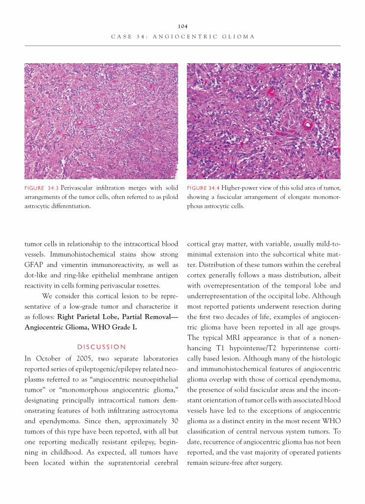
104
C A S E 3 4 : A N G I O C E N T R I C G L I O M A
tumor cells in relationship to the intracortical blood vessels. Immunohistochemical stains show strong GFAP and vimentin immunoreactivity, as well as dot-like and ring-like epithelial membrane antigen reactivity in cells forming perivascular rosettes.
We consider this cortical lesion to be repre-sentative of a low-grade tumor and characterize it as follows: Right Parietal Lobe, Partial Removal—Angiocentric Glioma, WHO Grade I.
D I S C U S S I O N
In October of 2005, two separate laboratories reported series of epileptogenic/epilepsy related neo-plasms referred to as “angiocentric neuroepithelial tumor” or “monomorphous angiocentric glioma,” designating principally intracortical tumors dem-onstrating features of both infi ltrating astrocytoma and ependymoma. Since then, approximately 30 tumors of this type have been reported, with all but one reporting medically resistant epilepsy, begin-ning in childhood. As expected, all tumors have been located within the supratentorial cerebral
cortical gray matter, with variable, usually mild-to-minimal extension into the subcortical white mat-ter. Distribution of these tumors within the cerebral cortex generally follows a mass distribution, albeit with overrepresentation of the temporal lobe and underrepresentation of the occipital lobe. Although most reported patients underwent resection during the fi rst two decades of life, examples of angiocen-tric glioma have been reported in all age groups. The typical MRI appearance is that of a nonen-hancing T1 hypointense/T2 hyperintense corti-cally based lesion. Although many of the histologic and immuno histochemical features of angiocentric glioma overlap with those of cortical ependymoma, the presence of solid fascicular areas and the incon-stant orientation of tumor cells with associated blood vessels have led to the exceptions of angiocentric glioma as a distinct entity in the most recent WHO classifi cation of central nervous system tumors. To date, recurrence of angiocentric glioma has not been reported, and the vast majority of operated patients remain seizure-free after surgery.
FIGURE 34.3 Perivascular infi ltration merges with solid arrangements of the tumor cells, often referred to as piloid astrocytic differentiation.
FIGURE 34.4 Higher-power view of this solid area of tumor, showing a fascicular arrangement of elongate monomor-phous astrocytic cells.

105
C A S E 3 4 : A N G I O C E N T R I C G L I O M A
References 1. Lellouch-Tubiana A, Boddaert N, Bourgeois M, et al.
Angiocentric neuroepithelial tumor (ANET): a new epilepsy-related clinicopathological entity with distinctive MRI. Brain Pathol 2005;15:281–6.
2. Preusser M, Hoischen A, Novak K, et al. Angiocentric glioma: report of clinicopathologic and genetic fi ndings in 8 cases. Am J Surg Pathol 2007;31:1709–18.
3. Shakur SF, McGirt MJ, Johnson MW, et al. Angiocentric glioma: a case series. J Neurosurg Pediatr 2009;3:197–202.
4. Wang M, Tihan T, Rojiani AM, et al. Monomorphous angiocentric glioma: a distinctive epileptogenic neoplasm with features of infi ltrating astrocytoma and ependymoma. J Neuropathol Exp Neurol 2005;64:875–81.

106
Case 35: Astroblastoma
C L I N I C A L I N F O R M A T I O N
The patient is a 19-year-old female who presents with a seizure. A well-demarcated, noncalcifi ed, lobulated mass with focal cystic change is noted on MRI stud-ies. The patient undergoes gross total resection, and histologic sections are reviewed.
O P I N I O N
Histologic sections shows proliferation of atypical-ap-pearing glial cells, with high nuclear- to- cytoplasmic ratio and irregular nuclear contours. Prominent perivascular rosetting structures, marked by short and broad cell processes, are seen. Focal subpial aggre-gation of tumor cells is present. Prominent mitotic activity, vascular proliferative changes, and necrosis are not seen. A Ki-67 labeling index of less than 1 is noted. There is no evidence of true rosettes.
We consider the lesion to be a glial neoplasm and characterize it as follows: Right Frontal Lobe, Excision—Astroblastoma.
C O M M E N T
The lack of prominent mitotic activity, marked cytologic atypia, vascular proliferative changes, and necrosis, along with the low Ki-67 labeling index, support a diagnosis of low-grade or well-differentiated astroblastoma.
D I S C U S S I O N
Astroblastomas are rare glial neoplasms that typi-cally present in the fi rst three decades of life. Most are located in the cerebral hemispheres and do not appear, like most ependymomas, to arise from or within the ventricles. The histologic hallmarks of this
FIGURE 35.2 Intermediate magnifi cation illustrating the appearance of astroblastomatous rosettes.
FIGURE 35.1 The tumor shows focal areas resembling low-grade astrocytoma.
107
C A S E 3 5 : A S T R O B L A S T O M A
tumor are its circumscription and characteristic roset-ting structures. Given the salient presence of rosettes, this tumor ostensibly mimics ependymoma. Occa-sionally, ependymomas may arise away from the ven-tricular system. In general, the cytoplasmic processes of the astroblastoma cells tend to be thicker and more broad-based, rather than the narrow and tapered cell processes more typical of the ependymoma. Vascular sclerosis, which is also fairly commonly observed in astroblastoma, is a relatively uncommon feature of ependymomas. The other differential diagnostic con-sideration is an ordinary diffuse fi brillary astrocytoma that has some secondary structuring around blood vessels. The astrocytoma tends to be more infi ltrative in nature, and the rosetting pattern not as structured as it is with the astroblastoma.
Like other gliomas, astroblastomas demonstrate S-100 and GFAP immunoreactivity. Focal membra-nous immunoreactivity with antibody to EMA may also be seen; however, cytokeratin immunostaining is typically not present. Ultrastructurally, astroblastoma cells usually exhibit fewer cilia and cell junctions than ependymomas.
Although there is no grade designation assigned to these tumors by the WHO, it is generally accepted that these neoplasms may have a low-grade/well- differentiated or high-grade appearance. Higher-grade tumors, in contrast to the current case, are usually marked by more prominent nuclear pleomorphism, increased mitotic activity, vascular proliferative changes, necrosis, and increased cell proliferation labeling indices. There is some suggestion in the lit-erature that tumors with low-grade histology have a better prognosis than higher-grade neoplasms. High-grade tumors may require radiotherapy and generally allow for a poor survival.
References 1. Bonnin JM, Rubinstein LJ. Astroblastomas: a pathological
study of 23 tumors with postoperative follow-up in 13 patients. Neurosurgery 1989;25:6–13.
2. Brat DJ, Hirose Y, Cohen KJ, et al. Astroblastoma. Clinicopathologic features and chromosome abnormalities defi ned by comparative genomic hybridization. Brain Pathol 2000;10:42–52.
3. Thiessen B, Finlay J, Kulkarni R, et al. Astroblastoma: does histology predict biologic behavior? J Neurooncol 1998;40:59–65.
FIGURE 35.3 Higher magnifi cation appearance of rosettes, showing broad, thickened cytoplasmic processes.
FIGURE 35.4 Focal subpial aggregation of tumor cells is evident.
108
Case 36: Dysplastic Cerebellar Gangliocytoma (Lhermitte-Duclos Disease)
C L I N I C A L I N F O R M A T I O N
The patient is an 18-year-old male who presents with dysmetria. The patient has had a history of previous seizures. Imaging studies show mild hydrocephalus and focal thickening of the cerebellar cortex, with increased signal intensity on a T2-weighted MRI study. The right cerebellar lesion is excised, and his-tologic sections are reviewed.
O P I N I O N
The histologic sections show focal enlargement of the cerebellar cortex, marked by an increased number of large ganglionic cells. There appears to be a sharp interface between the lesion and the adjacent gli-otic cerebellar parenchyma. The ganglionic cells are marked by eosinophilic-to-pale staining cytoplasm and an eccentrically placed nucleus with prominent
nucleolus. A gliomatous component is not noted. There is no evidence of calcifi cation. Microcystic changes are observed adjacent to the lesion.
We consider the lesion to be a low-grade gan-glionic cell proliferation, and we characterize it as follows: Right Cerebellum, Excision—Dysplastic Gangliocytoma of the Cerebellum (Lhermitte-Duclos Disease).
C O M M E N T
The absence of a gliomatous component in the lesion supports the diagnosis of a pure ganglion cell mass (gangliocytoma).
D I S C U S S I O N
Dysplastic gangliocytoma of the cerebellum is a rare disorder, well known to be associated with Cowden
FIGURE 36.1 Intermediate magnifi cation shows adjacent mild gliosis with preservation of the cerebellar architecture.
FIGURE 36.2 Focal microcystic changes and occasional reactive astrocytes are present adjacent to the lesion.
109
C A S E 3 6 : D Y S P L A S T I C C E R E B E L L A R G A N G L I O C Y T O M A
syndrome. It is unclear whether the gangliocytoma represents a true neoplasm or hamartoma. First described in 1920 by Lhermitte and Duclos, the entity also bears their name. Clinical presentation is usually related to signs and symptoms associated with the cerebellum. Frequent concommitent fi ndings in the central nervous system include megalencephaly, het-erotopic gray matter, hydrocephalus, seizures (noted in the current patient), and mental retardation.
Histologically, the lesion is marked by a proliferation of large ganglionic cells Although there is expansion of the cerebellum, the overall architecture is relatively preserved. Occasionally, calcifi cation, vascular dilatation, and microcystic or vacuolar changes may be noted in association with this entity. The ganglionic cells stain with antibodies to neural markers such as synaptophysin. Cell prolif-eration indices generally show negligible rates of cell proliferation. A gliomatous component, typical of ganglioglioma, is distinctly absent in this lesion.
The importance of the diagnosis rests in its association with Cowden disease. Cowden syndrome is an autosomal dominant condition associated with mutations in the PTEN gene. Patients with Cowden syndrome have an increased risk of developing cancers of the breast, thyroid (medullary carcinoma), and endometrium. Cutaneous trichilemmomas are also frequently noted.
References 1. Abel TW, Baker SJ, Fraser MM, et al. Lhermitte-Duclos dis-
ease: a report of 31 cases with immunohistochemical analysis of the PTEN/AKT/mTOR pathway. J Neuropathol Exp Neurol 2005;64:341–9.
2. Milbouw G, Born JD, Martin D, et al. Clinical and radiological aspects of dysplastic gangliocytoma (Lhermitte-Duclos disease): a report of two cases with review of the literature. Neurosurgery 1988;22(Part 1):124–8.
3. Robinson S, Cohen AR. Cowden disease and Lhermitte-Duclos disease: characterization of a new phakomatosis. Neurosurgery 2000;46:371–83.
FIGURE 36.3 Intermediate magnifi cation shows a sharp interface between the lesion and adjacent cerebellar parenchyma.
FIGURE 36.4 High magnifi cation appearance of ganglionic cells in the lesion. Note the absence of an atypical glioma-tous component in the lesion.

110
Case 37: Desmoplastic Infantile Astrocytoma/Ganglioglioma
C L I N I C A L I N F O R M A T I O N
The patient is a 1-month-old male who presents with bulging fontanelles and lethargy. Neuroimaging stud-ies are obtained that demonstrate a massive, multi-cystic, superfi cially located lesion in the right frontal lobe. The decision is made to resect the lesion, and histological sections are available for review.
O P I N I O N
Biopsies show a tumor superfi cially located in the leptomeninges, with a sharp interface with the
underlying cortex. Within the tumor, there are two different-appearing areas. The fi rst component is a spindle-cell tumor that is accompanied by a desmo-plastic (reticulin-rich) response. The cells show fea-tures of both fi broblastic-like spindle cells and more obvious glial cells with eosinophilic cytoplasm. The second population is that of poorly differentiated neuroepithelial tumor, composed of small blue cells with round, deeply basophilic nuclei and minimal cytoplasm. In some areas, these cells predominate. Few cells suggestive of neuronal cells are identifi ed on hematoxylin and eosin, but synaptophysin immu-nostaining discloses immunoreactivity in the desmo-plastic areas.
We consider the pathology to be that of a low-grade neoplasm, and, after immunohistochemical
FIGURE 37.1 MRI scan, demonstrating the large multicys-tic tumor of the right frontal lobe that occupies a large per-centage of the infant’s intracranial compartment.
FIGURE 37.2 Low-power microscopic view shows a spin-dled tumor, superfi cially located in leptomeninges, with at least focal sharp demarcation from the underlying cerebral cortex.

111
C A S E 3 7 : D E S M O P L A S T I C I N F A N T I L E A S T R O C Y T O M A / G A N G L I O G L I O M A
studies and correlation with clinical and neuroimag-ing features, we characterize it as follows: Right Fron-tal, Excision—Desmoplastic Infantile Astrocytoma/Ganglioglioma, WHO Grade I.
C O M M E N T
Desmoplastic infantile astrocytoma/ganglioglioma (DIA/DIG) is a rare neoplasm of childhood, which almost always occurs in patients under the age of
24 months. DIA/DIG invariably arises in supratento-rial areas, preferentially the frontal and parietal lobes, and often involves more than one lobe.
D I S C U S S I O N
DIA/DIG is a diagnosis that should be rendered only after correlation between neuropathologic, clini-cal, and neuroimaging features of the case. Because of the often massive size of the tumor, there may be enlargement of the skull and even midline shift, fea-tures that incorrectly suggest an ominous diagnosis if the observer is unaware of the existence of this WHO grade I neoplasm.
Histologically, three components have been described. The major component is that of a lepto-meningeal desmoplastic tumor, in which spindled cells with fi broblastic-like features are admixed with obvious glial cells, the latter of which are strongly GFAP-positive. Dense collagen is less frequent than abundant reticulin, and a reticulin network sur-rounds nearly every tumor cell, mimicking a primary mesenchymal tumor. In cases with an additional neoplastic neuronal component, immunostains for
FIGURE 37.3 High-power view of the spindled portion of the tumor shows that many of the cells have obviously eosinophilic cytoplasm and glial appearance.
FIGURE 37.4 A reticulin stain demonstrates abundant reticulin surrounding individual tumor cells.
FIGURE 37.5 GFAP is strongly positive in the glial portion of the tumor, but negative in blood vessel and mesenchy-mal cells in the desmoplastic areas.

112
C A S E 3 7 : D E S M O P L A S T I C I N F A N T I L E A S T R O C Y T O M A / G A N G L I O G L I O M A
FIGURE 37.6 Synaptophysin is expressed in this same area of tumor, even in cells that lack obvious neuronal differentiation.
synaptophysin, neurofi lament (heavy), and/or class III β-tubulin identify the neuronal population. Often, immunostaining for neuronal markers is seen, even in cells that lack obvious neuronal features on hematoxylin and eosin staining.
If these were the only features in the tumor, there would be little cause for alarm. However, by defi nition, these tumors also contain a poorly differentiated
neuroepithelial component that mimics small blue cell tumors. These hypercellular areas of tumor are composed of small cells with rounded hyperchromatic nuclei, minimal cytoplasm, and sometimes mitotic activity. When mitotic activity is identifi ed in the small blue cell component, it contrasts with the near-absence of mitoses in the desmoplastic astrocytic/ ganglioglioma component.
FIGURE 37.7 A small blue cell component of the tumor simulates a higher-grade lesion.
FIGURE 37.8 Small blue cell portions of the tumor usually lack reticulin fi bers.
FIGURE 37.9 Little GFAP immunoreactivity is present in the small blue cell areas.
113
C A S E 3 7 : D E S M O P L A S T I C I N F A N T I L E A S T R O C Y T O M A / G A N G L I O G L I O M A
A third cortical component may be seen in some tumors and is devoid of desmoplasia; that com-ponent is not evident in this case, at least in the por-tions of tumor that were resected.
The importance of clinicopathologic radio-graphic/neuroimaging correlation is that DIA/DIG is a WHO grade I tumor, whereas other small blue cell tumors of the central nervous system (medulloblas-toma, central nervous system primitive neuroecto-dermal tumors, pineoblastomas) are WHO grade IV. Thus, there is a radical difference in therapy. Most patients do well with gross total resection alone. Even with subtotal resection, some tumors are stable or
only grow slowly (although exceptional cases of DIA/DIG with a more malignant course have been docu-mented). The diagnosis of DIA/DIG should not be made without corroborating clinical and neuroimag-ing features in the patient.
References 1. Brat DJ, Vandenburg SR, Figarella-Branger D, et al.
Desmoplastic infantile astrocytoma and gangliglioma. In: Louis DN, Ohgaki H, Wiestler OD, Cavenee WK, Editors. WHO Classifi cation of Tumours of the Central Nervous System. Lyon, FR: IARC Press; 2007. p. 96–8.
2. Lonnrot K, Terho M, Kahara V, et al. Desmoplastic infantile ganglioglioma: novel aspects in clinical presentation and genetics. Surg Neurol 2007;68:304–8.

114
Case 38: Dysembryoplastic Neuroepithelial Tumor
C L I N I C A L I N F O R M A T I O N
The patient is a 9-year-old female who presents with chronic, pharmacologically resistant epilepsy. Elec-troencephalogram (EEG) studies localize the seizure activity to the left temporal lobe. T1-weighted MRI studies show a fairly well-demarcated multicystic mass involving the left temporal lobe. A gross total resection is performed, and histologic sections are reviewed.
O P I N I O N
Histologic sections show a multinodular, microcystic neoplasm. Many of the nodules are situated in the cortex. Histologically, these nodules are comprised primarily of cells with rounded nuclear contours and scant cytoplasm, resembling oligodendrocytes. Microcystic spaces contain light eosinophilic fl uid,
and occasional neuronal cells are noted fl oating in cystic areas. There is minimal cytologic atypia identi-fi ed in either component of the tumor. Focally, small gemistocytic-like cells are noted. Mitotic activity, vascular proliferative changes, and necrosis are not seen. A prominent arcuate capillary vascular pattern is present. The adjacent cortex shows architectural disorganization.
We consider the lesion to be a low-grade glio-neuronal neoplasm and characterize it as follows: Left Temporal Lobe, Excision—Dysembryoplastic Neuroepithelial Tumor, WHO Grade I.
C O M M E N T
The multinodular architecture, predominant cortical location of many of the nodules, and the presence of adjacent malformation cortical development (cortical
FIGURE 38.1 Low magnifi cation appearance of the tempo-ral lobe, showing a multinodular, cortical-based neoplasm.
FIGURE 38.2 Microcystic change is observable in many of the nodules.

115
C A S E 3 8 : D Y S E M B R Y O P L A S T I C N E U R O E P I T H E L I A L T U M O R
dysplasia) support a diagnosis of dysembryoplastic neuroepithelial tumor.
D I S C U S S I O N
The dysembryoplastic neuroepithelial tumor is a low-grade (WHO grade I) neoplasm most commonly asso-ciated with chronic epilepsy. Most patients are fairly young at the time of presentation, and the temporal lobe is the most common site of origin.
Salient histologic features of the tumor include the presence of multinodular, cortical-based foci of tumor. Tumor cells are arranged against a microcystic background, which grossly may be evident as a blister-like appearance on the surface of the involved cortex. The tumor is felt to be glioneuronal in nature and is comprised of a neuronal cell component devoid of appreciable cytologic atypia (in contrast to
FIGURE 38.3 Higher magnifi cation appearance of a micro-cystic area, highlighting oligodendroglial-like cells and benign-appearing neurons.
FIGURE 38.4 Cytologic atypia in the oligodendroglial-like and neuronal components of the tumor is distinctly absent.
FIGURE 38.5 Focal area of minigemistocytic cells is noted in this tumor.
FIGURE 38.6 An area adjacent to a microcystic nodule shows oligodendroglial-like cells with pericellular clearing (“fried egg” change), reminiscent of a low-grade oligoden-droglioma.
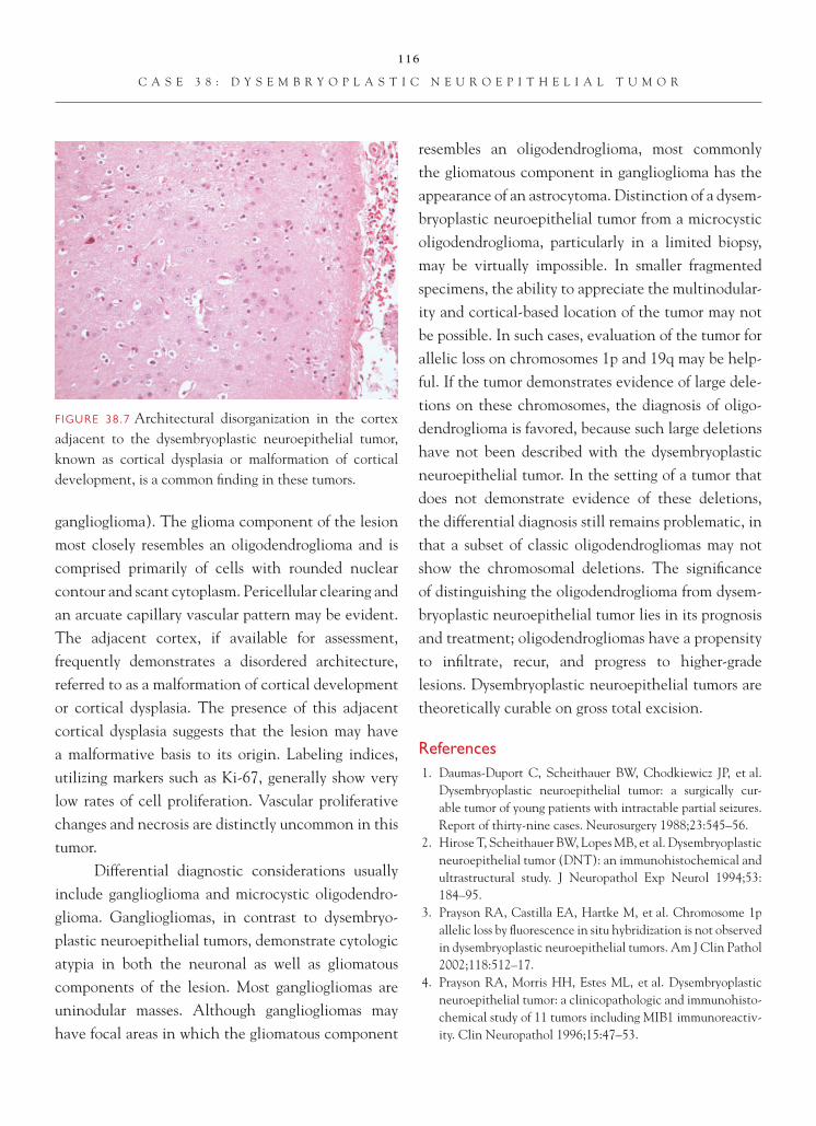
116
C A S E 3 8 : D Y S E M B R Y O P L A S T I C N E U R O E P I T H E L I A L T U M O R
ganglioglioma). The glioma component of the lesion most closely resembles an oligodendroglioma and is comprised primarily of cells with rounded nuclear contour and scant cytoplasm. Pericellular clearing and an arcuate capillary vascular pattern may be evident. The adjacent cortex, if available for assessment, frequently demonstrates a disordered architecture, referred to as a malformation of cortical development or cortical dysplasia. The presence of this adjacent cortical dysplasia suggests that the lesion may have a malformative basis to its origin. Labeling indices, utilizing markers such as Ki-67, generally show very low rates of cell proliferation. Vascular proliferative changes and necrosis are distinctly uncommon in this tumor.
Differential diagnostic considerations usually include ganglioglioma and microcystic oligodendro-glioma. Gangliogliomas, in contrast to dysembryo-plastic neuroepithelial tumors, demonstrate cytologic atypia in both the neuronal as well as gliomatous components of the lesion. Most gangliogliomas are uninodular masses. Although gangliogliomas may have focal areas in which the gliomatous component
resembles an oligodendroglioma, most commonly the gliomatous component in ganglioglioma has the appearance of an astrocytoma. Distinction of a dysem-bryoplastic neuroepithelial tumor from a microcystic oligodendroglioma, particularly in a limited biopsy, may be virtually impossible. In smaller fragmented specimens, the ability to appreciate the multinodular-ity and cortical-based location of the tumor may not be possible. In such cases, evaluation of the tumor for allelic loss on chromosomes 1p and 19q may be help-ful. If the tumor demonstrates evidence of large dele-tions on these chromosomes, the diagnosis of oligo-dendroglioma is favored, because such large deletions have not been described with the dysembryoplastic neuroepithelial tumor. In the setting of a tumor that does not demonstrate evidence of these deletions, the differential diagnosis still remains problematic, in that a subset of classic oligodendrogliomas may not show the chromosomal deletions. The signifi cance of distinguishing the oligodendroglioma from dysem-bryoplastic neuroepithelial tumor lies in its prognosis and treatment; oligodendrogliomas have a propensity to infi ltrate, recur, and progress to higher-grade lesions. Dysembryoplastic neuroepithelial tumors are theoretically curable on gross total excision.
References 1. Daumas-Duport C, Scheithauer BW, Chodkiewicz JP, et al.
Dysembryoplastic neuroepithelial tumor: a surgically cur-able tumor of young patients with intractable partial seizures. Report of thirty-nine cases. Neurosurgery 1988;23:545–56.
2. Hirose T, Scheithauer BW, Lopes MB, et al. Dysembryoplastic neuroepithelial tumor (DNT): an immunohistochemical and ultrastructural study. J Neuropathol Exp Neurol 1994;53:184–95.
3. Prayson RA, Castilla EA, Hartke M, et al. Chromosome 1p allelic loss by fl uorescence in situ hybridization is not observed in dysembryoplastic neuroepithelial tumors. Am J Clin Pathol 2002;118:512–17.
4. Prayson RA, Morris HH, Estes ML, et al. Dysembryoplastic neuroepithelial tumor: a clinicopathologic and immunohisto-chemical study of 11 tumors including MIB1 immunoreactiv-ity. Clin Neuropathol 1996;15:47–53.
FIGURE 38.7 Architectural disorganization in the cortex adjacent to the dysembryoplastic neuroepithelial tumor, known as cortical dysplasia or malformation of cortical development, is a common fi nding in these tumors.

117
Case 39: Ganglioglioma
C L I N I C A L I N F O R M A T I O N
The patient is a 7-year-old female who presents with seizures of 6.5 years duration. CT imaging shows a cir-cumscribed, partially cystic mass, with focal calcifi ca-tion situated in the left temporal lobe. The mass is resected, and histologic sections are reviewed.
O P I N I O N
Histologic sections show hypercellular parenchyma, marked by a proliferation of atypical, spindled glial cells with high nuclear-to-cytoplasmic ratio. Mitotic activity is not noted. Intermixed with the spindled cells is a second population of ganglionic type cells, with abundant eosinophilic cytoplasm and generally rounded nuclear contours with prominent nucleo-lation. Foci of perivascular chronic infl ammation,
consisting primarily of benign-appearing lymphocytes, are noted. Focal calcifi cations are also observed in the lesion. Vascular proliferative changes and necrosis are not seen. Focally, the mass appears to extend into the leptomeninges. Rosenthal fi bers and granular bodies are not noted in this lesion.
We consider the lesion to be a glioneuronal neoplasm and characterize it as follows: Left Temporal Lobe, Excision—Ganglioglioma, WHO Grade I.
C O M M E N T
The admixture of a gliomatous component resembling a low-grade fi brillary astrocytoma with an atypical ganglion cell component is consistent with a diagno-sis of ganglioglioma. Mitotic activity, necrosis, and vascular proliferative changes, suggesting a higher-grade lesion, are not observed.
FIGURE 39.1 Low magnifi cation appearance of the tumor, showing a moderately cellular neoplasm with focal calcifi cation.
FIGURE 39.2 Atypical ganglion cells are readily identifi able in the neoplasm.

118
C A S E 3 9 : G A N G L I O G L I O M A
D I S C U S S I O N
Gangliogliomas are a well-recognized glioneuronal neoplasm that has a peak incidence in the fi rst few decades of life. The tumors have been vari-ably described to arise in all areas of the brain, but they are most commonly located in the tempo-ral lobes. In patients with temporal lobe tumors, medically intractable epilepsy is often the clinical presentation.
Tumors may present as solid or cystic masses. Requisite for a diagnosis is identifi cation of an atypi-cal gliomatous component, which may resemble an ordinary fi brillary astrocytoma, pilocytic astrocytoma or, less commonly, an oligodendroglioma. Admixed with the gliomatous component is an atypical gan-glion cell component, marked by abnormal number
FIGURE 39.3 Cytologically atypical glial cells resembling neoplastic astrocytes are also noted in the tumor, associ-ated with occasional ganglionic cells.
FIGURE 39.4 Tumor is seen to extend focally, to involve the leptomeningeal space to the right. Occasional gangli-onic cells are present in the leptomeningeal tumor.
FIGURE 39.5 Foci of perivascular chronic infl ammation and calcifi cation are present.
FIGURE 39.6 An area of tumor consists exclusively of spin-dled glial cells resembling astrocytoma. Failure of adequate sampling in such a case may result in an erroneous diagno-sis of astrocytoma.

119
C A S E 3 9 : G A N G L I O G L I O M A
of ganglion cells, which are frequently cytologi-cally atypical (e.g., abnormal distribution of Nissl substance, neuronal cytomegaly, multinucleation). The two components of the tumor may not be evenly admixed throughout the neoplasm, and therefore, when the diagnosis is suspected, the tumor should be well sampled in order to guarantee that both com-ponents are identifi ed. Failure to do so may result in an erroneous diagnosis of a low-grade glioma, which carries with it a worse prognosis. Perivascular chronic infl ammation and calcifi cations are common fi ndings in these tumors. Occasionally, eosinophilic granular bodies or Rosenthal fi bers may also be observed in the neoplasm. Like dysembryoplastic neuroepithelial tumors, the adjacent uninvolved cortex may show architectural disorganization (malformation of corti-cal development or cortical dysplasia). Proliferative activity, as evidenced by Ki-67 immunostaining, is generally low, frequently <1. Immunohistochem-istry may be helpful in identifying the atypical gan-glion cell component in some tumors. Care should be taken not to overinterpret morphologically normal,
resident neurons, entrapped by an infi ltrating glioma, as an atypical ganglion cell component. As with the current case, gangliogliomas may occasionally extend to involve the leptomeninges; this has not necessarily been associated with a particularly worse prognosis.
The vast majority of gangliogliomas represent grade I neoplasms and are associated with an excel-lent prognosis. They generally do not require adju-vant therapy and are theoretically curable with gross total resection.
References 1. Diepholder HM, Schwechheimer K, Mohadjer M, et al. A
clinicopathologic and immunomorphologic study of 13 cases of ganglioglioma. Cancer 1991;68:2192–2201.
2. Lang FF, Epstein FJ, Ransohoff J, et al. Central nervous sys-tem gangliogliomas. Part 2: Clinical outcome. J Neurosurg 1993;79:867–73.
3. Prayson RA, Khajavi K, Comair YG. Cortical architectural abnormalities and MIB1 immunoreactivity in gangliogliomas: a study of 60 patients with intracranial tumors. J Neuropathol Exp Neurol 1995;54:513–20.
4. Zentner J, Wolf HK, Ostertun B, et al. Gangliogliomas: clini-cal, radiological, and histopathological fi ndings in 51 patients. J Neurol Neurosurg Psychiatry 1994;57:1497–1502.
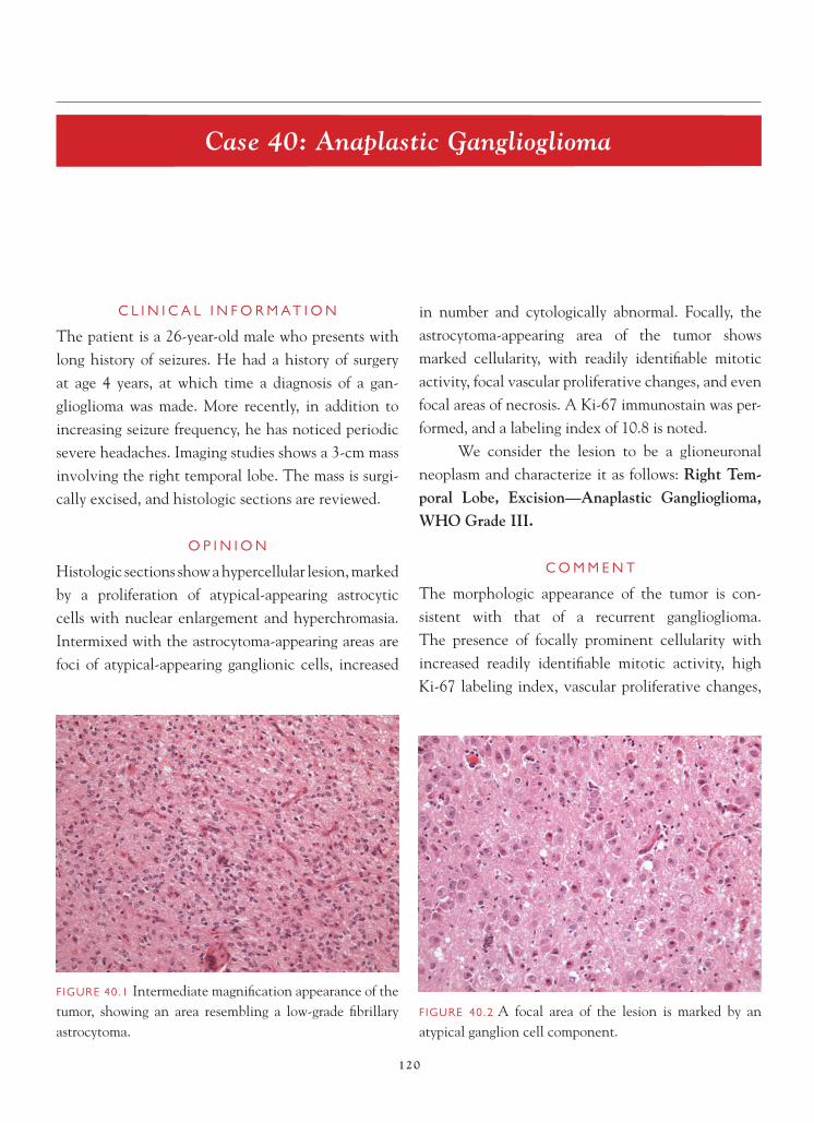
120
Case 40: Anaplastic Ganglioglioma
C L I N I C A L I N F O R M A T I O N
The patient is a 26-year-old male who presents with long history of seizures. He had a history of surgery at age 4 years, at which time a diagnosis of a gan-glioglioma was made. More recently, in addition to increasing seizure frequency, he has noticed periodic severe headaches. Imaging studies shows a 3-cm mass involving the right temporal lobe. The mass is surgi-cally excised, and histologic sections are reviewed.
O P I N I O N
Histologic sections show a hypercellular lesion, marked by a proliferation of atypical-appearing astrocytic cells with nuclear enlargement and hyperchromasia. Intermixed with the astrocytoma-appearing areas are foci of atypical-appearing ganglionic cells, increased
in number and cytologically abnormal. Focally, theastrocytoma-appearing area of the tumor shows marked cellularity, with readily identifi able mitotic activity, focal vascular proliferative changes, and even focal areas of necrosis. A Ki-67 immunostain was per-formed, and a labeling index of 10.8 is noted.
We consider the lesion to be a glioneuronal neoplasm and characterize it as follows: Right Tem-poral Lobe, Excision—Anaplastic Ganglioglioma, WHO Grade III.
C O M M E N T
The morphologic appearance of the tumor is con-sistent with that of a recurrent ganglioglioma. The presence of focally prominent cellularity with increased readily identifi able mitotic activity, high Ki-67 labeling index, vascular proliferative changes,
FIGURE 40.1 Intermediate magnifi cation appearance of the tumor, showing an area resembling a low-grade fi brillary astrocytoma.
FIGURE 40.2 A focal area of the lesion is marked by an atypical ganglion cell component.

121
C A S E 4 0 : A N A P L A S T I C G A N G L I O G L I O M A
and necrosis indicate a higher-grade, more aggressive-behaving tumor.
D I S C U S S I O N
Although the vast majority of gangliogliomas are low-grade, WHO grade I neoplasms, it has been rec-ognized that a small number of these tumors may degenerate into a higher-grade neoplasm (anaplas-tic ganglioglioma). Precise criteria for the diagnosis
of anaplastic ganglioglioma remain somewhat vague. In general, these tumors are marked by a morphology in the gliomatous component that resembles a malig-nant glioma, i.e., increased mitotic activity, vascular proliferative changes, and even necrosis. In the few cases that have been studied in the literature, these tumors also tend to demonstrate markedly increased rates of cell proliferation, as evidenced by Ki-67 or MIB-1 immunostaining. Although histologic sections from the original resection in the current case were
FIGURE 40.3 In areas, the gliomatous component is more cellular and marked by more nuclear pleomorphism, more akin to a higher-grade fi brillary astrocytoma.
FIGURE 40.4 Mitotic fi gures are readily identifi able in the gliomatous component of the tumor.
FIGURE 40.5 Focal vascular proliferative changes are also observed in the tumor.
FIGURE 40.6 Geographic necrosis is present in the neo-plasm.

122
C A S E 4 0 : A N A P L A S T I C G A N G L I O G L I O M A
FIGURE 40.7 Ki-67 immunostaining shows more than rare positive staining cells. The tumor had a labeling index of 10.8; usual low-grade gangliogliomas have lower labeling indices, typically <1.
of the tumor. In terms of differential diagnosis, simi-lar to ordinary low-grade gangliogliomas, distinction of this lesion from a high-grade glioma is important; documentation of the atypical ganglion cell compo-nent is critical to rendering the correct diagnosis.
References 1. Luyken C, Blumcke I, Fimmers R, et al. Supratentorial gan-
gliogliomas: histopathologic grading and tumor recurrence in 184 patients with a median follow-up of 8 years. Cancer 2004;101:146–55.
2. Prayson RA, Khajavi K, Comair YG. Cortical architectural abnormalities and MIB1 immunoreactivity in gangliogliomas: a study of 60 patients with intracranial tumors. J Neuropathol Exp Neurol 1995;54:513–20.
3. Suzuki H, Otsuki T, Iwasaki Y, et al. Anaplastic gangliogli-oma with sarcomatous component: an immunohistochemical study and molecular analysis of p53 tumor suppressor gene. Neuropathology 2002;22:40–7.
unavailable for examination, the prior diagnosis of ganglioglioma suggests an initial low-grade lesion. The recent increase in seizure frequency and development of headaches indicate a potential change in the status

123
Case 41: Papillary Glioneuronal Tumor
C L I N I C A L I N F O R M A T I O N
The patient is a 36-year-old male who presents with a 3-month history of recurrent headaches and new onset seizures. Imaging studies show a right temporal lobe mass with focal enhancement and cystic change. The lesion is resected, and histologic sections are reviewed.
O P I N I O N
Histologic sections show a neoplasm marked by a focal papillary architectural pattern. The centers of the papil-lae are formed by vessels that are variably sclerotic. Surrounding the vessels are rounded cells with a small amount of clear-to-eosinophilic cytoplasm. Intermixed between the papillary structures are a population of cells with generally rounded nuclei and scant cytoplasm. Hemosiderin-laden macrophages, indicative of prior hemorrhage, are observed. On immunohistochemical
staining, the cells lining the papillae demonstrate posi-tive immunoreactivity with antibody to GFAP. Focally, collections of cells situated between the papillae dem-onstrate synaptophysin immunoreactivity.
We consider the lesion to be a glioneuronal tumor and characterize it as follows: Right Temporal Lobe, Excision—Papillary Glioneuronal Tumor, WHO Grade I.
C O M M E N T
The presence of GFAP- and synaptophysin-positive staining of subpopulations of cells in the tumor sup-port a diagnosis of glioneuronal neoplasm.
D I S C U S S I O N
Papillary glioneuronal tumors are a relatively recently described entity. Most of the tumors described have
FIGURE 41.1 Lower-magnifi cation appearance of the tumor, showing a vaguely papillary architectural pattern.
FIGURE 41.2 Higher-magnifi cation view, showing sclerotic vessel changes in the center of papillary structures.

124
C A S E 4 1 : P A P I L L A R Y G L I O N E U R O N A L T U M O R
arisen in adults—in the cerebral hemispheres, with a predilection for the temporal lobe. On imaging stud-ies, a cystic mass with mural nodule(s) is frequently observable.
As its name suggests, the tumor consists of a papillary architecture, with vascular cores rimmed by GFAP-positive staining glial cells. Vascular scle-rotic changes may be prominently evident. Inter-spersed between the papillary areas is a population
of small, rounded, neuronal cells that demonstrate synaptophysin immunoreactivity. Occasionally, more mature-appearing ganglion cells may be evident. Prom-inent mitotic activity, vascular proliferative changes, and necrosis are distinctly uncommon in this tumor. The periphery of the lesion may show evidence of gliosis, with Rosenthal fi bers and eosinophilic granu-lar bodies. Microcalcifi cations have been described, as have hemosiderin-laden macrophages, evidence of prior hemorrhage. Cell proliferation markers, such as Ki-67, show very low rates of cell proliferation, typi-cally on the order of less than 2.
Of the tumors that have been reported thus far, all have been associated with long-term survival and have not required adjuvant chemotherapy or radia-tion therapy.
References 1. Bouvier-Labit C, Daniel L, Dufour H, et al. Papillary glioneuronal
tumour: clinicopathological and biochemical study of one case with 7-year follow up. Acta Neuropathol (Berl) 2000;99:321–6.
2. Kim DH, Suh YL. Pseudopapillary neurocytoma of tempo-ral lobe with glial differentiation. Acta Neuropathol (Berl) 1997;94:187–91.
3. Komori T, Scheithauer BW, Anthony DC, et al. Papillary glioneuronal tumor: a new variant of mixed neuronal-glial neoplasm. Am J Surg Pathol 1998;22:1171–83.
FIGURE 41.3 A more solid-appearing area of the tumor between papillae is marked by a proliferation of rounded cells and hemosiderin-laden macrophages.
FIGURE 41.4 GFAP immunoreactivity is observed in cells lining the papillae.
FIGURE 41.5 Synaptophysin immunoreactivity underscores the neural differentiation of the more solid foci of rounded cells situated between the papillae.

125
Case 42: Rosette-Forming Glioneuronal Tumor of the Fourth Ventricle
C L I N I C A L I N F O R M A T I O N
The patient is a 21-year-old male who presents with headaches and a mildly ataxic gait. On imaging studies, a midline fourth ventricular mass is noted. The lesion appears relatively circumscribed and shows increased signal intensity on T2-weighted images. The lesion is excised, and histologic sections are reviewed.
O P I N I O N
The histologic sections show a well-demarcated tumor, characterized by focal areas marked by rosetted structures. The rosetted structures contain central eosinophilic material, rimmed by a ring of cells with rounded nuclei, indistinct nucleoli, and scant cytoplasm. The majority of the tumor is marked by mildly hypercellular parenchyma, with atypical-appearing astrocytic cells. Mild focal vascular
proliferative changes are observed. In areas, the cells have more rounded nuclei and pericellular clearing. Rosenthal fi bers and eosinophilic granular bodies are not noted. Mitotic fi gures are not observed. There is no evidence of necrosis. On immunostaining, the tumor demonstrates positive staining with antibody to GFAP. The rosetted structures show positive staining, with antibody to synaptophysin, indicating neurocytic differentiation.
We consider the tumor to be a low-grade neo-plasm and characterize it as follows: Fourth Ventri-cle, Excision—Rosette-Forming Glioneuronal Tu-mor of the Fourth Ventricle, WHO Grade I.
C O M M E N T
The presence of synaptophysin-positive neurocytic rosettes, admixed with a GFAP-positive astrocytoma
FIGURE 42.1 Intermediate magnifi cation appearance of the tumor, showing a mildly hypercellular neoplasm resembling a low-grade glioma.
FIGURE 42.2 Focal vascular proliferative changes are present. There is no evidence of necrosis and prominent mitotic activity.

126
C A S E 4 2 : R O S E T T E - F O R M I N G G L I O N E U R O N A L T U M O R
component, supports the diagnosis of a mixed glion-euronal neoplasm.
D I S C U S S I O N
The rosette-forming glioneuronal tumor of the fourth ventricle is a new addition to the WHO classifi cation system. This is a rare tumor, which has been described in both children and adults. As its name suggests, the lesion generally arises in the fourth ventricle
(although anecdotal cases of origin in other loca-tions are reported in the literature). The patients typically present with signs and symptoms related to obstructive hydrocephalus, including headaches, or ataxia. The lesion is generally well circumscribed and microscopically is marked by the presence of synap-tophysin-positive neurocytic rosettes or perivascular pseudorosettes, admixed with areas of tumor resem-bling a low-grade astrocytoma, with elongated tumor
FIGURE 42.3 High-magnifi cation appearance, showing oli-godendroglial-like cells, with pericellular clearing in the gliomatous area of the tumor.
FIGURE 42.4 Characteristic neurocytic rosette-like structures are present in the tumor.
FIGURE 42.5 These rosette-like structures demonstrate positive staining with synaptophysin antibody, indicating neurocytic differentiation.
FIGURE 42.6 Much of the remaining tumor demon-strates GFAP immunoreactivity, indicating astrocytic differentiation.
127
C A S E 4 2 : R O S E T T E - F O R M I N G G L I O N E U R O N A L T U M O R
cell nuclei and GFAP-positive cytoplasm. Occasion-ally, background cells have rounded nuclei, with scant cytoplasm, and resemble oligodendroglial cells. Rosenthal fi bers, granular bodies, hemosiderin depo-sition, and microcalcifi cations have been variably described in the tumor. Mitotic activity and necro-sis are generally absent. Occasional tumors may show hyalinized or sclerosed vessels, vascular thrombosis, and vascular proliferation-type changes. Proliferation markers show low rates of proliferation, in keeping with the general low grade of the tumor. The key to the diagnosis is recognition of the rosette structures and demonstration of both glial and neuronal differ-entiation in the tumor.
Differential diagnostic considerations include pilocytic astrocytoma, dysembryoplastic neuroepi-thelial tumor, and neurocytoma. The presence of neurocytic differentiation in this tumor allows dis-tinction from pilocytic astrocytoma. The typical imaging study of a pilocytic astrocytoma (cystic neo-plasm with enhancing mural nodule) is not usually observed in the rosette-forming glioneuronal tumor. Dysembryoplastic neuroepithelial tumors usually
present as supratentorial lesions in younger patients, often have a multinodular architectural pattern, and generally lack a glial component that resembles pilocytic astrocytoma. Central neurocytomas are usu-ally situated in the lateral third ventricles, demon-strate diffuse neuronal differentiation, and do not contain geographic areas of astrocytic phenotype.
The relatively few cases of this tumor that have been reported in the literature have had a favorable outcome.
References 1. Albanese A, Mangiola A, Pompucci A, et al. Rosette-
forming glioneuronal tumor of the fourth ventricle: report of a case with clinical and surgical implications. J Neurooncol 2005;71:195–7.
2. Komori T, Scheithauer BW, Hirose T. A rosette-forming glio-neuronal tumor of the fourth ventricle: intratentorial form of dysembryoplastic neuroepithelial tumor? Am J Surg Pathol 2002;25:582–91.
3. Preusser M, Dietrich W, Czech T, et al. Rosette-forming glio-neuronal tumor of the fourth ventricle. Acta Neuropathol 2003;106:506–8.
4. Rosenblum MK. The 2007 WHO classifi cation of nervous system tumours: newly recognized members of the mixed glio-neuronal group. Brain Pathol 2007;17:308–13.

128
Case 43: Central Neurocytoma
C L I N I C A L I N F O R M A T I O N
The patient is a 32-year-old male who presents with increased headaches. On imaging, a heterogeneous 2.8-cm mass on T1-weighted MRI imaging is noted within the right lateral ventricle. The lesion is subto-tally resected, and histologic sections are reviewed.
O P I N I O N
Histologic sections show a proliferation of cells with rounded nuclei and scant eosinophilic cytoplasm. The nuclei have a salt-and-pepper chromatin pattern with an occasional small nucleolus evident. An arcuate cap-illary pattern is noted in the background of the tumor. Focal dystrophic calcifi cations are observed. Mitotic activity is not seen. Vascular proliferative changes or necrosis are not present. On immunostaining, the
tumor demonstrates diffuse positive staining, with antibody to synaptophysin, and does not stain with antibody to GFAP. A Ki-67 immunostain is per-formed, and a labeling index of 1.7 noted.
We consider the lesion to be a tumor with neu-ral differentiation and characterize it as follows: RightLateral Ventricle, Excision—Central Neurocytoma, WHO Grade II.
C O M M E N T
Diffuse synaptophysin immunoreactivity supports a diagnosis of central neurocytoma.
D I S C U S S I O N
Although central neurocytomas are classically tumors that arise in young adults and have a predilection
FIGURE 43.1 Low-magnifi cation appearance of a central neurocytoma, highlighting the monotony of the tumor cells.
FIGURE 43.2 A prominent arcuate capillary vascular pat-tern, similar to that seen in oligodendroglioma, may also be observed in central neurocytoma.

129
C A S E 4 3 : C E N T R A L N E U R O C Y T O M A
for the lateral ventricles, the clinical presentation is often related to symptoms associated with increased intracranial pressure. Morphologically, the tumors have a rather monomorphic appearance, consist-ing of rounded cells, with fi nely speckled chromatin nuclei and a prominent capillary vascular pattern. Morphologically, these tumors can resemble oligo-dendrogliomas, as well as rare clear cell ependymo-mas, particularly given the common intraventricular
location of the central neurocytoma. The key to the diagnosis rests on demonstrating evidence of neural differentiation by immunohistochemistry or ultra-structural examination of the tumor. Cell proliferation indices are generally low, typically less than 2. Central neurocytoma is considered a low-grade lesion; how-ever, the tumor may recur if incompletely resected.
Rare cases of neurocytomas arising outside the ventricular system (extraventricular neurocytomas) have also been described. These tumors are typically well-circumscribed, contrast-enhancing masses that often have a cystic appearance, like their intraven-tricular counterpart. The presence of a prominent vascular pattern and calcifi cations raises oligoden-droglioma as a differential diagnostic consideration. Again, immunohistochemistry is helpful in arriving at the proper diagnosis.
References 1. Brat DJ, Scheithauer BW, Eberhart CG, et al. Extraventricu-
lar neurocytomas: pathologic features and clinical outcome. Am J Surg Pathol 2001;25:1252–60.
2. Giangaspero F, Cenacchi G, Losi L, et al. Extraventricular neoplasms with neurocytoma features. A clinicopathological study of 11 cases. Am J Surg Pathol 1997;21:206–12.
FIGURE 43.3 High-magnifi cation appearance of the cells comprising central neurocytoma. Cells are marked by scant cytoplasm and nuclei with salt-and pepper chromatin pattern.
FIGURE 43.4 Focal calcifi cations are observed in the tumor.
FIGURE 43.5 Diffuse positive immunoreactivity with syn-aptophysin, indicating neural differentiation, is seen in this tumor.

130
C A S E 4 3 : C E N T R A L N E U R O C Y T O M A
3. Hassoun J, Soylemezoglu F, Gambarelli D, et al. Central neurocytoma: a synopsis of clinical and histological features. Brain Pathol 1993;3:297–306.
4. MacKenzie IR. Central neurocytoma: histologic atypia, proliferation potential, and clinical outcome. Cancer 1999;85:1606–10.
5. Schild SSE, Scheithauer BW, Haddock MG, et al. Central neurocytomas. Cancer 1997;79:790–5.
6. Yasargil MG, von Ammon K, von Deimling A, et al. Cen-tral neurocytoma: histopathological variants and therapeutic approaches. J Neurosurg 1992;76:32–7.

131
Case 44: Atypical Neurocytoma
C L I N I C A L I N F O R M A T I O N
The patient is a 39-year-old female who presents with increased confusion. A left lateral ventricular mass, measuring 2.1 cm, is identifi ed and shows marked enhancement after gadolinum injection. The tumor is subtotally resected, and histologic sections are reviewed.
O P I N I O N
Histologic sections show a somewhat monotonous proliferation of generally rounded cells, with scant eosinophilic cytoplasm and salt-and-pepper nuclear chromatin pattern. In focal areas, some degree of nuclear pleomorphism is observable. The back-ground of the tumor has a fi brillary-like appearance,
with a prominent arcuate capillary vascular pattern and focal calcifi cations. Up to 5 mitotic fi gures per 10 high-power fi elds are observed. Focal necrosis is also seen. Microvascular proliferation is noted in the tumor. Immunohistochemical staining shows diffuse positive staining with antibody to synaptophysin and paucity of GFAP immunoreactivity. A Ki-67 labeling index of 9.8 is observed.
We consider the lesion to be a tumor with neural differentiation and characterize it as follows: Left Lateral Ventricle, Excision—Atypical Central Neurocytoma.
C O M M E N T
The presence of increased mitotic activity, elevated Ki-67 labeling index, and focal necrosis suggest a more aggressive-behaving tumor.
FIGURE 44.1 Intermediate magnifi cation appearance of an atypical neurocytoma, showing a somewhat monotonous proliferation of rounded cells, with fi brillary-like back-ground and a prominent arcuate capillary vascular pattern.
FIGURE 44.2 Focal calcifi cations and vascular proliferative changes are evident in this tumor.

132
C A S E 4 4 : A T Y P I C A L N E U R O C Y T O M A
D I S C U S S I O N
Although the majority of central neurocytomas, as previously described in Case 43, represent low-grade neoplasms, a subset of tumors may behave in a more aggressive fashion. The morphologic correlates of more aggressive behavior in neurocytomas include
increased mitotic activity, vascular proliferative changes, and necrosis. Many of these tumors also have elevated Ki-67 or MIB-1 indices, in excess of 2–3, as in the current tumor. Central neurocy-tomas, particularly with increased cell prolifera-tion indices, have shown an increased likelihood of recurrence.
FIGURE 44.3 Focal perivascular pseudorosetting by cells with salt-and-pepper nuclear chromatin pattern is observed.
FIGURE 44.4 Focally, nuclear pleomorphism is evident.
FIGURE 44.5 Readily identifi able mitotic activity is observed in this tumor.
FIGURE 44.6 Focal necrosis is present in the neoplasm.

133
C A S E 4 4 : A T Y P I C A L N E U R O C Y T O M A
References 1. MacKenzie IR. Central neurocytoma: histologic atypia,
proliferation potential, and clinical outcome. Cancer 1999;85:1606–10.
2. Rades D, Schild SE, Fehlauer F. Prognostic value of the MIB-1 labeling index for central neurocytomas. Neurology 2004;62:987–9.
3. Soylemezoglu F, Scheithauer BW, Esteve J, et al. Atypical cen-tral neurocytoma. J Neuropathol Exp Neurol 1997;56:551–6.
FIGURE 44.7 Diffuse synaptophysin immunoreactivity is present in this tumor.

134
Case 45: Extraventricular Neurocytoma
C L I N I C A L I N F O R M A T I O N
The patient is a 27-year-old female who is found to have papilledema following work-up for headaches. An MRI scan shows a large left parieto-occipital cyst with an enhancing mural nodule. She undergoes complete gross total excision of the lesion.
O P I N I O N
Sections show a calcifi ed, cytologically bland, mod-erately hypercellular neoplasm, devoid of necrosis or microvascular proliferation. Numerous hyalinized blood vessels are present throughout the tumor, as is focal hemosiderin pigment. The tumor demonstrates diffuse positive staining, with synaptophysin antibody.
We consider the lesion to be a low-grade neurocytic tumor and characterize it as follows: Left
Parieto-Occipital Region, Excision– ExtraventricularNeurocytoma, WHO Grade II.
C O M M E N T
Given the fact that this tumor did not occur within the ventricular system, this neoplasm—which is otherwise histologically identical to central neuro-cytoma—would be considered an extraventricular neurocytoma. The cystic nature is apparent only on the neuroimaging studies, because the pathologist receives the solid mural nodule portion of the tumor.
D I S C U S S I O N
Extraventricular neurocytoma is a new entity in the 2007 World Health Organization classifi cation system. Although both central neurocytoma and extraventricular neurocytoma are composed of
FIGURE 45.1 Lower-magnifi cation appearance of the tumor, demonstrating moderate hypercellularity, hyalin-ized blood vessels, and vague orientation of tumor cells around blood vessels.
FIGURE 45.2 Intermediate magnifi cation shows the neuropil-like fi brillary background, as well as the slight variability in size of tumor nuclei.

135
C A S E 4 5 : E X T R A V E N T R I C U L A R N E U R O C Y T O M A
“uniform round cells that show immunohistochemi-cal and ultrastructural features of neuronal differen-tiation” (Figarella-Branger et al.), the term “central” neurocytoma is restricted to tumors lying within an intracerebral ventricle, usually the lateral ventricle. Both are WHO grade II neoplasms. Extraventricular neurocytoma may be solid or show a cyst with a mural nodule, as did this case. Tumors are variably contrast-enhancing, and some examples show limited infi ltra-tion of tumor cells into adjacent brain.
Making the diagnosis rests on recognizing the monotony of the tumor cells, delicate background fi bril-lar matrix simulating neuropil, and nuclei with a salt-and-pepper chromatin pattern and scant cytoplasm. The tumor may show intersecting vessels paralleling an endocrine tumor, calcifi cations, anuclear areas containing delicate fi brillary tumor processes simulating a large rosette, or even perivascular arrangement of tumor cells. Extraventricular neurocytomas are more likely to contain large or intermediate-sized ganglion cells than are central neurocytomas and may be less densely cellular. Suboptimal tissue preservation may even yield perinuclear haloes around the tumor cells. Not surprisingly, oligodendroglioma and ependymoma almost always enter the differential diagnosis.
Application of the immunohistochemical marker synaptophysin clarifi es that the tumor is com-posed of cells with neuronal lineage. Immunostaining is usually strong and diffuse, with maximal immuno-reactivity in the anuclear fi brillary zones. The nuclear neuronal marker NeuN is also usually positive. Most cases of extraventricular neurocytoma manifest low mitotic rate and low MIB-1 labeling index.
FIGURE 45.3 High-power magnifi cation depicts the salt-and-pepper chromatin pattern of the nuclei and scant cytoplasm.
FIGURE 45.4 Calcifi cations are seen in one-third to one-half of cases of extraventricular neurocytomas.
FIGURE 45.5 Strong immunoreactivity for synaptophysin is necessary to distinguish the tumor from an oligodendro-glioma or ependymoma.

136
C A S E 4 5 : E X T R A V E N T R I C U L A R N E U R O C Y T O M A
FIGURE 45.6 Many extraventricular neurocytomas contain larger ganglion cells.
References 1. Brat DJ, Parisi JE, Kleinschmidt-DeMasters BK, et al.
Neuropathology Committee, College of American Patholo-gists. Surgical neuropathology update: a review of changes introduced by the WHO classifi cation of tumours of the central nervous system, 4th edition. Arch Pathol Lab Med 2008;132:993–1007.
2. Brat DJ, Scheithauer BW, Eberhart CG, et al. Extraventricu-lar neurocytomas. Pathologic features and clinical outcome. Am J Surg Pathol 2001;25:1252–60.
3. Figarella-Branger D, Söylemezoglu, Burger PC. Central neurocytoma and extraventricular neurocytoma. In: Louis DN, Ohgaki H, Wiestler OD, Cavenee WK, Editors. WHO Classifi cation of Tumours of the Central Nervous System. Lyon, FR: IARC Press; 2007. p. 106–9.
FIGURE 45.7 Anti-neurofi lament immunostaining of the scattered individual ganglion cells highlights their neuritic processes.

137
Case 46: Paraganglioma
C L I N I C A L I N F O R M A T I O N
The patient is a 52-year-old male who presents with a history of progressively increasing lower back pain and sciatica. Imaging of the spinal cord shows a partially cystic, isointense mass in the cauda equina region of the cord on MR imaging. The patient undergoes gross total resection of the mass, and histologic sections are reviewed.
O P I N I O N
Histologic sections of the neoplasm are marked by a proliferation of generally rounded cells. The cells are focally arranged in loose nests, separated by deli-cate capillaries. Cells have a clear to lightly eosino-philic cytoplasm and nuclei with a salt-and-pepper
chromatin pattern. Scattered evidence of nuclear pleomorphism is also noted. The bulk of the tumor cells demonstrate positive staining, with antibod-ies to chromogranin and neuron-specifi c enolase. A smaller population of elongated cells at the periphery of the lobules demonstrate S-100 protein immunore-activity. The tumor does not stain with antibody to GFAP.
We consider the lesion to be a neuroendo-crine neoplasm and characterize it as follows: Cauda Equina Region, Excision—Paraganglioma, WHO Grade I.
C O M M E N T
The nested architectural pattern of the tumor and its immunohistochemical profi le support a diagnosis of paraganglioma.
FIGURE 46.1 A solid-appearing architectural pattern is present in this paraganglioma; the zellballen or nested pat-tern may not be always readily observable on hematoxylin-eosin stained sections.
FIGURE 46.2 An area of fi brosis is seen in the tumor, with an entrapped nerve twig.

138
C A S E 4 6 : P A R A G A N G L I O M A
D I S C U S S I O N
Paragangliomas associated with the spinal cord are similar to those that have been described elsewhere in the body. They typically are encapsulated, some-what well-circumscribed masses that are most com-monly encountered in the fi lum terminale region. Rare cases have been noted to arise in association with nerve roots or in the sellar and suprasellar areas. Most patients are adults at the time of presentation.
Histologically, the tumors are marked by classic nested architectural pattern (zellballen). Occasionally, tumors may have a solid appearance, where nesting is not readily evident, or a trabecular architecture. Scattered pleomorphic nuclei may be observable. Cells generally have a salt-and-pepper chromatin pattern and a variable amount of lightly eosinophilic cytoplasm. Occasional ganglionic cells may be observable in the tumor. By immunohisto-chemistry, the tumor demonstrates immunoreactivity, with antibodies to chromogranin and synaptophysin. Sustentacular cells, usually located around the perim-eter of the cell nests, demonstrate immunoreactivity with antibody to S-100 protein and may demonstrate some positive staining, with antibody to GFAP. Most tumors are quite amenable to surgical resection and have a good prognosis, although recurrence may occur in subtotally resected neoplasms.
Differential diagnostic considerations often involve other tumors commonly arising in the fi lum terminale region, which include myxopapillary ependy-moma, schwannoma, and ependymoma. Schwannomas generally lack a nested architectural pattern, demon-strate diffuse positive staining, with antibody to S-100
FIGURE 46.3 A prominent capillary vascular pattern is present.
FIGURE 46.4 Intermediate magnifi cation, showing cells with lightly eosinophilic-to-clear cytoplasm.
FIGURE 46.5 Eosinophilic cytoplasm is seen in cells arranged in a nested, or zellballen, architectural pattern.

139
C A S E 4 6 : P A R A G A N G L I O M A
FIGURE 46.6 High magnifi cation, showing scattered pleo-morphic nuclei and salt-and-pepper nuclear chromatin pattern.
protein, and generally do not stain with antibodies such as chromogranin and neuron-specifi c enolase. Similarly, diffuse immunoreactivity, with antibody to GFAP, is observable in myxopapillary ependymomas and low-grade ependymomas. Ependymal neoplasms lack im-munoreactivity with antibodies such as chromogranin. Rosettes and pseudorosettes that mark ependymomas and the mucin-positive microcystic areas of myxopapil-lary ependymoma are not features of paraganglioma.
References 1. Aggarwal S, Deck JH, Kucharczyk W. Neuroendocrine tumor
(paraganglioma) of the cauda equina: MR and pathologic fi ndings. Am J Neuroradiol 1993;14:1003–7.
2. Moran CA, Rush W, Mena H. Primary spinal paraganglio-mas: a clinicopathologic and immunohistochemical study of 30 cases. Histopathology 1997;31:167–73.
3. Sonneland PR, Scheithauer BW, LeChago J, et al. Paragan-glioma of the cauda equina region. Clinicopathologic study of 31 cases with special reference to immunocytology and ultra-structure. Cancer 1986;58:1720–35.
FIGURE 46.7 Neuron-specifi c enolase positivity is observed in a paraganglioma.
FIGURE 46.8 Positive staining with antibody to chromo-granin is characteristic of paraganglioma.
FIGURE 46.9 S-100 immunoreactivity highlights susten-tacular cells, primarily situated around the edge of the zellballen nests.

140
Case 47: Pineocytoma
C L I N I C A L I N F O R M A T I O N
The patient is a 57-year-old female with a long his-tory of intermittent headaches who recently develops nausea, vomiting, and paralysis of upward gaze. CT and MRI scanning demonstrates a noninvasive, well-circumscribed, homogeneously enhancing mass in the pineal region, measuring approximately 4 cm in greatest diameter. Histologic sections from the resec-tion specimen are available for review.
O P I N I O N
Sections demonstrate a lobulated mass composed of expanded nests of tumor cells within an anastomos-ing network of thin-walled blood vessels, with vari-able adventitial proliferation. Portions of the tumor
demonstrate an admixture of moderately pleomor-phic tumor cells and relatively abundant fi brillary matrix, with the formation of oversized Homer Wright-type rosettes (“pineocytomatous rosettes”). Other regions of the tumor show oversized nests of unipolar cells generating a delicate fi brillary matrix, demarcated by prominent fi brovascular septae con-taining chronic infl ammatory cells and adventitial proliferation. Mitotic fi gures are inconspicuous, and coagulative tumor necrosis is not identifi ed.
We consider the lesion to be a pineal tumor and characterize it as follows: Pineal Region, Removal—Pineocytoma, WHO Grade I.
FIGURE 47.1 The non-neoplastic pineal gland demon-strates nests of neuroendocrine cells within a delicate vas-cular stroma containing microcalcifi cations.
FIGURE 47.2 Glial cyst of the pineal gland with compres-sion of the pineal parenchyma, giving the appearance of hypercellularity, but without the architectural distortion and stromal reaction seen within true pineal parenchymal tumors.

141
C A S E 4 7 : P I N E O C Y T O M A
D I S C U S S I O N
Pineal region tumors in adults are unusual and consist predominately of meningiomas and astrocytic tumors. Pineal parenchymal tumors are rare lesions in adults but consist nearly exclusively of WHO grade I pine-ocytomas. These tumors consist of expanded nests of cells resembling those of the normal pineal, as well as regions containing diagnostic pineocytomatous
rosettes. The latter resemble Homer Wright rosettes, seen in primitive tumors containing neuroblastic differentiation, except that, in the case of pineocy-tomatous rosettes, there is a greater amount of fi bril-lary matrix, and the surrounding tumor nuclei are not mitotically active. The presence of these rosettes, in combination with the absence of primitive neuroec-todermal tumor-like differentiation, is highly predic-tive of benign (WHO grade I) behavior, without risk of leptomeningeal dissemination, and long postopera-tive survivals, even in patients with subtotal resec-tions. Analogous to “ancient change,” described in schwannomas, pineocytomas may contain regions demonstrating marked nuclear enlargement and hyperchromasia. When these changes occupy sig-nifi cant portions of the tumor, some authors refer to them as pleomorphic pineocytomas. Similar to schwannomas, the presence and degree of nuclear pleomorphism do not adversely affect the expected benign behavior of these tumors.
Although current neuroimaging techniques have reduced the number of pineal region biop-sies in patients with glial cysts of the pineal (pineal
FIGURE 47.3 Low magnifi cation view of expanded lobu-lar architecture comprising pineocytoma. A perivascular lymphocytic infi ltrate is present near the center of the photograph.
FIGURE 47.4 High magnifi cation view of the upper left region of the previous fi gure demonstrates pineocytoma-tous rosettes.
FIGURE 47.5 Expanded nests of neuroendocrine-type cells within a proliferative fi brovascular stroma contain-ing chronic infl ammatory cells and adventitial fi broblastic proliferation, are present.

142
C A S E 4 7 : P I N E O C Y T O M A
cysts), it is important to be familiar with the appear-ance of chronically compressed, non-neoplastic pi-neal parenchyma, in order to avoid the overdiagno-sis of tumor in patients who undergo biopsy for an atypical- appearing pineal region cyst. When portions of a pineal cyst are included in biopsy specimens, they are distinguished by the presence of geographic region of fi brillary glial matrix containing Rosenthal fi bers, which may be abundant.
References 1. Fakhran S, Escott EJ. Pineocytoma mimicking a pineal cyst
on imaging: true diagnostic dilemma or a case of incomplete imaging? AJNR Am J Neuroradiol 2008;29:159–63.
2. Fèvre-Montange M, Szathmari A, Champier J, et al. Pineocytoma and pineal parenchymal tumors of intermediate differentiation presenting cytologic pleomorphism: a multi-center study. Brain Pathol 2008;18:354–9.
3. Lekovic GP, Gonzalez LF, Shetter AG, et al. Role of gamma knife surgery in the management of pineal region tumors. Neurosurg Focus 2007;23:E12.

143
Case 48: Pineal Parenchymal Tumor of Intermediate Differentiation
C L I N I C A L I N F O R M A T I O N
The patient is a 46-year-old male who presents with progressively increasing headaches for several months. An MRI scan is obtained and shows an approximately 2.5-cm midline pineal mass that homogeneously enhances. The differential diagnosis includes menin-gioma versus pineal tumor. Because of the increasing headaches, the patient is felt to be a candidate for surgical resection. Sections are available for review.
O P I N I O N
Biopsies show a hypercellular, lobulated tumor, com-posed of aggregates of small blue cells with high nuclear-to-cytoplasmic ratio. Lobules are demarcated by a delicate vasculature devoid of microvascular proliferation; the appearance simulates an endo-crine tumor. There are no fi brillar, anuclear zones or
large pineocytomatous rosettes. Neither necrosis nor mitotic activity is present. Cytologically, the cells show hyperchromatic nuclei, indistinct nucleoli, and near-absence of cytoplasm.
We consider the pathology to be that of a pineal parenchymal tumor, albeit lacking the features of either a classic pineocytoma or pineoblas-toma. We characterize this tumor with intermediate features as follows: Pineal Gland, Excision—Pineal Parenchymal Tumor of Intermediate Differentia-tion, WHO Grade II–III.
C O M M E N T
Pineal parenchymal tumor of intermediate differen-tiation (PPTID) occupies a position intermediate between pineocytoma, a WHO grade I tumor usually found in adults, and pineoblastoma, a WHO grade IV
FIGURE 48.1 Low power shows a lobulated tumor devoid of necrosis with lobules defi ned by delicate vasculature.
FIGURE 48.2 Higher power shows that the tumor cells pos-sess stippled hyperchromatic nuclei, scant cytoplasm, and rare-to-absent mitoses.

144
C A S E 4 8 : P I N E A L P A R E N C H Y M A L T U M O R O F I N T E R M E D I A T E D I F F E R E N T I A T I O N
tumor usually found in children. Most patients with PPTID are adults. Defi nite grading criteria to establish whether PPTID should be given a WHO grade II or III designation have not been formally established.
D I S C U S S I O N
Given the rarity of all pineal parenchymal tumors and the often small size of the biopsies obtained from this region, the fi rst goal of the pathologist on a biopsy
from the pineal region is to exclude a metastasis to the pineal/tectal plate region, primary central ner-vous system germ cell tumor (especially germinoma), and a pineal cyst with adjacent normal pineal gland. Once the diagnosis of pineal parenchymal tumor is established, the next goal is to make a distinc-tion between pineocytoma (WHO grade I), pineal parenchymal tumor of intermediate differentiation (PPTID) (WHO grade II–III), and pineoblastoma (WHO grade IV), because the former is treated by surgical excision alone, and pineoblastoma is treated with radiation and chemotherapy. The PPTID has variable behavior; in general, however, outcome of PPTID is closer to that of pineocytoma than to that of pineoblastoma.
PPTID is the most frequent type of pineal parenchymal tumor, constituting over half of all cases in some series and showing higher frequency than either pineocytoma or pineoblastoma. Several different histological patterns exist. The least frequent is a truly mixed, biphasic tumor, with discrete areas of pineocytoma adjacent to areas of pure pineoblastoma.
More commonly, PPTID has intermediate histo-logic features. Unlike pineocytoma, PPTID lacks true
FIGURE 48.3 Strong immunostaining for neuron-specifi c enolase is present.
FIGURE 48.5 A MIB-1 labeling index is generally 10% or less in PPTID.
FIGURE 48.4 Strong immunostaining for synaptophysin establishes a neuronal lineage of PPTID and aids in exclud-ing other diagnostic considerations.
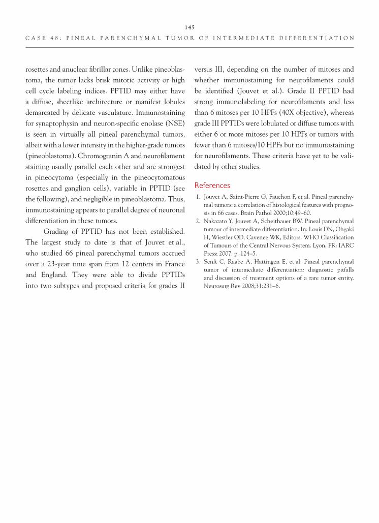
145
C A S E 4 8 : P I N E A L P A R E N C H Y M A L T U M O R O F I N T E R M E D I A T E D I F F E R E N T I A T I O N
rosettes and anuclear fi brillar zones. Unlike pineoblas-toma, the tumor lacks brisk mitotic activity or high cell cycle labeling indices. PPTID may either have a diffuse, sheetlike architecture or manifest lobules demarcated by delicate vasculature. Immunostaining for synaptophysin and neuron- specifi c enolase (NSE) is seen in virtually all pineal parenchymal tumors, albeit with a lower intensity in the higher-grade tumors (pineoblastoma). Chromogranin A and neurofi lament staining usually parallel each other and are strongest in pineocytoma (especially in the pineocytomatous rosettes and ganglion cells), variable in PPTID (see the following), and negligible in pineoblastoma. Thus, immunostaining appears to parallel degree of neuronal differentiation in these tumors.
Grading of PPTID has not been established. The largest study to date is that of Jouvet et al., who studied 66 pineal parenchymal tumors accrued over a 23-year time span from 12 centers in France and England. They were able to divide PPTIDs into two subtypes and proposed criteria for grades II
versus III, depending on the number of mitoses and whether immunostaining for neurofi laments could be identifi ed (Jouvet et al.). Grade II PPTID had strong immunolabeling for neurofi laments and less than 6 mitoses per 10 HPFs (40X objective), whereas grade III PPTIDs were lobulated or diffuse tumors with either 6 or more mitoses per 10 HPFs or tumors with fewer than 6 mitoses/10 HPFs but no immunostaining for neurofi laments. These criteria have yet to be vali-dated by other studies.
References 1. Jouvet A, Saint-Pierre G, Fauchon F, et al. Pineal parenchy-
mal tumors: a correlation of histological features with progno-sis in 66 cases. Brain Pathol 2000;10:49–60.
2. Nakazato Y, Jouvet A, Scheithauer BW. Pineal parenchymal tumour of intermediate differentiation. In: Louis DN, Ohgaki H, Wiestler OD, Cavenee WK, Editors. WHO Classifi cation of Tumours of the Central Nervous System. Lyon, FR: IARC Press; 2007. p. 124–5.
3. Senft C, Raabe A, Hattingen E, et al. Pineal parenchymal tumor of intermediate differentiation: diagnostic pitfalls and discussion of treatment options of a rare tumor entity. Neurosurg Rev 2008;31:231–6.
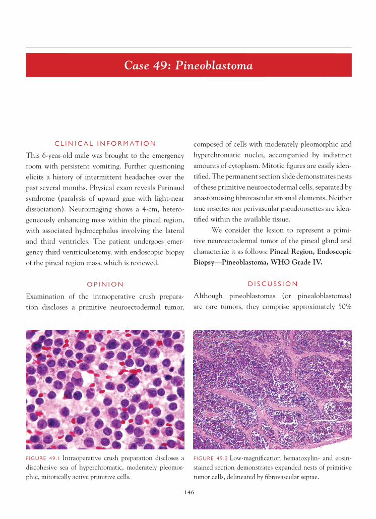
146
Case 49: Pineoblastoma
C L I N I C A L I N F O R M A T I O N
This 6-year-old male was brought to the emergency room with persistent vomiting. Further questioning elicits a history of intermittent headaches over the past several months. Physical exam reveals Parinaud syndrome (paralysis of upward gaze with light-near dissociation). Neuroimaging shows a 4-cm, hetero-geneously enhancing mass within the pineal region, with associated hydrocephalus involving the lateral and third ventricles. The patient undergoes emer-gency third ventriculostomy, with endoscopic biopsy of the pineal region mass, which is reviewed.
O P I N I O N
Examination of the intraoperative crush prepara-tion discloses a primitive neuroectodermal tumor,
composed of cells with moderately pleomorphic and hyperchromatic nuclei, accompanied by indistinct amounts of cytoplasm. Mitotic fi gures are easily iden-tifi ed. The permanent section slide demonstrates nests of these primitive neuroectodermal cells, separated by anastomosing fi brovascular stromal elements. Neither true rosettes nor perivascular pseudorosettes are iden-tifi ed within the available tissue.
We consider the lesion to represent a primi-tive neuroectodermal tumor of the pineal gland and characterize it as follows: Pineal Region, Endoscopic Biopsy—Pineoblastoma, WHO Grade IV.
D I S C U S S I O N
Although pineoblastomas (or pinealoblastomas) are rare tumors, they comprise approximately 50%
FIGURE 49.1 Intraoperative crush preparation discloses a discohesive sea of hyperchromatic, moderately pleomor-phic, mitotically active primitive cells.
FIGURE 49.2 Low-magnifi cation hematoxylin- and eosin-stained section demonstrates expanded nests of primitive tumor cells, delineated by fi brovascular septae.

147
C A S E 4 9 : P I N E O B L A S T O M A
of pineal parenchymal tumors. In addition, they are one of the most common pineal region tumors seen in the fi rst decade of life, second in incidence only to nongerminomatous germ cell tumors, especially immature teratoma. Because of their rarity, they are often included in studies of supratentorial primitive neuroectodermal tumors. However, evidence indi-cates that pineoblastomas, particularly those seen
in children over the age of 3 years, may have a more favorable prognosis and thus should be considered as distinct from a non-pineal region primitive neuroec-todermal tumors. Similar to other central nervous system primitive neuroectodermal tumors, pineoblas-tomas demonstrate a propensity to differentiate long neuronal and glial lineages. However, they may also, on occasion, demonstrate photosensory differen-tiation (Flexner-Wintersteiner rosettes, fl eurettes), similar to retinoblastomas, and often comprise the intracranial component of trilateral retinoblastoma.
Recent studies have demonstrated favorable outcome in children older than 3 years of age at presentation, especially with gross total resection, followed by craniospinal radiation and multiagent chemotherapy.
References 1. Cuccia V, Rodríguez F, Palma F, et al. Pinealoblastomas in
children. Childs Nerv Syst 2006;22:577–85. 2. Gilheeney SW, Saad A, Chi S, et al. Outcome of pediatric
pineoblastoma after surgery, radiation and chemotherapy. J Neurooncol 2008;89:89–95.
3. Hinkes BG, von Hoff K, Deinlein F, et al. Childhood pineoblastoma: experiences from the prospective multicenter trials HIT-SKK87, HIT-SKK92 and HIT91. J Neurooncol 2007;81:217–23.
FIGURE 49.3 Densely hypercellular sheets of mitotically active cells, with enlarged hyperchromatic pleomorphic nuclei and indistinct cytoplasm, are seen. Single cell necro-sis is readily identifi ed. Neither true nor pseudorosettes are present.

148
Case 50: Yolk Sac Tumor of the Pineal Gland
C L I N I C A L I N F O R M A T I O N
The patient is a 17-year-old male who presents with headaches. On imaging, he is noted to have a small mass situated in the pineal gland. A biopsy is per-formed, and histologic sections reviewed.
O P I N I O N
Histologic sections contain fragments of an epithe-lioid tumor. The neoplasm is characterized by ribbons and cords of cells and cystic spaces, resembling a vitell-ine pattern. The cells are somewhat homogeneous in appearance, contain small amounts of eosinophilic cytoplasm, and lack prominent nucleolation. Rare mitotic fi gures are observed. Eosinophilic droplets are not noted. Focal positive staining with antibody to alpha fetoprotein (AFP) is noted.
We consider the lesion to be a germ cell neoplasm and characterize it as follows: Pineal Gland, Biopsy—Yolk Sac Tumor (Endodermal Sinus Tumor).
C O M M E N T
AFP immunoreactivity supports the diagnosis of yolk sac tumor. Other germ cell tumor components are not identifi ed.
D I S C U S S I O N
Intracranial germ cell tumors are generally midline lesions that arise most commonly in the region of the pineal gland or suprasellar area. There is a male predilec-tion for development of these tumors. Symptomatology may be related to resulting hydrocephalus, Parinaud syn-drome (paralysis of upper gaze and conversion related to compression and invasion of the tectal plate), pituitary
FIGURE 50.1 Low-magnifi cation appearance of the entire biopsy, showing a loose arrangement of cells around blood vessels.
FIGURE 50.2 Higher-magnifi cation, highlighting solid foci, ribbons, and cords of tumor cells adjacent to blood vessels.

149
C A S E 5 0 : Y O L K S A C T U M O R O F T H E P I N E A L G L A N D
failure, visual fi eld defects in perioptic chiasmal tumors, and precocious puberty. Imaging appearances can be quite variable and somewhat dependent on the com-ponents of the neoplasm. Blood and the cerebrospinal fl uid-elevated levels of HCG (beta-human chorionic gonadotrophin), CEA (carcinoembryonic antigen), and AFP may provide clues to the diagnosis and to pos-sible components of the tumor.
The most commonly encountered pattern of intracranial germ cell tumor is germinoma. The tumors may be pure or contain multiple germ cell tumor elements (mixed cell tumors). Yolk sac tumor, or endodermal sinus tumor, is an epithelioid neoplasm in which cells are arranged in sheets, ribbons, cords, or papillae. Areas of the tumor have a loose micro-cystic appearance, resulting in what is described as a vitelline pattern. Schiller-Duval bodies, if present, are diagnostic; these structures are marked by ves-sels surrounded by tumor cells projecting into a clear
space also lined by tumor cells. Also, a useful diagnos-tic clue is the presence of eosinophilic droplets, which stain with Periodic acid-Schiff (PAS). Antibody to AFP can be useful in confi rming the diagnosis.
The prognosis is dependent on the components of the germ cells neoplasm, with germinomas being very radiosensitive, and along with teratomas, gener-ally associated with a good prognosis. Yolk sac tumors, along with choriocarcinomas and embryonal carcino-mas, tend to behave in a more aggressive fashion.
References 1. Bjornsson J, Scheithauer BW, Okasaki, H, et al. Intracranial
germ cell tumors: pathobiological and immunohistochemical aspects of 70 cases. J Neuropathol Exp Neurol 1985;44:32–46.
2. Ho DM, Liutt C. Primary intracranial germ cell tumor. Pathologic study of 51 patients. Cancer 1992;70:1577–1784.
3. Rueda P, Heifetz SA, Sesterhenn IA, et al. Primary intracra-nial germ cell tumors in the fi rst two decades of life. A clini-cal, light-microscopic, and immunohistochemical analysis of 54 cases. Perspect Pediatr Pathol 1987;10:160–207.
FIGURE 50.3 Intermediate magnifi cation, highlighting the microcystic areas in the tumor.
FIGURE 50.4 Focal AFP immunoreactivity confi rms the diagnosis of yolk sac tumor.
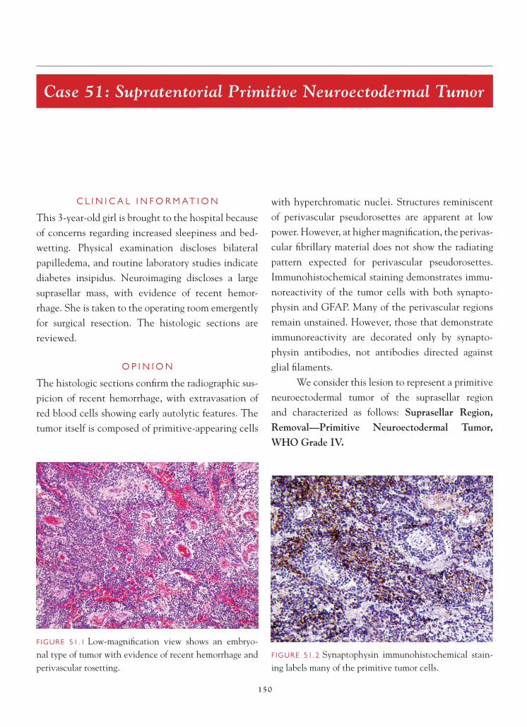
150
Case 51: Supratentorial Primitive Neuroectodermal Tumor
C L I N I C A L I N F O R M A T I O N
This 3-year-old girl is brought to the hospital because of concerns regarding increased sleepiness and bed-wetting. Physical examination discloses bilateral papilledema, and routine laboratory studies indicate diabetes insipidus. Neuroimaging discloses a large suprasellar mass, with evidence of recent hemor-rhage. She is taken to the operating room emergently for surgical resection. The histologic sections are reviewed.
O P I N I O N
The histologic sections confi rm the radiographic sus-picion of recent hemorrhage, with extravasation of red blood cells showing early autolytic features. The tumor itself is composed of primitive-appearing cells
with hyperchromatic nuclei. Structures reminiscent of perivascular pseudorosettes are apparent at low power. However, at higher magnifi cation, the perivas-cular fi brillary material does not show the radiating pattern expected for perivascular pseudorosettes. Immunohistochemical staining demonstrates immu-noreactivity of the tumor cells with both synapto-physin and GFAP. Many of the perivascular regions remain unstained. However, those that demonstrate immunoreactivity are decorated only by synapto-physin antibodies, not antibodies directed against glial fi laments.
We consider this lesion to represent a primitive neuroectodermal tumor of the suprasellar region and characterized as follows: Suprasellar Region, Removal—Primitive Neuroectodermal Tumor, WHO Grade IV.
FIGURE 51.1 Low-magnifi cation view shows an embryo-nal type of tumor with evidence of recent hemorrhage and perivascular rosetting.
FIGURE 51.2 Synaptophysin immunohistochemical stain-ing labels many of the primitive tumor cells.

151
C A S E 5 1 : S U P R A T E N T O R I A L P R I M I T I V E N E U R O E C T O D E R M A L T U M O R
C O M M E N T
Immunohistochemical stains with antibodies directed against synaptophysin and GFAP react with primitive tumor cells, consistent with divergent differentiation.
D I S C U S S I O N
As the name implies, supratentorial primitive neu-roectodermal tumors are among embryonal central
nervous system tumors occurring in the cerebral compartment. Although older studies combined pin-eoblastomas with other supratentorial primitive neu-roectodermal tumors, recently elucidated differences in the behavior of these tumors has led to narrowing of the category of supratentorial primitive neuroec-todermal tumors to those occurring within the cere-bral hemispheres or suprasellar region. Histologically and immunohistochemically, these tumors resemble the primitive pineal parenchymal tumor, in that they are composed of poorly differentiated neuroepithe-lial cells that may show divergent differentiation along neuronal and glial cell lineages. Supratentorial primitive neuroectodermal tumors are rare (approxi-mately 2.5% of childhood brain tumors) and occur within early childhood, demonstrating a peak inci-dence within the fi rst 3 years of life. They most com-monly present with symptoms and signs of increased intracranial pressure, as well as focal neurologic defi -cits dependent on anatomical localization. Dissemi-nated disease at presentation is seen in a minority of patients (approximately 20%), with these patients having an even poorer rate of survival in some
FIGURE 51.5 High-power review of the synaptophysin immunostained section demonstrates strong labeling of tumor cells, accompanied by weak but defi nite activity within perivascular regions.
FIGURE 51.4 High-magnifi cation view demonstrates both hyperchromatic pleomorphic primitive tumor cells and perivascular rarefaction, composed of processes resembling those seen with neuroblastic differentiation.
FIGURE 51.3 GFAP immunoreactivity is strongly positive within clusters and sheets of primitive tumor cells. Perivas-cular regions are unstained.

152
C A S E 5 1 : S U P R A T E N T O R I A L P R I M I T I V E N E U R O E C T O D E R M A L T U M O R
studies. The overall 5-year survival for children with supratentorial primitive neuroectodermal tumors is approximately 25%, which is much poorer than that for medulloblastomas. Similar to other primitive tumors of the nervous system, survival is improved by aggressive adjuvant therapy (and, therefore, in chil-dren old enough to tolerate such therapy). Supraten-torial primitive neuroectodermal tumors demonstrate a second age peak around 30 years, with a similarly dismal prognosis.
In all age groups, neuroimaging tends to demon-strate heterogeneously enhancing masses containing cysts, calcifi cation, necrosis, and hemorrhage. The
roles of complete versus partial resection, as well as potential relevance of genetic analyses, are still under investigation in this rare neoplasm.
References 1. Biswas S, Burke A, Cherian S, et al. Non-pineal supratento-
rial primitive neuroectodermal tumors (sPNET) in teenagers and young adults: Time to reconsider cisplatin-based chemo-therapy after cranio-spinal irradiation? Pediatr Blood Cancer 2009;52:796–803.
2. Johnston DL, Keene DL, Lafay-Cousin L, et al. Supratento-rial primitive neuroectodermal tumors: a Canadian pediatric brain tumor consortium report. J Neurooncol 2008;86:101–8.
3. Ohba S, Yoshida K, Hirose Y, et al. A supratentorial primitive neuroectodermal tumor in an adult: a case report and review of the literature. J Neurooncol 2008;86:217–24.

153
Case 52: Classic Medulloblastoma
C L I N I C A L I N F O R M A T I O N
The patient is a 6-year-old boy who presents with an approximately 1 year of episodic vomiting, with increased frequency over the past few months. The patient also reports episodic headache, and over the last two months, he has developed ataxia. Physical exam reveals bilateral papilledema and truncal ataxia. Neu-roimaging shows a large fourth ventricular tumor with noncommunicating hydrocephalus. Histopathologic sections from the resection specimen are reviewed.
O P I N I O N
Sections demonstrate expansive sheets of primitive tumor cells, typical of the “small round blue cell tumors” of childhood. Careful examination disclosed regions of slightly decreased cellularity, accompanied
by increased extracellular matrix reminiscent of intes-tinal Peyer’s patches, as well as areas within the tumor demonstrating classic Homer Wright rosettes. Immu-nohistochemical stains for synaptophysin and neuro-fi laments confi rm the identity of the Homer Wright rosettes. Immunohistochemical staining for GFAP highlights obviously reactive-appearing astrocytes throughout most of the tumor, with rare foci demon-strating immunoreactivity within less well-developed clusters of somewhat atypical-appearing cells.
We consider the lesion to be an embryonal tumor and characterize it as follows: Fourth Ventricle, Removal—Medulloblastoma, WHO Grade IV.
C O M M E N T
Homer Wright rosettes are identifi ed, consistent with primitive neuroblastic differentiation. GFAP
FIGURE 52.1 Low magnifi cation demonstrates a sea of nuclei without apparent cytoplasm or intervening stroma (the “small round blue cell tumor”).
FIGURE 52.2 Nodular foci, demonstrating visible intervening eosinophilic cytoplasm resembling intestinal Peyer’s patches.

154
C A S E 5 2 : C L A S S I C M E D U L L O B L A S T O M A
immunostaining discloses predominately reactive-appearing astrocytes, as well as a small focus of less well-differentiated cells, possibly representing astro-cytic differentiation within the tumor.
D I S C U S S I O N
Medulloblastomas are the most frequent malignant pediatric brain tumors and are particularly prevalent
during the fi rst decade of life. By defi nition, medullo-blastomas arise within the cerebellum. Patients often remained asymptomatic until the tumor grows to a point where the circulation of cerebrospinal fl uid is impaired, leading to hydrocephalus and increased intracranial pressure, with accompanying headaches and (often projectile) vomiting. When it becomes manifest, ataxia is often a late-presenting sign;
FIGURE 52.6 Rare foci within the tumor demonstrate GFAP labeling within less well-differentiated cells, consis-tent with true astrocytic differentiation within the tumor.
FIGURE 52.5 GFAP immunohistochemical staining dem-onstrates unmistakable reactive astrocytes throughout much of the primitive tumor.
FIGURE 52.4 Immunohistochemical staining for nonphos-phorylated neurofi laments establishes the fi brillary cores as primitive neuronal processes.
FIGURE 52.3 Classic Homer Wright rosettes, consisting of spherical arrangements of tumor cell nuclei around a fi bril-lary core, are present.

155
C A S E 5 2 : C L A S S I C M E D U L L O B L A S T O M A
children with medulloblastomas have often under-gone extensive and often repeated evaluation for gas-trointestinal disease. Younger children in particular may present with nonlocalizing signs of irritability and failure to thrive. In these patients, early diagnosis may only be possible by careful funduscopic examina-tion and/or neuroimaging.
Despite the status of medulloblastomas as WHO grade IV malignancies and the often protracted peri-ods between symptom onset and surgical resection, current 5-year survivals for patients with classic me-dulloblastomas range from 70 to 80%. Unfortunately, doses of radiation therapy required for tumor control often result in signifi cant physical and cognitive de-velopmental impairments. Efforts are being made to utilize radiation-sparing protocols in patients with standard risk medulloblastomas, and therefore par-allel efforts to defi ne standard and even biologically indolent variants are ongoing. Currently, we recog-nize classic medulloblastomas as cerebellar primitive neuroectodermal tumors capable of divergent differ-entiation along neuroblastic and primitive astrocytic lineages. Homer Wright rosettes are indicative of primitive neuroblastic differentiation within medullo-blastomas. Although distinctive in their appearance, they are seen in a minority of classic medulloblasto-mas, and then usually in small widely scattered foci. In the absence of desmoplasia and/or extensive nodu-larity, the presence of Homer Wright rosettes confers
neither improved nor worsened prognosis. Astrocytic differentiation within medulloblastomas can be quite diffi cult to appreciate, because reactive astrocytes often show some degree of atypia, related to the ad-verse environment to which they are reacting. True astrocytic differentiation among tumor cells generally is seen in less than 5% of the neoplastic cells. Medul-loblastomas demonstrating greater amounts of astro-cytic differentiation may demonstrate more aggressive behavior and respond less well to chemotherapeutic regimens tailored toward primitive neuroectodermal tumors, although such tumors are suffi ciently rare if they have not been systematically studied. Despite intensive efforts to defi ne histopathologic, immuno-histochemical, and molecular signatures related to biologic behavior, tumors not expressing histopatho-logic features excluding them from the classic medul-loblastoma category have so far been resistant to all of our attempts at prognostication.
References 1. Fattet S, Haberler C, Legoix P, et al. Beta-catenin status in
paediatric medulloblastomas: correlation of immunohisto-chemical expression with mutational status, genetic profi les, and clinical characteristics. J Pathol 2009;218:86–94.
2. Rossi A, Caracciolo V, Russo G, et al. Medulloblastoma: from molecular pathology to therapy. Clin Cancer Res 2008 Feb 15;14(4):971–6.
3. Takei H, Nguyen Y, Mehta V, et al. Low-level copy gain versus amplifi cation of myconcogenes in medulloblastoma: utility in predicting prognosis and survival. Laboratory investigation. J Neurosurg Pediatr 2009;3:61–5.

156
Case 53: Desmoplastic Medulloblastoma
C L I N I C A L I N F O R M A T I O N
A 7-year-old healthy boy develops generalized head-aches with intermittent vomiting. He then began experiencing diffi culties with balance and coordina-tion. A CT scan shows a large enhancing lesion in his posterior fossa, accompanied by hydrocephalus. A MRI discloses a 5-cm mass arising from the fl oor of the fourth ventricle. A gross total resection is accom-plished, and the histologic sections are reviewed.
O P I N I O N
Sections demonstrate a densely cellular tumor, punc-tuated by pale nodules containing similarly primitive-appearing cells disposed within a delicate fi brillary matrix, vaguely reminiscent of glomeruli within a primitive renal cortex. At higher power, a delicate
neuropil-like appearance of the pale islands is appre-ciated. Tumor cells between the paler islands appear to stream in an Indian fi le-like pattern. Synaptophysin immunohistochemical staining is strongly reactive within the pale islands, but not within the intervening primitive-appearing tumor cells; whereas, reticulin his-tochemical staining demonstrates the opposite pattern, a prominent pericellular reticulin, surrounding primi-tive tumor cells between the synaptophysin positive pale islands. Ki-67 immunostaining labels the majority of the cells between pale islands and a much smaller proportion of those within the islands themselves.
We consider this tumor to be a medulloblas-toma variant and characterize it as follows: PosteriorFossa, Resection—Desmoplastic Medulloblastoma, WHO Grade IV.
FIGURE 53.1 Low-magnifi cation appearance demonstrates pale islands within a densely hypercellular tumor (vaguely reminiscent of glomeruli within the renal cortex).
FIGURE 53.2 Pale islands stand out as sharply circumscribed regions of signifi cantly lower cellularity and mitotic activ-ity than the intervening primitive tumor cells.

157
C A S E 5 3 : D E S M O P L A S T I C M E D U L L O B L A S T O M A
D I S C U S S I O N
First recognized as a primitive, yet fi rm cerebellar tumor that often occurred off the midline in somewhat older children, the desmoplastic medulloblastoma was initially designated as “cerebellar arachnoidal sar-coma.” Since then, morphologic continuity with clas-sical medulloblastoma and desmoplasia occurring in
the absence of arachnoidal invasion have placed this tumor comfortably within the category of cerebellar medulloblastomas. Desmoplastic medulloblastomas demonstrate a somewhat biphasic age distribution, preferentially occurring both in the adolescent patients (often situated in the lateral lobes of the cerebellum)
FIGURE 53.4 High power, contrasting the fi ne fi brillary matrix seen within the better differentiated islands with the subtle yet coarse reticulin network present within the intervening tumor.
FIGURE 53.6 Reticulin histochemical staining shows a dominant pericellular network of reticulin (accounting for the fi rm consistency of the tumor) in which are disposed pale, reticulated, free islands of less densely cellular tumor.
FIGURE 53.5 Ki-67 immunohistochemical staining dem-onstrates brisk reactivity in the undifferentiated regions of the tumor and sparse clusters of reactivity within the pale islands.
FIGURE 53.3 Synaptophysin immunostaining highlights nodular areas of reactivity corresponding to the pale islands seen on hematoxylin- and eosin-stained slides.

158
C A S E 5 3 : D E S M O P L A S T I C M E D U L L O B L A S T O M A
and in the fi rst few years of life, they may represent a presenting feature of the nevoid basal cell carcinoma syndrome. The nevoid basal cell carcinoma syndrome, also known as “Gorlin syndrome,” is characterized by multiple basal cell carcinomas, odontogenic kerato-cysts of the jaws, hyperkeratosis of palms and soles, intracranial ectopic calcifi cations, and rib abnormali-ties (along with many other infrequently occurring developmental abnormalities and neoplasms). This syndrome results from mutations involving the PTCH1 gene on chromosome 9, which has subsequently also been demonstrated within both syndrome-associated and apparently sporadic desmoplastic medulloblasto-mas. Although specifi c therapy based on this molecu-lar defect is not yet available, further investigations
into the signaling pathways have provided informa-tion that may be useful in future therapeutic endeav-ors. Currently, desmoplasia within medulloblastomas does not appear to alter signifi cantly the generally favorable prognosis of treated medulloblastomas.
References 1. Crawford JR, Rood BR, Rossi CT, et al. Medulloblastoma
associated with novel PTCH mutation as primary manifesta-tion of Gorlin syndrome. Neurology 2009;72:1618.
2. Kool M, Koster J, Bunt J, et al. Integrated genomics identifi es fi ve medulloblastoma subtypes with distinct genetic profi les, pathway signatures and clinicopathological features. PLoS ONE 28 2008;3:e3088.
3. McManamy CS, Pears J, Weston CL, et al. Nodule formation and desmoplasia in medulloblastomas-defi ning the nodular/desmoplastic variant and its biological behavior. Brain Pathol 2007;17:151–64.

159
Case 54: Medulloblastoma with Extensive Nodularity
C L I N I C A L I N F O R M A T I O N
This 2-year-old boy comes to neurosurgical attention after extensive evaluation for delayed walking. A CT study shows a mass within the fourth ventricle. MRI scanning confi rms the fourth ventricular mass, which demonstrates a lobulated appearance. Gross total resection is accomplished, and histologic sections are reviewed.
O P I N I O N
Sections demonstrate large, anastomosing nodules of pale, moderately cellular islands, composed of mono-morphous cells, with round moderately hyperchromatic nuclei, within a neuropil-like matrix. These nodules comprise the vast majority of cross-sectional areas on the permanent section slides and are separated by
strands and islands of more primitive-appearing hyper-chromatic neuroectodermal cells. The pale areas react strongly with antibodies to synaptophysin and nonphos-phorylated neurofi laments, whereas the intervening primitive cells do not react with either antibody. Ki-67 immunostaining is positive within many of the primi-tive cells between pale islands, but only labels rare cells within these more differentiated-appearing areas.
We consider this tumor to represent a medul-loblastoma variant and characterize it as follows: Posterior Fossa, Removal—Medulloblastoma with Extensive Nodularity, WHO Grade IV.
C O M M E N T
Despite retention of the WHO grade IV designa-tion, a more favorable outcome is recognized for this medulloblastoma variant.
FIGURE 54.1 Low-magnifi cation section demonstrates grossly expanded nodules of tumor, demonstrating advanced neurocytic differentiation separated by intervening islands of less well-differentiated neuroectodermal cells.
FIGURE 54.2 Higher magnifi cation of the same section demonstrates the monomorphic nuclear appearance of the tumor cells, disposed within the pale islands of neuropil.

160
C A S E 5 4 : M E D U L L O B L A S T O M A W I T H E X T E N S I V E N O D U L A R I T Y
D I S C U S S I O N
First described by Giangaspero in 1999, the medullo-blastoma with extensive nodularity (described pre-viously as “cerebellar neuroblastoma”) is a tumor of early childhood, with most children undergoing sur-gery before the age of 3. This medulloblastoma variant appears similar to the desmoplastic medulloblastoma, but it demonstrates an overwhelming abundance of markedly expanded lobular architecture, with near elimination of intervening primitive neuroectodermal component. In addition, what remains of the less dif-ferentiated tumor is often devoid of reticulin. Medullo-blastoma with extensive nodularity is signifi cantly more common than either desmoplastic or classic medulloblastoma in patients under the age of 3 years. In addition, approximately 50% of these tumors in this age group are associated with Gorlin (nevoid basal cell carcinoma) syndrome. Because there are some indications that these very young patients with fully developed nodularity may not require aggressive radio-therapy to achieve long-term survival, the diagnosis is
an important one, if somewhat imprecisely defi ned. At present, the best defi nition requires radiographic/pathologic correlation: The grossly enlarged nodules should be radiologically visible, described commonly as resembling a “bunch of grapes.” In addition to a high association with Gorlin syndrome, association with other familial tumor predisposition syndromes, such as neurofi bromatosis, Li-Fraumeni, and fragile X syndrome, have been reported. Therefore, families of patients diagnosed with medulloblastoma with exten-sive nodularity should be investigated for tumor pre-disposition syndromes.
References 1. Garrè ML, Cama A, Bagnasco F, et. al. Medulloblastoma
variants: age-dependent occurrence and relation to Gorlin syndrome—a new clinical perspective. Clin Cancer Res 2009;15:2463–71.
2. Trembath D, Miller CR, Perry A. Gray zones in brain tumor classifi cation: evolving concepts. Adv Anat Pathol 2008;15:287–97.
3. Verma S, Tavaré CJ, Gilles FH. Histologic features and prognosis in pediatric medulloblastoma. Pediatr Dev Pathol 2008;11:337–43.
FIGURE 54.4 Immunohistochemical staining for Ki-67 labels a subpopulation of the primitive, intervening, neu-roectodermal tumor cells, but not the better differentiated cells, within the pale islands of neuropil.
FIGURE 54.3 Synaptophysin immunostaining is strongly and uniformly positive within the anastomosing islands of well-differentiated tumor.

161
Case 55: Anaplastic Medulloblastoma
C L I N I C A L I N F O R M A T I O N
This 19-year-old woman presents to the emergency room, complaining of severe headaches and inter-mittent vomiting that is not accompanied by nau-sea. Physical exam reveals bilateral papilledema, and neural imaging demonstrates a large enhancing lesion within her fourth ventricle. Histologic sections from the tumor resection are reviewed.
O P I N I O N
Sections show a densely cellular tumor, containing geographic areas of coagulative tumor necrosis. The tumor cells demonstrate an embryonal appearance, consisting nearly entirely of pleomorphic hyper-
chromatic nuclei, accompanied by barely discernible cytoplasm. At high power, tumor cells show unusual concentric arrangements, in which tumor cells appear to wrap around each other. Occasional microscopic fi elds contain tumor cells with more abundant cyto-plasm and larger nuclei, occasionally containing prominent nucleoli.
We consider this lesion to be a variant of me-dulloblastoma and characterize it as follows: PosteriorFossa, Removal—Anaplastic Medulloblastoma, WHO Grade IV.
C O M M E N T
Focal large cell differentiation is identifi ed. Because large cell and anaplastic medulloblastomas share many histopathologic features, as well as a dismal
FIGURE 55.1 Low power demonstrates a zonal pattern of differentiation, ranging from a nearly solid blue mass of tumor cells in the lower left corner through a hypercellular, but somewhat less densely packed, region in the center to geographic coagulative necrosis in the upper right.
FIGURE 55.2 Medium-power view shows numerous aggre-gates of concentrically arranged tumor cells, referred to as “cell wrapping.”

162
C A S E 5 5 : A N A P L A S T I C M E D U L L O B L A S T O M A
prognosis, some authors have suggested that these medulloblastoma subtypes be merged into a single entity that could be referred to as “large cell/anaplas-tic medulloblastoma.”
D I S C U S S I O N
Although all medulloblastomas share a WHO grade IV designation, the vast majority respond favorably to radiation and chemotherapy, resulting in long-term
survival for the majority of patients. Unfortunately, this has not been true for the 10 to 15% of patients whose medulloblastomas demonstrate marked anaplasia (characterized by increased nuclear size and pleomor-phism, accompanied by “cell wrapping”). Anaplastic medulloblastomas are characteristically encountered in very young patients, often within the fi rst few years of life, but may also be seen in adults, where they com-prise approximately 5% of all adult medulloblastomas. Clinical and radiographic features are indistinguishable from those seen in classic medulloblastomas. However, genetic analyses have demonstrated a high frequency of c-Myc overexpression in large cell and anaplastic medulloblastomas. Recent studies have also shown induction of cell cycle progression and increased cell size, by hepatocyte growth factor via activation of the c-Met pathway with consequent c-Myc overexpres-sion. Unfortunately, this has not yet been translated into effective therapy, and the majority of patients with anaplastic and/or large cell medulloblastomas experience recurrence, leptomeningeal and extraneu-ral dissemination, and die within several years of diag-nosis despite aggressive therapy.
FIGURE 55.5 Areas of large cell differentiation show a brisk mitotic rate, as well as cells with enlarged nuclei contain-ing prominent nucleoli.
FIGURE 55.4 Several regions of the tumor showed a more amphophilic appearance, secondary to the presence of cells with more abundant cytoplasm.
FIGURE 55.3 High power shows prominent cell wrapping, as well as distinct nucleoli, within several of the tumor cells.

163
C A S E 5 5 : A N A P L A S T I C M E D U L L O B L A S T O M A
References 1. Eberhart CG, Kepner JL, Goldthwaite PT, et al. Histopatho-
logic grading of medulloblastomas: a Pediatric Oncology Group study. Cancer 2002;94:552–60.
2. Pfi ster S, Remke M, Benner A, et al. Outcome prediction in pediatric medulloblastoma based on DNA copy-number aber-
rations of chromosomes 6q and 17q and the MYC and MYCN loci. J Clin Oncol 2009;27:1627–36.
3. Rodriguez FJ, Eberhart C, O’Neill BP, et al. Histopatho-logic grading of adult medulloblastomas. Cancer 2007;109:2557–65.

164
Case 56: Atypical Teratoid/Rhabdoid Tumor
C L I N I C A L I N F O R M A T I O N
The patient is a 16-month-old male who is brought to the Children’s Hospital after multiple visits to his pedi-atrician for unexplained weight loss, failure to thrive, and decreased muscle tone. After extensive work-up, neuroimaging studies are obtained that show a massive posterior fossa tumor, 3.4 × 2.5 × 3.4 cm in dimension, that extends through the foramen of Luschka on the left and Magendie in the midline. The tumor shows heterogeneous contrast enhancement and focal sub-acute hemorrhage. The decision is made to excise the mass, and histological sections are reviewed.
O P I N I O N
The specimen shows a hypercellular tumor, composed of small cells with vesicular nuclei, variably prominent
nucleoli, and eosinophilic cytoplasm. The cytoplasm is more prominent than is seen in most primitive neu-roectodermal tumors, including medulloblastomas. The tumor population is usually heterogeneous, but, focally, cells with larger “eye ball” nucleoli and eccentrically placed, globular, eosinophilic cytoplasmic inclusions can be identifi ed, meeting criteria for rhabdoid cells.
After immunohistochemical characteriza-tion, we characterize the tumor as follows: Posterior Fossa, Excision—Atypical Teratoid/Rhabdoid Tumor, WHO Grade IV.
C O M M E N T
Atypical teratoid/rhabdoid tumors most often occur in young children under the age of 3 years and may be found in infants.
FIGURE 56.1 This AT/RT manifests as a patternless embryo-nal tumor, composed of cells with spindled or rounded eosino-philic cytoplasm. The cytoplasm is usually more conspicuous than that seen in medulloblastomas or other CNS PNETs.
FIGURE 56.2 High power shows scattered mitoses, but these are few in number in comparison to the exceedingly high cell cycle labeling indices that are recognizable after immunostaining for MIB-1.

165
C A S E 5 6 : A T Y P I C A L T E R A T O I D / R H A B D O I D T U M O R
D I S C U S S I O N
Atypical teratoid/rhabdoid tumor (AT/RT) is a het-erogeneous tumor that usually presents in very young children and only rarely affects either older chil-dren (over age 6 years) or adults. Supratentorial sites are slightly more often affected than posterior fossa locations, with cerebral hemispheres and cerebello-
pontine angle being areas of predilection, respectively. AT/RT may seed the cerebrospinal fl uid pathways, and in approximately one-quarter of patients, cerebrospi-nal fl uid dissemination is identifi ed at the time of fi rst clinical presentation. Overall, prognosis is poor, and patient response to treatment is worse than is seen for medulloblastomas, even when very aggressive
FIGURE 56.6 Vimentin immunostaining usually shows strong cytoplasmic reactivity in the tumor cells and can highlight the ball-like cytoplasmic fi laments in the cyto-plasm of rhabdoid cells.
FIGURE 56.5 INI1 protein immunohistochemistry shows loss of immunoreactivity in tumor cell nuclei; positive immunostaining in endothelial cells from blood vessels in the tumor serves as an excellent internal control of stain-ing fi delity.
FIGURE 56.4 Rhabdoid cells, with eccentrically placed nuclei and eosinophilic, cytoplasmic ball-like inclusions, can be found in most well-sampled AT/RTs, but these cells can be quite few in number.
FIGURE 56.3 Nuclei are vesicular, and many, but not all, manifest large nucleoli.

166
C A S E 5 6 : A T Y P I C A L T E R A T O I D / R H A B D O I D T U M O R
FIGURE 56.7 Although polyphenotypic immunohistochem-ical expression is almost always detected, staining for some antibodies is more common; immunoreactivity for smooth muscle actin is one of the most frequently detected.
regimens are utilized. Hence, making the pathologic distinction between medulloblastoma and AT/RT is important.
The diagnosis can be diffi cult because of the rarity of the entity (constitutes 1–2% of all pediatric brain tumors) and the varied histologic appearance. Examples composed solely of classic rhabdoid cells
are rare. More often, AT/RT contains areas with small embryonal appearing cells showing glassy or ball-like eosinophilic cytoplasm, admixed with sheets of cells devoid of cytoplasm and manifesting “small blue cell” primitive neuroectodermal-like features. Mesenchy-mal areas with spindle cells and myxoid background are also fairly frequent. A purely epithelial appearance, with adenomatous or papillary areas, is uncommon but has been described, making distinction from choroid
FIGURE 56.8 EMA immunoreactivity is usually also pres-ent, in addition to that for vimentin and smooth muscle actin; note that cells with rhabdoid features may not be immunoreactive.
FIGURE 56.9 Patchy immunoreactivity for pancytokeratin (AE1/AE3) can often be seen, but often which cells are positive cannot be predicted based on morphological fea-tures at the hematoxylin and eosin level.
FIGURE 56.10 Focal immunoreactivity for GFAP is present in this case.

167
C A S E 5 6 : A T Y P I C A L T E R A T O I D / R H A B D O I D T U M O R
plexus carcinoma nearly impossible on hematoxylin- and eosin-stained sections alone.
Fortunately, an immunohistochemical antibody directed against INI1 protein has assisted greatly in making the diagnosis in problematic cases and is con-sidered to be a “sensitive and specifi c marker” for AT/RT. In most AT/RTs, there is loss of nuclear expres-sion of INI1 protein in the tumor, with retention of nuclear immunostaining in endothelial cells within the blood vessels of tumor. The latter serves as an excellent internal control of staining/technical fi del-ity. The overwhelming majority of AT/RTs show this nuclear INI1 protein loss, and 75% additionally show deletions or mutations of the INI1 gene on chromo-some 22.
An additional helpful feature in making the di-agnosis is the polyphenotypic expression of a variety of mesenchymal and epithelial immunohistochemi-cal markers in AT/RT. This polyphenotypic expres-sion pattern differs from the more strictly neuronal and glial lineage markers found in medulloblastomas and in most other primitive neuroectodermal tumors (PNETs).
Vimentin immunostaining is seen in all AT/RTs and can further highlight the cytoplasmic globu-lar fi lamentous inclusions in the subset of cells with classic rhabdoid features. Epithelial membrane anti-gen (EMA) and smooth muscle actin are also almost always expressed in AT/RTs, albeit in a patchy pat-tern. More variable immunoreactivity for cytokeratin, neurofi lament protein, GFAP, and synaptophysin is usually found. What is almost never seen is immuno-reactivity for germ cell markers, and true germinoma or mixed germ cell histologic features are not present. Hence, AT/RT is “teratoid,” or teratoma-reminiscent,
in its ability to show immunohistochemical differen-tiation along multiple epithelial, mesenchymal, neu-ronal, and glial lineages, but it does not meet criteria for a true germ cell tumor with teratoma elements.
As is the case with many embryonal tumors, the MIB-1 labeling index is often surprisingly high, in many instances exceeding 50%, even when only scat-tered mitoses are identifi able. Because a PNET-like small blue cell component is one of the most frequently encountered elements in true AT/RT, some laborato-ries routinely perform immunostaining for INI1 on all primary small blue neoplasms of the central nervous system (i.e., CNS PNETs) in children, in order not to miss the rare example of a PNET- dominant AT/RT that was perhaps undersampled and devoid of the rhabdoid elements. Haberler et al. have suggested that PNETs without nuclear immunoreactivity for INI1 protein are likely to be undersampled AT/RTs and behave in a more aggressive fashion than con-ventional PNETs.
References 1. Haberler C, Laggner U, Slavc I, et al. Immunohistochemical
analysis of INI1 protein in malignant pediatric CNS tumors: lack of INI1 in atypical teratoid/rhabdoid tumors and in a fraction of primitive neuroectodermal tumors without rhab-doid phenotype. Am J Surg Pathol 2006;30:1462–8.
2. Judkins AR, Burger PC, Hamilton RL, et al. INI1 protein expression distinguishes atypical teratoid/rhabdoid tumor from choroid plexus carcinoma. J Neuropathol Exp Neurol 2005;64:391–7.
3. Judkins AR, Eberhart CG, Wesseling P. Atypical teratoid/rhabdoid tumor. In: Louis DN, Ohgaki H, Wiestler OD, Cave-nee WK, Editors. WHO Classifi cation of Tumours of the Cen-tral Nervous System. Lyon, FR: IARC Press; 2007. p. 147–9.
4. Takei H, Bhattacharjee MB, Rivera A, et al. New immuno-histochemical markers in the evaluation of central nervous system tumors. A review of 7 selected adult and pediatric brain tumors. Arch Pathol Lab Med 2007;131:234–41.

168
Case 57: Embryonal Tumor with Abundant Neuropil and True Rosettes
C L I N I C A L I N F O R M A T I O N
This 3-year-old boy presents with macrocephaly, failure to thrive, and developmental delay. Neu-roimaging demonstrates a 9-cm well demarcated, focally enhancing mass within the right frontal and parietal lobes. The intraoperative consultation is read as “malignant glioma—primitive neuroecto-dermal tumor versus ependymoma.” The resection specimen is reviewed.
O P I N I O N
The tumor demonstrates a biphasic appearance and is predominately composed of moderately cellular sheets and nests of primitive-appearing cells, disposed within an abundant fi brillary matrix. Focal, relatively abrupt transitions to dense hypercellularity, with
abundant mitotic and apoptotic activity, are present. At the interface between these two regions, primitive rosettes can be identifi ed, many of which contain true lumens. The cells comprising the rosettes demonstrate pleomorphic, hyperchromatic, overlapping nuclei. Immunohistochemical staining for nonphosphorylated neurofi laments is strongly positive within the fi brillary matrix and between rosettes, but not within the cells forming the true rosettes. Scattered GFAP-positive cells and processes are identifi ed within stroma, but not within rosettes. A subpopulation of the rosette-forming cells reacts strongly with antibodies to vimentin.
We consider this lesion to represent a form of cerebral primitive neuroectodermal tumor and char-acterize it as follows: Right Frontal and Parietal Lobes, Subtotal Resection—Embryonal Tumor
FIGURE 57.1 Low magnifi cation, demonstrating biphasic morphology with an abundant fi brillary matrix present within much of the tumor.
FIGURE 57.2 Relatively abrupt transition into a densely cellular, stromal poor region containing several rosettes is observed.

169
C A S E 5 7 : E M B R Y O N A L T U M O R
with Abundant Neuropil and True Rosettes, WHO Grade IV.
D I S C U S S I O N
Embryonal tumor with abundant neuropil and true rosettes (ETANTR) is a recently distinguished entity still in the process of codifi cation within the classifi ca-tion of primitive central nervous system tumors. First
described in 2000 as “pediatric neuroblastic tumors containing abundant neuropil and true rosettes,” less than 40 cases have been described, which are briefl y discussed in the current WHO classifi cation of central nervous system tumors as an extremely aggressive type of central nervous system primitive neuroectodermal tumor. The average age at presentation is 24 months, with very few tumors being reported either before 1 year of age or after 4 years of age. There is a predilection
FIGURE 57.6 Vimentin immunohistochemical staining labels a subpopulation of the rosette-forming cells and can also be seen labeling the same stromal glial processes labeled by the GFAP antibodies.
FIGURE 57.5 Immunohistochemical staining for nonphos-phorylated neurofi laments demonstrates reactivity within the tumor stroma, but not within the cells composing the rosettes.
FIGURE 57.4 Immunohistochemical staining for GFAP demonstrates scant glial processes between rosettes.
FIGURE 57.3 High-power photomicrograph of a true rosette, surrounded by multiple layers of pleomorphic, hyperchro-matic, primitive tumor cells.
170
C A S E 5 7 : E M B R Y O N A L T U M O R
for supratentorial regions, particularly the frontal and parietal lobes, although signifi cant numbers have been described within the brainstem and cerebel-lum. To date, there appears to be an approximately 2 to 1 female predilection, which distinguishes this tumor from other aggressive central nervous system embryonal tumors of early childhood, in which males appear to be preferentially affected. ETANTRs are also distinctive among central nervous system primi-tive neuroectodermal tumors in their relative lack of enhancement. Although relatively recent in their description, it is likely that past cases were diagnosed
as ependymoblastomas or primitive neuroectodermal tumors with ependymal differentiation.
References 1. Eberhart CG, Brat DJ, Cohen KJ, et al. Pediatric neuroblastic
brain tumors containing abundant neuropil and true rosettes. Pediatr Dev Pathol 2000;3:346–52.
2. Gessi M, Giangaspero F, Lauriola L, et al. Embryonal tumors with abundant neuropil and true rosettes: a distinctive CNS primitive neuroectodermal tumor. Am J Surg Pathol 2009;33:211–17.
3. Judkins AR, Ellison DW. Ependymoblastoma: Dear, damned, distracting diagnosis, farewell! Brain Pathol 2008 Dec 17. [Epub ahead of print].

171
Case 58: Schwannoma with Ancient Change
C L I N I C A L I N F O R M A T I O N
The patient is a 72-year-old male who presents with hearing loss. On imaging, a 4-cm right cerebello- pontine angle mass is identifi ed and thought to be either a meningioma or an acoustic neuroma. Surgical resection is undertaken and yields histologic sections, which are reviewed.
O P I N I O N
The resection material shows a spindle-cell neoplasm, with mild hypercellularity and no necrosis. Cells dem-onstrate elongate nuclei, with pointed tapering ends and wavy eosinophilic cytoplasm. Occasional nuclei show enlargement and hyperchromasia. Hyalinized blood vessels, vascular thrombosis, and hemosiderin are seen. Mitotic fi gures are not observed.
We consider the fi ndings in this lesion to be consistent with those of a benign schwannoma with degenerative features (“ancient” change) and charac-terize it as follows: Right Cerebello-Pontine Angle, Excision—Vestibular Schwannoma, WHO Grade I.
C O M M E N T
There is no evidence of malignant features in this vestibular schwannoma (synonymous with acoustic neuroma, neurilemoma). Malignant transformation in schwannomas is rare.
D I S C U S S I O N
One of the more challenging diagnoses in surgi-cal neuropathology at the time of frozen section can be distinguishing a fi broblastic meningioma
FIGURE 58.1 Low-magnifi cation view of the resection material, showing a mildly hypercellular spindle-cell neo-plasm composed of intersecting bands of cells.
FIGURE 58.2 High-magnifi cation appearance, showing cells with nuclei showing tapering, pointed ends and wavy eosinophilic cytoplasm.

172
C A S E 5 8 : S C H W A N N O M A W I T H A N C I E N T C H A N G E
from a schwannoma, especially in the region of the cerebello-pontine angle. The presence of well-devel-oped whorls of cells or psammoma body calcifi cations makes the diagnosis of meningioma easy, if these fea-tures are present. More often, however, one encounters a relatively patternless spindle-cell neoplasm, devoid of even the densely cellular Antoni A tissue alternating with looser, hypocellular, degenerative Antoni B tissue that characterizes schwannomas. Another pitfall can
be in overinterpreting degenerative features, such as nuclear atypia (ancient change), vascular alterations, including thrombosis or clusters of malformation-like vessels, or macrophages in schwannomas. None of these features affect prognosis or grading, although occasionally clinically signifi cant bleeding can occur in benign schwannomas. Microhemorrhages, recent or remote, are more frequent in schwannomas than meningiomas, a feature recently appreciated by neuro-radiologists on more sensitive imaging techniques.
FIGURE 58.6 Clusters of macrophages can be present but are of no prognostic importance.
FIGURE 58.5 Occasional areas demonstrate a dense Antoni A area, juxtaposed to looser, degenerative Antoni B tumor tissue.
FIGURE 58.4 Prominent vascular hyalinization, vascular thrombosis, and focal hemosiderin pigment deposits are present.
FIGURE 58.3 High magnifi cation, showing other areas con-taining cells with more hyperchromatic enlarged nuclei, but no mitoses.

173
C A S E 5 8 : S C H W A N N O M A W I T H A N C I E N T C H A N G E
FIGURE 58.7 Antoni A tissue may contain rows of nuclei, aligned parallel to each other, so-called Verocay bodies.
References 1. Prayson RA. Non-glial tumors. In: Prayson RA, Editor.
Neuropathology. Philadelphia, PA: Elsevier Churchill Livingstone;2005. p. 489–586.
2. Scheithauer BW, Louis DN, Hunter S, et al. Schwannoma. In: Louis DN, Ohgaki H, Wiestler OD, Cavenee WK, Editors. WHO Classifi cation of Tumours of the Central Nervous System. Lyon, FR; IARC Press;2007. p. 152–5.
3. Thamburaj K. Radhakrishnan VV. Thomas B, et al. Intratumoral microhemorrhages on T2*-weighted gradient-echo imaging helps differentiate vestibular schwannoma from meningioma. AJNR 2008;29:552–7.
FIGURE 58.8 Rarely, unusual structuring of cells can be seen that show a faintly myxoid background.
FIGURE 58.9 Strong nuclear and cytoplasmic immunore-activity for S-100 protein is seen in schwannomas, albeit with less staining in Antoni B degenerative areas; reticulin fi bers are also fewer in these areas (red chromagen).

174
Case 59: Neurofibroma
C L I N I C A L I N F O R M A T I O N
The patient is a 19-year-old-male with known neu-rofi bromatosis, type I (NF I), who develops recent urinary retention and increasing right leg weakness. Neuroimaging discloses a neurofi broma of proxi-mal nerve root at the level of C2. Intraoperatively, the neurosurgeon thought multiple nerve roots were involved and felt that the patient had a plexiform neurofi broma. Limited resection was undertaken.
O P I N I O N
The resection material shows a bulbous mass, with background mucinous matrix, moderate cell den-sity, clusters of wavy cells with collagen formation
(so-called shredded carrots), and neither necrosis or mitotic activity. Cells posses thin processes and elongate to curved nuclei. Although large neurons are seen within the lesion, these are surrounded by satellite cells, contain cytoplasmic pigment typical of dorsal root ganglion cells, and lack cytological atypia. Thus, they are entrapped ganglion cells in the proxi-mal nerve root plexiform neurofi broma.
We consider the lesion to be a benign periph-eral nerve sheath tumor and characterize it as follows: Proximal Nerve Root, Excision—Neurofi broma, Plexiform, WHO Grade I.
C O M M E N T
The assessment of plexiform (“strands of rope,” “bag of worms”) confi guration is best accomplished intra-operatively or on neuroimaging studies, although
FIGURE 59.1 Lower-magnifi cation appearance of the nerve root mass, showing a relatively hypocellular tumor with spindle cells with wavy cytoplasm associated with bundles of collagen and embedded in a thin, bluish-gray mucoid background matrix; note absence of hyalinization of the small blood vessels in the tumor.
FIGURE 59.2 High magnifi cation shows the curved thin nuclei and wavy cytoplasm of the tumor cells, as well as the absence of mitotic activity.

175
C A S E 5 9 : N E U R O F I B R O M A
often the specimen received by the pathologist shows an involvement of multiple nerve branches or trunks that is necessary to diagnose as a “plexiform” neurofi broma.
D I S C U S S I O N
Neurofi bromas present as cutaneous nodules, circum-scribed mass in a peripheral nerve (localized intran-eural neurofi broma), or plexiform enlargement of a major nerve trunk. They are composed of Schwann
cells and fi broblasts, aggregated with wavy collagen and swimming in an Alcian-blue-positive, myxoid background. Extensive collagenization sometimes occurs, forming bundles with the “shredded carrot” appearance. Blood vessels are less hyalinized than they are in schwannomas, and hemorrhage is less frequent. Varying numbers of S-100 positive Schwann cells and neurofi lament-positive axons are present within neurofi bromas. In proximal nerve root examples such as this one, normal dorsal root ganglion cells can become entrapped. These should not be overinter-preted as being part of a ganglioneuroma. Neoplastic neurons in ganglioneuroma are not surrounded by sat-ellite cells.
Plexiform neurofi bromas, especially large ones, are usually associated with patients with NF1. Malignant transformation occurs in a minority of plexiform neurofi bromas, estimated to be 5%.
Many nonplexiform neurofi bromas are sporadic and unassociated with NF I, and, for the overwhelm-ing majority of cases, the cause is unknown. Rarely, benign neurofi bromas and schwannomas can occur as radiation-induced tumors.
FIGURE 59.5 High-power magnifi cation shows the absence of cytological atypia, the presence of normal satellite cells surrounding the neuron, and the fi ne brown cytoplasmic pigment typical of normal neuronal cells in this location.
FIGURE 59.4 In some areas of the mass, normal dorsal root ganglion cells are encompassed by the tumor.
FIGURE 59.3 Only extremely rare nuclear labeling is seen with MIB-1, yielding an index of <1.

176
C A S E 5 9 : N E U R O F I B R O M A
References 1. Ferner RE. Neurofi bromatosis 1 and neurofi bromatosis 2: a
twenty-fi rst century perspective. Lancet Neurology 2007;6:340–51.
2. Scheithauer BW, Louis DN, Hunter S, et al. Schwannoma. In: Louis DN, Ohgaki H, Wiestler OD, Cavenee WK, Editors.
WHO Classifi cation of Tumours of the Central Nervous System. Lyon, FR: IARC Press;2007. p. 156–7.
3. Zadeh G, Buckle C, Shannon P, et al. Radiation induced peripheral nerve tumors: case series and review of the litera-ture. J Neurooncol 2007;83:205–12.
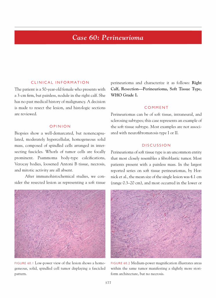
177
Case 60: Perineurioma
C L I N I C A L I N F O R M A T I O N
The patient is a 50-year-old female who presents with a 3-cm fi rm, but painless, nodule in the right calf. She has no past medical history of malignancy. A decision is made to resect the lesion, and histologic sections are reviewed.
O P I N I O N
Biopsies show a well-demarcated, but nonencapsu-lated, moderately hypercellular, homogeneous solid mass, composed of spindled cells arranged in inter-secting fascicles. Whorls of tumor cells are focally prominent. Psammoma body-type calcifi cations, Verocay bodies, loosened Antoni B tissue, necrosis, and mitotic activity are all absent.
After immunohistochemical studies, we con-sider the resected lesion as representing a soft tissue
perineurioma and characterize it as follows: RightCalf, Resection—Perineurioma, Soft Tissue Type, WHO Grade I.
C O M M E N T
Perineuriomas can be of soft tissue, intraneural, and sclerosing subtypes; this case represents an example of the soft tissue subtype. Most examples are not associ-ated with neurofi bromatosis type I or II.
D I S C U S S I O N
Perineurioma of soft tissue type is an uncommon entity that most closely resembles a fi broblastic tumor. Most patients present with a painless mass. In the largest reported series on soft tissue perineuriomas, by Hor-nick et al., the mean size of the single lesion was 4.1 cm (range 0.3–20 cm), and most occurred in the lower or
FIGURE 60.1 Low-power view of the lesion shows a homo-geneous, solid, spindled cell tumor displaying a fascicled pattern.
FIGURE 60.2 Medium-power magnifi cation illustrates areas within the same tumor manifesting a slightly more stori-form architecture, but no necrosis.

178
C A S E 6 0 : P E R I N E U R I O M A
upper limbs or the trunk. Nearly all were grossly well circumscribed. Of the 81 cases, 42 of 81 examples were located in subcutis, 25 of 81 in deep soft tissue, and 9 of 81 in dermis.
Most soft tissue perineuriomas are solid and composed of fascicles of cells, often with a stori-form and focally whorled growth pattern. Cells are elongate, wavy, and are surrounded by variable amounts of collagen. Nuclei are thin, elongated, have tapered ends, and devoid of conspicuous
nucleoli, in contrast to the larger, blunt-ended nuclei seen in smooth muscle tumors. Some authors have noted occasional nuclear atypia, but this seems to be degenerative in nature, similar to that seen in “ancient schwannoma,” and lacks prognos-tic signifi cance. Mitoses are usually rare to absent. Paralleling what is seen by electron microscopy, the eosinophilic, bipolar cell cytoplasmic processes are very long and thin. Myxoid background may be found in some cases.
FIGURE 60.6 Nuclei may be very thin is some planes of section, and the cytoplasm is bipolar and very thin as well.
FIGURE 60.5 Cytologically bland tumor cells have nuclei devoid of nucleoli or mitotic activity and resemble fi bro-blastic cells.
FIGURE 60.4 High-power view shows that blood vessels are delicate and usually devoid of thick hyalinization, as would be typical of schwannoma.
FIGURE 60.3 Medium-power magnifi cation highlights areas with whorls of cells, with resemblance to meningioma.

179
C A S E 6 0 : P E R I N E U R I O M A
The key feature for making the diagnosis is the strong immunoreactivity for EMA and negative im-munoreactivity for S-100 protein and muscle mark-ers. As noted by Giannini et al., however, “EMA reactivity may be diffi cult to demonstrate without the use of a higher concentration of antisera or of a longer incubation times than are routinely em-ployed in the diagnosis of epithelial neoplasms.” Diffi cult examples require electron microscopic confi rmation.
The differential for this benign, WHO grade I, nerve sheath tumor centers around fi broblastic tumors, such as dermatofi brosarcoma protuberans and desmoid tumor (fi bromatosis). The fi rst is stori-form in pattern and uniformly immunoreactive for CD34, but not for EMA. Desmoid tumors are more infi ltrative than most perineuriomas, and again, are EMA-negative. Myxoid soft tissue perineuriomas need to be distinguished from low-grade fi bromyx-oid sarcoma and low-grade myxofi brosarcoma, both of which lack EMA immunoreactivity. Absence of immunostaining for S-100 protein excludes schwan-noma, although both schwannoma and perineurioma have cells that are surrounded by collagen IV- and laminin- immunoreactive basement membrane. Soft
tissue perineuriomas also show immunostaining for newer markers, such GLUT-1 and claudin-1.
Intraneural perineuriomas present as enlarged nerve fascicles and used to be called “localized hyper-trophic neuropathy.” Histologically, they appear quite different than soft tissue perineurioma, in that they manifest tight concentric whorls of EMA- positive cells, yielding “pseudo-onion bulbs” within the affected nerve. Although the soft tissue and intraneural subtypes present very different histologic appearances, both appear to share abnormalities of chromosome 22.
References 1. Boyanton BL Jr, Jones JK, Shenaq SM, et al. Intraneural
perineurioma: a systematic review with illustrative cases. Arch Pathol Lab Med 2007;131:1382–92.
2. Giannini C, Scheithauer BW, Jenkins RB, et al. Soft-tissue perineurioma. Evidence for an abnormality of chromosome 22, criteria for diagnosis, and review of the literature. Am J Surg Pathol 1997;21:164–73.
3. Hornick JL, Fletcher CD. Soft tissue perineurioma: clinico-pathologic analysis of 81 cases including those with atypical histologic features. Am J Surg Pathol. 2005;29:845–58.
4. Macarenco RS, Ellinger F, Oliveira AM. Perineurioma: a dis-tinctive and underrecognized peripheral nerve sheath neo-plasm. Arch Pathol Lab Med 2007;131:625–36.
5. Pina-Oviedo S, Ortiz-Hidalgo C. The normal and neoplastic perineurium: a review. Adv Anat Pathol 2008;15:147–64.

180
Case 61: Malignant Peripheral Nerve Sheath Tumor
C L I N I C A L I N F O R M A T I O N
The patient is a 33-year-old-male with known neu-rofi bromatosis, type I, and a rapidly enlarging right popliteal mass, up to 9 cm in greatest dimension. The decision was made to resect the mass, and histological sections are reviewed.
O P I N I O N
The resection material shows a hypercellular, highly necrotic, focally hemorrhagic spindle neoplasm with brisk mitotic activity. Cells display prominent nucle-oli and variable amounts of cytoplasm. Slight palisad-ing of nuclei is seen around some areas of necrosis, a feature not completely exclusive to glioblastomas and
recognized in some sarcomas such as malignant periph-eral nerve sheath tumor (MPNST). The presence of a benign plexiform neurofi broma nearby ensures that the sarcoma is of peripheral nervous system origin.
We consider the lesion to be a sarcoma and characterize it as follows: Right Popliteal Nerve, Excision—Malignant Peripheral Nerve Sheath Tumor, WHO Grade IV.
C O M M E N T
The grading system for MPNST is similar to that employed for sarcomas in general. This example has very high mitotic activity and extensive necrosis.
D I S C U S S I O N
MPNSTs affect middle-aged adults. Patients with neurofi bromatosis, type I, generally are of younger
FIGURE 61.1 Lower-magnifi cation appearance of the nerve root mass, showing a hypercellular, spindled tumor with extensive necrosis and embedded in a thin, bluish-gray mucoid background matrix; note absence of hyalinization of the small blood vessels in the tumor.
FIGURE 61.2 Intermediate magnifi cation shows spindled cell areas.

181
C A S E 6 1 : M A L I G N A N T P E R I P H E R A L N E R V E S H E A T H T U M O R
age than patients with sporadic cases, explaining the young age of the current case. Larger nerves are more likely affected than smaller nerves, and common sites for MPNST include buttock and thigh, brachial plexus, paraspinal region, and sciatic nerve. Cranial nerve MPNSTs are rare. About one-third to one-half of all MPNSTs arise from neurofi bromas, usually plex-iform neurofi bromas, as in this case. Only rarely do
they arise from other types of benign peripheral nerve sheath tumors, such as schwannomas or ganglioneu-romas. It is estimated that 10% of MPNSTs arise years and decades following irradiation.
MPNSTs often begin their growth within the nerve fascicle of origin, but they commonly can break through the epineurium to involve adjacent soft tissues. Many are over 5 cm in diameter when they are removed. Grossly, they vary from being solid tan masses that are deceptively bland-appearing to overtly necrotic and hemorrhagic lesions of massive proportions.
Histologically, tumors exhibit varied patterns, ranging from intersecting fascicles of spindled cells to sheets of relatively patternless cells. Epithelioid cells may be found, with prominent nucleoli. Background mucoid matrix may be identifi ed. The fi nding of necrosis, often geographic and sometimes surrounded by palisading tumor cells, mandates a grading of WHO grade IV.
Additional neoplastic elements, such as rhab-domyosarcoma, chondrosarcoma, or osteosarcoma, or even intestinal or squamous epithelium, have been documented. These unusual elements tend to be seen
FIGURE 61.5 High-power magnifi cation of the benign plexiform neurofi broma shows dense collagenization and wavy, “shredded carrots” appearance in this area.
FIGURE 61.4 In some areas of the mass, the plexiform neu-rofi broma from which this MPNST arose can be identifi ed; note involvement of multiple nerve fascicles by the neu-rofi broma.
FIGURE 61.3 High-power magnifi cation illustrates areas in the same tumor with more prominent nucleoli and brisk mitotic activity.

182
C A S E 6 1 : M A L I G N A N T P E R I P H E R A L N E R V E S H E A T H T U M O R
in MPNSTs arising in patients with neurofi bromatosis, type I.
References 1. Ferner RE. Neurofi bromatosis 1 and neurofi bromatosis
2: a twenty-fi rst century perspective. Lancet Neurology 2007;16:340–51.
2. Scheithauer BW, Louis DN, Hunter S, et al. Schwannoma. In: Louis DN, Ohgaki H, Wiestler OD, Cavenee WK, Editors. WHO Classifi cation of Tumours of the Central Nervous System. Lyon, FR: IARC Press; 2007. p. 160–2.

183
Case 62: Cellular Schwannoma
C L I N I C A L I N F O R M A T I O N
The patient is a 50-year-old female who is found to have a mass involving the vagus nerve. Resection is undertaken, and histologic sections are examined.
O P I N I O N
The resection material shows an encapsulated, highly cellular, homogeneous solid mass, composed of spin-dled cells arranged in intersecting fascicles. Whorls of tumor cells are not identifi ed. Psammoma body-type calcifi cations and Verocay bodies are not seen. The lesion is composed of almost entirely dense Antoni A tissue, with less than 10% of the lesion showing identifi able Antoni B areas.
After immunohistochemical studies, we con-sider the resected lesion as representing a cellular
schwannoma and characterize it as follows: Vagus Nerve, Resection—Cellular Schwannoma, WHO Grade I.
C O M M E N T
Cellular schwannomas are benign and lack metastatic potential. Most examples are solitary and are not asso-ciated with neurofi bromatosis, types I or II.
D I S C U S S I O N
Cellular schwannoma is, by defi nition, a schwan-noma with high cellularity that is composed almost exclusively of dense Antoni A tissue without well-formed Verocay bodies. An Antoni B pattern may be focally present, but it constitutes no more than
FIGURE 62.1 Low-power view of the lesion shows a homo-geneous, solid, spindled cell tumor, devoid of loosened Antoni B areas or Verocay body formation.
FIGURE 62.2 Cellular schwannoma is more hypercellular than ordinary schwannoma and shows a more uniform pattern at low power. Note the absence of necrosis in this example.

184
C A S E 6 2 : C E L L U L A R S C H W A N N O M A
10% of the tumor area and is usually confi ned to sub-capsular areas. Cellular schwannomas are similar to ordinary schwannomas in that they are well circum-scribed, encapsulated, and microscopically share the presence of hyalinization of tumor vessels and occa-sional collections of lipid-laden histiocytes. They dif-fer in that they are more hypercellular, have a more uniform architectural pattern, and may show more
conspicuous subcapsular and capsular benign lympho-cytic collections than ordinary schwannoma. About one-third of cellular schwannomas arise from a recog-nizable nerve, as in this case.
Small foci of necrosis can rarely be encountered, but mitoses are found in the majority of cases, usually numbering 1–4 per 10 high-power microscopic fi elds. The key feature for making the diagnosis is the strong and diffuse immunoreactivity for S-100 protein. The
FIGURE 62.6 Mitoses are seen in the majority of cellular schwannomas but generally number from 1–4 per 10 high-power microscopic fi elds.
FIGURE 62.5 Cellular schwannomas possess many of the features of ordinary schwannomas, including hyalinization of tumor blood vessels.
FIGURE 62.4 Cells display elongate nuclei and eosinophilic cytoplasm without distinct cell borders, often identical to that seen in ordinary schwannoma.
FIGURE 62.3 The tumor is composed of intersecting fasci-cles of cells, and despite the hypercellularity, there is gener-ally an absence of degenerative nuclear atypia, as is often found in “ancient schwannomas.”

185
C A S E 6 2 : C E L L U L A R S C H W A N N O M A
FIGURE 62.7 Cellular schwannomas possess pericellular basement membrane, and cells are individually surrounded by reticulin fi bers.
uniform, abundant, parallel array of wavy, pericellular reticulin in a cellular schwannoma is also a helpful diagnostic feature.
The differential for this benign, WHO grade I, nerve sheath tumor centers around making the distinc-tion from MPNST. The latter is also hypercellular and composed of fascicles of cells, but the nuclei show a higher degree of hyperchromasia and cytologic atypia, and geographic necrosis and higher mitotic accounts are common. S-100 protein is seen in scattered cells in 50–70% of MPNSTs, but they generally lack the diffuse and strong S-100 staining pattern of cellular schwanno-mas. About 90% of MPNSTs are high-grade lesions, aiding in the distinction from cellular schwannoma.
In one of the original large series, “cellular schwannomas represented 4.6% of benign peripheral nerve tumors operated on at the Mayo Clinic. Median
patient age was 47.7 years (range, 15 to 80 years), and the female-to-male ratio was 1.6:1. The principle tumor locations were the para- and intraspinal regions, includ-ing the sacrum (64%), extremities (25%), and intracra-nial space (8%). . . . Surgery was the treatment in all cases. . . . Follow-up revealed recurrences in 11 patients (23.4%): no patient experienced metastasis or died of tumor.”
References 1. Casadei GP, Scheithauer BW, Hirose T, et al. Cellular
schwannoma. A clinicopathologic, DNA fl ow cytomet-ric, and proliferation marker study of 70 patients. Cancer 1995;75:1109–19.
2. Scheithauer BW, Woodruff JM, Erlandson RA. Atlas of Tumour Pathology: Tumors of the Peripheral Nervous System. Schwannoma, chapter 7. Washington DC: Armed Forces Institute of Pathology; 1997, p. 138–51.
FIGURE 62.8 Similar to ordinary schwannoma and differ-ing from most malignant peripheral nerve sheath tumors, cellular schwannomas show strong diffuse immunoreactiv-ity for S-100 protein.

186
Case 63: Melanotic Schwannoma
C L I N I C A L I N F O R M A T I O N
The patient is a 55-year-old male whose diagnosis is heralded by 2 years of subacute, progressive, left-hand paresthesias, which progressed to involve the entire left upper extremity, with occasional numbness in the left foot and toes. There is associated incoordination of the left hand, signifi cant diffi culty typing on a com-puter keyboard, and problems with dropping objects. Neuroimaging studies show a mass near the cervical spinal cord, at the dorsal root entry zone for C2. The decision is made to resect the lesion, and, intraopera-tively, the mass is noted to be jet black.
O P I N I O N
Sections show a heavily pigmented neoplasm, com-posed of fascicles of cells and devoid of distinct Antoni A and B areas or well-developed Verocay
bodies. The spindled and epithelioid cells manifest oval vesicular nuclei with occasional prominent nucleoli. There is no necrosis or mitotic activity. The most striking feature is the presence of dark cytoplasmic pigment, proven to be melanin, within the tumor cells. Unlike a primary or metastatic malignant melanoma in this site, reticulin fi bers lie between tumor cells, and minimal mitotic activity and no necrosis are found.
We consider the excised tumor as representing a peripheral nerve sheath tumor and characterize it as follows: Right Nerve Root, Excision—MelanoticSchwannoma, WHO Grade I.
C O M M E N T
There are no psammoma bodies in this example. Over half of patients with psammomatous melanotic
FIGURE 63.1 Intraoperative touch preparation, stained with hematoxylin and eosin, readily shows the heavily pig-mented cells in this tumor.
FIGURE 63.2 On low-power magnifi cation, the mass is com-posed of fascicles of heavily pigmented cells, but no necrosis, Verocay bodies, or distinct Antoni A and B tissue is seen.

187
C A S E 6 3 : M E L A N O T I C S C H W A N N O M A
schwannomas have Carney complex, a familial dis-order usually transmitted as an autosomal dominant condition. Psammomatous melanotic schwannomas may be multiple. In contrast, non-psammomatous melanotic schwannomas are usually single lesions, not associated with neurofi bromatosis, type II, or any other tumor predisposition syndrome.
D I S C U S S I O N
Melanotic schwannomas are rare, circumscribed, but not fully encapsulated tumors that, by immunohis-tochemistry and electron microscopy, have features of Schwann cell derivation. In addition, they also contain melanosomes and are immunoreactive for melanoma markers. Compared to ordinary schwan-nomas, these tumors are more highly cellular, show less Antoni A and B distinction, only rare Verocay bodies, a poorly developed reticulin pattern, and more cytologic atypia, including macronucleoli and hyperchromatic nuclei. Epithelioid cells may be present. The amount of melanin pigment is highly variable. Bleaching procedures may be necessary in some cases in order to appreciate fully the cytologic features, mitotic activity, and even degree of necrosis
in the tumor, because some examples are extremely heavily pigmented, and the melanin obscures features of the tumor. Melanin pigment can be confi rmed by the histochemical Fontana Masson stain. Basement membrane, even if more limited in amount, can be verifi ed by immunostaining for laminin and collagen type IV.
Melanotic schwannoma must be distin-guished from metastatic and primary malignant melanoma, mainly on the basis of increased mitot-ic activity and necrosis. The preoperative imaging
FIGURE 63.5 The absence of mitotic activity can be appre-ciated in less heavily pigmented areas.
FIGURE 63.4 On high-power magnifi cation, vesicular nuclei and delicate nucleoli are appreciated.
FIGURE 63.3 A close juxtaposition between the pigmented nerve sheath tumor (lower part of photograph) and a non-neoplastic nerve fascicle (upper part) can be seen.
188
C A S E 6 3 : M E L A N O T I C S C H W A N N O M A
FIGURE 63.6 Melanin totally obscures cytologic detail in some areas and makes the search for mitotic fi gures more diffi cult.
fi ndings of a dumbbell-shaped lesion involving a spinal nerve root (especially at cervical or thoracic levels, as in this patient) and the intraoperative and histologic recognition of a melanotic lesion arising from a nerve root are of great assistance in making this diagnosis. These features should sway the pathologist toward a diagnosis of melanotic schwannoma, not a metastasis. The differential diagnosis also includes meningeal melanocytoma
and intermediate-grade melanocytoma, the criteria for which have been published (see reference list). In general, less immunostaining for collagen type IV is seen in melanocytoma than in melanotic schwannoma, but electron microscopy may be nec-essary to confi rm the Schwann cell derivation of melanotic schwannoma.
Melanotic schwannomas are usually benign, but gross total surgical excision with negative margins
FIGURE 63.7 After bleaching procedures remove the mela-nin pigment, the fascicled arrangement of tumor cells is more clearly apparent.
FIGURE 63.9 Reticulin staining in this example shows the classic abundant pericellular reticulin of a Schwann cell tumor, corresponding to abundant basement membrane.
FIGURE 63.8 Fontana Masson histochemical staining confi rms that the fi nely granular, brown pigment was melanin—by the dark, black, positive staining.

189
C A S E 6 3 : M E L A N O T I C S C H W A N N O M A
is the optimal treatment. About 10–15% of mela-notic schwannomas follow a malignant course, with recurrences and metastases. Bony destruction is more often a feature of malignant than of benign mela-notic schwannoma. Unfortunately, it is not possible in individual cases to predict precisely which cases might metastasize, although, in general, tumors with mitotic activity and necrosis are more prone to this behavior. Very prominent macronucleoli are often also present in those cases that metastasize, although some tumors with this feature do not manifest malig-nant behavior.
References 1. Brat DJ, Giannini C, Scheithauer BW, et al. Primary mela-
nocytic neoplasms of the central nervous systems. Am J Surg Pathol 1999;23:745–54.
2. Brat DJ, Perry A. Melanocytic lesions. In: Louis DN, Ohgaki H, Wiestler OD, Cavenee WK, Editors. WHO Classifi cation of Tumours of the Central Nervous System. Lyon, FR: IARC Press;2007. p. 181–3.
3. Scheithauer BW, Louis DN, Hunter S, et al. Schwannoma. In: Louis DN, Ohgaki H, Wiestler OD, Cavenee WK, Editors. WHO Classifi cation of Tumours of the Central Nervous Sys-tem. Lyon, FR: IARC Press;2007. p. 154–5.
4. Scheithauer BW, Woodruff JM, Erlandson RA. Atlas of Tumour Pathology: Tumours of the Peripheral Nervous System. Schwannoma, chapter 7. Washington DC: Armed Forces Institute of Pathology; 1997. p. 156–70.

190
Case 64: Fibrous Meningioma
C L I N I C A L I N F O R M A T I O N
The patient is a 62-year-old woman with a prior history of breast carcinoma who presents with headaches and occasional episodes of dizziness. On imaging studies, she is noted to have a contrast-enhancing mass in the left cerebello-pontine angle region on MRI studies. The lesion appears to be separate from the eighth cranial nerve. The mass is excised, and histologic sections are reviewed.
O P I N I O N
Histologic sections show a spindle-cell neoplasm, marked by proliferation of cells with elongated nuclei with rounded nuclear ends and a scant amount of eosinophilic cytoplasm. Cells are arranged in interlacing bundles. Focal whorling of the cells around blood vessels is noted. Only rare mitotic fi gures are noted. There is no
evidence of calcifi cations or psammoma bodies. Verocay bodies are not identifi ed. There is no evidence of brain invasion, prominent nucleolation, small cell change, disordered architecture, necrosis, or hypercellularity.
We consider the lesion to be a meningioma and characterize it as follows: Left Cerebello-Pontine Angle Region, Excision—Fibrous (Fibroblastic) Meningioma, WHO Grade I.
C O M M E N T
Morphologic features suggestive of an atypical men-ingioma are not identifi ed in this tumor.
D I S C U S S I O N
Meningiomas are among the more common nonglial tumors of the central nervous system. Classically,
FIGURE 64.1 Low-magnifi cation appearance of the fi brous meningioma, showing spindled cells arranged in large fascicle.
FIGURE 64.2 The cells in fi brous meningioma are rather monotonous in appearance and demonstrate little in the way of nuclear pleomorphism.

191
C A S E 6 4 : F I B R O U S M E N I N G I O M A
they are dural-based in location and can arise at any age. There is a clear female predominance to cases that are reported in the literature. There is an asso-ciation of a meningioma with certain malignancies, including breast cancer (as in this patient) and prior radiation.
Histologically, fi brous meningioma is marked by a proliferation of spindled cells, which are often arranged in bundles or fascicles and associated with
a collagen-rich matrix. The whorling of cells around blood vessels is a fairly frequent fi nding. Psammoma bodies, which are not evident in the current tumor, may be present in this lesion. Occasionally, nuclear pleomorphism may be evident and has no impact on grading of the tumor or prognosis.
The main differential diagnostic consideration in this current case includes differentiating this lesion from a schwannoma. The cerebello-pontine angle region is also a frequent site of origin for schwanno-mas of cranial nerve eight derivation. In contrast to meningioma, schwannomas generally have a biphasic appearance, with Antoni A and Antoni B patterns and Verocay bodies. Schwannomas do not demonstrate much whorling of cells around blood vessels, and they generally lack psammoma bodies. Immunohistochemistry is useful in distinguishing these two tumor types when morphology is not clear. Schwannomas demonstrate diffuse, positive stain-ing with S-100 protein antibody; meningiomas may show weak, focal, positive staining in about 20% of cases. Most meningiomas demonstrate at least focal EMA immunoreactivity, in contrast to schwannomas that do not stain with EMA antibody. Occasionally, the fi brous pattern in meningioma may be admixed
FIGURE 64.3 High-magnifi cation appearance of tumor cells shows elongated nuclei with rounded nuclear ends.
FIGURE 64.5 Focal angiocentric whorling of tumor cells is a characteristic feature of many meningiomas.
FIGURE 64.4 Rare mitotic fi gures identifi ed in this fi brous meningioma. The number of mitotic fi gures was not high enough (4 mitotic fi gures of 10 high-power fi elds) to warrant consideration of atypical meningioma.

192
C A S E 6 4 : F I B R O U S M E N I N G I O M A
with the meningothelial pattern of tumor, in which case the diagnosis of transitional or mixed menin-gioma can be made. The patient’s history of breast carcinoma also raises metastasis in the clinical differ-ential diagnosis. Rare instances of metastatic breast carcinoma to a meningioma have been documented in the literature.
Fibrous meningiomas, along with meningothe-lial (syncytial) meningiomas, are among the more common patterns of grade I meningiomas. These tumors are quite amenable to surgical excision and have an excellent prognosis. Proliferation markers
generally show low rates of cell proliferation in the grade I variants.
References 1. Burger PC, Scheithauer BW. Meningioma. In: Tumors of the
Central Nervous System. Washington, DC: AFIP Fascicle 4th Series; 2007. p. 331–62.
2. Perry A, Louis DN, Scheithauer BW, et al. Meningiomas. In: Louis DN, Ohgaki H, Wiestler OD, Cavenee WK, Editors. WHO Classifi cation of Tumours of the Central Nervous System. Lyon, FR: IARC Press;2007. p. 164–72.
3. Prayson RA. Pathology of meningiomas. In: Lee JH, Editor. Meningiomas. Diagnosis, Treatment, and Outcome. London, UK: Springer-Verlag;2008. p. 31–43.

193
Case 65: Ectopic Meningioma
C L I N I C A L I N F O R M A T I O N
The patient is a 46-year-old female who presents with recurrent frontal sinus infections. On imaging studies, a mass lesion is noted, involving a frontal sinus with associated erosion of the adjacent bone. The mass is biopsied, and histologic sections are reviewed.
O P I N I O N
Histologic sections show a normal-appearing respi-ratory mucosa with a submucosal mass. The mass is comprised of a proliferation of spindled cells with elongated nuclei, scant cytoplasm, and blunt nuclear ends. There is minimal cytologic atypia or nuclear pleomorphism in the tumor cells. An occasional lymphoid aggregate is noted in association with the neoplasm. The adjacent submucosal soft tissue shows focal chronic infl ammation and reactive fi broblastic
response to the tumor. Mitotic activity, necrosis, prominent nucleolation, small cell change, marked hypercellularity, and disordered architecture are not noted. There is no evidence of psammoma bodies.
We consider the lesion to be an ectopic menin-gioma and characterize it as follows: Frontal Sinus,Excision—Fibrous (Fibroblastic) Meningioma, WHO Grade I.
C O M M E N T
The tumor morphologically resembles a meningioma arising in an ectopic location.
D I S C U S S I O N
Although the vast majority of meningiomas arise in the brain or spinal cord, most frequently associated
FIGURES 65.1 Low-magnifi cation appearance, showing respiratory mucosa overlying a submucosal tumor.
FIGURE 65.2 Adjacent submucosal tissue, uninvolved by tumor, shows chronic infl ammation and a fi broblastic reaction.

194
C A S E 6 5 : E C T O P I C M E N I N G I O M A
with dural tissue, ectopic locations for meningiomas have been described in the head and neck region, including the calvarium, orbit, middle ear, sinuses, and nasopharynx. Less frequently, meningiomas may arise in more distant locations, including skin, lung, and mediastinum; adrenal glands; and retroperito-neum. Because meningiomas in the ectopic locations are a relatively rare occurrence and often an unex-pected fi nding, tumors may present a particular diag-nostic challenge, particularly when they demonstrate
unusual morphologic features. This patient’s tumor shows morphology typical of a fi brous meningioma. The presence of psammoma bodies, whorling around vessels, and intranuclear pseudoinclusions may be useful clues that one is dealing with meningioma. The immunohistochemical profi le of meningioma tends to be somewhat nondescript. EMA immunoreactivity (observed in approximately 80% of meningiomas) is said to be the most useful in terms of differential diagnosis. Smaller numbers of meningiomas may variably stain with antibodies to S-100 protein and cytokeratin markers. These tumors are generally sub-typed and graded according to parameters defi ned in the central nervous system and spinal cord. Whether this approach is valid in ectopic sites is not known.
References 1. Perzin KH, Pushparaj N. Nonepithelial tumors of the nasal
cavity, paranasal sinuses, and nasopharynx: a clinicopatho-logic study. XIII: meningiomas. Cancer 1984;54:1860–9.
2. Sadar ES, Conomy SP, Benjamin SP, et al. Meningiomas of the paranasal sinuses, benign and malignant. Neurosurgery 1979;4:227–31.
3. Thompson LD, Gyure KA. Extracranial sinonasal tract meningioma: a clinicopathologic study of 30 cases with a review of the literature. Am J Surg Pathol 2000;24:640–50.
FIGURE 65.5 An occasional lymphoid aggregate is observed in the tumor.
FIGURE 65.4 The tumor cells show minimal nuclear pleomorphism. Nuclear contours are elongated with blunted ends.
FIGURE 65.3 The tumor is comprised of a proliferation of monomorphic-appearing spindled cells.

195
Case 66: Clear Cell Meningioma
C L I N I C A L I N F O R M A T I O N
The patient is a 42-year-old woman who presents with a mass in the right cerebello-pontine angle region. On imaging, the tumor appears to be a well-demarcated, dural-based mass. The lesion is excised, and histologic sections are reviewed.
O P I N I O N
Histologic sections show a dural-based mass, com-prised of a proliferation of cells with abundant clear cytoplasm. The tumor cell’s nuclei are frequently eccentrically placed and contain small nucleoli. The overall arrangement of the cells is somewhat disor-dered (sheeting). Scattered between tumor cells are sclerotic vessels. Bands of collagen are noted, separat-ing groups of cells. The edge of the neoplasm shows focal infi ltration of tumor into adjacent soft tissue.
Tumor cells in this region resemble the meningothe-lial pattern of meningioma, marked by cells with eosinophilic cytoplasm arranged in lobules.
We consider the lesion to be a meningioma and characterize it as follows: Right Cerebello-Pontine Angle Region, Excision—Clear Cell Meningioma, WHO Grade II.
C O M M E N T
The predominant pattern of this tumor is that of a clear cell meningioma. The clear cell pattern of men-ingioma has been associated with increased risk of recurrence.
D I S C U S S I O N
Although the vast majority of meningiomas are grade I lesions, a subset of tumors, because of their increased
FIGURE 66.1 Low-magnifi cation appearance of the clear meningioma, showing its dural attachment.
FIGURE 66.2 Intermediate-magnifi cation appearance of the tumor, showing abundant cleared cytoplasm and disor-dered architectural arrangement of cells.

196
C A S E 6 6 : C L E A R C E L L M E N I N G I O M A
risk of a recurrence, are designated as higher-grade lesions. Clear cell meningioma is one such variant that is marked by a proliferation of cells with abun-dant cleared cytoplasm. The clearing is related to increased cytoplasmic glycogen accumulation, which can be highlighted with PAS staining. The cells in this variant are often arranged in a haphazard fashion, and characteristic meningioma features, such as whorling of cells around vessels and the presence of psammoma bodies, are distinctly uncommon in the vast major-
ity of these tumors. Clear cell meningiomas are more commonly seen in younger patients and seem to have a predilection for arising in the cerebello-pontine angle and cauda equina regions. Again, the impor-tance of making this diagnosis is that these tumors show a propensity for recurrence in a shorter interval of time than grade I variants.
From a differential diagnostic standpoint, distinction of this tumor from other variants of meningioma may be important. This particular lesion, at its periphery, has areas more reminiscent of the grade I meningothelial meningioma pattern. There are no precise guidelines as to what percentage of the tumor must have a clear cell pattern in order to designate the tumor as clear cell meningioma. Because of this, if any appreciable portion of the tumor has clear cell morphology, it is important to indicate this in some way in the pathology report. Occasionally, distinction of clear cell meningioma from metastatic clear cell carcinomas, particularly renal cell carcino-mas, may be problematic. Immunomarkers targeted for renal cell carcinomas, such as carbonic anhydrase and RCC antibodies, may be helpful in sorting out this differential diagnosis.
FIGURE 66.5 The periphery of the tumor shows a more conventional-appearing meningothelial (syncytial) pattern of meningioma.
FIGURE 66.4 Focal sclerotic vessel change and bundles of collagen, deposited between groups of cells, are evident in this tumor.
FIGURE 66.3 Cleared cytoplasm and small nucleoli are highlighted in a clear cell meningioma.

197
C A S E 6 6 : C L E A R C E L L M E N I N G I O M A
References 1. Carlotti CG Jr, Neder L, Colli BO, et al. Clear cell menin-
gioma of the fourth ventricle. Am J Surg Pathol 2003;27:131–5.
2. Heth JA, Kirby P, Menezes AH. Intraspinal familial clear cell meningioma in a mother and child. Case report. J Neurosurg 2000;93:317–21.
3. Jallo GI, Kothbauer KF, Silvera VM, et al. Intraspinal clear cell meningioma: diagnosis and management: report of two cases. Neurosurgery 2001;48:218–21; discussion 221–2.
4. Zorludemir S, Scheithauer BW, Hirose T, et al. Clear cell menin-gioma. A clinicopathologic study of a potentially aggressive variant of meningioma. Am J Surg Pathol 1995;19:493–505.

198
Case 67: Chordoid Meningioma
C L I N I C A L I N F O R M A T I O N
The patient is a 46-year-old female who presents with headaches and a 3.8-cm falcine mass. The mass appears on imaging to be circumscribed. The lesion is excised, and histologic sections are reviewed.
O P I N I O N
Histologically, the mass is marked by a prolifera-tion of generally rounded-to-polygonal cells with eosinophilic cytoplasm and eccentrically placed nucleus. The cells are arranged in cords or trabe-culae with intervening mucoid matrix. Focal areas of fi brosis with collagen are observed. Some cells show prominent nucleolation. Occasional mitotic fi gures (up to 4 MF per 10 HPFs) are observed in
this tumor. Focal areas of the neoplasm show a more solid pattern with a less mucoid matrix. The cells in these areas are haphazardly arranged. There is no evidence of necrosis or brain invasion. Promi-nent small cell change or hypercellularity is not observed.
We consider the lesion to be a variant of meningioma and characterize it as follows: Falcine Region, Excision–Chordoid Meningioma, WHO Grade II.
C O M M E N T
The predominant pattern of the tumor is that of a chordoid meningioma. Chordoid meningiomas are associated with more aggressive behavior and a higher risk of recurrence.
FIGURE 67.1 Low-magnifi cation appearance of a chordoid meningioma, showing cords of cells arranged against a mucoid background.
FIGURE 67.2 Higher magnifi cation, showing characteristic architectural features of chordoid meningioma.

199
C A S E 6 7 : C H O R D O I D M E N I N G I O M A
D I S C U S S I O N
Chordoid meningioma, like clear cell meningioma, represents a morphologic variant of meningioma that has been associated with increased risk of recur-rence. Again, like clear cell meningioma, precise criteria are not provided in terms of what percent-age of tumor must have a chordoid phenotype to be designated as such. Subsequently, identifi cation of
any chordoid pattern in the tumor should be indi-cated in the pathology report. As the name suggests, the chordoid variant of meningioma resembles chordoma, in that the chordoid-like cells are often arranged in small clusters or trabeculae against a mucoid matrix background. More solid areas often show disordered architecture and, frequently, other features associated with more aggressive behavior in meningioma, such as prominent nucleolation and
FIGURE 67.5 A more solid focus in a chordoid meningioma is marked by decreased mucoid matrix and cells arranged in a disordered fashion. Many of the cells show prominent nucleolation.
FIGURE 67.4 Focal fi brosis is observed in this tumor.
FIGURE 67.6 Mitotic fi gures, up to 4 MF per 10 HPFs, are observed in this tumor. Using WHO grading criteria, the presence of 4 or more MF per 10 HPFs is also enough to diagnose the tumor as a grade II neoplasm.
FIGURE 67.3 Focal area, showing more discohesive arrange-ment of cells against a mucoid background.
200
C A S E 6 7 : C H O R D O I D M E N I N G I O M A
prominent mitotic activity (as in the current case), may be observable.
Differential diagnostic considerations typically include a metastatic adenocarcinoma and chordoma. The growth pattern of a chordoid meningioma is somewhat unusual for a metastatic carcinoma. Mucin-positive staining within cells or in glandular lumina is a feature of adenocarcinoma that is not observable in the chordoid meningioma. In contrast to chordoid meningiomas, the vast majority of metastatic muci-nous adenocarcinomas demonstrate diffuse positive staining with a variety of cytokeratin markers; cyto-keratin immunoreactivity in chordoid meningioma is generally focal. The majority of chordomas arise either in the clivus or fi lum terminale region. A falcine chordoma is extremely unusual. Additionally,
the physaliphorous cell change that is classic for chordoma is usually not observable in chordoid me-ningioma; however, small vacuoles may occasionally be observed in a subset of chordoid meningioma cells. Again, the immunohistochemical profi le of chordo-ma also allows for its distinction from the chordoid meningioma, i.e., a more widespread cytokeratin and S-100 protein immunoreactivity in chordoma.
References 1. Couce ME, Aker FV, Scheithauer BW. Chordoid meningi-
oma: a clinicopathologic study of 42 cases. Am J Surg Pathol 2000;24:899–905.
2. Kepes JJ, Chen WY, Connors MH, et al. “Chordoid” menin-geal tumors in young individuals with peritumoral lympho-plasmacellular infi ltrates causing systemic manifestations of the Castleman syndrome. A report of seven cases. Cancer 1988;62:391–406.

201
Case 68: Papillary Meningioma
C L I N I C A L I N F O R M A T I O N
The patient is an 8-year-old male who presents with confusion and headaches. On imaging studies, he is noted to have a dural-based mass overlying the right frontal-parietal convexity. The lesion is excised, and histologic sections are reviewed.
O P I N I O N
Histologic sections show a cellular, dural-based mass, marked by a disordered arrangement of cells. Tumor cells show a variable amount of eosinophilic cyto-plasm and somewhat eccentrically placed nucleus with prominent nucleolus. Prominent mitotic activ-ity (up to 16 mitotic fi gures per 10 high-power fi elds) is observed in the tumor. Areas of necrosis are also identifi ed. Focal areas of the tumor have a papillary architectural pattern, marked by fi brovascular cores
rimmed by tumor cells. Defi nite evidence of brain invasion is not identifi ed.
We consider the lesion to be a meningioma variant and characterize it as follows: Right Frontal-Parietal Convexity, Excision—Papillary Meningioma, WHO Grade III.
C O M M E N T
The focal presence of a papillary architectural pattern in meningioma indicates a more aggressive-behaving tumor, with increased risk of tumor recurrence and metastasis.
D I S C U S S I O N
Papillary meningioma is a rare variant that is par-ticularly aggressive in its behavior. The variant
FIGURE 68.1 Low-magnifi cation appearance, showing dural-based attachment of the tumor.
FIGURE 68.2 Intermediate magnifi cation, showing a disor-dered arrangement of tumor cells and readily identifi able mitotic activity.

202
C A S E 6 8 : P A P I L L A R Y M E N I N G I O M A
shows a predilection for younger patients and is typically marked by a propensity for recurrence and metastasis to distant sites. This variant, as its name suggests, is characterized by a papillary architectural pattern, marked by fi brovascular cores lined by meningothelial cells. More solid areas of the tumor generally show features of high-grade meningioma, including increased mitotic activity, necrosis, brain
invasion, hypercellularity, small cell change, promi-nent nucleolation, and a disordered architecture. In terms of differential diagnosis, care must be taken not to overinterpret discohesive clusters of menin-gothelial cells as papillary structures. Because of the aggressive behavior of this pattern, these tumors are often surgically excised and treated with adjuvant radiation therapy.
FIGURE 68.5 Low magnifi cation shows areas of tumor with a papillary architecture.
FIGURE 68.4 Focal necrosis is observed in the neoplasm.
FIGURE 68.6 Intermediate magnifi cation shows papillae, with fi brovascular cores lined by tumor cells.
FIGURE 68.3 Mitotic fi gures are readily identifi able in this tumor.

203
C A S E 6 8 : P A P I L L A R Y M E N I N G I O M A
References 1. Ludwin SK, Rubinstein LJ, Russell DS. Papillary meningi-
oma: a malignant variant of meningioma. Cancer 1975;36:1363–73.
2. Pasquier B, Gasnier F, Pasquier D, et al. Papillary meningi-oma. Clinicopathologic study of seven cases and review of the literature. Cancer 1986;58:299–305.
3. Piatt JH Jr, Campbell, GA, Oakes WJ. Papillary meningioma involving the oculomotor nerve in an infant. Case report. J Neurosurg 1986;64:808–12.
FIGURE 68.7 Another region of the tumor demonstrates the characteristic papillary architecture of papillary menin-gioma.
204
Case 69: Rhabdoid Meningioma
C L I N I C A L I N F O R M A T I O N
The patient is a 64-year-old female who presents with headaches of increasing intensity during the last two months. On imaging studies, a right frontal mass, overlying the convexity and attached to the dura, is observed. The lesion is resected, and histologic sec-tions are reviewed.
O P I N I O N
Histologic sections show a dural-based neoplasm, marked by a disordered proliferation of cells with abundant eosinophilic cytoplasm and eccentrically placed nuclei. Prominent nucleoli are observed in many of the cells’ nuclei. Occasional cells have an eosinophilic cytoplasmic inclusion. Other areas of the tumor are marked by a proliferation of cells with less cytoplasm and prominent nucleolation.
Mitotic activity is readily observable, up to 22 MF per 10 HPFs. There is no evidence of brain invasion. There is no evidence of necrosis or small cell change. In areas, the tumor cells appear somewhat disco-hesive, giving the tumor a focal pseudopapillary architectural pattern.
We consider the lesion to be a meningioma and characterize it as follows: Right Frontal Lobe, Excision—Rhabdoid Meningioma, WHO Grade III.
C O M M E N T
The tumor demonstrates focal rhabdoid morphol-ogy, which has been associated with more aggressive behavior in meningiomas. Additionally, the mitotic activity is on the order typically observed in anaplas-tic meningiomas.
FIGURE 69.1 Low-magnifi cation appearance of the tumor shows its relationship to dura and brain.
FIGURE 69.2 Intermediate magnifi cation of tumors shows rhabdoid cells with abundant eosinophilic cytoplasm. The cells are arranged in a disordered architectural pattern.

205
C A S E 6 9 : R H A B D O I D M E N I N G I O M A
D I S C U S S I O N
Rhabdoid meningioma represents a rare variant of aggressive meningioma, marked by the presence of cells with rhabdoid morphology. The rhabdoid morphology is characterized by cells with abundant eosinophilic cytoplasm, containing eosinophilic inclusion-like structures that ultrastructurally consist of aggregates of fi laments of intermediate molecular weight. Nuclei in these cells are often eccentrically
placed and associated with prominent nucleoli. Simi-lar cells are found in rhabdoid tumors arising elsewhere in the body, as well as the atypical teratoid/rhabdoid tumor of the central nervous system. Many rhabdoid meningiomas also demonstrate features of high-grade meningioma, including markedly increased mitotic activity (as in the current case), disordered architec-tural pattern arrangement of cells, prominent nucleo-lation, focal small cell change and hypercellularity,
FIGURE 69.6 A discohesive arrangement of cells creates a pseudopapillary appearance.
FIGURE 69.5 Prominent mitotic activity is observed in this tumor, in excess of 20 MF per 10 HPFs.
FIGURE 69.4 A disordered architectural pattern is observed in an area of the tumor that does not have a rhabdoid appearance.
FIGURE 69.3 High-magnifi cation appearance of rhab-doid cell shows cytoplasmic inclusions and prominent nucleolation.
206
C A S E 6 9 : R H A B D O I D M E N I N G I O M A
necrosis, and brain invasion. A subset of these tumors has also been known to metastasize. These tumors notoriously have a poor outcome, with increased risk of focal recurrence and distant metastasis.
References 1. Bannykh SI, Perry A, Powell HC, et al. Malignant rhabdoid
meningioma arising in the setting of preexisting gangliogli-oma: a diagnosis supported by fl uorescence in situ hybridiza-tion. Case report. J Neurosurg 2002;97:1450–5.
2. Kepes JJ, Moral LA, Wilkinson SB, et al. Rhabdoid transfor-mation of tumor cells in meningiomas: a histologic indication of increased proliferative activity: report of four cases. Am J Surg Pathol 1998;22:231–8.
3. Perry A, Scheithauer BW, Stafford SL, et al. “Rhabdoid” meningioma: an aggressive variant. Am J Surg Pathol 1998;22:1482–90.
4. Parwani AV, Mikolaenko I, Eberhart CG, et al. Rhabdoid meningioma: cytopathologic fi ndings in cerebrospinal fl uid. Diagn Cytopathol 2003;29:297–9.

207
Case 70: Brain-Invasive Meningioma
C L I N I C A L I N F O R M A T I O N
The patient is an 82-year-old female who presents with a seizure. Imaging studies show a 4.2-cm mass, attached to the dura in the right temporal-parietal lobe region. The lesion is resected, and histologic sec-tions are reviewed.
O P I N I O N
Histologic sections show a dural-based neoplasm, marked by a fairly uniform population of cells with oval nuclei and delicate chromatin pattern. Promi-nent nucleoli are observed focally throughout the tumor. Focal brain invasion is observed in the neo-plasm. Rare mitotic fi gures (up to 2 MF per 10 HPFs) are noted. There is no evidence of necrosis, small cell change, marked hypercellularity, or disordered
architectural pattern. Evidence of clear cell, chor-doid, rhabdoid, or papillary morphologies is not observed.
We consider the lesion to be a meningioma and characterize it as follows: Right Temporal-Parietal Lobe Region, Excision—Atypical Meningioma with Brain Invasion, WHO Grade II.
C O M M E N T
The presence of brain invasion warrants the diagnosis of a WHO grade II meningioma.
D I S C U S S I O N
Brain-invasive meningiomas are characterized by irreg-ular nests or islands of tumor cells protruding into the subjacent neural parenchyma. This invasion typically
FIGURE 70.1 Intermediate-magnifi cation appearance of the tumor, shows that it has the appearance of a meningo-thelial (syncytial) meningioma.
FIGURE 70.2 Tumor cells show focally prominent nucleo-lation, a feature usually associated with more aggressive behavior in meningiomas.

208
C A S E 7 0 : B R A I N - I N V A S I V E M E N I N G I O M A
elicits an adjacent reactive astrocytosis. Historically, the presence of brain invasion was equated with a high-grade WHO grade III neoplasm. The most recent updating of the WHO classifi cation in 2007 suggests that brain-invasive meningiomas behave in a fashion more akin to that of WHO grade II neoplasms, in the absence of other criteria that warrant a higher-grade
designation. Many of the invasive tumors demon-strate other histologic features associated with atypi-cal meningiomas, including increased mitotic activity, prominent nucleoli (as in the current case), necrosis, disordered architecture, small cell change, or hyper-cellularity. The search for the potential presence of an invasive component in meningioma should warrant a careful examination in all meningiomas of the tumor at its deep aspect. In other words, histologic sections should be taken at the interface between the tumor and brain, looking for evidence of invasion. In many of these cases, the surgeon indicates that the lesion is somewhat less circumscribed or is more attached to the brain tissue, warranting removal of brain tissue at the time of surgery.
References 1. Perry A, Louis DN, Scheithauer BW, et al. Meningiomas. In:
Louis DN, Ohgaki H, Wiestler OD, Cavenee WK, Editors. WHO Classifi cation of Tumours of the Central Nervous System. Lyon, FR: IARC Press;2007. p. 164–72.
2. Perry A, Stafford SL, Scheithauer BW, et al. Meningioma grading: an analysis of histologic parameters. Am J Surg Pathol 1997;21:1455–65.
FIGURE 70.5 An area of electrocautery artefact is associated with elongation of cells in the tumor. This artifactual elon-gation of cells can mimic a fi brous pattern of meningioma.
FIGURE 70.4 High-magnifi cation shows an area of brain invasion by tumor is present.
FIGURE 70.3 Intermediate-magnifi cation appearance, shows focal brain invasion by the tumor.

209
Case 71: Atypical Meningioma
C L I N I C A L I N F O R M A T I O N
The patient is a 68-year-old female who presents with headaches and a right falcine mass on imaging that measures 3.2 cm in greatest dimension. The lesion is resected, and histologic sections are reviewed.
O P I N I O N
Histologic sections show a neoplasm, characterized by a proliferation of cells arranged in a haphazard, patternless architecture. Prominent vasculature is observed in the tumor. Areas of prominent cellular-ity and nuclear pleomorphism are present. Individual tumor cells show prominent nucleolation and scat-tered nuclear cytoplasmic pseudoinclusions. Mitotic fi gures are readily observable in the tumor, and up to 6 MF per 10 HPFs are present. There is no evidence
of small cell change or necrosis. There is no evidence of brain invasion by the tumor.
We consider the lesion to be a meningioma and characterize it as follows: Right Falcine Region, Excision—Atypical Meningioma, WHO Grade II.
C O M M E N T
The increased mitotic activity (in excess of 4 MF per 10 HPFs), disordered architecture, hypercellularity, and prominent nucleolation are consistent with a diagnosis of atypical meningioma.
D I S C U S S I O N
In addition to the histologic subtypes of meningioma (chordoid, clear cell, papillary, and rhabdoid) that are particularly associated with more aggressive behavior,
FIGURE 71.1 Low-magnifi cation appearance of a hypercel-lular meningioma is marked by a haphazard or disordered arrangement of tumor cells and prominent vasculature.
FIGURE 71.2 Intermediate-magnifi cation appearance of the hypercellular, atypical meningioma, shows a disordered architectural pattern.

210
C A S E 7 1 : A T Y P I C A L M E N I N G I O M A
there are a number of other histologic features that portend an increased risk of recurrence and distant spread in meningiomas. The designation of atypical meningioma is used for a tumor that is marked by 4 or more MF per 10 HPFs (a HPF is defi ned as 0.16 mm2).Alternately, tumors that have three or more other histologic features—including increased cellularity, small cell change with tumor cells marked by high nuclear-to-cytoplasmic ratios, prominent nucleoli,
disordered sheetlike architectural pattern, and necro-sis—also warrant a diagnosis of atypical meningioma. As previously discussed in Case 70, tumors that dem-onstrate evidence of brain invasion are now desig-nated as grade II meningiomas by the WHO. Most of these tumors are well differentiated enough that it is known that they represent meningiomas. Stud-ies that have examined cell proliferation markers, such as Ki-67 or MIB-1, in these tumors have dem-onstrated an intermediate rate of cell proliferation, compared with the benign or WHO grade I variants of meningioma and the anaplastic or WHO grade III meningioma.
References 1. Abramovich CM, Prayson RA. Histopathologic features and
MIB-1 labeling indices in recurrent and nonrecurrent menin-giomas. Arch Pathol Lab Med 1999;123:793–800.
2. Perry A. Stafford SL, Scheithauer BW, et al. Meningioma grading: an analysis of histologic parameters. Am J Surg Pathol 1997;21:1455–65.
3. Nakasu S, Li DH, Okabe H, et al. Signifi cance of MIB-1 staining indices in meningiomas: comparison of two counting methods. Am J Surg Pathol 2001;25:472–8.
4. Perry A, Louis DN, Scheithauer BW, et al. Meningiomas. In: Louis DN, Ohgaki H , Wiestler OD, Cavenee WK, Editors. WHO Classifi cation of Tumours of the Central Nervous System. Lyon, FR: IARC Press;2007. p. 164–72.
FIGURE 71.5 Nuclear pleomorphism, which is evident in this tumor, is not used in assessing tumor grade in meningiomas. The presence of nuclear pseudoinclusions can be a useful diag-nostic clue to the meningothelial derivation of this tumor.
FIGURE 71.4 Mitotic fi gures are readily identifi able. Up to 6 MF per 10 HPFs are identifi ed in this tumor.
FIGURE 71.3 Prominent nucleoli are observed in tumor cells.
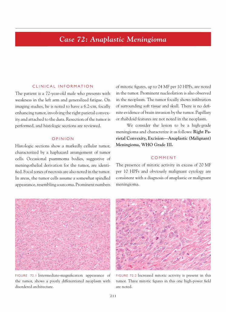
211
Case 72: Anaplastic Meningioma
C L I N I C A L I N F O R M A T I O N
The patient is a 72-year-old male who presents with weakness in the left arm and generalized fatigue. On imaging studies, he is noted to have a 6.2-cm, focally enhancing tumor, involving the right parietal convex-ity and attached to the dura. Resection of the tumor is performed, and histologic sections are reviewed.
O P I N I O N
Histologic sections show a markedly cellular tumor, characterized by a haphazard arrangement of tumor cells. Occasional psammoma bodies, suggestive of meningothelial derivation for the tumor, are identi-fi ed. Focal zones of necrosis are also noted in the tumor. In areas, the tumor cells assume a somewhat spindled appearance, resembling a sarcoma. Prominent numbers
of mitotic fi gures, up to 24 MF per 10 HPFs, are noted in the tumor. Prominent nucleolation is also observed in the neoplasm. The tumor focally shows infi ltration of surrounding soft tissue and skull. There is no defi -nite evidence of brain invasion by the tumor. Papillary or rhabdoid features are not noted in the neoplasm.
We consider the lesion to be a high-grade meningioma and characterize it as follows: Right Pa-rietal Convexity, Excision—Anaplastic (Malignant) Meningioma, WHO Grade III.
C O M M E N T
The presence of mitotic activity in excess of 20 MF per 10 HPFs and obviously malignant cytology are consistent with a diagnosis of anaplastic or malignant meningioma.
FIGURE 72.1 Intermediate-magnifi cation appearance of the tumor, shows a poorly differentiated neoplasm with disordered architecture.
FIGURE 72.2 Increased mitotic activity is present in this tumor. Three mitotic fi gures in this one high-power fi eld are noted.

212
C A S E 7 2 : A N A P L A S T I C M E N I N G I O M A
D I S C U S S I O N
Anaplastic meningiomas often show high-grade cytology, with tumor cells resembling those seen in carcinoma, melanoma, or high-grade sarcoma. These tumors often have exceedingly high mitotic rates, with mitotic indices of 20 or more MF per 10 HPFs. Tumors with either one of these features are generally classifi ed as anaplastic or malignant meningiomas. The signifi cance of the diagnosis is that many of these tumors prove fatal in a relatively short period, with
a median survival of less than 2 years in many cases. These lesions, in addition to surgical resection, often warrant adjuvant therapy.
Because these tumors are so poorly differentiat-ed, they often present a diagnostic challenge. Unless there are areas that are more well- differentiated in the tumor, recognition of the lesion as a meningioma can be challenging. The current case demonstrates psammoma body formations, which proves to be useful in delineating the lineage of the neoplasm. Distinction of these tumors from metastatic carci-nomas may require the use of keratin immunostains, which generally show diffuse positive staining in car-cinomas, and at most, focal weak staining in menin-giomas. Differentiation of anaplastic meningioma from metastatic malignant melanoma can also prove to be challenging. Again, immunohistochemistry can be helpful, in that melanomas generally show fairly diffuse S-100 immunoreactivity and immunos-taining with antibodies such as Melan-A or HMB 45, two markers that do not stain meningiomas. S-100 immunoreactivity, if present in meningiomas, is often focal and weak. Differentiation of high-grade meningioma from a spindled cell sarcoma can
FIGURE 72.5 Rare psammoma bodies in the tumor are help-ful in identifying the lesion as a meningioma.
FIGURE 72.4 Areas of the tumor have a spindled, sarcoma-like appearance, with increased mitotic fi gures.
FIGURE 72.3 Necrosis is present.

213
C A S E 7 2 : A N A P L A S T I C M E N I N G I O M A
FIGURE 72.6 Prominent hypercellularity is a feature of this tumor.
FIGURE 72.7 Infi ltration of the skull by tumor is evident.
be diffi cult. Again, unless there are other clues as to the meningothelial derivation of the tumor or other features suggesting a particular sarcoma type, immunohistochemistry is not helpful in these cases. Rare cases of a sarcomatoid prominent gliosarcoma should also be considered in the differential diagno-sis; these tumors generally show some areas of GFAP immunoreactivity, a fi nding that is not present in meningiomas.
References 1. Jaaskelainen J, Haltia M, Servo A. Atypical and anaplastic
meningiomas: radiology, surgery, radiotherapy, and outcome. Surg Neurol 1986;25:233–42.
2. Perry A, Louis DN, Scheithauer BW, et al. Meningiomas. In: Louis DN, Ohgaki H, Wiestler OD, Cavenee WK, Editors. WHO classifi cation of Tumours of the Central Nervous System. Lyon, FR: IARC Press;2007. p. 164–72.
3. Perry A, Scheithauer BW, Stafford SL, et al. “Malignancy” in meningiomas: a clinicopathologic study of 116 patients, with grading implications. Cancer 1999;85:2046–56.

214
Case 73: Angiomatous Meningioma
C L I N I C A L I N F O R M A T I O N
The patient is a 42-year-old female who presents with a right falcine mass, measuring 3.1 cm on imaging studies. The mass is resected, and histologic sections are reviewed.
O P I N I O N
Histologic sections show a dural-based mass, marked by a proliferation of blood vessels. In areas, the blood vessels appear to be almost back to back. Focally, prominent vascular sclerotic changes are noted. In other areas, small collections of atypical-appearing cells with rounded nuclei and eosinophilic cytoplasm are also seen. Intranuclear cytoplasmic pseudoin-clusions are noted in some of these cells. Scattered evidence of nuclear pleomorphism in this popula-tion of cells is also identifi ed. Mitotic activity, small cell change, necrosis, brain invasion, and prominent
nucleolation are not seen. Areas of microcystic degen-erative changes are noted. The tumor demonstrates evidence of intravascular embolization.
We consider the lesion to be a meningioma and characterize it as follows: Right Falcine Region, Resection—Angiomatous Meningioma, WHO Grade I.
C O M M E N T
The tumor is marked by a prominent proliferation of benign-appearing blood vessels. Worrisome histologic features indicative of an atypical or anaplastic menin-gioma are not identifi ed.
D I S C U S S I O N
The angiomatous variant of meningioma is char-acterized by a prominent proliferation of blood
FIGURE 73.1 The tumor is a dural-based mass.FIGURE 73.2 Back-to-back sclerotic vessels in the tumor can resemble a vascular malformation.

215
C A S E 7 3 : A N G I O M A T O U S M E N I N G I O M A
vessels. In areas of the tumor, the number of blood vessels may be so numerous as to raise vascular mal-formation as a differential diagnostic consideration. Careful search of these tumors, however, usually yields areas in which meningothelial cells, individ-ually or in clusters or nests, are situated between blood vessels. Vascular sclerotic changes, as in the
current tumor, may be observable in the angioma-tous meningioma as well. Hemangioblastomas can rarely arise in the falcine region; the hemangioblas-toma is often marked by a stromal cell component that has a vacuolated quality to the cytoplasm. By immunohistochemistry, hemangioblastomas do not demonstrate EMA immunoreactivity, whereas most meningiomas demonstrate at least focal EMA
FIGURE 73.6 High-magnifi cation appearance of meningo-thelial cells, demonstrates cytoplasmic pseudoinvagina-tions and nuclear pleomorphism.
FIGURE 73.5 Focal evidence of embolization is evident in the tumor. Embolization is used preoperatively to reduce intraoperative hemorrhage.
FIGURE 73.4 A more solid-appearing area of the tumor shows microcystic degenerative changes and nuclear pleo-morphism of the meningothelial cells.
FIGURE 73.3 Intermediate magnifi cation, shows a predom-inance of blood vessels, with rare atypical meningothelial cells situated between the vessels. The endothelial cells lining the vessels show minimal atypia.

216
C A S E 7 3 : A N G I O M A T O U S M E N I N G I O M A
staining. Hemangiopericytomas may also arise as dural-based masses and can present with a promi-nent vascular component. The cellular component of hemangiopericytomas is usually more atypical, and mitotic fi gures are frequently evident. Again, EMA does not usually stain hemangiopericytoma, and many of the cells in the solid areas of heman-giopericytoma frequently demonstrate CD34 immu-noreactivity.
As a historical side note, angiomatous menin-gioma, hemangioblastomas, and hemangiopericyto-mas used to be collectively referred to as angioblastic meningiomas. These tumors were thought to have a worse prognosis, probably because of inclusion of
hemangiopericytomas. With separation of heman-gioblastoma and hemangiopericytoma as distinctive entities, the remaining angiomatous meningioma are benign. Use of the term of angioblastic meningioma is no longer advised.
References 1. Burger PC, Scheithauer WB. Meningioma. In: Tumors of the
Central Nervous System. Washington, DC: AFIP Fascicle 4th
series; 2007. p. 331–62. 2. Hasselblatt M, Nolte K, Paulus W. Angiomatous meningi-
oma. A clinicopathologic study of 38 cases. Am J Surg Pathol 2004;28:390–3.
3. Prayson RA. Pathology of meningiomas. In: Lee JH, Editor. Meningiomas. Diagnosis, treatment, and outcome. London, UK: Springer-Verlag;2008. p. 31–43.
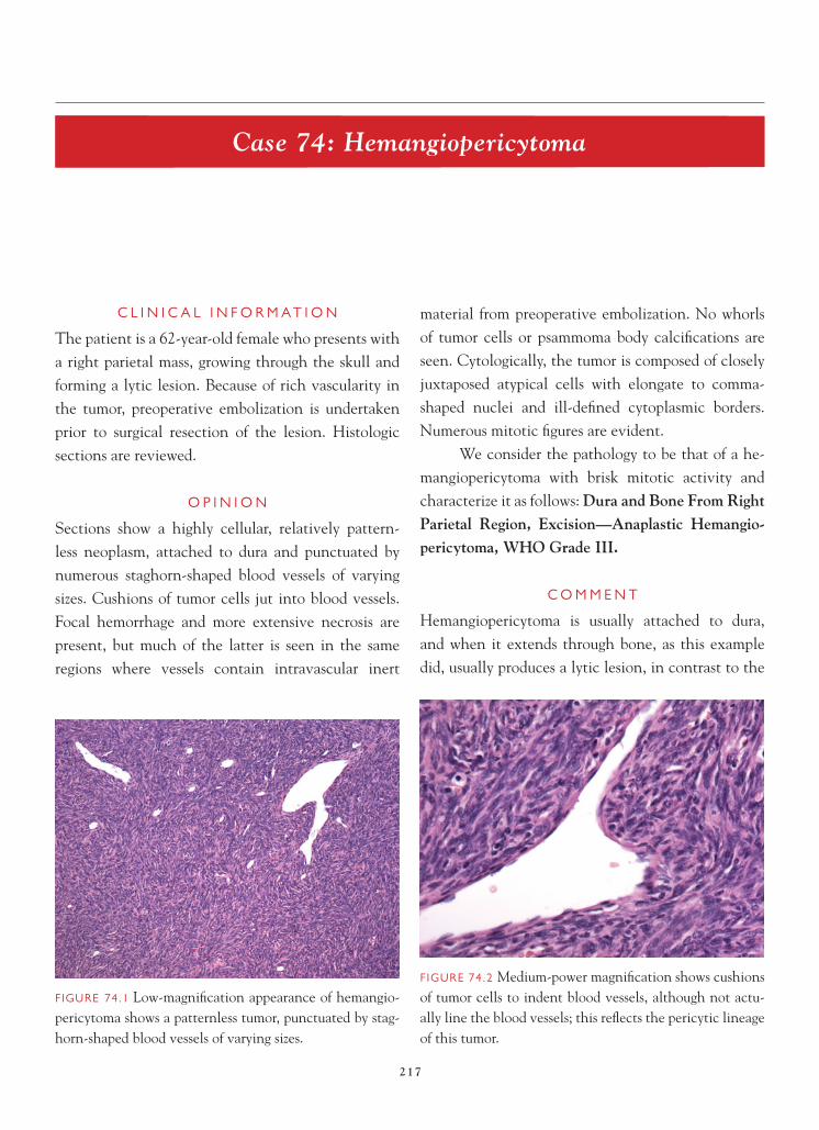
217
Case 74: Hemangiopericytoma
C L I N I C A L I N F O R M A T I O N
The patient is a 62-year-old female who presents with a right parietal mass, growing through the skull and forming a lytic lesion. Because of rich vascularity in the tumor, preoperative embolization is undertaken prior to surgical resection of the lesion. Histologic sections are reviewed.
O P I N I O N
Sections show a highly cellular, relatively pattern-less neoplasm, attached to dura and punctuated by numerous staghorn-shaped blood vessels of varying sizes. Cushions of tumor cells jut into blood vessels. Focal hemorrhage and more extensive necrosis are present, but much of the latter is seen in the same regions where vessels contain intravascular inert
material from preoperative embolization. No whorls of tumor cells or psammoma body calcifi cations are seen. Cytologically, the tumor is composed of closely juxtaposed atypical cells with elongate to comma-shaped nuclei and ill-defi ned cytoplasmic borders. Numerous mitotic fi gures are evident.
We consider the pathology to be that of a he-mangiopericytoma with brisk mitotic activity and characterize it as follows: Dura and Bone From Right Parietal Region, Excision—Anaplastic Hemangio-pericytoma, WHO Grade III.
C O M M E N T
Hemangiopericytoma is usually attached to dura, and when it extends through bone, as this example did, usually produces a lytic lesion, in contrast to the
FIGURE 74.1 Low-magnifi cation appearance of hemangio-pericytoma shows a patternless tumor, punctuated by stag-horn-shaped blood vessels of varying sizes.
FIGURE 74.2 Medium-power magnifi cation shows cushions of tumor cells to indent blood vessels, although not actu-ally line the blood vessels; this refl ects the pericytic lineage of this tumor.

218
C A S E 7 4 : H E M A N G I O P E R I C Y T O M A
hyperostotic lesion seen with meningiomas involving nearby bone.
D I S C U S S I O N
Hemangiopericytoma is usually a solitary lesion and unassociated with any familial tumor syndrome. It is far less common that meningioma and has a slight predilection for being located in the occipital region, near the confl uens of the sinuses. Despite the fact that hemangiopericytoma is usually well demar-cated and seemingly able to be totally resected, local recurrence is very frequent, especially if the patient is followed for a suffi ciently long period. Tumor may recur more than a decade after the initial resection. Hemangiopericytoma of the central nervous system (CNS) has the distinction of being one of the few primary CNS neoplasms with a propensity to metas-tasize outside the cranial vault or spinal column. Most patients eventually develop metastatic spread to bone, lung, or liver.
Diagnosis is made by identifying a patternless tumor, composed of closely packed cells and punc-tuated by staghorn-shaped blood vessels. A help-ful feature is the presence of each tumor cell being seemingly at equidistant spacing from its neighbor. This corresponds to the extracellular, basement membrane-like material that invests individual
FIGURE 74.5 Other areas show early zonal necrosis, imme-diately juxtaposed to vessels containing intravascular inert material from the preoperative embolization procedure.
FIGURE 74.4 Several foci of necrosis found in this tumor are likely as a result of the rapidly growing tumor itself, based on the high cellularity and cytologic atypia of the nearby tumor.
FIGURE 74.3 High magnifi cation illustrates the elongate-to-comma-shaped nuclei of tumor cells, the ill-defi ned cell borders, and the equidistant spacing of nuclei from each other as a result of the interspersed basement membrane. Note several mitoses in a single fi eld; this tumor had suf-fi cient mitotic activity to classify it as an anaplastic heman-giopericytoma, WHO grade III.

219
C A S E 7 4 : H E M A N G I O P E R I C Y T O M A
FIGURE 74.6 High magnifi cation illustrates the inert intra-vascular embolic material.
tumor cells and is highlighted by a rich reticulin net-work on reticulin stain.
Hemangiopericytoma (HPC) is a low-grade sar-coma of the dura, graded as WHO grade II or III. An-aplastic (WHO grade III) HPCs, according to WHO 2007 classifi cation, are characterized by “brisk mitotic activity (at least 5 mitoses per 10 high power fi elds) and/or necrosis, plus at least two of the following: hemorrhage, moderate to high nuclear atypia, and cellularity.” This example had all of these features,
although, as noted previously, some of the necrosis was best attributed to the preoperative embolization procedure.
Reticulin staining shows abundant reticulin in most cases. HPC is strongly and diffusely immunore-active for vimentin, shows patchy reactivity for CD34 (in contrast to the diffuse immunoreactivity of soli-tary fi brous tumor), and usually lacks the strong diffuse
FIGURE 74.7 Abundant reticulin fi bers surround individual and small groups of tumor cells.
FIGURE 74.8 Hemangiopericytomas always show strong diffuse immunoreactivity for vimentin.
FIGURE 74.9 Patchy CD34 immunoreactivity is usually seen in hemangiopericytoma, in contrast to the diffuse immunoreactivity seen in solitary fi brous tumor; CD34 also highlights the staghorn vasculature.

220
C A S E 7 4 : H E M A N G I O P E R I C Y T O M A
immunoreactivity for EMA expected in meningioma. However, high-grade (atypical, WHO grade II and anaplastic, WHO grade III) meningiomas also may lose EMA immunoreactivity, and conversely, focal EMA positivity can be seen in HPCs. CD99 and bcl-2 immunoreactivity is seen in the majority of HPCs.
References 1. Giannini C, Rushing EJ, Hainfellner JA. Haemangioperi-
cytoma. In: Louis DN, Ohgaki H, Wiestler OD, Cavenee,
WK, Editors. WHO Classifi cation of Tumours of the Central Nervous System. Lyon, FR: IARC Press;2007, p. 178–80.
2. Perry, A, Scheithauer BW, Nascimento AG. The immuno-phenotypic spectrum of meningeal hemangiopericytoma: a comparison with fi brous meningioma and solitary fi brous tumor of meninges. Am J Surg Pathol 1997;21:1354–60.
3. Prayson, RA. Non-glial tumors. In: Prayson RA, Editor. Neuropathology. Philadelphia, PA: Elsevier Churchill Livingstone;2005. p. 489–536.
4. Rajaram V, Brat DJ, Perry A. Anaplastic meningioma versus meningeal hemangiopericytoma: immunohistochemical and genetic markers. Hum Pathol 2004;35:1413–18.
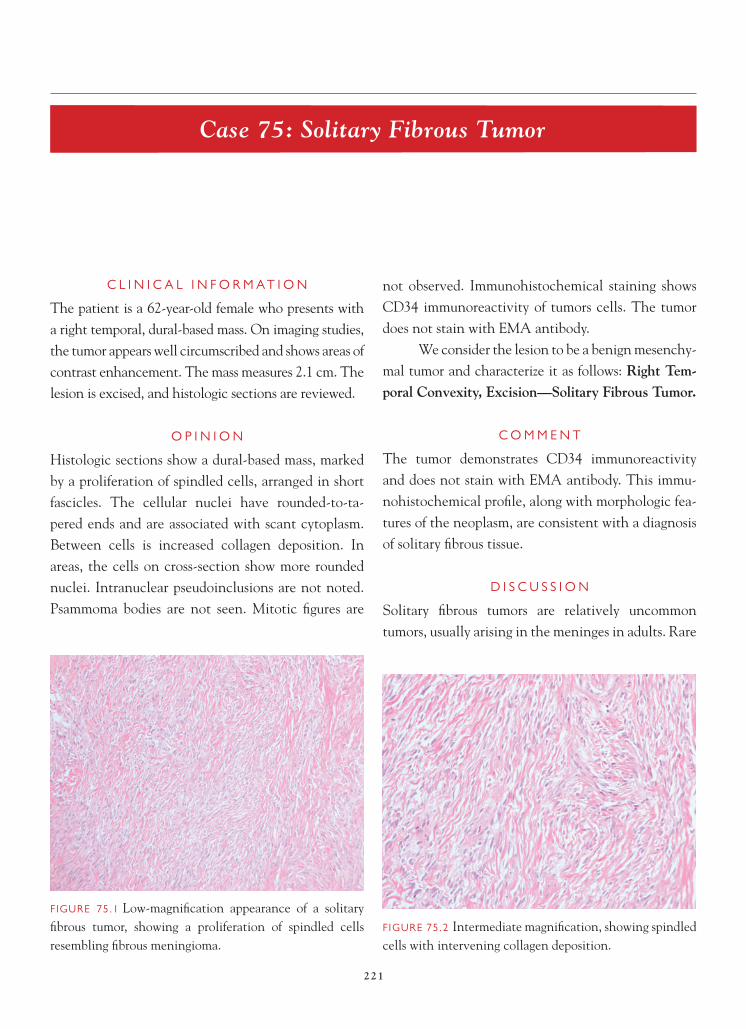
221
Case 75: Solitary Fibrous Tumor
C L I N I C A L I N F O R M A T I O N
The patient is a 62-year-old female who presents with a right temporal, dural-based mass. On imaging studies, the tumor appears well circumscribed and shows areas of contrast enhancement. The mass measures 2.1 cm. The lesion is excised, and histologic sections are reviewed.
O P I N I O N
Histologic sections show a dural-based mass, marked by a proliferation of spindled cells, arranged in short fascicles. The cellular nuclei have rounded-to-ta-pered ends and are associated with scant cytoplasm. Between cells is increased collagen deposition. In areas, the cells on cross-section show more rounded nuclei. Intranuclear pseudoinclusions are not noted. Psammoma bodies are not seen. Mitotic fi gures are
not observed. Immunohistochemical staining shows CD34 immunoreactivity of tumors cells. The tumor does not stain with EMA antibody.
We consider the lesion to be a benign mesenchy-mal tumor and characterize it as follows: Right Tem-poral Convexity, Excision—Solitary Fibrous Tumor.
C O M M E N T
The tumor demonstrates CD34 immunoreactivity and does not stain with EMA antibody. This immu-nohistochemical profi le, along with morphologic fea-tures of the neoplasm, are consistent with a diagnosis of solitary fi brous tissue.
D I S C U S S I O N
Solitary fi brous tumors are relatively uncommon tumors, usually arising in the meninges in adults. Rare
FIGURE 75.1 Low-magnifi cation appearance of a solitary fi brous tumor, showing a proliferation of spindled cells resembling fi brous meningioma.
FIGURE 75.2 Intermediate magnifi cation, showing spindled cells with intervening collagen deposition.

222
C A S E 7 5 : S O L I T A R Y F I B R O U S T U M O R
cases of intraventricular and spinal tumors have been also described. The lesions are generally well circum-scribed and marked by a proliferation of spindled-to-rounded cells, with abundant collagen deposition between individual cells and small groups of cells. Spindle-cell tumors can mimic fi brous meningioma, which is usually the major differential diagnostic con-sideration. In contrast to meningiomas, these tumors
generally lack intranuclear cytoplasmic invaginations or pseudoinclusions and lack psammoma bodies. Immunohistochemistry can be helpful in delineating the lesion from meningioma, in that the majority of these tumors demonstrate fairly extensive immunore-activity with antibody to CD34 and generally do not stain with antibody to EMA.
Although the majority of these tumors are low-grade lesions with a good prognosis, rare cases of anaplastic tumors with more aggressive histology
FIGURE 75.6 EMA immunostaining, characteristic of most meningiomas, is not present in this tumor.
FIGURE 75.5 CD34 immunoreactivity is present in this tumor.
FIGURE 75.4 An area of the tumor where cells are marked by more rounded nuclei.
FIGURE 75.3 High-magnifi cation appearance, showing bland nuclei with tapered ends and intervening collagen. Cytoplasmic intranuclear pseudoinclusions, a feature of many meningiomas, are not seen in solitary fi brous tumor.

223
C A S E 7 5 : S O L I T A R Y F I B R O U S T U M O R
have been described. These tumors are marked by increased mitotic activity and necrosis. The anaplastic tumors have a propensity to recur.
References 1. Cummings TJ, Burchette JL, McLendon RE. CD34 and dural
fi broblasts: the relationship to solitary fi brous tumor and meningioma. Acta Neuropathol (Berl) 2001;102:349–54.
2. Carneiro SS, Scheithauer BW, Nascimento AG, et al. Solitary fi brous tumor of the meninges: a lesion distinct from fi brous meningioma. A clinicopathologic and immunohistochemical study. Am J Clin Pathol 1996;106:217–24.
3. Tihan T, Viglione M, Rosenblum MK, et al. Solitary fi brous tumors in the central nervous system. A clinicopathologic review of 18 cases and comparison to meningeal hemangio-pericytomas. Arch Pathol Lab Med 2003;127:432–9.

224
Case 76: Primary Sarcoma of the Central Nervous System
C L I N I C A L I N F O R M A T I O N
The patient is a 44-year-old male who presents with headaches and is found by neuroimaging studies to have a right frontal lobe mass. The decision is made to excise the lesion surgically, and intraoperatively, no attachment to the dura is detected. Histologic sections are available for review.
O P I N I O N
Sections show a hypercellular, spindled cell neo-plasm, with cells arranged in a fascicular pattern. Multifocal zones of necrosis are present, and mitotic rate is very high, exceeding 20 mitoses per 10 high-power microscopic fi elds. Careful search fails
to disclose a coexistent glioblastoma. No smooth muscle, skeletal muscle, or malignant osteoid or cartilaginous elements are present in the sarcoma. The lesion has no continuity with a cranial periph-eral nerve.
We consider the lesion to be a high-grade mes-enchymal neoplasm and characterize it as follows: Right Frontal Lobe, Excision—Primary Sarcoma of the Central Nervous System, WHO Grade IV.
C O M M E N T
Mixed glioblastoma-sarcomas (gliosarcomas) with a dominant sarcomatous component are also uncom-mon, but more frequent than pure, nonmeningeal-based primary sarcomas of the brain.
FIGURE 76.1 Low-power magnifi cation appearance of the resected tissue shows a spindled neoplasm with cells arranged in fascicles. No muscle or osteocartilaginous elements are present.
FIGURE 76.2 Low-power magnifi cation allows identifi ca-tion of the extensive zonal, serpinginous necrosis in the high-grade sarcoma.
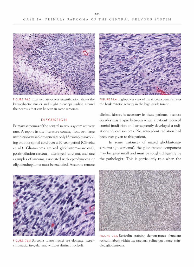
225
C A S E 7 6 : P R I M A R Y S A R C O M A O F T H E C E N T R A L N E R V O U S S Y S T E M
D I S C U S S I O N
Primary sarcomas of the central nervous system are very rare. A report in the literature coming from two large institutions was able to generate only 18 examples involv-ing brain or spinal cord over a 30-year period (Oliveira et al.). Gliosarcoma (mixed glioblastoma-sarcoma), postirradiation sarcoma, meningeal sarcoma, and rare examples of sarcoma associated with ependymoma or oligodendroglioma must be excluded. Accurate remote
clinical history is necessary in these patients, because decades may elapse between when a patient received cranial irradiation and subsequently developed a radi-ation-induced sarcoma. No antecedent radiation had been ever given to this patient.
In some instances of mixed glioblastoma- sarcoma (gliosarcoma), the glioblastoma component may be quite small and must be sought diligently by the pathologist. This is particularly true when the
FIGURE 76.6 Reticulin staining demonstrates abundant reticulin fi bers within the sarcoma, ruling out a pure, spin-dled glioblastoma.
FIGURE 76.5 Sarcoma tumor nuclei are elongate, hyper-chromatic, irregular, and without distinct nucleoli.
FIGURE 76.4 High-power view of the sarcoma demonstrates the brisk mitotic activity in the high-grade tumor.
FIGURE 76.3 Intermediate-power magnifi cation shows the karyorrhectic nuclei and slight pseudopalisading around the necrosis that can be seen in some sarcomas.

226
C A S E 7 6 : P R I M A R Y S A R C O M A O F T H E C E N T R A L N E R V O U S S Y S T E M
FIGURE 76.7 As is the case with many sarcomas, the only immunoreactivity seen in the tumor is diffuse staining for vimentin.
neurosurgeon has selectively removed the more obvi-ously abnormal, cohesive, solid portion of the mixed tumor, leaving the more infi ltrative glial portion of the lesion in the patient. No glioblastoma component was identifi ed in this case, and immunostaining for GFAP was negative in several sections.
Another consideration in some cases of primary sarcomas of the brain is a more spindled form of an ordinary glioblastoma, where the glial cells are elon-gate and mimic a mesenchymal neoplasm. Negative immunostaining for S-100 and GFAP in this case also negated that diagnosis. Very rare intracranial exam-ples of malignant peripheral nerve sheath tumor have been described, but without immunohistochemical evidence of peripheral nerve origin and without loca-tion near the skull base location of cranial peripheral nerves, that diagnosis is not possible. Finally, menin-geal sarcomas (now synonymous with anaplastic me-ningioma, WHO grade III) and metastatic sarcomas or intracranial extension of a head and neck sarcoma are considerations. All were excluded in this case.
This example lacked dural attachment and manifests a fi brosarcoma-like pattern, with intersect-
ing fascicles of spindled cells. The lesion is high grade, WHO grade IV, by virtue of its extensive necrosis and brisk mitotic activity. In the series of 18 cases reported by Oliveira et al., 6 of 18 were fi brosarcomas, 5 of 18 were malignant fi brous histiocytomas, and 3 of 16 were undifferentiated sarcomas. Two-thirds were high grade, based on degree of necrosis, mitotic activity, and extent of differentiation. Cerebrum was by far the most common location (83%). An immu-nohistochemical work-up includes immunostaining for GFAP, S-100, vimentin, desmin, smooth muscle actin, EMA, and CAM 5.2, with additional possible staining for other markers, depending on the light microscopic features.
Prognosis in the largest series of primary sarcomas of the brain and spinal cord was actually slightly better than that for glioblastomas, in that 28% of patients with high-grade sarcomas were alive at 5 years.
References 1. Duffner PK, Krischer JP, Horowitz ME, et al. Second malig-
nancies in young children with primary brain tumors follow-ing treatment with prolonged postoperative chemotherapy and delayed irradiation: a Pediatric Oncology Group study. Ann Neurol 1998;44:313–6.
FIGURE 76.8 S-100 protein immunostaining is negative.

227
C A S E 7 6 : P R I M A R Y S A R C O M A O F T H E C E N T R A L N E R V O U S S Y S T E M
2. Kleinschmidt-Demasters BK, Kang JS, Lillehei KO. The burden of radiation-induced central nervous system tumors: a single institution’s experience. J Neuropathol Exp Neurol 2006;65:204–16.
3. Oliveira AM, Scheithauer BW, Salomao DR, et al. Primary sarcomas of the brain and spinal cord: a study of 18 cases. Am J Surg Pathol 2002;26:1056–63.
4. Rodriguez FJ, Scheithauer BW, Perry A, et al. Ependymal tumors with sarcomatous change (“ependymosarcoma”): a clinicopathologic and molecular cytogenetic study. Am J Surg Pathol 2008;32:699–709.
5. Rodriguez FJ, Scheithauer BW, Jenkins R, et al. Gliosarcoma arising in oligodendroglial tumors (“oligosarcoma”): a clini-copathologic study. Am J Surg Pathol 2007;31:351–62.

228
Case 77: Meningioangiomatosis
C L I N I C A L I N F O R M A T I O N
The patient is a 16-year-old male who presents with seizures and a left temporal lobe lesion. On imaging, the lesion appears to be predominantly intracortical in location and has a plaque-like distribution, with low signal intensity on T2-weighted MRI studies. Calcifi cations are also noted on CT imaging of the lesion. A resection of the lesion is performed, and his-tologic sections are reviewed.
O P I N I O N
Histologic sections show a proliferation of blood ves-sels rimmed by spindled cells. Many of the spindled cells have elongated nuclei. The cells are piled up around the blood vessels, predominantly in the cor-tex, in a stratifi ed fashion. There is minimal cytologic
atypia or nuclear pleomorphism to these spindled cells. Occasional psammoma bodies are noted. Some of the vessels show prominent sclerotic changes. Mitotic fi gures are not identifi ed in the spindled or in the endothelial cells lining vascular lumina. Intervening parenchyma between the blood vessels and the cortex shows gliosis. The overlying menin-ges shows a tumor, marked by a solid proliferation of spindled cells.
We characterize the lesion as follows: Left Tem-poral Lobe, Excision—Meningioangiomatosis with Overlying Fibrous Meningioma, WHO Grade I.
C O M M E N T
A subset of meningioangiomatosis cases has been associated with overlying meningioma. There is a
FIGURE 77.1 Low-magnifi cation appearance of the cortex, shows collars of spindled cells arranged around blood ves-sels. Occasional psammoma bodies are observed.
FIGURE 77.2 Intermediate magnifi cation shows a perivas-cular arrangement of spindled meningothelial cells and fi broblastic cells.
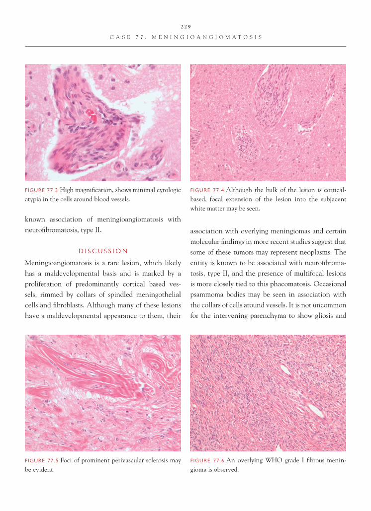
229
C A S E 7 7 : M E N I N G I O A N G I O M A T O S I S
known association of meningioangiomatosis with neurofi bromatosis, type II.
D I S C U S S I O N
Meningioangiomatosis is a rare lesion, which likely has a maldevelopmental basis and is marked by a proliferation of predominantly cortical based ves-sels, rimmed by collars of spindled meningothelial cells and fi broblasts. Although many of these lesions have a maldevelopmental appearance to them, their
association with overlying meningiomas and certain molecular fi ndings in more recent studies suggest that some of these tumors may represent neoplasms. The entity is known to be associated with neurofi broma-tosis, type II, and the presence of multifocal lesions is more closely tied to this phacomatosis. Occasional psammoma bodies may be seen in association with the collars of cells around vessels. It is not uncommon for the intervening parenchyma to show gliosis and
FIGURE 77.6 An overlying WHO grade I fi brous menin-gioma is observed.
FIGURE 77.5 Foci of prominent perivascular sclerosis may be evident.
FIGURE 77.4 Although the bulk of the lesion is cortical-based, focal extension of the lesion into the subjacent white matter may be seen.
FIGURE 77.3 High magnifi cation, shows minimal cytologic atypia in the cells around blood vessels.

230
C A S E 7 7 : M E N I N G I O A N G I O M A T O S I S
occasionally, neurofi brillary tangles and granulovacu-olar degeneration may be observable within cortical neurons in involved areas. Immunohistochemical studies have demonstrated low rates of cell prolifera-tion. EMA immunoreactivity, as seen in meningioma, can be observable in meningioangiomatosis.
The lesion has a distinctive appearance. Dif-ferential diagnostic consideration includes a brain-invasive meningioma. The architectural pattern of meningioangiomatosis, however, is unusual for the brain-invasive meningioma, which usually does not invade in a plaque-like fashion. Occasional cases of meningioangiomatosis may show prominent vascu-lar sclerosis; in these cases, the meningothelial and fi broblastic components of the tumor are not as clear-ly evident. In the lesions with prominent vascular
sclerosis, a vascular malformation may enter into the differential diagnosis.
References 1. Halper J, Scheithauer BW, Okazaki H, et al. Meningo-
angiomatosis: a report of six cases with special reference to the occurrence of neurofi brillary tangles. J Neuropathol Exp Neurol 1986;45:426–46.
2. Perry A, Kurt Kaya-Yapier O, Scheithauer BW, et al. Insights into meningioangiomatosis with and without meningioma: a clinicopathologic and genetic series of 24 cases with review of the literature. Brain Pathol 2005;15:55–65.
3. Prayson RA. Meningioangiomatosis. A clinicopathologic study including MIB1 immunoreactivity. Arch Pathol Lab Med 1995;119:1061–4.
4. Stemmer-Rachamimov AO, Horgan MA, Taratuto AL, et al. Meningioangiomatosis is associated with neurofi broma-tosis 2 but not with somatic alterations of the NF2 gene. J Neuropathol Exp Neurol 1997;56:485–9.

231
Case 78: Hemangioblastoma
C L I N I C A L I N F O R M A T I O N
The patient is a 36-year-old male who presents with an unsteady gait. His past medical history is notable for a father and older brother who both died of renal cell carcinoma. On imaging studies, the lesion is a cyst, with contrast-enhancing mural nodule in the wall of the cyst. The entire lesion measures 2.8 cm. The lesion is resected, and histologic sections are reviewed.
O P I N I O N
Histologic sections show a lesion marked by a pro-liferation of small caliber capillaries and venous vessels. Between blood vessels are cells with lightly eosinophilic or vacuolated, cleared cytoplasm. Focal microcystic changes are noted. In areas, the lesion shows a moderate degree of nuclear pleomorphism.
Focal dystrophic calcifi cation is observed. The lesion appears to be circumscribed, but not encapsulated. The adjacent parenchyma shows Rosenthal fi bers. There is no evidence of mitotic activity or necrosis in the lesion. EMA immunostaining is not observed.
We consider the lesion to be a neoplasm and characterize it as follows: Left Cerebellum, Excision—Hemangioblastoma, WHO Grade I.
C O M M E N T
The tumor does not stain with antibody to EMA. Hemangioblastomas are associated with von Hippel-Lindau disease.
D I S C U S S I O N
Hemangioblastomas are tumors that generally arise in adults and are somewhat more frequently
FIGURE 78.1 Low-magnifi cation appearance of hemangio-blastoma shows prominent numbers of small vessels with intravenous stromal cells.
FIGURE 78.2 Focal microcystic degenerative changes are observed in the tumor.

232
C A S E 7 8 : H E M A N G I O B L A S T O M A
encountered in males. The cerebellum is the most common location; however, tumors have been described in the spinal cord or in other dural-based locations in the central nervous system. There is a well-known association of hemangioblastoma with von Hippel-Lindau disease. In the current patient, the family history of a brother and father with renal cell carcinoma raises the possibility of von Hippel-Lindau. There is also a known association of second-ary polycythemia vera, with hemangioblastoma, as a
result of the production of erythropoietin in a subset of patients.
Imaging fi ndings demonstrate a characteris-tic cyst, with enhancing mural nodule confi gura-tion. Tumors are often well circumscribed, with gliosis and Rosenthal fi ber formations in the adja-cent parenchyma. Because pilocytic astrocytomas can also arise in the cerebellum and may have increased Rosenthal fi bers and a cyst with enhancing mural
FIGURE 78.6 The hemangioblastomas are well circum-scribed, and adjacent parenchyma may show piloid gliosis with Rosenthal fi ber formations.
FIGURE 78.5 Pleomorphic cells in a hemangioblastoma do not afford the tumor a worse prognosis.
FIGURE 78.4 High-magnifi cation appearance of stromal cells highlights lightly eosinophilic cytoplasm.
FIGURE 78.3 High-magnifi cation appearance of stromal cells shows vacuolated cytoplasm.

233
C A S E 7 8 : H E M A N G I O B L A S T O M A
nodule radiographic appearance, tissue taken from the edge of a hemangioblastoma may be misinterpreted as representing a pilocytic astrocytoma. The tumor itself is comprised of a prominent number of small capillar-ies and venous vessels. Vascular sclerotic changes are often present. The intervening stromal cells may have a light eosinophilic or cleared, vacuolar cytoplasm. Some degree of nuclear pleomorphism may be evident but has no prognostic signifi cance. Mitotic activity is rarely observed, and necrosis is distinctly uncommon in this tumor. These tumors generally demonstrate a low rate of cell proliferation.
The major differential diagnostic consider-ation is with a metastatic clear cell carcinoma, par-ticularly in patients who have von Hippel- Lindau disease. Antibodies that have been potentially used to distinguish the two tumors include EMA (which stains most renal cell carcinomas positively and hemangioblastomas negatively), inhibin (which stains many hemangioblastomas positively and most renal cell carcinomas negatively), and a variety of
renal cell carcinoma-directed antibodies, such as RCC or carbonic anhydrase 9. Metastatic renal cell carcinomas are also more likely to demonstrate areas of necrosis or mitotic activity. The importance of making the distinction lies in the differences in terms of biologic behavior. Hemangioblastomas are consid-ered low-grade lesions, presumably curable with gross total resection.
References 1. Conway JE, Chou D, Clatterbuck RE, et al. Hemangioblas-
tomas of the central nervous system in von Hippel-Lindau syndrome and sporadic disease. Neurosurgery 2001;48:55–62, discussion 62–3.
2. Hoang MP, Amirkhan RH. Inhibin alpha distinguishes hemangioblastoma from clear cell renal cell carcinoma. Am J Surg Pathol 2003;27:1152–6.
3. Neumann H, Eggert H, Weigel K, et al. Hemangioblastomas of the central nervous system. A 10-year study with spe-cial reference to von Hippel-Lindau syndrome. J Neurosurg 1989;70:24–30.
4. Wang C, Zhang J, Liu A, et al. Surgical management of medullary hemangioblastoma. Report of 47 cases. Surg Neurol 2001;56:218–26.

234
Case 79: Meningeal Melanocytoma
C L I N I C A L I N F O R M A T I O N
This 39-year-old man with a known right nevus of Ota presents with left homonymous hemianopsia, left hemi-paresis, and left hemiapraxia. A CT scan shows a homo-geneously enhancing, well-defi ned mass, involving the right occipital lobe, which is confi rmed by MRI. At surgery, the periosteum, bone, and dura mater surround-ing the tumor are deeply pigmented, as is the tumor itself. The tumor is densely adherent to the dura mater, but easily dissected away from the brain parenchyma. Sections of the resection specimen are reviewed.
O P I N I O N
The tumor is well demarcated, but not encapsulated. A small amount of neural parenchyma is adherent to
the tumor, but not invaded by it. The tumor is com-posed of fascicles and nests of monomorphous spindle and occasionally, epithelioid cells, with round-to-oval regular nuclei and indistinct nucleoli. Although scat-tered clusters of tumor cells contain large amounts of melanin pigment, the vast majority of the tumor cells appear nonpigmented. Rare mitotic fi gures are identifi ed. Coagulative tumor necrosis is absent. Immunohistochemical staining for EMA is negative. Immunostains for vimentin, S-100 protein, and gp100 (HMB 45 antibody) react with the tumor cells.
We consider the lesion to represent a low-grade meningeal melanocytic tumor and characterized as follows: Right Occipital Lobe of Brain, Excision—Meningeal Melanocytoma.
FIGURE 79.1 At low-power magnifi cation, the tumor is composed of fascicles of monomorphous cells without whorl formation. Melanin-containing tumor cells are pres-ent in widely scattered clusters.
FIGURE 79.2 High-power magnifi cation demonstrates a relatively monomorphous population of spindle shaped cells with small but distinct nucleoli. Although heavily pigmented tumor cells are uncommon, many of the tumor cells contain melanin granules.

235
C A S E 7 9 : M E N I N G E A L M E L A N O C Y T O M A
C O M M E N T
Fascicular growth pattern and low mitotic rate are con-sistent with “low-grade” meningeal melanocytoma. However, perivascular cortical permeation is considered to indicate “melanocytic tumor of intermediate differ-entiation.” The accumulated literature indicates that all primary melanocytic central nervous system tumors demonstrate a tendency to recur, even after complete surgical resection.
D I S C U S S I O N
Since the establishment of meningeal melanocytoma as a distinct tumor entity in 1972, approximately 100 primary melanocytic tumors of the central nervous system have been reported. Although they may occur throughout the neuraxis, spinal lesions are dispropor-tionately represented—more than half of reported cases have occurred within the spinal canal, usually presenting with radiculopathy secondary to nerve root compression. Primary melanocytic tumors of the cen-tral nervous system most commonly present in young adults; fewer than 10 cases have been reported in chil-dren. They are believed to be derived from neoplastic
transformation of pial melanocytes and, therefore, most commonly present as meningeal masses, although rare intramedullary melanocytomas have been reported. They can be distinguished from meningiomas immu-nohistochemically—these melanocytic tumors react with S-100 and HMB 45 antibodies, but not with antibody to EMA. Meningeal melanocytomas may be associated with dermal melanocytic lesions, such as oculodermal melanocytosis (nevus of Ota).
In 1999, Dan Brat and colleagues analyzed 33 of these rare tumors (comprising nearly half of the reported tumors at that time) and proposed a three-tiered grading system. Approximately half of their cases consisted of well-differentiated lesions (melanocytomas), characterized by variably pigment-ed cells arranged in nests and fascicles. This group of tumors demonstrated rare mitotic fi gures, inconspicu-ous nucleoli, and absence of parenchymal invasion. With a medium follow-up period of 3 years, none of these tumors recurred. Most of the other half of the tumors in the study were composed of poorly orga-nized, mitotically active cells, with more prominent nucleoli, necrosis, and parenchymal invasion. This
FIGURE 79.3 The tumor cells react strongly with the HMB 45 antibody, which also demonstrates scattered perivascu-lar melanocytes within the adjacent neural parenchyma.
FIGURE 79.4 In this patient, densely pigmented melanocytic cells are identifi ed within accompanying non- neoplastic dura mater, particularly along the epidural surface.
236
C A S E 7 9 : M E N I N G E A L M E L A N O C Y T O M A
group of tumors was classifi ed as primary central ner-vous system melanoma. Although half of these tumors recurred between 2 and 76 months after surgery, all but one were alive at the time of this publication. In addition, 4 out of 5 totally resected melanomas did not recur. Three of the tumors in the study (10%) could not be easily situated within either category and received the designation of intermediate-grade melanocytic tumor. Therefore, although the grading system proposed for primary central nervous system melanocytic tumors may be helpful in predicting the biologic behavior of these rare tumors, there is very
signifi cant overlap. In addition, with longer follow-up intervals, low-grade melanocytomas have shown a tendency toward radiographic recurrence.
References 1. Brat DJ, Giannini C, Scheithauer BW, et al. Primary
melanocytic neoplasms of the central nervous systems. Am J Surg Pathol 1999;23:745–6.
2. El-Khashab M, Koral K, Bowers DC, et al. Intermediate grade meningeal melanocytoma of cervical spine. Childs Nerv Syst 2009;25:407–10.
3. Rahimi-Movaghar V, Karimi M. Meningeal melanocytoma of the brain and oculodermal melanocytosis (nevus of Ota): case report and literature review. Surg Neurol 2003;59:200–10.

237
Case 80: Malignant Melanoma
C L I N I C A L I N F O R M A T I O N
The patient is a 15-month-old boy with congeni-tal nevi over 50–60% of his body surface. He had undergone neuroimaging 1 year previously, which had shown a tentorial dural lesion. He presents with recent medial deviation of his right eye, ataxia, vom-iting, and loss of consciousness. New imaging studies show a large hemorrhagic lesion of the superior vermis of cerebellum. Resection of his posterior fossa tumor is undertaken, and histologic sections are reviewed.
O P I N I O N
Biopsies show a hemorrhagic, highly necrotic, highly malignant epithelioid tumor. Widespread artifactual pigment is present as a result of the interaction between formalin fi xative and fresh blood. The tumor is pleo-morphic, amelanotic, and highly mitotically active.
We consider the pathology to be that of a malignant melanoma and characterize it as follows: Midline Cerebellum, Excision—Malignant Melano-ma, Possibly Primary to Central Nervous System, WHO Grade IV.
C O M M E N T
There is no obvious systemic or cutaneous primary source for this patient’s malignant melanoma; given his previously documented leptomeningeal melano-sis, this malignant melanoma may have arisen from leptomeningeal melanocytes.
D I S C U S S I O N
Primary melanocytic neoplasms occur in the cen-tral nervous system and can be either benign or malignant. This child displayed neurocutaneous
FIGURE 80.1 Low magnifi cation shows a highly necrotic and hemorrhagic epithelioid neoplasm.
FIGURE 80.2 The malignant melanoma is highly cellular and patternless.
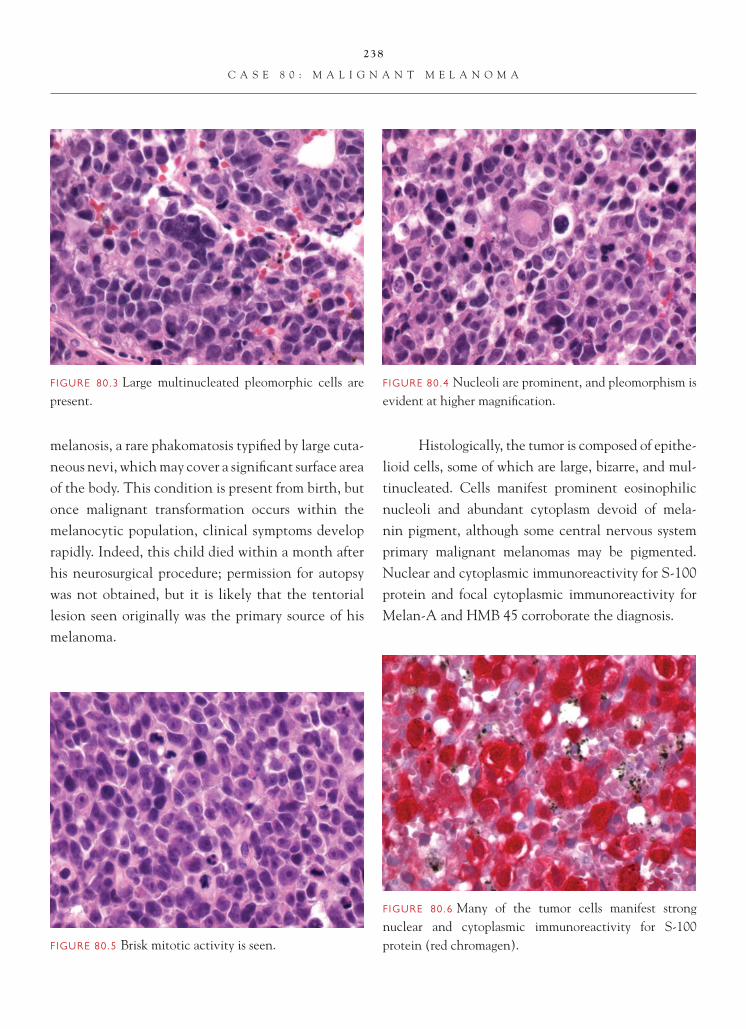
238
C A S E 8 0 : M A L I G N A N T M E L A N O M A
melanosis, a rare phakomatosis typifi ed by large cuta-neous nevi, which may cover a signifi cant surface area of the body. This condition is present from birth, but once malignant transformation occurs within the melanocytic population, clinical symptoms develop rapidly. Indeed, this child died within a month after his neurosurgical procedure; permission for autopsy was not obtained, but it is likely that the tentorial lesion seen originally was the primary source of his melanoma.
Histologically, the tumor is composed of epithe-lioid cells, some of which are large, bizarre, and mul-tinucleated. Cells manifest prominent eosinophilic nucleoli and abundant cytoplasm devoid of mela-nin pigment, although some central nervous system primary malignant melanomas may be pigmented. Nuclear and cytoplasmic immunoreactivity for S-100 protein and focal cytoplasmic immunoreactivity for Melan-A and HMB 45 corroborate the diagnosis.
FIGURE 80.6 Many of the tumor cells manifest strong nuclear and cytoplasmic immunoreactivity for S-100 protein (red chromagen).FIGURE 80.5 Brisk mitotic activity is seen.
FIGURE 80.4 Nucleoli are prominent, and pleomorphism is evident at higher magnifi cation.
FIGURE 80.3 Large multinucleated pleomorphic cells are present.

239
C A S E 8 0 : M A L I G N A N T M E L A N O M A
FIGURE 80.7 Signifi cant numbers of tumor cells also show immunoreactivity for Melan-A.
References 1. Brat DJ, Giannini C, Scheithauer BW, et al. Primary mela-
nocytic neoplasms of the central nervous systems. Am J Surg Path 1999;23(7):745–54.
2. Brat DJ, Perry A. Melanocytic lesions. In: Louis DN, Ohgaki H, Wiestler OD, Cavenee WK, Editors. WHO Classifi cation of Tumours of the Central Nervous System. Lyon, FR: IARC Press;2007. p. 181–3.
FIGURE 80.8 HMB 45 immunoreactivity is also present.
240
Case 81: Lymphoma with First Presentation as Spinal Cord Compression
C L I N I C A L I N F O R M A T I O N
The patient is an 18-year-old male who is a very active athlete. He notes worsening leg pain and numbness, as well as spasticity in his lower extremities. A MR scan of the spine demonstrates a large mass, involv-ing thoracic cord levels T3–T6. The mass appears to be located in the epidural space posterior to the spinal cord, with spinal cord compression, and is caus-ing signal changes at T6. Emergent decompression is undertaken, with extensive tumor removal. Histologic sections are reviewed.
O P I N I O N
Sections show sheets of cytologically monotonous, closely packed, hematopoietic tumor cells, occupy-ing the epidural soft tissue space as well as adjacent
vertebral bone. Cells manifest very high nuclear-to-cytoplasmic ratios, scant cytoplasm, hyperchromatic nuclei, and moderately distinct nucleoli. Mitotic activity is brisk.
We consider the lesion to be a hematopoietic neoplasm and, after special immunostaining and fl uo-rescence in situ hybridization (FISH) studies, char-acterize it as follows: Cerebral Hemispheric Clot, Evacuation—Acute B Lymphoblastic Lymphoma/Leukemia.
C O M M E N T
Acute B lymphoblastic leukemia usually presents with anemia or thrombocytopenia; bone pain or arthralgias are common. Presentation as a mass-like lymphoma is considerably less common.
FIGURE 81.1 Intermediate-power magnifi cation appear-ance shows patternless sheets of monomorphic tumor cells with little necrosis.
FIGURE 81.2 High-power magnifi cation illustrates the high nuclear-to-cytoplasmic ratio, scant cytoplasm, nuclear hyperchromasia, and mitotic activity of the tumor cells.

241
C A S E 8 1 : L Y M P H O M A W I T H F I R S T P R E S E N T A T I O N
D I S C U S S I O N
Criteria for diagnosis and classifi cation of leukemias involving the central nervous system are identical to those utilized in other tissues in the body. In this case, the histologic features were those of a lympho-blastic leukemia/lymphoma with very high MIB-1
index of greater than 80%, strong CD10 immunos-taining in the lymphoblasts (as is usually the case in acute lymphoblastic leukemia/lymphoma, along with CD24 immunoreactivity), and moderate staining for TdT and CD34. Although another consideration was Burkitt lymphoma, with the high MIB-1 rate and CD10 immunostaining, the additional positive stain-ing for the latter two immunostains negated that con-sideration. Immunostaining for B cell markers is also seen, with reactivity for CD19 and cytoplasmic CD79a
FIGURE 81.6 A very high MIB-1 cell cycle labeling index is present.
FIGURE 81.5 High-power view of the lymphoma within the bone demonstrates excellent cytologic preservation and the relative, moderate-sized nucleoli in the tumor, contrasting it with several other types of lymphoma, including Burkitt lymphoma.
FIGURE 81.4 Low-power magnifi cation also shows that adjacent vertebral bone is involved by the lymphoma.
FIGURE 81.3 Low-power magnifi cation illustrates the involvement of fi broadipose tissue by the lymphoma, including a strip of ligament from the epidural space (left lower area).
242
C A S E 8 1 : L Y M P H O M A W I T H F I R S T P R E S E N T A T I O N
FIGURE 81.7 Strong diffuse immunoreactivity for CD10 is seen in the lymphoblasts in most cases of acute lympho-blastic leukemia/lymphoma.
almost always being present, and variable expression of CD22 and CD20. In this example, FISH studies showed no c-MYC translocation, as would have been expected in Burkitt lymphoma, and did show multi-ple gains of various chromosomes, a feature seen in B lymphoblastic leukemia/lymphoma.
The epidural space is a well-known, albeit uncommon, location for lymphomatous involve-ment, estimated to occur in less than 3% of all sys-temic lymphomas. A 10-year retrospective search in one study generated 7 patients who presented initially with back pain, incontinence, and/or lower extremity weakness. By neuroimaging studies, these patients were found to have masses necessitating neurosurgical intervention. Lymphoma types includ-ed high-grade non-Hodgkin lymphoma, B cell type (n = 4), indolent B cell lymphoma (n = 1), nodu-lar sclerosing Hodgkin lymphoma (n = 1), and plas-macytoma (n = 1). Advanced disease (stage 4) was subsequently identifi ed in all 7 patients. Despite this, survival varied greatly with therapy, from 3 weeks to almost 6 years, underscoring the need for correct classifi cation of the lymphoma in order to optimize chemotherapeutic choices. The epidural space was
the site of presentation of disease in 4% of all lym-phomas diagnosed at our institution.
Epidural lymphomas are systemic lymphomas that present in a critical anatomic location that often requires emergent surgical decompression before a full systemic work-up can be undertaken. The lymphoma-tous diagnosis may be suspected but is often not proven by tissue biopsy at the time of the neurosurgical pro-cedure. Epidural lymphomas are distinct from both primary central nervous system lymphomas and from primary dural (MALT) lymphomas. Thus, a full work-up for lymphoma classifi cation should be undertaken on tissue received from the epidural space. Usually, adjacent bony involvement is also identifi ed. In some instances, this tissue received from the neurosurgical procedure may be the optimal or only tissue available from the patient on which to perform the work-up for classifi cation of the lymphoma. Therefore, it is impor-tant to make the diagnosis of lymphoma/leukemia at the time of intraoperative consultation to allow opti-mal triaging of tissue for the appropriate studies.
References 1. Brunning RD, Borowitz, M, Matutes E, et al. Precursor B
lymphoblastic leukaemia/lymphoblastic lymphoma. In: ES
FIGURE 81.8 Patchy immunoreactivity for CD20 is present in this case.

243
C A S E 8 1 : L Y M P H O M A W I T H F I R S T P R E S E N T A T I O N
Jaffe, NL Harris, H Stein, JW Vardiman, Editors. WHO Classifi cation of Tumours of Haematopoietic and Lymphoid Tissues. Lyon, FR: IARC Press;2001. p. 111–14.
2. Chahal S, Lagera JE, Ryder J, et al. Hematological neoplasms with fi rst presentation as spinal cord compression syndromes: a 10-year retrospective series and review of the literature. Clin Neuropathol 2003;22:282–90.
3. Landis DM, Aboulafi a DM. Granulocytic sarcoma: an unusual complication of a leukemic myeloid leukemia causing spinal cord compression. A case report and literature review. Leukemia & Lymphoma 2003;44:1753–60.

244
Case 82: Marginal Zone B-Cell Lymphoma
C L I N I C A L I N F O R M A T I O N
The patient is a 44-year-old woman who presents with a dural-based mass, involving the falx and overlying the right frontal convexity. The patient presents with headaches and episodic nausea and vomiting. On imaging, the lesion appears somewhat circumscribed, with contrast enhancement, and a diagnosis of menin-gioma is entertained. Gross total resection of the lesion is performed, and histologic sections are reviewed.
O P I N I O N
Histologic sections show a cellular lesion, marked by mixed infl ammatory infi ltrates, involving the dura and underlying leptomeninges. Sheets of predominantly small to medium-sized lymphocytes with a moder-ate amount of cytoplasm and nuclear irregularities are
observed. Some cells show a monocytoid appearance, with cleared cytoplasm and well-defi ned cell borders. Occasional immunoblastic-like cells, with large vesic-ular nuclei and prominent nucleoli, are present, as are scattered plasma cells. On low magnifi cation, the infl ammatory cells appear to be arranged in a nodular architectural pattern. Prominent numbers of mitotic fi gures are not evident. Focally, microcalcifi cations are noted, as are cholesterol clefts with giant cells, the latter indicating previous hemorrhage. Granulomas are not seen. There is no evidence of meningioma. By immu-nohistochemistry, the tumor cells demonstrate positive staining, with antibodies to CD20 and CD79a, and do not stain with antibodies to CD3, CD10, and CD5. Bcl-2 immunopositivity is also observed in the lesion.
We consider the lesion to be a lymphoprolifera-tive disorder and characterize it as follows: Falx and
FIGURE 82.1 Low-magnifi cation appearance of the dural mass shows a nodular architectural pattern.
FIGURE 82.2 Focal cholesterol cleft formation, with giant cells indicating previous hemorrhage in the lesion, is observed.

245
C A S E 8 2 : M A R G I N A L Z O N E B - C E L L L Y M P H O M A
Right Frontal Convexity, Excision—Marginal Zone B-Cell Lymphoma.
C O M M E N T
Marginal zone B-cell lymphomas in the central ner-vous system are typically indolent tumors that often present as meningioma-like, dural-based masses.
D I S C U S S I O N
Diffuse, large B-cell lymphomas are the most common primary central nervous system lympho-mas. Other lymphoma types have been less fre-quently observed to arise primarily in the central nervous system; among this group is the marginal zone B-cell lymphoma or low-grade B-cell lym-phoma of mucosa-associated lymphoid tissue (MALT). The lesion probably represents the most common primary low-grade intracranial lymphoma. Most of these tumors arise in association with the dura and leptomeninges, although rare cases of intracerebral and intraventricular tumor have been reported. Because of their dural location, imaging studies suggest meningioma and prompt surgical resection. In a series reported by Tu et al., 80% of patients were woman with a mean age at diagnosis of 55 years. Presenting symptoms varied and included headaches, nausea, vomiting, visual disturbances, seizures, motor or sensory defi cits, ataxia, or gait disturbance.
Histologically, the lesions are marked by proliferation of small lymphocytes, with cleared
FIGURE 82.5 High-magnifi cation appearance of cells shows irregular nuclear contour with prominent nuclei and cleared cytoplasm.
FIGURE 82.4 Intermediate magnifi cation shows a prolif-eration of small lymphocytic cells, with moderate amount of lightly eosinophilic to cleared cytoplasm, comprise the tumor.
FIGURE 82.3 Focal calcifi cations, likely representing entrapped psammoma bodies, are present.

246
C A S E 8 2 : M A R G I N A L Z O N E B - C E L L L Y M P H O M A
FIGURE 82.6 Diffuse positive staining with antibody to CD20 (B-cell lymphoid marker) is present.
FIGURE 82.7 The tumor demonstrates positive staining with Bcl-2 antibody.
cytoplasm and irregular, centrally located nuclei. Plasmacytic features are variably present. Focal areas of nodular architecture and areas marked by lym-phoid follicles may be present. A subset of tumors may demonstrate evidence of amyloid deposition. Similar to marginal zone B-cell lymphomas arising outside the central nervous system, these tumors demonstrate positive staining with B-cell markers (CD20 and CD79a) and do not stain with T-cell markers (CD3).
These tumors tend to have a long-term survival and favorable prognosis.
References 1. Kelley TW, Prayson RA, Barnett GH, et al. Extranodal
marginal zone B-cell lymphoma of mucosa-associated lym-phoid tissue arising in the lateral ventricle. Leuk Lymphoma 2005;46:1423–7.
2. Tu PH, Giannini C, Judkins AR, et al. Clinicopathologic and genetic profi le of intracranial marginal zone lymphoma: a pri-mary low grade CNS lymphoma that mimics meningioma. J Clin Oncol 2005;23:5718–27.
247
Case 83: Post-Transplant Lymphoproliferative Disorder
C L I N I C A L I N F O R M A T I O N
The patient is a 65-year-old male who presents with an approximately 2-month history of progressive short-term memory loss and problems with visual-spatial and executive functions. He has a signifi cant past medical history of having received a kidney trans-plant 3 years earlier. A medical opinion is sought, and a MRI scan demonstrates multifocal lesions in the brain, the largest of which involve the posterior corpus callosum and right parieto-occipital areas. A stereotactic biopsy of the corpus callosum lesion is undertaken, and sections are available for review.
O P I N I O N
Sections show a cytologically atypical hematopoeitic neoplasm, characterized by large immunoblast-like
cells, with vesicular nuclei, prominent nucleoli, and amphophilic cytoplasm surrounding blood vessels. Similar cells are also present in brain parenchyma and associated with even larger numbers of small, non-neoplastic lymphocytes of normal size. Necrosis and brisk astrocytic reaction are also identifi ed. The large cytologically atypical cells mark as B cells with CD20, whereas small non-neoplastic lymphocytes are predominantly CD3-positive T cells. Epstein-Barr virus is detected by in situ hybridization.
We consider the lesion to be a hematopoietic neoplasm and, after immunostaining and polymerase chain reaction (PCR) testing for clonality, characterize it as follows: Posterior Corpus Callosum, Stereotactic Biopsies—Post-Transplant Lymphoproliferative Dis-order, B-Cell Lymphoma, Epstein-Barr Virus-Positive, Monoclonal B-Cell Population Detected by PCR.
FIGURE 83.1 The hypercellular lesion shows perivascular collections of large, cytologically atypical cells, admixed with variable numbers of small, non-neoplastic lympho-cytes and occasional plasma cells.
FIGURE 83.2 High-power magnifi cation illustrates the vesicular nuclei and prominent nucleoli in the hematopoe-itic tumor, as well as mitoses.

248
C A S E 8 3 : P O S T - T R A N S P L A N T L Y M P H O P R O L I F E R A T I V E D I S O R D E R
C O M M E N T
Most post-transplant lymphoproliferative disorders (PTLDs) are driven by Epstein-Barr virus (EBV), and most are B-cell disorders, existing in a spectrum from polyclonal proliferations to monomorphic B-cell lymphomas. Diffuse, large B-cell lymphomas are usually immunoblastic in type. Far less commonly,
anaplastic lymphoma, T-cell lymphoma, plasma cell myeloma, Hodgkin lymphoma, and Hodgkin-like PTLD have been described.
D I S C U S S I O N
PTLD occurs in <2% of solid organ transplant recipients, of which the lowest frequency is in renal allograft recipients, with increasing frequency of PTLD in those who have undergone hepatic, car-diac, or complex heart-lung or liver-bowel allografts. PTLD tends to involve extranodal sites, including the transplanted organ itself and the central nervous sys-tem (CNS), especially if the patient was treated with azathioprine-based regimens. The use of tacrolimus-based regimens for immunosuppression has reduced the involvement of the CNS.
Most examples of PTLD in the CNS meet mor-phologic criteria for a diagnosis of diffuse, large B-cell lymphoma, based on the hypercellularity and cyto-logic atypia. In brain, tumor cells usually show both perivascular arrangement and sheet-like effacement of the parenchyma, with variable degrees of associated
FIGURE 83.5 There may be broad zones of necrosis that prompt consideration of an infectious process, especially infections more frequently seen in immunosuppressed patients, including fungal infection or toxoplasmosis.
FIGURE 83.4 Some perivascular cuffs of cells consist pre-dominantly of non-neoplastic lymphocytes (which prove to be T cells), with only a few recognizable, larger neoplas-tic B cells.
FIGURE 83.3 Tumor cells can be seen within vessel walls, but unlike vasculitis, the hematopoeitic cells manifest severe cytologic atypia.

249
C A S E 8 3 : P O S T - T R A N S P L A N T L Y M P H O P R O L I F E R A T I V E D I S O R D E R
FIGURE 83.6 CD20 identifi es the large neoplastic cells as being of B-cell origin.
FIGURE 83.7 In some areas of the biopsy, only isolated B cells are seen on CD20 staining, and even some smaller, cytologically normal-appearing B cells can be admixed.
necrosis. Because of the vasocentricity of the lympho-ma cells and the accompanying non-neoplastic T cells, vasculitis must be excluded, based on the severity of the cytologic atypia in the hematopoeitic population. The additional fi nding of a clonal B-cell tumor population on PCR testing supports the diagnosis of B-cell lympho-ma in a cytologically atypical process. Heavily necrotic cases of diffuse B-cell lymphoma can also raise consid-
eration of infectious process, especially fungal infection or toxoplasmosis of the CNS. Like most infections, these tend to have a preponderance of T cells rather than B cells, and staining for organisms is positive.
Immunostaining in PTLD-associated, EBV-driven, diffuse, large B-cell lymphomas usually shows positive expression of CD19, CD20, and CD79a. CD43 and CD45RO are also upregulated
FIGURE 83.8 Large numbers of non-neoplastic T cells often cuff the blood vessels and infi ltrate the parenchyma; these CD3-positive cells may overshadow the true, neoplastic, CD20-positive cells.
FIGURE 83.9 In situ hybridization for Epstein-Barr virus usually shows large numbers of EBV-positive cells with strong, blue-black, nuclear signal.

250
C A S E 8 3 : P O S T - T R A N S P L A N T L Y M P H O P R O L I F E R A T I V E D I S O R D E R
in EBV- infected B cells and can be expressed. Many are CD30-positive, even without anaplastic features. Reduction or withdrawal of immunosuppressive med-ications, with administration of chemotherapy or ra-diation therapy, can improve prognosis in some cases.
References 1. Harris NL, Swerdlow SH, Frizzera G, et al. Post-transplant
lymphoproliferative disorders. In: ES Jaffe, NL Harris, H Stein, JW Vardiman, Editors. WHO Classifi cation of Tumours of Haematopoietic and Lymphoid Tissues. Lyon, FR: IARC Press;2001. p. 264–9.

251
Case 84: Plasmacytoma
C L I N I C A L I N F O R M A T I O N
The patient is a 78-year-old male who presents with fl uctuating vision in his right eye. A MRI scan shows a dural-based mass, just superior and lateral to the optic foramen, suggestive of a meningioma. Work-up, how-ever, reveals lytic rib and sacral lesions. The patient is felt to be a candidate for decompression of his right optic nerve, and histologic sections are reviewed from the resected material.
O P I N I O N
Sections show sheets of cytologically monotonous, closely packed, hematopoietic tumor cells, occupy-ing the orbital soft tissue space. Cells manifest only moderate nuclear-to-cytoplasmic ratios, nuclei with clockwork chromatin pattern, moderate amounts of
faintly basophilic cytoplasm, and distinct nucleoli. Mitotic activity is brisk.
We consider the lesion to be a hematopoi-etic neoplasm and, after special immunostaining, characterize it as follows: Orbital Dura, Biopsy— Plasmacytoma.
C O M M E N T
Plasmacytoma is a localized tumoral collection of neoplastic plasma cells. This patient, who had a plas-macytoma on tissue biopsy (a major diagnostic crite-rion for plasma cell myeloma) and also had lytic bone lesions (a minor diagnostic criterion), met criteria for diffuse disease (plasma cell myeloma). He was subse-quently also found to have an M- component in the
FIGURE 84.1 Touch imprint, performed at the time of intra-operative consultation, shows that the cells in this lesion display cytologic monotony and abundant cytoplasm.
FIGURE 84.2 High-power magnifi cation illustrates the monotonous sheets of tumor cells with moderate nuclear-to-cytoplasmic ratio, basophilic cytoplasm, and clumped nuclear chromatin.

252
C A S E 8 4 : P L A S M A C Y T O M A
urine and serum (free kappa light chains) (another major criterion) and reduced normal serum immuno-globulins (a minor criterion).
D I S C U S S I O N
Making the diagnosis of plasma cell myeloma involves fi nding systemic body involvement, in addition to the localized tumoral collection of plasma cells seen on this biopsy. Plasmacytoma can be mistaken for a
metastatic carcinoma, especially breast carcinoma, thyroid carcinoma, or other relatively monotonous tumor type, such as a neuroendocrine tumor, includ-ing pituitary adenoma. Cytokeratin expression in these tumors verifi es the diagnosis of metastatic carcinoma and negates the diagnosis of plasmacy-toma. Plasmacytoma can also be mistaken for an olfactory neuroblastoma, if the mass occurs in the sinuses or skull base. Both plasmacytoma and olfac-tory neuroblastoma can be immunoreactive for neu-ron-specifi c enolase (NSE) and CD56 (neural cell adhesion molecule, NCAM), but only the former is strongly and diffusely immunoreactive for plasma cell markers such as CD38, CD79a, or CD138.
Benign infl ammatory conditions may contain large numbers of plasma cells, especially orbital in-fl ammatory pseudotumor, and thus, they are also di-agnostic considerations. However, the cytologic and immunohistochemical monotony of the process ar-gues against a reactive process in this case. Immunos-taining or in situ hybridization studies for light chain expression negate a benign reactive process when a monoclonal light chain population is identifi ed. This case expressed kappa light chains exclusively.
FIGURE 84.5 In situ hybridization signal shows a monoclo-nal kappa light chain expression in the plasmacytoma.
FIGURE 84.4 Immunoreactivity for CD138 is strong and diffuse and negates consideration of a metastatic carcinoma.
FIGURE 84.3 High-power magnifi cation shows the promi-nent nucleoli and mitotic activity in this tumor.

253
C A S E 8 4 : P L A S M A C Y T O M A
FIGURE 84.6 In situ hybridization signal shows no cells expressing lambda light chain expression in the plasmacytoma.
FIGURE 84.7 As is typically the case in most (but not all) plasma cell neoplasms, this plasmacytoma, as fi rst presen-tation of plasma cell myeloma, lacks expression for the mature B-cell marker CD20.
The orbital space is a known location for plasma cell myeloma or isolated plasmacytoma. Cranial or spinal dura in other anatomic locations in addition to the orbit is also a known location for plasmacytoma, as well as for various types of low-grade indolent dural lymphoma, including MALT lymphomas.
References 1. Grogan TM, Van Camp B, Kyle RA, et al. Plasma cell
neoplasms. In: Jaffe ES, Harris NL, Stein H, Vardiman JW, Editors. WHO Classifi cation of Tumours of Haematopoietic and Lymphoid Tissues. Lyon, FR: IARC Press;2001. p. 142–156.
2. Haegelen C, Riffaud L, Bernard M, et al. Dural plasmacy-toma revealing multiple myeloma. Case report. J Neurosurg 2006;104:608–10.
3. Lazaridou MN, Micallef-Eynaud P, Hanna IT. Soft tissue plasmacytoma of the orbit as part of the spectrum of multiple myeloma. Orbit 2007;26:315–18.
4. Sahin F, Saydam G, Ertan Y, et al. Dural plasmacytoma mim-icking meningioma in a patient with multiple myeloma. J Clin Neurosci 2006;13:259–61.
5. Thoumazet F, Donnio A, Ayeboua L, et al. Orbital and mus-cle involvement in multiple myeloma. Can J Ophthalmol 2006;41:733–6.

254
Case 85: Langerhans Cell Histiocytosis
C L I N I C A L I N F O R M A T I O N
The patient is a 9-year-old girl, otherwise healthy, who presents with headaches, occurring over a 2-month period. Neurologic evaluation is normal. MRI reveals a skull-centered mass, extending though the right fronto-parietal skull bone into the epidural space, with focal displacement of the brain in the area of the lesion, but without associated edema. Only a single lytic lesion is identifi ed. The decision is made to excise the skull lesion.
O P I N I O N
Biopsies show a hypercellular tumor, composed of a heterogeneous population of cells, including eosino-phils, neutrophils, large multinucleated histiocytes resembling osteoclasts, and a predominant population of medium-sized histiocytes with grooved, folded, or
lobulated nuclei. The latter Langerhans cells mani-fest inconspicuous nucleoli, delicate chromatin, and minimal hyperchromasia. Cytoplasm is devoid of prominent vacuoles, and mitoses are rare.
We consider the pathology to be that of a his-tiocytic neoplasm without malignant features and, after immunohistochemical studies, characterize it as follows: Right Fronto-Parietal Skull, Excision—Langerhans Cell Histiocytosis.
C O M M E N T
Langerhans cell histiocytosis was referred to in the past as histiocytosis X or Langerhans cell granulomatosis.
D I S C U S S I O N
Langerhans cell histiocytosis (histiocytosis X) is one of three overlapping syndromes, all with similar
FIGURE 85.1 The section shows a hypercellular tumor, with a heterogeneous population of hematopoeitic elements.
FIGURE 85.2 Small eosinophilic abscesses should not prompt misdiagnosis of a true granulomatous or parasitic infection.
255
C A S E 8 5 : L A N G E R H A N S C E L L H I S T I O C Y T O S I S
histologic features, but with the various forms of the disorder defi ned by the anatomic distribution pattern.
Langerhans cell histiocytosis is the unifocal form of the disease and is usually diagnosed in older children or adults. Bone is the most frequently af-fected site, particularly the skull (as in this case), but single lesions may also be found in femur, pelvic bones, or ribs.
In Hand-Schüller-Christian disease, there is multifocal involvement, but the multiple lesions are
confi ned to a single organ system—again, almost always the bone.
Letterer-Siwe disease represents the multifocal, multisystem form of involvement and usually is diag-nosed in infants. It may manifest in bones, skin, liver, spleen, and/or lymph nodes.
Prognosis is correlated with the number of organs involved. Although about 10% of patients
FIGURE 85.6 High-power view illustrates various elements in Langerhans cell histiocytosis: multinucleated cells, eosinophils, Langerhans cells with nuclear grooves, and neutrophils.
FIGURE 85.5 Multinucleated histiocytes may be prominent and resemble osteoclasts.
FIGURE 85.4 Langerhans cells manifest folded, indented, and grooved nuclei, delicate chromatin, and small nucleoli.
FIGURE 85.3 High magnifi cation illustrates the prominent eosinophils scattered throughout the lesion.

256
C A S E 8 5 : L A N G E R H A N S C E L L H I S T I O C Y T O S I S
FIGURE 85.7 CD1a immunoreactivity is consistently iden-tifi ed in Langerhans cell histiocytosis within the Langer-hans cell population.
with unifocal disease progress to multisystem disease, even patients with multiple bone lesions have a good prognosis. Overall survival of patients with unifocal disease is 95%.
Histologically, diagnosis rests on identifying the characteristic Langerhans cells, a special type of his-tiocyte, admixed with variable numbers of eosinophils, neutrophils, and multinucleated histiocytes. Small eo-sinophilic abscesses can be found in some cases, and such cases should not be mistaken for true infections, such as parasitic and granulomatous abscesses.
Early phases of the condition contain larger numbers of Langerhans cells, whereas older phases show fewer of these cells, with more fi brosis and more foamy macrophages. The identity of the Langerhans cells should be confi rmed by immunohistochemical studies. Langerhans cells show “consistent expression of CD1a and S-100 protein. They are usually posi-tive for vimentin, HLA-DR, peanut agglutinin lectin and placental alkaline phosphatase. They are variably
and weakly positive for CD45, CD68, and lysozyme. They are negative for most B-cell and T-cell lineage markers.”
Electron microscopy is less often necessary and may be negative because of Birbeck granules being present in a variable percentage of the Langerhans cells. Birbeck granules are rod-, to fl ask-, to tennis racket-shaped structures that show a median striated line. They are thought to represent a special type of endocytotic structure.
References 1. Weiss LM, Grogan TM, Müller-Hermelink H-K, et al.
Langerhans cell histiocytosis. In: Jaffe ES, Harris NL, Stein H, Vardiman JW, Editors. WHO Classifi cation of Tumours: Pathology and Genetics. Tumours of Haematopoietic and Lymphoid Tissues. Lyon, FR: IARC Press;2001. p. 280–2.
2. Ghadially FN. Diagnostic Ultrastructural Pathology: A Self-Evaluation and Self-Teaching Manual. Second Edition. Boston, MA: Butterworth-Heinemann;1998. p. 17.
FIGURE 85.8 Electron microscopy shows several intracyto-plasmic Birbeck granules, elongate, rodlike structures. The distal distention, forming a “tennis racket,” is not always identifi ed and is not present within the granules in this cell, although a median striated line can be seen clearly.

257
Case 86: Intravascular Lymphomatosis (Angiotropic Large Cell Lymphoma)
C L I N I C A L I N F O R M A T I O N
The patient is a 72-year-old female who presents with right-sided weakness; on imaging, she is noted to have a left-sided infarct. Over the next 2-week period, she develops multiple neural defi cits, and on imaging, shows six foci of infarcts in multiple vascular distribu-tions. A vasculitic process is suspected. A biopsy is performed, and histologic sections are reviewed.
O P I N I O N
Histologic sections show an intravascular process, marked by the presence of discohesive, atypical cells with high nuclear-to-cytoplasmic ratios. The adja-cent parenchyma shows evidence of an acute infarct, reactive vascular endothelial changes, macrophages, and ischemic-appearing neurons. The atypical cells are confi ned to vascular lumina. On immunostaining,
the intravascular cells demonstrate positive staining with CD20 antibody. These cells do not stain with antibodies to CD3, muramidase, TdT, cytokeratins AE1/3, or S-100 protein.
We consider the intravascular lesion to be a hematopoietic neoplasm and characterize it as follows: Left Parietal Lobe Region, Biopsy—Intravascular Lymphomatosis (Angiotropic Large Cell Lymphoma).
C O M M E N T
On immunostaining, the tumor cells demonstrates CD20 immunoreactivity, consistent with a B-cell lymphomatous process.
D I S C U S S I O N
Intravascular lymphomatosis is a relatively rare dis-order, marked by a proliferation of atypical lymphoid
FIGURE 86.1 Meningeal vessels, show atypical-appearing intravascular lymphoid cells.
FIGURE 86.2 White matter vessels are also involved by the lymphomatous process.

258
C A S E 8 6 : I N T R A V A S C U L A R L Y M P H O M A T O S I S
cells, confi ned primarily to vascular lumina. The clinical presentation is often quite variable, depend-ing on the organ involved. Neurologic symptoms include focal neural defi cits or dementia in a subset of patients. The characteristic intravascular growth pattern of this tumor has been conjectured to be related to a defect in homing receptors on neoplastic tumor cells. Morphologically, the cells show features
of atypical lymphoid cells, which can be confi rmed by immunostaining. The majority of the cases have a B-cell immunophenotype; however, rare cases of intravascular lymphoma of T-cell phenotype have also been reported.
Differential diagnostic considerations include other processes that may have an intravascular lo-cation, including leukemia, intravascular spread from a carcinoma, or metastasizing melanoma. The presence of negative immunostains characteristic of
FIGURE 86.6 CD3 immunoreactivity is not present in this tumor.
FIGURE 86.5 Most of the tumor cells demonstrate posi-tive staining, with antibody to CD20 (B-cell lymphoid marker).
FIGURE 86.4 An area of acute infarct is seen, likely second-ary to the intravascular tumor.
FIGURE 86.3 High-magnifi cation appearance of the atypical-appearing lymphoid cells shows a high nuclear- to-cytoplasmic ratio.

259
C A S E 8 6 : I N T R A V A S C U L A R L Y M P H O M A T O S I S
these various entities helps exclude them from the diagnosis.
The lesion has a particularly aggressive clini-cal course and does not usually respond well to che-motherapy. In many patients, because of the atypical presentations, the diagnosis is often not made until the time of autopsy.
References 1. Baehring JM, Longtine J, Hochberg FH. A new approach
to the diagnosis and treatment of intravascular lymphoma. J Neurooncol 2003;61:237–48.
2. Glass J, Hochberg FH, Miller DC. Intravascular lymphomato-sis. A systemic disease with neurologic manifestations. Cancer 1997;71:3156–64.
3. Ponzoni M, Ferreri AJ. Intravascular lymphoma: a neoplasm of ‘homeless’ lymphocytes? Hematol Oncol 2006;24:105–12.

260
Case 87: Germinoma
C L I N I C A L I N F O R M A T I O N
The patient is a 12-year-old male who presents with respiratory failure of unknown etiology and is found to have aspiration pneumonia. Neurologic evaluation identifi es nystagmus, dysmetria, wasting and fascicu-lations of the tongue, and voice changes suggestive of vocal cord paralysis. MRI shows an enhancing exo-phytic mass, 5.1 × 3.2 × 2.2 cm, in the medulla, as well as ventriculomegaly. MRI of the spine is negative for cerebrospinal fl uid dissemination. Given the site and enhancement characteristics, astrocytoma is favored. The patient subsequently undergoes excisional biopsy of the lesion, and histologic sections are reviewed.
O P I N I O N
Sections demonstrate sheets of large tumor cells, with round vesicular nuclei and prominent nucleoli. Some
areas are patternless, whereas, in other areas, a lobular arrangement is formed by delicate fi brovascular septae fi lled with non-neoplastic lymphocytes. Mitoses are easily appreciated in the large tumor cells. Necrosis is present in this example but is not commonly seen. No other germ cell tumor elements are seen.
We consider the lesion to be a germ cell tu-mor and characterize it as follows: Dorsal Brainstem/Pineal Region, Biopsies—Germinoma.
C O M M E N T
Most central nervous system primary germinomas develop in either the suprasellar region or pineal gland. This case is unusual in that it was an exophytic mass in the medulla, a much more common site for astrocytomas than germ cell tumors.
FIGURE 87.1 Low-power magnifi cation appearance of the excision specimen, shows patternless sheets of large cells.
FIGURE 87.2 Other areas in the resected material demon-strate lobules of tumor cells, demarcated by fi brovascular septae containing cytologically benign lymphocytes.

261
C A S E 8 7 : G E R M I N O M A
D I S C U S S I O N
Germinoma is the most common central nervous sys-tem germ cell tumor and often occurs in pure form. Large tumor cells can be arranged in patternless sheets or may be punctuated by cords of fi brovascular tissue, yielding a nested or lobular architectural pattern. Variable numbers of non-neoplastic lymphocytes may be present and are usually T cells. Occasional cen-tral nervous system germinomas show prominent
granulomatous reaction, simulating a tuberculoma or neurosarcoidosis. Granulomatous response may over-shadow the tumor cells, making diagnosis diffi cult.
Although mitoses are easily seen in well-fi xed specimens, necrosis is uncommon. Immunohisto-chemical staining aids in the diagnosis. Cytoplasmic immunoreactivity for c-kit and nuclear immunoreac-tivity for OCT4 are often seen, with cytoplasmic and cell membrane immunostaining for placental alkaline phosphatase (PLAP) slightly less frequent. NANOG has been shown in some studies to be another useful immunohistochemical marker for central nervous system germinoma.
References 1. Hattab EM, Pu PH, Wilson JD, et al. OCT4 immunohis-
tochemistry is superior to placental alkaline phosphatase (PLAP) in the diagnosis of central nervous system germi-noma. Am J Surg Pathol 2005;29:369–71.
2. Rosenblum MK, Nakazato Y, Matsutani M. CNS germ cell tumours. In: Louis DN, Ohgaki H, Wiestler OD, Cavenee WK, Editors. WHO Classifi cation of Tumours of the Central Nervous System. Lyon, FR: IARC Press;2007. p. 198–204.
3. Santagata S. Hornick JL. Ligon KL. Comparative analysis of germ cell transcription factors in CNS germinoma reveals diagnostic utility of NANOG. J Surg Pathol 2006;30:1613–8.
FIGURE 87.5 Cytoplasmic and membranous immuno- staining for PLAP is seen in many, but not all, examples of central nervous system germinomas.
FIGURE 87.4. High-power magnifi cation shows the large vesicular nuclei, prominent nucleoli, and mitoses in the neoplasm.
FIGURE 87.3 Small foci of necrosis are evident in this example of central nervous system germinoma.

262
Case 88: Pineal Teratoma
C L I N I C A L I N F O R M A T I O N
The patient is a 19-year-old male who presents with a 1.5-week history of double vision, headaches, nau-sea, and vomiting. On imaging, a pineal region mass, 4.1 × 2.7 × 3.4 cm, and hydrocephalus are found, and an emergent intraventricular shunt is placed. A decision is made to perform a small endoscopic biopsy, which yields a tumor composed of small blue cells. Pineoblastoma, versus immature nervous sys-tem tissue within an immature teratoma, cannot be distinguished on the limited material available. He is treated with radiation and chemotherapy. Subse-quently, there is enlargement of his pineal region mass, with a more complex appearance. A larger resection is undertaken, and histologic sections are reviewed.
O P I N I O N
The larger resection shows mature cartilage, loose connective tissue, central and peripheral nervous sys-tem tissue, fat and bone. No germ cell tumor or imma-ture nervous system tissue is present on the second surgical procedure.
We consider the lesion to have originally been an immature teratoma, now with maturation after radio- and chemotherapy, and characterize it as follows: Pineal Gland—Teratoma, Mature.
C O M M E N T
Immature teratomas may undergo “maturation” following use of radio- or chemotherapy. In retrospect, the “small blue cell tumor” came from immature, actively proliferating neuroectodermal tissue in the mass. The lesion was never a pineoblastoma. After
FIGURE 88.1 Lower-magnifi cation appearance of the resected material shows mature fat and connective tissue. FIGURE 88.2 Cartilage is often abundant in teratomas.

263
C A S E 8 8 : P I N E A L T E R A T O M A
the mitotically active portion of the immature tera-toma was ablated by the therapy, the residual, more differentiated portions remained and enlarged over time, with maturation of tissues and accumulation of cystic contents. This phenomenon is known as the “growing teratoma syndrome.”
D I S C U S S I O N
Teratomas of the central nervous system may be either mature, immature, or rarely, they may contain a true
malignant component. In mature teratomas, skin, brain, and choroid plexus tissues may be seen. Meso-dermal tissues include cartilage, bone, fat, and muscle. Cysts may be present and lined by respiratory or enteric type epithelium. Immature teratomas, in contrast, show hypercellular and mitotically active stoma, suggestive of embryonic mesenchymal and/or primitive neuroec-todermal elements. The latter may simulate neural
FIGURE 88.6 Mature nervous system tissue shows eosino-philic fi brillar background and easily recognized astrocytes. Note absence of immature neuroepithelium.
FIGURE 88.5 Bone can be easily identifi ed in many teratomas.
FIGURE 88.4 Cysts with squamous epithelium, keratohya-line layer, and abundant squamous cells simulate epider-moid cyst.
FIGURE 88.3 Cystic areas can be lined by fl attened, respiratory-type epithelium and fi lled with mucous contents.
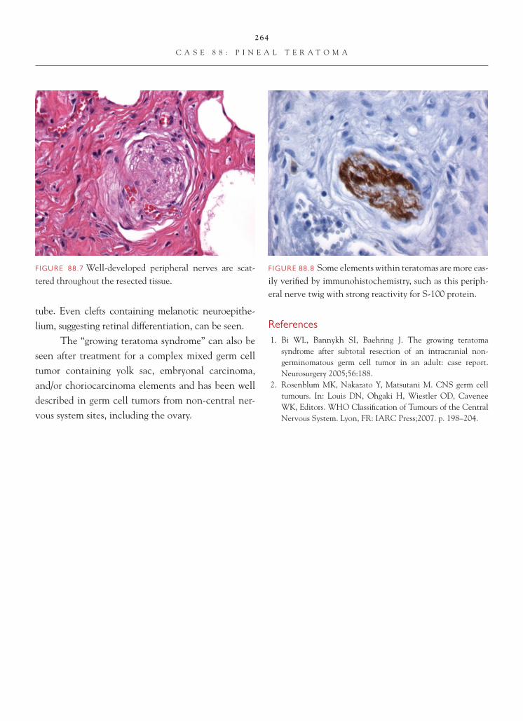
264
C A S E 8 8 : P I N E A L T E R A T O M A
tube. Even clefts containing melanotic neuroepithe-lium, suggesting retinal differentiation, can be seen.
The “growing teratoma syndrome” can also be seen after treatment for a complex mixed germ cell tumor containing yolk sac, embryonal carcinoma, and/or choriocarcinoma elements and has been well described in germ cell tumors from non-central ner-vous system sites, including the ovary.
References 1. Bi WL, Bannykh SI, Baehring J. The growing teratoma
syndrome after subtotal resection of an intracranial non-germinomatous germ cell tumor in an adult: case report. Neurosurgery 2005;56:188.
2. Rosenblum MK, Nakazato Y, Matsutani M. CNS germ cell tumours. In: Louis DN, Ohgaki H, Wiestler OD, Cavenee WK, Editors. WHO Classifi cation of Tumours of the Central Nervous System. Lyon, FR: IARC Press;2007. p. 198–204.
FIGURE 88.8 Some elements within teratomas are more eas-ily verifi ed by immunohistochemistry, such as this periph-eral nerve twig with strong reactivity for S-100 protein.
FIGURE 88.7 Well-developed peripheral nerves are scat-tered throughout the resected tissue.

265
Case 89: Cystic Craniopharyngioma
C L I N I C A L I N F O R M A T I O N
The patient is a 36-year-old male who presents with blurry and double vision, as well as headaches, nau-sea, fatigue, and decreased libido. Neuroimaging stud-ies show a cystic suprasellar and sellar mass, 2.3 by 1.6 cm. Near-total surgical resection is undertaken.
O P I N I O N
Sections show a collapsed, fi brotic cyst wall and a cyst lining that varies from an attenuated squamous epithelium to a more obvious adamantinomatous appearance. Fibrotic nodules containing cholesterol clefts are evident in some areas. Calcifi cation and islands of ghost cells (“wet keratin”) are noted.
We consider the lesion to be a cystic craniopha-ryngioma and characterize it as follows: Suprasellar
Region, Resection—Cystic Craniopharyngioma, WHO Grade I.
C O M M E N T
All symptoms can be attributed to optic chiasm com-pression and hypopituitarism, with hypogonadism leading to decreased libido and hypoadrenalism with nausea and fatigue.
D I S C U S S I O N
Craniopharyngiomas account for 2–5% of all primary intracranial neoplasms and have a bimodal peak age incidence: in children ages 5–14 years and in adults ages 50–74 years. These tumors arise along the path-way followed during embryological periods by the craniopharyngeal duct before it involutes. Most have a suprasellar component.
FIGURE 89.1 Low magnifi cation shows collapsed cyst wall; complexity and cyst contents may not be fully apparent on the specimen received by the pathologist.
FIGURE 89.2 Adamantinomatous epithelium and squamous epithelium can be seen lining the cyst.

266
C A S E 8 9 : C Y S T I C C R A N I O P H A R Y N G I O M A
Although craniopharyngiomas are WHO grade I neoplasms, their location near the hypothalamus often precludes gross total removal. Many patients require postoperative adjuvant radiation therapy to achieve tumor control. Histologically, almost all
pediatric, and most adult, craniopharyngiomas possess calcifi cation and adamantinomatous epithelium. The latter is recognized by intersecting strands and islands of epithelium, with a palisaded basal layer of cells; an intermediate stellate reticulum, which may loosen and degenerate, forming cysts with watery cyst fl uid con-tents; and nodules of “wet keratin.” The wet keratin has the appearance of squamous cells, but with loss of
FIGURE 89.6 Nodules of cholesterol clefts, embedded in a fi brotic background, indicate previous, often subclinical bleeding into craniopharyngiomas.
FIGURE 89.5 Adamantinomatous epithelium is character-ized by a crowded basal layer of cells; loosening within the epithelium with fl uid accumulation may also give rise to cysts in the craniopharyngioma.
FIGURE 89.4 Calcifi cation and clusters of ghost cells characterize most adamantinomatous and cystic cranio-pharyngiomas.
FIGURE 89.3 The attenuated epithelium lining the cyst in some areas is best appreciated by immunostaining for keratins, in this instance CAM 5.2. Note the squamous appearance to the epithelium in this area and the nonim-munoreactive chronic infl ammatory cells in the underlying stroma.

267
C A S E 8 9 : C Y S T I C C R A N I O P H A R Y N G I O M A
hematoxylin staining, leaving eosinophilic outlines of the nucleus. Dystrophic calcifi cation occurs within these nodules of “ghost cells” (wet keratin). Degen-eration of the wet keratin yields the machine oil con-tent in some of the craniopharyngioma cysts. Finding nodules of wet keratin in a small biopsy of a suprasel-lar mass, even without the viable adamantinomatous epithelium, is diagnostic for craniopharyngioma.
The cholesterol debris and histiocytic reaction, in contrast, are also typical of cystic
craniopharyngioma but are only suggestive of the diagnosis of craniopharyngioma if found in isolation without the diagnostic epithelium.
References 1. Burger PC, Scheithauer BW. Tumors of the Central Nervous
System. AFIP Atlas of Tumor Pathology. Fourth series, fascicle 7. Washington DC: American Registry of Pathology;2007. p. 461–9.
2. Karavitaki N, Cudlip S, Adams CBT, Wass JAH. Craniopha-ryngiomas. Endocr Rev 2006;27:371–97.
3. Prayson RA. Non-glial tumors. In: Prayson RA, Editor. Neuropathology. Philadelphia, PA: Elsevier Publishing; 2005. p. 528–30.
FIGURE 89.8 CD68 immunostaining highlights the foreign body giant cell reaction to the cholesterol debris.
FIGURE 89.7 Higher-power magnifi cation of the cho-lesterol clefts shows the adjacent hemosiderin pigment deposition, as well as the foreign body giant cell reaction to the material.
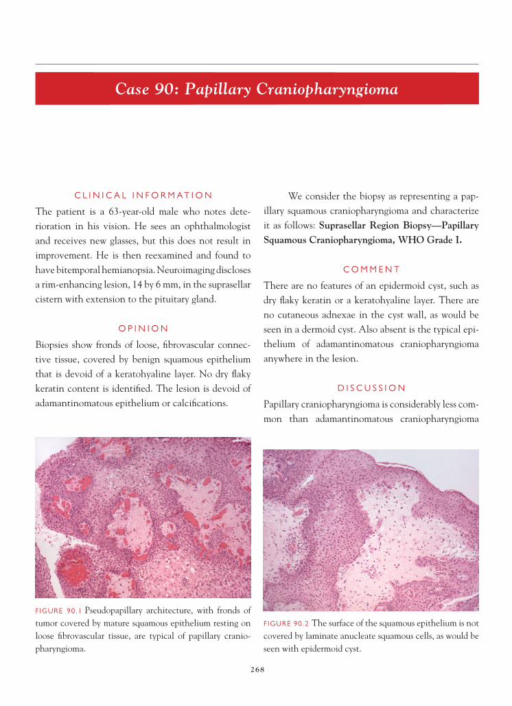
268
Case 90: Papillary Craniopharyngioma
C L I N I C A L I N F O R M A T I O N
The patient is a 63-year-old male who notes dete-rioration in his vision. He sees an ophthalmologist and receives new glasses, but this does not result in improvement. He is then reexamined and found to have bitemporal hemianopsia. Neuroimaging discloses a rim-enhancing lesion, 14 by 6 mm, in the suprasellar cistern with extension to the pituitary gland.
O P I N I O N
Biopsies show fronds of loose, fi brovascular connec-tive tissue, covered by benign squamous epithelium that is devoid of a keratohyaline layer. No dry fl aky keratin content is identifi ed. The lesion is devoid of adamantinomatous epithelium or calcifi cations.
We consider the biopsy as representing a pap-illary squamous craniopharyngioma and characterize it as follows: Suprasellar Region Biopsy—Papillary Squamous Craniopharyngioma, WHO Grade I.
C O M M E N T
There are no features of an epidermoid cyst, such as dry fl aky keratin or a keratohyaline layer. There are no cutaneous adnexae in the cyst wall, as would be seen in a dermoid cyst. Also absent is the typical epi-thelium of adamantinomatous craniopharyngioma anywhere in the lesion.
D I S C U S S I O N
Papillary craniopharyngioma is considerably less com-mon than adamantinomatous craniopharyngioma
FIGURE 90.1 Pseudopapillary architecture, with fronds of tumor covered by mature squamous epithelium resting on loose fi brovascular tissue, are typical of papillary cranio-pharyngioma.
FIGURE 90.2 The surface of the squamous epithelium is not covered by laminate anucleate squamous cells, as would be seen with epidermoid cyst.
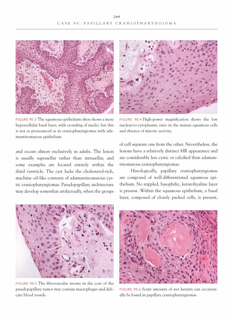
269
C A S E 9 0 : P A P I L L A R Y C R A N I O P H A R Y N G I O M A
and occurs almost exclusively in adults. The lesion is usually suprasellar rather than intrasellar, and some examples are located entirely within the third ventricle. The cyst lacks the cholesterol-rich, machine oil-like contents of adamantinomatous cys-tic craniopharyngiomas. Pseudopapillary architecture may develop somewhat artifactually, when the groups
of cell separate one from the other. Nevertheless, the lesions have a relatively distinct MR appearance and are considerably less cystic or calcifi ed than adaman-tinomatous craniopharyngiomas.
Histologically, papillary craniopharyngiomas are composed of well-differentiated squamous epi-thelium. No stippled, basophilic, keratohyaline layer is present. Within the squamous epithelium, a basal layer, composed of closely packed cells, is present,
FIGURE 90.6 Scant amounts of wet keratin can occasion-ally be found in papillary craniopharyngiomas.
FIGURE 90.5 The fi brovascular stroma in the core of the pseudopapillary tumor may contain macrophages and deli-cate blood vessels.
FIGURE 90.4 High-power magnifi cation shows the low nuclear-to-cytoplasmic ratio in the mature squamous cells and absence of mitotic activity.
FIGURE 90.3 The squamous epithelium often shows a more hypercellular basal layer, with crowding of nuclei, but this is not as pronounced as in craniopharyngiomas with ada-mantinomatous epithelium.

270
C A S E 9 0 : P A P I L L A R Y C R A N I O P H A R Y N G I O M A
but this crowded basal layer is less prominent than that seen in adamantinomatous craniopharyngioma. Nodules of wet keratin, calcifi cations, and keratin pearls are rare to absent. On very small biopsies, distinction from a Rathke cleft cyst with abundant squamous metaplasia may be impossible without neuroimaging correlation.
References 1. Burger PC, Scheithauer BW. Tumors of the Central Nervous
System. AFIP Atlas of Tumor Pathology. Fourth series, fasci-cle 7. Washington DC: American Registry of Pathology;2007. p. 461–9.
2. Crotty TB. Scheithauer BW. Young WF Jr., et al. Papillary craniopharyngioma: a clinicopathological study of 48 cases. J Neurosurg. 1995;83:206–14.
3. Sartoretti-Schefer S, Wichmann W, Aguzzi A, et al. MR differentiation of adamantinous [sic] and squamous-papillary craniopharyngiomas. AJNR 1997;18:77–87.

271
Case 91: Granular Cell Tumor of the Pituitary Gland
C L I N I C A L I N F O R M A T I O N
The patient is a 62-year-old male who presents with headaches and decreased visual acuity. Imaging studies show a small mass, located in the pituitary gland, that is interpreted as representing a possible adenoma. The lesion is resected, and histologic sections are reviewed.
O P I N I O N
Histologic sections show a circumscribed, nonencap-sulated mass, situated in the neurohypophysis. The mass is comprised of a sheetlike proliferation of large rounded cells, with granular eosinophilic cytoplasm. Nuclei are eccentrically placed, with a delicate chro-matin pattern. Mitotic fi gures are not observed. There is no evidence of necrosis. On immunohistochemi-cal staining, the tumor demonstrates diffuse positive
staining, with antibody to S-100 protein. The tumor does not stain with antibodies to GFAP or to anterior pituitary hormones.
We consider the lesion to be a low-grade neoplasm and characterize it as follows: PituitaryNeurohypophysis, Excision—Granular Cell Tumor, WHO Grade I.
C O M M E N T
Granular cell tumors represent low-grade neoplasms, which are potentially curable with gross total resection.
D I S C U S S I O N
Granular cell tumor represents the most common neo-plasm arising in the neurohypophysis and infundibu-lum of the pituitary gland. Many of these tumors are
FIGURE 91.1 Low-magnifi cation appearance of the neu-rohypophysis shows a circumscribed, eosinophilic nodule representing the tumor.
FIGURE 91.2 Intermediate magnifi cation shows a circum-scribed interface between the tumor and adjacent neuro-hypophysis.

272
C A S E 9 1 : G R A N U L A R C E L L T U M O R O F T H E P I T U I T A R Y G L A N D
small and represent incidental fi ndings at autopsy. Occasionally, the tumors may be large enough to pres-ent clinically with decreased visual acuity or endocrine defi ciency. On imaging, they appear as contrast-en-hancing, circumscribed lesions. Microscopically, they resemble their counterparts elsewhere in the body and are marked by a proliferation of large cells with distinc-tive granular cytoplasm. Nuclei are often eccentrically placed, with delicate chromatin. The granules may be highlighted with PAS staining. Occasional tumors may demonstrate a focal spindled cell pattern. Perivas-cular chronic infl ammation may also be evident. On immunohistochemistry, the tumors demonstrate dif-fuse positive staining with antibodies to S-100 and vimentin, but do not stain with antibody to GFAP or to antibodies targeting hormones produced in the adenohypophysis. Ultrastructurally, the granular qual-ity of the tumor cells cytoplasm is related to increased number of large lysosomes.
Differential diagnostic considerations most commonly include pituitary adenoma, particularly when the exact location of the lesion is not evident. The granular quality of the cytoplasm is a distinctive characteristic of this tumor, which is not present in
the adenomas. In spindled cell areas of granular cell tumor, the neoplasm may resemble pituicytoma or pilocytic astrocytoma. Both of these entities demon-strate GFAP immunoreactivity and are also marked by other fi ndings, including Rosenthal fi bers and granular bodies. Rare cases of granular cell differen-tiation in astrocytomas has been described, particu-larly in a small number of glioblastoma. Granular cell differentiation in glioblastoma may resemble the cells seen in a granular cell tumor, and key to recognition is identifying the more recognizable glioblastomatous portion of the tumor.
Granular cell tumors of the pituitary gland rep-resent low-grade neoplasms and are curable with gross total resection.
References 1. Cohen-Gadol AA, Pichelmann MA, Link MJ, et al. Granular
cell tumor of the sellar and suprasellar region: Clinicopatho-logic study of 11 cases and literature review. Mayo Clin Proc 2003;78:567–73.
2. Nishioka H, Ii k, Llena JF, et al. Immunohistochemical study of granular cell tumors of the neurohypophysis. Virchows Arch B Cell Pathol Incl Mol Pathol 1991;60:413–17.
3. Schaller B, Kirsch E, Tolnay M, et al. Symptomatic granular cell tumor of the pituitary gland: case report and review of the literature. Neurosurgery 1998;42:166–70; discussion 170–1.
FIGURE 91.4 The tumor demonstrates diffuse positive staining with antibody to S-100 protein.
FIGURE 91.3 High-magnifi cation appearance of the tumor cells shows abundant granular eosinophilic cytoplasm and eccentrically placed nucleus.

273
Case 92: Pituicytoma
C L I N I C A L I N F O R M A T I O N
The patient is a 55-year-old male who presents with adrenal insuffi ciency and a serum sodium of 111 ng/dl. He is begun on cortisol, and neuroimaging is obtained. MRI shows an enhancing lesion in the sella, which extends upward and measures 1.8 cm in diameter. He has visual problems, and visual fi eld test shows bitem-poral hemianopsia. A decision is made to resect the lesion, and histologic sections are reviewed.
O P I N I O N
Sections show a mildly hypercellular neoplasm, com-prised of aggregates of plump, eosinophilic cells with abundant cytoplasm. Nuclei manifest an open chro-matin pattern, are oval, and contain small nucleoli.
No tight whorls of cells, psammoma body calcifi ca-tions, mitoses, or necrosis are found.
We consider this lesion to be a low-grade tumor and characterize it as follows: Suprasellar Region, Excision—Pituicytoma, WHO Grade I.
C O M M E N T
This neoplasm is a rare, low-grade tumor that is distinct from other pituitary region masses, such as pituitary adenoma, craniopharyngioma, Rathke cleft cysts, or pilocytic astrocytoma.
D I S C U S S I O N
Pituicytoma is a newly described entity in the 2007 World Health Organization (WHO) classifi cation system. Most patients are adults (mean age 48 years),
FIGURE 92.1 Low-magnifi cation appearance of the neo-plasm shows the absence of necrosis, tight whorls of cells, or calcifi cation. Note the small aggregates and nests of tumor cells.
FIGURE 92.2 Higher magnifi cation highlights the bland open nuclei and delicate small nucleoli in the tumor cells of pituictyoma.

274
C A S E 9 2 : P I T U I C Y T O M A
and MR shows solid, discrete, enhancing lesions, either in the sella or suprasellar space, often mis-taken for pituitary adenoma. Histologically, the tumor is composed of sheets and nests of plump spindle cells with oval nuclei and small nucleoli. Unlike pilocytic astrocytomas that can also occur in this area, pituicytoma lacks Rosenthal fi bers, eosino-philic granular bodies, or a biphasic architectural pattern. Mitoses are rare to absent, and MIB-1 cell cycle labeling is low.
On immunohistochemistry, tumor cells are always strongly immunoreactive for vimentin and S-100 protein, but only variably positive for GFAP. No immunostaining for neuronal markers, such as synaptophysin or neurofi lament protein, is ever found. Thus, unlike normal posterior pituitary gland, no axons are seen in the tissue on neurofi la-ment immunostaining. Immunostaining for EMA is focal and weak, in rare instances, and usually not present at all.
FIGURE 92.6 MIB-1 cell cycle labeling is very low.FIGURE 92.5 Unlike meningioma, immunostaining for EMA is almost always negative.
FIGURE 92.4 Absence of abundant reticulin fi bers between individual tumor cells negates consideration of schwannoma.
FIGURE 92.3 Strong immunoreactivity for S-100 protein is seen in all cases.

275
C A S E 9 2 : P I T U I C Y T O M A
Although pituicytoma is strongly immunoreac-tive for S-100 protein, no abundant reticulin fi bers are seen in the tumor between individual tumor cells, as would be expected in schwannoma. No meningothelial, ependymal, or Schwann cell features are found on ultrastructural studies. An origin from specialized glial cells known as “pituicytes” is thought most likely.
References 1. Brat DJ, Scheithauer BW, Fuller GN, et al. Newly codifi ed
glial neoplasms of the 2007 WHO Classifi cation of Tumours of the Central Nervous System: angiocentric glioma, pilomyxoid astrocytoma and pituicytoma. Brain Pathol 2007;17:319–24.
2. Brat DJ, Scheithauer BW, Staugaitis SM, et al. Pituicytoma: a distinctive low-grade glioma of the neurohypophysis. Am J Surg Pathol 2000;24:362–8.
3. Ulm AJ, Yachnis AT, Brat DJ, et al. Pituicytoma: report of two cases and clues regarding histogenesis. Neurosurgery 2004;54:753–7.
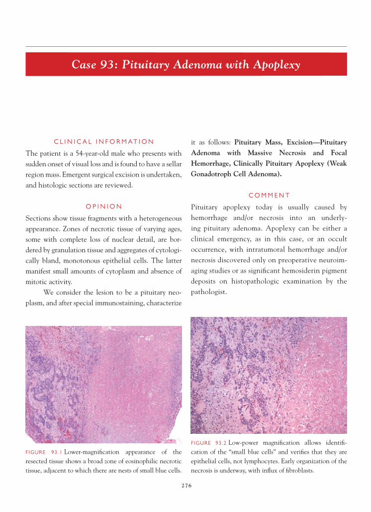
276
Case 93: Pituitary Adenoma with Apoplexy
C L I N I C A L I N F O R M A T I O N
The patient is a 54-year-old male who presents with sudden onset of visual loss and is found to have a sellar region mass. Emergent surgical excision is undertaken, and histologic sections are reviewed.
O P I N I O N
Sections show tissue fragments with a heterogeneous appearance. Zones of necrotic tissue of varying ages, some with complete loss of nuclear detail, are bor-dered by granulation tissue and aggregates of cytologi-cally bland, monotonous epithelial cells. The latter manifest small amounts of cytoplasm and absence of mitotic activity.
We consider the lesion to be a pituitary neo-plasm, and after special immunostaining, characterize
it as follows: Pituitary Mass, Excision— PituitaryAdenoma with Massive Necrosis and Focal Hemorrhage, Clinically Pituitary Apoplexy (Weak Gonadotroph Cell Adenoma).
C O M M E N T
Pituitary apoplexy today is usually caused by hemorrhage and/or necrosis into an underly-ing pituitary adenoma. Apoplexy can be either a clinical emergency, as in this case, or an occult occurrence, with intratumoral hemorrhage and/or necrosis discovered only on preoperative neuroim-aging studies or as signifi cant hemosiderin pigment deposits on histopathologic examination by the pathologist.
FIGURE 93.1 Lower-magnifi cation appearance of the resected tissue shows a broad zone of eosinophilic necrotic tissue, adjacent to which there are nests of small blue cells.
FIGURE 93.2 Low-power magnifi cation allows identifi -cation of the “small blue cells” and verifi es that they are epithelial cells, not lymphocytes. Early organization of the necrosis is underway, with infl ux of fi broblasts.

277
C A S E 9 3 : P I T U I T A R Y A D E N O M A W I T H A P O P L E X Y
D I S C U S S I O N
Pituitary apoplexy is almost always the result of bleed-ing into, or infarction of, a pituitary adenoma. The swelling and mass effect within the pituitary adenoma compresses surrounding structures and results in sud-den severe headache, nausea and vomiting, visual defects, oculomotor palsies, and impairment in pituitary function. Although supportive therapy
alone (including steroids) may result in clinical improvement, some patients required urgent surgical decompression of their mass. Most pituitary tumor apoplexy is spontaneous, but precipitating factors have included pituitary endocrine stimulation testing, surgery (especially coronary artery surgery), and coagulopathy. Rarely, apoplexy may be a result
FIGURE 93.6 Reticulin stain of the necrotic adenoma shows the expected disruption of normal acinar pattern and looks identical to what a reticulin stain shows in a non- apoplectic adenoma.
FIGURE 93.5 High-power view of an area with more advanced necrosis and greater loss of cell detail.
FIGURE 93.4 Intermediate-power magnifi cation of a recently necrotic portion of the adenoma allows discern-ment of nuclear detail and verifi es the absence of normal anterior pituitary gland architecture.
FIGURE 93.3 Intermediate-power magnifi cation shows the preservation of a pituitary adenoma architectural pattern within the coagulative necrosis; note brisk surrounding granulation tissue response.

278
C A S E 9 3 : P I T U I T A R Y A D E N O M A W I T H A P O P L E X Y
of bleeding into a Rathke cleft cyst or other sellar region mass.
Pituitary adenomas with several different types of hormonal secretory patterns may present with apoplexy, including growth hormone- secreting adenomas, corticotrophin-secreting adenomas, and clinically nonfunctioning adenomas, including those with patchy weak immunostaining for alpha subunit, f ollicle-stimulating hormone (FSH), or
luteinizing hormone (LH) (i.e., weak gonadotroph cell adenomas), such as this one.
Histologically, either necrosis or hemorrhage, or both, may be present in cases of apoplexy. The necrosis may be of slightly differing ages and may show varying degrees of preservation of nuclear detail and architectural pattern, but usually the basic
FIGURE 93.8 CAM5.2 immunostaining is seen in the pre-served, viable aggregates of adenoma cells.
FIGURE 93.7 Reticulin stain of normal anterior pitu-itary gland from another patient, for comparison with Figure 93.6.
FIGURE 93.10 Immunostaining for alpha subunit in the viable adenoma (upper left), as well as in a few of the cells in necrotic areas (lower right); the latter should be inter-preted with caution, unless the staining is also seen in the viable cells.
FIGURE 93.9 Synaptophysin immunostaining is seen in the preserved, viable aggregates of adenoma cells.

279
C A S E 9 3 : P I T U I T A R Y A D E N O M A W I T H A P O P L E X Y
architectural outline of the adenoma can be detected. Often, the necrosis appears subacute in age and may show a modest neutrophilic cell response, similar to the cellular response seen in other infarctions of 48–96 hours in duration. At the edge of the necrotic areas, granulation tissue, hemosiderin deposits, and residual clusters of viable adenoma may be found.
Even within the necrotic areas, the alteration in reticulin pattern that is diagnostic for pituitary adenoma can still be detectable for a considerable length of time and allow confi dent diagnosis of an underlying adenoma. Loss of reticulin pattern is the hallmark of an adenoma and contrasts with the hon-eycomb pattern of reticulin outlining normal acini in non-adenomatous anterior pituitary gland. Small viable islands of adenoma are often found and should not be mistaken for lymphocytes or histiocytes. The viable adenoma retains co-expression of CAM5.2 and synaptophysin, verifying the diagnosis of pituitary
adenoma. Immunostaining for these antibodies, or any of the anterior pituitary hormones is, of course, less interpretable in necrotic portions of the lesion, but in the most acute/recent areas of necrosis, some bona fi de immunostaining may be retained in some of the adenoma cells.
References 1. Binning MJ, Liu JK, Gannon J, et al. Hemorrhagic and non-
hemorrhagic Rathke cleft cysts mimicking pituitary apoplexy. J Neurosurg 2008;108:3–8.
2. Kleinschmidt-DeMasters BK, Lillehei KO. Pathological correlates of pituitary adenomas presenting with apoplexy. Hum Pathol 1998;29:1255–65.
3. Nawar RN, AbdelMannan D, Selman WR, et al. Pituitary tumor apoplexy: a review. J Intens Care Med 2008;23:75–90.
4. Nielsen EH, Lindholm J, Bjerre P, et al. Frequent occur-rence of pituitary apoplexy in patients with non-functioning pituitary adenoma. Clin Endo 2006;64:319–22.
5. Semple PL, Jane JA, Lopes MBS, et al. Pituitary apoplexy: correlation between magnetic resonance imaging and histo-pathological results. J Neurosurg 2008;108:909–15.

280
Case 94: Pituitary Adenoma in an Ectopic Site
C L I N I C A L I N F O R M A T I O N
The patient is a 56-year-old male with a lesion of the left cavernous sinus/posterior sphenoid area, found on work-up for headache. The lesion has been followed by multiple MRI scans, has shown no change in size over time, and does not fi ll the cavernous sinus. The patient does not have cranial nerve palsies. Accord-ing to the patient’s neurosurgeon, there is no well-de-fi ned intrasellar or pituitary region mass, and systemic metastatic work-up is negative. Prior to surgery, the patient has undergone an extensive endocrinologic work-up that shows a mildly elevated prolactin of 28 ng/dl. A decision is made to biopsy the lesion.
O P I N I O N
Biopsies show fi brous tissue, in which are embedded nests of monomorphic epithelial cells. These nests
show neither necrosis or mitotic activity, and cells manifest bland nuclei and scant cytoplasm.
We consider the biopsy as representing a pi-tuitary adenoma, albeit in the wall of the cavernous sinus and not the pituitary gland. We characterize it as follows: Left Cavernous Sinus, Biopsy—Pituitary Adenoma, Ectopic.
C O M M E N T
By immunohistochemistry, the adenoma displayed diffuse immunoreactivity for prolactin, explaining the mild elevation noted preoperatively.
D I S C U S S I O N
The usual site of pituitary adenomas is the sellar region, although adenomas can occasionally arise
FIGURE 94.1 Nests of monomorphic cells are seen embed-ded within the connective tissue of the dura.
FIGURE 94.2 Intermediate-power magnifi cation shows absence of necrosis, mitotic activity, as well as a fi brillar matrix.

281
C A S E 9 4 : P I T U I T A R Y A D E N O M A I N A N E C T O P I C S I T E
anywhere along the migration pathway followed by the craniopharyngeal duct/Rathke pouch during the embryologic period. Reported ectopic sites for pituitary adenoma include the sphenoid sinus, cav-ernous sinus, third ventricle, and even nasopharynx.
Sections show small clusters of monomor-phic cells within the fi brotic dura. The small cells manifest prominent nucleoli, clumpy nuclear chromatin, and moderate amounts of chromophobic cytoplasm by periodic acid Schiff/Orange G stain. No ganglion cell differentiation is identifi ed, and no fi brillar matrix is interspersed within the cell clusters, as might be seen in an olfactory neuroblastoma. Immunostains show the tumor to be positive for synaptophysin and pan-cytokeratin—as can be seen with pituitary adenomas in any site—and negative for GFAP and CD45 (leukocyte common antigen). Additional immunostains showed no encirclement of cell nests by S-100 positive cells, as might be seen with a paraganglioma.
An immunoperoxidase panel for anterior pituit-ary hormones showed strong, diffuse immunoreactivity for prolactin, with negative immunoreactivity for other pituitary hormones, including growth hormone. MIB-1 cell cycle labeling index was negligible.
References 1. Collie RB. Collie MJ. Extracranial thyroid-stimulating
hormone-secreting ectopic pituitary adenoma of the naso-pharynx. Otolaryngology—Head & Neck Surgery 2005;133: 453–4.
2. Gondim JA, Schops M, Ferreira E, et al. Acromegaly due to an ectopic pituitary adenoma in the sphenoid sinus. Acta Radiologica 2004;45:689–91.
3. Hou L, Harshbarger T, Herrick MK, et al. Suprasellar adrenocor-ticotropic hormone-secreting ectopic pituitary adenoma: case report and literature review. Neurosurgery 2002;50:618–25.
4. Kleinschmidt-DeMasters BK, Winston KR, Rubinstein D, et al. Ectopic pituitary adenoma of the third ventricle. Case report. J Neurosurg 1990;72:139–42.
5. Mitsuya K, Nakasu Y, Nioka H, et al. Ectopic growth hormone-releasing adenoma in the cavernous sinus—case report. Neurologia Medico-Chirurgica 2004;44:380–5.
FIGURE 94.4 Diffuse immunoreactivity for prolactin is seen.FIGURE 94.3 High-power magnifi cation highlights the endocrine features of the cells, with clumpy coarse nuclear chromatin pattern and moderate amounts of cytoplasm.

282
Case 95: Metastatic Small Cell Carcinoma of Lung
C L I N I C A L I N F O R M A T I O N
The patient is a 54-year-old male with known diagno-sis of small cell lung cancer and previously diagnosed brain metastases. He has been treated in the past with whole-brain irradiation, in addition to stereotactic radiosurgery. However, over the previous months, there has been slow but progressive increase in the size of the right frontal and right temporal brain lesions. Resection of both lesions is undertaken, and histologic sections are reviewed.
O P I N I O N
Extensively necrotic tissue is seen, containing small collections of small malignant cells with hyperchro-matic nuclei and scant cytoplasm. The tumor is relatively sharply demarcated from the surrounding
gliotic brain tissue. Necrosis in the lesion is associated with fi brinoid vascular necrosis and other features of radiation-induced damage.
We consider the pathology to be that of a metastatic carcinoma and characterize it as follows: Right Frontal Lobe and Right Temporal Lobe, Excision—Metastatic Small Cell Carcinoma of Lung, with Extensive Radionecrosis.
C O M M E N T
This tumor is particularly necrotic as a result of the patient having received both whole-brain radiation and stereotactic radiosurgery prior to removal of his brain metastases. The continued growth of the lesions, despite therapy in this patient, is attributable to residual nests of viable, growing tumor cells.
FIGURE 95.1 Low-power view shows the extensive necrosis in this lesion as a result of prior treatment with stereotactic radiosurgery; note small islands of viable tumor cells.
FIGURE 95.2 Low-power view shows the sharp interface between the viable metastatic tumor and the adjacent gliotic brain, rather than individual tumor cell infi ltration, as would be seen with most primary high-grade neoplasms of brain.
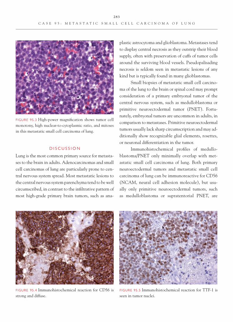
283
C A S E 9 5 : M E T A S T A T I C S M A L L C E L L C A R C I N O M A O F L U N G
D I S C U S S I O N
Lung is the most common primary source for metasta-ses to the brain in adults. Adenocarcinomas and small cell carcinomas of lung are particularly prone to cen-tral nervous system spread. Most metastatic lesions to the central nervous system parenchyma tend to be well circumscribed, in contrast to the infi ltrative pattern of most high-grade primary brain tumors, such as ana-
plastic astrocytoma and glioblastoma. Metastases tend to display central necrosis as they outstrip their blood supply, often with preservation of cuffs of tumor cells around the surviving blood vessels. Pseudopalisading necrosis is seldom seen in metastatic lesions of any kind but is typically found in many glioblastomas.
Small biopsies of metastatic small cell carcino-ma of the lung to the brain or spinal cord may prompt consideration of a primary embryonal tumor of the central nervous system, such as medulloblastoma or primitive neuroectodermal tumor (PNET). Fortu-nately, embryonal tumors are uncommon in adults, in comparison to metastases. Primitive neuroectodermal tumors usually lack sharp circumscription and may ad-ditionally show recognizable glial elements, rosettes, or neuronal differentiation in the tumor.
Immunohistochemical profi les of medullo-blastoma/PNET only minimally overlap with met-astatic small cell carcinoma of lung. Both primary neuroectodermal tumors and metastatic small cell carcinoma of lung can be immunoreactive for CD56 (NCAM, neural cell adhesion molecule), but usu-ally only primitive neuroectodermal tumors, such as medulloblastoma or supratentorial PNET, are
FIGURE 95.5 Immunohistochemical reaction for TTF-1 is seen in tumor nuclei.
FIGURE 95.4 Immunohistochemical reaction for CD56 is strong and diffuse.
FIGURE 95.3 High-power magnifi cation shows tumor cell monotony, high nuclear-to-cytoplasmic ratio, and mitoses in this metastatic small cell carcinoma of lung.
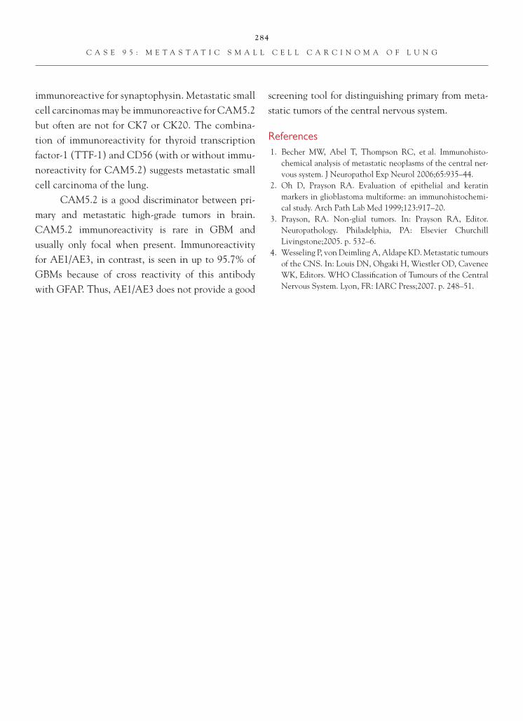
284
C A S E 9 5 : M E T A S T A T I C S M A L L C E L L C A R C I N O M A O F L U N G
immunoreactive for synaptophysin. Metastatic small cell carcinomas may be immunoreactive for CAM5.2 but often are not for CK7 or CK20. The combina-tion of immunoreactivity for thyroid transcription factor-1 (TTF-1) and CD56 (with or without immu-noreactivity for CAM5.2) suggests metastatic small cell carcinoma of the lung.
CAM5.2 is a good discriminator between pri-mary and metastatic high-grade tumors in brain. CAM5.2 immunoreactivity is rare in GBM and usually only focal when present. Immunoreactivity for AE1/AE3, in contrast, is seen in up to 95.7% of GBMs because of cross reactivity of this antibody with GFAP. Thus, AE1/AE3 does not provide a good
screening tool for distinguishing primary from meta-static tumors of the central nervous system.
References 1. Becher MW, Abel T, Thompson RC, et al. Immunohisto-
chemical analysis of metastatic neoplasms of the central ner-vous system. J Neuropathol Exp Neurol 2006;65:935–44.
2. Oh D, Prayson RA. Evaluation of epithelial and keratin markers in glioblastoma multiforme: an immunohistochemi-cal study. Arch Path Lab Med 1999;123:917–20.
3. Prayson, RA. Non-glial tumors. In: Prayson RA, Editor. Neuropathology. Philadelphia, PA: Elsevier Churchill Livingstone;2005. p. 532–6.
4. Wesseling P, von Deimling A, Aldape KD. Metastatic tumours of the CNS. In: Louis DN, Ohgaki H, Wiestler OD, Cavenee WK, Editors. WHO Classifi cation of Tumours of the Central Nervous System. Lyon, FR: IARC Press;2007. p. 248–51.

285
Case 96: Leukemic Involvement of the Central Nervous System
C L I N I C A L I N F O R M A T I O N
The patient is a 29-year-old male who presents to the emergency department with right ankle pain and is found to have an elevated white blood cell count of 159,000/µl and thrombocytopenia, with a plate-let count of 20,000/µl . Hours after admission to the hospital, the patient is found unresponsive, and an emergent CT scan reveals a massive intracerebral hemorrhage. Clot is evacuated, and histologic sec-tions from the clot are reviewed.
O P I N I O N
Sections show massive acute clot within brain paren-chyma, containing sheets of discohesive neoplastic hematopoietic cells, with kidney bean-shaped to bi-lobed irregular nuclei, high nuclear-to-cytoplasmic
ratio, and only mildly granular cytoplasm. Nucleoli are moderate in size. Myeloperoxidase reaction is strongly and diffusely positive.
We consider the lesion to be a hematopoietic neoplasm, and after special immunostaining and fl uo-rescence in situ hybridization (FISH) studies, charac-terize it as follows: Cerebral Hemispheric Clot, Evac-uation—Acute Leukemia with Hemorrhage (Acute Promyelocytic Leukemia, Hypogranular Form).
C O M M E N T
The clot likely occurred as a result of a combination of the very low platelet count coupled with the very high circulating white cell count in this patient. This period in acute leukemia, when the platelet count falls below 25,000/µl and the white count exceeds
FIGURE 96.1 Lower-magnifi cation appearance shows a clot within the brain parenchyma, accompanied by the sheets of neoplastic hematopoietic cells.
FIGURE 96.2 High-power magnifi cation illustrates the kidney bean-shaped nuclei and hypogranular cytoplasm of the neoplastic hematopoietic cells.

286
C A S E 9 6 : L E U K E M I C I N V O L V E M E N T O F T H E C E N T R A L N E R V O U S S Y S T E M
100,000/µl, has been termed the “critical period” for risk of central nervous system (CNS) hemorrhage.
D I S C U S S I O N
Criteria for diagnosis and classifi cation of leukemias involving the CNS are identical to those utilized in other tissues in the body. In this case, the histologic features were those of a myelocytic leukemia, with
bean-shaped nuclei and abundant cytoplasm, rather than that of a B-cell lymphoma, with rounded nuclei and more prominent nucleoli. The immunohis-tochemical reaction for CD45RB (leukocyte common antigen) was patchy, and no staining for CD20 (B-cell marker) was detected. In contrast, the cytoplasmic reaction for myeloperoxidase was diffuse, strong, and almost obliterated the nuclear detail of the cells. In addition, FISH was performed to look for the PML/RARA rearrangement associated with the (15;17)(q22;q21.1) translocation found in acute promyelo-cytic leukemia, and it was found in this case. The cytoplasm is relatively hypogranular in this example but may show coarse granulation in some examples of acute promyelocytic leukemia.
Leukemias of several different types can involve the CNS. Direct tumor involvement manifests as intra-parenchymal or extra-axial mass lesion(s) or as infi ltra-tions of the leptomeninges or dura. CNS hemorrhage is less common. However, in cases where there is extensive systemic and bone marrow involvement by the leuke-mia, coagulopathy and thrombocytopenia can result, and CNS hemorrhage may be the cause of demise, as it was in this patient soon after his clot evacuation.
FIGURE 96.5 As is the case with most acute promyelo-cytic leukemias, only patchy weak immunoreactivity for CD45RB (leukocyte common antigen) is present.
FIGURE 96.4 High-power magnifi cation shows the absence of immunostaining for the B-cell marker CD20 in the tumor cells.
FIGURE 96.3 High-power magnifi cation depicts the strong, diffuse, cytoplasmic immunoreactivity for myeloperoxidase in the tumor cells.

287
C A S E 9 6 : L E U K E M I C I N V O L V E M E N T O F T H E C E N T R A L N E R V O U S S Y S T E M
The most likely leukemia types to develop CNS hemorrhage are, in descending order of fre-quency, acute myeloblastic leukemia, blastic phase of chronic myelogenous leukemia, and lymphoblastic leukemia (ALL). Acute promyelocytic leukemia con-stitutes a minority (5–8%) of all acute myelogenous leukemias. The current case is unusual, in that the patient had severe thrombocytopenia and massively elevated white blood cell count on his initial clini-cal presentation and developed CNS bleeding within hours after hospital admission. Similar cases of intrac-erebral hemorrhage as the fi rst clinical manifestation of acute promyelocytic leukemia have been reported; however, CNS hemorrhage may also develop during relapse or as a late complication of radiation therapy, in children with ALL who have received whole-brain radiotherapy. The hemorrhage in the latter instance
develops years after cranial irradiation treatment and is a result of the formation of intracerebral cavernous malformations that subsequently rupture.
References 1. Chamberlain MC. Leukemia and the nervous system. Curr
Oncol Reports 2005;7:66–73. 2. Glass J. Neurologic complications of lymphoma and leuke-
mia. Sem Oncol 2006;33:342–7. 3. Larson JJ, Ball WS, Bove KE, et al. Formation of intracere-
bral cavernous malformations after radiation treatment for central nervous system neoplasia in children. J. Neurosurg 1998;88:51–6.
4. Marcheta MP, Kok Shun J, Liu Yin JA. Central nervous sys-tem relapse of acute promyelocytic leukaemia in a patient with cerebral haemorrhage at diagnosis. Brit J Haematol 2001;114:954–5.
5. Nowacki P, Zdziarska B, Fryze C, et al. Co-existence of thrombo-cytopenia and hyperleukocytosis (“critical period”) as a risk fac-tor of haemorrhage into the central nervous system in patients with acute leukaemias. Haematologia 2002;31:347–55.

288
Case 97: Chordoma
C L I N I C A L I N F O R M A T I O N
The patient is a 58-year-old male who fi rst develops diplopia about 1 year ago but puts off seeking medical attention until the onset of blurred vision 2 months ago. Physical examination shows his visual acuity to be 20/40 on the left and 20/20 on the right. Exami-nation of cranial nerves III, IV, and VI reveal a lack of upward gaze on the left side, as well as anisocoria, with the left pupil being larger than the right. Neu-roimaging studies demonstrate a large, enhancing mass in the sellar and suprasellar regions, posterior to the clivus and to the left of midline. The decision is made to attempt surgical debulking of the lesion, and histologic sections are available for review.
O P I N I O N
Biopsies show a moderately hypercellular, slightly lobulated mass, with epithelioid cells arranged in cords and rows. Cells are contiguous to each other and show touching borders, rather than being arranged in individual lacunae. Many cells manifest a vacu-olated cytoplasm (physaliphorous cells), and cells are embedded in abundant, extracellular, gray-blue mucin. Mitoses or necrosis are not seen, but slight nuclear atypia is apparent.
After immunohistochemical studies, we con-sider the resected lesion as representing a bone-based mass and characterize it as follows: Left Clivus, Re-section-Chordoma.
FIGURE 97.1 Low-power view of the lesion shows epithe-lioid cells arranged in rows and cords and embedded in a mucinous matrix.
FIGURE 97.2 High-power magnifi cation illustrates the extensive vacuolization within tumor cytoplasm and absence of mitotic activity.

289
C A S E 9 7 : C H O R D O M A
C O M M E N T
Chordomas are relatively uncommon tumors, but about a third of them occur in the clivus or spheno-occipital region.
D I S C U S S I O N
Chordoma is a tumor of notochordal derivation that can occur anywhere along the neural axis, includ-ing skull base, sacrococcygeal region, and vertebral
bodies. Virtually all involve intraosseous sites, and most cause signifi cant bone destruction. Symptoms relate to the location of the lesion, and in the skull base, problems with visual acuity, cranial nerve pal-sies of cranial nerves III, IV, and VI (resulting in diplopia), and headache are typical symptoms.
In the skull base, chordomas must be differen-tiated from chondrosarcomas, almost all of which are well differentiated. In addition, a variant denoted as chondroid chordoma was fi rst described in 1973
FIGURE 97.6 Chordoma may also be only focally positive for S-100 protein.
FIGURE 97.5 Chordoma may show far more focal immuno-reactivity for EMA.
FIGURE 97.4 Chordomas are generally strongly and dif-fusely immunoreactive for keratin (AE1/AE3).
FIGURE 97.3 High-power magnifi cation highlights occa-sional, very large, physaliphorous cells and modest nuclear atypia.
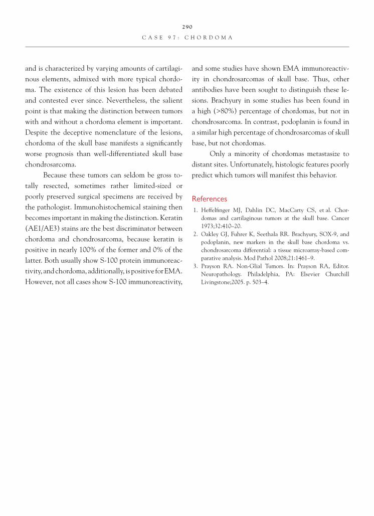
290
C A S E 9 7 : C H O R D O M A
and is characterized by varying amounts of cartilagi-nous elements, admixed with more typical chordo-ma. The existence of this lesion has been debated and contested ever since. Nevertheless, the salient point is that making the distinction between tumors with and without a chordoma element is important. Despite the deceptive nomenclature of the lesions, chordoma of the skull base manifests a signifi cantly worse prognosis than well-differentiated skull base chondrosarcoma.
Because these tumors can seldom be gross to-tally resected, sometimes rather limited-sized or poorly preserved surgical specimens are received by the pathologist. Immunohistochemical staining then becomes important in making the distinction. Keratin (AE1/AE3) stains are the best discriminator between chordoma and chondrosarcoma, because keratin is positive in nearly 100% of the former and 0% of the latter. Both usually show S-100 protein immunoreac-tivity, and chordoma, additionally, is positive for EMA. However, not all cases show S-100 immunoreactivity,
and some studies have shown EMA immunoreactiv-ity in chondrosarcomas of skull base. Thus, other antibodies have been sought to distinguish these le-sions. Brachyury in some studies has been found in a high (>80%) percentage of chordomas, but not in chondrosarcoma. In contrast, podoplanin is found in a similar high percentage of chondrosarcomas of skull base, but not chordomas.
Only a minority of chordomas metastasize to distant sites. Unfortunately, histologic features poorly predict which tumors will manifest this behavior.
References 1. Heffelfi nger MJ, Dahlin DC, MacCarty CS, et al. Chor-
domas and cartilaginous tumors at the skull base. Cancer 1973;32:410–20.
2. Oakley GJ, Fuhrer K, Seethala RR. Brachyury, SOX-9, and podoplanin, new markers in the skull base chordoma vs. chondrosarcoma differential: a tissue microarray-based com-parative analysis. Mod Pathol 2008;21:1461–9.
3. Prayson RA. Non-Glial Tumors. In: Prayson RA, Editor. Neuropathology. Philadelphia, PA: Elsevier Churchill Livingstone;2005. p. 503–4.

291
Case 98: Metastatic Papillary Carcinoma of the Thyroid
C L I N I C A L I N F O R M A T I O N
The patient is a 56-year-old female who presents with a history of papillary thyroid cancer, diagnosed 2 years previously. At the time of initial surgery, the patient has metastases noted in several lymph nodes. Most recently, the patient presents with episodes of confu-sion. On imaging, a right temporal lobe mass, measur-ing 1.6 cm, is identifi ed. The lesion is excised, and histologic sections are reviewed.
O P I N I O N
Histologic sections show a lesion that is sharply demar-cated from the adjacent gliotic parenchyma. The lesion itself is marked by a tumor that demonstrates a papil-lary architectural pattern. Fibrovascular cores are lined by tumor cells. The tumor cells show nuclear clearing
and occasional nuclear grooves. Rare mitotic fi gures are seen. Adjacent to the tumor is a large area of geographic necrosis. Focal microcalcifi cations are seen in viable tumor. On immunohistochemical analysis, the tumor demonstrates positive staining, with antibody to thyro-globulin and to thyroid transcription factor-1 (TTF-1).
We consider the lesion to be a metastatic neo-plasm and characterize it as follows: Right Temporal Lobe, Excision—Metastatic Papillary Carcinoma of the Thyroid.
C O M M E N T
The tumor is consistent with a metastasis from the patient’s previously diagnosed papillary carcinoma of the thyroid. Thyroglobulin and TTF-1 immunore-activity support an origin of the metastasis from the thyroid tumor.
FIGURE 98.1 The interface between the tumor and the adjacent gliotic brain parenchyma is sharply delineated, typical of a metastasis.
FIGURE 98.2 Intermediate magnifi cation showing geo-graphic necrosis adjacent to a papillary neoplasm.

292
C A S E 9 8 : M E T A S T A T I C P A P I L L A R Y C A R C I N O M A O F T H E T H Y R O I D
D I S C U S S I O N
Metastatic disease from the thyroid gland is relatively uncommon. Most frequently, tumors that metastasize to the CNS include primaries from the lung, breast, kidney, and skin (melanoma). Metastases are fre-quently (although not always) multifocal. The tumor in this case presents as a single mass. Morphologically, metastases, in contrast to gliomas, are well circum-
scribed, in that there is often a sharp interface between the tumor and the adjacent gliotic parenchyma.
In many cases, determining the site of origin by examining the metastasis is not possible. In the current case, the history of previous papillary carcinoma of the thyroid and the papillary architecture and cytology of the metastatic tumor are useful in suggesting the site of origin. This impression was confi rmed by immuno-histochemical staining. Thyroglobulin is fairly specifi c for a thyroid carcinoma. TTF-1, although an excellent
FIGURE 98.6 Thyroglobulin immunoreactivity is present in the tumor.
FIGURE 98.5 High-magnifi cation appearance of the tumor shows nuclear clearing and occasional cleaved or grooved nuclei, cytologically consistent with papillary carcinoma of thyroid origin.
FIGURE 98.4 Focal calcifi cations are seen in papillary areas of the tumor.
FIGURE 98.3 Focal hemosiderin deposition indicates previ-ous hemorrhage associated with the tumor.

293
C A S E 9 8 : M E T A S T A T I C P A P I L L A R Y C A R C I N O M A O F T H E T H Y R O I D
marker for thyroid cancer, is also well-known to stain a signifi cant subset of metastatic tumors of pulmonary origin and therefore is not specifi c.
References 1. Aguiar PH, Agner C, Tavares FR, et al. Unusual brain
metastases from papillary thyroid carcinoma: case report. Neurosurgery 2001;49:1008–13.
2. DeYoung BR, Wick MR. Immunohistologic evaluation of metastatic carcinomas of unknown origin: an algorithmic approach. Semin Diagn Pathol 2000;17:184–93.
3. Oliveira AM, Tazelaar HD, Myers JL, et al. Thyroid transcription factor-1 distinguishes metastatic pulmonary from well-differentiated neuroendocrine tumors of other sites. Am J Surg Pathol 2001;25:815–19.

294
Case 99: Leptomeningeal Carcinomatosis
C L I N I C A L I N F O R M A T I O N
The patient is a 52-year-old male with a diagnosis of stage IV non-small cell lung cancer metastatic to bone. Approximately 1 month prior to surgery, the patient began to have symptoms of diffi culty with memory and with dragging both feet when walking. This led to an MRI that showed meningeal enhance-ment, but no mass lesions in the brain. As a result of persistent symptoms, a decision is made to biopsy the patient, and histologic sections are reviewed.
O P I N I O N
Biopsies of cerebral cortex show leptomeninges and Virchow-Robin spaces, fi lled with metastatic neoplastic cells. These cells are arranged in cohesive groups, and where small, cortical, rounded metastases are formed
in the superfi cial cortex, gland formation is evident in the metastatic adenocarcinoma. High power shows cells with large vesicular nuclei, prominent nucleoli, and vacuolated, mucin-fi lled cytoplasm. An additional biopsy of overlying dura was negative for tumor cells.
We consider the biopsy to be metastatic carcino-ma, with involvement of the leptomeninges, and char-acterize it as follows: Right Frontal Cortex, Biopsy—Leptomeningeal Carcinomatosis, Metastatic Non-Small Cell Carcinoma of Lung, WHO Grade IV.
C O M M E N T
Leptomeningeal carcinomatosis, synonymous with car-cinomatous meningitis, often occurs without coexistent parenchymal brain metastases or dural involvement.
FIGURE 99.1 Section of cerebral cortex, shown at inter-mediate magnifi cation, demonstrates clusters of metastatic neoplastic cells confi ned to the subarachnoid space.
FIGURE 99.2 Adjacent sections show a thicker layer of tumor cells in the meninges that dips down into cor-tex along Virchow-Robin spaces; note reactive gliosis in underlying subpial tissue.
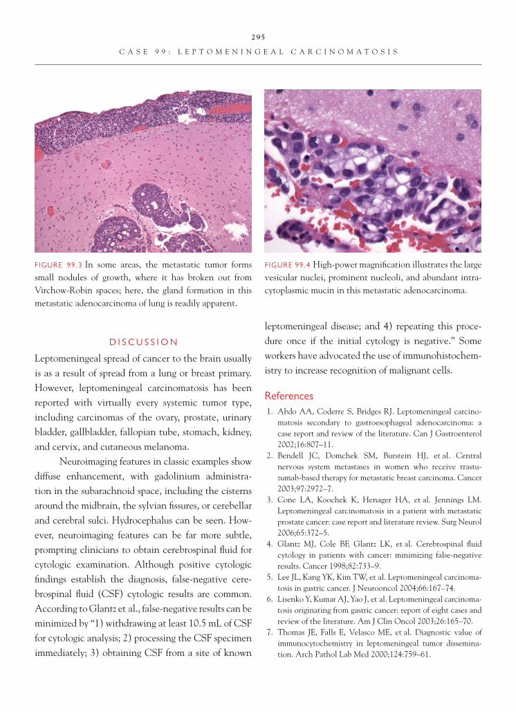
295
C A S E 9 9 : L E P T O M E N I N G E A L C A R C I N O M A T O S I S
D I S C U S S I O N
Leptomeningeal spread of cancer to the brain usually is as a result of spread from a lung or breast primary. However, leptomeningeal carcinomatosis has been reported with virtually every systemic tumor type, including carcinomas of the ovary, prostate, urinary bladder, gallbladder, fallopian tube, stomach, kidney, and cervix, and cutaneous melanoma.
Neuroimaging features in classic examples show diffuse enhancement, with gadolinium administra-tion in the subarachnoid space, including the cisterns around the midbrain, the sylvian fi ssures, or cerebellar and cerebral sulci. Hydrocephalus can be seen. How-ever, neuroimaging features can be far more subtle, prompting clinicians to obtain cerebrospinal fl uid for cytologic examination. Although positive cytologic fi ndings establish the diagnosis, false-negative cere-brospinal fl uid (CSF) cytologic results are common. According to Glantz et al., false-negative results can be minimized by “1) withdrawing at least 10.5 mL of CSF for cytologic analysis; 2) processing the CSF specimen immediately; 3) obtaining CSF from a site of known
leptomeningeal disease; and 4) repeating this proce-dure once if the initial cytology is negative.” Some workers have advocated the use of immunohistochem-istry to increase recognition of malignant cells.
References 1. Abdo AA, Coderre S, Bridges RJ. Leptomeningeal carcino-
matosis secondary to gastroesophageal adenocarcinoma: a case report and review of the literature. Can J Gastroenterol 2002;16:807–11.
2. Bendell JC, Domchek SM, Burstein HJ, et al. Central nervous system metastases in women who receive trastu-zumab-based therapy for metastatic breast carcinoma. Cancer 2003;97:2972–7.
3. Cone LA, Koochek K, Henager HA, et al. Jennings LM. Leptomeningeal carcinomatosis in a patient with metastatic prostate cancer: case report and literature review. Surg Neurol 2006;65:372–5.
4. Glantz MJ, Cole BF, Glantz LK, et al. Cerebrospinal fl uid cytology in patients with cancer: minimizing false-negative results. Cancer 1998;82:733–9.
5. Lee JL, Kang YK, Kim TW, et al. Leptomeningeal carcinoma-tosis in gastric cancer. J Neurooncol 2004;66:167–74.
6. Lisenko Y, Kumar AJ, Yao J, et al. Leptomeningeal carcinoma-tosis originating from gastric cancer: report of eight cases and review of the literature. Am J Clin Oncol 2003;26:165–70.
7. Thomas JE, Falls E, Velasco ME, et al. Diagnostic value of immunocytochemistry in leptomeningeal tumor dissemina-tion. Arch Pathol Lab Med 2000;124:759–61.
FIGURE 99.4 High-power magnifi cation illustrates the large vesicular nuclei, prominent nucleoli, and abundant intra-cytoplasmic mucin in this metastatic adenocarcinoma.
FIGURE 99.3 In some areas, the metastatic tumor forms small nodules of growth, where it has broken out from Virchow-Robin spaces; here, the gland formation in this metastatic adenocarcinoma of lung is readily apparent.
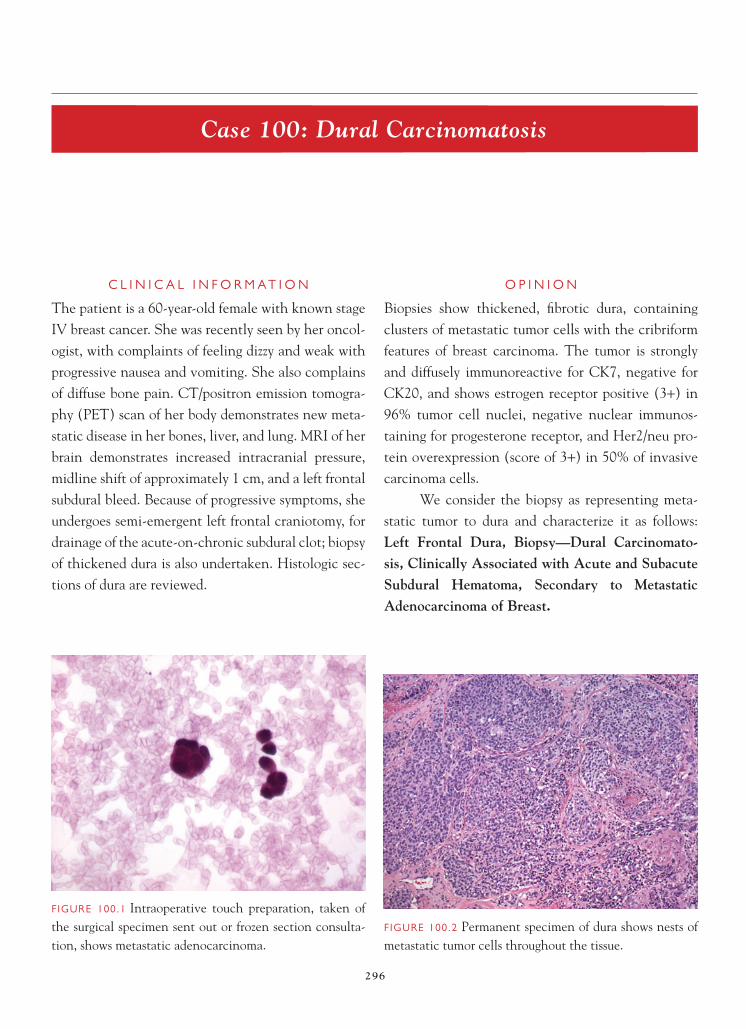
296
Case 100: Dural Carcinomatosis
C L I N I C A L I N F O R M A T I O N
The patient is a 60-year-old female with known stage IV breast cancer. She was recently seen by her oncol-ogist, with complaints of feeling dizzy and weak with progressive nausea and vomiting. She also complains of diffuse bone pain. CT/positron emission tomogra-phy (PET) scan of her body demonstrates new meta-static disease in her bones, liver, and lung. MRI of her brain demonstrates increased intracranial pressure, midline shift of approximately 1 cm, and a left frontal subdural bleed. Because of progressive symptoms, she undergoes semi-emergent left frontal craniotomy, for drainage of the acute-on-chronic subdural clot; biopsy of thickened dura is also undertaken. Histologic sec-tions of dura are reviewed.
O P I N I O N
Biopsies show thickened, fi brotic dura, containing clusters of metastatic tumor cells with the cribriform features of breast carcinoma. The tumor is strongly and diffusely immunoreactive for CK7, negative for CK20, and shows estrogen receptor positive (3+) in 96% tumor cell nuclei, negative nuclear immunos-taining for progesterone receptor, and Her2/neu pro-tein overexpression (score of 3+) in 50% of invasive carcinoma cells.
We consider the biopsy as representing meta-static tumor to dura and characterize it as follows: Left Frontal Dura, Biopsy—Dural Carcinomato-sis, Clinically Associated with Acute and Subacute Subdural Hematoma, Secondary to Metastatic Adenocarcinoma of Breast.
FIGURE 100.1 Intraoperative touch preparation, taken of the surgical specimen sent out or frozen section consulta-tion, shows metastatic adenocarcinoma.
FIGURE 100.2 Permanent specimen of dura shows nests of metastatic tumor cells throughout the tissue.

297
C A S E 1 0 0 : D U R A L C A R C I N O M A T O S I S
C O M M E N T
Dural carcinomatosis often occurs as a late-stage complication in patients who also manifest bone and other systemic metastatic disease. Patients with sys-temic carcinomas who present with acute or subacute subdural hematomas should be suspected of having dural tumor involvement.
D I S C U S S I O N
Dural metastases are usually more discrete masses, rather than diffuse, involving dura in a thickened, diffuse layer of tumor. The most common tumor types to involve the dura are breast carcinoma (as this case), prostate cancer, lung cancer, and hematopoi-etic tumors. Even at biopsy or autopsy tissue exami-nation, less than one-half of cases show coexistent brain parenchymal metastatic disease. Both anteced-ent pulmonary lymphangitic spread and bony disease seem to be associated with development of dural met-astatic disease. Hence, usually the patient is known to have systemic malignancy before presenting with dural involvement, making the diagnosis easier for the pathologist.
Nevertheless, documentation of identity of the metastatic tumor with the alleged primary neo-plasm is important. An algorithmic approach for the work-up of metastatic disease of known type to the CNS has been outline by Becher et al. This involves a “fi rst round” of antibodies for epithelial tumors or known adenocarcinomas, using CAM5.2, CK7, CK20, and thyroid transcription factor-1
FIGURE 100.5 Tumor is strongly and diffusely immunoreac-tive for cytokeratin 7, but it was negative for CK20.
FIGURE 100.4 High-power magnifi cation demonstrates cells with high nuclear-to-cytoplasmic ratio and many mitoses.
FIGURE 100.3 Focal necrosis is seen within the metastatic tumor cell nests.

298
C A S E 1 0 0 : D U R A L C A R C I N O M A T O S I S
FIGURE 100.6 Strong, 3+, nuclear immunoreactivity is seen for estrogen receptor.
(TTF-1). Tumors that are positive for CAM5.2 and CK7 and negative for CK20 are then additionally immunostained for GCFDP-15, estrogen receptor, and CA125, to differentiate breast carcinoma from endometrial carcinoma. In contrast, for very poorly differentiated CNS metastases, the suggested initial battery of immunostains is broader and includes GFAP, CAM5.2, synaptophysin, CK7, CK20, Melan-A, CD3, and CD20.
References 1. Becher MW, Abel T, Thompson RC, et al. Immunohisto-
chemical analysis of metastatic neoplasms of the central nervous system. J Neuropathol Exp Neurol 2006;65:935–44.
2. Jamjoom AB. Cast IP. Subdural haematoma [sic] caused by metastatic duralcarcinomatosis. Br J Neurosurg 1987;1:385–8.
3. Kleinschmidt-DeMasters BK. Dural metastases. A retro-spective surgical and autopsy series. Arch Pathol Lab Med 2001;125:880–7.
4. Minette SE. Kimmel DW. Subdural hematoma in patients with systemic cancer. Mayo Clinic Proc 1989;64:637–42.
FIGURE 100.7 Strong membranous immunostaining is found for Her2neu indicative of overexpression of protein.

299
Case 101: Mesenchymal Chondrosarcoma
C L I N I C A L I N F O R M A T I O N
The patient is a 39-year-old female who presents with severe thoracic region pain. Preoperative neuroimag-ing shows a tumor anterior to the spinal cord, at the T4-5 level, with signifi cant cord compression. The decision is made to resect portions of the mass. Intra-operatively, no intradural tumor was identifi ed by the neurosurgeon.
O P I N I O N
Sections show a hypercellular neoplasm, punctuated by staghorn blood vessels and composed of small, elon-gated to comma-shaped nuclei with scant cytoplasm. The tumor is attached to dense connective tissue and associated with reactive cartilage and bone. However, in addition, there are islands of more cytologically atypical and irregular cartilage and bone, blending
intimately with undifferentiated myxoid tissue and undifferentiated tumor composed of small blue cells.
We consider the tumor to be a sarcoma and characterize it as follows: Spinal Thoracic T4–5 Levels, Excision—Mesenchymal Chondrosarcoma.
C O M M E N T
Mesenchymal chrondrosarcoma is a rare neoplasm, constituting 10% or less of all chrondrosarcomas.
D I S C U S S I O N
Mesenchymal chrondrosarcoma has a peak incidence in young adults in the second and third decades. Maxilla, mandible, orbit, ribs, ilium, and vertebrae (as in this case) are the most frequent bony sites of involvement. Soft tissue may also be the primary site of involvement, or tumor may break through bone
FIGURE 101.1 Low-power magnifi cation illustrates the sheets of undifferentiated small blue cells.
FIGURE 101.2 High-power magnifi cation shows the stag-horn blood vessels typically seen in this tumor type.
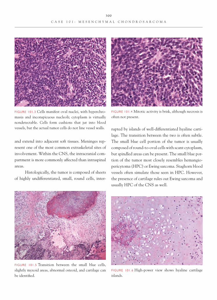
300
C A S E 1 0 1 : M E S E N C H Y M A L C H O N D R O S A R C O M A
and extend into adjacent soft tissues. Meninges rep-resent one of the most common extraskeletal sites of involvement. Within the CNS, the intracranial com-partment is more commonly affected than intraspinal areas.
Histologically, the tumor is composed of sheets of highly undifferentiated, small, round cells, inter-
rupted by islands of well-differentiated hyaline carti-lage. The transition between the two is often subtle. The small blue cell portion of the tumor is usually composed of round-to-oval cells with scant cytoplasm, but spindled areas can be present. The small blue por-tion of the tumor most closely resembles hemangio-pericytoma (HPC) or Ewing sarcoma. Staghorn blood vessels often simulate those seen in HPC. However, the presence of cartilage rules out Ewing sarcoma and usually HPC of the CNS as well.
FIGURE 101.5 Transition between the small blue cells, slightly myxoid areas, abnormal osteoid, and cartilage can be identifi ed.
FIGURE 101.4 Mitotic activity is brisk, although necrosis is often not present.
FIGURE 101.3 Cells manifest oval nuclei, with hyperchro-masia and inconspicuous nucleoli; cytoplasm is virtually nondetectable. Cells form cushions that jut into blood vessels, but the actual tumor cells do not line vessel walls.
FIGURE 101.6 High-power view shows hyaline cartilage islands.

301
C A S E 1 0 1 : M E S E N C H Y M A L C H O N D R O S A R C O M A
FIGURE 101.7 The cartilage component is S-100 immuno-reactive.
Immunoreactivity for vimentin and CD99 in the small cell component can be seen in mesenchy-mal chondrosarcoma, HPC, and Ewing. Chondroid areas are immunoreactive for S-100 protein. Osteoid and bone can also be found.
References 1. Devoe K, Weidner N. Immunohistochemistry of small round-
cell tumors. Sem Diagn Pathol 2000;17:216–24.
2. Nakashima Y, Park YK, Sugano O. Mesenchymal chondrosar-coma. In: Fletcher CDM, Unni KK, Mertens F, Editors. WHO Classifi cation of Tumours: Tumours of Soft Tissue and Bone. Lyon, FR: IARC Press;2002. p. 255–6.
3. Rajaram V, Brat DJ, Perry A. Anaplastic meningioma versus meningeal hemangiopericytoma: immunohistochemical and genetic markers. Hum Pathol 2004;35:1413–8.
4. Rushing EJ, Armonda RA, Ansari Q, et al. Mesenchymal chondrosarcoma: a clinicopathologic and fl ow cytometric study of 13 cases presenting in the central nervous system. Cancer 1996;77:1884–91.
FIGURE 101.8 The malignant, undifferentiated small blue cell component is immunoreactive for CD99.

This page intentionally left blank

303
I n d e x
Note: Page numbers followed by f refer to fi gures.
Acute lymphoblastic leukemia (ALL)in central nervous system, 287in spinal cord compression, 240
Acute myeloblastic leukemia, 287Acute promyelocytic leukemia, 286, 287Adamantinomatous versus papillary craniopharyngioma,
268–269Adjacent cortex
dysembryoplastic neuroepithelial tumor, 114, 116pleomorphic xanthoastrocytoma, 31
Adjuvant radiation therapyanaplastic meningioma, 212cystic craniopharyngioma, 266papillary glioneuronal tumor, 124papillary meningioma, 202supratentorial primitive neuroectodermal tumor, 152
Adults, tumors inchoroid plexus papilloma, 91cystic craniopharyngioma, 265hemangioblastoma, 231Langerhans cell histiocytosis, 255metastatic small cell carcinoma of lung, 283papillary craniopharyngioma, 269paraganglioma, 138pineal parenchymal tumor of intermediate
differentiation, 143, 144pituicytoma, 273–274pleomorphic xanthoastrocytoma, 32rosette-forming glioneuronal tumor of the fourth
ventricle, 126solitary fi brous tumor, 221
AE1/AE3 cytokeratins, immunoreactivity ofchordoma, 290
epithelioid glioblastoma, 40, 42intravascular lymphomatosis, 257
Anaplastic astrocytomacharacteristics, 13diagnosis, 14–15diagnostic images of, 13f, 14f, 15fgrading, 14treatment, 15
Anaplastic ependymomacharacteristics of, 80defi ned, 81diagnosis of, 81diagnostic images of, 80f, 81f, 82f, 83fgrading, 81–82location, 80–81prognosis, 82–83
Anaplastic gangliogliomacharacteristics, 120diagnosis, 121–122diagnostic images of, 120f, 121f, 122fgrading, 121
Anaplastic medulloblastomacharacteristics, 161diagnosis, 162diagnostic images of, 162f, 163fgrading, 162histological features, 162
Anaplastic meningioma, 204, 214, 226characteristics, 211diagnosis, 212–213diagnostic images of, 211f, 212f, 213fhistological features, 212
Anaplastic oligoastrocytomacharacteristics of, 65diagnosis of, 66

304
I N D E X
Anaplastic oligoastrocytoma (continued)diagnostic images of, 65f, 66fgrading, 65–66location, 65
Anaplastic oligodendroglioma, 38characteristics, 59diagnosis, 60–61diagnostic images of, 59f, 60f, 61fgrading, 61treatment, 61
Angioblastic meningiomas, 216Angioblastoma. See HemangioblastomaAngiocentric glioma
characteristics, 103–104diagnostic images of, 103f, 104fgrading, 104histological features, 104location, 103recurrence of, 104
Angiocentric neuroepithelial tumor, 104. See alsoAngiocentric glioma
Angiomatous meningiomacharacteristics, 214diagnosis, 215–216diagnostic images of, 214f, 215fhistological features, 214–215
Angiotropic large cell lymphoma. See Intravascular lymphomatosis
Antibody to alpha fetoprotein (AFP), 148, 149Antoni A and B tissues
cellular schwannoma, 183fi brous meningioma, 191melanotic schwannoma, 186, 187schwannoma with ancient change, 172tanycytic ependymoma, 84
Astroblastomacharacteristics, 106diagnosis, 107diagnostic images of, 106f, 107fgrading, 107histological features, 106–107location, 106
Astrocytic cellsin gemistocytic astrocytoma, 22, 23
in glioneuronal tumor with neuropil-like islands, 16in granular cell glioblastoma, 25in low-grade astrocytoma, 10, 11in pleomorphic xanthoastrocytoma, 32fin SEGA, 54in subependymal giant cell astrocytoma, 53, 53f
Astrocytomasanaplastic, 13–15desmoplastic infantile, 110–113gemistocytic, 22–24high-grade, 11low-grade, 10–12pilocytic, 45–48pilomyxoid, 49–51subependymal giant cell, 52–54
AT/RT. See Atypical teratoid/rhabdoid tumor (AT/RT)Atypical choroid plexus papilloma
characteristics of, 94diagnosis, 94–96diagnostic images of, 94f, 95fgrading, 95–96prognostic features of, 95recurrence, 95
Atypical meningiomaversus brain-invasive meningioma, 208characteristics, 209diagnosis, 210diagnostic images of, 209f, 210fgrading, 210histological features, 209–210
Atypical neurocytomacharacteristics, 131diagnostic images of, 131f, 132f, 133fhistological features, 132recurrence, 132
Atypical teratoid/rhabdoid tumor (AT/RT), 98versus CPC, 98–99characteristics, 164diagnosis, 166–167histological features, 165–166location, 165
B cell markers, 241, 246Bcl-2 antibody, 220, 244

305
I N D E X
CD30-positive, diffuse B-cell lymphoma, 2249CD34 antibody, 222
CD34 immunostaining, 179, 216, 219, 241CD38 immunostaining, 252CD43 immunostaining, 249CD45RB immunostaining
pituitary adenoma in an ectopic site, 281small cell glioblastoma, 38
CD45RB (leukocyte common antigen), 286CD45RO immunostaining, 249CD56 immunostaining, 283, 284CD56 (neural cell adhesion molecule), 252CD68 immunostaining, 44CD79a immunostaining, 241–242, 246, 249, 252CD99 immunostaining, 220, 301CD138 immunostaining, 252CDX2 immunostaining, 99Cell proliferation indices, 109Cell proliferation markers, 11Cellularity, 4Cellular schwannoma
characteristics, 183diagnosis, 184–185diagnostic images of, 183f, 184f, 185fgrading, 185histological features, 183–184location, 185versus MPNSTs, 185versus ordinary schwannomas, 184treatment, 185
Central nervous system (CNS) tumorsectopic meningioma, 194ETANTRs, 170fi brous meningioma, 190hemangiopericytoma, 218intracranial compartment, 110f, 300leukemic involvement of, 285–287malignant melanoma, 237meningeal melanocytoma, 235PNETs, 167primary sarcoma of, 224–227PTLDs, 248–249rhabdoid meningioma, 205supratentorial primitive neuroectodermal tumor, 151
Birbeck granules, 256Bone
chordoma, 289dural carcinomatosis, 297hemangiopericytoma, 217–218Langerhans cell histiocytosis, 254, 255mesenchymal chondrosarcoma, 299pineal teratoma, 263, 263fplasmacytoma, 251
Brachyury, 290Brain-invasive meningioma
characteristics, 207diagnostic images of, 207f, 208fgrading, 208histological features, 208surgery, 208
Brainstemembryonal tumor with abundant neuropil and true
rosettes, 170germinoma, 260gliomatosis cerebri, 43pilocytic astrocytoma, 46
Breast cancer, and meningioma, 191, 192Burkitt lymphoma, 241, 242
CA125, 99, 298CAM5.2 immunostaining
choroid plexus carcinoma, 99choroid plexus papilloma, 93dural carcinomatosis, 297, 298epithelioid glioblastoma, 42metastatic small cell carcinoma of lung, 284pituitary adenoma with apoplexy, 279primary sarcoma of the central nervous system, 226small cell glioblastoma, 38
Carbonic anhydrase, 196, 233Carcinoembryonic antigen (CEA), 149Carney complex, 187CD3 immunostaining, 246, 257, 258f, 298CD10 immunostaining, 99, 241CD19 immunostaining, 241, 249CD20 (B-cell marker), 286CD20 immunostaining, 242, 246, 249, 257, 298CD22 immunostaining, 242

306
I N D E X
clear cell ependymoma, 87Langerhans cell histiocytosis, 235medulloblastoma with extensive nodularity, 160pilocytic astrocytoma, 46pineal parenchymal tumor of intermediate
differentiation, 144pineoblastoma, 147pleomorphic xanthoastrocytoma, 32rosette-forming glioneuronal tumor of the fourth
ventricle, 126supratentorial primitive neuroectodermal
tumor, 152Chondrosarcoma versus chordoma, 289–290Chordoid glioma
characteristics of, 100diagnosis, 102diagnostic images of, 100f, 101fhistological features, 101–102location, 100treatment, 102
Chordoid meningioma, 102characteristics, 198diagnosis, 200diagnostic images of, 198f, 199fhistological feature, 199–200recurrence, 199
Chordoma, 102characteristics, 288diagnosis, 290diagnostic images of, 288f, 289fhistological features, 289–290location, 289symptoms, 289
Choroid plexus carcinoma (CPC)characteristics of, 97diagnosis, 98–99diagnostic images of, 97f, 98fgrading, 98histological features, 98location/prevalence, 97
Choroid plexus papillomacharacteristics, 91diagnosis, 92–93diagnostic images of, 91f, 92f, 93f
Central neurocytomacharacteristics, 128diagnosis, 129diagnostic images of, 128f, 129fhistological features, 128–129
Cerebellar arachnoidal sarcoma. See Desmoplastic medulloblastoma
Cerebellar neuroblastoma. See Medulloblastoma with extensive nodularity
Cerebellar parenchyma, 1, 45, 108, 109fCerebellum tumors, 43
biopsy, 1fchoroid plexus carcinoma, 97, 97fdysplastic cerebellar gangliocytoma, 108, 109embryonal tumor with abundant neuropil and true
rosettes, 170gliomatosis cerebri, 51hemangioblastoma, 232malignant melanoma, 237medulloblastomas, 154, 159pilocytic astrocytoma, 45, 46pilomyxoid astrocytoma, 50
Cerebral cortexangiocentric glioma, 103, 104desmoplastic infantile astrocytoma/
ganglioglioma, 110fleptomeningeal carcinomatosis, 294, 294f
Cerebrospinal fl uid (CSF), 295Cerebrum, 226Chemotherapy
for anaplastic medulloblastoma, 162for anaplastic oligodendroglioma, 61for classic medulloblastoma, 155for intravascular lymphomatosis, 259for low-grade oligodendroglioma, 58for pineal teratoma, 262for pineoblastoma, 147for post-transplant lymphoproliferative disorder, 250
Children, tumors inanaplastic ependymoma, 81atypical teratoid/rhabdoid tumor, 164, 165choroid plexus carcinoma, 97, 98choroid plexus papilloma, 91classic medulloblastoma, 155

307
I N D E X
of meningeal melanocytoma, 234of meningioangiomatosis, 228of pineocytoma, 140
Corpora amylacea, 2f, 3Cortical dysplasia, 116Cowden syndrome, and dysplastic cerebellar
gangliocytoma, 108–109CPC. See Choroid plexus carcinoma (CPC)Cranial nerve, tumors in
chordoma, 288, 289fi brous meningioma, 191MPNSTs, 181
Critical period, hemorrhage, 286CT. See Computed tomography (CT)Cutaneous trichilemmomas, 109Cystic craniopharyngioma
characteristics, 265diagnosis, 267diagnostic images of, 265f, 266f, 267fgrading, 266histological features, 266–267location, 266prognosis, 265–266symptoms, 265
Dense collagen, 111, 2180fDesmin, 226Desmoid tumors, 179Desmoplastic infantile astrocytoma/ganglioglioma (DIA/
DIG)characteristics, 110–111diagnosis, 111–113diagnostic images of, 110f, 111f, 112fgrading, 113histological features, 112–113location, 111treatment, 113
Desmoplastic medulloblastomacharacteristics, 156diagnostic images of, 156f, 157fhistological features, 157–158treatment, 158
DIA/DIG. See Desmoplastic infantile astrocytoma/ganglioglioma (DIA/DIG)
grading, 92locations, 91
Chromogranin, 138, 145Chromosomal deletions, 57–58, 61Chromosome 1p, 57, 61, 116Chromosome 19q, 57, 116Cilia, 89CK7, 99, 298CK7-positive cytokeratin, 92CK20, 298CK20-negative cytokeratin, 92c-kit, 261Classic medulloblastoma
charactersitics, 153diagnosis, 155diagnostic images of, 153f, 154fgrading, 155histological features, 154–155location, 154treatment, 155
Claudin-1, 179Clear cell ependymoma
characteristics of, 87diagnosis of, 88–89diagnostic images of, 87f, 88f, 89fgrading, 90locations, 87
Clear cell meningiomacharacteristics, 195diagnosis, 196diagnostic images of, 195f, 196fgrading, 195–196histological features, 196
c-Met pathway, 162c-Myc overexpression, 162Collagen IV, 85, 179, 188Computed tomography (CT)
of anaplastic oligodendroglioma, 59of desmoplastic medulloblastoma, 156dural carcinomatosis, 296of ganglioglioma, 117of leukemic involvement of the central nervous system, 285of low-grade astrocytoma, 10of medulloblastoma with extensive nodularity, 159

308
I N D E X
Embryonal tumor with abundant neuropil and true rosettes (ETANTR)
characteristics, 168diagnosis, 170diagnostic images of, 168f, 169fhistological features, 169–170
Encephalitis, 2Endodermal sinus tumor. See Yolk sac tumor of the pineal
glandEosinophilic cells, 25, 27, 273Eosinophilic granular bodies (EGBs), 48, 49f, 85, 119,
124, 274Ependymal differentiation, 81, 85, 88, 170Ependymoma
anaplastic, 80–83versus astroblastoma, 107characteristics of, 76clear cell, 87–90diagnosis of, 77–79diagnostic images of, 76f, 77f, 78fgrading, 79location, 76myxopapillary, 73–75tanycytic, 84–86
Epidermal growth factor receptor (EGFR), 31, 38, 61Epidural lymphomas, 242Epithelial cells
chordoid glioma, 100choroid plexus papilloma, 91, 92, 92fpituitary adenoma in an ectopic site, 280pituitary adenoma with apoplexy, 276, 276fsupratentorial primitive neuroectodermal tumor, 151
Epithelial membrane antigen (EMA)of angiomatous meningioma, 215–216of AT/RTs, 167of chordoid glioma, 102of choroid plexus carcinoma, 98of clear cell ependymomas, 89of ectopic meningioma, 194of ependymoma, 79of fi brous meningioma, 190of HPCs, 220of meningeal melanocytoma, 234of perineurioma, 179
Dural carcinomatosischaracteristics, 296diagnosis, 297–298diagnostic images of, 296f, 297f, 298fhistological features, 297prevalence, 297
Dysembryoplastic neuroepithelial tumorcharacteristics, 114diagnosis, 116diagnostic images of, 114f, 115f, 116fgrading, 115histological features of, 115–116location, 115treatment, 116
Dysplastic cerebellar gangliocytomacharacteristics, 108diagnosis, 109diagnostic images of, 108f, 109fhistological features, 108–109
Dystrophic mineralization, 7f, 57f
EBV. See Epstein-Barr virus (EBV)Ectopic meningioma
characteristics, 193diagnosis, 194diagnostic images of, 193f, 194fhistological features, 194location, 193–194
EGBs. See Eosinophilic granular bodies (EGBs)EGFR. See Epidermal growth factor receptor (EGFR)Electron microscopy
of clear cell ependymoma, 89, 89fof ependymoma, 79of Langerhans cell histiocytosis, 256of melanotic schwannoma, 188of tanycytic ependymoma, 86
EMA. See Epithelial membrane antigen (EMA)EMA immunostaining
of chordoma, 290of hemangioblastoma, 233of meningioangiomatosis, 230of pituicytoma, 274of primary sarcoma of the central nervous system, 226
Embryonal tumors, 153, 167, 283

309
I N D E X
ependymoma, 76, 77rosette-forming glioneuronal tumor, 97, 125–127subependymoma, 70
Gadolinium, 295Ganglioglioma
characteristics, 117diagnosis, 118–119diagnostic images of, 117f, 118fversus dysembryoplastic neuroepithelial tumor, 116grading, 119histological features, 118treatment, 119
Ganglionic cells, 108, 109f, 118f, 138GBM. See Glioblastoma multiforme (GBM)GCDFP immunostaining, 99, 298Gemistocytes, 23, 23f, 24f, 61Gemistocytic astrocytoma
diagnosis, 24diagnostic images of, 22f, 23f, 24fgrading, 23histological features of, 22and subependymal giant cell astrocytomas, 54
Genetic alterations, 61Germinoma, 149
characteristics, 260diagnosis, 261diagnostic images of, 260f, 261fhistological features, 261location, 260
GFAP immunostaininganaplastic ependymoma, 81astroblastoma, 107chordoid glioma, 102choroid plexus carcinoma, 98choroid plexus papilloma, 93clear cell ependymomas, 89desmoplastic infantile astrocytoma/ganglioglioma, 111dural carcinomatosis, 298ependymoma, 79epithelioid glioblastoma, 42gemistocytic astrocytoma, 22giant cell glioblastoma, 29gliomatosis cerebri, 44
Epithelioid glioblastomacharacteristics, 40diagnosis, 41–42diagnostic images of, 40f, 41flocation, 41versus pleomorphic xanthoastrocytoma, 41
Epstein-Barr virus (EBV), 249Erythropoietin, 232Estrogen receptors, 99, 298ETANTR. See Embryonal tumor with abundant neuropil
and true rosettes (ETANTR)Ewing sarcoma, 300. See also Hemangiopericytoma
(HPC)Extraventricular neurocytoma
characteristics, 134diagnosis, 135, 136fdiagnostic images of, 134f, 134f, 136fgrading, 135histological features, 134–135
Fibroblasts, 175Fibrous meningioma
characteristics, 190diagnosis, 191–192diagnostic images of, 190f, 191fgrading, 192histologic feature, 190–191treatment, 192
Fluorescence in situ hybridization (FISH)for Epstein-Barr virus, 249fin leukemic involvement of the central nervous system,
285, 286in low-grade oligodendroglioma, 55, 57fin lymphoblastic leukemia/lymphoma, 242in lymphoma with fi rst presentation as spinal cord
compression, 290in plasmacytoma, 252, 252f, 253ffor post-transplant lymphoproliferative disorder, 247
Follicle-stimulating hormone (FSH), 278Fontana Masson, 187, 2187fFourth ventricle, tumors of
anaplastic ependymoma, 81choroid plexus carcinoma, 97classic medulloblastoma, 153
310
I N D E X
grading, 18Gliosarcoma
characteristics, 34–35diagnosis, 35–36diagnostic images of, 34f, 35f, 36f
Gliosischaracteristics, 4diagnosis, 4–5diagnostic images of, 4f, 5fversus glioma, 10and hemangioblastomas, 232, 232fversus low-grade astrocytoma, 11and papillary glioneuronal tumor, 124
Glomeruloid vessels, 48GLUT-1, 179Gorlin syndrome. See Nevoid basal cell carcinoma
syndromegp100 antibody, 234Granular cell glioblastoma
characteristics, 27diagnosis, 28–29diagnostic images of, 25f, 26f, 27f
Granular cell tumor of the pituitary glandcharacteristics, 271diagnosis, 272diagnostic images of, 271f, 272fhistological features, 271–272
Growing teratoma syndrome, 263, 264
Hand-Schüller-Christian disease, 255HCG (beta-human chorionic gonadotrophin), 149Hemangioblastoma, 89, 215
characteristics, 231diagnosis, 233diagnostic images of, 231f, 232fhistological features, 232–233location, 232prevalence, 231and von Hippel-Lindau disease, 232
Hemangiopericytoma (HPC), 216, 300characteristics, 217diagnosis, 219–220diagnostic images of, 217f, 218f, 219fgrading, 219
GFAP immunostaining (continued)glioneuronal tumor with neuropil-like islands, 18gliosarcoma, 35granular cell tumor of the pituitary gland, 272myxopapillary ependymoma, 74paraganglioma, 139pilomyxoid astrocytoma, 51pituitary adenoma in an ectopic site, 281pleomorphic xanthoastrocytoma, 32primary sarcoma of the central nervous system, 226small cell glioblastoma, 38tanycytic ependymoma, 85
Ghost cells (wet keratin), 266f, 267Giangaspero, 160Giant cell glioblastoma
characteristics, 28diagnosis, 29–30diagnostic images of, 28f, 29f, 30fhistological features, 29
Glioblastomaepithelioid, 40–42giant cell, 28–30granular cell, 25–27small cell, 37–40
Glioblastoma multiforme (GBM), 13, 54, 284characteristics, 19diagnosis, 19–21diagnostic images of, 19f, 20f, 21fgrading, 20radiation therapy, 20
Glioblastoma with oligodendroglial componentcharacteristics of, 67diagnosis, 68–69diagnostic images of, 67f, 68f, 69fgrading, 68
Gliomatosis cerebricharacteristics, 43diagnosis, 43–44diagnostic images of, 43f, 44fgrading, 44
Glioneuronal tumor with neuropil-like islandscharacteristics, 16diagnosis, 17–18diagnostic images of, 16f, 17f, 18f

311
I N D E X
subependymal giant cell astrocytomas, 54Inhibin, 233INI1 protein, immunohistochemical analysis of
in AT/RT, 165f, 167in choroid plexus carcinoma, 99, 167
In situ hybridization. See Fluorescence in situ hybridization (FISH)
Intravascular lymphomatosischaracteristics, 257diagnosis, 258–259diagnostic images of, 257f, 258fhistological features, 257–258treatment, 259
Ki-67 immunostaininganaplastic astrocytoma, 14anaplastic ganglioglioma, 121anaplastic oligodendroglioma, 61atypical meningioma, 210ganglioglioma, 119gemistocytic astrocytoma, 22giant cell glioblastoma, 29–30glioblastoma multiforme, 21low-grade astrocytoma, 11low-grade oligodendroglioma, 57papillary glioneuronal tumor, 124subependymal giant cell astrocytoma, 52
Laminin, 85, 179, 187Langerhans cell granulomatosis. See Langerhans cell
histiocytosisLangerhans cell histiocytosis
characteristics, 254diagnosis, 256diagnostic images of, 254f, 255fhistological features, 254–255location, 255prognosis, 255–256
Large cell medulloblastoma. See Anaplastic medulloblastoma
Leptomeningeal carcinomatosischaracteristics, 294diagnosis, 295diagnostic images of, 294f, 295f
histological features, 217–218recurrence, 218
Histiocytosis X. See Langerhans cell histiocytosisHMB 45 antibody, immunoreactivity of
in anaplastic meningioma, 212in epithelioid glioblastoma, 42, 212, 238in malignant melanoma, 238, 239fin meningeal melanocytoma, 235, 235f
Homer Wright rosettesin classic medulloblastoma, 153, 154f, 155in pineocytoma, 140, 141, 155
HPC. See Hemangiopericytoma (HPC)Hydrocephalus, 148, 295Hypercellularity, 10Hypercellular parenchyma, 10f, 11, 43Hypothalamus, tumors in
chordoid gliomas, 35cystic craniopharyngioma, 266pilomyxoid astrocytoma, 50
IHC. See Immunohistochemistry (IHC)Immunohistochemistry (IHC)
anaplastic meningiomas, 212anaplastic oligodendroglioma, 61angiocentric glioma, 104central neurocytoma, 129chordoid glioma, 102chordoma, 290choroid plexus carcinoma, 98–99ependymoma, 79epithelioid glioblastoma, 42fi brous meningioma, 190ganglioglioma, 119gemistocytic astrocytoma, 24germinoma, 261gliomatosis cerebri, 44glioneuronal tumor with neuropil-like islands, 17granular cell tumor of the pituitary gland, 272leptomeningeal carcinomatosis, 295melanotic schwannoma, 187meningioangiomatosis, 230paraganglioma, 138small cell glioblastoma, 38solitary fi brous tumor, 222

312
I N D E X
Magnetic resonance imaging (MRI)anaplastic ependymoma, 80angiocentric glioma, 104choroid plexus papilloma, 91desmoplastic infantile astrocytoma/ganglioglioma, 110fdesmoplastic medulloblastoma, 156dural carcinomatosis, 296dysembryoplastic neuroepithelial tumor, 114dysplastic cerebellar gangliocytoma, 108epithelioid glioblastoma, 40extraventricular neurocytoma, 134fi brous meningioma, 190gemistocytic astrocytoma, 22gliomatosis cerebri, 43glioneuronal tumor with neuropil-like islands, 16gliosarcoma, 34Langerhans cell histiocytosis, 254leptomeningeal carcinomatosis, 295low-grade oligodendroglioma, 55for lymphoma with fi rst presentation as spinal cord
compression, 240medulloblastoma with extensive nodularity, 159meningeal melanocytoma, 234meningioangiomatosis, 228myxopapillary ependymoma, 73paraganglioma, 137pineal parenchymal tumor of intermediate
differentiation, 143pineocytoma, 140pituicytoma, 273pituitary adenoma in an ectopic site, 280plasmacytoma, 251tanycytic ependymoma, 84
Malformation of cortical development. See Cortical dysplasia
Malignant melanoma, 42, 187, 212characteristics, 237diagnosis, 238diagnostic images of, 237f, 238f, 239fhistological features, 238location, 237prevalence, 238
Malignant meningioma. See Anaplastic meningiomaMalignant peripheral nerve sheath tumor (MPNST)
versus cellular schwannoma, 185
histological features, 295location, 295
Leptomeningeal desmoplastic tumor, 111Letterer-Siwe disease, 255Leukemias, classifi cation of, 286–287Leukemic involvement of the central nervous system
characteristics, 285diagnosis, 286–287diagnostic images of, 285f, 286fhistological features, 286
Lhermitte-Duclos disease. See Dysplastic cerebellar gangliocytoma
Localized hypertrophic neuropathy, defi ned, 179Low-grade astrocytomas, 5
characteristics, 10diagnosis, 11–12diagnostic images of, 10f, 11f, 12flocation, 11
Low-grade oligoastrocytomacharacteristics, 62diagnostic criteria, 63diagnostic images of, 62f, 63fgrading, 63prognosis, 64
Low-grade oligodendrogliomacharacteristic feature of, 56–57characteristics, 55diagnosis, 57–58diagnostic images of, 55f, 56f, 57flocation, 56prognosis, 58
Lung tumorsectopic meningioma, 194hemangiopericytoma, 218leptomeningeal carcinomatosis, 295metastatic small cell carcinoma of lung, 282–284
Luteinizing hormone, 278Lymphoma with fi rst presentation as spinal cord
compression (Leukemia)characteristics, 240classifi cation of, 242diagnosis, 242diagnostic images of, 240f, 241f, 242fhistological features, 241locations, 242
313
I N D E X
diagnostic images of, 299f, 300f, 301fhistological features, 300location, 299–300
Metastatic papillary carcinoma of the thyroidcharacteristics, 291diagnostic images of, 291f, 291fhistological features, 292–293location, 292
Metastatic small cell carcinoma of lungcharacteristics, 282diagnosis, 283–284diagnostic images of, 282f, 283fhistological features, 283location, 283
MIB-1, immunohistochemical analysisanaplastic astrocytoma, 14AT/RTs, 167atypical choroid plexus papilloma, 96atypical meningioma, 210extraventricular neurocytoma, 135lymphoblastic leukemia/lymphoma, 241pilocytic astrocytoma, 48, 48fpituitary adenoma in an ectopic site, 281
Microcysts, 71, 71fMicrohemorrhages, 172Microvilli, 89, 89fMinigemistocytes, 24, 59, 61Mixed glioma, anaplastic. See Anaplastic oligoastrocytomaMixed glioma, low-grade. See Low-grade oligoastrocytomaMonomorphous angiocentric glioma. See Angiocentric gliomaMPNST. See Malignant peripheral nerve sheath tumor
(MPNST)MRI. See Magnetic resonance imaging (MRI)Mucin staining
basophilic, 71, 71fin chordoid meningioma, 200eosinophilic, 47f, 48in leptomeningeal carcinomatosis, 295in myxopapillary ependymoma, 74, 75
Mucosa-associated lymphoid tissue (MALT), 242, 245, 253Myxopapillary ependymoma
characteristics of, 73diagnosis, 73–74diagnostic images of, 73f, 74f, 75fgrading, 75
characteristics, 180diagnostic images of, 180f, 181fgrading, 181histological features, 181–182location, 181prevalence, 180–181
MALT. See Mucosa-associated lymphoid tissue (MALT)Marginal zone B-cell lymphoma
characteristics, 244–245diagnostic images of, 244f, 245f, 246fhistological features, 245–246location, 245prevalence, 245prognosis, 246symptoms, 245
Medulloblastoma with extensive nodularitycharacteristics, 159diagnosis, 160diagnostic images of, 159f, 160fhistological features, 160
Melan-A, 42, 212, 238, 298Melanin pigment, 187, 188f, 238Melanocytic tumors. See Meningeal melanocytomaMelanotic schwannoma
characteristics, 186diagnosis, 188diagnostic images of, 186f, 187f, 188fhistological features, 187treatment, 188–189
Meningeal melanocytomacharacteristics, 234diagnosis, 236diagnostic images of, 234f, 235fhistological features, 235–236prevalence, 235recurrence, 236
Meningioangiomatosischaracteristics, 228diagnosis, 230diagnostic images of, 228f, 229fhistological features, 229–230
Meningothelial (syncytial) meningiomas, 192Mesenchymal chondrosarcoma
characteristics, 299diagnosis, 301
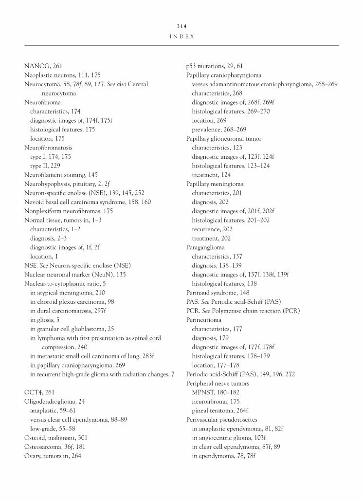
314
I N D E X
p53 mutations, 29, 61Papillary craniopharyngioma
versus adamantinomatous craniopharyngioma, 268–269characteristics, 268diagnostic images of, 268f, 269fhistological features, 269–270location, 269prevalence, 268–269
Papillary glioneuronal tumorcharacteristics, 123diagnostic images of, 123f, 124fhistological features, 123–124treatment, 124
Papillary meningiomacharacteristics, 201diagnosis, 202diagnostic images of, 201f, 202fhistological features, 201–202recurrence, 202treatment, 202
Paragangliomacharacteristics, 137diagnosis, 138–139diagnostic images of, 137f, 138f, 139fhistological features, 138
Parinaud syndrome, 148PAS. See Periodic acid-Schiff (PAS)PCR. See Polymerase chain reaction (PCR)Perineurioma
characteristics, 177diagnosis, 179diagnostic images of, 177f, 178fhistological features, 178–179location, 177–178
Periodic acid-Schiff (PAS), 149, 196, 272Peripheral nerve tumors
MPNST, 180–182neurofi broma, 175pineal teratoma, 264f
Perivascular pseudorosettesin anaplastic ependymoma, 81, 82fin angiocentric glioma, 103fin clear cell ependymoma, 87f, 89in ependymoma, 78, 78f
NANOG, 261Neoplastic neurons, 111, 175Neurocytoma, 58, 78f, 89, 127. See also Central
neurocytomaNeurofi broma
characteristics, 174diagnostic images of, 174f, 175fhistological features, 175location, 175
Neurofi bromatosistype I, 174, 175type II, 229
Neurofi lament staining, 145Neurohypophysis, pituitary, 2, 2fNeuron-specifi c enolase (NSE), 139, 145, 252Nevoid basal cell carcinoma syndrome, 158, 160Nonplexiform neurofi bromas, 175Normal tissue, tumors in, 1–3
characteristics, 1–2diagnosis, 2–3diagnostic images of, 1f, 2flocation, 1
NSE. See Neuron-specifi c enolase (NSE)Nuclear neuronal marker (NeuN), 135Nuclear-to-cytoplasmic ratio, 5
in atypical meningioma, 210in choroid plexus carcinoma, 98in dural carcinomatosis, 297fin gliosis, 5in granular cell glioblastoma, 25in lymphoma with fi rst presentation as spinal cord
compression, 240in metastatic small cell carcinoma of lung, 283fin papillary craniopharyngioma, 269in recurrent high-grade glioma with radiation changes, 7
OCT4, 261Oligodendroglioma, 24
anaplastic, 59–61versus clear cell ependymoma, 88–89low-grade, 55–58
Osteoid, malignant, 301Osteosarcoma, 36f, 181Ovary, tumors in, 264
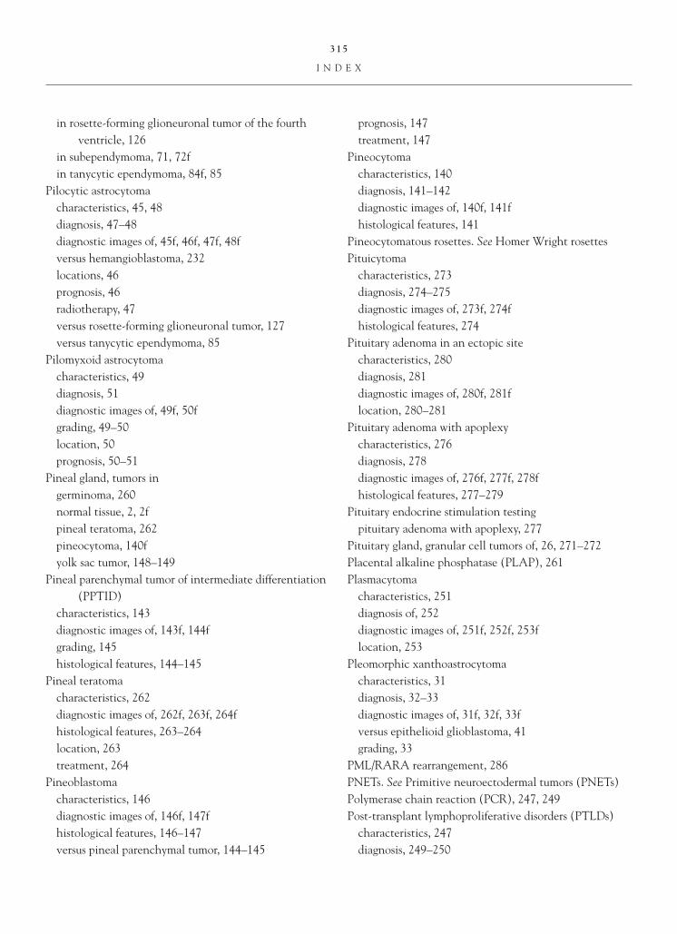
315
I N D E X
prognosis, 147treatment, 147
Pineocytomacharacteristics, 140diagnosis, 141–142diagnostic images of, 140f, 141fhistological features, 141
Pineocytomatous rosettes. See Homer Wright rosettesPituicytoma
characteristics, 273diagnosis, 274–275diagnostic images of, 273f, 274fhistological features, 274
Pituitary adenoma in an ectopic sitecharacteristics, 280diagnosis, 281diagnostic images of, 280f, 281flocation, 280–281
Pituitary adenoma with apoplexycharacteristics, 276diagnosis, 278diagnostic images of, 276f, 277f, 278fhistological features, 277–279
Pituitary endocrine stimulation testingpituitary adenoma with apoplexy, 277
Pituitary gland, granular cell tumors of, 26, 271–272Placental alkaline phosphatase (PLAP), 261Plasmacytoma
characteristics, 251diagnosis of, 252diagnostic images of, 251f, 252f, 253flocation, 253
Pleomorphic xanthoastrocytomacharacteristics, 31diagnosis, 32–33diagnostic images of, 31f, 32f, 33fversus epithelioid glioblastoma, 41grading, 33
PML/RARA rearrangement, 286PNETs. See Primitive neuroectodermal tumors (PNETs)Polymerase chain reaction (PCR), 247, 249Post-transplant lymphoproliferative disorders (PTLDs)
characteristics, 247diagnosis, 249–250
in rosette-forming glioneuronal tumor of the fourth ventricle, 126
in subependymoma, 71, 72fin tanycytic ependymoma, 84f, 85
Pilocytic astrocytomacharacteristics, 45, 48diagnosis, 47–48diagnostic images of, 45f, 46f, 47f, 48fversus hemangioblastoma, 232locations, 46prognosis, 46radiotherapy, 47versus rosette-forming glioneuronal tumor, 127versus tanycytic ependymoma, 85
Pilomyxoid astrocytomacharacteristics, 49diagnosis, 51diagnostic images of, 49f, 50fgrading, 49–50location, 50prognosis, 50–51
Pineal gland, tumors ingerminoma, 260normal tissue, 2, 2fpineal teratoma, 262pineocytoma, 140fyolk sac tumor, 148–149
Pineal parenchymal tumor of intermediate differentiation (PPTID)
characteristics, 143diagnostic images of, 143f, 144fgrading, 145histological features, 144–145
Pineal teratomacharacteristics, 262diagnostic images of, 262f, 263f, 264fhistological features, 263–264location, 263treatment, 264
Pineoblastomacharacteristics, 146diagnostic images of, 146f, 147fhistological features, 146–147versus pineal parenchymal tumor, 144–145

316
I N D E X
diagnostic images of, 6f, 7f, 8ftreatment, 7
Renal cell carcinomas, 196, 233Reticulin staining
in desmoplastic infantile astrocytoma/ganglioglioma, 111in gliosarcoma, 35in hemangiopericytoma, 219in pleomorphic xanthoastrocytoma, 32in tanycytic ependymoma, 85
Rhabdoid meningiomacharacteristics, 204diagnostic images of, 204f, 205fhistological features, 205–206recurrence, 206
Rosenthal fi bers, 47, 100, 232, 272Rosette-forming glioneuronal tumor of the fourth ventricle
characteristics, 125diagnosis, 127diagnostic images of, 125f, 126fhistological features, 126–127
S-100 protein, staining ofin anaplastic meningioma, 212in astroblastoma, 107in cellular schwannoma, 185in chordoid meningioma, 200chordoma, 290in choroid plexus carcinoma, 98in clear cell ependymomas, 89in ectopic meningioma, 194in ependymoma, 79in epithelioid glioblastoma, 42in granular cell tumor of the pituitary gland, 272in malignant melanoma, 238in meningeal melanocytoma, 234in mesenchymal chondrosarcoma, 301in myxopapillary ependymoma, 74in paraganglioma, 138–139in perineurioma, 179in pituicytoma, 275in pituitary adenoma in an ectopic site, 281in primary sarcoma of the central nervous system, 226in tanycytic ependymoma, 85
Schiller-Duval bodies, 149
Post-transplant lymphoproliferative disorders (PTLDs) (continued)
diagnostic images of, 247f, 248f, 249fhistological features, 248–249prevalence, 248treatment, 250
PPTID. See Pineal parenchymal tumor of intermediate differentiation (PPTID)
Primary sarcoma of the central nervous systemcharacteristics, 224diagnosis, 226diagnostic images of, 224f, 225f, 226fgrading, 226histological features, 224–225location, 226prognosis, 226
Primitive neuroectodermal tumors (PNETs), 167, 283Prolactin, 280, 281, 281fPsammoma bodies, 212, 229Psammomatous versus non-psammomatous melanotic
schwannomas, 187Pseudopalisading necrosis, 81, 283Pseudotumor, infl ammatory, 252PTCH1 gene, 158PTEN gene, 109PTLDs. See Post-transplant lymphoproliferative disorders
(PTLDs)
Radiation therapyfor anaplastic astrocytoma, 15for anaplastic oligodendroglioma, 61for astroblastoma, 107for children with ALL, 287for classic medulloblastoma, 155for glioblastoma multiforme, 20for medulloblastoma with extensive nodularity, 160for pineal teratoma, 262for pineoblastoma, 147for post-transplant lymphoproliferative disorder, 250for recurrent high-grade glioma, 6–9
RCC antibodies, 196, 233Recurrent high-grade glioma with radiation changes
characteristics, 6diagnosis, 7–9

317
I N D E X
grading, 71Subpial aggregation of tumor cells
astroblastoma, 107fgliomatosis cerebri, 44flow-grade astrocytoma, 11, 12flow-grade oligodendroglioma, 57f
Supratentorial primitive neuroectodermal tumorcharacteristics, 150diagnostic images of, 150f, 151fhistological features, 151–152location, 151
Surgeryfor anaplastic meningioma, 212for brain-invasive meningioma, 208for cellular schwannoma, 185for melanotic schwannoma, 188–189for papillary meningioma, 202for pituitary adenoma with apoplexy, 277
Synaptophysin immunoreactivity, 18, 112, 124, 145, 279, 283, 298
Tanycytic ependymomacharacteristics of, 84diagnosis, 85–86diagnostic images of, 84f, 85fgrading, 85location, 85
T-cell markers, 246T cells, non-neoplastic, 249TdT, 241Temporal lobe tumors, 115, 118Teratoma, immature
pineal teratoma, 262, 263pineoblastoma, 147
Third ventricle, tumors ofchordoid glioma, 101choroid plexus papilloma, 92papillary craniopharyngioma, 269pituitary adenoma in an ectopic site, 281rosette-forming glioneuronal tumor of the fourth
ventricle, 127Thrombocytopenia, 286, 287Thyroglobulin, 291, 292
Schwann cells, 175Schwannoma with ancient change
characteristics, 171diagnosis, 171–172, 2172fdiagnostic images of, 171f, 172f, 173fhistological features, 172versus tanycytic ependymoma, 85
SEGA. See Subependymal giant cell astrocytoma (SEGA)
Small blue cell tumors, 166, 276f, 300Small cell glioblastoma
characteristics, 37diagnosis, 37–38diagnostic images of, 37f, 38f, 39f
Smooth muscle actin (SMA) stainingin AT/RTs, 2165f, 167in choroid plexus carcinoma, 99in primary sarcoma of the central nervous system, 226
Solitary fi brous tumorcharacteristics, 221diagnosis, 222–223diagnostic images of, 221f, 222fhistological features, 221–222
Spinal cord, tumors incervico-thoracic, 76ectopic meningioma, 193ependymomas, 77paragangliomas, 138tanycytic ependymoma, 86
Spindle cells, 34, 44, 172Squamous epithelium, 269Steroids, 277Subependymal giant cell astrocytoma (SEGA)
characteristics, 52diagnosis, 54diagnostic images of, 52f, 53flocations, 53and tuberous sclerosis, 52–53
Subependymal zone, 2, 2fSubependymoma
characteristics of, 70diagnosis, 71–72diagnostic images of, 70f, 71f, 72f

318
I N D E X
WHO grade II tumorsanaplastic astrocytoma, 14brain-invasive meningioma, 208clear cell ependymomas, 90HPCs, 220pilomyxoid astrocytoma, 50subependymoma, 71tanycytic ependymoma, 85
WHO grade III tumorsanaplastic ependymoma, 81atypical choroid plexus papilloma, 96clear cell ependymoma, 90CPC, 98gemistocytic astrocytoma, 23glioneuronal tumor with neuropil-like islands, 18pineal parenchymal tumor of intermediate
differentiation, 145WHO grade IV tumors
anaplastic astrocytoma, 14anaplastic medulloblastoma, 162classic medulloblastoma, 155desmoplastic infantile astrocytoma/ganglioglioma, 113malignant peripheral nerve sheath tumor, 181medulloblastoma with extensive nodularity, 159MPNST, 181pineal parenchymal tumor of intermediate
differentiation, 144primary sarcoma of the central nervous system, 226
Yolk sac tumor of the pineal glandcharacteristics, 148diagnosis, 149diagnostic images of, 148f, 149fhistological features, 148–149location, 148prognosis, 149
Transthyretin, 98TTF-1 (thyroid transcription factor-1) staining
in choroid plexus carcinoma, 99in dural carcinomatosis, 284, 292–293, 297–298, 298in metastatic papillary carcinoma of the thyroid, 291,
292in metastatic small cell carcinoma of lung, 283, 284
Tuberous sclerosis, and SEGA, 52–53
Vascular sclerosis, 8f, 107, 230Verocay bodies, 173f, 183, 187Vimentin immunostaining
in AT/RTs, 167in chordoid glioma, 102in choroid plexus carcinoma, 98in clear cell ependymomas, 89in ependymoma, 79in granular cell tumor of the pituitary gland, 272in mesenchymal chondrosarcoma, 301in myxopapillary ependymoma, 74in pituicytoma, 274in primary sarcoma of the central nervous system, 226in tanycytic ependymoma, 85
von Hippel-Lindau disease, 232. See alsoHemangioblastoma
Wet keratin, 266, 267, 269f, 270WHO classifi cation of tumors, 17, 81, 95, 208, 273WHO grade I tumors
cellular schwannoma, 185clear cell meningioma, 195cystic craniopharyngioma, 266desmoplastic infantile astrocytoma/ganglioglioma, 113fi brous meningiomas, 192myxopapillary ependymoma, 75