8 Benign Neoplasms and Tumor-Like Lesions
52
8 Benign Neoplasms and Tumor-Like Lesions Roberto Maroldi, Marco Berlucchi, Davide Farina, Davide Tomenzoli, Andrea Borghesi, and Luca Pianta R. Maroldi, Professor, MD; D. Farina, MD; A. Borghesi, MD Department of Radiology University of Brescia, Piazzale Spedali Civili 1, Brescia, 25123, Italy M. Berlucchi, MD; D. Tomenzoli, MD; L. Pianta, MD Department of Otorhinolaryngology University of Brescia, Piazzale Spedali Civili 1, Brescia, 25123, Italy 8.1 Osteoma 8.1.1 Definition, Epidemiology, Pattern of Growth Osteoma is a benign, slow-growing osteoblastic tu- mor, more commonly located in the outer table of the calvarium, the mandible, the paranasal sinuses, and occasionally in tubular bones (Samy and Mostafa 1971; Kransdorf et al. 1991; Sayan et al. 2002). It is the most frequent benign tumor of the nose and sinonasal, since being found in 1% of patients who undergo plain sinus radiographs and in 3% of CT examinations obtained for sinonasal symptoms (Earwaker 1993). Osteoma usually presents between the third and sixth decade of life, with a male pre- dominance of 1.5–2.6:1 (Metha and Grewal 1963; Atallah and Jay 1981). About 80% of osteomas oc- cur in the frontal sinus, while ethmoid and maxillary sinus are affected in about 20% of cases (Earwaker 1993). Isolated cases of sphenoid osteoma have been observed (Fu and Perzin 1974). CONTENTS 8.1 Osteoma 107 8.1.1 Definition, Epidemiology, Pattern of Growth 107 8.1.2 Clinical and Endoscopic Findings 108 8.1.3 Treatment Guidelines 108 8.1.4 Key Information to Be Provided by Imaging 109 8.1.5 Imaging Findings 109 8.1.6 Follow-Up 110 8.2 Fibrous Dysplasia and Ossifying Fibroma 110 8.2.1 Definition, Epidemiology, Pattern of Growth 110 8.2.2 Clinical and Endoscopic Findings 111 8.2.3 Treatment Guidelines 112 8.2.4 Key Information to Be Provided by Imaging 112 8.2.5 Imaging Findings 112 8.2.6 Follow-Up 114 8.3 Aneurysmal Bone Cyst 114 8.3.1 Definition, Epidemiology, Pattern of Growth 114 8.3.2 Clinical and Endoscopic Findings 115 8.3.3 Treatment Guidelines 115 8.3.4 Key Information to Be Provided by Imaging 115 8.3.5 Imaging Findings 115 8.3.6 Follow-Up 116 8.4 Sinonasal Mucocele 117 8.4.1 Definition, Epidemiology, Pattern of Growth 117 8.4.2 Clinical and Endoscopic Findings 117 8.4.3 Treatment Guidelines 117 8.4.4 Key Information to Be Provided by Imaging 118 8.4.5 Imaging Findings 118 8.4.6 Follow-Up 121 8.5 Abnormal Sinus Pneumatization 121 8.5.1 Definition and Epidemiology 121 8.5.2 Pathogenesis 121 8.5.3 Clinical and Endoscopic Findings 121 8.5.4 Treatment Guidelines 122 8.5.5 Key Information to Be Provided by Imaging 122 8.5.6 Imaging Findings 122 8.6 Inverted Papilloma and Other Schneiderian Papillomas 122 8.6.1 Definition, Epidemiology, Pattern of Growth 122 8.6.2 Clinical and Endoscopic Findings 124 8.6.3 Treatment Guidelines 125 8.6.4 Key Information to Be Provided by Imaging 126 8.6.5 Imaging Findings 126 8.6.6 Follow-Up 131 8.7 Juvenile Angiofibroma 131 8.7.1 Definition, Epidemiology, Pattern of Growth 131 8.7.2 Clinical and Endoscopic Findings 132 8.7.3 Treatment Guidelines 132 8.7.4 Key Information to Be Provided by Imaging 133 8.7.5 Imaging Findings 133 8.7.6 Follow-Up 141 8.8 Pyogenic Granuloma (Lobular Capillary Hemangioma) 145 8.8.1 Definition, Epidemiology, Pattern of Growth 145 8.8.2 Treatment Guidelines 145 8.8.3 Key Information to Be Provided by Imaging 146 8.8.4 Imaging Findings 146 8.9 Unusual Benign Lesions 147 8.9.1 Pleomorphic Adenoma (Mixed Tumor) 147 8.9.2 Schwannoma 149 8.9.3 Leiomyoma 150 8.9.4 Paraganglioma 151 References 152
-
Upload
khangminh22 -
Category
Documents
-
view
0 -
download
0
Transcript of 8 Benign Neoplasms and Tumor-Like Lesions

Benign Neoplasms and Tumor-Like Lesions 107
8 Benign Neoplasms and Tumor-Like Lesions Roberto Maroldi, Marco Berlucchi, Davide Farina, Davide Tomenzoli, Andrea Borghesi, and Luca Pianta
R. Maroldi, Professor, MD; D. Farina, MD; A. Borghesi, MDDepartment of Radiology University of Brescia, Piazzale Spedali Civili 1, Brescia, 25123, ItalyM. Berlucchi, MD; D. Tomenzoli, MD; L. Pianta, MDDepartment of Otorhinolaryngology University of Brescia, Piazzale Spedali Civili 1, Brescia, 25123, Italy
8.1 Osteoma
8.1.1 Defi nition, Epidemiology, Pattern of Growth
Osteoma is a benign, slow-growing osteoblastic tu-mor, more commonly located in the outer table of the calvarium, the mandible, the paranasal sinuses, and occasionally in tubular bones (Samy and Mostafa 1971; Kransdorf et al. 1991; Sayan et al. 2002).
It is the most frequent benign tumor of the nose and sinonasal, since being found in 1% of patients who undergo plain sinus radiographs and in 3% of CT examinations obtained for sinonasal symptoms (Earwaker 1993). Osteoma usually presents between the third and sixth decade of life, with a male pre-dominance of 1.5–2.6:1 (Metha and Grewal 1963; Atallah and Jay 1981). About 80% of osteomas oc-cur in the frontal sinus, while ethmoid and maxillary sinus are affected in about 20% of cases (Earwaker 1993). Isolated cases of sphenoid osteoma have been observed (Fu and Perzin 1974).
CONTENTS
8.1 Osteoma 1078.1.1 Defi nition, Epidemiology, Pattern of Growth 1078.1.2 Clinical and Endoscopic Findings 1088.1.3 Treatment Guidelines 1088.1.4 Key Information to Be Provided by Imaging 1098.1.5 Imaging Findings 1098.1.6 Follow-Up 1108.2 Fibrous Dysplasia and Ossifying Fibroma 1108.2.1 Defi nition, Epidemiology, Pattern of Growth 1108.2.2 Clinical and Endoscopic Findings 1118.2.3 Treatment Guidelines 1128.2.4 Key Information to Be Provided by Imaging 1128.2.5 Imaging Findings 1128.2.6 Follow-Up 1148.3 Aneurysmal Bone Cyst 1148.3.1 Defi nition, Epidemiology, Pattern of Growth 1148.3.2 Clinical and Endoscopic Findings 1158.3.3 Treatment Guidelines 1158.3.4 Key Information to Be Provided by Imaging 1158.3.5 Imaging Findings 1158.3.6 Follow-Up 1168.4 Sinonasal Mucocele 1178.4.1 Defi nition, Epidemiology, Pattern of Growth 1178.4.2 Clinical and Endoscopic Findings 1178.4.3 Treatment Guidelines 1178.4.4 Key Information to Be Provided by Imaging 1188.4.5 Imaging Findings 1188.4.6 Follow-Up 1218.5 Abnormal Sinus Pneumatization 1218.5.1 Defi nition and Epidemiology 1218.5.2 Pathogenesis 1218.5.3 Clinical and Endoscopic Findings 1218.5.4 Treatment Guidelines 1228.5.5 Key Information to Be Provided by Imaging 1228.5.6 Imaging Findings 1228.6 Inverted Papilloma and Other Schneiderian Papillomas 1228.6.1 Defi nition, Epidemiology, Pattern of Growth 1228.6.2 Clinical and Endoscopic Findings 1248.6.3 Treatment Guidelines 1258.6.4 Key Information to Be Provided by Imaging 1268.6.5 Imaging Findings 126
8.6.6 Follow-Up 1318.7 Juvenile Angiofi broma 1318.7.1 Defi nition, Epidemiology, Pattern of Growth 1318.7.2 Clinical and Endoscopic Findings 1328.7.3 Treatment Guidelines 1328.7.4 Key Information to Be Provided by Imaging 1338.7.5 Imaging Findings 1338.7.6 Follow-Up 1418.8 Pyogenic Granuloma (Lobular Capillary Hemangioma) 1458.8.1 Defi nition, Epidemiology, Pattern of Growth 1458.8.2 Treatment Guidelines 1458.8.3 Key Information to Be Provided by Imaging 1468.8.4 Imaging Findings 1468.9 Unusual Benign Lesions 1478.9.1 Pleomorphic Adenoma (Mixed Tumor) 1478.9.2 Schwannoma 1498.9.3 Leiomyoma 1508.9.4 Paraganglioma 151 References 152
108 R. Maroldi et al.
Histologically, osteomas are divided into three categories: (1) the ivory or “eburnated” osteoma is composed of hard, dense bone, lacking a haversian system and containing only a minimal amount of fi brous tissue; (2) the osteoma spongiosum (mature osteoma) contains more trabecular tissue and often marrow component. It is surrounded by mature can-cellous bone with small haversian systems; (3) the mixed osteoma contains both aspects of ivory and mature subtypes (Fu and Perzin 1974).
The etiology of the lesion is still unknown and speculative. Three main hypotheses have been proposed: (1) the embryologic theory, which pos-tulates that osteoma arises at the junction be-tween the embryonic cartilaginous ethmoid and the membranous frontal bones, does not explain the large number of osteomas located far from the fronto-ethmoid junction; (2) the traumatic theory identifies a previous trauma as the possible patho-genetic event; (3) the infective theory is related to the concomitant observation of osteoma and rhi-nosinusitis in about 30% of patients. Other theo-ries consider osteomas osteodysplastic lesions, osteogenic hamartomas, embryonic bone rests, or as the result of sinus polyp ossification (Namdar et al. 1998).
Osteomas can occur in conjunction with Gardner’s syndrome, which is characterized by the association of osteomas with intestinal polyposis and multiple soft tissue tumors, such as dermoid cysts and fi bro-mas (Atallah and Jay 1981).
Osteomas of the paranasal sinuses are often as-ymptomatic and may be serendipitously detected on routine radiologic examinations. Furthermore, tu-mor growth is slow and occasionally characterized by a tendency to displace and compress the surround-ing structures (about 10% of cases). Intracranial and intraorbital extent of sinus osteomas may occur as a late event (Rappaport and Attia 1994; Namdar et al. 1998; Huang et al. 2001).
8.1.2 Clinical and Endoscopic Findings
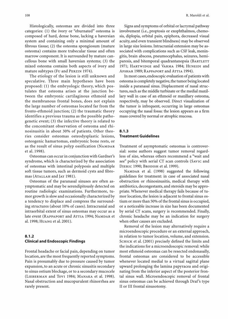
Frontal headache or facial pain, depending on tumor location, are the most frequently reported symptoms. Pain is presumably due to pressure caused by tumor expansion, to an acute or chronic sinusitis secondary to sinus ostium blockage, or to a secondary mucocele (Lieberman and Tovi 1984; Manaka et al. 1998). Nasal obstruction and mucopurulent rhinorrhea are rarely present.
Signs and symptoms of orbital or lacrymal pathway involvement (i.e., proptosis or exophthalmos, chemo-sis, diplopia, orbital pain, epiphora, decreased visual acuity, and even transient blindness) may be observed in large size lesions. Intracranial extension may be as-sociated with complications such as CSF leak, menin-gitis, brain abscess, pneumocephalus, seizures, hemi-paresis, and bitemporal quadrantanopsia (Bartlett 1971; Hartwidge and Varma 1984; Huneidi and Afshar 1989; Rappaport and Attia 1994).
In most cases, endoscopic evaluation of patients with osteoma is completely negative, the tumor being located inside a paranasal sinus. Displacement of nasal struc-tures, such as the middle turbinate or the medial maxil-lary wall in case of an ethmoid or maxillary osteoma, respectively, may be observed. Direct visualization of the tumor is infrequent, occurring in large osteomas occupying the nasal fossa: the lesion appears as a fi rm mass covered by normal or atrophic mucosa.
8.1.3 Treatment Guidelines
Treatment of asymptomatic osteomas is controver-sial: some authors suggest tumor removal regard-less of size, whereas others recommend a “wait and see” policy with serial CT scan controls (Savic and Djeric 1990; Brodish et al. 1999).
Namdar et al. (1998) suggested the following guidelines for treatment: in case of associated nasal obstruction or rhinosinusitis, medical therapy with antibiotics, decongestants, and steroids may be appro-priate. Whenever medical therapy fails because of tu-mor location, the lesion is adjacent to frontal sinus os-tium or more than 50% of the frontal sinus is occupied, or a noticeable increase in size has been documented by serial CT scans, surgery is recommended. Finally, chronic headache may be an indication for surgery when other causes are excluded.
Removal of the lesion may alternatively require a microendoscopic procedure or an external approach, in relation to tumor location, volume, and extension. Schick et al. (2001) precisely defi ned the limits and the indications for a microendoscopic removal: while most ethmoid osteomas can be resected endonasally, frontal osteomas are considered to be accessible whenever located medial to a virtual sagittal plane upward prolonging the lamina papyracea and origi-nating from the inferior aspect of the posterior fron-tal sinus wall. Microendoscopic removal of frontal sinus osteomas can be achieved through Draf ’s type II or III frontal sinusotomy.
Benign Neoplasms and Tumor-Like Lesions 109
Tumor extension beyond the paranasal sinuses, or large osteomas which can not be entirely exposed through the nose must be removed via an external approach (Smith and Calcaterra 1989; Namdar et al. 1998; Brodish et al. 1999; Schick et al. 2001). Osteoplastic frontal sinusotomy (with or without fat obliteration), with a coronal or uni-/bilateral brow incision, allows direct access to the frontal sinus and removal of osteomas with anterior and/or posterior wall involvement. Craniofacial resection has been described for the removal of giant fronto-ethmoid osteomas with intracranial complications (Blitzer et al. 1989).
8.1.4 Key Information to Be Provided by Imaging
� Location, extent, and site of origin � Potential impairment of drainage pathways� Extension beyond paranasal sinuses� Position in respect to the lamina papyracea and
inferior and posterior frontal sinus wall (endona-sal accessibility)
� Presence of possible complications (intracranial and/or intraorbital)
8.1.5 Imaging Findings
Diagnosis of Osteoma is generally simple diagnosis, based on the detection – either on X-ray fi lms or
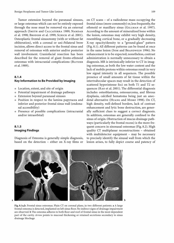
on CT scans – of a radiodense mass occupying the frontal sinus (more commonly) or, less frequently, the ethmoid or maxillary sinus (Gillman et al. 1997). According to the amount of mineralized bone within the lesion, osteomas may exhibit very high density, resembling cortical bone, or a gradually decreasing X-ray opacity/density to a “ground-glass” pattern (Fig. 8.1). All different patterns can be found at once in the same lesion (Som and Brandwein 1996). No enhancement is to be expected; nonetheless, contrast administration is normally unnecessary to obtain a diagnosis. MR is intrinsically inferior to CT in imag-ing osteomas, as both the low water content and the lack of mobile protons within osteomas result in very low signal intensity in all sequences. The possible presence of small amounts of fat tissue within the intertrabecular spaces may result in the detection of scattered hyperintense foci on both T1 and T2 se-quences (Rao et al. 2001). The differential diagnosis includes osteoblastoma, osteosarcoma, and fi brous dysplasia, calcifi ed hematoma being just an anec-dotal alternative (Huang and Misko 1998). On CT, high density, well-defi ned borders, lack of contrast enhancement and lytic bone destruction, are gener-ally suffi cient clues to suggest a correct diagnosis. In addition, osteomas are generally confi ned to the sinus of origin. Obstruction of mucus drainage path-ways (particularly the frontal recess) is the more fre-quent concern in sinonasal osteomas (Fig. 8.2). High quality CT multiplanar reconstructions – obtained with multidetector equipment – may be necessary to precisely identify the sinusal wall from which the lesion arises, to fully depict course and patency of
Fig. 8.1a,b. Frontal sinus osteomas. Plain CT on coronal plane, in two different patients. a A large frontal osteoma is detected, implanted on left sinus fl oor. No indirect signs of drainage impairment are observed. b The osteoma adheres to both fl oor and roof of frontal sinus in the most dependent part of the cavity. Arrow points to mucosal thickening or retained secretions secondary to sinus drainage blockage
ba

110 R. Maroldi et al.
all sinus paths, and to correctly assess the integrity of thin bony walls such as the lamina papyracea or the cribriform plate.
8.1.6 Follow-Up
Osteomas do not show a propensity to recur after radical excision (Smith and Calcaterra 1989). Patients not undergoing surgery or having incom-plete tumor removal require periodical radiologic evaluations (Atallah and Jay 1981; Spencer and Mitchell 1987; Koiuvnen et al. 1997). The maximal rate of growth of osteoma is observed during the pe-riod of skeletal development: for this reason, growth of untreated or residual lesions is much slower when skeletal growth is completed.
8.2 Fibrous Dysplasia and Ossifying Fibroma
Fibrous dysplasia and ossifying fi broma are two clearly distinct diseases. For a long period, they have been considered as different varieties of the same disease, but since 1963, they have been differenti-ated into two separate entities (Reed 1963). Fibrous
dysplasia is a developmental anomaly of the bone-forming mesenchyme with a defect in osteoblastic differentiation and maturation and the tendency of stabilize after puberty, whereas ossifying fi broma is a true benign neoplasm with varying aggressiveness depending on the clinical form (Marvel et al. 1991). They will both be discussed in this chapter since they share very similar radiologic fi ndings.
8.2.1 Defi nition, Epidemiology, Pattern of Growth
Fibrous dysplasia is a non-neoplastic, slowly pro-gressing disorder of unknown etiology. The disease is characterized by resorption of normal bony tis-sue, which is replaced by fi brous tissue and im-mature woven bone histologically corresponding to different stages of bone metaplasia (Simovic et al. 1996; Commins et al. 1998). Several causes (i.e., endocrine anomalies, trauma, defects of bone growth, development from a hamartoma) have been suggested to explain fi brous dysplasia pathogenesis (Lichtenstein and Jaffe 1942; Pound et al. 1965; Smith 1965), but to date none of them has been widely accepted.
Ossifying fi broma is a slow-growing benign neo-plasm composed of a fi broblastic and osseous compo-nent (Shanmugaratnam 1991), which is considered
Fig. 8.2a,b. Fronto-ethmoidal osteoma. Plain CT on coronal (a) and sagittal reformatted plane (b). a A large fronto-ethmoidal osteoma exhibits a mixed pattern, ivory in its lateral part, ground glass in the medial portion. Lamina papyracea is encroached. The lesion contacts the medial rectus muscle (arrowhead). b Unexpectedly, sagittal reformation demonstrates patency of the frontal recess, all along its course (black and white arrows)
ba

Benign Neoplasms and Tumor-Like Lesions 111
to arise from mesenchymal blast cells giving origin to the periodontal ligament (Hamner et al. 1968).
Both fi brous dysplasia and ossifying fi broma are more common in females than in males. Fibrous dysplasia is usually diagnosed within the fi rst two decades of life (Commins et al. 1998; Muraoka et al. 2001), whereas ossifying fi broma have been more frequently observed in the third and fourth decades (Hamner et al. 1968; Eversole et al. 1985), and a higher incidence has been reported in the black population (Harrison and Lund 1993). The head and neck area is involved in 25% of cases of fi brous dysplasia (Brodish et al. 1999); maxilla and man-dible are the most frequently affected sites, followed by frontal, parietal, and occipital bones (Nager et al. 1982). The molar and premolar periapical regions of the mandible and maxilla are the most frequently involved sites by ossifying fi broma (Hamner et al. 1968; Schmaman et al. 1970; Waldron and Ginsanti 1973; Eversole et al. 1985; Hyams et al. 1988; Slootweg et al. 1994; Su et al. 1997). The de-velopment of the lesion in the sinonasal tract distant from dental alveoli has been explained by the pres-ence of primitive mesenchymal cell rests, ectopic periodontal rests, or by an incomplete migration of the medial part of the nasal anlage (Krausen et al. 1977; Fujimoto et al. 1987).
Histologically, fi brous dysplasia may be divided into three groups (active form, potentially active form, inactive form) in relation to the number of mitoses, cellular matrix, and osseous component (Stamm et al. 2000). Sarcomatous transformation is a rare event, which occurs in about 0.5% of patients and is usu-ally related to previous radiotherapy (Schwartz and Alpert 1964).
There are two main histological variants of os-sifying fi broma: the classic and the cementiform or psammomatoid (Shanmugaratnam 1991). The latter is characterized by round calcifi c masses re-sembling psammomatoid bodies of meningiomas (Shanmugaratnam 1991).
From the histological point of view, fi brous dys-plasia and ossifying fi broma have similar histological features but the former does not have a capsule and is characterized by a more immature bone without osteoblastic activity (Hyams et al. 1988).
In 1968, Ramsey et al. suggested classifying fi -brous dysplasia into three forms: monostotic, poly-ostotic, and disseminated. The fi rst one, which af-fects a single osseous site, is the most common type (accounting for 70% of cases) and generally regresses with puberty. Craniofacial involvement is usually unilateral and occurs in about 30% of pa-
tients. The polyostotic form is characterized by mul-tiple lesions involving several bones and accounts for approximately 30% of cases. This type occurs earlier than the monostotic form and involves head and neck structures in 50% of patients. The dissemi-nated form, also called McCune-Albright syndrome, is the most rare and prevalently involves young girls. It presents with multiple lesions and extraskeletal manifestations such as cutaneous and mucosal pig-mentation, and endocrine abnormalities (i.e., sexual precocity, growth hormone, and prolactin hyperse-cretion) (Pacini et al. 1987).
A clinical variant of ossifying fi broma, called “juvenile ossifying fi broma,” is more frequently ob-served in the sinonasal tract, where it predominantly affects male subjects (Johnson et al. 1991; El-Mofty 2002). It usually belongs to the cementiform type and is considered to have an aggressive biological behav-ior, mimicking a malignant neoplasm (Marvel et al. 1991).
8.2.2 Clinical and Endoscopic Findings
Clinical features of fi brous dysplasia and ossifying fi -broma are aspecifi c and determined by a submucosal slow expansion. A diagnostic delay of up to 10 years has been reported for ossifying fi broma (Boysen et al. 1979).
Even though painless facial and skull deformities are the most frequently observed signs, symptoms such as nasal obstruction, headache, epistaxis, an-osmia, loosening of teeth, facial paralysis, hearing loss, trigeminal neuralgia-like pain, and recurrent rhinosinusitis due to drainage impairment may develop (Bollen et al. 1990; Camilleri 1991; Ferguson 1994; Slootweg et al. 1994; Wenig et al. 1995; Redaelli De Zinis et al. 1996; Chong and Tang 1997; Commins et al. 1998; Muraoka et al. 2001; Cheng et al. 2002). Diplopia, proptosis, loss of visual acuity due to optic nerve compression, epiphora, limitation of ocular motility are other im-portant symptoms and signs indicating an involve-ment of the orbit and/or of the lacrymal pathways (Moore et al. 1985; Osguthorpe and Gudeman 1987; Johnson et al. 1991; Slootweg et al. 1994; Wenig et al. 1995; Redaelli De Zinis et al. 1996). Since both diseases display a submucosal pattern of growth, nasal endoscopy is often negative or shows a lesion covered by intact mucosa.
112 R. Maroldi et al.
8.2.3 Treatment Guidelines
The management of fi brous dysplasia may be delayed when the patient is asymptomatic; vice versa, when clinical manifestations occur, surgical treatment is re-quired. However, one should keep in mind that radi-cal excision is not the fi rst goal of surgery, which is instead meant to relieve symptoms. Selection of the surgical technique and extension of the resection de-pend on the site and size of the lesion, its closeness to vital structures (i.e., internal carotid artery, op-tic nerve, orbital cavity, middle and anterior cranial fossa), age of the patient, severity of signs and symp-toms, and possibility of sarcomatous degeneration (Kessler et al. 1998; Stamm et al. 2000). Although external approaches (Simovic et al. 1996; Commins et al. 1998; Muraoka et al. 2001) have been exten-sively used, recently some authors reported anecdotal cases treated through a more conservative transna-sal microendoscopic approach (Kessler et al. 1998; Brodish et al. 1999; Pasquini et al. 2002).
Radiotherapy is strongly contraindicated due to the risk of malignant transformation. Other factors, which should discourage its use, are a poor radiosen-sitivity and its negative effects on the growth centers in young patients.
Based on the concept that fi brous dysplasia is related to an excess of osteoclastic activity, some authors reported successful results using bisphos-phonates, a well-known family of drugs that inhibit osteoclast reabsorption (Liens et al. 1994; Lane et al. 2001).
Ossifying fi broma requires radical excision, which can be obtained by a simple microendoscopic ap-proach (London et al. 2002) or can require, in very large lesions, even an anterior craniofacial resection (Redaelli De Zinis et al. 1996). Aggressive surgery is justifi ed by the high percentage of recurrences, which has been estimated to account for approxi-mately 30% in a large series of patients including all anatomic sites (Johnson et al. 1991). If only the ethmoid is considered, the overall recurrence rate is around 44% (Redaelli De Zinis et al. 1996).
8.2.4 Key Information to Be Provided by Imaging
� Location of the disease (in fi brous dysplasia monostotic, polyostotic, and disseminated forms must be accurately differentiated)
� Assessment of lesion extent
� Relationships with adjacent structures, particu-larly with the orbit, internal carotid artery, optic nerve, cavernous sinus, middle and anterior cra-nial fossa
� Evidence of sarcomatous transformation (fi brous dysplasia)
8.2.5 Imaging Findings
Imaging fi ndings of fi brous dysplasia refl ect the pathophysiology of this lesion, (i.e., focal or complete replacement of medullary bone by woven fi broosse-ous tissue.) As a general rule, the cortex is unaffected by this process (Kransdorf et al. 1990; Som and Lidov 1992).
The degree of mineralization of the tissue will de-termine radiographic and CT density of fi brous dys-plasia. This may range from radiolucent, diffi cult to differentiate from a simple bone cyst (particularly in the monostotic form of the disease), to ground glass (equal proportions of fi brous and osseous tissue), or even sclerotic (predominance of dense osseous tissue) (Kransdorf et al. 1990; Wenig et al. 1995) (Figs. 8.3, 8.4).
MR signal on SE T2 sequence is rather vari-able. Fibrous dysplasia has been reported as hav-ing overall hypointense signal and cystic/necrotic hyperintense areas (Casselman et al. 1993; Som and Lidov 1992), but also hyper- or isointense sig-nal as compared to subcutaneous fat tissue (Utz et al. 1989). SE T1 signal is unanimously reported as hypointense (Som and Lidov 1992; Utz et al. 1989; Kransdorf et al. 1990); non-homogeneous enhancement may be obtained after contrast ad-ministration.
As the amount of fi brous tissue within the lesion progressively increases, expansile remodeling of the affected bone can occur (Fig. 8.5). In the max-illo-facial area, this may result in encroachment of optic canal and skull base foramina and fi ssures (Sterling et al. 1993), entailing the risk of poten-tially severe neurologic complications (Commins et al. 1998).
Imaging fi ndings are usually insuffi cient to pre-cisely discriminate between fi brous dysplasia and ossifying fi broma. As for fi brous dysplasia, CT den-sity ranges between radiolucency (Engelbrecht et al. 1999), ground glass (Sterling et al. 1993), and sclerosis (Marvel et al. 1991). Nonetheless, in most

Benign Neoplasms and Tumor-Like Lesions 113
Fig. 8.4a–c. Fibrous dysplasia. Plain CT on axial (a) and coronal plane (b,c). Polyostotic form of the disease involves the tem-poral, sphenoid, frontal, maxillary, and palatine bone. Note the different degrees of mineralization of the expanded medullary cavities, ranging from ivory (temporal bone), to ground glass (sphenoid) and radiolucent (maxillary) appearance. Vidian canal, indicated by black arrow on the left, is not detected on the affected site (white arrow)
Fig. 8.5a,b. Fibrous dysplasia. Plain CT (a), SE T2 (b) on axial plane. a At CT, a high density ground glass lesion is detected in an 8-year-old boy with right proptosis. The right ethmoidal mass appears non-homogeneously hypointense on T2. The lesion abuts the right orbit, with no signs of invasion present (arrows)
a b c
Fig. 8.3a,b. Fibrous dysplasia. Plain CT on coronal plane, in two different patients. a Lesion involves both the right frontal and ethmoid bone. Note expansion of crista galli and middle turbinate, the latter contralaterally displacing the nasal septum. b Monostotic form involves the sphenoid bone, expanding the left pterygoid root and laminae. Note initial narrowing of both foramen rotundum (white arrow) and vidian canal (black arrowheads), at its anterior opening
ba
ba

114 R. Maroldi et al.
cases ossifying fi broma is reported as a multilocu-lated lesion, bordered by a peripheral eggshell-like dense rim (Han et al. 1991). Whether the outer rim is a proliferating part of the tumor or just reactive hyperostosis is a matter of debate in the literature. At MR, ossifying fi broma shows hyperintensity on T2 sequence, whereas its T1 pattern consists of inter-mediate-to-hyperintense signal in the central part combined with hypointensity of the outer shell. The latter strongly enhances after paramagnetic con-trast administration (Fig. 8.6), a fi nding impossible to appreciate at CT due to the bone-like density of this component of the lesion.
Better than a density/signal pattern, the site of the lesion may sometimes help to rule out a differential diagnosis. Isolated sphenoid or temporal bone le-sions as well as diffuse craniofacial involvement bet-ter apply to fi brous dysplasia, whereas the suspicion of ossifying fi broma is raised in the presence of zygo-matic or mandibular tumor. Fronto-ethmoid lesions are, conversely, rather unpredictable (Som and Lidov 1992).
8.2.6 Follow-Up
Since complete resection of fi brous dysplasia is very diffi cult to achieve and the recurrence rate ranges from 25% to 75% (Ramsey et al. 1968), it is manda-tory to follow patients every 6 months during the fi rst year after surgery and, subsequently, every year. Moreover, an ophthalmologic, neurologic, and endo-
crinologic evaluation at 6-month intervals is recom-mended in patients affected by McCune-Albright syndrome (Uzun et al. 1999).
8.3 Aneurysmal Bone Cyst
8.3.1 Defi nition, Epidemiology, Pattern of Growth
Aneurysmal bone cyst is a benign bone lesion char-acterized by several sponge-like, blood- or serum-fi lled, generally non-endothelialized spaces of vari-ous diameters. The fi rst description dates back to the early 1940s (Jaffe and Lichtenstein 1942), but only in 1950 was the term aneurysmal bone cyst intro-duced by Lichtenstein (1950). However, the term seems to be inaccurate, since the lesion is neither a true aneurysm nor a cyst. Several hypotheses on the pathogenesis of aneurysmal bone cyst have been proposed. These include: (1) alterations of local os-seous hemodynamics with elevated venous pressure (Lichtenstein 1950); (2) predisposing factors such as trauma (Levy et al. 1975); (3) local thrombosis of veins and arterovenous malformations (Bernier and Bhasker 1958); (4) vascular alterations due to a preexisting bony disorder (i.e., angioma, chondro-blastoma, chondromyxoid fi broma, fi brosarcoma, fi brous dysplasia, giant cell tumor, hemangioendo-thelioma, histiocytoma, nonossifying fi broma, non-osteogenic fi broma, osteoblastoma, osteoclastoma,
Fig. 8.6a,b. Ossifying fi broma. Gd-DTPA SE T1 on coronal (a) and sagittal (b) plane. Huge, brightly enhancing ethmoido-maxillary mass encroaching both the nasal septum (black arrowhead) and the anterior cranial fossa fl oor (white arrow). Medial orbital wall is laterally displaced (black ar-row) but not invaded
ba
Benign Neoplasms and Tumor-Like Lesions 115
osteosarcoma, unicameral bone cyst). (Citardi et al. 1996; Kransdorf and Sweet 1995; Buraczewski and Dabska 1971)
Aneurysmal bone cyst is slightly more frequent in
females and develops in about 90% of patients during
the fi rst two decades of life (Jaffe and Lichtenstein1942; Calliauw et al. 1985). Most lesions involve
long bone metaphyses, vertebrae, and pelvis, whereas
the occurrence in the head and neck area is sporadic
(2%). The mandible and maxilla are involved in 66%
and 33% of cases, respectively, while rarely aneurys-
mal bone cyst has been observed in the orbitoethmoid
complex (Citardi et al. 1996; Chateil et al. 1997).
As described by Buraczewski and Dabska (1971), the development of aneurysmal bone cyst fol-lows three different stages: the initial phase (I stage), which is characterized by osteolysis without peculiar fi ndings; the growth phase (II stage), showing a rapid increase in size with osseous erosion and enlarge-ment of involved bone associated with formation of a shell around the central part of the lesion; the stabilization phase (III stage), with a fully developed radiological pattern.
Histologically, aneurysmal bone cyst is classi-fi ed into two variants. The classic form, which is the most common (about 95% of cases), is composed of blood-fi lled clefts among bony trabeculae as-sociated with osteoid tissue in the stromal ma-trix (Kershisnik and Batsakis 1994). The solid form, which affects 5% of patients, is characterized by osteoid production, fi broblastic proliferation, and degenerated calcifying fi bromyxoid elements (Sankerkin et al. 1983).
8.3.2 Clinical and Endoscopic Findings
In the initial phase, the lesion may be asymptomatic or may cause aspecifi c symptoms. Subsequently, pa-tients usually complain of local pain, nasal obstruc-tion, epistaxis, headache, and progressive anosmia. Moreover, different ocular signs and symptoms (i.e., epiphora, recurrent dacryocystitis, blurry vision, proptosis, diplopia, alteration of the extraocular motility, central scotomas, decreased visual acuity, retroorbital headache, and even blindness) may oc-cur (Kimmelman et al. 1982; Som et al. 1991; Patel et al. 1993; Citardi et al. 1996; Chateil et al. 1997; Saito et al. 1998; Pasquini et al. 2002). Very rarely the lesion can present with meningitis and/or pneu-mocephalus due to intradural extension (Saito et al. 1998).
Physical examination generally shows a submuco-sal expansile lesion with different location (maxilla, medial ocular canthus, frontal bone, cheek) depend-ing on the site of origin (Patel et al. 1993; Citardi et al. 1996; Suzuki et al. 2001).
Nasal endoscopy may reveal only a mild mucosal congestion of the lateral nasal wall. In other cases, it shows a fl eshy mass in the nasal cavity or a pinkish bulging of the medial wall of the maxillary sinus with secondary defl ection of the nasal septum (Hady et al. 1990; Som et al. 1991; Patel et al. 1993; Pasquini et al. 2002).
8.3.3 Treatment Guidelines
Although several alternative options such as curet-tage with or without bone grafting, cryotherapy in association with surgery, intratumoral sclerotherapy, and local injection of calcitonin have been advocated, radical surgical excision must be considered the treat-ment of choice (Kimmelman et al. 1982; Hady et al. 1990; Chartrand-Lefebvre et al. 1996; Citardi et al. 1996; De Minteguiaga et al. 2001). Whenever possible, performing radical surgery instead of cu-rettage is mandatory since the recurrence rate af-ter complete excision is indeed very low. Different surgical approaches ranging from minimally inva-sive endoscopic treatment (De Minteguiaga et al. 2001; Pasquini et al. 2002) to external techniques (Kimmelman et al. 1982; Hady et al. 1990; Som et al. 1991; Patel et al. 1993) have been employed in relation to the site and extent of the lesion.
In view of the possible occurrence of post-irradia-tion sarcomas (Tillman et al. 1968), radiotherapy is contraindicated.
8.3.4 Key Information to Be Provided by Imaging
� Site, extent, and vascularization of the lesion� Involvement of the surrounding structures (i.e.
orbital cavity, cranial fossa)� Concomitant presence of other osseous lesions
8.3.5 Imaging Findings
Imaging fi ndings of aneurysmal bone cyst closely mirror the macroscopic structure of the lesion (i.e.,

116 R. Maroldi et al.
multiple cavernous blood-fi lled spaces expanding and remodeling the host bone) (Som et al. 1991).
Conventional X-ray fi lms demonstrate a well-de-fi ned lytic lesion, demarcated by a “ballooned” bony contour, probably refl ecting new bone formation by the periosteum. In about 30% of cases, irregular den-sities can be seen within the lesion corresponding to calcifi ed chondroid-like material (Kransdorf and Sweet 1995).
CT scan allows additional information such as fo-cal cortical breaches and fl uid-on-fl uid levels (in up to 35% of cases) (Senol et al. 2002). Their identifi -cation requires, in some cases, a few minutes delay before scanning (to allow them to reform in the su-pine decubitus) and to display images with narrow window settings.
MR shows a well-defi ned expansile lesion demar-cated by a thin rim and crossed by multiple internal septa. Both these display hypointense signal related to the presence of fi brous tissue. Fluid material re-tained within the cyst exhibits non-homogeneous signal, generally hypointense on T1 and hyperin-tense on T2. Nonetheless, focal areas of spontane-ously hyperintense T1 signal are also described, refl ecting the presence of different byproducts of hemoglobin (Hrishikesh et al. 2002). Fluid-on-fl uid levels are much more commonly identifi ed than on CT; they are thought to represent the layer-ing of blood-tinged serous material above the un-clotted liquid blood, laying in the dependent part of the cyst (Fig. 8.7).
After contrast administration, bright enhancement is observed exclusively along both peripheral rim and internal septa (De Minteguiaga et al. 2001).
Digital subtraction angiography also demon-strates the hypervascularity of the peripheral part of the lesion; however, this technique may play a
more relevant role in the pre-treatment work-up. As signifi cant blood loss may occur during surgery, preoperative embolization may grant easier resec-tion of the lesion. Direct intraoperative sclerother-apy has also been reported (Chartrand-Lefebvre et al. 1996).
The differential diagnosis of aneurysmal bone cyst is rather complex, refl ecting the controversy still ex-isting about the real nature of this lesion. In fact, the propensity to create blood-fi lled spaces is shared by a list of extremely different lesions including fi brous dysplasia, chondromyxoid fi broma, non-ossifying fi -broma, osteoblastoma, angioma, chondroblastoma, telangiectatic osteosarcoma. This observation has led several authors to consider aneurysmal bone cyst as a pathophysiologic change in a preexisting lesion, rather than a distinct entity (Kransdorf and Sweet 1995; Citardi et al. 1996). The main weak point of this theory is represented by the low rate of associ-ated lesions ranging between 29% and 35%.
This could be interpreted as a progressive involution of the primary lesion that gradually looses its charac-teristic structure. Anyway, whenever an aneurysmal bone cyst is suspected, the main task of imaging is to accurately detect any coexisting bone change: in this regard, CT may be preferred to MR, particularly when areas with complex bone anatomy are assessed.
8.3.6 Follow-Up
Since treatment of aneurysmal bone cysts is associ-ated with a 20% recurrence rate, most by (90%) oc-curring within 2 years, patients require a close endo-scopic and radiological follow-up for at least 5 years after surgery (Ariel et al. 1992).
Fig. 8.7a,b. Aneurysmal bone cyst. Enhanced SE T1 (a) and plain post-surgery CT (b) on coronal plane. a A polypoid mass oc-cupies the left ethmoid, the middle turbinate is medially displaced. Fluid–fl uid level is present (arrowheads) with the inferior component exhibiting bright enhancement. b At 6 months after microendoscopic resection, submucosal recurrent lesion is associated with thickening and demineralization of lamina papyracea and fovea (arrows)
ba
Benign Neoplasms and Tumor-Like Lesions 117
8.4 Sinonasal Mucocele
8.4.1 Defi nition, Epidemiology, Pattern of Growth
Mucocele may be defi ned as an accumulation of se-cretion products, desquamation, and infl ammation within a paranasal sinus with expansion of its bony walls. The lesion is limited by a wall (“sac”) made by respiratory mucosa with a pseudostratifi ed columnar epithelium. According to Lloyd et al. (2000a), who reported data on 70 mucoceles, 89% of the lesions were located in the fronto-ethmoid area, while max-illary, ethmoid and sphenoid mucoceles were rarely observed. Mucocele more frequently occurs in the fourth and fi fth decades of life, with approximately the same distribution in men and women.
The development of the lesion is thought to occur as the result of sinus ostium blockage. The accumula-tion of mucus creates a positive pressure inside the cavity (Kass et al. 1999), which might explain resorp-tion of the surrounding bony walls. Nevertheless, these factors cannot be considered fully responsible for the process, since sinus occlusion can also re-sult in chronic sinus atelectasia (Kass et al. 1997). According to Wurster et al. (1986) and Lund et al. (1988), there is a dynamic process occurring at the interface between mucocele and bone, as suggested by high levels of bone re-absorbing factors such as PGE2, IL-1, and TNF, which have been demonstrated in the mucosa of the affected sinuses. Furthermore, histology more often demonstrates an active secre-tory columnar ciliated epithelium instead of a thin atrophic cuboidal epithelium, as would be expected from the positive pressure existing inside the lesion (Lund and Milroy 1991). As mucus continues to be produced, mucocele gradually expands, resulting in remodeling and/or erosion of the surrounding bone, with possible intraorbital and intracranial exten-sion. Secondary infection (pyomucocele) can lead to a rapid expansion with signifi cant risk of complica-tions mostly involving the orbit.
Sinus ostia obstruction may be the consequence of chronic rhinosinusitis, allergic rhinitis, previous ra-diotherapy, trauma, scarring due to previous surgical procedures, or, more rarely, of sinonasal neoplasms. In recent years, some authors have alluded to a pos-sible increase in the occurrence of mucoceles related to previous surgery (Rambaux et al. 2000); however, these data must be interpreted very cautiously.
8.4.2 Clinical and Endoscopic Findings
The clinical presentation of a mucocele varies in re-lation to the anatomical area involved. Occasionally, patients may present vague and non-specifi c com-plaints similar to those of chronic rhinosinusitis, but there are indeed symptoms and signs suggesting the diagnosis. When the frontal sinus is involved, frontal headache and proptosis may be the herald-ing manifestations; displacement of the ocular globe in a downward and outward direction may result in diplopia (Ikeda et al. 2000). If an erosion of the anterior or posterior wall of the frontal sinus is pres-ent, a Pott’s puffy tumor or neurological symptoms may occur, respectively. When a mucocele arises in the ethmoid and/or sphenoid sinus the most fre-quent complaints are vertex or occipital headache, associated to various ophthalmologic symptoms (Moriyama et al. 1992; Benninger and Marks 1995). Among these, one should bear in mind that sudden loss of vision may be the fi rst symptom of a mucocele involving the sphenoid sinus. Finally, a lesion localized into the maxillary sinus may pres-ent with cheek pressure or pain, maxillary nerve hyperesthesia, dental pain, unilateral nasal obstruc-tion, mucous or purulent rhinorrhea (Busaba and Salman 1999).
At endoscopy, the appearance varies according to the phase of growth and to the site of the lesion. In fact, while in the intrasinusal phase no alterations are generally visible, expansion of the mucocele may lead to bony alterations of the lateral nasal wall, as anterior dislocation of the uncinate process, me-dialization of middle turbinate, bulging of the agger nasi cells or of the infundibular area. Furthermore, in a mucocele of the sphenoid sinus, a submucosal remodeling or a bulging in the sphenoethmoid re-cess or in the posterior ethmoid may be appreciated. In a purely frontal mucocele, endoscopy is usually negative.
8.4.3 Treatment Guidelines
Until the 1980s, mucocele was invariably treated by an external approach, which was meant not only to drain the mucus or pus collection, but also to completely remove the affected sinus mucosa. In 1989, Kennedy et al. (1989) published a series of 18 sinus mucoceles treated by endoscopic mar-supialization without recurrences after a mean
118 R. Maroldi et al.
follow up of 18 months. The philosophy behind microendoscopic operation is to marsupialize the lesion leaving untouched its epithelial-lined sac. Most surgeons are currently approaching muco-celes with a microendoscopic technique, which avoids any facial incision and gives similar results compared to transfacial operations (Lund 1998; Busaba and Salman 1999; Hartley and Lund 1999; Ikeda et al. 2000; Har-El 2001; Ichimura et al. 2001).
However, a limited opening may not always war-rant a steady drainage of mucocele, particularly in patients with a frontal localization who present one or more unfavorable conditions (diffuse polyp-oid rhinosinusitis, small frontal sinus, presence of abundant scar tissue due to previous surgery, facial trauma). In these cases, resorting to a type III drain-age according to Draf (1991), which involves the removal of the fl oor of both frontal sinuses together with the superior third of the nasal septum and drill-ing of the intrasinusal septum, may ensure a wider permanent drainage. Finally, mucoceles located in the far lateral extremity of a hyperpneumatized fron-tal sinus may be diffi cult to reach transnasally and require an external approach through an osteoplastic frontal sinusotomy. Whenever a mucocele is second-ary to a tumor, selection of the surgical approach is obviously dictated by the nature, site, and extent of the tumor.
Diagnosis and treatment of a mucocele located in the sphenoid sinus and impairing visual acuity or causing visual loss must be considered an emer-gency, since only a prompt drainage within a few hours from the onset of symptoms may revert the defi cit.
8.4.4 Key Information to Be Provided by Imaging
� Confi rmation of the clinical diagnosis� Assessment of the site and extension of the disease
as well as of bony erosion entity (with special ref-erence to orbital walls and skull base)
� Identifi cation of anatomical variants, hyperostotic changes, or of a concomitant neoplasm causing the mucocele
� In case of frontal mucocele, information on the size of the sinus and the confi guration of the fron-tal recess which can help in selecting the best sur-gical approach
8.4.5 Imaging Findings
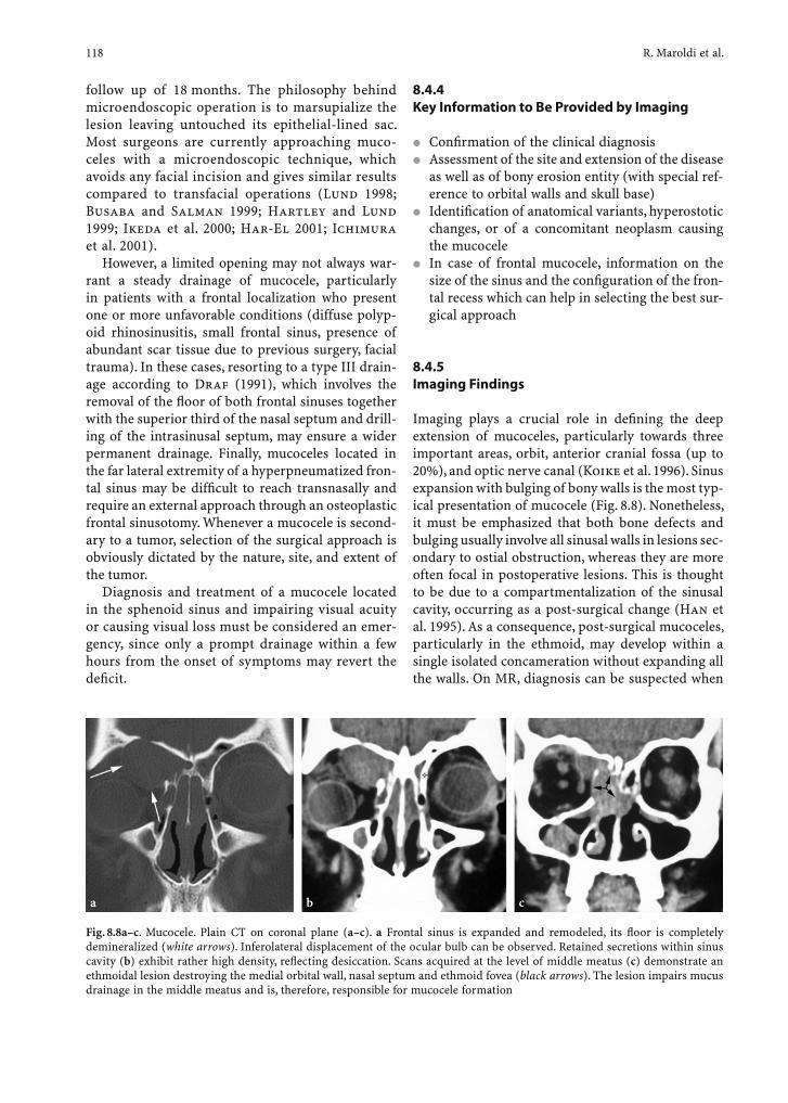
Imaging plays a crucial role in defi ning the deep extension of mucoceles, particularly towards three important areas, orbit, anterior cranial fossa (up to 20%), and optic nerve canal (Koike et al. 1996). Sinus expansion with bulging of bony walls is the most typ-ical presentation of mucocele (Fig. 8.8). Nonetheless, it must be emphasized that both bone defects and bulging usually involve all sinusal walls in lesions sec-ondary to ostial obstruction, whereas they are more often focal in postoperative lesions. This is thought to be due to a compartmentalization of the sinusal cavity, occurring as a post-surgical change (Han et al. 1995). As a consequence, post-surgical mucoceles, particularly in the ethmoid, may develop within a single isolated concameration without expanding all the walls. On MR, diagnosis can be suspected when
Fig. 8.8a–c. Mucocele. Plain CT on coronal plane (a–c). a Frontal sinus is expanded and remodeled, its fl oor is completely demineralized (white arrows). Inferolateral displacement of the ocular bulb can be observed. Retained secretions within sinus cavity (b) exhibit rather high density, refl ecting desiccation. Scans acquired at the level of middle meatus (c) demonstrate an ethmoidal lesion destroying the medial orbital wall, nasal septum and ethmoid fovea (black arrows). The lesion impairs mucus drainage in the middle meatus and is, therefore, responsible for mucocele formation
a b c

Benign Neoplasms and Tumor-Like Lesions 119
signal a intensity differing from typical fl uid and not solid is detected within a cell.
In fact, both CT and MR appearance of mucoceles largely depends on the composition of the entrapped material. At MR, the basic expected signal pattern of fl uids (hyper T2, hypo T1) may be greatly altered by progressive dehydration and consequent increase of viscosity and protein concentration (Lim et al. 1999; Sievers et al. 2000). In detail, protein concentration within the range 20%–25% will result in hyperintensity at both T2 and T1. Further increase of concentration changes the signal pattern, which turns to hypointen-sity, respectively at 30% (T2) and 40% (T1) (Som et al. 1989) (Fig. 8.9). Similarly to the various signal patterns observed on MR, CT density ranges from fl uid-like (cystic appearance) to progressively higher values as far as the entrapped material desiccates. Hypointense signal and high density on plain T1 can be observed also in the case of eosinophilic fungal rhinosinusitis (Van Tassel et al. 1989). After contrast administra-tion, enhancement is observed exclusively at the pe-riphery, along the thin mucosal layer, both at CT and MR (Lloyd et al. 2000a; Hejazi et al. 2001).
Anterior ethmoid mucoceles are more frequent than those arising from posterior ethmoid cells, probably because anterior ethmoid cells have smaller ostia, more easily obstructed in pathologic conditions. Ethmoid mucoceles tend to thin and remodel the adjacent lamina papyracea. They may also arise in cells resulting from the extensive pneumatization of adjacent structures, as in mucoceles developed within a concha bullosa.
Multiple mucoceles are infrequent, more often ob-served after facial fractures, complex facial surgery, and
in patients with severe allergy (particularly in those with aspirin intolerance) (Price et al. 1981). Furthermore, an increased number of mucoceles secondary to en-doscopic sinus surgery procedures has been reported (Raynal et al. 1999). These mucoceles tend to develop earlier (<22 months) than after open sinus surgery or trauma (<10 years). Different causes of sinus obstruc-tion have been demonstrated by endoscopy and CT. Fronto-ethmoid mucoceles were caused by frontal re-cess occlusion due to fi brotic/osteogenic scar tissue, or anterior ethmoid synechia; maxillary sinus mucoceles were related to stenosis/occlusion of the ethmoid in-fundibulum because of uncinate process fragments or scar tissue. Besides, mucoceles of the maxillary sinus may develop after Caldwell-Luc procedures due to the entrapment of mucosa (Weber et al. 2000).
Although infrequent, sphenoid sinus mucoceles entail complex clinical problems, as they may erode the optic nerve canal, the sinusal wall separating from the cavernous sinus, or largely extend into a pneuma-tized pterygoid root. Expansion of the whole sphe-noid sinus, remodeling, and focal erosion indicate the slow-growing development of a mucocele (Fig. 8.10). Signal patterns on MR and CT density may vary in relation to the protein content.
The differential diagnosis of mucocele is strictly related to the pattern of growth and site of origin of the lesion. In the early phase – not associated with si-nus expansion and bone destruction – retention cyst, rhinosinusitis and polyposis should be considered. Though rather uncommon, the presence of intrasinusal calcifi cations could raise the suspect of non-invasive fungal rhinosinusitis (Sievers et al. 2000) (Fig. 8.11).
Fig. 8.9a–c. Post-surgical mucocele. MR SE T2 on axial plane (a), SE T1 on coronal plane (b), and enhanced SE T1 on sagittal plane (c). At 2 years after craniofacial resection for ethmoid adenocarcinoma, (a) MR demonstrates a mucocele occupying the left frontal sinus. b This is related to the presence of scar tissue impairing mucus drainage (white arrows). Note hypointensity on T2 and spontaneous T1 hyperintensity of the retained material in the inferior and inner portion of the mucocele (asterisk). c After contrast agent administration no enhancement of the mucocele or of the mucosa within frontal sinus is seen. Note regular and ossifi ed margins of frontal sinusotomy (white arrow). Mild enhancement of the meningo-galeal complex is demonstrated at the level of the reconstructed anterior skull base fl oor (black arrows)
a b c

120 R. Maroldi et al.
Fig. 8.11a–d. Fungal superinfection of residual cavity. Plain CT on axial plane (a,b), enhanced SE T1 on axial plane (c), fat-suppressed SE T2 on sagittal plane (d). At 3 years after craniotomic evacuation of a left frontal sinus mucocele, a residual cavity is demonstrated by both techniques, exhibiting expanded and remodeled bony walls. Retained material within the cavity shows spon-taneous CT hyperdensity and hypointensity on all MR sequences in its central part. Findings are consistent with fungal superinfection. The peripheral rim lining the cavity (arrows) probably corresponds to the mucosal layer
Fig. 8.10a,b. Mucocele. CT (a) and MR T2 (b), coronal plane. The lesion occupies and markedly expands the left sphenoid sinus. Bone remodeling is detected consisting of demineralization of the greater sphenoid wing (arrows), base of the pterygoid process (asterisk), and pterygoid laminae. Note the sharp margin of the residual part of the sphenoid wing, pointing out resorption rather than destruction. Eggshell calcifi cations are present at the periphery of the lesion (white arrow-heads). Retained secretions show hypointense MR signal in the caudal part of the lesion (black arrowheads), probably due to dehydration and increase in protein content
ba
ba
c d
Benign Neoplasms and Tumor-Like Lesions 121
Actually, in mycotic lesions these are more frequently seen in the central part of the sinus, whereas in a mu-cocele calcifi cations develop close to the mucosal layer (Yoon et al. 1999).
A list of more critical differentials must be consid-ered when extensive bone remodeling and destruc-tion are observed, including necrotic squamous cell carcinoma, cystadenocarcinoma, plasmacytoma, and (for sphenoid sinus mucoceles) pituitary adenoma (Skoulakis et al. 1999; Hejazi et al. 2001). At MR, enhancement pattern – better than T2 and plain T1 signal – helps to obtain the diagnosis: absent or ex-clusively peripheral contrast uptake enables one to predict mucocele with a sensitivity of up to 83%–93% and a specifi city of up to 86%–95% (Yousem 1993; Atasoy et al. 2001).
8.4.6 Follow-Up
The follow up of patients treated through endonasal or open surgery for a mucocele entails a close clinical examination with endoscopic survey. Imaging is indi-cated when the patient presents ocular symptoms or when the cavity cannot be accessed or assessed (i.e., mucoceles previously located laterally in the frontal sinus, recurrence of nasal polyps).
8.5 Abnormal Sinus Pneumatization
8.5.1 Defi nition and Epidemiology
The observation of an abnormal pneumatization of a sinus, a condition fi rst described by Meyes (1898), is a very rare event. According to the classifi cation proposed by Urken et al. (1987), three different cat-egories are encompassed under the name of enlarged aerated sinuses: hypersinus, pneumosinus dilatans, and pneumocele. The fi rst term refers to an abnor-mally aerated sinus which is increased in size but still within the range of normal limits and not associated with bony thinning or erosion. Pneumosinus dilatans is a sinus dilatation beyond the normal range but still with normal bony thickness. When focal bony erosion or diffuse thinning is detected, the defi nition of pneumocele is adopted. Some authors (Chan et al. 1992) distinguish pneumocele from pneumatocele, the latter being characterized by the presence of air in
soft tissues. It is worth mentioning that this classifi ca-tion is not universally adopted, so that discrepancies in nomenclature may be encountered.
One or more paranasal sinuses can be involved at the same time. Frontal and sphenoid sinuses are most frequently involved (Bachor et al. 1994), whereas the occurrence in the maxillary sinus and ethmoid is more rare.
Due to the infrequency of the disease, epidemio-logic data are lacking. Nevertheless, the reported age of observation ranges between 12 and 72 years, with the highest peak of occurrence in the third and fourth decade (Stretch and Poole 1992; Tellado et al. 2002)
8.5.2 Pathogenesis
Several hypotheses have been put forward to explain the occurrence of an abnormal sinusal pneumatiza-tion. According to some authors (Benedikt et al. 1991), a spontaneously drained mucocele could lead to this unusual dilation. One of the most credited theories is based on the existence of a valve obstruc-tive mechanism, which could explain the intrasinusal entrapment of air with consequent pressure in-crease and sinus dilatation (Wolfensberger 1984). However, this hypothesis does not explain why the abnormal pneumatization does not evolve in sinus-itis; secretions and infl amed mucosa are in fact com-pletely absent. Also, an alteration in bone turnover mechanism have been purposed (Rosenberg 1994). In some cases a remodeling action on bony tissues due to a meningioma of the optic nerve (Hirst et al. 1979), or to a bone disease, such as fi brous dys-plasia (Lloyd 1985) or Melnick-Needle syndrome (Stretch and Poole 1992), was considered the main pathogenetic factor. It seems appropriate to defi ne these latter cases as “secondary” abnormal pneuma-tization, differentiating them from the “idiopathic” ones (Trimarchi et al. 2003).
8.5.3 Clinical and Endoscopic Findings
Signs and symptoms typical of abnormal paranasal sinus pneumatization are very different, according to the site of origin, the number of sinuses involved, and the entity of dilation. When the maxillary si-nus is involved, facial pain, possibly increased by atmospheric pressure changes, nasal obstruction,

122 R. Maroldi et al.
facial deformity, are the most frequent presenting complaints (Komori and Sugisaki 1988; Breidhal et al. 1997; Mauri et al. 2000; Karlidag et al. 2003; Trimarchi et al. 2003). Also hearing loss and ear pain (Morrison et al. 1976), and enlargement of the swelling after exposure to sunlight have been detected (Mauri et al. 2000). Involvement of the lamina papy-racea may lead to the onset of ocular disturbances, like eye ball displacement, diplopia or exophthalmos. In the presence of frontal sinus dilation, nasal ob-struction, frontal headache, facial deformity, and eye ball displacement are frequently evident at diagnosis (Adams et al. 1998; Klossek et al. 2000; Walker and Jones 2002). Sphenoid abnormal pneumatiza-tion may present with visual disturbances (Bachor et al. 1994; Skolnick et al. 2000), headache, pain in the proximity of the ocular bulb (Adams et al. 1998), abnormal secretion of pituitary hormones (Reicher et al. 1986). These clinical presentation modalities are sometimes hardly distinguishable from those typical of other expansile lesions.
At endoscopy, displacement of sinusal bony walls is commonly detected; conversely, no mucosal lesion is present. Nevertheless, facial deformities or sinusal wall displacement do not allow one to rule out a mass growing within the sinus and/or along a submucosal plane. The diagnosis of abnormal pneumatization is therefore based upon imaging evaluation.
8.5.4 Treatment Guidelines
The treatment of abnormal sinus pneumatization is surgery, which is meant to restore a normal ven-tilation of the sinus through a wide sinusotomy. Microendoscopic surgery, a minimally invasive ap-proach, appears to be the most appropriate and ef-fective option (Trimarchi et al. 2003). In maxillary localization, a wide meatotomy avoids further si-nus enlargement and allows cessation of symptoms. When a cosmetic correction of facial deformity is required, an external approach may be necessary. A coronal approach for frontal sinus enlargement (Klossek et al. 2000; Tellado et al. 2002) or an infratemporal approach for maxillary enlargement and orbital involvement may be accomplished (Trimarchi et al. 2003). Bifrontal craniotomy has been attempted also with the aim to decom-press optic nerves in case of sphenoid involvement (Stretch and Poole 1992).
8.5.5 Key Information to Be Provided by Imaging
� Demonstration that the expansile submucosal subcutaneus lesion is due to an abnormal pneu-matization of a sinusal cavity
� Assessment of the actual size of the involved sinus(es)
� Precise defi nition of bony wall thickness, with spe-cial reference to the presence of areas of resorp-tion (partial or complete)
� Involvement of critical areas (i.e., orbit, optic canal)
� Possible presence of lesions arising from bone or neoplasms associated with the abnormal pneuma-tization
8.5.6 Imaging Findings
Aberrant enlargement of a normally aerated sinus cavity is an obvious diagnosis, both at plain radiog-raphy and at CT. The latter provides additional infor-mation needed to differentiate pneumosinus dilatans from pneumocele and hypersinus (Trimarchi et al. 2003). According to Urken et al.’s (1987) classifi ca-tion, pneumosinus dilatans is an abnormal sinus ex-pansion that may cause deformity – such as frontal bossing – in the absence of any abnormality of bony sinus walls. Detection of focal or generalized thinning of sinus walls switches the diagnosis to pneumocele (Fig. 8.12). Hypersinus lacks both deformities and bone changes, as a consequence its diagnosis is based on the detection of sinus size exceeding statistically calculated normal limits.
The absence of inherent contrast between air and cortical bone (both hypointense on all sequences), re-stricts the role of MR in these conditions.
8.6 Inverted Papilloma and Other Schneiderian Papillomas
8.6.1 Defi nition, Epidemiology, Pattern of Growth
According to Shanmugaratnam (1991), inverted papilloma is an epithelial benign neoplasm com-
Benign Neoplasms and Tumor-Like Lesions 123
posed of invaginating crypts, thick ribbons or islands of non-keratinizing squamous epithelium which may alternate with or be covered by pseudostratifi ed co-lumnar (cylindrical) or ciliated respiratory epithe-lium. The infolding of the mucosa may result in the presence of apparently discontinuous cell masses ly-ing deep in the epithelial surface, but the basement membrane is intact and may be shown to be continu-ous with that of surface epithelium.
Inverted papilloma originates from the schneide-rian membrane, which is the ectodermally derived mucosa that lines the nasal cavity and paranasal si-nuses. The lesion is estimated to represent 0.4%–4.7% of the surgically removed nasal tumors, with an inci-dence ranging from 0.6 to 1.5 cases per 100,000 in-habitants per year (Buchwald et al. 1995; Outzen et al. 1996). Males are four to fi ve times more frequently affected than females (Hyams 1971; Phillips et al. 1990; Winter et al. 2000). Inverted papilloma is prev-alent in the fi fth and sixth decades of life, although isolated observations in the pediatric age group have been reported (Cooter et al. 1998).
Even though its etiology is still unknown, recent studies using in situ hybridization and polymerase chain reaction have detected human papilloma virus in up to 86% of the lesions (Tang et al. 1994). In par-ticular, viral subtypes 6, 11, 16, and 18 were the most frequently found (Brandwein et al. 1989; Furuta et al. 1991; Gaffey et al. 1996).
The lateral wall of the nasal fossa and the maxil-lary sinus are the most frequent sites of origin of in-verted papilloma, whereas its exclusive localization to the frontal (Shohet and Duncavage 1996; Chee and Sethi 1999) or the sphenoid sinus (Yiotakis et al. 2001a; Lee et al. 2003) is exceedingly rare. In
1971 Hyams reported that in the majority of cases with multiple site involvement the histologic fi nd-ings suggested the spread to result from one single lesion by metaplasia of the adjacent mucosa. In this way, inverted papilloma could easily extend through sinus ostia to the surrounding cavities without de-stroying the bony walls. Intracranial invasion is a rare event, mostly noted in lesions recurring at the level of the cribriform plate or ethmoid roof (Vural et al. 1999). Intraorbital extension may be observed in lesions with extensive ethmoid involvement; how-ever, the tumor usually laterally displaces the orbital content without transgressing the periorbit (Elner et al. 1995; Bajaj and Pushker 2002). The presenta-tion of inverted papilloma is generally unilateral, but bilateral involvement of the sinonasal tract has been reported in a percentage of patients ranging from less than 1% (Phillips et al. 1990) to 9% (Weissler et al. 1986).
The association between inverted papilloma and squamous cell carcinoma, reported to be as high as up to 56% (Yamaguchi et al. 1979), is likely to be overestimated. In fact, recent accurate reviews of the literature (Pelausa and Fortier 1992; Mansell and Bates 2000; Krouse 2001) and data coming from large series (Hyams 1971; Weissler et al 1986; Kapadia et al. 1993) concur to indicate that the two diseases are concomitantly diagnosed in 2% to 13% of patients. A further 1%–1.5% of patients has been shown to present a metachronous sinonasal malig-nant lesion (Fechner and Alford 1968; Weissler et al. 1986).
Staging systems have been developed for a range of benign and malignant lesions of the nose and pa-ranasal sinuses with the intent to guide selection of
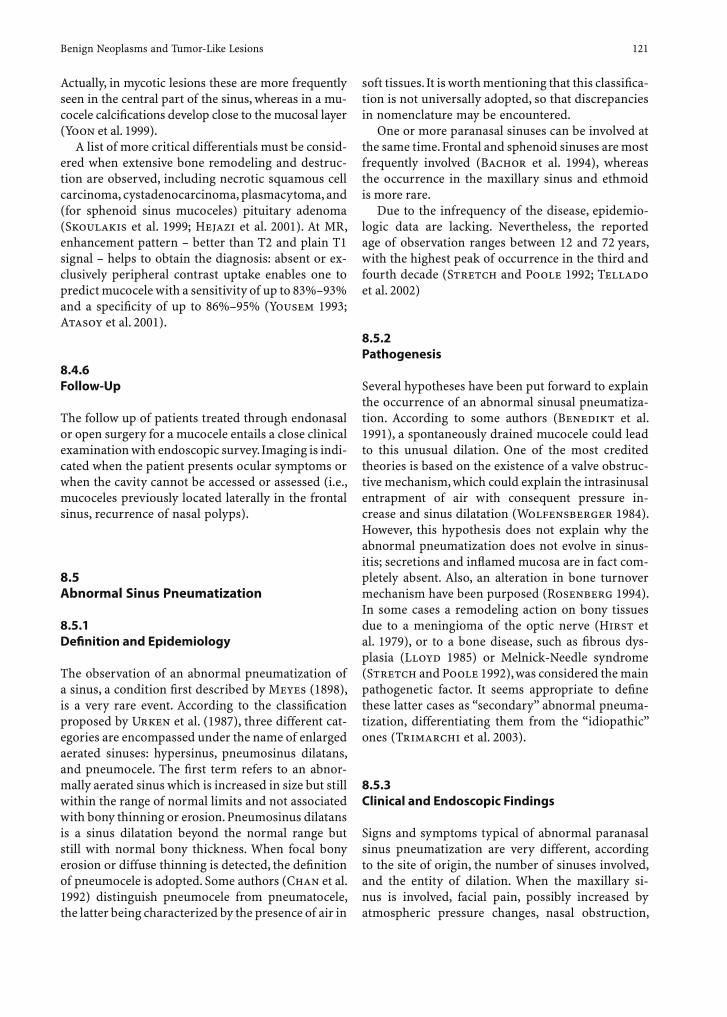
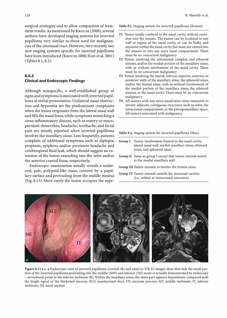
Fig. 8.12a,b. Pneumosinus dilatans. Plain CT on coronal plane (a) and, after surgery, on axial plane (b). Abnormal enlargement of the right maxillary sinus, medially contacting the nasal septum (arrow), no bone destruction is appreciated. After surgical resection of the medial maxillary sinus wall and middle turbinate, normal caliber of the nasal fossa is restored (asterisks)
ba
124 R. Maroldi et al.
surgical strategies and to allow comparison of treat-ment results. As mentioned by Krouse (2000), several authors have developed staging systems for inverted papilloma very similar to those used for malignan-cies of the sinonasal tract. However, very recently two new staging systems specifi c for inverted papilloma have been introduced (Krouse 2000; Han et al. 2001) (Tables 8.1, 8.2).
8.6.2 Clinical and Endoscopic Findings
Although nonspecifi c, a well-established group of signs and symptoms is associated with inverted papil-loma at initial presentation. Unilateral nasal obstruc-tion and hyposmia are the predominant complaints when the lesion originates from the lateral nasal wall and fi lls the nasal fossa, while symptoms mimicking a sinus infl ammatory disease, such as watery or muco-purulent rhinorrhea, headache, toothache, and facial pain are mostly reported when inverted papilloma involves the maxillary sinus. Less frequently, patients complain of additional symptoms such as diplopia, proptosis, epiphora and/or persistent headache and cerebrospinal fl uid leak, which should suggest an ex-tension of the lesion extending into the orbit and/or the anterior cranial fossa, respectively.
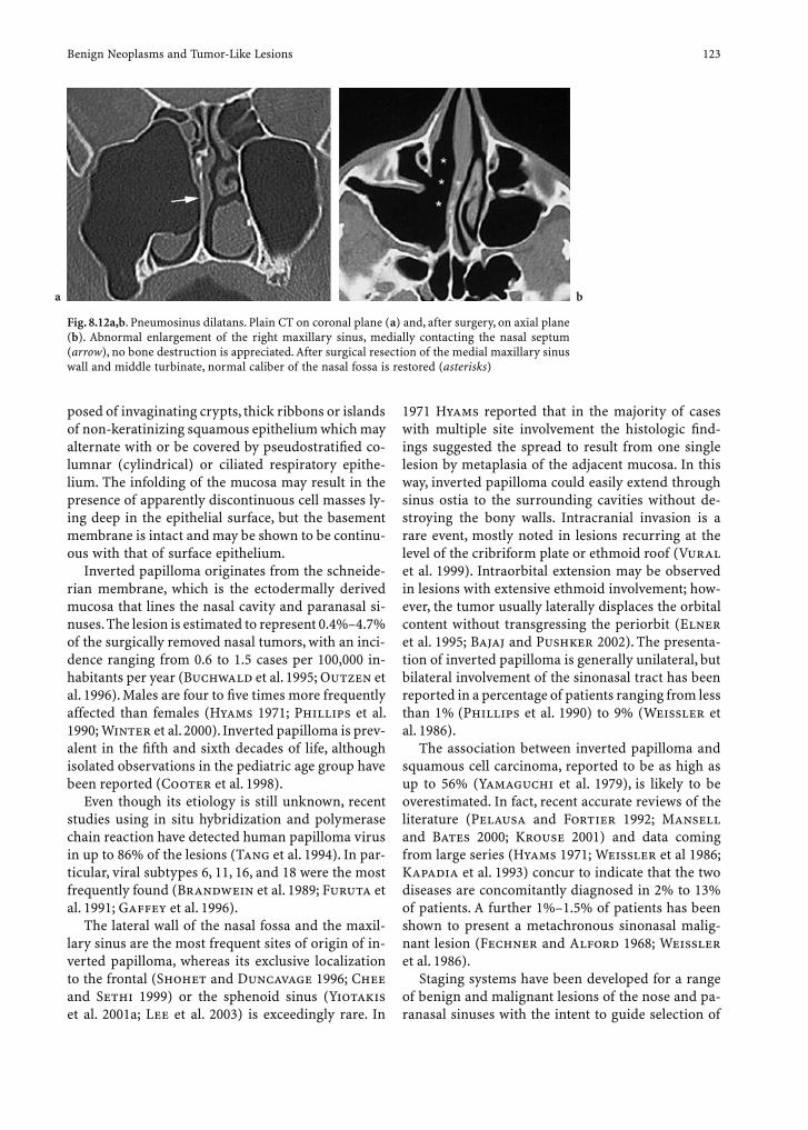
Endoscopic examination usually reveals a unilat-eral, pale, polypoid-like mass, covered by a papil-lary surface and protruding from the middle meatus (Fig. 8.13). More rarely the lesion occupies the supe-
a b c
Figure 8.13 a-c. a Endoscopic view of inverted papilloma. Coronal (b) and axial (c) TSE T2 images show that only the small por-tion of the inverted papilloma protruding into the middle (MM) and inferior (IM) meati is actually demonstrated by endoscopy – arrowheads point to the inferior turbinate (b). Within the maxillary sinus, the main part appears hypointense compared with the bright signal of the thickened mucosa. NLD, nasolacrimal duct; UP, uncinate process; MT, middle turbinate; IT, inferior turbinate; NS, nasal septum
Table 8.1. Staging system for inverted papilloma (Krouse)
T1 Tumor totally confi ned to the nasal cavity, without exten-sion into the sinuses. The tumor can be localized to one wall or region of the nasal cavity, or can be bulky and extensive within the nasal cavity, but must not extend into the sinuses or into any extra nasal compartment. There must be no concurrent malignancy
T2 Tumor involving the ostiomeatal complex, and ethmoid sinuses, and/or the medial portion of the maxillary sinus, with or without involvement of the nasal cavity. There must be no concurrent malignancy
T3 Tumor involving the lateral, inferior, superior, anterior, or posterior walls of the maxillary sinus, the sphenoid sinus, and/or the frontal sinus, with or without involvement of the medial portion of the maxillary sinus, the ethmoid sinuses, or the nasal cavity. There must be no concurrent malignancy
T4 All tumors with any extra-nasal/extra-sinus extension to involve adjacent, contiguous structures such as orbit, the intracranial compartment, or the pterygomaxillary space. All tumors associated with malignancy
Table 8.2. Staging system for inverted papilloma (Han)
Group I Tumor involvement limited to the nasal cavity, lateral nasal wall, medial maxillary sinus, ethmoid sinus, and sphenoid sinus
Group II Same as group I except that tumor extends lateral to the medial maxillary wall
Group III Tumor extends to involve the frontal sinus
Group IV Tumor extends outside the sinonasal cavities (i.e., orbital or intracranial extension)

Benign Neoplasms and Tumor-Like Lesions 125
rior meatus. The concomitant presence of infl amma-tory polyps may be misleading and can cause a delay in establishing a proper diagnosis. Therefore, when-ever the clinician is faced with a unilateral polypoid lesion, multiple biopsies are mandatory.
8.6.3 Treatment Guidelines
Surgery is unanimously considered the treatment of choice for inverted papilloma. Features repeatedly emphasized in the literature such as multicentricity, frequent association with squamous cell carcinoma, and high incidence of recurrences have prompted most of the authors to identify medial maxillectomy (by lateral rhinotomy or midfacial degloving) as the surgical technique of choice (Myers et al. 1990; Lawson et al. 1995; Outzen et al. 1996). However, the introduction in the early 1980s of microendo-scopic surgery, along with the subsequent refi nement of instrumentation and the increasing experience in endonasal surgery for infl ammatory diseases, have led to successful results in the treatment of inverted papilloma even with more conservative techniques (Brors and Draf 1999; Lund 2000; Winter et al. 2000). According to Lund (2000), there is no single right or wrong surgical solution but rather a range of procedures from which a choice should be made in any individual case.
In our experience, based on the management of 47 patients (Tomenzoli et al. 2004), a microendoscopic approach is contraindicated when one of the follow-ing situations is present: extensive involvement of the frontal sinus; massive bone erosion (except for the medial wall of the maxillary sinus and the anterior wall of the sphenoid sinus); intradural invasion; in-traorbital invasion; abundant scar tissue due to pre-vious surgery; association with squamous cell carci-noma. Different microendoscopic resections may be adopted in relation to the site of origin and the extent of the lesion. When inverted papilloma is limited to the middle meatus, anterior and posterior ethmoid, and/or spheno-ethmoid recess, a type 1 resection, including anterior ethmoidectomy with clearance of the frontal recess, posterior ethmoidectomy, a large middle antrostomy, sphenoidotomy, partial or middle turbinectomy (according to tumor extent) is performed. In such a situation, an “en bloc” resection is easily obtained, making sure that the dissection is carried out in the subperiosteal plane.
Whenever the lesion extends from the middle me-atus into the maxillary sinus or originates from the
medial wall of the maxillary sinus, a type 2 resection is performed. In addition to all surgical steps of a type 1 resection, the operation includes a medial maxil-lectomy with or without section of the nasolacrimal duct, in relation to the anterior extent of the tumor.
Inverted papillomas that originate from or involve the posterolateral, anterior and/or inferior wall of the maxillary sinus are better managed through a type 3 resection, which corresponds to the technique in-dicated by Brors and Draf (1999) as an “endonasal Denker operation”.
In patients undergoing a Type 2 or 3 resection, “en bloc” removal is rarely feasible due to the large extent of the lesion. Therefore, debulking of the nasal por-tion of the mass is fi rst performed to subsequently focus on the most critical areas involved by the lesion, where dissection is always carried out in the subperi-osteal plane. Drilling of the bone underlying the dis-eased mucosa is then performed to ensure surgical radicality.
In the era when transnasal resection without mi-croendoscopic assistance was the only available tech-nique, “recurrences” ranged from 40% (Oberman 1964) to 78% (Calcaterra et al. 1980). We concur with other authors (Hyams 1971; Lund 2000) that most of these “recurrences” were probably “residual” lesions, since the exposure offered by a transnasal ap-proach did not guarantee an adequate radicality of the resection and “recurrent” lesions prevalently oc-curred at the same site of the primary (Lund 2000).
By using external medial maxillectomy or microen-doscopic dissection the occurrence of recurrences has dropped down to a prevalence ranging from 0% (Weissler et al. 1986) to 29% (Bielamowicz et al. 1993), and from 0% (Kamel 1995) to 33% (Stankiewicz and Girgis 1993), respectively.Even though most recurrent inverted papillomas present within 2 years following treatment, late re-currences may also occur. Therefore, it is mandatory to prospectively follow patients with endoscopic con-trols every 4 months during the fi rst postoperative year and subsequently every 6 months for at least 4 years. In contrast to other benign diseases such as juvenile angiofi broma, which requires radiologic evaluation for early detection of recurrent submu-cosal lesions (Nicolai et al. 2003), imaging should be obtained only in patients with clear endoscopic evidence of disease or with a complete stenosis of sinus ostium/a precluding a full endoscopic sinus in-spection. In the latter situation, imaging evaluation is aimed not only at detecting recurrent lesions, but also to diagnose possible sequelae such as mucocele.

126 R. Maroldi et al.
regular parallel folds made of a highly cellular meta-plastic epithelium and of an underlying less cellular stroma. On CT examination, when surrounded by air, these folds give inverted papilloma its typical lobu-lated contour, which is consistent with the endoscopic appearance of a polypoid lesion with several micro-digitations on its surface (Dammann et al. 1999). MR does more, because the inner macroscopic arrange-ment is demonstrated as a septate striated pattern or a convoluted cerebriform pattern both on T2 and contrast-enhanced T1 (Yousem et al. 1992; Ojiri et al. 2000). Thus, on T2 these parallel folds of inverted papilloma appear as thick striations of hyperintense signal alternate with thinner ones, closer to fat inten-sity. The thinner striations have been correlated with the metaplastic epithelium, while the thicker ones have been considered to correspond to the less cellu-lar edematous stroma. On contrast-enhanced T1, the stroma shows a strong enhancement while the thin-ner epithelium has a lesser enhancement (Fig. 8.17).
This pattern was described by Yousem et al. (1992) in 5/10 patients while, recently, Ojiri et al. (2000) demonstrated the striated appearance in 8/10 pa-tients on both T2 and contrast-enhanced T1.
In our experience of 20 inverted papillomas (their size ranging from 1 to 6 cm) the pattern was dem-onstrated in all studies on contrast-enhanced T1, while on T2 it was detectable only for lesions more than 3 cm in size (Maroldi et al. 2004). One possible explanation for the increasing observation of this fi nding could be the improved spatial resolution. In fact, Yousem et al. (1992) acquired images with a 256 matrix, Ojiri et al. (2000) obtained slices of 3-mm of
8.6.4 Key Information to Be Provided by Imaging
� Information on the nature of the lesion� Sites involved by the lesion� Presence and location of bony erosion� Possible association of inverted papilloma with a
malignant neoplasm
8.6.5 Imaging Findings
In general, there are two clinical settings in which imaging is faced with the diagnosis of inverted pap-illoma, namely the evaluation of a unilateral nasal obstruction or the assessment of the local spread of a lesion already identifi ed by the endoscopic ex-amination (Lehnerdt et al. 2001; Woodruff and Vrabec 1994).
In the fi rst case, there is a high probability that the lesion is detected on a screening CT for rhinosinus-itis, while in the second one the patient is more fre-quently imaged by MR.
Almost all inverted papillomas are unilateral. Bilateral involvement has been reported; it is more likely related to the perforation of the nasal septum rather than to an actual multifocal origin (Yousem et al. 1992; Dammann et al. 1999).
The imaging features typical of inverted papilloma are based on the site of origin, the pattern of changes of the lateral nasal wall framework, and – respectively – the lobulated surface contour on CT and the striated inner pattern on MR.
Most inverted papillomas arise from both surfaces of the lateral nasal wall, i.e., the nasal and maxillary. Those originating from the middle meatus spread early into the maxillary sinus (Savy et al. 2000) via remodeling and destruction of its very thin or dehis-cent medial bony wall. Those originating from the maxillary sinus tend to fi ll the cavity and further gain access into the nasal cavity via the accessory ostium or via the physiologic ostium.
On CT, high densities within the inverted papil-loma are shown in up to 50% of cases; more fre-quently they appear multiple and discrete. It has been demonstrated by Som and Lidov (1994) that in most cases they represent residual bone included within the lesion rather than calcifi cations (Figs. 8.14, 8.15). Nevertheless, displacement and remodeling of sinusal walls may be observed at the same time (Fig. 8.16).
The peculiar macroscopic arrangement of inverted papilloma is characterized by the alternation of quite
Fig. 8.14 At CT, an inverted papilloma arising from left su-perior turbinate (long arrows) fi lls the superior meatus, and abuts the ground lamella of the middle turbinate that shows irregular sclerotic changes (short arrows)

Benign Neoplasms and Tumor-Like Lesions 127
Fig. 8.16. At CT, a maxillary sinus inverted pap-illoma fi lls the whole paranasal cavity and dis-places all sinusal walls. Focal remodeling and thinning of the posterior aspect of medial si-nus wall is seen (arrows), suggesting the extent through the posterior fontanelle area, a path of lesser resistance
thickness. Our protocol entails 512 matrix with a 210-mm FOV on axial planes; slices of 2–3 mm of thick-ness are used, therefore providing a higher resolution (Maroldi et al. 2004).
The combined use of thin slices and multiple planes of examination makes it possible to recognize that the geometry of the lesion (i.e., the orientation of the septa) depends on the growth along least resis-tance areas (Fig. 8.18). Moreover, this striated appear-ance is particularly evident in those narrow spaces (i.e., ethmoid infundibulum, fontanelle, meatus) where the lesion is constricted by adjacent structures. In effect, the cerebriform convoluted pattern can be resolved by this examination technique in a complex combination of several parallel or fan-shaped groups of folds (exiting/entering holes/fi ssures), merging the concept of striated septa with the one of curvi-linear convoluted cerebriform pattern. In addition, it is sometimes feasible to identify the site of origin – or the path of growth – by tracing back the quite parallel septa to their center. Besides showing a high sensitivity, this pattern proves to be highly predic-tive of inverted papilloma. In our series it was actu-ally observed only in two malignant tumors: in both cases pathologic examination of the surgical speci-men demonstrated areas consistent with inverted
Fig. 8.15a–d. Inverted papilloma of left posterior ethmoid sinus. At CT, the lesion extends into both sphenoid sinuses. Lobulated contours are well detectable on bone-window im-ages (a,b). Non-homogeneous density and irregular bone den-sities are more evident on soft-tissue windows (c,d)
a
d
c
b
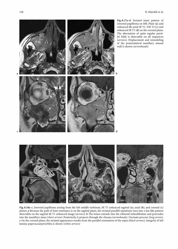
128 R. Maroldi et al.
Fig. 8.17a–d. Striated inner pattern of inverted papilloma on MR. Plain (a) and enhanced (b) axial SE T1, TSE T2 (c) and enhanced SE T1 (d) on the coronal plane. The alternation of quite regular paral-lel folds is detectable on all sequences (arrows). Displacement and remodeling of the posterolateral maxillary sinusal wall is shown (arrowheads)
a b c
Fig. 8.18a–c. Inverted papilloma arising from the left middle turbinate, SE T1 enhanced sagittal (a), axial (b), and coronal (c) planes. a Because the path of least resistance is on the sagittal plane, the striated parallel septations turn into a fan-like pattern detectable on the sagittal SE T1 enhanced image (arrows). b The lesion extends into the ethmoid infundibulum and protrudes into the maxillary sinus (short arrow). Posteriorly, it projects through the choana (arrowheads). Uncinate process (long arrow). c On the coronal plane, the striated appearance results from the parallel orientation of the septa (black arrows). Integrity of left lamina papyracea/periorbita is shown (white arrows)
ba
c d

Benign Neoplasms and Tumor-Like Lesions 129
a b
Fig. 8.19a,b. Enhanced SE T1 on axial (a) and sagittal (b) planes. a Squamous cell carcinoma of the left ethmoid (1) with erosion of left nasal bone (2), displacement and remodeling of left lamina papyracea/periorbita (3). The posterior part of the lesion (black arrows), located in the sphenoid sinus, shows more intense enhancement. Thin hypointense and parallel linear densities, arranged in a roughly septation pattern, can be detected within the tumor (short black arrows). Pathologic examination of the specimen demonstrated foci of IP in this area. On sagittal plane (b), two different sites of anterior cranial fossa fl oor abnormali-ties are demonstrated. Anteriorly, extradural intracranial neoplastic invasion (1) is suggested by the presence of enhancing tissue extending through the bone/periosteum, covered by thickened enhancing dura. A few millimeters posteriorly, the fl uid signal intensity of a mucocele secondary to the more lobulated component (black arrows) remodels both the planum sphenoidalis (2) and the roof/posterior wall of the sphenoid sinus (3)
papilloma where MR showed the pattern (Fig. 8.19) (Maroldi et al. 2004). Conversely, foci of squamous cell carcinoma within the inverted papilloma require the histological examination to be identifi ed (Yousem et al. 1992).
8.6.5.1 Diff erential Diagnosis
Inverted papilloma arising from the maxillary sinus has to be distinguished from antrochoanal polyp, fungus ball, and from malignant neoplasms (Savy et al. 2000).
The antrochoanal polyp, though exhibiting a simi-lar pathway of extent, is usually diagnosed in adoles-cents, rather than in adults, and shows a homogeneous cystic content. Nevertheless, its variant – i.e., the an-giomatous polyp – may be more diffi cult to differen-tiate. In fact, a strangled antrochoanal polyp passing through a constrictive ostium generally shows a more complex signal pattern. Its constricted mucosal folds and vessels resemble linear and parallel/fan-shaped
structures exiting the ostium (Fig. 8.20). Moreover, the intranasal portion of the sinochoanal polyp ex-hibits bright enhancement, probably due to stasis. If a mainly cystic intra-sinusal component is present, an angiomatous polyp is more probable.
On CT, fungus balls may show fi ndings compa-rable to those of inverted papilloma, namely remod-eling and destruction of the medial antral wall and discrete densities within the lesion. They are easily differentiated on MR because almost totally hypoin-tense on T2 sequences.
Generally, squamous cell carcinomas – being the most frequent antral malignant lesion – are charac-terized by a more extensive destruction of the bony walls, and do not show the striated pattern on MR.
The differential diagnosis is more complex for inverted papilloma presenting as a unilateral nasal mass. Of course, sinonasal polyposis typically in-volves both nasal cavities; therefore the detection of a unilateral polypoid lesion in the adult raises the suspicion of inverted papilloma among other benign and malignant neoplasms.

130 R. Maroldi et al.
8.6.5.2 Pathways of Spread
There are two problems to solve when assessing the extent of inverted papilloma: to distinguish it from intrasinusal-retained secretions, and to defi ne its relationship with both the orbit and the skull base (Yousem et al. 1992; Savy et al. 2000). MR can ad-equately answer both issues (Oikawa et al. 2003).
Three relevant aspects regarding inverted pap-illoma pathways of spread have to be outlined: ad-jacent bony structures are destroyed by mean of pressure erosion; extension into adjacent sinusal cavities occurs through a centrifugal pattern; in-volvement of skull base is, usually, late, being ob-served after several failed surgical exccisions. As most inverted papillomas arise from the lateral nasal wall or from the maxillary sinus, they grow centrifugally into the ethmoid labyrinth and later into the sphenoid or the frontal sinus (Fig. 8.21). Thinning and bowing of adjacent bony structures and of the nasal septum is frequent (approximately 80%–90%).
Erosion is common at the level of the lateral nasal wall or of the turbinates (more than 80%); it is also seen in nearly 50% of cases at the level of the eth-moid. Extensive erosion of the lamina papyracea or of the cribriform plate is more often associated with a coexistent squamous cell carcinoma (Dammann et al. 1999).
a
b
c
Fig. 8.21a–c. Inverted papilloma of left frontal sinus, TSE T2 on the axial plane. The lesion has lobulated contours, inter-mediate signal intensity, clearly distinguished from surround-ing retained hyperintense mucus. Focal areas of posterior wall erosion are demonstrated [arrows on (a) and (b)]
a b
Fig. 8.20a,b. Angiomatous polyp of the right maxillary sinus, TSE T2 and enhanced VIBE on the axial plane

Benign Neoplasms and Tumor-Like Lesions 131
8.6.6 Follow-Up
At the end of surgical treatment in almost all inverted papillomas the removal of middle turbinate, medial maxillary sinus and anterior sphenoid sinus walls creates a wide cavity easy to access at endoscopy dur-ing the follow-up (Dammann et al. 1999; Petit et al. 2000) (Fig. 8.22).
For this reason, imaging is indicated mainly to defi ne the extent of a recurrent inverted papilloma, particularly to demonstrate its relationship with the skull base or the orbit. Of course, recurrences located deep in the frontal sinus can be detected by imaging only. The same diagnostic criteria used to identify
the primary inverted papilloma apply to relapsing le-sions. Also in this setting, MR provides a sensitivity superior to CT (Petit et al. 2000).
8.7 Juvenile Angiofi broma
8.7.1 Defi nition, Epidemiology, Pattern of Growth
Juvenile angiofi broma is a lesion composed of vas-cular and fi brous elements in varying proportion, which accounts for 0.5% of all head and neck tumors
a b
c d
Fig. 8.22a–d. Post-microendoscopic removal of left nasal wall inverted papilloma. Endoscopy (a), TSE T2 (b), plain (c) and enhanced (d) SE T1 on the axial plane. a Endoscopy shows complete healing of the nasal fossa and posterior maxillary sinus wall, which is characterized by mild bulging. b On T2 se-quence, the bulging corresponds to submucosal thickened connective tissue – due to previous surgery – showing intermediate signal (asterisk). Thickened hyperintense mucosa invests the zygomatic recess of left maxillary sinus (white arrows). Periosteal thickening of posterolateral antral wall presents with a double layer pattern (arrowheads). c On the axial plane, obtained a few millimeters cranial, scar tissue partially fi lls the maxillary sinus. d After GD-DTPA administration minimal enhancement of the scar tissue is shown. SS, sphenoid sinus; NS, nasal septum

132 R. Maroldi et al.
Coronal
and typically occurs in adolescent males. Recent im-munohistochemical and electron microscopy stud-ies suggest that the lesion must be considered a vascular malformation (or hamartoma) instead of a tumor (Schick et al. 2002). The point of origin is identifi ed by some authors in the area of the spheno-palatine foramen, while others, based on the results of CT or MR imaging, indicate that the lesion takes origin in the pterygopalatine fossa, at the aperture of the vidian canal. Juvenile angiofi broma has the peculiar tendency to grow in the submucosal plane into the nasopharynx, along the vidian canal into the basisphenoid, and laterally towards the ptery-gopalatine and infratemporal fossa. Another impor-tant feature of juvenile angiofi broma is the early in-vasion of the cancellous bone of the pterygoid root; from here, the lesion can extend laterally to involve the greater wing of the sphenoid bone. Intracranial extension mainly occurs into the middle cranial fossa along the maxillary nerve into the parasellar region and the cavernous sinus; another pattern in-volves growth through the inferior and the superior orbital fi ssures. Erosion of the anterior skull base is seldom observed. It is noteworthy that most of the lesions with intracranial extension are extradural; only very rare instances of juvenile angiofi bromas crossing the dura have been documented in the lit-erature (Danesi et al. 2000).
8.7.2 Clinical and Endoscopic Findings
Nasal obstruction and epistaxis are the typical symp-toms of juvenile angiofi broma; their occurrence in a young male should always arouse the suspicion of such a disease. Additional symptoms such as diplo-pia, cheek swelling, and headache can be reported by patients with advanced lesions extending into the superior orbital fi ssure, the infratemporal fossa, and the cranial fossa, respectively. The endoscopic fi nd-ing of a smooth-surfaced, clearly vascularized, ex-pansile lesion growing behind the middle turbinate is another important element highly suggestive for juvenile angiofi broma (Fig. 8.23). Since epidemio-logic and endoscopic fi ndings are so typical, biopsy is almost unanimously considered contraindicated, since it carries a considerable and undue risk of hem-orrhage.
8.7.3 Treatment Guidelines
Even though several methods (i.e., embolization, cryotherapy, hormonal therapy, chemotherapy) have been proposed for the treatment of juvenile angiofi -broma, surgery is commonly considered the main-
Fig. 8.23a,b. Juvenile angiofi broma (JA). a Coronal TSE T2 shows a hypointense mass fi lling the left nasal cavity. It projects among nasal septum (S), middle (arrowheads) and inferior (I) turbinates. Retained fl uid within the obstructed nasal cavity appears as hyperintense signal surrounding the inferior turbinate. b Endoscopy demonstrates a bluish and vascularized polyp. I, Inferior turbinate; M, middle turbinate; S nasal septum
ba
Benign Neoplasms and Tumor-Like Lesions 133
stay. Radiotherapy has also been shown to be effective in isolated institutions; however, the observation of a few cases of radioinduced tumors, as well as the possible alterations of maxillary growth, bring the validity of this treatment into question.
Since many years, transfacial techniques (through a lateral rhinotomy or a midfacial degloving) and the infratemporal approach are considered the best options. More recently, the rapid spread of endonasal techniques using endoscopes or both a microscope and endoscopes has prompted many surgeons to treat even small and intermediate-size juvenile angiofi bromas with such a conservative ap-proach with apparently satisfactory results. Even in our experience, juvenile angiofi bromas involv-ing the nasopharynx, the nasal cavities, the sphe-noid, the ethmoid, the maxillary sinus, and/or the pterygomaxillary fossa can be managed successfully through endoscopic surgery (Nicolai et al. 2003). Lesions extending into the infratemporal fossa and/or the cavernous sinus frequently require an external approach. The head and neck surgeon is therefore faced with a wide spectrum of surgical techniques that can be selected in the single patient mainly in relation to the size and the extent of the lesion. An informed consent to switch intraoperatively to an external procedure should always be obtained from the patient.
8.7.4 Key Information to Be Provided by Imaging
� Confi rmation of the suspected nature of the dis-ease
� Extent of the lesion, with specifi c emphasis on orbit, infratemporal fossa, intracranial (cavernous sinus, ICA, dura) and intraosseous involvement
� Pattern of vascularization (unilateral vs. bilateral; recruitment of internal carotid artery branches)
8.7.5 Imaging Findings
The diagnosis of juvenile angiofi broma on CT and MR is based on three features: the site of origin of the lesion (Lloyd et al. 2000b); its hypervascular ap-pearance after contrast enhancement (Schick and Kahle 2000); and its pattern of growth.
First, as juvenile angiofi broma arises close to the sphenopalatine foramen – either just on its submu-cosal aspect or from the adjacent pterygopalatine
fossa – it usually presents as a mass expanded medi-ally into the nasal cavity, and eroding the root of the pterygoid process.
It has been suggested that the actual site of ori-gin should be a recess of the pterygopalatine fossa behind the sphenopalatine ganglion, at the exit ap-erture of the vidian canal (Fig. 8.24). The evidence that up to 96% of the 72 juvenile angiofi bromas in the series of Lloyd caused enlargement or erosion of the anterior part of the vidian canal should support this hypothesis.
Second, as juvenile angiofi broma mainly consists of a vascular histological component, the adminis-tration of contrast agent – either iodinated or para-magnetic – causes a strong and usually quite homo-geneous, enhancement on CT or MR T1 sequences (Fig. 8.25). Moreover, the diagnosis of juvenile an-giofi broma on MR is suggested by the presence – on both T1 and T2 sequences – of several signal voids within the lesion, indicating major intra-lesion ves-sels (Fig. 8.26).
As the mass increases in size, enlargement of the internal maxillary artery can be directly detected by CT or MR. Cystic changes are rare; those reported have been correlated to spontaneous or post-chemo-therapy tumor regression (Schick and Kahle 2000) (Fig. 8.27).
Third, bone involvement by juvenile angiofi-broma consists either of bone remodeling – for example the thinning and anterior displacement of the postero-superior antral wall – and bone ero-sion, typically at the level of the pterygoid root. In the latter case, access to the cancellous content of the sphenoid bone enables the juvenile angiofi-broma to extend deeply into the medullary spaces of the basisphenoid or, even, to replace the diploe of the greater wing of the sphenoid (Lloyd et al. 1999). Although bone erosion can be more easily shown by CT, MR is adequate in demonstrating cortical erosion and cancellous replacement by tu-mor (Fig. 8.28).
Therefore, the typical juvenile angiofi broma is diagnosed by CT or MR with almost total certainty, making pre-operative biopsy unnecessary. Juvenile angiofi broma arising outside the lateral margins of the posterior nares is very rare. Approximately 50 cases have been reported in the literature, the maxil-lary sinus being the commonest atypical site (Schick and Kahle 2000).

134 R. Maroldi et al.
Fig. 8.24a,b. Juvenile angiofi broma. Plain CT (a), and plain SE T1 (b) in the axial plane (same patient as in Fig. 8.23). Both tech-niques demonstrate a lesion arising from the medial aspect of the left pterygopalatine fossa in the region of the sphenopalatine foramen (black arrows). The exit aperture of the pterygoid/vidian canal is markedly enlarged, whereas its posterior portion is normal (arrowheads). Chronically retained secretions fi ll the sphenoid sinus, being hyperintense on T1 sequence because of the increased protein concentration. MR shows a small part of the juvenile angiofi broma abutting the sinusal lumen (white arrows). Reactive sclerosis of the left pterygoid root spongiosa is demonstrated on both techniques
ba
Fig. 8.25a,b. Juvenile angiofi broma: pattern of enhancement. CT before (a) and after (b) contrast administration, coronal plane. After contrast injection a bright and quite uniform enhancement is observed. Intracranial extent at the cavernous sinus level appears equally as a hyperdense nodule (arrows). The epicenter of juvenile angiofi broma is located at the sphe-nopalatine foramen
ba

Benign Neoplasms and Tumor-Like Lesions 135
Fig. 8.26a–c. Juvenile angiofi broma: MR signal inten-sity and pattern of enhancement. The juvenile an-giofi broma exhibits intermediate signal, comparable to brain parenchyma, on both TSE T2 (a) and plain SE T1 (b) sequences. a Part of the lesion extends into the left nasal fossa, scarcely separable on plain T1 from the middle turbinate (b, white arrows). Intense enhancement after paramagnetic contrast injection (c). Major intra-lesion vessels are detected on MR as serpiginous fl ow voids (arrowheads). A small part of juvenile angiofi broma abuts the left pterygopalatine fossa (black arrows)
ba
c

136 R. Maroldi et al.
Fig. 8.28a–c. Juvenile angiofi broma. CT after contrast adminis-tration, axial plane (a); TSE T2 (b) and fat saturated SE T1 (c) after contrast administration, coronal plane. a The lesion com-pletely fi lls the nasopharynx and the nasal fossae, hypodense cystic areas are detectable (asterisks). CT clearly depicts ero-sion of the medial pterygoid plate and tumor growth into the cancellous bone of pterygoid process (black arrows); fi nger-like projections of the mass spread into the infratemporal fossa (white arrows). MR shows invasion of cancellous bone on both TSE T2 and enhanced fat-sat T1, with the lesion extending into the pterygoid root and into the greater sphenoid wing (white arrows). These fi nding are more clear-cut on the fat saturated sequence (c). The lesion grows along the path of the foramen rotundum (black arrows) and, submucosally, along the under surface of sphenoid bone (black arrowheads), without invad-ing cavernous sinus (single black arrowhead in b)
Fig. 8.27 Juvenile angiofi broma. TSE T2, coronal plane. Hyperintensity of the intra-sphenoidal component of the le-sion refl ects spontaneous cystic changes
b
a
c

Benign Neoplasms and Tumor-Like Lesions 137
8.7.5.1 Pathways of Spread
Of course, the pathways of spread of juvenile an-giofi broma will infl uence the choice of the surgical approach (Fig. 8.29).
Owing to its nature, juvenile angiofi broma tends to grow along the paths of least resistance, causing displacement of adjacent soft tissues, rather than in-vasion. Adherence can be present, particularly when the lesion contacts the dura, but dural or brain in-fi ltration is rare (Danesi et al. 2000; Scholtz et al. 2001).
Its peculiar dual pattern of bone involvement (re-modeling and destruction) is likely to result from two different mechanisms of interaction between juvenile angiofi broma and bony structures. Displacement of the periosteal invested cortical surfaces causes re-modeling and thinning, with only late breakthrough and destruction, whereas the direct growth of juve-nile angiofi broma along perforating arteries into the cancellous root of the sphenoid lets the lesion extend into the medullary content of the sphenoid (fl oor of sphenoid sinus, greater wing). This could explain why the vidian canal (invested by a periosteal layer), though the closest to the growing lesion, shows more frequently enlargement of its anterior third, rather
Fig. 8.29. Pathways of spread of juvenile angiofi broma. A, ex-tent through the sphenopalatine foramen into the nasal fossa (1) and – via the choana – into the nasopharynx (2); through the erosion of the sphenoid sinus fl oor into the sphenoid sinus (3). B, extent from the pterygopalatine fossa along the foramen rotundum (actually a groove or a complete bone canal) (4), into the cancellous bone of the greater wing of the sphenoid (5), and into the masticator space (6)
than destruction. Moreover, further posterior exten-sion into the canal is infrequent, and usually observed in advanced lesions.
Due to the knowledge of the elementary interac-tions of juvenile angiofi broma with surrounding structures and its constant site of origin, the patterns of spread are highly predictable (Lloyd et al. 2000b; Schick and Kahle 2000).
From its site of origin in the pterygopalatine fossa, the juvenile angiofi broma extends medially into the nasal cavity and nasopharynx – the areas of least re-sistance – via enlargement and erosion of the spheno-palatine foramen. Growth of tumor anteriorly indents the postero-superior maxillary sinus wall, resulting in anterior bowing of the sinusal wall, the so-called antral sign, described by Holman and Miller on lateral plain X-ray (Lloyd and Phelps 1986) (Fig. 8.30) .The lateral extent, via an enlarged pterygo-maxil-lary fi ssure, gives rise to infratemporal fossa spread. Extension into this space is demonstrated by detect-ing the “fi nger-like projections” of the enhancing juvenile angiofi broma characterized by sharp and lobulated margins (Fig. 8.31). In this area, the least resistant structure consists of the fat tissue between the pterygoid muscles, usually splayed.
From the pterygo-maxillary fi ssure, the lesion can also access the apex of the orbit through the in-ferior orbital fi ssure, and further extends into the middle cranial fossa via the superior orbital fi ssure (Fig. 8.32).
Posterior spread from pterygopalatine fossa is almost certainly the most dangerous for the patient because it enables the juvenile angiofi broma to pen-etrate the cancellous bone of the root of pterygoid process. From this site, juvenile angiofi broma ex-tends both medially, into the fl oor of the sphenoid sinus, and laterally, into the greater wing (Lloyd et al. 1999). As the center of growth of the intrasinusal component of juvenile angiofi broma is located at the intersection between the fl oor and the lateral wall, there is evidence to suggest that the intrasinusal ex-tension comes from the root of the pterygoid, rather than being due to upward extension from the choana. Lateral spread allows the juvenile angiofi broma to re-place the diploe of the greater wing, usually with late erosion of the inner table.
The key to detecting the diploic invasion consists of differentiating its medullary content from the le-sion on the basis of CT density and MR signal. On CT, this is achieved by the strong enhancement of juvenile angiofi broma within the diploe. On MR, the optimal discrimination is obtained by combining a plain T1 with a post-contrast T1 with or without fat
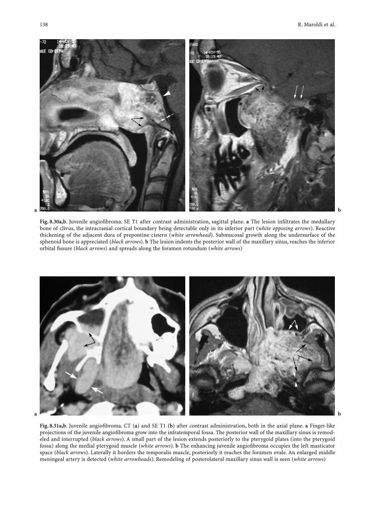
138 R. Maroldi et al.
Fig. 8.31a,b. Juvenile angiofi broma. CT (a) and SE T1 (b) after contrast administration, both in the axial plane. a Finger-like projections of the juvenile angiofi broma grow into the infratemporal fossa. The posterior wall of the maxillary sinus is remod-eled and interrupted (black arrows). A small part of the lesion extends posteriorly to the pterygoid plates (into the pterygoid fossa) along the medial pterygoid muscle (white arrows). b The enhancing juvenile angiofi broma occupies the left masticator space (black arrows). Laterally it borders the temporalis muscle, posteriorly it reaches the foramen ovale. An enlarged middle meningeal artery is detected (white arrowheads). Remodeling of posterolateral maxillary sinus wall is seen (white arrows)
Fig. 8.30a,b. Juvenile angiofi broma. SE T1 after contrast administration, sagittal plane. a The lesion infi ltrates the medullary bone of clivus, the intracranial cortical boundary being detectable only in its inferior part (white opposing arrows). Reactive thickening of the adjacent dura of prepontine cistern (white arrowhead). Submucosal growth along the undersurface of the sphenoid bone is appreciated (black arrows). b The lesion indents the posterior wall of the maxillary sinus, reaches the inferior orbital fi ssure (black arrows) and spreads along the foramen rotundum (white arrows)
ba
ba
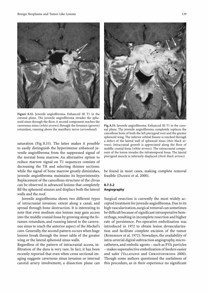
Benign Neoplasms and Tumor-Like Lesions 139
saturation (Fig. 8.33). The latter makes it possible to easily distinguish the hyperintense enhanced ju-venile angiofi broma from the suppressed signal of the normal bone marrow. An alternative option to reduce marrow signal on T1 sequences consists of decreasing the TR and selecting thinner sections: while the signal of bone marrow greatly diminishes, juvenile angiofi broma maintains its hyperintensity. Replacement of the cancellous structure of the clivus can be observed in advanced lesions that completely fi ll the sphenoid sinuses and displace both the lateral walls and the roof.
Juvenile angiofi broma shows two different types of intracranial invasion: extent along a canal, and spread through bone destruction. It is interesting to note that even medium size lesions may gain access into the middle cranial fossa by growing along the fo-ramen rotundum, and running lateral to the cavern-ous sinus to reach the anterior aspect of the Meckel’s cave. Generally, the second pattern occurs when huge lesions break through the inner table of the greater wing or the lateral sphenoid sinus walls.Regardless of the pattern of intracranial access, in-fi ltration of the dura is very rare. In fact, it has been recently reported that even when cross sectional im-aging suggests cavernous sinus invasion or internal carotid artery involvement, a dissection plane can
Figure 8.32. Juvenile angiofi broma. Enhanced SE T1 in the coronal plane. The juvenile angiofi broma invades the sphe-noid sinus through the fl oor. A second component reaches the cavernous sinus (white arrows) through the foramen (groove) rotundum, running above the maxillary nerve (arrowhead)
Fig. 8.33. Juvenile angiofi broma. Enhanced SE T1 in the coro-nal plane. The juvenile angiofi broma completely replaces the cancellous bone of both the left pterygoid root and the greater sphenoid wing. The inferior orbital fi ssure is reached through a defect of the lateral wall of sphenoid sinus (thin black ar-rows). Intracranial growth is appreciated along the fl oor of middle cranial fossa (white arrows). The extracranial compo-nent of the lesion invades the infratemporal fossa. The lateral pterygoid muscle is inferiorly displaced (thick black arrows)
be found in most cases, making complete removal feasible (Danesi et al. 2000).
8.7.5.2 Angiography
Surgical resection is currently the most widely ac-cepted treatment for juvenile angiofi broma. Due to its high vascularization, surgical removal can sometimes be diffi cult because of signifi cant intraoperative hem-orrhage, resulting in incomplete resection and higher rate of persistence. Pre-operative embolization was introduced in 1972 to obtain lesion devasculariza-tion and facilitate complete excision of the tumor (Roberson et al. 1972). Nowadays, the availability of intra-arterial digital subtraction angiography, micro-catheters, and embolic agents – such as PVA particles – makes superselective embolization of feeders easier and safer (Valavanis and Christoforidis 2000). Though some authors questioned the usefulness of this procedure, as in their experience no signifi cant

140 R. Maroldi et al.
difference in surgical bleeding was observed, there is increasing evidence that embolization is a safe and effective method to reduce intra-operative blood loss (Siniluoto et al. 1993; Moulin et al. 1995; Li et al. 1998). Nevertheless, the shrinkage of lesion achieved by embolization has been indicated as a contribu-tory cause to incomplete excision of juvenile angio-fi broma by McCombe et al. (1990).
At present, the role of angiography is to provide a detailed map of feeders, demonstrating the recruit-ment of internal carotid artery, vertebral or contra-lateral external carotid artery branches, and to obtain preoperative devascularization.
According to Lasjaunias et al. (1980), the angio-graphic fi ndings of juvenile angiofi broma consist of
moderate enlargement of feeding arteries, intense “parenchymal” blush, absence of large arteriovenous shunts, or early venous return (Fig. 8.34).
The pattern of arterial feeders recruited is predict-able in most cases. It is strictly related to the path-ways of spread, but not to the actual size of the lesion, though most large lesions are multi-compartmental. In our experience of 15 patients treated by exclusive endonasal excision, there was no correlation between the volume of juvenile angiofi broma and the num-ber of feeding vessels (Nicolai et al. 2003). However, recruitment of internal carotid artery, vertebral branches was signifi cantly more frequent among lesions with several external carotid artery feed-ers. Notably, though internal carotid artery feeders
Fig. 8.34a,b. Juvenile an-giofi broma. Enhanced SE T1 in the coronal plane (a); intra-arterial DSA (b). a On MR, the lesion shows a prevalent endoluminal growth within the naso-pharynx. Upwards exten-sion in the pterygoid root and within the sphenoid sinus is also appreciated. b DSA demonstrates vas-cular feeders arising from the distal part of the sphe-nopalatine artery; a more prominent blush is ob-served at the level of the nasal part of the lesion
Fig. 8.35a,b. Juvenile an-giofi broma. Enhanced SE T1 in the sagittal plane (a); intra-arterial DSA (b). a The lesion fi lls the sphe-noid sinus. Infi ltration of the clivus is demonstrated by the encroachment of both its cortical layers and replacement of the medullary content. b DSA demonstrates the several subtle feeders from the internal carotid artery, not embolized
ba
ba

Benign Neoplasms and Tumor-Like Lesions 141
were demonstrated in approximately 47% of cases (Fig. 8.35), intracranial extent was present only in 13%.
During the last decade the availability of small particles and microcatheters has made it possible to reach even peripheral small branches of the external carotid artery, preserving adjacent normal vessels from being devascularized by the more proximal occlusion obtained by Gelfoam or Spongel emboli-zation (Fig. 8.36). In fact, the goal of embolization is to achieve vessel occlusion at the capillary level. Consequently, polyvinyl-alcohol particles with a minimal size of 150 µm have been suggested, as signifi cant arteriovenous shunts have been demon-strated for particles of 50 µm or less by nuclear medi-cine techniques (Schroth et al. 1996).
The rate of minor and major complications for embolization of external carotid artery branches is negligible, approximately 4% (Ungkanont et al. 1996).
It is evident that the major challenge to angiog-raphy regards the management of juvenile angio-fi broma vascularization by internal carotid artery feeders. Advanced lesions with intracranial extent have been successfully excised after pre-operative embolization of external carotid artery branches with acceptable blood loss, despite involvement of the internal carotid artery branches in the blood supply. Devascularization by direct tumor punc-ture and embolization, advocated by Casasco et al. (1999), entails an unacceptable risk of major neuro-logic complications. Balloon occlusion and sacrifi ce of the internal carotid artery is required in rare cases (Casasco et al. 1999).
8.7.6 Follow-Up
There are two different types of persistent lesions: those left intentionally because their resection would require unacceptable neural damage, and those left due to intraoperative oversight (Figs. 8.37, 8.38). In both cases, proper management mandates a precise assessment of site, size, and extent of the lesion.
Recurrences are a peculiar characteristic of ju-venile angiofi broma. In fact, most authors doubt this theory and consider more likely the hypoth-esis of incomplete excision leaving lesion remnants (Andrews et al. 1989; Chagnaud et al.1998; Lloyd et al. 2000b; ).
To support this hypothesis is the fact that most persistent lesions occur within months or a few years
of primary treatment and more commonly are found in anatomical areas diffi cult to reach at surgery. This presumption is consistent with Chagnaud et al. (1998) who demonstrated that lesion remnants were already detectable at the fi rst follow-up with cross sectional imaging.
Persistent lesions are more frequent when the pri-mary juvenile angiofi broma invades the infratempo-ral fossa, sphenoid sinus, pterygoid root, clivus, and cavernous sinus (Herman et al. 1999; Howard et al. 2001). According to McCombe et al. (1990), “recur-rences” are also related to primary juvenile angiofi -broma size, being more frequent in large lesions.
Fig. 8.36a,b. Juvenile angiofi broma. DSA, before (a) and after (b) embolization. The juvenile angiofi broma is fed by termi-nal branches of the sphenopalatine artery, it occupies the na-sal fossa and the nasopharynx; upwards it extends into the sphenoid sinus, downwards it reaches the superior part of the oropharyngeal lumen. Embolization permits complete devas-cularization of the mass
b
a
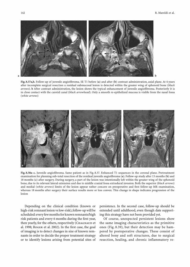
142 R. Maroldi et al.
Depending on the clinical condition (known or high-risk remnant lesion vs low-risk), follow-up will be scheduled: every few months for known remnants/high risk patients and every 6 months during the fi rst year, then yearly, for the others, respectively (Chagnaud et al. 1998; Roger et al. 2002). In the fi rst case, the goal of imaging is to detect changes in size of known rem-nants in order to decide the proper treatment strategy or to identify lesions arising from potential sites of
persistence. In the second case, follow-up should be extended until adulthood, even though date support-ing this strategy have not been provided yet.
Of course, unexpected persistent lesions show the same imaging characteristics as the primitive ones (Fig. 8.39), but their detection may be ham-pered by postoperative changes. These consist of altered bony and soft structures, due to surgical resection, healing, and chronic inflammatory re-
Fig. 8.38a–c. Juvenile angiofi broma. Same patient as in Fig. 8.37. Enhanced T1 sequences in the coronal plane. Pretreatment examination for planning sub-total resection of the residual juvenile angiofi broma (a). Follow-up study after 13 months (b) and 18 months (c) after surgery. During surgery, a part of the lesion was intentionally left within the greater wing of the sphenoid bone, due to its relevant lateral extension and due to middle cranial fossa extradural invasion. Both the superior (black arrows) and medial (white arrows) limits of the lesion appear rather concave on preoperative and fi rst follow-up MR examination, whereas 18 months after surgery their surface results more or less convex. This change in shape indicates progression of the lesion
a b c
Fig. 8.37a,b. Follow-up of juvenile angiofi broma. SE T1 before (a) and after (b) contrast administration, axial plane. At 4 years after incomplete surgical resection a residual submucosal lesion is detected within the greater wing of sphenoid bone (black arrows). b After contrast administration, the lesion shows the typical enhancement of juvenile angiofi broma. Posteriorly it is in close contact with the carotid canal (black arrowhead). Only a smooth re-epithelized mucosa is visible from the nasal fossa (white arrows)
ba

Benign Neoplasms and Tumor-Like Lesions 143
action of the sinonasal mucosa (Chagnaud et al. 1998).
Bony defects of the sinonasal framework and skull base mainly depend both on the specifi c surgical ap-proach and on the extent of the erosion caused by juvenile angiofi broma.
However, some constant postoperative fi ndings appear to be the partial or total excision of the pos-terior, and medial antral walls, of the pterygoid root and of the fl oor of the sphenoid sinus, detectable in almost half of the patients, regardless of the surgical approach (Fig. 8.40).
The scar tissue replacing the site previously occu-pied by juvenile angiofi broma – pterygopalatine fossa, inferior orbital fi ssure, infratemporal fossa – appears usually hypointense on both T1 and T2 and does not enhance on CT or MR after contrast agent adminis-tration. Conversely, enhancement is shown either by the thickened infl amed sinonasal mucosa or by lesion
remnants. Of course, infl ammatory mucosal changes appear hyperintense also on T2 sequences, whereas juvenile angiofi broma has intermediate signal inten-sity. In addition, signal voids may be observed, their detection probably depending on the overall size of the persistent juvenile angiofi broma.
Nevertheless, it is not infrequent to image enhanc-ing submucosal areas fi lling previous site(s) of the le-sion. Serial examinations appear necessary to effec-tively estimate their growth, a fi nding that should be consistent with residual lesions (Fig. 8.41). However, known lesion remnants – left in place during surgery – may not show increase in size (Deschler et al. 1992). Angiography has been advocated to obtain a defi nite diagnosis, but it is hampered by false positive results (Bremer et al. 1986).
Owing to this limitation, and because the surgical treatment of small residuals – particularly those in-tracranially located – is rather controversial, the role
Fig. 8.39a–d. Recurrent/persistent juvenile angiofi bromas in two different patients. CT after contrast administration, axial plane (a); endoscopic view (b); enhanced VIBE sequence in sagittal and coronal planes (c,d). a A thin plaque of non-enhancing scar tissue is detected at the level of sphenopalatine foramen (white arrow). The submucosal relapse (black arrows) has its epicenter at the pterygoid root level where the medullary bone is replaced by the enhanced juvenile angiofi broma (asterisk). Erosion of pterygoid bony boundaries is seen (arrowheads). The encircled area corresponds to the endoscopic view (b), which does not show any abnormality. c,d In a different patient, a relapsing juvenile angiofi broma is detected 2 years after endonasal surgery (arrows) located submucosally within residual sphenoid sinus fl oor
ba
c d
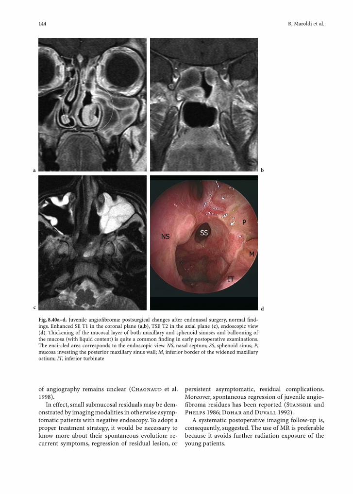
144 R. Maroldi et al.
of angiography remains unclear (Chagnaud et al. 1998).
In effect, small submucosal residuals may be dem-onstrated by imaging modalities in otherwise asymp-tomatic patients with negative endoscopy. To adopt a proper treatment strategy, it would be necessary to know more about their spontaneous evolution: re-current symptoms, regression of residual lesion, or
persistent asymptomatic, residual complications. Moreover, spontaneous regression of juvenile angio-fi broma residues has been reported (Stansbie and Phelps 1986; Dohar and Duvall 1992).
A systematic postoperative imaging follow-up is, consequently, suggested. The use of MR is preferable because it avoids further radiation exposure of the young patients.
Fig. 8.40a–d. Juvenile angiofi broma: postsurgical changes after endonasal surgery, normal fi nd-ings. Enhanced SE T1 in the coronal plane (a,b), TSE T2 in the axial plane (c), endoscopic view (d). Thickening of the mucosal layer of both maxillary and sphenoid sinuses and ballooning of the mucosa (with liquid content) is quite a common fi nding in early postoperative examinations. The encircled area corresponds to the endoscopic view. NS, nasal septum; SS, sphenoid sinus; P, mucosa investing the posterior maxillary sinus wall; M, inferior border of the widened maxillary ostium; IT, inferior turbinate
ba
c d

Benign Neoplasms and Tumor-Like Lesions 145
8.8 Pyogenic Granuloma (Lobular Capillary Hemangioma)
8.8.1 Defi nition, Epidemiology, Pattern of Growth
First reported in the nineteenth century by Poncet and Dor (1897) with the term of botryomycoma, pyo-genic granuloma is a benign, rapidly growing lesion, characterized by a lobular proliferation of capillaries (Mills et al. 1980), also known with several other names (i.e., telangiectatic granuloma, granuloma pe-dunculatum, infected granuloma). Even though the term “pyogenic granuloma” is the most commonly used, it does not address the true nature of the lesion, which is neither the result of a bacterial infection nor a true granuloma (El-Sayed and Al-Serhani 1997).
In 1980, the synonym “lobular capillary heman-gioma” was proposed as an adequate term to indi-cate a lesion which consists of capillaries arranged in lobules and separated by a loose connective tis-sue stroma, often infi ltrated by infl ammatory cells (Mills et al. 1980).
Lobular capillary hemangioma mainly affects the female population, with a peak incidence in the third decade (range: 11–65 years) (Leyden and Master 1973; El-Sayed and Al-Serhani 1997). The lesion may involve skin and mucosa, most often
in the oral cavity, where the lips are more frequently affected (about 38% of patients). Sinonasal localiza-tion ranges from 7% to 29%, the anterior portion of the nasal septum and the turbinates being the most frequently involved areas (Jafek et al. 1977; Mills et al. 1980).
The pathogenesis of the lesion is still unclear. Nasal trauma and hormonal imbalances have been postulated as possible etiologic factors (Lance et al. 1992). The second hypothesis seems to play a role in a variant of lobular capillary hemangioma known as granuloma gravidarum, which usually appears on the gingival mucosa during pregnancy (Miller et al. 1999).
8.8.2 Treatment Guidelines
The ideal treatment for lobular capillary heman-gioma is radical surgery, which in most cases can be performed through a microendoscopic approach even in very extensive lesions. Other alternative treatments (i.e., electrocoagulation, cryotherapy, Nd:YAG laser) have been successfully employed (Mayer 1962; Leopard 1975; Powell et al. 1994). Routine use of preoperative microembolization, which was suggested to improve bleeding control in oral lesions (Forman and Goldberg 1990), does not seem routinely justifi ed for sinonasal localiza-tions.
Fig. 8.41a–c. Recurrent/persistent juvenile angiofi broma (JA). SE T1 after contrast administration, coronal plane (a,b); endo-scopic view (c). a Normal postsurgical MR fi ndings are demonstrated in the fi rst follow-up examination. b Patient did not follow a regular imaging and clinical follow-up. He presented 3 years after surgery showing a recurrent/persistent lesion at the level of the pterygoid root (b) with a prevalently exophytic pattern of growth, detectable on endoscopic examination (c)
a cb

146 R. Maroldi et al.
8.8.3 Key Information to Be Provided by Imaging
� Site, size, and vascularization of the lesion
8.8.4 Imaging Findings
Pyogenic granuloma has no distinctive imaging features. CT shows a soft tissue density mass with lobulated contours more commonly arising from the nasal septum (Simo et al. 1998) (Fig. 8.42). Sinus opacifi cation can be observed when the mass impairs
any of the mucus drainage pathways. Bony remodel-ing is usually observed, while bony destruction has been described in a single case (Lance et al. 1992).
On MR, pyogenic granuloma exhibits intermedi-ate to bright signal on T2 and hypointense signal on T1 sequences (Fig. 8.43). Relevant enhancement is observed after either Gd-based or iodine contrast administration (El-Sayed and Al-Serhani 1997). Angiography demonstrates the presence of several arteries converging into the lesion.
The differential diagnosis should be restricted to highly vascularized sinonasal masses: hemangioma, hemangiopericytoma, juvenile angiofi broma, para-ganglioma and vascularized metastases (kidney, thy-
Fig. 8.43a–c. Pyogenic granuloma. Endoscopy (a), TSE T2 (b) on axial plane, enhanced T1 on coronal plane (c). a Endoscopy of the right nasal fossa shows a pale polypoid mass completely fi lling the inferior meatus and displacing superiorly the inferior turbinate (IT). NS, nasal septum; MT, middle turbinate. The lobular capillary hemangioma has a hyperintense signal on T2 sequence, it turns around the septum to extend into the contralateral choana. At surgery, the lesion was demonstrated to arise from posterior nasal septum (arrows). c On the enhanced coronal T1 image a focal irregularity on the right surface of the nasal septum (arrows) is seen. The inferior turbinate is displaced superiorly (arrowheads)
a cb
Fig. 8.42a,b. Pyogenic granuloma. Plain CT, enhanced VIBE, axial plane. A polypoid mass occupies the right nasal fossa, indenting the medial maxillary sinus wall. This appears almost completely demineralized on CT (white arrowheads), whereas MR shows permeation of a residual hypointense periosteal layer (black arrows). CT density is unremarkable, bright and uniform enhancement is demonstrated on Gd-DTPA VIBE sequence
ba

Benign Neoplasms and Tumor-Like Lesions 147
roid, lung, breast). Unfortunately, among these, exclu-sively juvenile angiofi broma holds pathognomonic features (site of origin, presence of intralesional vas-cular fl ow voids, age, and male sex of patients). As a consequence, pyogenic granuloma can be suspected only by matching imaging fi ndings and clinical his-tory (female sex, age in the range 20–40 years, preg-nancy, site of origin of the lesion).
8.8.4.1 Diff erential Diagnosis with Cavernous Hemangioma
On CT, cavernous hemangioma may be very diffi cult to be suspected. Plain study can detect a unilateral hypodense mass, that may be associated with destruc-tion of the sinusal bony walls, showing poor to mild, non-homogeneous enhancement (Loh et al. 1994; Kim et al. 1995; Dufour et al. 2001). On MR, cavernous hemangioma usually shows intermediate T1 and hy-perintense T2 signal, prior to contrast administration (Fig. 8.44). The enhancement is remarkable. Signal voids corresponding to enlarged vessels may also be detected within the lesion. Angiography further de-fi nes the vascular supply to the mass. Preoperative embolization has been recommended to prevent un-controllable hemorrhage (Kilde et al. 2003).
The differential diagnosis includes capillary hem-angioma (pyogenic granuloma), hemangioendothe-lioma, angiosarcoma, and Kaposi’s sarcoma. Most
cavernous hemangiomas arise from the maxillary sinus (Kulkarni et al. 1989), while capillary heman-gioma more frequently is seen at the nasal septum (Iwata et al. 2002; Kilde et al. 2003) and is charac-terized by a dense enhancement on both CT and MR. Furthermore, adjacent bony structures are usually remodeled rather than eroded (Dillon et al. 1991).
8.9 Unusual Benign Lesions
A large spectrum of unusual benign lesions taking origin from minor salivary glands (i.e., pleomorphic adenoma, monomorphic adenoma) (Compagno and Wong 1977; Cano Cuenca et al. 2000; London et al. 2002) and soft tissues (i.e., benign fi brous his-tiocytoma, hemangioma, infl ammatory myofi bro-blastic tumor, leiomyoma, myxoma, paraganglioma, schwannoma) (Gregor and Loftus-Coll 1994; Basak et al. 1998; Soysal et al. 2001; Bloom et al. 2001; Kilde et al. 2003; Cakmak et al. 2003; Ketabchi et al. 2003) has been reported in the sinonasal tract (Fig. 8.45).
The predominant symptom is unilateral nasal ob-struction, which is mostly related to the presence of a polypoid or sessile lesion of variable macroscopic appearance. When the lesion entirely fi lls the nasal cavity, determination of the site of origin can be very diffi cult even resorting to endoscopic examination.
Information provided by imaging rarely suggests the histologic diagnosis, but it is essential to discrim-inate between liquid vs. solid content of the lesion, to-gether with the degree of vascularization. Moreover, imaging may detect connection or extension to the anterior cranial fossa.
Apart from the very rare lesions extending intra-cranially (Nicolai et al. 1996), benign tumors are generally amenable with microendoscopic surgery.
Since most of the aforementioned lesions share the same epidemiologic, clinical and radiologic profi le, as well as the same treatment guidelines, only pleo-morphic adenoma, schwannoma, leiomyoma, and paraganglioma, which are more frequently observed in the sinonasal tract, will be discussed in detail.
8.9.1 Pleomorphic Adenoma (Mixed Tumor)
With more than 100 cases reported in the litera-ture (Compagno and Wong 1977; Freeman et al.
Fig. 8.44. Cavernous hemangioma. TSE T2 fat-sat coronal plane. In the right nasal fossa a large hemangioma extends deeply into the maxillary sinus (black arrows) and displaces the nasal septum contralaterally (white arrows)

148 R. Maroldi et al.
1990; Prager et al. 1991; Liao and Chong 1993; Wakami et al. 1996; Golz et al. 1997; Jassar et al. 1999; Makeieff et al. 1999; Facon et al. 2002), pleo-morphic adenoma is the most common benign tu-mor of the sinonasal tract after osteoma and inverted papilloma. Histologically, the neoplasm displays a pleomorphic architecture, being made up of lumi-
nal-type ductal epithelial cells, myoepithelial cells and tissue of mucoid, myxoid, or chondroid appear-ance (Shanmugaratnam 1991). Most pleomorphic adenomas take origin from major salivary glands, whereas only about 10% affect minor salivary glands, more commonly of the hard and soft palate (Prager et al. 1991; Liao and Chong 1993). The tumor may
Fig. 8.45a–d. Myofi broblastic infl ammatory tumor. TSE T2 (a) and enhanced T1 (b) on coronal plane. a On left side, the ethmoid is occupied by an hypointense mass (asterisk) abutting the adjacent lamina papyracea. Extensive thickening of the nasal septum due to spread of lesion is demonstrated (arrows). Retained secretions within frontal sinus are characterized by bright sig-nal. b Posteriorly, the mass shows intermediate signal intensity (white arrows), it causes blockage of left maxillary sinus. On the opposite side, the maxillary sinus is partially fi lled by signal in-tensity consistent with lesion (1). Thickening of anterior infraorbital canal is present (2). c Same level as (a) after Gd-DTPA: non-homogeneous enhancement of the left ethmoid (asterisk) and septal lesions is shown. Effacement of intraorbital fat is also present (arrows). d Same level as (b) after Gd-DTPA: enhancement of the lesion extending into right maxillary sinus is demonstrated (1). Compared to the left side (3), the right infraorbital nerve presents perineural thickening and enhancement (2). Focal interruption of hypointense periosteal/bony layer (arrowhead) of the left fovea is associated with enhanced thickened dura (4)
ba
c d

Benign Neoplasms and Tumor-Like Lesions 149
rarely occur in other sites such as the lacrimal gland, larynx, pharynx, sinonasal tract, and trachea. Data from two major reviews of patients with pleomor-phic adenoma of the sinonasal area (Compagno and Wong 1977; Wakami et al. 1996) clearly identify the nasal septum as the most frequently affected site, fol-lowed by the maxillary sinus. Pleomorphic adenoma usually affects patients in the fi fth decade of life, with a slight female predominance. At present, no one of the histogenetic hypotheses (i.e., misplaced embryo-genic ectodermal epithelial cells, residual of the vom-eronasal organ, fully developed salivary gland tissue) postulated to explain the origin of the lesion from the nasal septum is fully accepted (Stevenson 1932; Matthew et al. 1944; Evans and Cruickshank 1970). Strangely enough, fewer minor salivary glands are present in the septal submucosa than in other sinonasal anatomic areas (Wallace et al. 1990).
A recurrence rate of 10% after surgical treatment has been specifi cally reported for sinonasal localiza-tions by Compagno and Wong (1977). According to the authors, this low recurrence rate might be related to the histologic profi le of sinonasal pleomorphic adenoma, which presents more cells and less myxoid stroma than the major salivary gland counterpart. Distant metastases and malignant degeneration have been rarely observed (Freeman et al. 1990; Cho et al. 1995).
8.9.1.1 Imaging Findings
Sinonasal pleomorphic adenoma generally appears as a well-defi ned soft tissue mass, most commonly
arising from the nasal septum (Pisani et al. 1998) (Fig. 8.46). Bone destruction is unexpected, unless malignant transformation has occurred (Yiotakis et al. 2001b). Ossifi cation has been described in an isolated report (Lee et al. 1992).
As for lesions arising in more typical sites (such as the parotid gland), MR fi ndings consist of T2 hy-perintensity, low to intermediate T1 signal and non-homogeneous enhancement (Yiotakis et al. 2001b; Unlu et al. 2003).
Nonetheless, none of the above-mentioned fea-tures support a defi nitive diagnosis.
8.9.2 Schwannoma
According to the WHO classifi cation (Shanmu-garatnam 1991), schwannoma is a benign tumor taking origin from Schwann cells. Even though this tumor has been in the past indicated with other synonyms such as neurilemmoma, neurinoma, neu-rolemmoma, neuroma, perineural fi broblastoma, peripheral fi bromatosis, peripheral glioma and schwannoglioma, schwannoma nowadays is univer-sally considered the most adequate term. From a macroscopic standpoint, schwannomas are well en-capsulated, gelatinous, or cystic lesions.
Schwannoma frequently occurs in the head and neck area (25%–45%), but only about 4% of the le-sions involve the sinonasal tract (Younis et al. 1991; Donnelly et al. 1992). In this region, the ethmoid complex is the more frequently affected site, followed by the maxillary sinus, nasal septum, and sphenoid
Fig. 8.46a,b. Pleomorphic adenoma. Plain CT on axial (a) and coronal plane (b). Non-homoge-neous soft tissue mass centered on the anterior part of the septum and occupying both nasal vestibules. Both displacement and erosion of the septum are demonstrated (arrows). (Courtesy of Castelnuovo, MD and Di Giulio, MD, Pavia, Italy)
ba
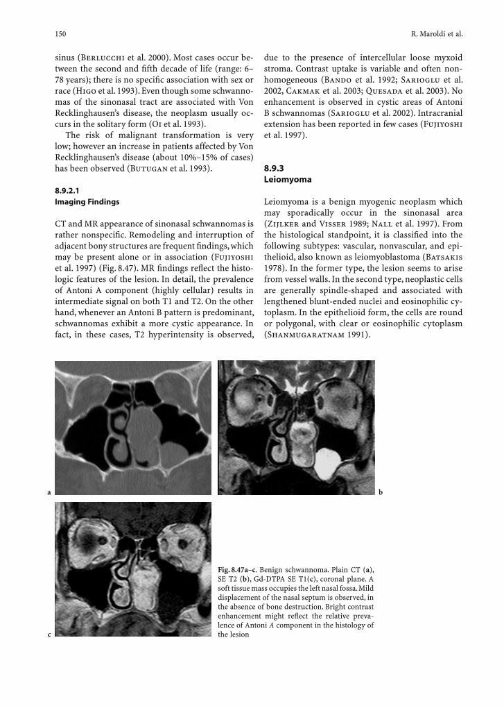
150 R. Maroldi et al.
sinus (Berlucchi et al. 2000). Most cases occur be-tween the second and fi fth decade of life (range: 6–78 years); there is no specifi c association with sex or race (Higo et al. 1993). Even though some schwanno-mas of the sinonasal tract are associated with Von Recklinghausen’s disease, the neoplasm usually oc-curs in the solitary form (Oi et al. 1993).
The risk of malignant transformation is very low; however an increase in patients affected by Von Recklinghausen’s disease (about 10%–15% of cases) has been observed (Butugan et al. 1993).
8.9.2.1 Imaging Findings
CT and MR appearance of sinonasal schwannomas is rather nonspecifi c. Remodeling and interruption of adjacent bony structures are frequent fi ndings, which may be present alone or in association (Fujiyoshi et al. 1997) (Fig. 8.47). MR fi ndings refl ect the histo-logic features of the lesion. In detail, the prevalence of Antoni A component (highly cellular) results in intermediate signal on both T1 and T2. On the other hand, whenever an Antoni B pattern is predominant, schwannomas exhibit a more cystic appearance. In fact, in these cases, T2 hyperintensity is observed,
due to the presence of intercellular loose myxoid stroma. Contrast uptake is variable and often non-homogeneous (Bando et al. 1992; Sarioglu et al. 2002, Cakmak et al. 2003; Quesada et al. 2003). No enhancement is observed in cystic areas of Antoni B schwannomas (Sarioglu et al. 2002). Intracranial extension has been reported in few cases (Fujiyoshi et al. 1997).
8.9.3 Leiomyoma
Leiomyoma is a benign myogenic neoplasm which may sporadically occur in the sinonasal area (Zijlker and Visser 1989; Nall et al. 1997). From the histological standpoint, it is classifi ed into the following subtypes: vascular, nonvascular, and epi-thelioid, also known as leiomyoblastoma (Batsakis 1978). In the former type, the lesion seems to arise from vessel walls. In the second type, neoplastic cells are generally spindle-shaped and associated with lengthened blunt-ended nuclei and eosinophilic cy-toplasm. In the epithelioid form, the cells are round or polygonal, with clear or eosinophilic cytoplasm (Shanmugaratnam 1991).
Fig. 8.47a–c. Benign schwannoma. Plain CT (a), SE T2 (b), Gd-DTPA SE T1(c), coronal plane. A soft tissue mass occupies the left nasal fossa. Mild displacement of the nasal septum is observed, in the absence of bone destruction. Bright contrast enhancement might refl ect the relative preva-lence of Antoni A component in the histology of the lesion
ba
c

Benign Neoplasms and Tumor-Like Lesions 151
Hachisuga et al. (1984), in a large series of 562 patients affected by leiomyoma, observed only fi ve cases of sinonasal involvement. Nicolai et al. (1996), in a review of the literature, identifi ed about 30 in-stances of well-documented sinonasal leiomyomas. The neoplasm usually affects subjects between the fi fth and sixth decade of life, with a slight predomi-nance for females.
The rarity of such a lesion in the sinonasal tract may be explained by the paucity of vascular smooth muscle fi bers, which are considered the elements of origin (Batsakis 1978; Mccaffrey et al. 1978; ). According to other theories, the lesion could stem from aberrant undifferentiated mesenchymal cells or from piloerector or sweat gland muscles (Batsakis 1978; Barr et al. 1990).
8.9.3.1 Imaging Findings
Both the lack of specifi city of CT or MR fi ndings and the extreme rarity of sinonasal leiomyoma make an imaging based diagnosis nearly impossible. As an aggressive pattern of growth has sometimes been de-scribed for this benign lesion – including bone de-struction and orbit encroachment (Harcourt and Gallimori 1993; Trott et al. 1994) – the main role of imaging is to precisely defi ne the deep extension of the lesion.
8.9.4 Paraganglioma
Paraganglioma is a benign neoplasm made up of cells originating from the neural crest and later migrating together with ganglionic cells of the autonomic ner-vous system. Histologically, the lesion is characterized by nests of epithelioid cells (Zellballen), surrounded by a capillary network (Shanmugaratnam 1991). Other terms such as glomus tumor, chemodectoma and apu-doma have been used, but at present paraganglioma is the recommended name. Paragangliomas are com-monly divided into adrenal (i.e., pheochromocytomas) and extra-adrenal in relation to the site of origin. The latter group includes cervicofacial paragangliomas, which rarely secrete catecholamines and therefore are classifi ed as non-chromaffi n. Most cases take origin in the temporal bone (glomus jugulare and glomus tym-panicum), above the bifurcation of the carotid artery (carotid body), and from the nodose ganglion (glomus intravagale) (Lack et al. 1977), whereas localization in the sinonasal tract is exceedingly rare, with approxi-
mately 30 cases reported in the literature (Ketabchi et al. 2003). In this area, the lesion more frequently involves the ethmoid and the middle turbinate; fur-thermore, there is a slight predominance in females and in the fi fth decade of life (Ketabchi et al. 2003).
As in other anatomic sites, paraganglioma of the si-nonasal tract may display an aggressive behavior and an infi ltrating growth pattern (Welkoborsky et al. 2000). According to a recent review of the literature (Ketabchi et al. 2003), six patients with paraganglioma of the si-nonasal tract presented distant or regional metastases. Increased mitotic fi gures, necrosis, vascular invasion, aneuploidy and a high proliferation rate may be impor-tant indicators for identifying lesions with a potentially aggressive biological behavior (Shanmugaratnam 1991; Welkoborsky et al. 2000)
8.9.4.1 Imaging Findings
Sinonasal paragangliomas exhibit no distinctive im-aging feature. On both CT and MR sinonasal para-gangliomas appear as markedly enhancing lesions; bone destruction can be observed (Myssiorek 2001) (Fig. 8.48). The differential diagnosis is therefore re-stricted to highly vascularized lesions, such as lobu-lar capillary hemangioma, hemangiopericytoma, schwannoma, and metastases (particularly from kid-ney, breast, and thyroid cancer).
Fig. 8.48. Paraganglioma. Gd-DTPA SE T1 on sagittal plane. Brightly enhancing ethmoid lesion eroding the anterior cranial fossa fl oor. Note encroachment of the hyperintense signal of the dura (arrows), indicating intradural extension. The lesion also invades both the frontal and sphenoid sinus (arrowheads)

152 R. Maroldi et al.
References
Adams WM, Jones RI, Chavda SI et al (1998) Pneumosinus dilatans: a discussion of four cases and the possible aetiol-ogy. Rhinology 36:40–42
Andrews JC, Fisch U, Valavanis A et al (1989) The surgical management of extensive nasopharyngeal angiofi bromas with the infratemporal fossa approach. Laryngoscope 99:429–437
Ariel M, De Dios V, Bond JR et al (1992) Aneurysmal bone cyst: a clinicopathologic study of 238 cases. Cancer 69:2921–2931
Atasoy C, Ustuner E, Erden I et al (2001) Frontal sinus muco-cele: a rare complication of craniofacial fi brous dysplasia. Clin Imaging 25:388-391
Attallah N, Jay MM (1981) Osteomas of the paranasal sinus. J Laryngol Otol 95:291–304
Bachor E, Weber R, Kahle R et al (1994) Temporary unilat-eral amaurosis with pneumosinus dilatans of the sphenoid sinus. Skull Base Surg 4:169–175
Bajaj MS, Pushker N (2002) Inverted papilloma invading the orbit. Orbit 21:155–159
Bando K, Obayashi M, Tsuneharu F (1992) A case of subfrontal schwannoma. No Shinkei Geka 20:1189–1194
Barr GD, More IAR, McCallum HM (1990) Leiomyoma of the nasal septum. J Laryngol Otol 104:891–893
Bartlett JR (1971) Intracranial complications of frontal and ethmoid osteomas. Br J Surg 58:607–609
Basak S, Mutlu C, Erkus M et al (1998) Benign fi brous histio-cytoma of the nasal septum. Rhinology 36:133–135
Batsakis JG (1978) Tumors of the head and neck: clinical and pathological considerations, 2nd edn. Williams & Wikins, Baltimore
Benedikt RA, Brown DC, Roth MK et al (1991) Spontaneous drainage of an ethmoidal mucocele: a possible cause of pneumosinus dilatans. AJNR Am J Neuroradiol 12:729–731
Benninger MS, Marks S (1995) The endoscopic management of sphenoid and ethmoid mucoceles with orbital and intra-cranial extension. Rhinology 33:157–161
Berlucchi M, Piazza C, Blanzuoli L et al (2000) Schwannoma of the nasal septum: a case report with review of the literature. Eur Arch Otorhinolaryngol 257:402–405
Bernier JL, Bhasker SN (1958) Aneurysmal bone cyst of man-dibula. Oral Surg 11:1018
Bielamowicz S, Calcaterra TC, Watson D (1993) Inverting pap-illoma of the head and neck: the UCLA update. Otolaryngol Head Neck Surg 109:71–76
Blitzer A, Kalmon D, Conley J (1989) Craniofacial resection of ossifying fi bromas and osteomas of the sinuses. Arch Otolaryngol Head Neck Surg 115:1112–1115
Bloom DC, Finley JC Jr, Broberg TG et al (2001) Leiomyoma of the nasal septum. Rhinology 39:233–235
Bollen E, Vielvoye J, Van Dijk JG et al (1990) Trigeminal neu-ralgia-like pain in an aged woman with fi brous dysplasia of the skull. Headache 30:277–279
Boysen ME, Olving JH, Vatne K et al (1979) Fibro-osseous lesions of the cranio-facial bones. J Laryngol Otol 93:793–807
Brandwein M, Steinberg B, Thung S et al (1989) Human papil-lomavirus 6/11 and 16/18 in Schneiderian inverted papil-lomas. In situ hybridiziation with human papillomavirus RNA preobes. Cancer 63:1708–1713
Breidahl AF, Szwajkun P, Chen YR (1997) Pneumosinus dilat-ans of the maxillary sinus: a report of two cases. Br J Plast Surg 50:33–39
Bremer JW, Neel HB 3rd, DeSanto LW et al (1986) Angiofi -broma: treatment trends in 150 patients during 40 years. Laryngoscope 96:1321–1329
Brodish BN, Morgan CE, Sillers MJ (1999) Endoscopic resec-tion of fi brous-osseous lesions of the paranasal sinuses. Am J Rhinol 13:111–116
Brors D, Draf W (1999) The treatment of inverted papilloma. Curr Opin Otolaryngol Head Neck Surg 7:33–38
Buchwald C, Franzmann MB, Tos S (1995) Sinonasal papillo-mas: a report of 82 cases in Copenhagen Country, including a longitudinal epidemiological and clinical study. Laryngo-scope 105:72–79
Buraczewski J, Dabska M (1971) Pathogenesis of aneurysmal bone cyst. Cancer 28:597–604
Busaba NY, Salman SD (1999) Maxillary sinus mucoceles: clinical presentation and long-term results of endoscopic surgical treatment. Laryngoscope 109:1446–1449
Butugan O, Grazel SS, de Almeida ER et al (1993) Schwannoma of the nasal septum: report of the two cases. Rev Laryngol 114:33–36
Cakmak O, Yavuz H, Yucel T (2003) Nasal and paranasal sinus schwannomas. Eur Arch Otorhinolaryngol 260:195–197
Calcaterra TC, Thompson JW, Paglia DE (1980) Inverting pap-illomas of the nose and paranasal sinuses. Laryngoscope 90:53–60
Calliauw L, Roels H, Caemaert J (1985) Aneurysmal bone cysts in the cranial vault and base of skull. Surg Neurol 23:193–198
Camilleri AE (1991) Craniofacial fi brous dysplasia. J Laryngol Otol 105:662–666
Cano Cuenca B, Gimenez Vaillo F, Perez Climent F et al (2000) Mucinous cystadenoma of a minor salivary gland of the nasal fossa. An Otorrinolaringol Ibero Am 27:469–476
Casasco A, Houdart E, Biondi A et al (1999) Major complica-tions of percutaneous embolization of skull-base tumors. AJNR Am J Neuroradiol 20:179–181
Casselman JW, De Jonge I, Neyt L et al (1993) MRI in craniofa-cial fi brous dysplasia. Neuroradiology 35:234–237
Chagnaud C, Petit P, Bartoli J et al (1998) Postoperative follow-up of juvenile nasopharyngeal angiofi bromas: assessment by CT scan and MR imaging. Eur Radiol 8:756–764
Chan FL, Chow SK, Sham JST (1992) Infratemporal pneumato-cele arising from maxillary sinus. Clin Radiol 45:27–30
Chartrand-Lefebvre C, Dubois J, Roy D et al (1996) Direct intraoperative sclerotherapy of an aneurysmal bone cyst of the sphenoid. AJNR Am J Neuroradiol 17:870–872
Chateil JF, Dousset V, Meyer P et al (1997) Cranial aneurysmal bone cysts presenting with raised intracranial pressure: report of two cases. Neuroradiology 39:490–494
Chee LWJ, Sethi DS (1999) The endoscopic management of sinonasal inverted papillomas. Clin Otolaryngol 24:61–66
Cheng C, Takahashi H, Yao K et al (2002) Cemento-ossifying fi broma of maxillary and sphenoid sinuses: case report and literature review. Acta Otolaryngol Stockh (Suppl) 547:118–122
Cho KJ, el-Naggar AK, Mahanupab P et al (1995) Carcinoma ex-pleomorphic adenoma of the nasal cavity: a report of two cases. J Laryngol Otol 109:677–679
Chong VF, Tang LH (1997) Maxillary sinus ossifying fi broma. Am J Otolaryngol 18:419–424

Benign Neoplasms and Tumor-Like Lesions 153
Citardi MJ, Janjua T, Abrahams JJ et al (1996) Orbitoeth-moid aneurysmal bone cyst. Otolaryngol Head Neck Surg 114:466–470
Commins DJ, Tolley NS, Milford CA (1998) Fibrous dysplasia and ossifying fi broma of the paranasal sinuses. J Laryngol Otol 112:964–968
Compagno J, Wong RT (1977) Intranasal mixed tumours (pleomorphic adenomas): a clinicopathologic study of 40 cases. Am J Clin Pathol 68:213–218
Cooter MS, Charlton SA, Lafreniere D et al (1998) Endoscopic management of an inverted nasal papilloma in a child. Oto-laryngol Head Neck Surg 118:876–879
Dammann F, Pereira P, Laniado M et al (1999) Inverted papil-loma of the nasal cavity and the paranasal sinuses: using CT for primary diagnosis and follow-up. AJR Am J Roent-genol 172:543–548
Danesi G, Panizza B, Mazzoni A et al (2000) Anterior approaches in juvenile nasopharyngeal angiofi bromas with intracranial extension. Otolaryngol Head Neck Surg 122:277–283
De Minteguiaga C, Portier F, Guichard JP et al (2001) Aneu-rysmal bone cyst in the sphenoid bone: treatment with minimally invasive surgery. Ann Otol Rhinol Laryngol 110:331–334
Deschler DG, Kaplan MJ, Boles R (1992) Treatment of large juvenile nasopharyngeal angiofi broma. Otolaryngol Head Neck Surg 106:278–284
Dillon WP, Som PM, Rosenau W (1991) Hemangioma of the nasal vault: MR and CT features. Radiology 180:761–765
Dohar JE, Duvall AJ 3rd (1992) Spontaneous regression of juvenile nasopharyngeal angiofi broma. Ann Otol Rhinol Laryngol 101:469–471
Donnelly M, Al-Seder MH, Blayney AW (1992) Benign nasal schwannoma. J Laryngol Otol 106:1011–1015
Draf W (1991) Endonasal micro-endoscopic frontal sinus sur-gery: the Fulda concept. Op Tech Otolaryngol Head Neck Surg 4:234–240
Dufour H, Fesselet J, Metellus P et al (2001) Cavernous heman-gioma of the sphenoid sinus: case report and review of the literature. Surg Neurol 55:169–173; discussion 173
Earwaker J (1993) Paranasal sinus osteomas: A review of 46 cases. Skeletal Radiol 22:417–423
El-Mofty S (2002) Psammomatoid and trabecular juvenile ossifying fi broma of the craniofacial skeleton: two distinct clinicopathologic entities. Oral Surg Oral Med Oral Pathol Oral Radiol Endod 93:296–304
Elner VM, Burnstine MA, Goodman ML et al (1995) Inverted papillomas that invade the orbit. Arch Ophthalmol 113:1178–1183
El-Sayed Y, Al-Serhani A (1997) Lobular capillary haeman-gioma (pyogenic granuloma) of the nose. J Laryngol Otol 111:941–945
Engelbrecht V, Preis S, Hassler W et al (1999) CT and MRI of congenital sinonasal ossifying fi broma. Neuroradiology 41:526–529
Evans RW, Cruickshand AH (1970) Major problems in pathol-ogy. Epithelial tumors of the salivary glands. Saunders WB, Philadelphia
Eversole LR, Leider AS, Nelson K (1985) Ossifying fi broma: a clinicopathologic study of sixty-four cases. Oral Surg Oral Med Oral Pathol 60:505–511
Facon F, Paris J, Ayache S et al (2002) Adènome pléomorphic des fosses nasals: une localisation à la cloison inter sinuso nasale. Rev Laryngol Otol Rhinol 123:103–107
Fechner RE, Alford DO (1968) Inverted papilloma and squa-mous carcinoma. An unusual case. Arch Otolaryngol 88:507–512
Ferguson BJ (1994) Fibrous dysplasia of the paranasal sinuses. Am J Otolaryngol 15:227–230
Forman D, Goldberg HI (1990) Microembolization and resec-tion of a highly vascular pyogenic granuloma. J Oral Maxillofac Surg 48:415–418
Freeman SB, Kennedy KS, Parker GS et al (1990) Metastasizing pleomorphic adenoma of the nasal septum. Arch Otolaryn-gol Head Neck Surg 116:1331–1333
Fu YS, Perzin KH (1974) Non-epithelial tumors of the nasal cavity, paranasal sinuses, and nasopharynx: a clinicopatho-logic study. Cancer 33:1289–1305
Fujimoto Y, Katoh M, Miyata M et al (1987) Cystic cemento-ossifying fi broma of the ethmoidal cells. J Laryngol Otol 101:946–952
Fujiyoshi F, Kajiya Y, Nakajo M (1997) CT and MR imaging of nasoethmoid schwannoma with intracranial extension. AJR Am J Roentgenol 169:1754–1755
Furuta Y, Shinohara T, Sano K et al (1991) Molecular patho-logic study of human papillomavirus infection in inverted papilloma and squamous cell carcinoma of nasal cavities and paranasal sinuses. Laryngoscope 101:79–85
Gaffey MJ, Frierson HF, Weiss LM et al (1996) Human papillo-mavirus and Epstein-Barr virus in sinonasal Schneiderian papillomas. An in situ hybridization and polymerase chain reaction study. Am J Clin Pathol 106:475–482
Gillman GS, Lampe HB, Allen LH (1997) Orbitoethmoid osteoma: case report of an uncommon presentation of an uncommon tumor. Otolaryngol Head Neck Surg 117:S218–220
Golz A, Ben-Arie Y, Fradis M (1997) Pleomorphic nasoseptal adenoma. J Otolaryngol 26:399–401
Gregor RT, Loftus-Coll B (1994) Myxoma of the paranasal sinuses. J Laryngol Otol. 108:679–681
Hachisuga T, Hashimoto O, Enjoji A (1984) Angioleiomyoma: a clinicopathological reappraisal of 562 cases. Cancer 54:126–130
Hady MR, Ghanaam B, Hady MZ (1990) Aneurysmal bone cyst of the maxillary sinus. J Laryngol Otol 104:501–503
Hamner JE, Scofeild HH, Cornyn J (1968) Benign fi bro-osse-ous jaw lesions of periodontal membrane origin. Cancer 22:861–878
Han JK, Smith TL, Loehrl T et al (2001) An evolution in the management of sinonasal inverting papilloma. Laryngo-scope 111:1395–1400
Han MH, Chang KH, Lee CH et al (1991) Sinonasal psam-momatoid ossifying fi bromas: CT and MR manifestations. AJNR Am J Neuroradiol 12:25–30
Han MH, Chang KH, Lee CH et al (1995) Cystic expansile masses of the maxilla: differential diagnosis with CT and MR. AJNR Am J Neuroradiol 16:333–338
Harcourt JP, Gallimore AP (1993) Leiomyoma of the paranasal sinuses. J Laryngol Otol 107:740–741
Har-El G (2001) Endoscopic management of 108 sinus muco-celes. Laryngoscope 111:2131–2134
Harrison D, Lund VJ (1993) Tumours of the Upper Jaw. London, Churchill Livingstone
Hartley BE, Lund VJ (1999) Endoscopic drainage of pediatric paranasal sinus mucoceles. Int J Pediatr Otorhinolaryngol 50:109–111
Hartwidge C, Varma TR (1984) Intracranial aeroceles as a

154 R. Maroldi et al.
complication of frontal sinus osteoma. Surg Neurol 24:401–404
Hejazi N, Witzmann A, Hassler W (2001) Ocular manifesta-tions of sphenoid mucoceles: clinical features and neu-rosurgical management of three cases and review of the literature. Surg Neurol 56:338–343
Herman P, Lot G, Chapot R et al (1999) Long-term follow-up of juvenile nasopharyngeal angiofi bromas: analysis of recur-rences. Laryngoscope 109:140–147
Higo R, Yamasoba T, Kikuchi S (1993) Nasal neurinoma: case report and review of literature. Auris Nasus Larynx 20:297–301
Hirst LW, Miller NR, Allen GS (1979) Sphenoidal pneumosinus dilatans with bilateral optic nerve meningiomas. J Neuro-surg 51:402–407
Howard DJ, Lloyd G, Lund V (2001) Recurrence and its avoidance in juvenile angiofi broma. Laryngoscope 111:1509–1511
Hrishikesh KA, Narlawar RS, Deasi SB et al (2002) Case report: Aneurysmal bone cyst of the ethmoid bone. Br J Radiol 75:916–918
Huang HM, Liu CM, Lin KN et al (2001) Giant ethmoid oste-oma with orbital extension, a nasoendoscopic approach using an intranasal drill. Laryngoscope 111:430–432
Huang PC, Misko G (1998) Imaging quiz case 1. Osteoma of the ethmoidal sinus. Arch Otolaryngol Head Neck Surg 124:602, 604–605
Huneidi A, Afshar F (1989) Chronic spontaneous tension pneumocephalus due benign frontal osteoma. Br J Neuro-surg 3:389–392
Hyams VJ (1971) Papillomas of the nasal cavity and paranasal sinuses. A clinicopathological study of 315 cases. Ann Otol Rhinol Laryngol 80:192–206
Hyams VJ, Batsakis JG, Michaels L (1988) Tumors of the upper respiratory tract and ear. Washington, Armed Forces Insti-tute of Pathology
Ichimura K, Ohta Y, Maeda YI et al (2001) Mucoceles of the paranasal sinuses with intracranial extension. Postopera-tive course. Am J Rhinol 15:243–247
Ikeda K, Takahashi C, Oshima T et al (2000) Endonasal endo-scopic marsupialization of paranasal sinus mucoceles. Am J Rhinol 14:107–111
Iwata N, Hattori K, Nakagawa T et al (2002) Hemangioma of the nasal cavity: a clinicopathologic study. Auris Nasus Larynx 29:335–339
Jafek BW, Wood RP 2nd, Dion M (1977) Granuloma pyogeni-cum. Ear Nose Throat J 56:228–233
Jaffe HL, Lichtenstein L (1942) Solitary unicameral bone cyst with emphasis on the roentgen picture, the pathologic appearance, and the pathogenesis. Arch Surg 44:1004–1025
Jassar P, Stafford ND, MacDonald AW (1999) Pleomorphic ade-noma of the nasal septum. J Laryngol Otol 113:483–485
Johnson LC, Yousefi M, Vinh TN et al (1991) Juvenile active ossifying fi broma: its nature, dynamics and origin. Acta Otolaryngol Stockh (Suppl) 488:1–40
Kamel RH (1995) Transnasal endoscopic medial maxillectomy in inverted papilloma. Laryngoscope 105:847–853
Kapadia SB, Barnes L, Pelzman K et al (1993) Carcinoma ex oncocytic schneiderian (cylindrical cell) papilloma. Am J Otolaryngol 14:332–338
Karlidag T, Yalcin S, Kaygusuz I et al (2003) Bilateral pneu-mosinus dilatans of the maxillary sinuses. Br J Oral Maxil-lofac Surg 41:122–123
Kass ES, Fabian RL, Montgomery WW (1999) Manometric study of paranasal sinus mucoceles. Ann Otol Rhinol Lar-yngol 108:63–66
Kass ES, Salman S, Rubin PA et al (1997) Chronic maxillary atelectasis. Ann Otol Rhinol Laryngol 106:109–116
Kennedy DW, Josephson JS, Zinreich SJ et al (1989) Endoscopic sinus surgery for mucoceles: a viable alternative. Laryngo-scope 99:885–895
Kershisnik M, Bataskis JG (1994) Aneurysmal bone cysts of the jaws. Ann Otol Rhinol Laryngol 103:164–165
Ketabchi S, Massi D, Santoro R et al (2003) Paraganglioma of the nasal cavity: a case report. Eur Arch Otorhinolaryngol 260:336–340
Kilde JD, Rhee JS, Balla AA et al (2003) Hemangioma of the sphenoid and ethmoid sinuses: two case reports. Ear Nose Throat J 82:217–221
Kim HJ, Kim JH, Hwang EG (1995) Bone erosion caused by sinonasal cavernous hemangioma: CT fi ndings in two patients. AJNR Am J Neuroradiol 16:1176–1178
Kimmelman CP, Potsic WP, Schut L (1982) Aneurysmal bone cyst of the sphenoid in a child. Ann Otol Rhinol Laryngol 91:339–341
Kessler A, Berenholz LP, Segal S (1998) Use of intranasal endo-scopic surgery to relieve ostiomeatal complex obstruction in fi brous dysplasia of the paranasal sinuses. Eur Arch Oto-rhinolaryngol 255:454–456
Klossek JM, Dufour X, Toffel P et al (2000) Pneumosinus dilat-ans: a report of three new cases and their surgical manage-ment. Ear Nose Throat J 79:48–51
Koike Y, Tokoro K, Chiba Y et al (1996) Intracranial extension of paranasal sinus mucocele: two case reports. Surg Neurol 45:44–48
Koiuvnen P, Lopponen H, Fors AP et al (1997) The growth rate of osteomas of the paranasal sinuses. Clin Otolaryn-gol 22:111–114
Komori E, Sugisaki M (1988) Ectopic pneumosinus maxillary dilatans. A case report. J Craniomaxillofac Surg 16:240–242
Kransdorf MJ, Moser RP Jr, Gilkey FW (1990) Fibrous dyspla-sia. Radiographics 10:519–537
Kransdorf MJ, Stull MA, Gilkey FW et al (1991) Osteoid oste-oma. Radiographics 11:671–696
Kransdorf MJ, Sweet DE (1995) Aneurysmal bone cyst: con-cept, controversy, clinical presentation, and imaging. AJR Am J Roentgenol 164:573–580
Krausen AS, Gulman S, Zografakis G (1977) Cementomas. II. Aggressive cemento-ossifying fi broma of the ethmoid region. Arch Otolaryngol Head Neck Surg 103:371–373
Krouse JH (2000) Development of a staging system for inverted papilloma. Laryngoscope 110:965–968
Krouse JH (2001) Endoscopic treatment of inverted papilloma: safety and effi cacy. Am J Otolaryngol 22:87–99
Kulkarni MV, Bonner FM, Abdo GJ (1989) Maxillary sinus hemangioma: MR and CT studies. J Comput Assist Tomogr 13:340–342
Lack EE, Cubilla AL, Woodruff JM et al (1977) Paragangliomas of the head and neck region. Cancer 39:397–409
Lance E, Schatz C, Nach R et al (1992) Pyogenic granuloma gravidarum of the nasal fossa: CT features. J Comput Assist Tomogr 16:663–664
Lane JM, Khan SN, O’Connor WJ et al (2001) Bisphospho-nate therapy in fi brous dysplasia. Clin Orthop Rel Reser 382:6–12

Benign Neoplasms and Tumor-Like Lesions 155
Lasjaunias P, Picard L, Manelfe C et al (1980) Angiofi broma of the nasopharynx. A review of 53 cases treated by embolisa-tion. The role of pretherapeutic angiography. Pathophysi-ological hypotheses. J Neuroradiol 7:73–95
Lawson W, Ho BT, Shaari CM et al (1995) Inverted papilloma: a report of 112 cases. Laryngoscope 105:282–288
Lee JT, Bhuta S, Lufkin R et al (2003) Isolated inverting papil-loma of the sphenoid sinus. Laryngoscope 113:41–44
Lee KC, Chan JK, Chong YW (1992) Ossifying pleomorphic ade-noma of the maxillary antrum. J Laryngol Otol 106:50–52
Lehnerdt G, Weber J, Dost P (2001) Die einseitige verschat-tung der nasennebenhöhlen im CT oder MRT – Hinweis für einen seltenen histologischen befund. Laryngorhinoot-ologie 80:141–145
Leopard PJ (1975) Cryosurgery, and its application to oral sur-gery. Br J Oral Surg 13:128–152
Levy WM, Miller AS, Bonakdarpour A et al (1975) Aneurysmal bone cyst secondary to other osseous lesions: report of 57 cases. Am J Clin Pathol 63:1–8
Leyden JJ, Master GH (1973) Oral cavity pyogenic granuloma. Arch Dermatol 108:226–228
Li JR, Qian J, Shan XZ et al (1998) Evaluation of the effec-tiveness of preoperative embolization in surgery for naso-pharyngeal angiofi broma. Eur Arch Otorhinolaryngol 255:430–432
Liao BS, Chong E (1993) Septal pleomorphic adenoma mas-querading as squamous cell carcinoma. Ear Nose Throat J 72:781–782
Lichtenstein L (1950) Aneurysmal bone cyst: A pathological entity commonly mistaken for giant cell tumor and occa-sionally for hemangioma and osteogenic sarcoma. Cancer 3:279–289
Lichtenstein L, Jaffe HL (1942) Fibrous dysplasia of bone. Arch Pathol 33:777–781
Lieberman A, Tovi F (1984) A small osteoma of the frontal sinus causing headaches. J Laryngol Otol 98:1147–1149
Liens D, Delmas PD, Meunier PJ (1994) Long term effects of intravenous pamidronate in fi brous dysplasia of bone. Lancet 343:953–954
Lim CC, Dillon WP, McDermott MW (1999) Mucocele involv-ing the anterior clinoid process: MR and CT fi ndings. AJNR Am J Neuroradiol 20:287–290
Lloyd G, Lund VJ, Savy L et al (2000a) Optimum imaging for mucoceles. J Laryngol Otol 114:233–236
Lloyd G, Howard D, Lund VJ et al (2000b) Imaging for juvenile angiofi broma. J Laryngol Otol 114:727–730
Lloyd G, Howard D, Phelps P et al (1999) Juvenile angiofi -broma: the lessons of 20 years of modern imaging. J Lar-yngol Otol 113:127–134
Lloyd GA (1985) Orbital pneumosinus dilatans. Clin Radiol 36:381–386
Lloyd GA, Phelps PD (1986) Juvenile angiofi broma: imaging by magnetic resonance, CT and conventional techniques. Clin Otolaryngol 11:247–259
Loh LE, Ho KH, Thoo A et al (1994) Hemangioma of the eth-moidal sinuses. Singapore Med J 35:211–214
London SD, Schlosser RJ, Gross CW (2002) Endoscopic man-agement of benign sinonasal tumors: a decade of experi-ence. Am J Rhinol 16:221–227
Lund VJ (1998) Endoscopic management of paranasal sinus mucocoeles. J Laryngol Otol 112:36–40
Lund VJ (2000) Optimum management of inverted papilloma. J Laryngol Otol 114:194–197
Lund VJ, Milroy CM (1991) Fronto-ethmoidal mucocoeles: a histopathological analysis. J Laryngol Otol 105:921–923
Lund VJ, Wilson H, Sajeda M et al (1988) Prostaglandin syn-thesis in the pathogenesis of fronto-ethmoidal mucocoeles. Acta Otolaryngol (Stockh) 106:145–151
Makeieff M, Youssef B, Gardiner Q et al (1999) Adénome plèo-morphe de la cloison nasale. A propos d’un cas. Ann Oto-laryngol Chir Cervicofac 116:368–371
Manaka H, Tokoro K, Sakata K et al (1998) Intradural exten-sion of mucocele complicating frontoethmoid sinus oste-oma: case report. Surg Neurol 50:453–456
Mansell NJ, Bates GJ (2000) The inverted schneiderian papil-loma: a review and literature report of 43 new cases. Rhi-nology 38:97–101
Maroldi R, Farina D, Palvarini L et al (2004) MRI fi ndings of inverted papilloma: differential diagnosis with malignant sinonasal tumors. Am J Rhinol in press
Marvel JB, Marsh MA, Catlin FI (1991) Ossifying fi broma of the mid-face and paranasal sinuses: diagnostic and therapeutic considerations. Otolaryngol Head Neck Surg 104:803–808
Matthew S, Ersner MD, Saltzman M (1944) A mixed tumor of the nasal septum. Report of a case. Laryngoscope 54:287–296
Mauri M, de Oliveira CO, Franche G (2000) Pneumosinus dilatans of the maxillary sinus. Ann Otol Rhinol Laryngol 109:278–280
Mayer I (1962) Electrocoagulation of oral lesions. Oral Surg 16:522–528
McCaffrey TV, McDonald TJ, Unni KK (1978) Leiomyoma of the nasal cavity. J Laryngol Otol 92:817–819
McCombe A, Lund VJ, Howard DJ (1990) Recurrence in juve-nile angiofi broma. Rhinology 28:97–102
Metha BS, Grewal GS (1963) Osteoma of the paranasal sinuses along with a case of an orbito-ethmoid osteoma. J Laryngol Otol 77:601–610
Meyes J (1898) Pneumosinus dilatans. Nederlands Tijdsschrift voor Geneeskunde 11:143
Miller FR, D’Agostino MA, Schlack K (1999) Lobular capillary hemangioma of the nasal cavity. Otolaryngol Head Neck Surg 120:783–784
Mills SE, Cooper PH, Fechner RE (1980) Lobular capillary hemangioma: the underlying lesion of pyogenic granu-loma. A study of 73 cases from the oral and nasal mucous membranes. Am J Surg Pathol 4:470–479
Moore AT, Buncic JR, Muro IR (1985) Fibrous dysplasia of the orbit in childhood. Clinical features and management. Ophthalmology 92:12–20
Morrison MD, Tchang SP, Maber BR (1976) Pneumocele of the maxillary sinus. Report of a case. Arch Otolaryngol 102:306–307
Moriyama H, Hesaka H, Tachibana T et al (1992) Mucoceles of ethmoid and sphenoid sinus with visual disturbance. Arch Otolaryngol Head Neck Surg 118:142–146
Moulin G, Chagnaud C, Gras R et al (1995) Juvenile nasopha-ryngeal angiofi broma: comparison of blood loss during removal in embolized group versus nonembolized group. Cardiovasc Intervent Radiol 18:158–161
Muraoka H, Ishihara A, Kumagai J (2001) Fibrous dysplasia with cystic appearance in maxillary sinus. Auris Nasus Larynx 28:103–105
Myers EN, Fernau JL, Johnson JT et al (1990) Management of inverted papilloma. Laryngoscope 100:481–490

156 R. Maroldi et al.
Myssiorek D (2001) Head and neck paragangliomas: an over-view. Otolaryngol Clin North Am 34:829–836
Nager GT, Kennedy DW, Kopstein E (1982) Fibrous dysplasia: a review of the disease and its manifestations in the temporal bone. Ann Otol Rhinol Laryngol Suppl 92:1–52
Nall AV, Stringer SP, Baughman RA (1997) Vascular leiomyoma of the superior turbinate: fi rst reported case. Head Neck 19:63–67
Namdar I, Edelstein DR, Huo J et al (1998) Management of oste-omas of the paranasal sinuses. Am J Rhinol 12:393–398
Nicolai P, Berlucchi M, Tomenzoli D et al (2003) Endoscopic surgery for juvenile angiofi broma: when and how. Laryn-goscope 113:775–782
Nicolai P, Redalli de Zinis LO, Facchetti F et al (1996) Cranio-facial resection for vascular leiomyoma of the nasal cavity. Am J Otolaryngol 17:340–344
Oberman HA (1964) Papillomas of the nose and paranasal sinuses. Am J Clin Pathol 42:245–258
Oi H, Watanabe Y, Shojaku H et al (1993) Nasal septum neuri-noma. Acta Otolaryngol (Stockh) Suppl 504:151–154
Oikawa K, Furuta Y, Oridate N et al (2003) Preoperative stag-ing of sinonasal inverted papilloma by magnetic resonance imaging. Laryngoscope 113:1983–1987
Ojiri H, Ujita M, Tada S et al (2000) Potentially distinctive fea-tures of sinonasal inverted papilloma on MR imaging. AJR Am J Roentgenol 175:465–468
Osguthorpe JD, Gudeman SK (1987) Orbital complications of fi brous dysplasia. Otolaryngol Head Neck Surg 97:403–405
Outzen KE, Grontveld A, Jorgensen K et al (1996) Inverted papilloma: incidence and late results of surgical treatment. Rhinology 34:114–118
Pacini F, Perri G, Bagnolesi P et al (1987) McCune-Albright syndrome with gigantism and hyperprolactinemia. J Endo-crinol Invest 10:417–420
Pasquini E, Ceroni Compadretti G, Sciarretta V et al (2002) Transnasal endoscopic surgery for the treatment of fi brous dysplasia of maxillary sinus associated to aneurysmal bone cyst in a 5-year-old child. Int J Pediatr Otorhinolaryngol 62:59–62
Patel BC, Sabir DI, Flaharty PM et al (1993) Aneurysmal bone cyst of the orbit and ethmoid sinus. Arch Ophthalmol 111:586–587
Pelausa EO, Fortier MAG (1992) Schneiderian papilloma of the nose and paranasal sinuses: the University of Ottawa experience. J Otolaryngol 21:9–15
Petit P, Vivarrat-Perrin L, Champsaur P et al (2000) Radiologi-cal follow-up of inverted papilloma. Eur Radiol 10:1184–1189
Phillips PP, Gustafson RO, Facer GW (1990) The clinical behavior of inverting papilloma of the nose and paranasal sinuses: report of 112 cases and review of the literature. Laryngoscope 100:463–469
Pisani P, Dosdegani R, Ramponi A et al (1998) Adenoma pleo-morfo del setto nasale. Acta Otorhinolaryngol Ital 18:30–33
Poncet A, Dor L (1897) Botryomycose humaine. Rev Chir (Paris) 18:966
Pound E, Pickrell K, Hunger W et al (1965) Fibrous dysplasia of the maxilla. Ann Surg 161:406–410
Powell JL, Bailey CL, Coopland AT et al (1994) Nd:YAG laser excision of a giant gingival pyogenic granuloma of preg-nancy. Lasers Surg Med 14:178–183
Prager DA, Weiss MH, Buchalter WL et al (1991) Pleomorphic adenoma of the nasal cavity. Ann Otol Rhinol Laryngol 100:600
Price HI, Batnitzky S, Karlin CA et al (1981) Multiple paranasal sinus mucoceles. J Comput Assist Tomogr 5:122–125
Quesada JL, Enrique A, Lorente J et al (2003) Sinonasal schwannoma treated with endonasal microsurgery. Oto-laryngol Head Neck Surg 129:300–302
Rambaux P, Bertrand B, Eloy P et al (2000) Endoscopic endo-nasal surgery for paranasal sinus mucoceles. Acta Otorhi-nolaryngol Belg 54:115–122
Ramsey HE, Strong EW, Frazel EL (1968) Fibrous dysplasia of the craniofacial bones. Am J Surg 116:542–547
Rao VM, Sharma D, Madan A (2001) Imaging of frontal sinus disease. Concepts, interpretation and technology. Otolar-yngol Clin North Am 34:23–39
Rappaport JM, Attia EL (1994) Pneumocephalus in frontal sinus osteoma. J Otolaryngol 23:430–436
Raynal M, Peynegre R, Beautru R et al (1999) Mucocèles sinusiennes et iatrogénie chirurgicale. Ann Otolaryngol Chir Cervicofac 116:85–91
Redaelli de Zinis LO, Ansarin M, Galli G et al (1996) Approccio chirurgico transbasale-subfrontale in un caso di fi broma ossifi cante etmoidale. Gior Pat Chir Cr Fa 2:42–45
Reed RJ (1963) Fibrous dysplasia of bone: a review of 25 cases. Arch Pathol 75:480–495
Reicher MA, Bentson JR, Halbach VV et al (1986) Pneumosi-nus dilatans of the sphenoid sinus. AJNR Am J Neuroradiol 7:865–868
Roberson GH, Biller H, Sessions DG et al (1972) Presurgical internal maxillary artery embolization in juvenile angiofi -broma. Laryngoscope 82:1524–1532
Roger G, Tran Ba Huy P, Froehlich P et al (2002) Exclusively endoscopic removal of juvenile nasopharyngeal angio-fi broma: trends and limits. Arch Otolaryngol Head Neck Surg 128:928–935
Rosenberg AE (1994) Skeletal system and soft tissues tumors. In: Cotran RS, Kumar V, Robbins SL (eds) Robbins patho-logic basis of disease, 5th edn. WB Saunders, Philadelphia, pp 1213–1271
Saito K, Fukuta K, Takahashi M et al (1998) Benign fi broosse-ous lesions involving the skull base, paranasal sinuses, and nasal cavity. J Neurosurg 88:1116–1119
Samy LL, Mostafa H (1971) Osteoma of the nose and para-nasal sinuses with a report of 21 cases. J Laryngol Otol 85:449–469
Sankerkin NG, Mott MG, Roylance J (1983) An unusual intraos-seous lesion with fi broblastic, osteoclastic, osteoblastic, aneurysmal and fi bromyxoid elements: “solid” variant of aneurysmal bone cyst. Cancer 51:2278–2286
Sarioglu S, Ozkal S, Guneri A et al (2002) Cystic schwannoma of the maxillary sinus. Auris Nasus Larynx 29:297–300
Savic D, Djeric DR (1990) Indications for surgical treatment of osteomas of the frontal and ethmoid sinuses. Clin Oto-laryngol 15:397–404
Savy L, Lloyd G, Lund VJ et al (2000) Optimum imaging for inverted papilloma. J Laryngol Otol 114:891–893
Sayan NB, Ucok C, Karasu HA et al (2002) Peripheral osteoma of the oral and maxillofacial region: a study of 35 new cases. J Oral Maxillofac Surg 60:1299–1301
Schick B, Plinkert PK, Prescher A (2002) Die vaskulare kompo-nente: gedanken zur entstehung des Angiofi broms. Laryn-gorhinootologie 81:280–284
Benign Neoplasms and Tumor-Like Lesions 157
Schick B, Kahle G (2000) Radiological fi ndings in angiofi -broma. Acta Radiol 41:585–593
Schick B, Steigerwald C, El Tahan AER et al (2001) The role of endonasal surgery in the management of frontoethmoid osteoma. Rhinology 39:66–70
Schmaman A, Smith I, Ackerman LV (1970) Benign fi bro-osse-ous lesions of the mandible and maxilla: a review of 35 cases. Cancer 26:303–312
Scholtz AW, Appenroth E, Kammen-Jolly K et al (2001) Juvenile nasopharyngeal angiofi broma: management and therapy. Laryngoscope 111:681–687
Schroth G, Haldemann AR, Mariani L et al (1996) Preopera-tive embolization of paragangliomas and angiofi bromas. Measurement of intratumoral arteriovenous shunts. Arch Otolaryngol Head Neck Surg 122:1320–1325
Schwartz DR, Alpert MA (1964) The malignant transformation of fi brous dysplasia. Am J Med Sci 274:35–54
Senol U, Karaali K, Akyuz M et al (2002) Aneurysmal bone cyst of the orbit. AJNR Am J Neuroradiol 23:319–321
Shanmugaratnam K (1991) Histological typing of tumours of the upper respiratory tract and ear. Springer-Verlag, Berlin Heidelberg New York
Shohet JA, Duncavage JA (1996) Management of the frontal sinus with inverted papilloma. Otolaryngol Head Neck Surg 114:649–652
Sievers KW, Greess H, Baum U et al (2000) Paranasal sinuses and nasopharynx CT and MRI. Eur J Radiol 33:185–202
Simo R, De Carpentier J, Rejali D et al (1998) Paediatric pyo-genic granuloma presenting as a unilateral nasal polyp. Rhinology 36:136–138
Simovic S, Klapan I, Bumber Z et al (1996) Fibrous dysplasia in paranasal cavities. ORL J Otorhinolaryngol Relat Spec 58:55–58
Siniluoto TM, Luotonen JP, Tikkakoski TA et al (1993) Value of pre-operative embolization in surgery for nasopharyngeal angiofi broma. J Laryngol Otol 107:514–521
Skolnick CA, Mafee MF, Goodwin JA (2000) Pneumosinus dilatans of the sphenoid sinus presenting with visual loss. J Neuroophthalmol 20:259–263
Skoulakis CE, Velegrakis GA, Doxas PG et al (1999) Mucocele of the maxillary antrum in an eight-year-old boy. Int J Pedi-atr Otorhinolaryngol 47:283–287
Slootweg PJ, Panders AK, Nikkels PGJ (1994) Juvenile ossifying fi broma: an analysis of 33 cases with emphasis on histo-pathological aspects. J Oral Pathol 23:385–388
Smith JF (1965) Fibrous dysplasia of the jaws. Arch Otolaryn-gol 81:592–603
Smith ME, Calcaterra TC (1989) Frontal sinus osteoma. Ann Otol Rhinol Laryngol 98:896–900
Som PM, Brandwein M (1996) Sinonasal cavities: infl amma-tory diseases, Tumors, fractures, and postoperative fi nd-ings. In: Som PM, Curtin HD (eds) Head and neck imaging. Mosby Year Book, St. Louis, pp 227-229
Som PM, Dillon WP, Fullerton GD et al (1989) Chronically obstructed sinonasal secretions: observations on T1 and T2 shortening. Radiology 172:515–520
Som PM, Lidov M (1992) The benign fi broosseous lesion: its association with paranasal sinus mucoceles and its MR appearance. J Comput Assist Tomogr 16:871–876
Som PM, Lidov M (1994) The signifi cance of sinonasal radiodensities: ossifi cation, calcifi cation, or residual bone? AJNR Am J Neuroradiol 15:917–922
Som PM, Schatz CJ, Flaum EG et al (1991) Aneurysmal bone
cyst of the paranasal sinuses associated with fi brous dys-plasia: CT and MR fi ndings. J Comput Assist Tomogr 15:513–515
Soysal V, Yigitbasi OG, Kontas O et al (2001) Infl ammatory myofi broblastic tumor of the nasal cavity: a case report and review of the literature. Int J Pediatr Otorhinolaryn-gol 61:161–165
Spencer MG, Mitchell DB (1987) Growth of a frontal sinus osteoma. J Laryngol Otol 101:726–728
Stamm AC, Watashi CH, Malheiros PF et al (2000) Micro-endo-scopic surgery of benign sino-nasal tumors. In: Stamm AC, Draf W, eds. Micro-endoscopic surgery of paranasal sinuses and the skull base. Springer, Berlin Heidelberg New York, pp 489-514
Stankiewicz JA, Girgis SJ (1993) Endoscopic surgical treatment of nasal and paranasal sinus inverted papilloma. Otolaryn-gol Head Neck Surg 109:988–995
Stansbie JM, Phelps PD (1986) Involution of residual juvenile nasopharyngeal angiofi broma (a case report). J Laryngol Otol 100:599–603
Sterling KM, Stollman A, Sacher M et al (1993) Ossifying fi broma of sphenoid bone with coexistent mucocele: CT and MRI. J Comput Assist Tomogr 17:492–494
Stevenson HN (1932) Mixed tumor of the septum. Ann Otol Rhinol Laryngol 41:563–570
Stretch JR, Poole MD (1992) Pneumosinus dilatans as the aetiology of progressive bilateral blindness. Br J Plast Surg 45:469–473
Su L, Weathers DR, Waldron CA (1997) Distinguishing features of focal cemento-osseous dysplasia and cemento-ossifying fi bromas. II. A clinical and radiologic spectrum of 316 cases. Oral Surg Oral Med Oral Pathol Oral Radiol Endod 84:540–549
Suzuki F, Fukuda S, Yagi K et al (2001) A rare aneurysmal bone cyst of the maxillary sinus: a case report. Auris Nasus Larynx 28:131–137
Tang AC, Grignon DJ, MacRae DL (1994) The association of human papillomavirus with Schneiderian papillomas: a DNA in situ hybridization study. J Otolaryngol 23:292–297
Tellado MG, Mendez R, Lopez-Cedrun JL et al (2002) Pneu-mosinus dilatans of the frontal and ethmoidal sinuses: case report. J Craniomaxillofac Surg 30:62–64
Tillman BP, Dahlin DC, Lipscomb PR et al (1968) Aneurysmal bone cyst: an analysis of ninety nine cases. Mayo Clin Proc 43:478–495
Tomenzoli D, Castelnuovo P, Pagella F et al (2004) Differ-ent endoscopic surgical strategy in the management of inverted papilloma of the sinonasal tract: experience with 47 patients. Laryngoscope 114:193–200
Trimarchi M, Lombardi D, Tomenzoli D et al (2003) Pneu-mosinus dilatans of the maxillary sinus: a case report and review of the literature. Eur Arch Otorhinolaryngol 260:386–389
Trott MS, Gewirtz A, Lavertu P et al (1994) Sinonasal leiomyo-mas. Otolaryngol Head Neck Surg 111:660–664
Ungkanont K, Byers RM, Weber RS et al (1996) Juvenile naso-pharyngeal angiofi broma: an update of therapeutic man-agement. Head Neck 18:60–66
Unlu HH, Celik O, Demir MA et al (2003) Pleomorphic ade-noma originated from the inferior nasal turbinate. Auris Nasus Larynx 30:417–420
Urken ML, Som PM, Lawson W et al (1987) Abnormally large

158 R. Maroldi et al.
frontal sinus. II. Nomenclature, pathology and symptoms. Laryngoscope 97:606–611
Utz JA, Kransdorf MJ, Jelinek JS et al (1989) MR appearance of fi brous dysplasia. J Comput Assist Tomogr 13:845–851
Uzun C, Adali MK, Koten M et al (1999) McCune-Abright syn-drome with fi brous dysplasia of the paranasal sinus. Rhi-nology 37:122–124
Valavanis A, Christoforidis G (2000) Applications of interven-tional neuroradiology in the head and neck. Semin Roent-genol 35:72–83
Van Tassel P, Lee YY, Jing BS et al (1989) Mucoceles of the paranasal sinuses: MR imaging with CT correlation. AJR Am J Roentgenol 153:407–412
Vural E, Suen JY, Hanna E (1999) Intracranial extension of inverted papilloma: an unusual and potentially fatal com-plication. Head Neck 21:703–706
Wakami S, Muraoka M, Nakai Y (1996) Two cases of pleo-morphic adenoma of the nasal cavity. Nippon Jibiinkoka Gakkai Kaiho 99:38–45
Waldron CA, Ginsanti JS (1973) Benign fi bro-osseous lesions of the jaws: a clinical-radiologic-histologic review of sixty-fi ve cases. II. Benign fi bro-osseous lesions of periodontal liga-ment origin. Oral Surg Oral Med Oral Pathol 35:340–350
Walker JL, Jones NS (2002) Pneumosinus dilatans of the fron-tal sinuses: two cases and a discussion of its aetiology. J Laryngol Otol 116:382–385
Wallace RD, Ardent MD, Irene RT (1990) Pathologic quiz case 1. Pleomorphic adenoma. Arch Otolaryngol Head Neck Surg 116:486–488
Weber R, Keerl R, Draf W (2000) Endonasale endoskopische chirurgie von kieferhöhlenmukozelen nach Caldwell-Luc operation. Laryngorhinootologie 79:532–535
Weissler MC, Montogmery WW, Turner PA et al (1986) Inverted papilloma. Ann Otol Rhinol Laryngol 95:215–221
Welkoborsky HJ, Gosepath J, Jacob R et al (2000) Biologic characteristic of paragangliomas of the nasal cavity and paranasal sinuses. Am J Rhinol 14:419–426
Wenig BM, Vinh TN, Smirniotopoulos JG et al (1995) Aggres-sive psammomatoid ossifying fi bromas of the sinonasal region: a clinicopathologic study of a distinct group of fi bro-osseous lesions. Cancer 76:1155–1165
Winter M, Rauer RA, Gode U et al (2000) Invertierte papillome der nase und ihrer nebenhöhlen. Langzeitergebnisse nach endoskopischer endonasaler resektion. HNO 48:568–572
Wolfensberger M (1984) Zur pathogenese des pneumosinus maxillaris dilatans. HNO 32:518–520
Woodruff WW, Vrabec DP (1994) Inverted papilloma of the nasal vault and paranasal sinuses: spectrum of CT fi ndings. AJR Am J Roentgenol 162:419–423
Wurster CF, Levine TM, Sisson GA (1986) Mucocele of the sphenoid sinus causing sudden onset of blindness. Otolar-yngol Head Neck Surg 94:257–259
Yamaguchi KT, Shapsay SM, Incze JS et al (1979) Inverted papilloma and squamous cell carcinoma. J Otolaryngol 8:171–178
Yiotakis I, Psarommatis I, Manolopoulos L et al (2001a) Iso-lated inverted papilloma of the sphenoid sinus. J Laryngol Otol 115:227–230
Yiotakis I, Dinopoulou D, Ferekidis E et al (2001b) Pleomor-phic adenoma of the nose. Rhinology 39:55–57
Yoon JH, Na DG, Byun HS et al (1999) Calcification in chronic maxillary sinusitis: comparison of CT findings with histopathologic results. AJNR Am J Neuroradiol 20:571–574
Younis RT, Gross CW, Lazar RH (1991) Schwannomas of the paranasal sinuses. Arch Otolaryngolo Head Neck Surg 117:677–680
Yousem DM (1993) Imaging of sinonasal infl ammatory dis-ease. Radiology 188:303–314
Yousem DM, Fellows DW, Kennedy DW et al (1992) Inverted papilloma: evaluation with MR imaging. Radiology 185:501–505
Zijlker TD, Visser R (1989) A vascular leiomyoma of the eth-moid. Report of case. Rhinology 27:129–135