Myeloproliferative neoplasms working group consensus ...
14
Indian Journal of Medical and Paediatric Oncology | Jan-Mar 2015 | Vol 36 | Issue 1 3 Myeloproliferative neoplasms working group consensus recommendations for diagnosis and management of primary myelofibrosis, polycythemia vera, and essential thrombocythemia INTRODUCTION The philadelphia chromosome (Ph)-or bcr-abl negative myeloproliferative neoplasms (MPNs) represent a range of clonal hematological diseases with overlapping clinicopathological features. The main entities, polycythemia vera (PV), essential thrombocythemia (ET), and primary Access this article online Quick Response Code: Website: www.ijmpo.org DOI: 10.4103/0971-5851.151770 M. B. Agarwal, Hemant Malhotra 1 , Prantar Chakrabarti 2 , Neelam Varma 3 , Vikram Mathews 4 , Jina Bhattacharyya 5 , Tulika Seth 6 , K. Gayathri 7 , Hari Menon 8 , P. G. Subramanian 9 , Ajay Sharma 10 , Maitreyee Bhattacharyya 11 , Jay Mehta 12 , A. K. Vaid 13 , Sandeep Shah 14 , Shyam Aggarwal 15 , P. K. Gogoi 16 , Reena Nair 17 , Usha Agarwal 18 , Subhash Varma 19 , S. V. S. S. Prasad 20 , Marie Therese Manipadam 21 Department of Hematology, Bombay Hospital Institute of Medical Sciences, 8 Department of Medical Oncology, Tata Memorial Hospital, 9 Hematopathology Laboratory, Tata Memorial Hospital, 12 Centre of Excellence in Histopathology, SRL Diagnostics, 18 Ashirwad Hematology Centre, Dadar, Mumbai, Maharashtra, 1 Division of Medical Oncology, RK Birla Cancer Center, SMS Medical College Hospital, Jaipur, Rajasthan, 2 Department of Hematology, NRS Medical College, 11 Institute of Hematology and Transfusion Medicine, Medical College, 17 Department of Clinical Hematology, Tata Medical Centre, Kolkata, West Bengal, Departments of 3 Hematology and Pathology and 21 Internal Medicine, Postgraduate Institute of Medical Education and Research, Chandigarh, Departments of 4 Hematology and 19 Pathology, Christian Medical College, Vellore, Tamil Nadu, 5 Department of Clinical Hematology, Guwahati Medical College and Hospital, 16 East India Hematological Centre, Rajgarh Road, Guwahati, Assam, 6 Department of Hematology, All India Institute of Medical Sciences, 10 Department of Hematology and Center for Stem Cell Transplantation and Research, Army Research and Referral Hospital, Delhi Cantonment, 13 Cancer Institute-Division of Medical Oncology and Haemotology, Medanta-The Medicity, 15 Department of Medical Oncology, Sir Ganga Ram Hospital, New Delhi, 7 Department of Hematopathology, Lifeline Tapadia Diagnostic Centre, 20 Division of Medical Oncology, Apollo Cancer Hospitals, Hyderabad, Telangana, 14 Department of Medical Oncology, Gujarat Cancer and Research Institute, Ahmedabad, Gujarat, India Address for correspondence: Dr. Hemant Malhotra, 1 Division of Medical Oncology, RK Birla Cancer Center, SMS Medical College Hospital, Jaipur, Rajasthan, India. E-mail: drmalhotrahemant@ gmail.com EXECUTIVE SUMMARY • According to the 2008 revision of the World Health Organization (WHO) classification of myeloid malignancies, philadelphia chromosome (Ph)-negative myeloproliferative neoplasms (MPNs) include clonal, hematologic disorders such as polycythemia vera, primary myelofibrosis, and essential thrombocythemia. • Recent years have witnessed major advances in the understanding of the molecular pathophysiology of these rare subgroups of chronic, myeloproliferative disorders. Identification of somatic mutations in genes associated with pathogenesis and evolution of these myeloproliferative conditions (Janus Kinase 2; myeloproliferative leukemia virus gene; calreticulin) led to substantial changes in the international guidelines for diagnosis and treatment of Ph-negative MPN during the last few years. • The MPN-Working Group (MPN-WG), a panel of hematologists with expertise in MPN diagnosis and treatment from various parts of India, examined applicability of this latest clinical and scientific evidence in the context of hematology practice in India. • This manuscript summarizes the consensus recommendations formulated by the MPN-WG that can be followed as a guideline for management of patients with Ph-negative MPN in the context of clinical practice in India. Key words: Essential thrombocythemia, myeloproliferative neoplasms, polycythemia vera, primary myelofibrosis POSITION PAPER Article published online: 2021-07-12
-
Upload
khangminh22 -
Category
Documents
-
view
3 -
download
0
Transcript of Myeloproliferative neoplasms working group consensus ...
Indian Journal of Medical and Paediatric Oncology | Jan-Mar 2015 | Vol 36 | Issue 1 3
Myeloproliferative neoplasms working group consensus recommendations for diagnosis and management of primary myelofibrosis, polycythemia vera, and essential thrombocythemia
INTRODUCTION
The philadelphia chromosome (Ph)-or bcr-abl negative myeloproliferative neoplasms (MPNs) represent a range of clonal hematological diseases with overlapping clinicopathological features. The main entities, polycythemia vera (PV), essential thrombocythemia (ET), and primary
Access this article online
Quick Response Code:Website: www.ijmpo.org
DOI: 10.4103/0971-5851.151770
M. B. Agarwal, Hemant Malhotra1, Prantar Chakrabarti2, Neelam Varma3, Vikram Mathews4, Jina Bhattacharyya5, Tulika Seth6, K. Gayathri7, Hari Menon8, P. G. Subramanian9, Ajay Sharma10, Maitreyee Bhattacharyya11, Jay Mehta12, A. K. Vaid13, Sandeep Shah14, Shyam Aggarwal15, P. K. Gogoi16, Reena Nair17, Usha Agarwal18, Subhash Varma19, S. V. S. S. Prasad20, Marie Therese Manipadam21
Department of Hematology, Bombay Hospital Institute of Medical Sciences, 8Department of Medical Oncology, Tata Memorial Hospital, 9Hematopathology Laboratory, Tata Memorial Hospital, 12Centre of Excellence in Histopathology, SRL Diagnostics, 18Ashirwad Hematology Centre, Dadar, Mumbai, Maharashtra, 1Division of Medical Oncology, RK Birla Cancer Center, SMS Medical College Hospital, Jaipur, Rajasthan, 2Department of Hematology, NRS Medical College, 11Institute of Hematology and Transfusion Medicine, Medical College, 17Department of Clinical Hematology, Tata Medical Centre, Kolkata, West Bengal, Departments of 3Hematology and Pathology and 21Internal Medicine, Postgraduate Institute of Medical Education and Research, Chandigarh, Departments of 4Hematology and 19Pathology, Christian Medical College, Vellore, Tamil Nadu, 5Department of Clinical Hematology, Guwahati Medical College and Hospital, 16East India Hematological Centre, Rajgarh Road, Guwahati, Assam, 6Department of Hematology, All India Institute of Medical Sciences, 10Department of Hematology and Center for Stem Cell Transplantation and Research, Army Research and Referral Hospital, Delhi Cantonment, 13Cancer Institute-Division of Medical Oncology and Haemotology, Medanta-The Medicity, 15Department of Medical Oncology, Sir Ganga Ram Hospital, New Delhi, 7Department of Hematopathology, Lifeline Tapadia Diagnostic Centre, 20Division of Medical Oncology, Apollo Cancer Hospitals, Hyderabad, Telangana, 14Department of Medical Oncology, Gujarat Cancer and Research Institute, Ahmedabad, Gujarat, India
Address for correspondence: Dr. Hemant Malhotra, 1Division of Medical Oncology, RK Birla Cancer Center, SMS Medical College Hospital, Jaipur, Rajasthan, India. E-mail: drmalhotrahemant@ gmail.com
E X E C U T I V E S U M M A R Y
• According to the 2008 revision of theWorld Health Organization (WHO)classification ofmyeloidmalignancies, philadelphia chromosome (Ph)-negativemyeloproliferativeneoplasms(MPNs)includeclonal,hematologicdisorderssuchaspolycythemiavera,primarymyelofibrosis,andessentialthrombocythemia.
• Recentyearshavewitnessedmajoradvancesintheunderstandingofthemolecularpathophysiologyoftheseraresubgroupsofchronic,myeloproliferativedisorders.Identification of somaticmutations in genes associatedwith pathogenesis andevolutionofthesemyeloproliferativeconditions(JanusKinase2;myeloproliferativeleukemia virus gene; calreticulin) led to substantial changes in the internationalguidelinesfordiagnosisandtreatmentofPh-negativeMPNduringthelastfewyears.
• TheMPN-WorkingGroup(MPN-WG),apanelofhematologistswithexpertiseinMPNdiagnosisandtreatmentfromvariouspartsofIndia,examinedapplicabilityofthislatestclinicalandscientificevidenceinthecontextofhematologypracticeinIndia.
• ThismanuscriptsummarizestheconsensusrecommendationsformulatedbytheMPN-WGthatcanbefollowedasaguidelineformanagementofpatientswithPh-negativeMPNinthecontextofclinicalpracticeinIndia.
Key words: Essential thrombocythemia, myeloproliferative neoplasms, polycythemia vera, primary myelofibrosis
P O S I T I O N P A P E R
Article published online: 2021-07-12

Agarwal, et al.: Management of Ph-negative MPNs: Consensus recommendations from Indian MPN-WG
4 Indian Journal of Medical and Paediatric Oncology | Jan-Mar 2015 | Vol 36 | Issue 1
myelofibrosis (PMF), are characterized by clonal excess hematopoiesis in one or more cell lines.[1] Both PV and ET can progress to myelofibrosis (MF), and all three entities can transform into acute myeloid leukemia (AML).[2,3] The incidence rates for classic, Ph-negative MPNs fall in the range of 0.1-2.8 cases/100,000/year in United States and European Union where systematic registry studies are available. Estimated incidence rates are higher for PV (0.4-2.8/100,000/year) compared to ET (0.38-1.7/100,000/year) and MF (0.1-1/100,000/year) based on data from various registries from European Union. According to a recent study based on US registries, prevalence rates are higher for PV (44-57/100,000) and ET (38-57/100,000) compared to MF (4-6/100,000). This may be due to longer median survival in PV and ET (8-10 years) compared to MF (2-5 years).[4,5] Specific studies addressing incidence and prevalence of Ph-negative MPNs are not available in the context of developing countries like India.
Recent advances in the understanding of the molecular pathogenesis of Ph-negative MPNs offer scope for improving diagnosis of these rare hematologic malignancies with the help of universally applicable simple tests. In 2005, many research groups simultaneously identified mutations in Janus Kinase 2 (JAK2), a protein tyrosine kinase playing a central role in cytokine signal transduction, as the most frequent molecular abnormality in PV (~90% of cases), ET and MF (~50% of cases). These findings have modified the landscape of diagnosis, risk stratification, treatment and response assessment in Ph-negative MPNs.[6-8] The MPN Working Group (MPN-WG) has discussed how these latest developments can be applied to the context of hematology practice in India and formulated consensus recommendations to improve diagnosis and prognostic stratification of patients with Ph-negative MPNs.
Traditional treatment approaches for PV and ET focus on prevention of thrombotic events with aspirin along with antiproliferative agents. Treatment strategies for MF are driven by the clinical presentations like anemia and/or splenomegaly. Few randomized controlled trials are available in the field of Ph-negative MPN to guide decision-making for individual patients. In clinical practice, many issues remain uncertain, demanding high degree of professional experience. Improved understanding of the molecular pathophysiology of these entities in recent years has prompted an investigation of targeted treatments in MF and PV. This article summarizes extensive discussions held during the MPN-WG meetings regarding relevance and applicability of these new treatment options in the Indian scenario. Based on these discussions, the MPN-WG has formulated recommendations for diagnosis and treatment of Ph-negative MPNs in order to assist clinicians in optimizing patient care.
METHODS
The MPN-WG is a panel of expert physicians from India, selected for their expertise in research and clinical management of Ph-negative classical MPNs. The panel met twice during November 2013 and February 2014 with the objective of formulating the consensus recommendations for diagnosis and management of Ph-negative MPNs along with various other objectives including raising awareness about MPNs through continuing medical education programs, conducting pathology workshops to improve diagnosis and collaborating on MPN related research. In order to draft the recommendations for Ph-negative classical MPNs, the panel formed two committees; one for diagnosis and the other for management of Ph-negative MPNs. The panel members searched databases like PubMed for available literature on Ph-negative classical MPNs for formulating the guidelines. For each recommendation, references from international guidelines are provided for the level of evidence. As there is limited information available on treatment practices in the Indian context, some of the recommendations are based on the experience of the authors. The draft prepared by each committee was circulated to all the members of the group for their critical feedback. Subsequently, the group met face-to-face to have a final discussion on each subject and formulate the consensus recommendations. Evidence levels and recommendation grades are as per Appendix 1.[9]
Diagnostic criteria for Ph-negative classical myeloproliferative neoplasmsWorld Health Organization (WHO) 2008 classification and diagnosis criteria for Ph-negative MPNs have been widely accepted as standardized, uniform diagnostic criteria to be followed for clinical research, case reporting, and clinical practice. The panel agreed that these criteria can be adapted in the Indian context with a few additional measures [Table 1]. Even though red cell mass measurement has been cited as a valid diagnostic tool for PV in the WHO 2008 diagnostic criteria, this test is not commonly available. In the context of hematology practice in India, diagnostic workup for MPNs should be performed taking into account the fact that hemoglobin levels may not be in the normal range due to nutritional deficiencies. When marginal hemoglobin deficiency is observed, common nutritional causes should be diagnosed by simple blood tests and corrective measures should be taken such as iron supplementation in patients where required. Application of morphology criteria to distinguish ET from prefibrotic PMF will be challenging in India due to shortage of specialized centers. WHO 2008 classification system for Ph-negative MPNs addresses this issue by incorporating biologically relevant minor criteria to confirm histologic

Agarwal, et al.: Management of Ph-negative MPNs: Consensus recommendations from Indian MPN-WG
Indian Journal of Medical and Paediatric Oncology | Jan-Mar 2015 | Vol 36 | Issue 1 5
pattern indicative of prefibrotic PMF.[10] The panel suggested that pathology slides should be referred to MPN reference centers to establish confirmed diagnosis.
Baseline workup for patients with Ph-negative classical myeloproliferative neoplasmsThe panel reached a consensus regarding baseline diagnostic workup to be followed in the case of suspected Ph-negative MPN [Figure 1]. General physical examination, biochemical and pathological assessments, cytogenetics, and mutation analysis should be done for all patients
with Ph-negative classical MPNs. Molecular testing for JAK2 V617F mutation in peripheral blood sample by polymerase chain reaction can be incorporated in the baseline workup to guide risk-adapted therapy in patients with Ph-negative classical MPNs. In JAK2 V617F negative patients, myeloproliferative leukemia and calreticulin mutation testing can be performed to assist further in diagnosis and to exclude other etiologies as cause of reactive thrombocytosis.
Primary myelofibrosisTreatment goalsTreatment strategies in patients with PMF aim at improving survival, possible cure in patients eligible for allogeneic stem-cell transplantation (allo-SCT), minimizing risk of transformation to acute leukemia, avoiding first occurrence or recurrence of thrombotic and bleeding complications, treating anemia and other cytopenias, managing symptomatic splenomegaly, and treating symptoms (weight loss, night sweats, fever, pruritus, fatigue etc.). Potential high-risk situations such as surgery should be anticipated and well-managed. Therapy should be directed toward improving quality-of-life.[9,10]
Risk stratificationTreatment decisions in PMF are often challenging, particularly with regards to timing of allo-SCT or participation in clinical trials. Therefore, accurate risk stratification of patients in terms of overall and leukemia-free survival is critical. In this regard, survival from the time of diagnosis is best assessed
Appendix 1: Evidence levels and recommendations grades[9]
Where possible and appropriate, recommendation grade (A, B and C) and evidence level (I-IV) are given. Grade A does not imply that a treatment is more recommendable than a grade B, but implies that the given recommendation regarding the use of a specific treatment is based on at least one randomized trial.
A) Levels of evidence
Level Type of evidence
Ia Evidence obtained from meta-analysis of randomized trials
Ib Evidence obtained from at least one randomized controlled trial
IIa Evidence obtained from at least one well-designed controlled study without randomization
IIb Evidence obtained from at least one other type of well-designed quasi-experimental study
III Evidence obtained from well-designed nonexperimental descriptive studies, such as comparative studies, correlation studies and case-control studies
IV Evidence obtained from expert committee reports and/or clinical experiences of respected authorities
B) Grades of recommendation
Grade Evidence level Recommendation
A Ia, Ib Required: At least one randomized controlled trial as part of the body of literature of overall good quality and consistency addressing specific recommendation
B IIa, IIb, III Required: Availability of well-conducted clinical studies but no randomized clinical trials on the topic of recommendation
C IV Required: Evidence obtained from expert committee reports or opinions and/or clinical experiences of respected authorities
Indicates an absence of directly applicable studies of good quality
Figure 1: Baseline workup for classical philadelphia chromosome-negative myeloproliferative neoplasms

Agarwal, et al.: Management of Ph-negative MPNs: Consensus recommendations from Indian MPN-WG
6 Indian Journal of Medical and Paediatric Oncology | Jan-Mar 2015 | Vol 36 | Issue 1
by the International Prognostic Scoring System (IPSS)[11] whereas a dynamic IPSS (DIPSS) model is used for estimating survival at any point in the disease course [Table 2].[12] Both IPSS and DIPSS utilize the same five risk factors for survival (age >65 years, hemoglobin <10 g/dL, leukocyte count >25 × 109/L, circulating blasts ≥1%, and constitutional symptoms) in order to classify patients into four risk groups: Low, intermediate-1 (Int-1), Int-2, and high-risk. More recently, IPSS-independent prognostic factors for survival in PMF have been described and include red cell transfusion need, unfavorable karyotype, and thrombocytopenia.[13] The DIPSS plus effectively combines prognostic information from DIPSS, karyotype, platelet count, and transfusion status to predict overall survival in PMF.[13]
Clinical managementAfter reviewing the available evidence for treatment outcome in controlled trials, the panel reached a consensus that clinical management of PMF should combine available treatment options directed toward the control of anemia, thromobocytopenia, and splenomegaly. As a result of these discussions, a comprehensive treatment algorithm was compiled for PMF, integrating traditional and innovative targeted treatment strategies [Figure 2].
Treatment of anemiaAs a general guideline, pharmacological treatment of anemia should be initiated at hemoglobin levels approximately <11 g/dL in symptomatic patients, and should be considered in asymptomatic patients with hemoglobin levels <10 g/dL.[9,10] Anemia in PMF is
Table 1: Diagnostic criteria for MPN (adapted from WHO 2008 diagnostic criteria)Primary MyelofibrosisMajor criteria Megakaryocytic proliferation and atypia accompanied by either reticulin and/or collagen fibrosis, or in the absence
of reticulin fibrosis, the megakaryocytic changes must be accompanied by increased marrow cellularity, granulocytic proliferation and often decreased erythropoiesis (i. e., prefibrotic PMF)
Not meeting WHO criteria for CML, PV, MDS or other myeloid neoplasm
Demonstration of JAK2 V617F or other clonal marker (which includes MPL/CALRa exon 9 mutations etc.,) or no evidence of reactive marrow fibrosis (infective/inflammatory etiologies must be excluded as cause of reactive marrow fibrosis)
Infective/inflammatory etiologies must be excluded as the cause of reactive marrow fibrosisb
Minor criteria Leukoerythroblastosis
Increased serum LDH
Anemia
Palpable splenomegaly
Diagnosis Presence of all 3 major + 2 minor criteria
Polycythemia Verac
Major criteria Hb >18.5 g/dL (men), >16.5 g/dL (women) or
Hb or Hct >99th percentile of the reference range for age, sex, or altitude of residence
or
Hb > 17 g/dL (men), or >15 g/dL (women) if associated with a sustained increase of >2 g/dL from baseline that cannot be attributed to correction of iron deficiency
Iron status should be established as Hb levels may not be in the normal range because of iron deficiency. A trial of iron supplementation should be given in cases with borderline elevation in Hb or Hctb
Presence of JAK2 V617F or similar mutation
Minor criteria BM trilineage myeloproliferation (If JAK2 V617F mutation not done or it is not positive)
Subnormal serum erythropoietin level
Diagnosis Presence of major + 1 minor or 1st major + 2 minor criteria
Essential thrombocythemia
Major criteria Platelet count ≥450×109/L
Megakaryocytic proliferation with large and mature morphology. No or little granulocytic or erythroid proliferation
Other etiologies (iron deficiency, infection/inflammation, and nonmyeloid malignancies) must be excluded as the cause of reactive thrombocytosisb
Not meeting WHO criteria for CML, PV, PMF, MDS, or other myeloid neoplasm
Demonstration of JAK2 V617F or other clonal marker (which includes MPL/CALR exon 9 mutations etc.,) or no evidence of reactive thrombocytosis
Minor criteria Nil
Diagnosis Presence of all 4 Major criteriaMPN - Myeloproliferative neoplasms; Hct - Hematocrit; MPL - Myeloproliferative leukemia; CALR - Calreticulin; JAK2 - Janus kinase 2; WHO - World Health Organization; PMF – Primary myelofibrosis; CML – Chronic myeloid leukemia; PV – Polycythemia vera; MDS – Myelodysplastic syndrome; Hb – Hemoglobin. aCALR mutation testing is not yet commercially available; bRecommendations of the MPN-WGF; cElevated red cell mass >25% above mean normal value: RBC mass test is an obsolete test now. This test is neither needed nor available anymore

Agarwal, et al.: Management of Ph-negative MPNs: Consensus recommendations from Indian MPN-WG
Indian Journal of Medical and Paediatric Oncology | Jan-Mar 2015 | Vol 36 | Issue 1 7
multifactorial and deficiency of iron, Vitamin B12, and folic acid should always be ruled out and corrected before considering other therapies. Furthermore, reticulocyte count and Coomb’s test should be performed to rule out immunohemolytic conditions. Since baseline reticulocyte count can be higher in patients with PMF, these results need to be interpreted with caution.
ErythropoietinErythropoietin (EPO) levels should be measured before starting therapy with recombinant human EPO.
If EPO levels are <500 IU, then EPO replacement therapy can be considered. The starting dose of EPO is 10,000 U once weekly. The response should be monitored for 2 weeks, and if no response is observed, the dose can be escalated up to 40,000 IU once weekly. If no further response is seen after the maximum recommended dose for 8-12 weeks of EPO, therapy should be discontinued. Darbepoietin-α administered bi-monthly is equally effective, and the recommended dose is in the range 150-300 μg/fortnight. The goal is to maintain hemoglobin level at approximately 12 g/dL (11 g/dL for females) in patients with PMF. Recombinant human EPO should be stopped, or the dose reduced when hemoglobin is above 12 g/dL to avoid risk of thrombosis.[9]
RecommendationErythropoietin is recommended as first-line therapy for treatment of anemia in PMF only for patients with erythropoietin levels <500 IU.[9,14] Grade B recommendation, evidence level III.[9]
DanazolDanazol is administered at a dose of 200 mg twice daily. Monitoring of liver function is regularly recommended (once monthly). Most patients respond within the first 2-3 months. The therapy should be discontinued if no response is observed after a 4 months trial of these agents. A synergistic effect between human recombinant EPO and danazol treatment has been reported.[9,15]
RecommendationDanazol is recommended as an alternative first-line therapy in the treatment of anemia in PMF.[9] Grade B recommendation, evidence level III.[9]
Glucocorticoids (mostly used in combination with thalidomide)Glucocorticoid therapy is recommended in patients with hemolytic activity but may also be useful at dose of 10-20 mg/day in some patients with anemia without hemolytic activity. In patients with hemolysis and a positive Coomb’s test, an initial test dose of 1 mg/kg is recommended. Grade C recommendation, evidence level IV.[9]
ThalidomideThalidomide can increase the hemoglobin level and decrease spleen size in PMF patients. Low-dose thalidomide (50 mg/day preferably given at bedtime) in combination with prednisolone can improve anemia in 20-30% of patients.[16] However, thalidomide is associated with nonhematological toxicity (constipation, sedation, depression, peripheral neuropathy), and is contraindicated in pregnancy. It is to be used with caution and effective contraception in women of child-bearing age.
RecommendationLow-dose thalidomide (maximum 50 mg/day) in combination with prednisolone (10-20 mg/day for 2 weeks and afterward tapering to the lowest dose necessary for maintaining an adequate hemoglobin-concentration) can be considered for patients not responding to EPO or danazol.
Table 2: Risk stratification for patients with MFVariable IPSS DIPSS DIPSS plusa
Age >65 years 1 1 —
Constitutional symptoms 1 1 —
Hb <10 g/dL 1 2 —
Leukocyte count >25×109/L 1 1 —
Circulating blasts ≥1% 1 1 —
Platelet count <100×109/L — — 1
RBC transfusion need — — 1
Unfavorable karyotype — — 1
Risk groups IPSS risk score
DIPSS risk score
DIPSS plus risk scoreb
Low-risk 0 0 0
Intermediate-1 risk 1 1 or 2 1
Intermediate-2 risk 2 3 or 4 2 or 3
High-risk ≥3 5 or 6 ≥4MF – Myelofibrosis; RBC – Red blood cell; Hb – Hemoglobin; IPSS – International Prognostic Scoring System; DIPSS – Dynamic IPSS. aFor DIPSS pIus, the score is derived from the DIPSS score, and additional points added as per the table; b1 adverse point was assigned to DIPSS intermediate-1 risk. DIPSS intermediate-2 and high-risk were assigned 2 and 3 adverse points, respectively
Figure 2: Treatment algorithm for primary myelofibrosis
Agarwal, et al.: Management of Ph-negative MPNs: Consensus recommendations from Indian MPN-WG
8 Indian Journal of Medical and Paediatric Oncology | Jan-Mar 2015 | Vol 36 | Issue 1
Therapy should be discontinued if no response is seen after 4 months. Grade B recommendation, evidence level III.[9]
The panel agreed that standard transfusion guidelines should be applied for patients eligible for transfusion to correct anemia associated with MF.
Treatment of thrombocytopeniaThe panel has examined the available evidence for treatment of thrombocytopenia associated with MF and concluded that in the context of hematology practice in India, thalidomide in combination with prednisolone should be the choice of therapy.[17] If no response is observed with this combination, danazol is recommended.
Treatment of symptomatic splenomegalyHydroxyureaThe efficacy and safety of hydroxyurea (0.5-2 g/d) in the treatment of PMF have been reported in very few studies with relatively small number of patients.[18,19]
RecommendationHydroxyurea is recommended as cytoreductive therapy in older PMF patients not eligible for transplantation. Grade B recommendation, evidence level III.[9]
Janus kinase 2 inhibitorsSeveral JAK2 inhibitors have been tested, but until now ruxolitinib is the only approved drug available. Ruxolitinib has been shown to reduce spleen volume by at least 35% in 40% of patients with Int-2 or high-risk disease in two large, randomized controlled trials; and 50% reduction in spleen length in 50%, 15%, and 48% of patients with Int-1, Int-2 and high-risk disease, respectively in a phase 2 trial.[20-22] So far, there is no clear evidence that ruxolitinib can slow disease progression. However, a survival benefit has been reported in patients on ruxolitinib when compared to patients on placebo or best available therapy.[20,21] Ruxolitinib is the current treatment of choice for constitutional prognostic symptoms of the disease (weight loss, fever, and night sweats), spleen related symptoms (such as abdominal discomfort, pain under left ribs etc.), and disease-related symptoms (such as itching, fatigue) as no other therapy has been shown to significantly improve symptom burden and quality-of-life in MF.[23] A common misconception is that JAK inhibition is effective primarily in patients who have the JAK2 V617F mutation. However, ruxolitinib demonstrated comparable efficacy in patients with or without the V617F mutation. Since JAK2 is involved in thrombopoietin and EPO signaling, dose-dependent thrombocytopenia and anemia should be anticipated and managed appropriately in MF patients undergoing treatment with JAK2 inhibitors such as ruxolitinib. Starting dose of ruxolitinib should be personalized according to baseline platelet counts [Table 3].
In MF patients with baseline platelet counts between 100 and 200 × 109/L, ruxolitinib starting dose of 15 mg twice daily is recommended along with frequent monitoring of platelet and absolute neutrophil counts.[24-26]
RecommendationRuxolitinib should be considered as first-choice of treatment in patients with symptomatic splenomegaly [Table 3].[22-26] Grade A recommendation, evidence level 1b.[9]
SplenectomyEven though splenectomy procedure is associated with significant morbidity (25-30%) and mortality (7-10%), in a restricted subgroup of MF patients with refractory splenomegaly, this option should be carefully evaluated. Significant surgical expertise, fastidious surgical hemostasis, and careful control of postoperative thrombocytosis have been found to be essential in trying to minimize the risk of this procedure. Critical factors to be assessed before splenectomy include type of conditioning used, timing of the intervention and expertise of the surgeon.[27]
RecommendationSplenectomy should not be considered for symptomatic splenomegaly. Splenectomy should be considered only in patients who are not responsive to hydroxyurea, interferon-α, and/or ruxolitinib, with marked splenomegaly associated complications such as splenic infarct, splenic abscess, or rupture, repeated upper gastrointestinal bleeding episodes due to portal hypertension and/or cytopenias secondary to hypersplenism. Grade B recommendation, evidence level III.[9]
Splenic irradiationSplenic irradiation should be reserved for patients in the indications mentioned above who are not responsive to conventional and novel therapies and patients not eligible for splenectomy. Grade B recommendation, evidence level III.[9] Splenic embolization is not recommended for symptomatic splenomegaly in MF.
Treatment of symptom burdenRuxolitinib is the current treatment of choice as no other therapy has been shown to significantly improve symptom burden and quality-of-life in MF. Grade A recommendation, evidence level 1b.[9]
Combination therapyAs further details of molecular mechanisms leading to Ph-negative MPNs emerge, combinations of targeted agents are under investigation to improve outcomes in these patients. In preclinical studies, pan-histone deacetylase inhibitor panobinostat has shown a synergistic effect when

Agarwal, et al.: Management of Ph-negative MPNs: Consensus recommendations from Indian MPN-WG
Indian Journal of Medical and Paediatric Oncology | Jan-Mar 2015 | Vol 36 | Issue 1 9
combined with ruxolitinib especially due to deacetylation of protein chaperone heat shock protein 90 involved in JAK-signal transducers and activators of transcription signaling.[28] Preliminary results from a Phase1b study in MF patients have provided supportive evidence for combination treatment strategies in MF.[29] However, such therapy should only be considered in the clinical trial setting.
Stem-cell transplantationSince allo-SCT is the only curative treatment in PMF, this option should be considered in all PMF patients at the time of diagnosis. It is recommended in eligible patients (no/minimal associated comorbidities, younger age, availability of HLA-matched donor) with Int-2 or high-risk MPN at diagnosis, and during follow-up of younger low/Int-1 patients that progress to a higher-risk using DIPSS or DIPSS plus.[11-13] Outcome is better for patients with low-risk disease, but due to the high procedure-related morbidity and mortality, transplants should only be performed in patients with an expected survival of <5 years which includes patients with IPSS, DIPSS, or DIPSS plus risk score of Int-2 or high-risk.[11-13,30] Patients above 45 years have a very poor survival on myeloablative conditioning.[31]
RecommendationAllogeneic stem-cell transplant with myeloablative or reduced-intensity conditioning is indicated in young (<40 years of age), Int-2 or high-risk patients with PMF. Reduced intensity transplantation should be considered for patients aged 40-60 years with Int-2 or high-risk at diagnosis or later during the course of the disease. Grade B recommendation, evidence level III.[9]
Avoiding thrombotic and bleeding complicationsRetrospective analyses indicate that the incidence of thrombotic complications was similar in PMF and ET.[32,33]
No prospective trials of platelet reducing agents or aspirin have been performed in PMF. The panel suggested that clinicians should follow the guidelines provided for ET regarding thrombosis and bleeding prevention in PMF. Other factors contributing to atherosclerosis should be addressed. General care guidelines for preventing thrombosis should be followed.
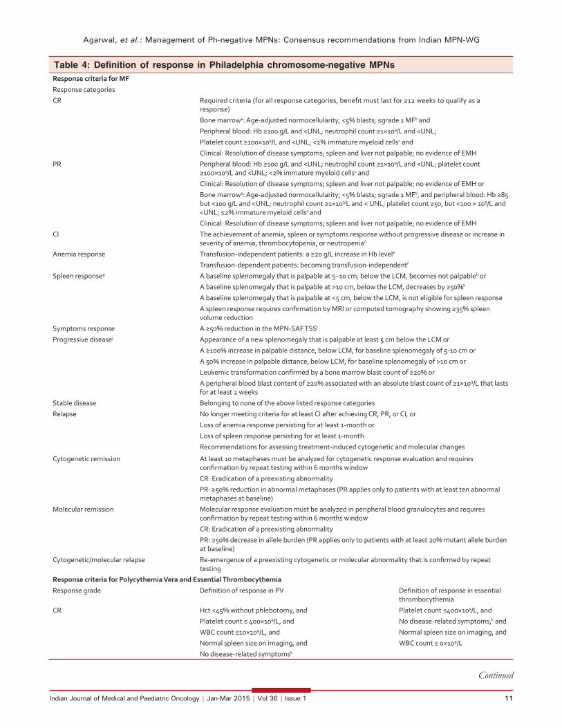
Evaluation of response and follow-upClinical assessment should be done at regular intervals to monitor response to therapy. The revised International WG-MPNs Research and Treatment and the European LeukemiaNet (ELN) criteria are used to objectively assess treatment outcomes in MF [Table 4].[34] Since PMF patients are especially susceptible to marrow suppression, evaluation of full blood count is recommended at least on a weekly basis while starting a new treatment that can potentially lower blood counts. Once the patient is stable, complete blood count can be done once in 3 months. Bone marrow biopsy should be performed on an annual basis in transplant eligible patients to look for clonal evolution. Bone marrow examination is essential in assessing the transformation to acute leukemia. For the transplant ineligible patients, routine bone marrow aspiration is not indicated unless there is evidence of leukemic transformation. Other than allo-SCT or a clinical trial setting, monitoring of molecular response is not recommended for clinical use. Symptomatic burden compromises quality-of-life in MF patients leading to the negative impact on clinical outcomes. Therefore, symptom assessment should be performed routinely in MF patients, and the use of MPNs Symptom Assessment Form Total Symptom Score (SAF TSS) is recommended to evaluate outcomes.[35] The MPN-SAF TSS is a reliable subset of MPN-SAF (10 items) tool that concisely assesses the prevalence and severity of symptoms in PMF, PV, and ET patients [Appendix 2].
Table 3: Ruxolitinib dose in primary MFRecommended starting doses based on platelet count
Platelet count Starting dose<200×109/L 20 mg orally twice daily
100×109/L-200×109/L 15 mg orally twice daily
50×109/L-≤100×109/L 5 mg orally twice daily
Dose modifications (with starting platelet count of at least 100×109/L)
Current platelet count Dose at time of decline in platelet count Maximum dose based on platelet count after prior treatment interruption or dose reduction20 mg BID 15 mg BID 10 mg BID 5 mg BID
New dose to be used
≥125×109/L No change No change No change No change 20 mg BID
100-≤125×109/L 15 mg BID No change No change No change 15 mg BID
75-≤100×109/L 10 mg BID 10 mg BID No change No change 10 mg BID for 2 weeks; if stable, may increase to 15 mg BID
50-≤75×109/L 5 mg BID 5 mg BID 5 mg BID No change 5 mg BID for 2 weeks; if stable, may increase to 10 mg BID
MF – Myelofibrosis

Agarwal, et al.: Management of Ph-negative MPNs: Consensus recommendations from Indian MPN-WG
10 Indian Journal of Medical and Paediatric Oncology | Jan-Mar 2015 | Vol 36 | Issue 1
Polycythemia veraTreatment goalsMajor treatment objectives in patients with PV include minimizing the risk of thrombosis and progression; reducing constitutional symptoms, maintaining hematocrit <0.45, and managing special situations like pregnancy or cardiovascular risk.
Risk stratificationCurrent risk stratification strategies in PV are designed to estimate the likelihood of thrombotic complications and do not necessarily estimate survival or risk of leukemic/fibrotic transformation.[36] High-risk is defined by age above 60 years, previous thrombosis or platelets more than 1500 × 109/L. These patients should be treated with cytoreductive therapy. Low-risk is defined by the absence of these risk factors and patients in general, should not receive cytoreductive therapy.
Clinical managementThe panel examined clinical evidence for traditional treatment approaches in PV and recommendations were formulated based on the consensus. The treatment algorithm for PV is provided in Figure 3.
PhlebotomyThe hematocrit should be maintained at <0.45 and <0.42 in male and female patients, respectively.[37] Phlebotomy is recommended for hematocrit levels >0.48. Grade A recommendation, evidence Level Ib.[9]
Aspirin therapyAspirin has been shown to reduce both arterial and venous thrombosis in PV.[38] Aspirin should not be given to patients with platelets more than 1,500 × 109/L due to an increased risk of bleeding, instead cytoreductive therapy should be initiated. In the case of aspirin allergy,
clopidogrel can be used. Grade A recommendation, evidence level Ib.[9]
Choice of cytoreductive therapy in polycythemia veraInterferon-αInterferon-α can be considered in younger patients (<40 years). For patients >40 years of age, hydroxyurea should be considered as the first line of treatment. Grade B recommendation, evidence level III.[9]
HydroxyureaHydroxyurea is the best-documented drug in PV having been used in large randomized trials.[39,40] Hydroxyurea is recommended as a first-line cytoreductive therapy in PV patients >60 years or younger patients who do not tolerate interferon-α (Grade A recommendation, evidence level Ib). Increased risk of leukemic transformation has not been observed despite long-term hydroxyurea treatment.
Appendix 2: Myeloproliferative neoplasm symptom assessment form total symptom score[35]
Symptom 1-10 (0 if absent) ranking 1 is most favorable and 10 least favorable
Please rate your fatigue (weariness, tiredness) by circling the one number that best describes your worst level of fatigue during past 24 h
(No fatigue) 0 1 2 3 4 5 6 7 8 9 10 (worst imaginable)
Circle the one number that describes how, during the past week how much difficulty you have had with each of the following symptoms
Filling up quickly when you eat (early satiety) (Absent) 0 1 2 3 4 5 6 7 8 9 10 (worst imaginable)
Abdominal discomfort (Absent) 0 1 2 3 4 5 6 7 8 9 10 (worst imaginable)
Inactivity (Absent) 0 1 2 3 4 5 6 7 8 9 10 (worst imaginable)
Problems with concentration - compared to prior to my myeloproliferative disorder
(Absent) 0 1 2 3 4 5 6 7 8 9 10 (worst imaginable)
Numbness/tingling (in my hands and feet) (Absent) 0 1 2 3 4 5 6 7 8 9 10 (worst imaginable)
Night sweats (Absent) 0 1 2 3 4 5 6 7 8 9 10 (worst Imaginable)
Itching (pruritus) (Absent) 0 1 2 3 4 5 6 7 8 9 10 (worst imaginable)
Bone pain (diffuse not joint pain or arthritis) (Absent) 0 1 2 3 4 5 6 7 8 9 10 (worst imaginable)
Fever (>100F) (Absent) 0 1 2 3 4 5 6 7 8 9 10 (daily)
Unintentional weight loss last 6 months (Absent) 0 1 2 3 4 5 6 7 8 9 10 (worst imaginable)
Figure 3: Treatment algorithm for polycythemia vera
Agarwal, et al.: Management of Ph-negative MPNs: Consensus recommendations from Indian MPN-WG
Indian Journal of Medical and Paediatric Oncology | Jan-Mar 2015 | Vol 36 | Issue 1 11
Table 4: Definition of response in Philadelphia chromosome-negative MPNsResponse criteria for MFResponse categories
CR Required criteria (for all response categories, benefit must last for ≥12 weeks to qualify as a response)
Bone marrowa: Age-adjusted normocellularity; <5% blasts; ≤grade 1 MFb and
Peripheral blood: Hb ≥100 g/L and <UNL; neutrophil count ≥1×10⁹/L and <UNL;
Platelet count ≥100×109/L and <UNL; <2% immature myeloid cellsc and
Clinical: Resolution of disease symptoms; spleen and liver not palpable; no evidence of EMH
PR Peripheral blood: Hb ≥100 g/L and <UNL; neutrophil count ≥1×10⁹/L and <UNL; platelet count ≥100×109/L and <UNL; <2% immature myeloid cellsc and
Clinical: Resolution of disease symptoms; spleen and liver not palpable; no evidence of EMH or
Bone marrowa: Age-adjusted normocellularity; <5% blasts; ≤grade 1 MFb, and peripheral blood: Hb ≥85 but <100 g/L and <UNL; neutrophil count ≥1×10⁹/L and < UNL; platelet count ≥50, but <100 × 109/L and <UNL; ≤2% immature myeloid cellsc and
Clinical: Resolution of disease symptoms; spleen and liver not palpable; no evidence of EMH
CI The achievement of anemia, spleen or symptoms response without progressive disease or increase in severity of anemia, thrombocytopenia, or neutropeniad
Anemia response Transfusion-independent patients: a ≥20 g/L increase in Hb levele
Transfusion-dependent patients: becoming transfusion-independentf
Spleen responseg A baseline splenomegaly that is palpable at 5–10 cm, below the LCM, becomes not palpableh or
A baseline splenomegaly that is palpable at >10 cm, below the LCM, decreases by ≥50%h
A baseline splenomegaly that is palpable at <5 cm, below the LCM, is not eligible for spleen response
A spleen response requires confirmation by MRI or computed tomography showing ≥35% spleen volume reduction
Symptoms response A ≥50% reduction in the MPN-SAF TSSi
Progressive diseasej Appearance of a new splenomegaly that is palpable at least 5 cm below the LCM or
A ≥100% increase in palpable distance, below LCM, for baseline splenomegaly of 5-10 cm or
A 50% increase in palpable distance, below LCM, for baseline splenomegaly of >10 cm or
Leukemic transformation confirmed by a bone marrow blast count of ≥20% or
A peripheral blood blast content of ≥20% associated with an absolute blast count of ≥1×10⁹/L that lasts for at least 2 weeks
Stable disease Belonging to none of the above listed response categories
Relapse No longer meeting criteria for at least CI after achieving CR, PR, or CI, or
Loss of anemia response persisting for at least 1-month or
Loss of spleen response persisting for at least 1-month
Recommendations for assessing treatment-induced cytogenetic and molecular changes
Cytogenetic remission At least 10 metaphases must be analyzed for cytogenetic response evaluation and requires confirmation by repeat testing within 6 months window
CR: Eradication of a preexisting abnormality
PR: ≥50% reduction in abnormal metaphases (PR applies only to patients with at least ten abnormal metaphases at baseline)
Molecular remission Molecular response evaluation must be analyzed in peripheral blood granulocytes and requires confirmation by repeat testing within 6 months window
CR: Eradication of a preexisting abnormality
PR: ≥50% decrease in allele burden (PR applies only to patients with at least 20% mutant allele burden at baseline)
Cytogenetic/molecular relapse Re-emergence of a preexisting cytogenetic or molecular abnormality that is confirmed by repeat testing
Response criteria for Polycythemia Vera and Essential Thrombocythemia
Response grade Definition of response in PV Definition of response in essential thrombocythemia
CR Hct <45% without phlebotomy, and
Platelet count ≤ 400×109/L, and
WBC count ≤10×109/L, and
Normal spleen size on imaging, and
No disease-related symptomsk
Platelet count ≤400×109/L, and
No disease-related symptoms,k and
Normal spleen size on imaging, and
WBC count ≤ 0×109/L
Continued

Agarwal, et al.: Management of Ph-negative MPNs: Consensus recommendations from Indian MPN-WG
12 Indian Journal of Medical and Paediatric Oncology | Jan-Mar 2015 | Vol 36 | Issue 1
Hydroxyurea should be continued till patients develop intolerance or progression. Grade A recommendation, evidence level Ib.[9]
Janus kinase 2 inhibitorsRuxolitinib provided rapid and durable clinical benefits in patients with advanced PV who were refractory or intolerant to hydroxyurea in a phase 2 clinical trial.[41] These findings were recently confirmed in a phase 3 trial in hydroxyurea refractory or intolerant PV patients that compared 10 mg twice daily ruxolitinib treatment to best available therapy.[42] However, ruxolitinib is presently not approved for use in PV and should not be used outside of the context of clinical trials.
AnagrelideAnagrelide has powerful platelet reducing activity that could be helpful in the management of patients’ intolerant or refractory to hydroxyurea, interferon-α, or ruxolitinib. Grade C recommendation, evidence level IV.[9]
Radioactive phosphorusDue to its leukemogenic effect, radioactive phosphorus is not a recommended option for patients <75 years.[9]
Evaluation of response and follow-upThe goal of therapy should be normalization of peripheral blood counts and the ELN criteria for PV should be used for evaluation of response [Table 4].[43] Patients on phlebotomy alone should be monitored with complete blood counts every 4-6 weeks. There is no
indication for repeated bone marrow trephine biopsies during routine follow-up in PV but is essential in assessing the transformation to MF or acute leukemia. Monitoring of molecular response, including sequential assessment of the JAK2 V617F allele burden is at the moment not recommended for routine clinical use.
Essential thrombocythemiaTreatment goalsSimilar to PV, main treatment goals in ET include the minimizing risk for thrombosis and progression, normalizing peripheral blood counts, reducing constitutional symptoms, and managing special situations like pregnancy.
Risk stratificationRisk stratification in ET is based on the assessment of risk of thrombosis, as the current therapy in ET is aimed at lowering the risk of thrombosis. True ET diagnosed according to the 2008 WHO classification has not been reported to affect the life expectancy of patients.[32] High-risk is defined as the presence of age above 60 years or history of previous thrombosis, or a platelet count more than 1,500 × 109/L; and these patients should be treated with cytoreductive therapy.[9] Low-risk is defined by the absence of these 3 factors and should not be treated with cytoreductive therapy except in patients with uncontrolled cardiovascular risk factors.[9]
Clinical managementPatients with ET are at high-risk for thrombosis. Hence, vigorous treatment is required for managing
Table 4: (Continued)PR In patients who do not fulfill the criteria for complete response
Hct < 45% without phlebotomy. or
Response in ≥ 3 of the other criteria
In patients who do not fulfill the criteria for complete response: Platelet count ≤600×109/L or decrease of > 50% from baseline
No response Any response that does not satisfy partial response Any response that does not satisfy partial response
CT – Computed tomography; PRBC – Packed red blood cell; MF – Myelofibrosis; WBC – White blood cell; Hct – Hematocrit; PV – Polycythemia vera; TSS – Total symptom score; SAF – Symptom assessment form; MRI – Magnetic resonance imaging; CI – Clinical improvement; CR – Complete response; PR – Partial response; MPN – Myeloproliferative neoplasm; EMH – Extramedullary hematopoiesis (no evidence of EMH implies the absence of pathology or imaging study-proven nonhepatosplenic EMH); LCM – Left costal margin; UNL – Upper normal limit. aBaseline and posttreatment bone marrow slides are to be interpreted at one sitting by a central review process. Cytogenetic and molecular responses are not required for CR assignment; bGrading of MF is according to the European classification. Thiele et al. European consensus on grading bone marrow fibrosis and assessment of cellularity. Haematologica. 2005;90:1128. It is underscored that the consensus definition of a CR bone marrow is to be used only in those patients in which all other criteria are met, including resolution of leukoerythroblastosis. It should also be noted that it was a particularly difficult task for the working group to reach a consensus regarding what represents a complete histologic remission; cImmature myeloid cells constitute blasts + promyelocytes + myelocytes + metamyelocytes + nucleated red blood cells. In splenectomized patients, <5% immature myeloid cells is allowed; dSee above for definitions of anemia response, spleen response, and progressive disease. Increase in severity of anemia constitutes the occurrence of new transfusion dependency or a ≥20 g/L decrease in Hb level from pretreatment baseline that lasts for at least 12 weeks. Increase in severity of thrombocytopenia or neutropenia is defined as a 2-grade decline, from pretreatment baseline, in platelet count or absolute neutrophil count, according to the Common Terminology Criteria for Adverse Events version 4.0. In addition, assignment to CI requires a minimum platelet count of ≥25,000×109/L and absolute neutrophil count of ≥0.5×10(9)/L; eApplicable only to patients with baseline Hb of <100 g/L. In patients not meeting the strict criteria for transfusion dependency at the time of study enrollment (see as follows), but have received transfusions within the previous month, the pretransfusion Hb level should be used as the baseline; fTransfusion dependency before study enrollment is defined as transfusions of at least 6 units of PRBCs, in the 12 weeks prior to study enrollment, for a Hb level of <85 g/L, in the absence of bleeding or treatment-induced anemia. In addition, the most recent transfusion episode must have occurred in the 28 days prior to study enrollment. Response in transfusion-dependent patients requires absence of any PRBC transfusions during any consecutive “rolling” 12 weeks interval during the treatment phase, capped by a Hb level of ≥85 g/L; gIn splenectomized patients, palpable hepatomegaly is substituted with the same measurement strategy; hSpleen or liver responses must be confirmed by imaging studies where a ≥35% reduction in spleen volume, as assessed by MRI or CT, is required. Furthermore, a ≥35% volume reduction in the spleen or liver, by MRI or CT, constitutes a response regardless of what is reported with physical examination; iSymptoms are evaluated by the MPN-SAF TSS; jProgressive disease assignment for splenomegaly requires confirmation my MRI or computed tomography showing a ≥25% increase in spleen volume from baseline. Baseline values for both physical examination and imaging studies refer to pretreatment baseline and not to posttreatment measurements; kDisease-related symptoms include microvascular disturbances, pruritus, and headache

Agarwal, et al.: Management of Ph-negative MPNs: Consensus recommendations from Indian MPN-WG
Indian Journal of Medical and Paediatric Oncology | Jan-Mar 2015 | Vol 36 | Issue 1 13
Figure 4: Treatment algorithm for essential thrombocythemia
cardiovascular risk factors. It is important to emphasize that before star ting therapy patients should be evaluated for eventual progression to MF, if they show symptomatic or progressive splenomegaly, other evidence of disease progression such as weight loss, night sweats, progressive leukocytosis, and/or thrombocytosis.
Stem-cell transplantation is almost never performed in ET due to unfavorable risk-benefit profile. The treatment algorithm for ET is provided in Figure 4.
AspirinAntiplatelet therapy with aspirin 75-150 mg/day is recommended, unless otherwise contraindicated, for all ET patients.
Choice of cytoreductive therapy in essential thrombocythemiaHydroxyureaHydroxyurea is the best-documented therapy in ET and is recommended as a first-line therapy in the majority of ET patients. Hydroxyurea markedly reduces thrombotic complications compared to aspirin alone.[44]
Interferon-αInterferon-α treatment is well-documented and safe in ET and is not considered leukemogenic or teratogenic.[43]
RecommendationInterferon-α is the recommended first-line therapy in younger patients. It can be used in older patients if long-term use of hydroxyurea is not suitable and in patients who do not tolerate hydroxyurea (Grade B recommendation, evidence level III).[9] Interferon-α is the treatment of choice if cytoreductive therapy is indicated during pregnancy or when pregnancy is planned.
Janus kinase 2 inhibitorsSince no JAK2 inhibitor has been studied extensively in ET to date, these drugs are at the present time experimental and should not be used outside of the context of clinical trials.
Other drugsOther drugs, such as busulfan and radioactive phosphorus, are not relevant in ET. Anagrelide has many complications and may not be as effective as hydroxyurea, but may be used in some patients, if necessary, in combination with hydroxyurea to control platelet counts.
Response monitoring in essential thrombocythemiaThe goal of therapy should be to normalize peripheral blood counts in patients who can tolerate pharmacological intervention. The ELN response criteria for ET should be used for evaluation of response [Table 4].[43] There is currently no absolute evidence for a correlation between platelet levels <400 × 109/L and the reduced risk of thrombosis. Therefore, in patients who develop anemia on hydroxyurea treatment, lowering the dose to allow for higher platelet number in order to avoid anemia is acceptable. There is no indication for repeated bone marrow trephine biopsies in routine follow-up in ET but is essential in assessing the transformation to MF or acute leukemia. Monitoring of molecular response, including sequential assessment of the JAK2 V617F allele burden, is at the moment not recommended for clinical use.
Management of complications of myeloproliferative neoplasmsAcute thrombotic events and secondary prophylaxisIn general, acute thrombotic events should be treated as in non-MPN patients. Control of hematocrit and platelet count should be optimized. In emergency situations such as acute cerebrovascular complications or severe digital ischemia, acute platelet apheresis or erythropheresis can be used in order to achieve a rapid reduction in blood counts. Since the effect is brief, cytoreductive therapy, preferably with hydroxyurea should be started as soon as possible in patients, not on cytoreductive therapy. Prevention of re-thrombosis should be independently achieved in patients with previous venous thrombosis by both oral anticoagulants and antiplatelet drugs. Since no prospective trials exist, it remains unclear whether it is better to give a short course of warfarin or to continue with long-term therapy for secondary prevention of venous thromboembolism.
BleedingThe most important cause of bleeding in ET and PV is acquired von Willebrand´s syndrome associated with high platelet counts (>1,500 × 109/L).[45,46] Therefore, the most important therapeutic intervention to manage acute bleeding in the thrombocythemic patient is platelet reduction, and the recommended agent is hydroxyurea.
Platelet apheresis is indicated when extreme thrombocytosis is accompanied by an urgent need to reduce platelet counts, that is, severe or life-threatening bleeding.[47]

Agarwal, et al.: Management of Ph-negative MPNs: Consensus recommendations from Indian MPN-WG
14 Indian Journal of Medical and Paediatric Oncology | Jan-Mar 2015 | Vol 36 | Issue 1
PruritusPruritus, typically aquagenic, can be a severe clinical problem in PV. Antihistamines may be of benefit. Selective serotonin re-uptake inhibitors can also lead to improvement of pruritus. Benefit has been shown with phototherapy using psoralen and ultraviolet A light.
Transformation to acute myeloid leukemiaThe results after conventional AML induction chemotherapy are dismal in patients developing AML after PV, ET, or PMF, with a very short median survival.[48] If possible, it is recommended that patients undergo allo-SCT after induction chemotherapy. Hypomethylating agents can be used as a bridge to transplant.[49,50]
Special issuesPregnancyThere is limited information in the medical literature about the management of MPNs in pregnancy.[51] The live birth rate is about 60% due to an overall incidence of first trimester miscarriage of 31-36% (about twice the normal rate) and an increased risk of intrauterine growth retardation, intrauterine death, and stillbirth (8%). Major maternal complications are less common and occur in approximately 8% of ET patients.[52]
Pregnancy is likely to be accompanied by a high-risk of complication for the mother and/or fetus if any of the following factors are present: Previous venous or arterial thrombosis in mother, previous hemorrhage attributed to PV/ET, previous pregnancy complication that may have been caused by PV/ET (these include significant ante-or postpartum hemorrhage, severe preeclampsia, unexplained recurrent first trimester loss (≥3), intrauterine growth retardation (<5% for gestation), intrauterine death or stillbirth with no other cause identified, placental abruption), and platelet count above 1,000 × 109/L.[53]
Therapeutic options include antithrombotic treatment, phlebotomy in PV and cytoreductive agents. The optimal hematocrit in pregnancy has yet to be established, but the current recommendation is to maintain the hematocrit within the normal range appropriate for gestation. The increased plasma volume often results in a reduced hematocrit and platelet count during the second trimester. The levels rise again during the postpartum period contributing to an increased thrombotic risk during the first 6 weeks after delivery. Close monitoring of blood counts is important during this period.[53] Low dose aspirin is safe and seems advantageous during pregnancy in ET. Starting on the day of the delivery, aspirin is substituted by a prophylactic dose of low molecular weight heparin (LMWH) which is given until
6 weeks after delivery.[51] The doses of LMWH that have been reported are dalteparin 5000 U or enoxaparin 40 mg daily.
Pediatric myeloproliferative neoplasmThe incidence of different MPN in patients aged <20 years is so low that formal evidence-based recommendations are impossible to provide.
DISCUSSION
Limited information is available from controlled, randomized clinical trials for the management of Ph-negative MPNs and consequently, clinical decision making is extremely challenging. The recommendations presented in this manuscript are mainly based on the clinical experience and knowledge of experts in the field of Ph-negative classical MPNs and a group decision making process was adopted while interpreting available clinical evidence in the context of hematology practice in India. Wherever possible, an expert panel has provided practical suggestions to assist clinicians in diagnosing and treating patients with PMF, PV, or ET. The goals of current therapy for Ph-negative classical MPNs are to address major clinical issues in PMF and to prevent the risk of thrombosis in PV and ET. The challenges in diagnosis are addressed by the recently revised WHO classification, which integrates hematologic, morphologic, and molecular parameters to separate the three clinical entities. Better understanding of molecular mechanisms contributing to the pathogenesis and progression of Ph-negative MPNs during the recent years will translate into more accurate diagnosis and targeted treatment approaches in the near future. It is the intention of this group to update periodically and modify this consensus statement as and when more data become available.
REFERENCES
1. Tefferi A, Vainchenker W. Myeloproliferative neoplasms:Molecularpathophysiology,essentialclinicalunderstanding,andtreatmentstrategies.JClinOncol2011;29:573-82.
2. CervantesF,TassiesD,SalgadoC,RoviraM,PereiraA,RozmanC.Acutetransformationinnonleukemicchronicmyeloproliferativedisorders:Actuarialprobabilityandmaincharacteristicsinaseriesof218patients.ActaHaematol1991;85:124-7.
3. MesaRA,NiblackJ,WadleighM,VerstovsekS,CamorianoJ,BarnesS,et al. The burden of fatigue and quality of life inmyeloproliferativedisorders(MPDs):AninternationalInternet-basedsurveyof1179MPDpatients.Cancer2007;109:68-76.
4. MoulardO,MehtaJ,FryzekJ,OlivaresR,IqbalU,MesaRA.Epidemiology of myelofibrosis, essential thrombocythemia,andpolycythemiaveraintheEuropeanUnion.EurJHaematol2014;92:289-97.
5. Mehta J, Wang H, Iqbal SU, Mesa R. Epidemiology ofmyeloproliferative neoplasms in the United States. LeukLymphoma2014;55:595-600.
Agarwal, et al.: Management of Ph-negative MPNs: Consensus recommendations from Indian MPN-WG
Indian Journal of Medical and Paediatric Oncology | Jan-Mar 2015 | Vol 36 | Issue 1 15
6. ScottLM,TongW,LevineRL,ScottMA,BeerPA,StrattonMR,et al. JAK2 exon 12 mutations in polycythemia vera andidiopathicerythrocytosis.NEnglJMed2007;356:459-68.
7. OhST,SimondsEF,JonesC,HaleMB,GoltsevY,GibbsKDJr,et al.NovelmutationsintheinhibitoryadaptorproteinLNKdriveJAK-STATsignalinginpatientswithmyeloproliferativeneoplasms.Blood2010;116:988-92.
8. Pikman Y, Lee BH, Mercher T, McDowell E, Ebert BL,Gozo M, et al. MPLW515L is a novel somatic activatingmutationinmyelofibrosiswithmyeloidmetaplasia.PLoSMed2006;3:e270.
9. Andersen CL, Andreasson B, Hasselbalch H, HultcrantzM,KnutsenH,LindgrenM,et al.Nordicguidelineson thediagnosisand treatmentofpatientswithMyeloproliferativeNeoplasms. January 2013. Available from: http://www.nmpn.org/index.php/guidelines/1-nmpn-guidelines-2013/file.[Lastaccessedon2014Dec04].
10. Barbui T, Barosi G, Birgegard G, Cervantes F, Finazzi G,Griesshammer M, et al. Philadelphia-negative classicalmyeloproliferative neoplasms: Critical concepts andmanagementrecommendationsfromEuropeanLeukemiaNet.JClinOncol2011;29:761-70.
11. CervantesF,DupriezB,PereiraA,PassamontiF,ReillyJT,MorraE,et al.NewprognosticscoringsystemforprimarymyelofibrosisbasedonastudyoftheInternationalWorkingGroup for Myelofibrosis Research and Treatment. Blood2009;113:2895-901.
12. PassamontiF,CervantesF,VannucchiAM,MorraE,RumiE,Pereira A, et al. A dynamic prognostic model to predictsurvival in primarymyelofibrosis:A study by the IWG-MRT(InternationalWorkingGroupforMyeloproliferativeNeoplasmsResearchandTreatment).Blood2010;115:1703-8.
13. Gangat N, Caramazza D, Vaidya R, George G, Begna K,SchwagerS,et al.DIPSSplus:ArefinedDynamicInternationalPrognostic Scoring System for primary myelofibrosis thatincorporatesprognosticinformationfromkaryotype,plateletcount,andtransfusionstatus.JClinOncol2011;29:392-7.
14. HuangJ,TefferiA. Erythropoiesis stimulating agentshavelimitedtherapeuticactivityintransfusion-dependentpatientswithprimarymyelofibrosisregardlessofserumerythropoietinlevel.EurJHaematol2009;83:154-5.
15. HasselbalchHC,ClausenNT,JensenBA.Successfultreatmentof anemia in idiopathic myelofibrosis with recombinanthumanerythropoietin.AmJHematol2002;70:92-9.
16. ThapaliyaP,TefferiA,PardananiA,SteensmaDP,CamorianoJ,WuW,et al.Internationalworkinggroupformyelofibrosisresearch and treatment response assessment and long-termfollow-upof50myelofibrosispatientstreatedwiththalidomide-prednisonebasedregimens.AmJHematol2011;86:96-8.
17. BarosiG,RostiV,VannucchiAM.Therapeuticapproachesinmyelofibrosis.ExpertOpinPharmacother2011;12:1597-611.
18. Löfvenberg E, Wahlin A. Management of polycythaemiavera, essential thrombocythaemia and myelofibrosis withhydroxyurea.EurJHaematol1988;41:375-81.
19. ManoharanA.Managementofmyelofibrosiswithintermittenthydroxyurea.BrJHaematol1991;77:252-4.
20. VerstovsekS,MesaRA,GotlibJ,LevyRS,GuptaV,DiPersioJF,et al.Adouble-blind,placebo-controlledtrialofruxolitinibformyelofibrosis.NEnglJMed2012;366:799-807.
21. HarrisonC,KiladjianJJ,Al-AliHK,GisslingerH,WaltzmanR,StalbovskayaV,et al.JAKinhibitionwithruxolitinibversusbest available therapy for myelofibrosis. N Engl J Med2012;366:787-98.
22. MeadA,ClarkR,ChackoJ,KnapperS,YinJ,MilojkovicD,et al.Responsetoruxolitinibinpatientswithintermediate-1,intermediate-2 and high-risk myelofibrosis: Results of theUK Robust trial. Haematologica 2014;99 suppl 1:414.[Abstract].
23. Cervantes F. How I treat myelofibrosis. Blood2014;124:2635-42.
24. Jakavi.SummaryofProductCharacteristicslastupdatedontheeMC:29/11/2013.Availablefrom:http://www.medicines.org.uk/emc/medicine/26991/SPC/Jakavi+5 g,+15 mg+and+20mg+Tablets/#INDICATIONS.[Lastaccessedon2014Jul01].
25. Jakavi[packageinsert].18December2013.Novartis IndiaLimited,Mumbai,India.
26. Mesa RA, Cortes J. Optimizing management of ruxolitinibin patients with myelofibrosis: The need for individualizeddosing.JHematolOncol2013;6:79.
27. Mesa RA, Nagorney DS, Schwager S, Allred J, Tefferi A.Palliativegoals,patientselection,andperioperativeplateletmanagement: Outcomes and lessons from 3 decades ofsplenectomyformyelofibrosiswithmyeloidmetaplasiaattheMayoClinic.Cancer2006;107:361-70.
28. EvrotE,EbelN,RomanetV,RoelliC,AndraosR,QianZ,et al. JAK1/2andPan-deacetylaseinhibitorcombinationtherapyyieldsimprovedefficacyinpreclinicalmousemodelsofJAK2V617F-drivendisease.ClinCancerRes2013;19:6230-41.
29. RibargV,HarrisonCN,HeidelFH,KiladjianJJ,AcharyyaS,Mu S, et al. A phase 1b, dose-finding study of ruxolitinibplus panobinostat in patients with primary myelofibrosis(PMF), post-polycythemia vera MF (PPV-MF), or post-essentialthrombocythemiaMF(PET-MF):Identificationoftherecommendedphase2dose.Blood2013;122:4045.
30. BallenKK,ShresthaS,SobocinskiKA,ZhangMJ,BasheyA,BolwellBJ,et al.Outcomeoftransplantationformyelofibrosis.BiolBloodMarrowTransplant2010;16:358-67.
31. AbelssonJ,MerupM,BirgegårdG,WeisBjerrumO,BrinchL,BruneM,et al.Theoutcomeofallo-HSCT for92patientswith myelofibrosis in the Nordic countries. Bone MarrowTransplant2012;47:380-6.
32. Barbui T, Thiele J, Passamonti F, Rumi E, Boveri E,RuggeriM,et al.Survivalanddiseaseprogressioninessentialthrombocythemia are significantly influenced by accuratemorphologicdiagnosis:Aninternationalstudy.JClinOncol2011;29:3179-84.
33. CarobbioA,ThieleJ,PassamontiF,RumiE,RuggeriM,RodeghieroF,et al.Risk factors forarterialandvenousthrombosis in WHO-defined essential thrombocythemia:An international study of 891 patients. Blood2011;117:5857-9.
34. Tefferi A, Cervantes F, Mesa R, Passamonti F,Verstovsek S, Vannucchi AM, et al. Revised responsecriteria for myelofibrosis: International Working Group-Myeloproliferative Neoplasms Research and Treatment(IWG-MRT) and European LeukemiaNet (ELN) consensusreport.Blood2013;122:1395-8.
35. EmanuelRM,DueckAC,GeyerHL,KiladjianJJ,SlotS,et al.Myeloproliferativeneoplasm(MPN)symptomassessmentformtotal symptom score: Prospective international assessmentof an abbreviated symptom burden scoring system amongpatientswithMPNs.JClinOncol2012;30:4098-103.
36. FinazziG,BarbuiT.Evidenceandexpertiseinthemanagementof polycythemia vera and essential thrombocythemia.Leukemia2008;22:1494-502.
37. Marchioli R, Finazzi G, Specchia G, Cacciola R,Cavazzina R, Cilloni D, et al. Cardiovascular events andintensityoftreatmentinpolycythemiavera.NEnglJMed2013;368:22-33.
38. Landolfi R, Marchioli R, Kutti J, Gisslinger H, Tognoni G,PatronoC,et al.Efficacyandsafetyof low-doseaspirin inpolycythemiavera.NEnglJMed2004;350:114-24.
39. Najean Y, Rain JD. Treatment of polycythemia vera: Useof32Paloneor incombinationwithmaintenance therapyusing hydroxyurea in 461 patients greater than 65 yearsof age. The French Polycythemia Study Group. Blood1997;89:2319-27.
40. Kiladjian JJ, Rain JD, Bernard JF, Briere J, Chomienne C,Fenaux P. Long-term incidence of hematological evolutionin three French prospective studies of hydroxyurea
Agarwal, et al.: Management of Ph-negative MPNs: Consensus recommendations from Indian MPN-WG
16 Indian Journal of Medical and Paediatric Oncology | Jan-Mar 2015 | Vol 36 | Issue 1
and pipobroman in polycythemia vera and essentialthrombocythemia.SeminThrombHemost2006;32:417-21.
41. VerstovsekS,PassamontiF,RambaldiA,BarosiG,RosenPJ,RumiE,et al.Aphase2studyofruxolitinib,anoralJAK1andJAK2Inhibitor,inpatientswithadvancedpolycythemiaverawhoarerefractoryorintoleranttohydroxyurea.Cancer2014;120:513-20.
42. Verstovsek S, Kiladjian JJ, Griesshammer M, Masszi T,DurrantSTS,PassamontiF,et al.Resultsofaprospective,randomized, open-label phase 3 study of ruxolitinib (RUX)inpolycythemiavera(PV)patientsresistanttoorintolerantof hydroxyurea (HU): The RESPONSE trial. J Clin Oncol2014;32suppl:5s.(abstr7026).
43. BarosiG,BirgegardG,FinazziG,GriesshammerM,HarrisonC,Hasselbalch HC, et al. Response criteria for essentialthrombocythemiaandpolycythemiavera:ResultofaEuropeanLeukemiaNetconsensusconference.Blood2009;113:4829-33.
44. Cortelazzo S, Finazzi G, Ruggeri M, Vestri O, Galli M,RodeghieroF,et al.Hydroxyureaforpatientswithessentialthrombocythemia and a high risk of thrombosis. N Engl JMed1995;332:1132-6.
45. Barbui T, Barosi G, Grossi A, Gugliotta L, Liberato LN,Marchetti M, et al. Practice guidelines for the therapy ofessential thrombocythemia. A statement from the ItalianSocietyofHematology, the ItalianSocietyofExperimentalHematology and the Italian Group for Bone MarrowTransplantation.Haematologica2004;89:215-32.
46. BuddeU,SchaeferG,MuellerN,EgliH,DentJ,RuggeriZ,et al. AcquiredvonWillebrand’sdisease in themyeloproliferativesyndrome.Blood1984;64:981-5.
47. GreistA.Theroleofbloodcomponentremovalinessentialandreactivethrombocytosis.TherApher2002;6:36-44.
48. BjörkholmM,DerolfAR,HultcrantzM,KristinssonSY,EkstrandC,GoldinLR,et al.Treatment-relatedriskfactorsfortransformationto acutemyeloid leukemia andmyelodysplastic syndromes inmyeloproliferativeneoplasms.JClinOncol2011;29:2410-5.
49. KundrandaMN,TibesR,MesaRA.Transformationofachronicmyeloproliferativeneoplasmtoacutemyelogenousleukemia:Doesanythingwork?CurrHematolMaligRep2012;7:78-86.
50. Thepot S, Itzykson R, Seegers V, Raffoux E, Quesnel B,Chait Y, et al. Treatment of progression of Philadelphia-negative myeloproliferative neoplasms to myelodysplasticsyndromeoracutemyeloidleukemiabyazacitidine:Areporton54casesonthebehalfoftheGroupeFrancophonedesMyelodysplasies(GFM).Blood2010;116:3735-42.
51. Griesshammer M, Struve S, Harrison CM. Essentialthrombocythemia/polycythemia vera and pregnancy: Theneed for an observational study in Europe. Semin ThrombHemost2006;32:422-9.
52. Harrison CN, Robinson SE. Myeloproliferative disorders inpregnancy.HematolOncolClinNorthAm2011;25:261-75,vii.
53. Barbui T, Finazzi G. Special issues in myeloproliferativeneoplasms.CurrHematolMaligRep2011;6:28-35.
How to cite this article: Agarwal MB, Malhotra H, Chakrabarti P, Varma N, Mathews V, Bhattacharyya J, et al. Myeloproliferative neoplasms working group consensus recommendations for diagnosis and management of primary myelofibrosis, polycythemia vera, and essential thrombocythemia. Indian J Med Paediatr Oncol 2015;36:3-16.Source of Support: Nil. Conflict of Interest: None declared.
New features on the journal’s website
Optimized content for mobile and hand-held devicesHTML pages have been optimized of mobile and other hand-held devices (such as iPad, Kindle, iPod) for faster browsing speed.Click on [Mobile Full text] from Table of Contents page.This is simple HTML version for faster download on mobiles (if viewed on desktop, it will be automatically redirected to full HTML version)
E-Pub for hand-held devices EPUB is an open e-book standard recommended by The International Digital Publishing Forum which is designed for reflowable content i.e. the text display can be optimized for a particular display device.Click on [EPub] from Table of Contents page.There are various e-Pub readers such as for Windows: Digital Editions, OS X: Calibre/Bookworm, iPhone/iPod Touch/iPad: Stanza, and Linux: Calibre/Bookworm.
E-Book for desktopOne can also see the entire issue as printed here in a ‘flip book’ version on desktops.Links are available from Current Issue as well as Archives pages. Click on View as eBook