a consensus study and st - Nature
24
A checklist for assessing the methodological quality of concurrent tES-fMRI studies (ContES checklist): a consensus study and statement Hamed Ekhtiari 1 ✉ , Peyman Ghobadi-Azbari 2,3 , Axel Thielscher 4,5 , Andrea Antal 6 , Lucia M. Li 7,8 , A. Duke Shereen 9 , Yuranny Cabral-Calderin 10 , Daniel Keeser 11,12,13 , Til Ole Bergmann 14,15,16 , Asif Jamil 17 , Ines R. Violante 18 , Jorge Almeida 19,20 , Marcus Meinzer 21,22 , Hartwig R. Siebner 4,23,24 , Adam J. Woods 25 , Charlotte J. Stagg 26,27 , Rany Abend 28 , Daria Antonenko 22 , Tibor Auer 18 , Marc Bächinger 29,30 , Chris Baeken 31,32,33 , Helen C. Barron 26,27 , Henry W. Chase 34 , Jenny Crinion 35 , Abhishek Datta 36,37 , Matthew H. Davis 38 , Mohsen Ebrahimi 3 , Zeinab Esmaeilpour 39 , Brian Falcone 40 , Valentina Fiori 41 , Iman Ghodratitoostani 42 , Gadi Gilam 43,44 , Roland H. Grabner 45 , Joel D. Greenspan 46 , Georg Groen 47 , Gesa Hartwigsen 48 , Tobias U. Hauser 49,50 , Christoph S. Herrmann 51,52,53 , Chi-Hung Juan 54,55 , Bart Krekelberg 56 , Stephanie Lefebvre 57 , Sook-Lei Liew 58,59,60,61 , Kristoffer H. Madsen 4,62 , Rasoul Mahdavifar-Khayati 2 , Nastaran Malmir 3 , Paola Marangolo 63,64 , Andrew K. Martin 21,65 , Timothy J. Meeker 66 , Hossein Mohaddes Ardabili 67,68 , Marius Moisa 69 , Davide Momi 70 , Beni Mulyana 1 , Alexander Opitz 71 , Natasza Orlov 72,73,74,75 , Patrick Ragert 76,77 , Christian C. Ruff 69 , Giulio Ruffini 78,79 , Michaela Ruttorf 80 , Arshiya Sangchooli 3 , Klaus Schellhorn 81 , Gottfried Schlaug 82 , Bernhard Sehm 77,83 , Ghazaleh Soleimani 84 , Hosna Tavakoli 3,85 , Benjamin Thompson 86,87,88 , Dagmar Timmann 89 , Aki Tsuchiyagaito 1 , Martin Ulrich 47 , Johannes Vosskuhl 51 , Christiane A. Weinrich 6,90 , Mehran Zare-Bidoky 3,91 , Xiaochu Zhang 92 , Benedikt Zoefel 38,93,94 , Michael A. Nitsche 17,95 and Marom Bikson 39 Low-intensity transcranial electrical stimulation (tES), including alternating or direct current stimulation, applies weak electrical stimulation to modulate the activity of brain circuits. Integration of tES with concurrent functional MRI (fMRI) allows for the mapping of neural activity during neuromodulation, supporting causal studies of both brain function and tES effects. Methodological aspects of tES-fMRI studies underpin the results, and reporting them in appropriate detail is required for reproducibility and interpretability. Despite the growing number of published reports, there are no consensus-based checklists for disclosing methodological details of concurrent tES-fMRI studies. The objective of this work was to develop a consensus-based checklist of reporting standards for concurrent tES-fMRI studies to support methodological rigor, transparency and reproducibility (ContES checklist). A two-phase Delphi consensus process was conducted by a steering committee (SC) of 13 members and 49 expert panelists through the International Network of the tES-fMRI Consortium. The process began with a circulation of a preliminary checklist of essential items and additional recommendations, developed by the SC on the basis of a systematic review of 57 concurrent tES-fMRI studies. Contributors were then invited to suggest revisions or additions to the initial checklist. After the revision phase, contributors rated the importance of the 17 essential items and 42 additional recommendations in the final checklist. The state of methodological transparency within the 57 reviewed concurrent tES-fMRI studies was then assessed by using the checklist. Experts refined the checklist through the revision and rating phases, leading to a checklist with three categories of essential items and additional recommendations: (i) technological factors, (ii) safety and noise tests and (iii) methodological factors. The level of reporting of checklist items varied among the 57 concurrent tES-fMRI papers, ranging from 24% to 76%. On average, 53% of checklist items were reported in a given article. In conclusion, use of the ContES checklist is expected to enhance the methodological reporting quality of future concurrent tES-fMRI studies and increase methodological transparency and reproducibility. A full list of affiliations appears at the end of the paper. 596 NATURE PROTOCOLS | VOL 17 | MARCH 2022 | 596–617 | www.nature.com/nprot CONSENSUS STATEMENT https://doi.org/10.1038/s41596-021-00664-5 1234567890():,; 1234567890():,;
-
Upload
khangminh22 -
Category
Documents
-
view
3 -
download
0
Transcript of a consensus study and st - Nature

A checklist for assessing the methodologicalquality of concurrent tES-fMRI studies(ContES checklist): a consensus study andstatementHamed Ekhtiari 1, Peyman Ghobadi-Azbari 2,3, Axel Thielscher4,5, Andrea Antal6,Lucia M. Li7,8, A. Duke Shereen 9, Yuranny Cabral-Calderin10, Daniel Keeser 11,12,13,Til Ole Bergmann14,15,16, Asif Jamil 17, Ines R. Violante18, Jorge Almeida19,20,Marcus Meinzer21,22, Hartwig R. Siebner4,23,24, Adam J. Woods25, Charlotte J. Stagg26,27,Rany Abend28, Daria Antonenko22, Tibor Auer 18, Marc Bächinger29,30, Chris Baeken31,32,33,Helen C. Barron26,27, Henry W. Chase34, Jenny Crinion 35, Abhishek Datta36,37,Matthew H. Davis38, Mohsen Ebrahimi 3, Zeinab Esmaeilpour39, Brian Falcone40,Valentina Fiori41, Iman Ghodratitoostani 42, Gadi Gilam43,44, Roland H. Grabner45,Joel D. Greenspan46, Georg Groen47, Gesa Hartwigsen48, Tobias U. Hauser 49,50,Christoph S. Herrmann 51,52,53, Chi-Hung Juan54,55, Bart Krekelberg 56, Stephanie Lefebvre57,Sook-Lei Liew58,59,60,61, Kristoffer H. Madsen 4,62, Rasoul Mahdavifar-Khayati2,Nastaran Malmir3, Paola Marangolo63,64, Andrew K. Martin21,65, Timothy J. Meeker 66,Hossein Mohaddes Ardabili67,68, Marius Moisa69, Davide Momi70, Beni Mulyana1,Alexander Opitz71, Natasza Orlov72,73,74,75, Patrick Ragert76,77, Christian C. Ruff69,Giulio Ruffini78,79, Michaela Ruttorf80, Arshiya Sangchooli 3, Klaus Schellhorn81,Gottfried Schlaug82, Bernhard Sehm77,83, Ghazaleh Soleimani84, Hosna Tavakoli3,85,Benjamin Thompson86,87,88, Dagmar Timmann 89, Aki Tsuchiyagaito1, Martin Ulrich47,Johannes Vosskuhl51, Christiane A. Weinrich6,90, Mehran Zare-Bidoky 3,91, Xiaochu Zhang 92,Benedikt Zoefel38,93,94, Michael A. Nitsche 17,95 and Marom Bikson39
Low-intensity transcranial electrical stimulation (tES), including alternating or direct current stimulation, applies weakelectrical stimulation to modulate the activity of brain circuits. Integration of tES with concurrent functional MRI (fMRI)allows for the mapping of neural activity during neuromodulation, supporting causal studies of both brain function and tESeffects. Methodological aspects of tES-fMRI studies underpin the results, and reporting them in appropriate detail is requiredfor reproducibility and interpretability. Despite the growing number of published reports, there are no consensus-basedchecklists for disclosing methodological details of concurrent tES-fMRI studies. The objective of this work was to develop aconsensus-based checklist of reporting standards for concurrent tES-fMRI studies to support methodological rigor,transparency and reproducibility (ContES checklist). A two-phase Delphi consensus process was conducted by a steeringcommittee (SC) of 13 members and 49 expert panelists through the International Network of the tES-fMRI Consortium. Theprocess began with a circulation of a preliminary checklist of essential items and additional recommendations, developed bythe SC on the basis of a systematic review of 57 concurrent tES-fMRI studies. Contributors were then invited to suggestrevisions or additions to the initial checklist. After the revision phase, contributors rated the importance of the 17 essentialitems and 42 additional recommendations in the final checklist. The state of methodological transparency within the 57reviewed concurrent tES-fMRI studies was then assessed by using the checklist. Experts refined the checklist through therevision and rating phases, leading to a checklist with three categories of essential items and additional recommendations:(i) technological factors, (ii) safety and noise tests and (iii) methodological factors. The level of reporting of checklist itemsvaried among the 57 concurrent tES-fMRI papers, ranging from 24% to 76%. On average, 53% of checklist items werereported in a given article. In conclusion, use of the ContES checklist is expected to enhance the methodological reportingquality of future concurrent tES-fMRI studies and increase methodological transparency and reproducibility.
A full list of affiliations appears at the end of the paper.
596 NATURE PROTOCOLS | VOL 17 |MARCH 2022 | 596–617 |www.nature.com/nprot
CONSENSUS STATEMENThttps://doi.org/10.1038/s41596-021-00664-5
1234
5678
90():,;
1234567890():,;

The advent of functional neuroimaging techniques allowsone to investigate the neural correlates of behavior andunderlying processes. However, functional neuroimaging
techniques cannot by themselves establish causal evidence forbrain-behavior relationships. Non-invasive brain stimulationtechniques, including low-intensity transcranial electricalstimulation (tES), can be used in combination with functionalneuroimaging, such as functional MRI (fMRI), to directlymodulate patterns of neuronal activity and to establish causalevidence for the involvement of particular neural regions ornetworks in a specific behavior and cognitive process1–18. Overthe last 20 years, low-intensity tES has been used extensivelyto study and modulate the neural mechanisms underlyingbasic physiological and cognitive processes19–27. Initial studiescombining tES with fMRI were limited to sequential tES-fMRIrecording, which primarily provides an avenue to investigate theneural mechanisms underlying tES offline (after) effects28–34.
Over the last decade, advances in tES technology have madeconcurrent tES-fMRI (i.e., simultaneous acquisition of fMRIdata during tES) in principle technically feasible, thus enablingmonitoring of immediate (online) tES effects. ConcurrenttES-fMRI recording poses specific technical challenges35;however, these issues can be minimized when standardprotocols are followed29,36–39. As a ‘perturb-and-measure’approach40, applications are rapidly diversifying such thatconcurrent tES-fMRI is being used increasingly as a proxymeasure for local and global neuro-metabolic activity toaddress causal mechanistic25,41–44 and predictive45,46 questionsabout underlying physiology and therapeutic effects.
Online integration of tES with fMRI recordings is, however,associated with technical and theoretical challenges, whichinclude the risk of electrode heating due to the radio frequency(RF) pulses of the scanner47,48 and susceptibility-related echo-planar imaging (EPI) artifacts under the electrodes36,49. Fur-thermore, evidence is increasing for the significant impact ofdifferent methodological procedures on online fMRI responsesto tES, including the localization of electrodes49–51, MRI-conditional stimulator setup29,37,38, the amount/type of contactmedium35 and the timing of concurrent tES within the fMRIparadigm52,53. Given the variability in fMRI responses to tES,as well as tolerability/safety/noise concerns and methodologicalvariations, there is an urgent need to clearly and systematicallyplan, measure, report and control as many of these metho-dological factors as possible. To ensure a robust interpretationof the data and to increase the potential for future replication, areporting guideline and checklist is required. Methodologicalchecklists not only improve the transparent reporting of studymethodology and quality of data collection analysis, but alsoreduce design and reporting biases, factors with clear impli-cations for future interpretation and use of the data. Thesechecklists could also assist peer review and critical appraisal ofresearch methodology54–56.
A limited number of methodological checklists are availablein the field of human brain mapping for transcranial magneticstimulation (TMS) studies55, transcranial direct current sti-mulation (tDCS) studies56 and MRI/fMRI studies57,58. One ofthe most well-known checklists in the field of human brainmapping is the COBIDAS (Committee on Best Practices in
Data Analysis and Sharing) statement, which was developed toprovide an evidence-based minimum set of recommendationsto prepare best practices for data analysis, result reporting,algorithms and data sharing in neuroimaging research, topromote transparency, reliability and collaboration57. Giventhe potential for variability of the neural responses elicited bytES and the growing number of concurrent tES-fMRI studies,guidelines on factors that should be reported and/or controlledin concurrent tES-fMRI studies are essential to ensure thatresearch findings are correctly interpreted and reproducible59.In addition, to facilitate meta-analyses, studies should beconsistent in both methodology and reporting practice. Hence,we aimed to address these issues by conducting a Delphi studyto reach a consensus on the essential items that are mandatoryto be reported or recommendations that should be consideredwhen reporting a concurrent tES-fMRI study (ContESchecklist).
Research methodologyThe Delphi method is a questionnaire-based approachdesigned to facilitate reaching a consensus, based on the fun-damental principles of purposive sampling of experts in thefield of interest, panelist anonymity, iterative questionnairepresentation and feedback of statistical analysis60–62. Like otherexpert consensus methods, the Delphi method is sensitive toexpert sampling and opinion aggregation choices and is relianton subjective expert judgement inherently, necessitating theuse of other complementary empirical evidence62,63. However,rigorously collected and synthesized expert opinion constitutesan important source of information when empirical data arescarce and issues of interest are complex and multifaceted64,65.
This study was designed, implemented and coordinatedwithin the International Network of tES-fMRI (INTF) and asteering committee (SC) that supervised the process ofchecklist development, data analysis and determining theinitial criteria for item consensus and survey termination.The flowchart of the Delphi method adapted for this study isillustrated in Fig. 1. The development of the ContES checklistby using the Delphi technique involved the following steps:(i) formation of the SC, (ii) selection of the expert panel (EP),(iii) checklist development and revision and (iv) data collectionand analysis. The protocol of this study is pre-registered inOSF66, and its questionnaires and databases are publiclyavailable in the study’s Open Science Framework (OSF) page(https://osf.io/f9j8z/).
SCThe role of the SC—Jorge Almeida, Andrea Antal, MaromBikson, Hamed Ekhtiari, Lucia M. Li, Marcus Meinzer,Michael Nitsche, Duke Shereen, Hartwig Siebner, CharlotteStagg, Axel Thielscher, Ines Violante and Adam Woods—wasto determine the aim of the research, produce items and selectadditional experts for the Delphi process. Peyman Ghobadi-Azbari served as the Delphi facilitator to implement the pre-registered methods within and between the SC and EP. The SCgrew out of the INTF collaborative group after a series ofwebinars (28 March 2019, 27 June 2019 and 26 September2019; recorded videos of the webinars are available on the
NATURE PROTOCOLS CONSENSUS STATEMENT
NATURE PROTOCOLS | VOL 17 |MARCH 2022 | 596–617 |www.nature.com/nprot 597

INTF YouTube channel at https://youtube.com/channel/UCKcEYDmyqTipDW7OzuoVSlg), in which considerableheterogeneities of technical/methodological aspects in studiescombining tES with fMRI were discussed along with strategiesto help to bridge respective knowledge gaps and reduceheterogeneity.
EPThe project involved the recruitment of a group of experts onthe basis of a systematic review of 57 concurrent tES-fMRIstudies (published before 1 January 2020). We reviewed theconcurrent tES-fMRI literature in the PubMed research data-base from inception up to 1 January 1 2020 to select evidence-based concurrent tES-fMRI studies and experts who conducted
those studies. The Preferred Reporting Items for SystematicReviews and Meta-Analyses (PRISMA)67 flow diagram for thesystematic review is provided in Extended Data Fig. 1. Thesearch included the terms (tDCS OR transcranial direct currentstimulation OR tACS OR transcranial alternating current sti-mulation) AND (functional magnetic resonance imaging ORfMRI OR functional MRI OR fcMRI OR functional connectivityMRI OR rsfMRI OR resting-state fMRI). 57 articles wereselected on the basis of the PRISMA. The inclusion criteriaused to invite the experts included being the first, last or cor-responding author in ≥1 of 57 published studies in the field. Inaddition, the members of the SC were asked to nominateadditional experts in the field of concurrent tES-fMRI to jointhe EP. All SC members agreed on the list of experts before theinvitation process was started. Potential candidates for the EPbased on the above-mentioned inclusion criteria (n = 54) wereinvited to participate in the Delphi study by using the contactinformation provided in each publication (the e-mail addressof the respective contributor). Furthermore, the committeeinvited 21 additional experts to join the EP. The final list of EPinvitees included 75 potential candidates with expertise acrossa range of backgrounds (i.e., medicine, neuroscience and bio-medical and electrical engineering) and geographical areas(United States, United Kingdom, Germany, Denmark, Iran andCanada). Over 65% of the invitees (49 experts) accepted to jointhe EP.
Checklist development phaseThe checklist aimed to facilitate an in-depth consensus amongthe tES-fMRI experts regarding the technical/methodologicalaspects necessary to be followed and reported to safelyand successfully perform acquisition of fMRI during tESdelivery and to enable critical appraisal and systematicreporting of concurrent tES-fMRI studies. The initial draft ofthe checklist was developed on the basis of currently availableevidence in the field. The concurrent tES-fMRI studies wereoperationally defined as ‘studies that apply tES in the bore ofthe magnet while acquiring fMRI data during stimulation’.Studies using tES-fMRI in offline or sequential approaches (i.e.,imaging only before and after stimulation) to evaluate theshort- and long-term after-effects of brain stimulation were notincluded.
As the first step of the Delphi process, an initial emailcirculation started within the SC by asking each SC member tosuggest a list of the specific technical/methodological aspects ofthe interaction between fMRI and tES that they consideredvery likely to influence a concurrent tES-fMRI study and itsreport. Repeated responses were merged, and the remainingitems were thematically categorized into technological factors,safety and noise tests and methodological factors. The SC alsosuggested additional recommendations for each main item thatshould be considered to increase the quality of reporting. Afteragreement on the checklist format by the SC, the initial draft ofthe checklist was tested by rating five sample concurrent tES-fMRI articles with Yes/No ratings on whether the item wasreported in the article or not, to ensure the checklist’s objec-tivity and clarity. After the pilot test, the SC reworded and/orcombined items that were deemed unclear for inclusion in the
Preliminary phase
Initial checklist development
Summarizing commentsand developing checklist
17 items endorsed inrating phase
42 recommendationsendorsed in rating phase
Checklist revision phase(n = 49 EP, n = 13 SC)
Checklist rating phase(n = 45 EP, n = 9 SC)
Analysis and reporting
16 items endorsed inrevision phase
28 recommendationsendorsed in revision phase
EP(n = 49)
SC(n = 13)
Research problem definition
Systematic review ofconcurrent tES-fMRI
(n = 57 studies)
Fig. 1 | Flowchart diagram of the Delphi process to develop thechecklist. The Delphi process started with members of the SCdefining the research problem. Then, the field of concurrent tES-fMRIstudies was systematically explored to find eligible people to invite tothe SC and expert panel (EP). The checklist was then developed by theSC and sent for revisions to the EP. After this phase, the checklist wasrevised by the SC and sent for the rating phase. At the final stage, theratings were analyzed. ‘n’ indicates the number of participants ineach group.
CONSENSUS STATEMENT NATURE PROTOCOLS
598 NATURE PROTOCOLS | VOL 17 |MARCH 2022 | 596–617 |www.nature.com/nprot
revision phase. The results of each phase were summarized anddisplayed on the study’s OSF page (https://osf.io/f9j8z/).
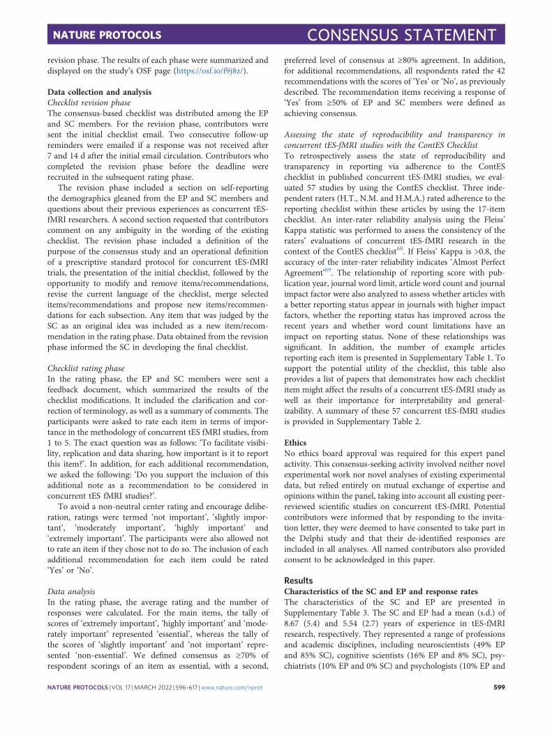
Data collection and analysisChecklist revision phaseThe consensus-based checklist was distributed among the EPand SC members. For the revision phase, contributors weresent the initial checklist email. Two consecutive follow-upreminders were emailed if a response was not received after7 and 14 d after the initial email circulation. Contributors whocompleted the revision phase before the deadline wererecruited in the subsequent rating phase.
The revision phase included a section on self-reportingthe demographics gleaned from the EP and SC members andquestions about their previous experiences as concurrent tES-fMRI researchers. A second section requested that contributorscomment on any ambiguity in the wording of the existingchecklist. The revision phase included a definition of thepurpose of the consensus study and an operational definitionof a prescriptive standard protocol for concurrent tES-fMRItrials, the presentation of the initial checklist, followed by theopportunity to modify and remove items/recommendations,revise the current language of the checklist, merge selecteditems/recommendations and propose new items/recommen-dations for each subsection. Any item that was judged by theSC as an original idea was included as a new item/recom-mendation in the rating phase. Data obtained from the revisionphase informed the SC in developing the final checklist.
Checklist rating phaseIn the rating phase, the EP and SC members were sent afeedback document, which summarized the results of thechecklist modifications. It included the clarification and cor-rection of terminology, as well as a summary of comments. Theparticipants were asked to rate each item in terms of impor-tance in the methodology of concurrent tES fMRI studies, from1 to 5. The exact question was as follows: ‘To facilitate visibi-lity, replication and data sharing, how important is it to reportthis item?’. In addition, for each additional recommendation,we asked the following: ‘Do you support the inclusion of thisadditional note as a recommendation to be considered inconcurrent tES fMRI studies?’.
To avoid a non-neutral center rating and encourage delibe-ration, ratings were termed ‘not important’, ‘slightly impor-tant’, ‘moderately important’, ‘highly important’ and‘extremely important’. The participants were also allowed notto rate an item if they chose not to do so. The inclusion of eachadditional recommendation for each item could be rated‘Yes’ or ‘No’.
Data analysisIn the rating phase, the average rating and the number ofresponses were calculated. For the main items, the tally ofscores of ‘extremely important’, ‘highly important’ and ‘mode-rately important’ represented ‘essential’, whereas the tally ofthe scores of ‘slightly important’ and ‘not important’ repre-sented ‘non-essential’. We defined consensus as ≥70% ofrespondent scorings of an item as essential, with a second,
preferred level of consensus at ≥80% agreement. In addition,for additional recommendations, all respondents rated the 42recommendations with the scores of ‘Yes’ or ‘No’, as previouslydescribed. The recommendation items receiving a response of‘Yes’ from ≥50% of EP and SC members were defined asachieving consensus.
Assessing the state of reproducibility and transparency inconcurrent tES-fMRI studies with the ContES ChecklistTo retrospectively assess the state of reproducibility andtransparency in reporting via adherence to the ContESchecklist in published concurrent tES-fMRI studies, we eval-uated 57 studies by using the ContES checklist. Three inde-pendent raters (H.T., N.M. and H.M.A.) rated adherence to thereporting checklist within these articles by using the 17-itemchecklist. An inter-rater reliability analysis using the Fleiss’Kappa statistic was performed to assess the consistency of theraters’ evaluations of concurrent tES-fMRI research in thecontext of the ContES checklist68. If Fleiss’ Kappa is >0.8, theaccuracy of the inter-rater reliability indicates ‘Almost PerfectAgreement’69. The relationship of reporting score with pub-lication year, journal word limit, article word count and journalimpact factor were also analyzed to assess whether articles witha better reporting status appear in journals with higher impactfactors, whether the reporting status has improved across therecent years and whether word count limitations have animpact on reporting status. None of these relationships wassignificant. In addition, the number of example articlesreporting each item is presented in Supplementary Table 1. Tosupport the potential utility of the checklist, this table alsoprovides a list of papers that demonstrates how each checklistitem might affect the results of a concurrent tES-fMRI study aswell as their importance for interpretability and general-izability. A summary of these 57 concurrent tES-fMRI studiesis provided in Supplementary Table 2.
EthicsNo ethics board approval was required for this expert panelactivity. This consensus-seeking activity involved neither novelexperimental work nor novel analyses of existing experimentaldata, but relied entirely on mutual exchange of expertise andopinions within the panel, taking into account all existing peer-reviewed scientific studies on concurrent tES-fMRI. Potentialcontributors were informed that by responding to the invita-tion letter, they were deemed to have consented to take part inthe Delphi study and that their de-identified responses areincluded in all analyses. All named contributors also providedconsent to be acknowledged in this paper.
ResultsCharacteristics of the SC and EP and response ratesThe characteristics of the SC and EP are presented inSupplementary Table 3. The SC and EP had a mean (s.d.) of8.67 (5.4) and 5.54 (2.7) years of experience in tES-fMRIresearch, respectively. They represented a range of professionsand academic disciplines, including neuroscientists (49% EPand 85% SC), cognitive scientists (16% EP and 8% SC), psy-chiatrists (10% EP and 0% SC) and psychologists (10% EP and
NATURE PROTOCOLS CONSENSUS STATEMENT
NATURE PROTOCOLS | VOL 17 |MARCH 2022 | 596–617 |www.nature.com/nprot 599

0% SC). Their professional settings were primarily universities(59% EP and 69% SC), hospitals (18% EP and 8% SC), uni-versity hospitals (0% EP and 8% SC), independent researchinstitutes (6% EP and 15% SC) and businesses/industries (6%EP and 0% SC), and the most commonly held academicdegrees were PhD (76% EP and 69% SC), MD-PhD (6% EPand 15% SC) and MD (4% EP and 15% SC). 49 EP members,along with 13 SC members completed the revision phase of theDelphi questionnaire, and 45 EP members and 9 SC memberscompleted the rating phase. Retention was very high, with 54(87.1%) revision phase contributors also completing therating phase.
Results of the Delphi processChecklist development phaseFour members of the SC (A.D.S., I.R.V., J.A. and H.E.) pro-duced an initial list of items for the overall structure of thechecklist on the basis of suggestions derived from the con-current tES-fMRI study literature. After the discussions withinthe SC, the checklist was expanded from 14 items to 16 items.Thus, for the revision phase, nine items in the TechnologicalFactors category, four items in the Safety and Noise Testscategory and three items in the Methodological Factors cate-gory were provided within the checklist. Furthermore, an‘Additional Recommendations’ column was added to theContES checklist by the SC with 28 additional recommenda-tions for experimental parameters and practices. These addi-tional recommendations provide guidance to the requirements
for adequate and appropriately documented simultaneousconduction of fMRI and tES.
Checklist revision phaseIn the revision phase, one item was added to the ContESchecklist (tES-fMRI Setting Test—Subjective IntoleranceReporting). The additional recommendations were expandedby the contributors from 28 items to 42 items. The finalchecklist includes 9 items and 19 recommendations in theTechnological Factors category, 5 items and 12 recommenda-tions in the Safety and Noise Tests category, 3 items and 9recommendations in the Methodological Factors categoryand 2 general recommendations. Different versions of thechecklist in its development process are provided by the study’sOSF page (https://osf.io/f9j8z/).
Checklist rating phaseThe collected responses of the rating phase are shown inFigs. 2,3 Tables 1–3. Respondents had a high rate of agreementabout most of the checklist items. However, three items(marked with † in Fig. 2), Amount of Contact Medium(Paste/Gel/Electrolyte), Electrode Placement Visualization andWire Routing Pattern, did not reach the 80% consensusthreshold (rated as moderately, highly or extremely importantby >80% of the respondents). Of these, one item, Amountof Contact Medium (Paste/Gel/Electrolyte), did not reachthe ≥70% consensus (marked with ‡ in Fig. 2 and Supple-mentary Table 4). However, the draft ContES checklist met
Category 1: Technological Factors
1.1 . Manufacturer of MR Conditional Stimulator
1.2. MR Conditional Electrode Details
1.3. Electrode Positioning
1.4. MR Conditional Skin-Electrode Interface
1.5. Amount of Contact Medium (Paste/Gel/Electrolyte) †,‡
1.6. Electrode Placement Visualization †
1.7. RF Filter
1.8. Wire Routing Pattern †
1.9. tES-fMRI Machine Synchronization/Communication
Category 2: Safety and Noise Tests
2.1. MR Conditionality Specifics for tES Setting
2.2. tES-fMRI Setting Test–Safety Testing
2.3. tES-fMRI Setting Test–Subjective Intolerance Reporting
2.4. tES-fMRI Setting Test–Noise/Artifact
2.5. Impedance Testing
Category 3: Methodological Factors
3.1. Concurrent tES-fMRI Timing
3.2. Imaging Session Timing
3.3. tES Experience Report
Extremely important Highly important Moderately important Slightly important
0% 20% 40% 60% 80% 100%
Not important
Fig. 2 | Collected responses from contributors regarding the importance of the main items (rating phase). This figure depicts the rating of thechecklist items by 54 respondents in the rating phase. Each item was rated from 1 to 5 (not important to extremely important). 14 items reached the80% threshold (rated as moderately, highly or extremely important by >80% of the respondents). The items that did not reach this threshold aremarked with ‘†’). 16 items reached the 70% threshold (rated as moderately, highly or extremely important by >70% of the respondents). The one itemthat did not reach this threshold is marked with ‘‡’. Full text of the items is provided in Tables 1–3. MR, magnetic resonance.
CONSENSUS STATEMENT NATURE PROTOCOLS
600 NATURE PROTOCOLS | VOL 17 |MARCH 2022 | 596–617 |www.nature.com/nprot
Category 1: Technological Factors
1.2.1. MR Conditional Electrode Conductive Properties
1.3.1. Electrode Positioning for Improving Reproducibility
1.3.2. Level of Localization of Electrode Position
1.3.3. Method for Individualized Electrode Positioning
1.3.4. Reproducibility of Localization of Electrode Position in Multiple tES Sessions
1.3.5. Securing Electrode Placement inside Scanner
1.3.6. Electrode Cable Placement inside Scanner
1.3.7. Post hoc Validation of Electrode Positioning
1.4.1. MR Conditional Skin Electrode Interlace Visualization
1.4.2. MR Conditional Skin Electrode Interlace Control
1.5.1. Control of Amount of Contact Medium #
1.7.1. Attenuation Characteristic of RF Filter #
1.7.2. Restrictions Regulations for RF Filtering Method #
1.8.1. Cable Testing inside Scanner
1.8.2. MR Safe Cable Details
1.8.3. Securing Cables Filter Boxes during Imaging
1.8.4. Wire Routing Pattern Modifications from Manufacturer Recommendations
1.8.5. Restrictions Regulations for Wire Routing Pattern #
1.9.1. tES MRI Synchronization
Category 2: Safety and Noise Tests
2.1.1. MR Technical Specifications
2.1.2. MR Conditionality Specifications based on tES Manufacturer Guideline
2.3.1. Safety Tests for tES fMRI Setting
2.3.1. Safety Incidents for tES fMRI Setting
2.3.1. Reasons for Subjective tES fMRI Intolerance
2.4.1. Manufacturer s Statement for Signal to Noise Ratio
2.4.2. Exclusion Criteria from Data Analysis Artifact
2.4.3. Quantification of Artifact Noise Caused by Task fMRI Devices
2.4.4. Quantification of Artifact Noise Caused by tES Setup with Pre tES fMRI
2.4.5. Image Processing Assessments for tES Induced Imaging Artifacts
2.5.1. Characteristics of Impedance Recorded
2.5.2. Current Delivered Assessment inside Scanner
Category 3: Methodological Factors
3.1.1. Schematic Diagram for Concurrent tES fMRI Timing
3.1.2. Carry Over Effects for Stimulation Condition Brain State
3.2.1. Timing of Imaging Events
3.2.2. Placement Time of tES Setup on Subject
3.2.3. Frequency Matching for tACS Studies
3.3.1. Assessment of tES Subjective Experience inside Scanner
3.3.2. Electric Current Tolerance
3.3.3. Training for Subject Convenience before tES fMRI Session
3.3.4. Subjective Experiences Questionnaires of Receiving tES inside Scanner
3.0.1. Handedness
3.0.2. Electrode Placement Verification
Yes No
0% 50% 100%
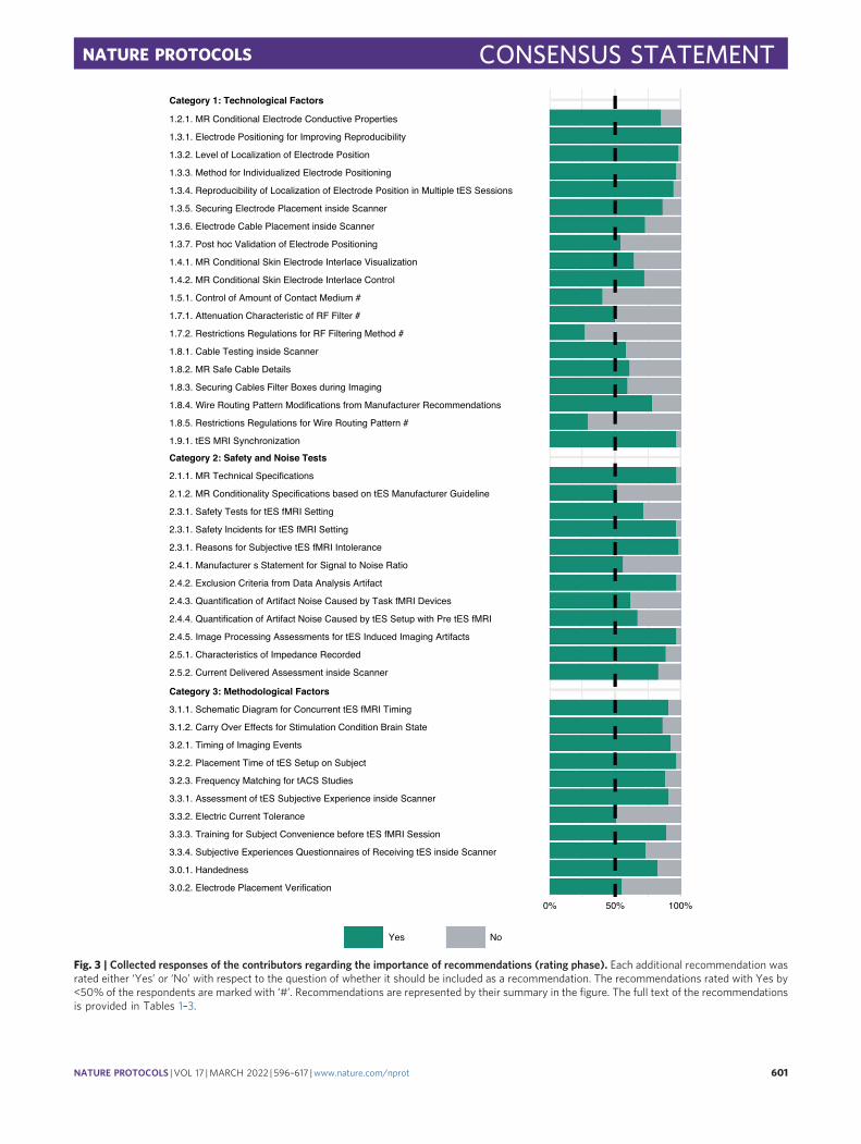
Fig. 3 | Collected responses of the contributors regarding the importance of recommendations (rating phase). Each additional recommendation wasrated either ‘Yes’ or ‘No’ with respect to the question of whether it should be included as a recommendation. The recommendations rated with Yes by<50% of the respondents are marked with ‘#’. Recommendations are represented by their summary in the figure. The full text of the recommendationsis provided in Tables 1–3.
NATURE PROTOCOLS CONSENSUS STATEMENT
NATURE PROTOCOLS | VOL 17 |MARCH 2022 | 596–617 |www.nature.com/nprot 601

Tab
le1|C
oncurren
ttES-fM
RI(Con
tES20
21)checklist:mainitem
san
drecommen
dation
sof
theTechn
olog
ical
Factorscatego
ryto
repo
rtin
concurrent
tES-fM
RIresea
rch
Categ
ories/subcateg
ories
Mainitem
sto
repo
rtItem
impo
rtan
ceSp
ecificrecommen
dation
Recom
men
dation
forinclusion
1.1.Manufacturerof
MR
Con
ditio
nalStim
ulator
Thebrandandmodel
(ifabrandis
providingdifferent
MRconditional
models)
fortheMRconditionalstimulator
4.37(0
.96)
––
1.2.
MRCon
ditio
nal
Electrod
eDetails
The
MRcond
ition
alelectrod
etype
(i.e.,
cond
uctiv
epo
lymer
with
orwith
outa
spon
geor
othe
rcond
uctiv
emed
ium
holders)
4.06(1.01)
1.2.1.Rep
ortcond
uctiv
eprop
ertie
sof
theMRcond
ition
alelectrod
es,cables,
contactmed
ium
andothe
rcond
uctiv
eelem
ents,including
thepo
sitio
nand
materialsused
fortheelectrod
e-cableconn
ectio
ns71 .Thisis
espe
cially
impo
rtantifthey
areno
tfrom
anestablishe
dmanufactureror
notwell
describe
din
thepriorliterature.
How
ever,e
venforwell-establishe
deq
uipm
ent,thesede
tails
arecriticalto
repo
rtto
ensure
replicability
44(85)
1.3.
Electrod
ePo
sitio
ning
The
metho
dforelectrod
eplacem
ent
over
thehe
adinside
thescanne
r(i.e.,
targetingsoftware,
10-20conven
tion
with
orwith
outEEG
cap,
functio
nal
targeting(fMRI),c
ompu
tatio
nalhe
admod
elsor
othe
rs)
4.83(0
.60)
1.3.1.Rep
ortelectrod
epo
sitio
ning
aspreciselyas
possible
tofacilitate
reprod
uctio
n.Itisusually
inadeq
uate
tosimplyrepo
rtan
anatom
ical
target
(e.g.,‘th
eanod
alelectrod
ewas
placed
over
M1’)
51(100)
1.3.2.
Rep
ortwhe
ther
electrod
epo
sitio
ning
isbasedon
theindividu
alanatom
yor
agrou
ptemplateifim
agingor
head
mod
elingisused
for
electrod
epo
sitio
ning
49(98)
1.3.3.
Rep
ortho
welectrod
epo
sitio
ning
ispe
rformed
attheindividu
alparticipantlevel.Forexam
ple,
was
ane
uron
avigationsystem
used
orthe
EEG
10-20system
orsomething
else
49(96)
1.3.4.R
eportthemetho
dsto
ensure
that
thesameelectrod
elocatio
nswere
used
againiftherearemultip
lesessions
48(94)
1.3.5.
Rep
ortclearlyho
wtheelectrod
esarehe
ldin
placeinside
thescanne
r,includ
inguseof
headgear
orcustom
ized
supp
orts
42(86)
1.3.6.R
eportho
welectrod
esandtheirconn
ectin
gcables
over
thehe
adare
locatedin
relatio
nshipto
theMRhe
adcoilwhile
thesubjectislyingdo
wn
inside
thescanne
randho
wthehe
adwas
held
inplace(e.g.,pillows,foam
,etc.)to
ensure
that
thepo
sitio
nof
thehe
ad/electrode
sremainin
thesame
placedu
ring
thescanswhile
theconven
ienceof
theparticipantisen
sured
37(73)
1.3.7.
Rep
ortapo
st-hoc
validationof
theelectrod
epo
sitio
ning
basedon
anatom
ical
images
with
theelectrod
esin
place,
ifpractic
al.Forop
timal
validation,
curren
tde
nsity
mod
elsbasedon
anatom
icalim
ages
may
beused
(e.g.,ROAST
,Sim
NIBS,etc.).Itwou
ldbe
even
better
todirectlymeasure
the
electric
fields
byusingMRCDIandMREIT103;h
owever,M
REITandMRCDI
arestill
notavailablein
mostinstitu
tes
27(54)
1.4.M
RCon
ditio
nalSkin-
Electrod
eInterface
The
MRcond
ition
alskin-electrode
interface(salinesolutio
n,cond
uctiv
epaste,
gel,etc.)
4.09(0
.90)
1.4.1.R
eportaph
otoor
asche
matic
figu
reor
technicald
etailsshow
ingin
areprod
ucible
way
how
theelectrod
ewith
theMRcond
ition
alskin-electrode
interfaceisconn
ectedto
thecranium
(including
aview
from
unde
rneath
the
electrod
eifne
eded
).Ifhe
adgear
orhe
adstraps
obscuretheelectrod
es,y
oumay
providean
imagewith
outthehe
adstraps
32(64)
1.4.2.R
eportanyothe
rMR-spe
cificstrategies
torestrict
thecontactmed
ium
(suchas
with
inan
electrod
eho
lder)to
avoidshortcircuits
36(72)
Table
continue
d
CONSENSUS STATEMENT NATURE PROTOCOLS
602 NATURE PROTOCOLS | VOL 17 |MARCH 2022 | 596–617 |www.nature.com/nprot

Tab
le1(con
tinu
ed)
Categ
ories/subcateg
ories
Mainitem
sto
repo
rtItem
impo
rtan
ceSp
ecificrecommen
dation
Recom
men
dation
forinclusion
1.5.
Amou
ntof
Con
tact
Med
ium
(Paste/G
el/
Electrolyte)
The
amou
ntor
thicknessof
med
ium
that
isused
foreach
electrod
eor
ametho
dto
controlthis
confou
nding
variable
2.91(1.13)
1.5.1.Rep
orttechnicalde
tails/d
ifficulties
inmeasuring
thethicknessof
the
layerof
cond
uctiv
ematerialu
nderne
aththeelectrod
esandho
wcream/g
elun
derneath
theelectrod
esiseven
lydistribu
ted.
Alth
ough
this
canbe
impo
rtant,mainlywhe
nhaving
bigelectrod
es,in
practic
e,theam
ount
ofcream/g
elun
derneath
theelectrod
esmay
notbe
even
lydistribu
ted.
Develop
ingne
wmetho
dsto
measure,controlandrepo
rtthisim
portant
variable
isde
sired.
Rep
ortin
gtheim
pedance(before,
during
andafter
stim
ulation)
provides
insigh
ton
electrod
econtactqu
ality
butisno
tinitselfa
substituteforcontrolling
andrepo
rtingcontactmed
ium
parameters
21(40)
1.6.E
lectrode
Placem
ent
Visualization
Any
photo/
diagram/fi
gure
toprecisely
visualizetheelectrod
emon
tage
inside
thescanne
randmakereplication
possible
3.56
(1.17)
––
1.7.
RFFilte
rThe
RFfilte
ring
metho
d(stim
ulator
device
conn
ectedto
thesubjectviaa
pene
trationpane
l(e.g.,RFfilte
rsfrom
differen
tbrands)or
conn
ectedviaa
waveguide
with
RFbo
xeson
either
end)
3.50
(1.07)
1.7.1.Rep
orttheattenu
ationcharacteristic
oftheRFfilte
ring
25(50)
1.7.2.
Rep
ortanypo
tentialregu
latory
consideration/
limita
tionat
the
institu
te/u
niversity/cou
ntry
level
13(27)
1.8.W
ireRou
tingPa
ttern
Wireroutingpattern(out
theback
ofthebo
reandarou
ndthecontrolroom
orstraight
downthefron
tof
thebo
reto
thecontrolroom
)
3.43(1.22)
1.8.1.R
eportwhe
ther/h
owthestateof
thecables
ischeckedafterthesubject
enters
thescanne
r,to
avoidcreatin
ganyloop
s29
(58)
1.8.2.R
eporttheleng
thof
thecables
requ
ired
toconn
ecttheinne
rbo
xwith
theou
terbo
xby
usingbo
xcable,
how
thecables
areconn
ectedto
the
electrod
es,in
which
directionthecables
leavethehe
ad,ho
wmultip
leconn
ectin
gcables
aremanaged
together
and,de
pend
ingon
thegeom
etry
ofthehe
adcoil,ho
wthecables
enterinto
thecoil.Asketch
might
behe
lpfulto
visualizethesede
tails
31(61)
1.8.3
Rep
ortho
wthecables
andfilte
rbo
xesaresecuredto
preven
tmotion
during
thescan
(i.e.,sand
bag,
tape
,etc.)
29(59)
1.8.4.Rep
ortifthereareanymod
ificatio
nsfrom
manufacturer
recommen
datio
ns39
(78)
1.8.5.R
eportanypo
tentialregu
latory
consideration/
limita
tionat
the
institu
te/u
niversity/cou
ntry
level
14(29)
1.9.tES
-fMRIMachine
Synchron
ization/
Com
mun
ication
The
synchron
ization/
commun
ication
metho
dbe
tweenthetESdevice,the
stim
ulus
deliveryPC
andthescanne
r
3.70
(1.09)
1.9.1.R
eportanysynchron
izationbe
tweentESandMRI.Synchron
ization/
commun
icationcanbe
aTTLscanne
rsync
pulseto
trigger/sync
(tES
and/
orno
n-tES)
stim
ulus
recorded
viaUSB
/parallelpo
rt/N
Idevice,the
useof
markers
fortESor
manualtriggering
oftheTES
device
49(96)
Ratings
forite
ms(scores1–5)
arerepo
rted
asmean(s.d.),and
ratin
gsforrecommen
datio
ns(Y
es/N
o)arerepo
rted
asfreq
uency(percentage)
ofYes
repo
rts.MRCDI,magne
ticresonancecurren
tde
nsity
imaging;MREIT,m
agnetic
resonanceelectricalim
pedance
tomog
raph
y;NI,NationalInstrumen
ts;ROAST
,realistic
volumetricapproach
tosimulatetranscranial
electric
stim
ulation;
SimNIBS,
simulationof
noninvasivebrainstim
ulation;
TTL,
transistor–transistorlogic;
USB
,un
iversalserial
bus.
NATURE PROTOCOLS CONSENSUS STATEMENT
NATURE PROTOCOLS | VOL 17 |MARCH 2022 | 596–617 |www.nature.com/nprot 603

Tab
le2|Con
curren
ttES-fM
RI(C
ontES20
21)checklist:mainitem
san
drecommen
dation
sof
theSa
fety
andNoise
Tests
catego
ryto
repo
rtin
concurrent
tES-fM
RI
research
Categ
ories/
subcateg
ories
Mainitem
sto
repo
rtItem
impo
rtan
ceSp
ecificrecommen
dation
torepo
rtRecom
men
dation
forinclusion
2.1.MRCon
ditio
nality
Specifics
fortES
Setting
The
technicalspecificatio
nsof
theMRscanne
r,theapplied
fMRIsequ
encesandtheused
tESsettings
andconfi
guratio
nto
fallwith
inthespecifics
ofMRcond
ition
ality
onthebasis
ofthetESmanufacturer
guideline
4.37(0
.99)
2.1.1.Rep
ortthetechnicalspe
cificatio
nsof
theMRscanne
r,includ
ingfieldstreng
th,R
Ftransm
itcoiltype
,maxim
altransm
itpo
wer
andthenu
mbe
rof
head
coilchanne
ls.S
tand
ardgu
idelines
forprop
errepo
rting
onMRI/fM
RIparametersshou
ldbe
considered
57,58,104
50(96)
2.1.2.
Rep
ortthede
tails
ofMRcond
ition
ality
that
arede
mon
stratedby
themanufacturerof
thetES
equipm
entforspecificcond
ition
sof
use
24(51)
2.2.
tES-fM
RISetting
Test—
Safety
Testin
gThe
safety
ofthetES-fM
RI
setting
3.93(1.23)
2.2.1.Rep
ortsafety
testsandrespectiv
ede
tails,w
hich
includ
ebu
tareno
tlim
itedto
impe
dancetesting,
tempe
rature
testing(any
tempe
rature
change
unde
relectrod
es)andelectric
curren
ttolerancetesting
with
realhu
man
subjects
orph
antoms.W
hene
verthesafety
testingisreferred
toaprevious
stud
y,itis
still
recommen
dedto
provideabriefde
scriptionof
thesafety
teststhat
have
been
considered
37(71)
2.2.2.
Rep
orttheoccurren
ce/absen
ceof
anysafety
incide
nts
48(96)
2.3.
tES-fM
RISetting
Test—
Subjectiv
eIntoleranceRep
ortin
g
The
numbe
rof
casesthat
have
nottoleratedthetES/
fMRI
session(evenifitis
zero)
4.33(0
.92)
2.3.1.Rep
ortthereason
sthat
participants
have
nottoleratedthetES/
fMRIsessionifany(i.e.,bu
rning
sensation,
increasedtempe
rature,pain,shortnessof
breath,nausea,etc.)
50(98)
2.4.tES
-fMRISetting
Test—
Noise/A
rtifact
The
noise/artifactindu
cedby
thetESsettingin
thefM
RI
sign
alwith
real
human
subjects
orph
antomsbe
fore
startin
gthestud
y(itcanbe
repo
rted
orreferred
toprevious
stud
ieswith
thesame
setting)
3.91(0
.99)
2.4.1.R
eportor
cite
prioranalysison
thede
gree
towhich
theeq
uipm
entalon
eandtheeq
uipm
entdu
ring
stim
ulationaffectstheSN
R.Impo
rtantly
,suchanalysisisspecificto
theprotocol
(electrode
prep
aration
andim
agingsequ
ence)such
that
claimscann
otbe
automaticallygene
ralized
with
outanalysis(e.g.,~8%
asde
scribe
din
ref.
49(‘SN
Rwas
hardly
redu
cedwith
decreasesrang
ingfrom
3to
8%
forthedifferen
tROIs
andsetups,even
inthegray
matterROIin
M1targeted
bytD
CS’))
29(56)
2.4.2.Rep
ortho
wmanyparticipants
orruns
wereexclud
edfrom
theanalysis
becauseof
artifacts.
Exclusioncrite
riashou
ldbe
repo
rted
aswell(e.g.,basedon
visual
inspectio
nor
anydata
analysis
tool
that
might
detect
artifacts
forsing
leruns)
50(96)
2.4.3.Rep
ortthequ
antificatio
nof
thepo
ssible
increase
inartifactor
noiseifthetask-related
fMRI
requ
ires
theuseof
someothe
rde
vices,such
astactile/p
ainstim
ulators,olfactoryor
juicemachine
s,etc.
(e.g.,compare
theno
ise/artifacts
ofthetES-fM
RIsetup
alon
ewith
thetES-fM
RIsetup
with
theadditio
nof
therespectiv
ede
vice)
32(62)
2.4.4.R
eportbaseline‘pre-tES
’fMRIa
sapartof
thedata
acqu
isition
sequ
ence
intheim
agingsessionto
investigatetheeffects/no
iseintrod
uced
bythetESsetuppe
rse
(with
outanystim
ulationandwith
inthe
subject).H
owever,thiswillno
tbe
sufficien
tto
fully
controlfor
noiseindu
cedby
tESadministration,
with
prob
lemssuch
asscanne
rdrift
andtheorde
reffect
34(67)
2.4.5.Rep
ortanyspecialfM
RIprocessing
measuresor
assessmen
tsthat
areused
tode
alwith
tES-
indu
cedim
agingartifacts
ifapplicable
50(96)
2.5.
Impe
dance
Testin
gIm
pedancemon
itoring
(i.e.,
before
entering
thescanne
rroom
and/
orin
thescanne
rroom
and/
orinside
the
scanne
rand/
ordu
ring
scanning
)
4.17(1.00)
2.5.1.Rep
orttheim
pedance(i.e.,cutoffcrite
rion
prog
rammed
inthede
vice
ormeasureson
anindividu
albasiswith
mean/
rang
eacross
grou
psbe
fore,du
ring
andafterscanning
)45(88)
2.5.2.
Rep
ortthemetho
dsappliedto
verify
thecurren
tde
livered
inside
thescanne
r(ifany).So
me
devicesalreadyinclud
ean
inde
pend
entcurren
tmeter,andsomeinvestigatorsusetheirow
nexternal
devices
42(82)
Ratings
forite
ms(scores1–5)
arerepo
rted
asmean(s.d.),a
ndratin
gsforrecommen
datio
ns(Y
es/N
o)arerepo
rted
asfreq
uency(percentage)
ofYes
repo
rts.SN
R,signal-to-noise
ratio
.
CONSENSUS STATEMENT NATURE PROTOCOLS
604 NATURE PROTOCOLS | VOL 17 |MARCH 2022 | 596–617 |www.nature.com/nprot

Tab
le3|Con
curren
ttES-fM
RI(C
ontES20
21)checklist:mainitem
san
drecommen
dation
sof
theMetho
dologicalFactorscatego
ryto
repo
rtin
concurrent
tES-fM
RI
research
Categ
ories/
subcateg
ories
Mainitem
sto
repo
rtItem
impo
rtan
ceSp
ecificrecommen
dation
torepo
rtRecom
men
dation
forinclusion
3.1.Con
curren
ttES-
fMRITim
ing
The
timingof
concurrent
tESwith
inthefM
RIparadigm
4.52(0
.81)
3.1.1.Providingasche
maticdiagram
isstrong
lyen
couraged
toachievemaxim
umclarity
for
thereader
46(90)
3.1.2.
Rep
ortcarryovereffectsbe
tweendifferen
tstim
ulationcond
ition
sanddifferen
tbrain
states.How
such
effectshave
been
considered
ormitigatedshou
ldbe
discussed
42(86)
3.2.
Imaging
SessionTim
ing
The
imagingeven
tsbe
fore
andafter
concurrent
tES-fM
RI
andrespectiv
esequ
ences
4.09(0
.99)
3.2.1.Rep
orttheexacttim
ingof
allim
agingeven
ts(structuralor
functio
nal)be
fore
and
afterconcurrent
tES-fM
RI
47(92)
3.2.2.
Rep
ortwhe
nthetESsetupisplaced
ontheparticipant(e.g.,ifthetESsetupwas
placed
ontheparticipantat
thestartof
thetES-fM
RIsession(and
was
thereforeon
the
participantdu
ring
othe
rno
n-fM
RIsequ
ences))
50(96)
3.2.3.
IntA
CSstud
ies,repo
rtho
wstim
ulationfreq
uencyismatched
with
TR.Toredu
cepo
tentialsou
rces
ofbiases
intA
CS-fM
RIstud
ies,thestim
ulationfreq
uencyshou
ldbe
set
such
that
afullnu
mbe
rof
cycles
fits
into
theTRof
thefunctio
nalm
easuremen
t36(post-
mortem
stud
y).O
therwise,thetissuepo
larizatio
nmight
beaveraged
over
thetim
eof
one
volumemeasured
43(88)
3.3.
tES
Expe
rien
ceRep
ort
The
assessmen
tof
thesubjectiv
eexpe
rien
ceof
receivingtESinside
thescanne
r
4.06(0
.97)
3.3.1.Re
portthegeneralexperience(com
fort/fatigue)andtheparticipant’s
otherexperie
nces
with
thestim
ulation,
becausesometESmon
tages/protocolsmight
bemoreun
comfortable/
perceptib
lethan
others
whenlyinginside
thescanner,andthiscouldbe
aconfou
nder
when
comparin
gacross
stim
ulationmon
tages.Options
includ
eassessingparticipantratin
gsof
symptom
sforeach
cond
ition
,askingparticipants
whether
they
perceivedstim
ulationor
not
foreach
cond
ition
,reportin
gon
thepresence
andintensity
ofph
osph
enes/tactilesensation
(inthecase
oftACS),etc.T
hisisim
portantbecauseitcouldshow
whether
participants
can
differentiate
betw
eenstim
ulationcond
ition
s(e.g.,betw
eenactiveandsham
stim
ulationor
betw
eendifferentfrequencies(inthecase
oftACS).H
avingdifferentside
effectsbetw
een
sessions
does
notnecessarily
meanthat
subjects
candiscernandareun
blinded
47(90)
3.3.2.Rep
ortelectriccurren
ttoleranceforsubjectcomfort(i.e.,be
fore
entering
thescanne
rroom
(iftechnically
possible)and/
orin
thescanne
rroom
andinside
thescanne
rand/
ordu
ring
scanning
(asapprop
riate))
26(51)
3.3.3.Rep
ortanyinstructions,trainingor
expo
sure
provided
before
thetES-fM
RIsession
tomaketheexpe
rimen
tmoreconven
ient
fortheparticipants
46(88)
3.3.4.R
eporttheexactwording
orprovidecitatio
nsof
thequ
estio
nsor
questio
nnairesused
torepo
rton
thesubjectiv
eexpe
rien
ceof
receivingtESinside
thescanne
rintheartic
leor
itssupp
lemen
ts
38(73)
Gen
eral
recommen
datio
ns0.0.1.R
eportthehand
edne
ssof
thesubjectas
apo
tentialsou
rceof
variability
oftES-fM
RI
stud
ies.Thisinteractioncouldbe
addressedin
relevant
contexts
either
bylim
iting
the
sampleto
righ
t-hand
edindividu
als,repo
rtinghand
edne
sswith
quantitativestandard
instrumen
tsor
throug
hmetho
dological/analytical
approaches
that
shou
ldbe
repo
rted
41(82)
0.0.2.Ifpo
ssible,presen
ttheon
linetESelectrod
esas
additio
nalbu
mps
inthesurface/
meshreconstructio
n.Thisisago
odpo
ssibility
tode
term
inetheexactlocatio
nof
theon
line
electrod
es.H
owever,thisno
n-biolog
ical
reconstructio
nmay
also
influe
ncesimulations,so
performance
ofadditio
nalstructuralT
1wandT2w
scanswith
outtheelectrod
eswhe
never
possible
isadvantageo
us
28(55)
Ratings
forite
ms(scores1–5)
arerepo
rted
asmean(s.d.),a
ndratin
gsforrecommen
datio
ns(Y
es/N
o)arerepo
rted
asfreq
uency(percentage)
ofYes
repo
rts.TR,rep
etition
time.
NATURE PROTOCOLS CONSENSUS STATEMENT
NATURE PROTOCOLS | VOL 17 |MARCH 2022 | 596–617 |www.nature.com/nprot 605

the consensus level for all 17 items with a 65% threshold. Therating phase also included scoring of each of the additionalrecommended items by the scoring choices of Yes and No.The results showed that 38 (90%) of the recommendationsreached the 50% threshold (rated as Yes by >50% of the con-tributors), but the following four recommendations didnot (10%) (Fig. 3): Control of Amount of Contact Medium,Attenuation Characteristic of RF Filter, Restrictions/Regula-tions for RF Filtering Method and Restrictions/Regulations forWire Routing Pattern.
The ratings of the items and recommendations of theContES checklist are outlined in Tables 1–3. The full version ofthe ContES checklist that includes 17 essential items and 42additional recommendations and a short version that includesessential items only are provided in Supplementary Tables 4–7to be used by authors and reviewers. The reporting items thatdid not meet the 70% and 80% thresholds and additionalrecommendations that did not meet the 50% thresholds aremarked in the final checklist. On the basis of this information,researchers can decide to choose more stringent or more liberalthresholds when using the checklist.
The state of reproducibility and transparency in concurrenttES-fMRI studies with the ContES checklistThree independent raters evaluated the adherence of theconcurrent tES-fMRI articles to the finalized reporting check-list items. The consistency of the raters’ responses resulted in aFleiss’ Kappa of 0.85, indicating that the consistency is AlmostPerfect Agreement69.
Inclusion of information about the main items of theContES checklist varied widely, ranging from fully reported(100%; Manufacturer of MR Conditional Stimulator, Con-current tES-fMRI Timing and Imaging Session Timing) torarely reported (5.3%; Magnetic Resonance ConditionalitySpecifics for tES Setting and Amount of Contact Medium). Thepattern of adherence to the checklist items varied relevantly
between articles, ranging from 24% to 76%, averaging 53% ofchecklist items reported in a given article (Fig. 4).
All studies (100%) reported the Manufacturer of MRConditional Stimulator (Item 1), and the Electrode Positioning(Item 3) was described clearly in 89% of articles, but details onthe MR Conditional Electrode (Item 2) were included in only56% of the reviewed articles. A relatively high number of papers(84%) reported the MR Conditional Skin-Electrode Interface(Item 4), but the Amount of Contact Medium (Item 5) wasmentioned less frequently (5%). The Electrode PlacementVisualization (Item 6) was shown in only 25% of the articles,and the RF Filter (Item 7) was included in 35% of the articles.The Wire Routing Pattern (Item 8) was described clearly in only32% of the articles, and the tES-fMRI Machine Synchronization/Communication (Item 9) was rarely described (23%).
Only 5% of the articles reported information regarding MRConditionality Specifics for tES Setting (Item 10). Few articlesdescribed details on the tES-fMRI setting test, ranging between28% and 33% in Items 11–13 (33% Safety Testing, 33% Sub-jective Intolerance Reporting and 28% Noise/Artifact). Impe-dance Testing (Item14) information was included in only 28%of the articles. Concurrent tES-fMRI Timing (Item 15) and theImaging Session Timing (Item 16) were reported in all 57articles; however, tES Experience was reported less frequently(Item 17; 63%).
The highest reporting score was 76%, and six articles had ascore >70%. One article reported >75% of the checklist items70.The lowest reporting score was 24%; 28 studies failed to meet areporting threshold of 50%. The correlations of study reportingstatus with journal word limit, article word count and journalimpact factor were not significant, and relevant graphs arepresented in Extended Data Fig. 2.
DiscussionThe goal of this study was to develop a consensus-basedchecklist of methodological details to facilitate the evaluation of
Category 1: Technological Factors1.1. Manufacturer of MR Conditional Stimulator1.2. MR Conditional Electrode Details1.3. Electrode Positioning1.4. MR Conditional Skin-Electrode Interface1.5. Amount of Contact Medium (Paste/Gel/Electrolyte)1.6. Electrode Placement Visualization1.7. RF Filter1.8. Wire Routing Pattern1.9. tES-fMRI Machine Synchronization/CommunicationCategory 2: Safety and Noise Tests2.1. MR Conditionality Specifics for tES Setting2.2. tES-fMRI Setting Test–Safety Testing2.3. tES-fMRI Setting Test–Subjective Intolerance Reporting2.4. tES-fMRI Setting Test–Noise/Artifact2.5. Impedance TestingCategory 3: Methodological Factors3.1. Concurrent tES-fMRI Timing3.2. Imaging Session Timing3.3. tES Experience Report
0%
2 2
8
7
9
13
8
2
1
5
0% 25%
50%
Articles that report a checklist item (%)
75%
100%
100%63.2%
100%
89.5%56.1%
5.3%
5.3%
84.2%
24.6%35.1%
31.6%22.8%
33.3%33.3%
28.1%28.1%
100%
0
5
10
Fre
quen
cy
15ba
24%
29%
35%
41%
47
%
Checklist items reported in a article (%)
53%
59% 65
%71
% 7
6%10
0%
Fig. 4 | State of reproducibility/transparency in concurrent tES-fMRI research in the context of the ContES checklist. Assessments by threeindependent raters are based on 57 tES-fMRI papers, from the first published study up to January 1, 2020. a, Percentage of the articles that adhered toeach checklist item. b, The checklist items adhered to by the 57 articles.
CONSENSUS STATEMENT NATURE PROTOCOLS
606 NATURE PROTOCOLS | VOL 17 |MARCH 2022 | 596–617 |www.nature.com/nprot

concurrent tES-fMRI studies in terms of methodologicaltransparency and reproducibility (ContES checklist). Wesuccessfully developed the ContES checklist to guide authors inreporting the minimum information necessary to ensurereproducibility by using the 17 essential items. The 42 addi-tional recommendations should be considered to furtherenhance the quality of future research in this field. Thischecklist can be used by editors and reviewers for criticalappraisal of future studies. The checklist will also be helpful forresearchers who are in the process of setting up a concurrenttES-fMRI study. Indeed, our systematic literature review andappraisal of 57 published concurrent tES-fMRI studies revealed
a general lack of sufficient information to fully reproducecritical methodological details of these studies. Overall, thischecklist offers a methodological framework for understandingand replicating previous studies and provides journal reviewersand editors with an efficient tool to gauge and promote con-current tES-fMRI reproducibility. Figure 5 summarizes theitems that are deemed important to be considered when con-ducting and reporting a concurrent tES-fMRI study.
Technological factorsThe technical features of the stimulator and accessories,including setup on the subject´s head and configuration inside
Technological Itemsa b
c
Safety and Noise Tests
Methodological Items
MR Conditionality Specificsfor tES Setting
Subjective IntoleranceReportingPre-acquisition steps
tES-fMRI setup
Electrode Positioning Amount of Contact Medium Electrode PlacementVisualization
tES-fMRI Setting Test–safety Testing/lmpedance Testing
tES-fMRI Setting Test–Noise/Artifact
Concurrent tES-fMRI Timing
Imaging Session Timing
tES Experience Report
Contact Medium(gel, saline, electrolyte)
MR ConditionalStimulator
RF FilterMethod
Wire Routing Pattern
MR ConditionalElectrode
Electrode
Filter box
Block A Block A Block ARestRest
tES experience
Task
Current(mA)
2
0M
R s
cann
er r
oom
MR
con
trol
roo
m
RFwaveguide/penetration
panel
MRI controlcomputer
Pro
ject
or
Presentationcomputer
TriggerModule
RF
Freq
tES
tES
Menu
,
WithouttES-equipment
Impedance TestingSafety Testing
°C
WithtES-equipment
Anode electrode
3 5 6
12
1411
13
15
16
17
10
2–
–
+
+
4
9
17
8
Cathode electrode
A1 T3
T5P3
O1 O2
INION
Pz P4T6
T4 A2C3 Cz
Fz F4 F8
Fp2
NASION
Fp1
C4
F3F7
α α α α
Menu
tES
tES
Menu
Menu
Ω
Fig. 5 | Scheme of the concurrent tES-fMRI approach in the context of the ContES checklist. a, Summary of technological considerations. The MRconditional stimulator (1, item 1.1) is connected to the head through the RF waveguide or RF penetration panel (7, item 1.7). The box cable should bealigned with the wall of the scanner room and run parallel to the bore axis (8, item 1.8). The MR conditional stimulator is connected to the outer filterbox or RF band-stop filter adapter as well as to the presentation computer trigger output cable. The synchronization module (9, item 1.9) should beconnected to the presentation computer as well as to the MRI control computer. Electrode positioning (3, item 1.3) is used to accurately stimulatecortical target regions and exert neuromodulatory effects. A method allowing quantification of contact medium (e.g., syringes) should be used toachieve a consistent and appropriate amount of contact medium (5, item 1.5). An MR conditional skin-electrode (e.g., saline solution, conductive pasteor gel) (2, item 1.2) is used to facilitate delivery of current to the scalp (4, item 1.4). Electrode placement visualization can be used to reproduciblycenter each electrode on the head so that intrascanner stimulation allows verification of correct positioning of the electrodes on the head (6, item 1.6).b, Summary of safety considerations. MR Conditionality Specifics for tES Setting include the technical specifications of the MR scanner, the appliedfMRI sequences and the used tES settings and configuration to fall within the specifics of MR conditionality on the basis of tES manufacturer guideline(10, item 2.1). The Safety of the tES-fMRI Setting includes electrode temperature testing, electric current tolerance testing, etc. with real humansubjects or phantoms (11, item 2.2). tES-fMRI Setting Test - Subjective Intolerance Reporting shows the number of cases that have not tolerated thetES-fMRI session (12, item 2.3). tES-fMRI Setting Test - Noise/Artifact shows the noise/artifact induced by the tES setting in the fMRI signal with realhuman subjects or phantoms before starting the study (13, item 2.4). The impedance is monitored before entering the scanner room and/or in thescanner room and/or inside the scanner and/or during scanning (14, item 2.5). c, Summary of methodological considerations. Concurrent tES-fMRITiming shows the timing of concurrent tES within the fMRI paradigm (15, item 3.1). Imaging Session Timing shows the imaging events before and afterconcurrent tES-fMRI and respective sequences (16, item 3.2). tES Experience Report includes the assessment of the subjective experience of receivingtES inside the scanner (17, item 3.3).
NATURE PROTOCOLS CONSENSUS STATEMENT
NATURE PROTOCOLS | VOL 17 |MARCH 2022 | 596–617 |www.nature.com/nprot 607

the scanner, underpin rigor and reproducibility—which in turninforms how these elements should be reported (Table 1).Manufacturer make and model should be reported, but thedegree to which this satisfies items on the checklist varies. Forexample, although the material composition of an electrodemay not always be explicitly specified, indicating a uniqueelectrode part number would allow reproduction and refer-encing to other documentation. For other items, the amount ofdetail given beyond the part number can vary depending onthe specific approach. For example, the thickness of paste oramount of other electrolytes is determined by the operator(setup) for large pad electrodes but controlled by the electrodeholder for high-definition (HD) electrodes. The item numberdoes, however, not explain setup details such as cablearrangements or ad-hoc steps to support electrode positioning.These aspects are important to state. The degree to which priorpapers can be referenced for these methodological details (e.g.,‘we applied tES-fMRI used methods as reported in these otherreferences’) should be qualified. Our analysis suggests that, todate, only a limited number of papers documented these detailsin sufficient detail. To the extent that these technical factorsunderpin reproducibility, expanding on them in any givenpublication supports rigor.
Electrodes used for stimulation inside the MR environmentneed to be at minimum MR conditional. Manufacturer andmodel details, electrode size and shape and materials andconductive properties of electrodes (conductive polymer, Ag/AgCl, etc.), connectors (often residually ferromagnetic), cablesand other conductive materials (e.g., a specific brand of elec-trode paste and NaCl concentration) need to be provided. Therelevant item in the checklist was considered highly important(item 1.2, average rating score: 4.06), and the inclusion of theadditional recommendation was recommended by 85% of thecontributors (recommendation 1.2.1). In addition, the positionof the connector on the electrode should be reported, because itcan significantly influence the homogeneity of current dis-tribution within the electrode71. As revealed by concurrenttDCS-magnetoencephalography (MEG) experiments72, someconductive polymer (rubber) electrodes are magnetized, pos-sibly during the production process, whereas others from thesame brand are not. It remains to be determined whether thisproperty is related to MR imaging artifacts.
Electrode positions and size are crucial parameters thatdetermine the distribution of current flow in the brain tissue. Itis therefore recommended unanimously by the experts toreport this information (item 1.3, average rating score: 4.83) asprecisely as possible. It should be distinguished between howthe intended montage is determined and how this is practicallyimplemented. The former may be based on the literature,theoretical considerations or dedicated E-field modeling ingeneric or personalized head models, whereas the latter mayinvolve TMS hotspot-search (for M1), 10-20 electro-encephalography (EEG) system head measurements or MR-based neuronavigation. The reported details should include themethod of electrode positioning (e.g., with or without an EEGcap), the position of the electrode center and, if applicable, itsorientation in case of non-circularly shaped electrodes. Insteadof ‘the electrode was positioned on the left M1’, one would
preferably state, for example, that ‘the electrode was centeredon the first dorsal interosseous (FDI) motor hotspot as deter-mined by TMS, with the longer sides of the 7 × 5 cm2 rec-tangular electrode pointing in anteromedial and posterolateraldirections, respectively, and the connector inserted at thecenter of the electrode pointing toward one shorter side in theanteromedial direction’. If MRI-based head modeling was used,it should be stated whether electrode position had beendetermined on the basis of individual anatomy or a grouptemplate and how electrode positioning was performed (e.g., byusing a neuronavigation system or EEG 10-20 coordinates).For MR-based neuronavigation, Montreal Neurological Insti-tute (MNI) coordinates may be reported for electrode centersand/or corners. In the case of multi-session experiments,measures taken to ensure consistency of electrode placementacross sessions have to be described, such as co-registration ofstimulation electrodes with the individual MRI by using neu-ronavigation or the use of EEG caps and/or the 10-20 system73.The accuracy of the stimulation montage can be judged only ifthis information is provided, and the detailed informationfurther allows post-hoc current modeling and replicationstudies.
It is also recommended to report the position of electrodesand, in particular, how the cables are directed (intertwined orseparated) relative to the MR head coil, as well as informationon how electrodes were affixed to the head in the MR and howthe head was stabilized to prevent movement of electrodesrelative to either the head or MR coil during recordings toprevent discomfort, impedance issues and imaging artifacts,respectively. A post-hoc validation of electrode positions canalso be achieved by the acquisition of anatomical images withthe stimulation electrodes in place, even though such imagesmight not be easy to use for E-field modeling itself (because ofthe challenges of segmentation between the electrodes andskin), for which anatomical images without electrodes (andrelated artifacts) are preferred.
For the sake of reproducibility, it is also important toprovide proper visualization of electrode position (item 1.6,average rating score: 3.56), which may be a photo, a sufficientlydetailed schematic figure or, preferably, the precisely modeledrepresentations on a 3D-rendered head surface as provided byE-field modeling software, such as simulation of noninvasivebrain stimulation (SimNIBS)74 or realistic volumetric approachto simulate transcranial electric stimulation (ROAST)75.Besides the electrode position itself, it is also considered highlyimportant to provide visual information (a photo or suffi-ciently detailed schematic figure) regarding the skin-electrodeinterface (item 1.4, average rating score: 4.09), that is, whichconducting medium was used (e.g., gel/paste or saline solutionwith sponges), how contact with the skin was ensured if thehair was in between and measures taken to restrict the locationof the contact medium to control the effective size of thestimulation surface and prevent short circuits. Although theamount of conductive medium (volume of saline solution orthickness of the layer of electrode paste) was rated of mediumimportance (item 1.5, average rating score: 2.91), this infor-mation, together with the evenness of its distribution across theelectrode surface, is relevant for the impedance as well as the
CONSENSUS STATEMENT NATURE PROTOCOLS
608 NATURE PROTOCOLS | VOL 17 |MARCH 2022 | 596–617 |www.nature.com/nprot

current distribution in the skin (and potentially the brain). Itcan, however, be very difficult in practice to control thisvariable, given that gel is squeezed between the electrode andthe head and that saline solution flows away or evaporates, andit is thus helpful to also report potential countermeasures takento control or measure this influence. In any case, electrodeimpedances should be measured directly before and after theexperiment and be reported.
The introduction of any electrical wire into the MRI magnetbore may result in undesired artifacts and/or noise. Whereasthe magnetic fields induced by the current in wires and elec-trodes during tDCS have been known to lead to false-positiveactivation in blood oxygenation level–dependent (BOLD)fMRI, tACS is far less prone to this artifact because the ACinduces relatively rapid polarity-switching magnetic fields thattime-average to zero net effect36. However, any electricalcabling used in tES-MRI experiments may act as a transmitterof RF energy from outside the MRI shielded environment andtherefore may potentially increase electromagnetic RF inter-ference with the MRI signal—even with the stimulator swit-ched off. It is therefore extremely important to use an RFfiltering method to suppress any external electromagnetic noisethat may find its way into the scanner room by using thestimulator’s cabling as a tunnel. Currently, there are twohardware configurations for addressing this issue29: (i) the RFwaveguide setup, which includes two filter boxes positionedoutside and inside of the scanner room and cables runningfrom the MRI control room through the RF waveguidetube37,38 and (ii) the RF penetration panel setup, whichincludes an RF filter adapter connected directly to the RFpenetration panel and MRI ground, positioned outside of thescanner room with cables running from the stimulator in theMRI control room through the filter and RF penetration paneland to the electrode leads in the scanner room76.
It is therefore recommended unanimously by the con-tributors to report this information, which was rated to bemoderately important (item 1.7, average rating score: 3.50), asprecisely as possible. In addition, it is recommended that theauthors provide details regarding the attenuation characteristicof RF filtering (recommendation 1.7.1, recommended by 50%of the contributors)49,51. For instance, ‘in the case of con-current tDCS-fMRI, the characteristic bandwidth of the stopband of the filters on the DC path have been chosen to providean approximate attenuation of 60 dB within a frequency rangeof 20–200 MHz to mitigate the RF noise, protecting commonstrength MRI scanners such as 1.5T and 3T, which operate atLarmor frequencies of ~64 MHz and ~128 MHz during fMRI(proton imaging)49’.
The wire routing pattern is also an important methodolo-gical detail when using transcranial stimulation simultaneouslywith fMRI measurement to increase replicability and validity ofa study. However, this factor was rated as moderately impor-tant overall (item 1.8, average rating score: 3.43). It is impor-tant to make sure that the wires/cables do not create loops andrun parallel to the bore axis as they approach and exit thescanner. It is also recommended by 58% of the contributors toensure that after the subject enters the scanner, no loop can becreated subsequent to entry because of wire movements, and
practical measures to avoid them should be stated explicitly(recommendation 1.8.1). An example is a protocol reported byWilliams and colleagues in which they emphasize thatregarding stimulator setup, they ensured that no loop wasmade by the wire and that it was placed along the wall ofthe room38. It might be of importance (recommendation 1.8.2,recommended by 61% of the contributors) to include a figureillustrating the wiring details, such as the length of thecables required to connect inner and outer filter boxes, howthe cables are connected to the electrodes, in which directionthe cables leave the head, how multiple connecting cables aremanaged together and depending on the geometry of the headcoil, how the cables enter the coil. Researchers are alsoencouraged (recommendation 1.8.3, recommended by 59% ofthe contributors) to report how they controlled cable motioninside the scanner (e.g., via sandbag, tape, etc.). One reason fordoing so is to make sure that no loop is created by move-ments37. The contributors stress the importance of reporting ifthere were any deviations from the device manufacturers’recommendations due to study purposes (recommendation1.8.4, recommended by 78% of the contributors). There aredifferent institutional policies in various countries regardingthe use of electrical stimulators during MR imaging (e.g.,permission to transfer electrical current through the penetra-tion panel); however, only 29% of responders recommendedreporting limitations at the levels of institutions/countriesbased on regulations or policies (recommendation 1.8.5). Thisinformation might not be required, because it does not affectthe results of the study if the methods are transparent.
The full potential of simultaneous tES and fMRI acquisition,such as dynamic monitoring of the brain during tES, can beexplored only if the data of both systems are temporally syn-chronized. Because the analysis depends critically on properlytimed stimulation, it is crucial to synchronize imaging andstimulation. It is therefore recommended by the contributorsto report this information, which was rated to be moderatelyimportant (item 1.9, average rating score: 3.70), as precisely aspossible. In general, to address this issue, the presentationcomputer receives a volume trigger transistor–transistor logic(TTL) output from the MRI scanner and also sends outputTTL triggers to the stimulator at desired stimulation timesthrough a stimulus presentation software77. In addition,because synchronization protocols vary from center to center,it is recommended to clearly specify which method was usedwhen sending the trigger pulse (recommendation 1.9.1,recommended by 96% of the contributors). There are severalmethods for addressing this issue, e.g., (i) universal serial bus(USB), (ii) parallel port or (iii) other additional devices. Twodevices most commonly used for sending the trigger pulseinclude a USB data acquisition (DAQ) device, which workswell for the Psychtoolbox software package78, and a USB-to-Serial port device, which works well for the E-Prime softwarepackage79.
Safety and noise testsReporting technical parameters that can be safety relevant wasconsidered highly important (item 2.1, average rating score:4.37). Ensuring the safety of the equipment for all possible MR
NATURE PROTOCOLS CONSENSUS STATEMENT
NATURE PROTOCOLS | VOL 17 |MARCH 2022 | 596–617 |www.nature.com/nprot 609

environments and applications is usually not possible. Rather,most equipment is demonstrated to be MR conditional, i.e.,safe under specific usage conditions in specific MR environ-ments80. This implies that the same equipment might still posesafety risks when used in untested scenarios, requiring a re-evaluation of its safety.
Manufacturers of tES equipment should clearly documentthe safety-relevant technical parameters and settings used fortheir testing to ensure that users can replicate those appro-priately. Although it was less frequently recommended torepeat these parameters in the paper (recommendation 2.1.2,recommended by 51% of the contributors), deviations shouldbe clearly reported, including the measures that were taken toensure that safety was not compromised. To provide someguidance, the following paragraph gives a brief overview ofaspects that can be safety relevant and thus warrant con-sideration. Generally, external equipment brought inside theMR scanner might cause harmful effects via interaction withthe static magnetic field, the magnetic gradient fields and thetransmitted RF field81:(1) The static magnetic field exerts strong accelerating forces
on ferromagnetic materials. In the case of tES-fMRI, usingonly non-magnetic materials for the cables and electrodesis a straightforward way that should be taken by theequipment manufacturer to prevent safety risks.
(2) The time-varying magnetic gradient fields can create eddycurrents in a conductive material that in turn result inmechanical forces via their interaction with the static field.This effect seems less relevant in the case of tES-fMRI forwhich the cables are the only high conductive parts.Because they do not form closed high-conductive loops atthe low-electromagnetic frequencies corresponding to thetime-varying gradient fields and are interrupted by thehead, the electrodes, the stimulator and often also thesafety resistors, the currents induced by gradient fieldswitching are weak. This effect might, however, causevibrations of the cables and might contribute to localnerve stimulation underneath the electrodes, althoughserious adverse effects such as burns due to tissue heatingare unlikely81.
(3) Interactions of cables and electrodes with the transmittedRF field can potentially lead to local tissue heating andburns, which has been described, for example, forelectrocardiogram equipment82,83. The MR scanner con-trols the transmit power to ensure that the specificabsorption rate (SAR) (i.e., the mean power depositionper unit of tissue weight) stays within safe limitseverywhere in the body. When cables are brought intothe scanner, they can absorb and redistribute RF energy.By that, they might heat up and additionally locally focusRF energy in close-by body tissue. Both mechanisms cancause burns. They can occur for wire loops, but also formore-or-less straight cables that act as antennas, depend-ing on several parameters including wire length and path,the terminal conditions at the electrodes, the frequency ofthe RF transmit field (linearly scaling with the MR fieldstrength as long as only hydrogen nuclei are imaged), thespatial extent of the RF transmit field and the head and
body position inside the field. Some of these parametersare difficult to standardize in practice, so it is worth notingthat the absence of heating in a test scan might notnecessarily generalize. The safety of cables can berelevantly improved by adding resistors or cable traps orusing lower conductive carbon instead of copper wires tosystematically reduce or fully prevent the occurrence ofstanding waves. Although these measures can be veryeffective, expert knowledge is required when implement-ing them to ensure that they work as intended and in awide range of practical scenarios84. When space allows, asimple measure to reduce the risk of burns is to ensurea physical distance between the cables and the skin.However, this does not help to prevent burns aroundpoints of high resistance, for example, at the connection tothe electrode, which is generally more likely.
The electrodes and gel are far less conductive than metal sothat their interaction with the RF transmit field is relevantlysmaller. However, because the rubber electrodes still havebetter ohmic conductance than body tissue (e.g., ~30 S/m forthe silicon rubber), they can cause a redistribution of theelectric field that is created by the RF transmit field insidethe head85. This effect can change the local SAR distributionand potentially cause local skin heating. Its strength dependson the size, shape and position of the electrodes, with thetendency that heating will be stronger for larger and thickerrubber electrodes.
The strength and duty cycle of the RF transmit fielddepends on the MR sequence type, which translates to theamount of local SAR increases that might occur due to elec-trodes or cables. Standard gradient-echo EPI used for func-tional brain imaging has comparatively low SAR. The SAR ofnewer multiband EPI and, in particular, turbo spin echosequences (rapid acquisition with relaxation enhancement(RARE), turbo spin echo (TSE), fast spin echo (FSE), fluid-attenuated inversion recovery (FLAIR), T2-weighted samplingperfection with application-optimized contrasts using differentflip angle evolutions (SPACE)) for T2-weighted structuralimaging can be close to the allowed limits and might exceedthese limits locally when cables and electrodes are present.
To summarize, interactions of the tES cables and electrodeswith the RF transmit field depend on several parameters, whichcan make it difficult to generally ensure that local heating ofthe skin is kept within safe limits. Measures such as resistorsadded to the cables can reduce the risk of inducing adverseeffects, but it remains important that the tES equipment is usedwithin the technical parameter ranges that are cleared by themanufacturer. These parameters include the MR field strength,the type of transmit coil (body coil versus birdcage coil ortransmit array), the MR sequence type and settings, the cablepaths, the electrode sizes and their shape, position andmaterial.
According to our knowledge, with the concurrent applica-tion methods, no higher number of reported adverse eventscompared to conventional tES applications and no seriousadverse events have been reported86. Nevertheless, the studyprotocol must always comply with the safety standards forboth tES and MRI, and these parameters should be carefully
CONSENSUS STATEMENT NATURE PROTOCOLS
610 NATURE PROTOCOLS | VOL 17 |MARCH 2022 | 596–617 |www.nature.com/nprot

documented in the protocol/paper. More detailed suggestionsand recommendations of experts can be found in Table 2.
Experiments should always start with safety testing when anew protocol is applied. These safety tests should include, butare not limited to, impedance testing, temperature testing (anytemperature change under electrodes)87 and electric currenttolerance testing (recommendation 2.2.1, recommended by71% of the contributors). As suggested by ≥45 respondents(Figs. 2, 3), it is highly recommended to report impedancechanges before and during the course of scanning and use a gelunder the electrode (and not saline-soaked sponges) to avoidimpedance increase (recommendation 2.5.1, recommended by88% of the contributors).
The measurement of signal-to-noise ratio (SNR) was rated3.91 (item 2.4), reflecting an important aspect in tES-fMRIstudies. A small number of papers reported SNRs duringthe concurrent application of tES and fMRI, although it is wellknown that electrical equipment can compromise imageSNR via several mechanisms, resulting in distorted imagesand false-positive changes87. The stimulator is connected to theMR-compatible electrodes by specially designed leads. In somedevices, the stimulating leads are passed through a waveguidetube in the MR cabin wall and through an RF filter moduleconsisting of two filter boxes. In other stimulators, there is onlyone filter attached to the patch (penetration) panel of the MRI(to ensure that the Faraday cage of the MRI room is notopened), and there is no noise induced during the normal MRIimage acquisition. In spite of these safeguards, a small amountof noise is frequently present.
At least two papers reported susceptibility artifacts under-neath the electrodes restricted to the skull layer with no visualevidence of any distortion in brain EPI images39,49. Anotherstudy using fMRI measurements during tES in cadaversobserved significant BOLD signal changes36. Therefore, carefulinspection of the SNR in different conditions during dataacquisition is of critical importance to diminish errors andissues related to false-positive results. However, sometimes it isvery difficult to deal with tES-fMRI artifacts because theymight emerge sporadically, can be stimulation protocol andmontage specific (e.g., tDCS seems to induce more noise thantACS) and often are not reproducible. Artifacts can be causedby many factors, by the noise of the stimulator itself, by theelectrode/cable positions relative to the direction of the mag-netic field or by individual anatomical differences. Artifactremoval is not trivial and may depend on the applied task inthe scanner and processing methods. Beyond manual inspec-tion, in a recent study, independent component analysiswas used to automatically remove noise in concurrenttDCS-fMRI88. Manual inspection suggested that by applyingthis method, noise was successfully removed from the voxel’stime series.
As suggested in Table 2, our recommendations are as follows:1 tES manufacturers should state in the manual to what
degree SNR changes during stimulation. Because SNR willdepend on the local settings, the type of scanner (e.g., itsshimming performance) and the MR sequences, severaltests are suggested at different locations. This scan can beachieved by using phantoms and in human subjects by
targeting different ROIs, tES doses and electrode posi-tions49. Basing the tests on the spherical agar phantom andthe procedures outlined in the Function BiomedicalInformatics Research Network protocol would be a goodstarting point to ensure that the results of the quality testsare comparable between different MR sites and tESequipment89. They should be complemented by measure-ments of the RF noise spectrum using the standard testsequences provided by scanner manufacturers and by fieldmapping sequences to quantify the distortion of the staticmagnetic field induced by tES equipment90.
2 When a new stimulator or protocol is tested, pilotin-scanner investigations, first using phantoms and laterhealthy human participants, are necessary, and any incidentor the absence of incidents should be reported.
3 SNR testing should always be done before the study starts(pilot measurements).
4 Later, during the study phase, when artifacts/SNR changesoccur, it should be reported how many participants or runswere excluded from the analysis because of artifacts.Visualization of the artifacts is suggested.
5 If other devices are involved during the tES-fMRI session, itshould be tested whether these devices or the interactionsmodify the SNR. In the protocol, it should be clearly statedhow tES-induced noise can be or was separated from othertypes of noise.
Subjective tolerance was reported in only 33% of the con-current tES-fMRI articles in our systematic review. A gradualchange in intolerance/side effects (itching sensation, burningand pain) may be the source of non-tES-induced BOLDchanges. This is particularly important for online tES studiesbut may also have an impact on offline tES. Subjective intol-erance that leads to study discontinuation should always bereported. In addition, it is recommended that gradual sub-jective intolerance is reported (recommendation 2.3.1, recom-mended by 98% of the contributors). The Comfort RatingQuestionnaire offers a good way to do this91. It measuressensations such as pain, tingling, burning, fatigue, nervousness,concentration, vision, sleep disturbances, headaches andflashes of light before, during and after stimulation, whereverpossible as a visual analog scale between 1 (not at all) and 10(extreme). Subjective intolerance reporting (item 2.3) was rated4.33 by the contributors. This indicates consensus that it isimportant to report this item in publications.
Methodological factorsIt is crucial for studies to be precise about the timing of tESapplication relative to fMRI acquisition and also relative to anybehavioral task performed, for both technical and experimentalreasons. The checklist contains two items related to this spe-cific point: items 3.1 (‘Concurrent tES-fMRI Timing’) and 3.2(‘Imaging Session Timing’). It is the committee’s position thatthese items should be reported with precise details (Table 3).
This is to address three issues in particular. First, tES-fMRIstudies targeting the sensorimotor cortex have clearly shownthat acute stimulation effects during tDCS are not the same asits post-stimulation effects6,7. Therefore, knowledge aboutfMRI effects during stimulation cannot be simply extrapolated.
NATURE PROTOCOLS CONSENSUS STATEMENT
NATURE PROTOCOLS | VOL 17 |MARCH 2022 | 596–617 |www.nature.com/nprot 611

Second, it is also increasingly recognized that brain state is animportant determinant of the BOLD response to tES77,88. Thisis not surprising, given that tES is thought to modulatespontaneous neuronal activity via subthreshold changes ofmembrane polarization without directly eliciting actionpotentials. Thus, it is vital that studies report exactly whenstimulation was applied during the task, so that findings can beinterpreted with knowledge of the underlying brain state. Onefinal issue is that there is still relatively little known about theduration and nature of after-effects of tES. Early tDCS studiesused the classical bipolar montage to stimulate the motor handarea and measured the motor-evoked potential, rather thanfMRI, as the physiological outcome. These seminal studiessuggest that ≥3 min of continuous stimulation are needed toproduce after-effects on corticomotor excitability21 and thatprolongation of stimulation within specific windows can pro-long after-effects92,93. However, these dose–response relation-ships have been less frequently studied for other brainareas94,95 and have yielded somewhat divergent results. Thisalso applies to the concurrent tES-fMRI approach27.
We recommend that the timeline of experimentation isreported in detail together with other design-related informa-tion, such as counterbalancing of scans for within-subjectstudies, and whether subjects are repositioned in between scanruns should also be reported. This level of detail helps thereader to evaluate the results in the appropriate context andwill enable the replication of techniques by other researchers.
For concurrent tES/fMRI studies, besides the importance ofreporting the timing of tES relative to the timing of fMRI, it isalso important to report the precise timing of tES within aparticular fMRI imaging sequence (item 3.2, average ratingscore: 4.09). This is critical for assessing the temporal rela-tionship between tES and physiological activity acquired fromfMRI, particularly in scenarios in which the stimulation itself isdynamic (e.g., tACS, or during the ramping up/down of tDCS),which may then lead to different dynamics in fMRI-recordedphysiological activity. For example, in a tDCS–functional near-IR spectroscopy study on a small sample of stroke patients,anodal tDCS resulted in non-stationary changes in bloodoxygenation at the start of stimulation, possibly becauseof stimulation-induced changes in blood vessel dilation orneurovascular coupling96,97. This issue deserves additionalconsideration in block designs in which stimulation is appliedin an on-off-on sequence. Here, physiological effects obtainedby fMRI could possibly further be confounded by carryoveror homeostatic effects due to repeated stimulation98,99.Researchers interested in using such a block design orrepeated-stimulation approach may want to consider assessingthe temporal stability of their stimulation protocol on the fMRIsignal of interest. In all cases, the experts advise that careshould be taken to report the precise stimulation start time inrelation to the start of the imaging sequence and that a diagramor schematic be included along with the methodologicaldescription to provide maximum clarity to readers.
Reporting tES-associated sensations is crucial when usingtES in any experimental or clinical setting, both for safety andmethodological reasons. This item was rated at 4.06, whichshows a high agreement within the panel regarding its
relevance when reporting the methods in tES-fMRI studies.Different stimulation protocols can induce different sensoryexperiences and associated brain activity changes, which can inprinciple be confounded with true direct tES effects. Experi-menters should consider this as a possible confound, e.g., whencomparing between stimulation protocols and/or montages.For example, in the case of tACS, cutaneous sensation andphosphene perception (i.e., perceiving an illusionary flash-likelight evoked by electric or magnetic pulses) are frequencyspecific100 and differ between brain states (e.g., lighting con-ditions and eyes open versus eyes closed53,101). Moreover,phosphene intensities have been shown to correlate with tACS-induced BOLD signal changes in the insular cortex, during10-Hz stimulation53. In addition to phosphenes or cutaneoussensations, different tES montages can potentially inducedifferent levels of discomfort, especially while participants arelying in the MRI (e.g., depending on the distance between theelectrodes and the RF coil and on electrode location (e.g., iflocated on the back of the head)). When interpreting tESeffects, it is important to carefully evaluate associated experi-ences to separate secondary from direct tES effects.
Reporting tES-associated sensory experiences is also crucialfor safety reasons (see also section 4.2, subjective intoleranceitem). Asking participants to report on several factors such aselectric current tolerance, headache, nausea, burning sensationand pain can help experimenters to better monitor unwantedtES side effects, which will help to guarantee the safety ofconcurrent tES-fMRI protocols. Therefore, we recommendassessing and reporting tES-associated experiences before,during and/or after tES (as appropriate). As stated in Table 3,we specifically offer three recommendations. First, tES-associated sensory experience (e.g., tactile sensation, phos-phene perception, burning sensation and others) should bereported by using rating scales or questionnaires (e.g.,refs. 91,102); in addition, participants should report whetherthey can differentiate between active and sham stimulationconditions (to assess the effectiveness of blinding wheneverappropriate). The latter could be done by asking the partici-pants to assign conditions in a forced-choice manner. Thiswould allow testing whether they perform above chance levelin detecting real stimulation, even when consciously not beingable to state a difference. Second, electric current toleranceshould be reported before entering the scanner room (iftechnically possible), inside the scanner and/or before/duringscanning (as appropriate). Third, experimenters should reportany instructions or additional training/tests that were con-ducted before the tES-fMRI session to make the experimentmore suitable for the participant.
ConclusionThe ContES checklist is a consensus-based product that aimsto promote best practices in reporting the relevant methodo-logical details of concurrent tES-fMRI studies. We hope thatthe ContES checklist will encourage researchers to consider thescientific reasoning behind each methodological choice morethoroughly and report detailed methodological parameters oftheir studies more completely. This will improve the technicaland scientific standard of concurrent tES-fMRI studies and help
CONSENSUS STATEMENT NATURE PROTOCOLS
612 NATURE PROTOCOLS | VOL 17 |MARCH 2022 | 596–617 |www.nature.com/nprot

with the interpretability of the results and the reproduction ofexperiments. This checklist could also be useful when con-current tES-fMRI study protocols are being designed andmethodological parameters decided upon. Addressing thechecklist in pre-registered protocols will enhance the scientificrigor and increase the replicability of protocols. As technologicaland methodological aspects of concurrent tES-fMRI studiesdiversify and the field advances over time, the SC of the checklistwill work on future versions of the checklist to keep its detailsup to date. To ensure the feasibility of checklist application,we suggest considering reporting the ‘items’ (SupplementaryTable 6) as a ‘routine’ requirement in concurrent tES-fMRIstudies and considering ‘additional recommendations’ as ‘sug-gestions’ to improve the methodological design and reporting ofconcurrent tES-fMRI studies (Supplementary Table 7). As withany checklist, the exact importance of each item will ultimatelydiffer for each study, and it is the responsibility of the investi-gator, with support from regulatory and supervising bodies, toadapt the standards appropriately. It is impossible to anticipateevery possible experimental setup, equipment or subject char-acteristic and how these factors interact to influence importantmethodological and reporting considerations. Nonetheless, thedevelopment of generalized checklists provides standards andreferences for the research field and a common language todiscuss methodological and reporting concerns with a baselineframework. Ultimately, the impact of this checklist will dependon its use by authors, reviewers and editors in the reporting,editing and peer-review processes.
Data availabilityThe data presented and discussed in this paper are available onthe study’s OSF page (https://osf.io/f9j8z/). We provide dif-ferent versions of the checklist produced during the data-collection process on the OSF repository (https://osf.io/f9j8z/)to allow readers to follow the Delphi procedure on which thechecklist is based.
References1. Ligneul, R., Obeso, I., Ruff, C. C. & Dreher, J.-C. Dynamical
representation of dominance relationships in the human ros-tromedial prefrontal cortex. Curr. Biol. 26, 3107–3115 (2016).
2. Zhang, Y., Yu, H., Yin, Y. & Zhou, X. Intention modulates theeffect of punishment threat in norm enforcement via the lateralorbitofrontal cortex. J. Neurosci. 36, 9217–9226 (2016).
3. Baker, J. M., Rorden, C. & Fridriksson, J. Using transcranialdirect-current stimulation to treat stroke patients with aphasia.Stroke 41, 1229–1236 (2010).
4. Wang, Y. et al. Neural substrates of updating the predictionthrough prediction error during decision making. Neuroimage157, 1–12 (2017).
5. Frangou, P., Correia, M. & Kourtzi, Z. GABA, not BOLD, revealsdissociable learning-dependent plasticity mechanisms in thehuman brain. eLife 7, e35854 (2018).
6. Sehm, B., Kipping, J., Schäfer, A., Villringer, A. & Ragert, P. Acomparison between uni-and bilateral tDCS effects on functionalconnectivity of the human motor cortex. Front. Hum. Neurosci.7, 183 (2013).
7. Sehm, B. et al. Dynamic modulation of intrinsic functionalconnectivity by transcranial direct current stimulation. J. Neu-rophysiol. 108, 3253–3263 (2012).
8. Barron, H. C. et al. Unmasking latent inhibitory connections inhuman cortex to reveal dormant cortical memories. Neuron 90,191–203 (2016).
9. Koolschijn, R. S. et al. The hippocampus and neocortical inhi-bitory engrams protect against memory interference. Neuron101, 528–541.e6 (2019).
10. Yang, L.-Z. et al. Electrical stimulation reduces smokers’ cravingby modulating the coupling between dorsal lateral prefrontalcortex and parahippocampal gyrus. Soc. Cogn. Affect. Neurosci.12, 1296–1302 (2017).
11. Mizuguchi, N., Uehara, S., Hirose, S., Yamamoto, S. & Naito, E.Neuronal substrates underlying performance variability in well-trained skillful motor task in humans. Neural Plast. 2016,1245259 (2016).
12. Hu, J. et al. How do self-interest and other-need interact in thebrain to determine altruistic behavior? Neuroimage 157, 598–611(2017).
13. Wu, Q. et al. A critical role of temporoparietal junction in theintegration of top‐down and bottom‐up attentional control.Hum. Brain Mapp. 36, 4317–4333 (2015).
14. Nihonsugi, T., Ihara, A. & Haruno, M. Selective increase ofintention-based economic decisions by noninvasive brainstimulation to the dorsolateral prefrontal cortex. J. Neurosci. 35,3412–3419 (2015).
15. Ashizuka, A. et al. Functional relevance of the precuneus inverbal politeness. Neurosci. Res. 91, 48–56 (2015).
16. Woods, A. J. et al. Space, time, and causality in the human brain.Neuroimage 92, 285–297 (2014).
17. Xue, G., Juan, C.-H., Chang, C.-F., Lu, Z.-L. & Dong, Q. Lateralprefrontal cortex contributes to maladaptive decisions. Proc. NatlAcad. Sci. USA. 109, 4401–4406 (2012).
18. Clark, V. P. et al. TDCS guided using fMRI significantly accel-erates learning to identify concealed objects. Neuroimage 59,117–128 (2012).
19. Stagg, C. J. & Nitsche, M. A. Physiological basis of transcranialdirect current stimulation. Neuroscientist 17, 37–53 (2011).
20. Kuo, M.-F., Polanía, R. & Nitsche, M. Physiology of transcranialdirect and alternating current stimulation. In Transcranial DirectCurrent Stimulation in Neuropsychiatric Disorders 29–46(Springer, 2016).
21. Nitsche, M. A. & Paulus, W. Excitability changes induced in thehuman motor cortex by weak transcranial direct current sti-mulation. J. Physiol. 527, 633–639 (2000).
22. Minhas, P. et al. Electrodes for high-definition transcutaneousDC stimulation for applications in drug delivery and electro-therapy, including tDCS. J. Neurosci. Methods 190, 188–197(2010).
23. Bachtiar, V., Near, J., Johansen-Berg, H. & Stagg, C. J. Modula-tion of GABA and resting state functional connectivity bytranscranial direct current stimulation. eLife 4, e08789 (2015).
24. Keeser, D. et al. Prefrontal transcranial direct current stimulationchanges connectivity of resting-state networks during fMRI. J.Neurosci. 31, 15284–15293 (2011).
25. Zoefel, B., Archer-Boyd, A. & Davis, M. H. Phase entrainment ofbrain oscillations causally modulates neural responses to intel-ligible speech. Curr. Biol. 28, 401–408.e5 (2018).
26. Bikson, M. & Rahman, A. Origins of specificity during tDCS:anatomical, activity-selective, and input-bias mechanisms. Front.Hum. Neurosci. 7, 688 (2013).
27. Jamil, A. et al. Current intensity- and polarity-specific online andaftereffects of transcranial direct current stimulation: an fMRIstudy. Hum. Brain Mapp. 41, 1644–1666 (2020).
28. Ghobadi-Azbari, P. et al. fMRI and transcranial electrical sti-mulation (tES): a systematic review of parameter space andoutcomes. Prog. Neuropsychopharmacol. Biol. Psychiatry 107,110149 (2021).
NATURE PROTOCOLS CONSENSUS STATEMENT
NATURE PROTOCOLS | VOL 17 |MARCH 2022 | 596–617 |www.nature.com/nprot 613

29. Esmaeilpour, Z. et al. Methodology for tDCS integration withfMRI. Hum. Brain Mapp. 41, 1950–1967 (2019).
30. Almeida, J. et al. Polarity-specific transcranial direct currentstimulation effects on object-selective neural responses in theinferior parietal lobe. Cortex 94, 176–181 (2017).
31. Lee, D., Mahon, B. Z. & Almeida, J. Action at a distance onobject-related ventral temporal representations. Cortex 117,157–167 (2019).
32. Antal, A. et al. Transcranial electrical stimulation modifies theneuronal response to psychosocial stress exposure. Hum. BrainMapp. 35, 3750–3759 (2014).
33. Meeker, T. J. et al. Non-invasive motor cortex neuromodulationreduces secondary hyperalgesia and enhances activation of thedescending pain modulatory network. Front. Neurosci. 13, 467(2019).
34. Ruttorf, M., Kristensen, S., Schad, L. R. & Almeida, J. Tran-scranial direct current stimulation alters functional networkstructure in humans: a graph theoretical analysis. IEEE Trans.Med. Imaging 38, 2829–2837 (2019).
35. Woods, A. J. et al. A technical guide to tDCS, and related non-invasive brain stimulation tools. Clin. Neurophysiol. 127,1031–1048 (2016).
36. Antal, A. et al. Imaging artifacts induced by electrical stimulationduring conventional fMRI of the brain. Neuroimage 85,1040–1047 (2014).
37. Meinzer, M. et al. Transcranial direct current stimulation andsimultaneous functional magnetic resonance imaging. J. Vis. Exp.2014, 51730 (2014).
38. Williams, K. A. et al. Simultaneous transcranial alternatingcurrent stimulation and functional magnetic resonance imaging.J. Vis. Exp. 2017, 55866 (2017).
39. Gbadeyan, O., Steinhauser, M., McMahon, K. & Meinzer, M.Safety, tolerability, blinding efficacy and behavioural effects of anovel MRI-compatible, high-definition tDCS set-up. Brain Sti-mul. 9, 545–552 (2016).
40. Paus, T. Inferring causality in brain images: a perturbationapproach. Philos. Trans. R. Soc. Lond. B Biol. Sci. 360, 1109–1114(2005).
41. Bachinger, M. et al. Concurrent tACS-fMRI reveals causalinfluence of power synchronized neural activity on resting statefMRI connectivity. J. Neurosci. 37, 4766–4777 (2017).
42. Gilam, G. et al. Attenuating anger and aggression with neuro-modulation of the vmPFC: a simultaneous tDCS-fMRI study.Cortex 109, 156–170 (2018).
43. Weinrich, C. A. et al. Modulation of Long-Range ConnectivityPatterns via Frequency-Specific Stimulation of Human Cortex.Curr. Biol. 27, 3061–3068.e3 (2017).
44. Orlov, N. D. et al. Stimulating thought: a functional MRI study oftranscranial direct current stimulation in schizophrenia. Brain140, 2490–2497 (2017).
45. Lin, R. L. et al. Structural connectivity variances underlie func-tional and behavioral changes during pain relief induced byneuromodulation. Sci. Rep. 7, 41603 (2017).
46. Abend, R. et al. Modulating emotional experience using electricalstimulation of the medial-prefrontal cortex: a preliminary tDCS-fMRI study. Neuromodulation 22, 884–893 (2018).
47. Lemieux, L., Allen, P. J., Franconi, F., Symms, M. R. & Fish, D. R.Recording of EEG during fMRI experiments: patient safety.Magn. Reson. Med. 38, 943–952 (1997).
48. Saiote, C., Turi, Z., Paulus, W. & Antal, A. Combining functionalmagnetic resonance imaging with transcranial electrical stimu-lation. Front. Hum. Neurosci. 7, 435 (2013).
49. Antal, A., Polania, R., Schmidt-Samoa, C., Dechent, P. &Paulus, W. Transcranial direct current stimulation over theprimary motor cortex during fMRI. Neuroimage 55, 590–596(2011).
50. Meinzer, M., Lindenberg, R., Antonenko, D., Flaisch, T. &Floel, A. Anodal transcranial direct current stimulation tem-porarily reverses age-associated cognitive decline and functionalbrain activity changes. J. Neurosci. 33, 12470–12478 (2013).
51. Vosskuhl, J., Huster, R. J. & Herrmann, C. S. BOLD signal effectsof transcranial alternating current stimulation (tACS) in thealpha range: a concurrent tACS-fMRI study. Neuroimage 140,118–125 (2016).
52. Li, L. M. et al. Cognitive enhancement with Salience Networkelectrical stimulation is influenced by network structural con-nectivity. Neuroimage 185, 425–433 (2019).
53. Cabral-Calderin, Y. et al. Transcranial alternating current sti-mulation affects the BOLD signal in a frequency and task-dependent manner. Hum. Brain Mapp. 37, 94–121 (2016).
54. Baker, M. Fostering reproducible fMRI research. Nat. Neurosci.20, 298 (2017).
55. Chipchase, L. et al. A checklist for assessing the methodologicalquality of studies using transcranial magnetic stimulation tostudy the motor system: an international consensus study. Clin.Neurophysiol. 123, 1698–1704 (2012).
56. Buch, E. R. et al. Effects of tDCS on motor learning and memoryformation: a consensus and critical position paper. Clin. Neu-rophysiol. 128, 589–603 (2017).
57. Nichols, T. E. et al. Best practices in data analysis and sharing inneuroimaging using MRI. Nat. Neurosci. 20, 299–303 (2017).
58. Poldrack, R. A. et al. Guidelines for reporting an fMRI study.Neuroimage 40, 409–414 (2008).
59. Baker, M. Reproducibility crisis: blame it on the antibodies.Nature 521, 274–276 (2015).
60. Vernon, W. & Vernon, W. The Delphi technique: a review. Int. J.Ther. Rehabil. 16, 69–76 (2009).
61. Hsu, C.-C. & Sandford, B. A. The Delphi technique: makingsense of consensus. Practical Assessment, Research, and Evalua-tion vol. 12 (2007).
62. Keeney, S., Hasson, F. & McKenna, H. P. A critical review of theDelphi technique as a research methodology for nursing. Int. J.Nurs. Stud. 38, 195–200 (2001).
63. Kennedy, H. P. Enhancing Delphi research: methods and results.J. Adv. Nurs. 45, 504–511 (2004).
64. Bojke, L. et al. Developing a reference protocol for structuredexpert elicitation in health-care decision-making: a mixed-methods study. Health Technol. Assess. 25, 1–124 (2021).
65. Lund, B. D. Review of the Delphi method in library and infor-mation science research. J. Doc. 76, 929–960 (2020).
66. Ekhtiari, H. & International Network of tES-fMRI. Developing aChecklist for Assessing the Methodological Quality of ConcurrenttES-fMRI (CTF) Studies: A Consensus Study. https://osf.io/f9j8z/(2020).
67. Moher, D., Liberati, A., Tetzlaff, J. & Altman, D. G. Preferredreporting items for systematic reviews and meta-analyses: thePRISMA statement. Int. J. Surg. 8, 336–341 (2010).
68. Fleiss, J. L. Measuring nominal scale agreement among manyraters. Psychol. Bull. 76, 378–382 (1971).
69. Landis, J. R. & Koch, G. G. The measurement of observeragreement for categorical data. Biometrics 33, 159–174 (1977).
70. Sotnikova, A., Soff, C., Tagliazucchi, E., Becker, K. & Siniatchkin,M. Transcranial direct current stimulation modulates neuronalnetworks in attention deficit hyperactivity disorder. BrainTopogr. 30, 656–672 (2017).
71. Saturnino, G. B., Antunes, A. & Thielscher, A. On the impor-tance of electrode parameters for shaping electric field patternsgenerated by tDCS. Neuroimage 120, 25–35 (2015).
72. Marshall, T. R., Esterer, S., Herring, J. D., Bergmann, T. O. &Jensen, O. On the relationship between cortical excitability andvisual oscillatory responses—a concurrent tDCS–MEG study.Neuroimage 140, 41–49 (2016).
CONSENSUS STATEMENT NATURE PROTOCOLS
614 NATURE PROTOCOLS | VOL 17 |MARCH 2022 | 596–617 |www.nature.com/nprot

73. Padberg, F. et al. Prefrontal transcranial direct current stimula-tion (tDCS) as treatment for major depression: study design andmethodology of a multicenter triple blind randomized placebocontrolled trial (DepressionDC). Eur. Arch. Psychiatry Clin.Neurosci. 267, 751–766 (2017).
74. Thielscher, A., Antunes, A. & Saturnino, G. B. Field modeling fortranscranial magnetic stimulation: a useful tool to understandthe physiological effects of TMS? In Proceedings of the AnnualInternational Conference of the IEEE Engineering in Medicineand Biology Society, EMBS 222–225 (Institute of Electrical andElectronics Engineers Inc., 2015).
75. Huang, Y., Datta, A., Bikson, M. & Parra, L. C. Realisticvolumetric-approach to simulate transcranial electric stimulation—ROAST—a fully automated open-source pipeline. J. NeuralEng. 16, 056006 (2019).
76. Lefebvre, S. et al. Differences in high-definition transcranial directcurrent stimulation over the motor hotspot versus the premotorcortex on motor network excitability. Sci. Rep. 9, 17605 (2019).
77. Violante, I. R. et al. Externally induced frontoparietal synchro-nization modulates network dynamics and enhances workingmemory performance. eLife 6, e22001 (2017).
78. Kleiner, M. et al. What’s new in Psychtoolbox-3? A free cross-platform toolkit for Psychophysics with Matlab & GNU/Octave.Available at http://www.psychtoolbox.org (2007).
79. Schneider, W., Eschman, A. & Zuccolotto, A. E-Prime ReferenceGuide http://psy.swan.ac.uk/staff/lucignoli/E-Prime/ReferenceGuideV2_2012.pdf (2002).
80. Shellock, F. G., Woods, T. O. & Crues, J. V. MR labeling infor-mation for implants and devices: explanation of terminology.Radiology 253, 26–30 (2009).
81. Panych, L. P. & Madore, B. The physics of MRI safety. J. Magn.Reson. Imaging 47, 28–43 (2018).
82. Dempsey, M. F., Condon, B. & Hadley, D. M. Investigation ofthe factors responsible for burns during MRI. J. Magn. Reson.Imaging 13, 627–631 (2001).
83. Dempsey, M. F. & Condon, B. Thermal injuries associated withMRI. Clin. Radiol. 56, 457–465 (2001).
84. Kozlov, M. et al. RF Safety of Transcranial Direct Current Sti-mulation Equipment During MRI http://indexsmart.mirasmart.com/ISMRM2018/PDFfiles/4061.html (2018).
85. Kozlov, M., Horner, M., Kainz, W., Weiskopf, N. & Möller, H. E.Modeling radio-frequency energy-induced heating due to thepresence of transcranial electric stimulation setup at 3T. Magn.Reson. Mater. Phy. 33, 793–807 (2020).
86. Antal, A. et al. Low intensity transcranial electric stimulation:safety, ethical, legal regulatory and application guidelines. Clin.Neurophysiol. 128, 1774–1809 (2017).
87. Moisa, M., Polania, R., Grueschow, M. & Ruff, C. C. Brainnetwork mechanisms underlying motor enhancement by tran-scranial entrainment of gamma oscillations. J. Neurosci. 36,12053–12065 (2016).
88. Li, L. M. et al. Brain state and polarity dependent modulation ofbrain networks by transcranial direct current stimulation. Hum.Brain Mapp. 40, 904–915 (2019).
89. Friedman, L. & Glover, G. H. Report on a multicenter fMRI qualityassurance protocol. J. Magn. Reson. Imaging 23, 827–839 (2006).
90. Moisa, M., Pohmann, R., Ewald, L. & Thielscher, A. New coilpositioning method for interleaved transcranial magnetic sti-mulation (TMS)/functional MRI (fMRI) and its validation in amotor cortex study. J. Magn. Reson. Imaging 29, 189–197 (2009).
91. Palm, U. et al. The role of contact media at the skin-electrodeinterface during transcranial direct current stimulation (tDCS).Brain Stimul. 7, 762–764 (2014).
92. Nitsche, M. A. & Paulus, W. Sustained excitability elevationsinduced by transcranial DC motor cortex stimulation in humans.Neurology 57, 1899–1901 (2001).
93. Nitsche, M. A. et al. Modulation of cortical excitability by weakdirect current stimulation—technical, safety and functionalaspects. Suppl. Clin. Neurophysiol. 56, 255–276 (2003).
94. Antal, A., Kincses, T. Z., Nitsche, M. A., Bartfai, O. & Paulus, W.Excitability changes induced in the human primary visual cortex bytranscranial direct current stimulation: direct electrophysiologicalevidence. Investig. Ophthalmol. Vis. Sci. 45, 702–707 (2004).
95. Matsunaga, K., Nitsche, M. A., Tsuji, S. & Rothwell, J. C. Effect oftranscranial DC sensorimotor cortex stimulation on somato-sensory evoked potentials in humans. Clin. Neurophysiol. 115,456–460 (2004).
96. Dutta, A., Jacob, A., Chowdhury, S. R., Das, A. & Nitsche, M. A.EEG-NIRS based assessment of neurovascular coupling duringanodal transcranial direct current stimulation—a stroke caseseries. J. Med. Syst. 39, 205 (2015).
97. Durand, S., Fromy, B., Bouyé, P., Saumet, J. L. & Abraham, P.Vasodilatation in response to repeated anodal current applica-tion in the human skin relies on aspirin-sensitive mechanisms.J. Physiol. 540, 261–269 (2002).
98. Fricke, K. et al. Time course of the induction of homeostaticplasticity generated by repeated transcranial direct current sti-mulation of the human motor cortex. J. Neurophysiol. 105,1141–1149 (2011).
99. Monte-Silva, K. et al. Induction of late LTP-like plasticity in thehuman motor cortex by repeated non-invasive brain stimulation.Brain Stimul. 6, 424–432 (2013).
100. Turi, Z. et al. Both the cutaneous sensation and phospheneperception are modulated in a frequency-specific manner duringtranscranial alternating current stimulation. Restor. Neurol.Neurosci. 31, 275–285 (2013).
101. Kanai, R., Chaieb, L., Antal, A., Walsh, V. & Paulus, W.Frequency-dependent electrical stimulation of the visual cortex.Curr. Biol. 18, 1839–1843 (2008).
102. Misselhorn, J., Schwab, B. C., Schneider, T. R. & Engel, A. K.Synchronization of sensory gamma oscillations promotes mul-tisensory communication. eNeuro 6, ENEURO.0101-19.2019(2019).
103. Göksu, C. et al. Human in-vivo brain magnetic resonancecurrent density imaging (MRCDI). Neuroimage 171, 26–39 (2018).
104. Grainger, D. Safety Guidelines for Magnetic Resonance ImagingEquipment in Clinical Use https://assets.publishing.service.gov.uk/government/uploads/system/uploads/attachment_data/file/958486/MRI_guidance_2021-4-03c.pdf (2014).
AcknowledgementsR.A. is partially supported by the National Institute of Mental Health IntramuralResearch Program. J.A. is supported by the European Research Council (ERC) underthe European Union’s Horizon 2020 research and innovation programme (grantagreement no. 802553 – ‘ContentMAP’) and by grant PTDC/PSI-GER/30745/2017from Fundação para a Ciência e a Tecnologia Portugal and Programa COMPETE. A.A. is supported by the Ministry for Science and Culture of Lower Saxony Germany(76251-12-7/19 ZN 3456) and by the BMBF (STIMCODE 01GP2124B). D.A. issupported by the Deutsche Forschungsgemeinschaft (DFG, German ResearchFoundation (AN 1103/3-1)). C.B. is supported by the Research Foundation–Flanders(FWO) and the Queen Elisabeth Medical Foundation for Neurosciences. H.C.B. issupported by the Medical Research Council (MRC) UK (MC_UU_00003/4 andMC_UU_12024/3). T.O.B. is supported by the Boehringer Ingelheim Foundation andthe DFG (Grant 362546008). M.B. is supported by grants from Harold Shames andthe National Institutes of Health: NIH-NIDA UG3DA048502, NIH-NIGMST34GM137858, NIH-NINDS 1R01NS112996, NIH-NINDS 1R01NS101362, NIH-NIMH 1R01MH111896 and NIH-NINDS 1R01NS095123. J.C. is supported by aWellcome Trust Senior Clinical Fellowship (106161/Z/14/Z). M.H.D. is supported bythe UK MRC (SUAG044/G101400). H.E. is supported by the Laureate Institute forBrain Research (LIBR), Warren K. Family Foundation, Oklahoma Center forAdvancement of Science and Technologies (OCAST, #HR18-139) and Brain andBehavior Foundation (NARSAD Young Investigator Award #27305). V.F. is sup-ported by the Italian Ministry of Health (grant GR-2018-12365991). P.G.-A. is
NATURE PROTOCOLS CONSENSUS STATEMENT
NATURE PROTOCOLS | VOL 17 |MARCH 2022 | 596–617 |www.nature.com/nprot 615

supported by the Cognitive Science and Technologies Council (grant CSTC-7761).I.G. is supported (grant number: 2013/07375-0) by Innovation and Diffusion ofMathematical Sciences Center Applied to Industry (CEPID-CeMEAI) of Sao PauloResearch Foundation (FAPESP), the University of Sao Paulo. G. Gilam is supportedby the Redlich Pain Research Endowment and the Feldman Family Foundation PainResearch Fund. G.H. is supported by the Max Planck Society and by the DFG (HA6314/3-1, HA 6314/4-1 and HA 6314/9-1). T.U.H. is supported by a Sir Henry DaleFellowship (211155/Z/18/Z) from Wellcome Trust and the Royal Society, an ERCStarting Grant, a grant from the Jacobs Foundation (2017-1261-04), the MedicalResearch Foundation and a 2018 NARSAD Young Investigator grant (27023) fromthe Brain & Behavior Research Foundation. C.S.H. is supported by the GermanFederal Ministry of Education and Research (BMBF, grant nos. 16SV7787 and13GW0273D) as well as the DFG under Germany’s Excellence Strategy (grant no.EXC 2177/1 – Project ID 390895286). C.-H.J. is supported by the Ministry of Scienceand Technology, Taiwan (grant nos.108-2639-H-008-001-ASP and 108-2321-B-075-004 -MY2) and sponsored by a Taiwan Ministry of Education’s ‘Academic StrategicAlliance: Taiwan and Oxford University’ project grant. D.K. is supported by theGerman Center for Brain Stimulation (GCBS) research consortium (Work Package 5,grant no. 01EE1403E), funded by the BMBF. B.K. is supported by the NationalInstitute of Neurological Disorders and Stroke and the National Institute of MentalHealth under awards R01MH111766 and R21MH113917. L.M.L. is supported by anNIHR Clinical Lectureship, Academy of Medical Sciences Starter Grant and theNIHR Brain Injury MedTech Cooperative. S.-L.L. is supported by the NationalInstitutes of Health (K01 HD091283 and R01 NS115845). T.J.M. is supported by aPostdoctoral Scholarship from the Neurosurgery Pain Research Institute at the JohnsHopkins Medical Institute and National Institutes of Health (R01 NS107602). M.Meinzer is supported by the DFG (ME 3161/3-1). M.A.N. is supported by DFGProjektnummer 316803389 – SFB 1280, the German Federal Ministry of Educationand Research (BMBF, GCBS grant 01EE1501) and the EU (NEUROTWIN, grant101017716). A.O. is supported by RF1MH117428 and RF1MH124909. C.C.R. issupported by an ERC Consolidator grant (BRAINCODES, grant #725355) and by theSwiss National Science Foundation (grant #100019L_173248). M.R. was supportedby Deutscher Akademischer Austauschdienst (DAAD - Project-ID 57212180).G. Schlaug acknowledges support by U01NS102353 and R01MH111874. A.D.S. issupported by National Institute of Neurological Disorders and Stroke under awardR21NS115018. H.R.S. holds a 5-year professorship in precision medicine at theFaculty of Health Sciences and Medicine, University of Copenhagen, which issponsored by the Lundbeck Foundation (grant no. R186-2015-2138). H.R.S. hasreceived a Lundbeckfonden ‘Collaborative Alliance’ grant (grant no. R336-2020-1035). H.R.S. and A.T. are supported by Innovationfund Denmark grant agreementnumber 9068-00025B. C.J.S. holds a Sir Henry Dale Fellowship, funded by theWellcome Trust and the Royal Society (102584/Z/13/Z). B.T. is supported by CIHR
grant 390283, CFI grant 34095 and NSERC grants RPIN-05394 and RGPAS-477166.A. Thielscher was supported by the Lundbeck foundation (R244-2017-196 and R313-2019-622). D.T. is supported by the DFG - Projektnummer 316803389 - SFB 1280.I.R.V. and T.A. are supported by the BBSRC (BB/S008314/1). B.Z. is supported bythe European Union’s Horizon 2020 research and innovation programme under theMarie Sklodowska-Curie grant agreement number 743482. The views presented inthis manuscript represent those of the authors and not necessarily those of thefunding agencies.
Competing interestsThe City University of New York holds patents on brain stimulation with M.B. asinventor. M.B. has equity in Soterix Medical Inc. M.B. consults for, received grants from,assigned inventions to and/or serves on the Scientific Advisory Board of BostonScientific, GlaxoSmithKline, Mecta and Halo Neuroscience. C.S.H. holds a patent fortES. M.A.N. is on the Scientific Advisory Boards of Neuroelectrics and Neurodevice.G.R. is a co-founder and works for Neuroelectrics, a company dedicated to providingnoninvasive stimulation solutions for patients. K.S. is managing director of neuroConnGmbH and a shareholder in neurocare group AG. H.R.S. has received honoraria as aspeaker from Sanofi Genzyme, Denmark, and Novartis, Denmark; as a consultant fromSanofi Genzyme, Denmark, and Lundbeck AS, Denmark; and as an editor-in-chief(NeuroImage: Clinical) and senior editor (NeuroImage) from Elsevier Publishers,Amsterdam, the Netherlands. He has received royalties as a book editor from SpringerPublishers, Stuttgart, Germany, and from Gyldendal Publishers, Copenhagen, Denmark.
Additional informationExtended data is available for this paper at https://doi.org/10.1038/s41596-021-00664-5.
Supplementary information The online version contains supplementary materialavailable at https://doi.org/10.1038/s41596-021-00664-5.
Correspondence and requests for materials should be addressed to Hamed Ekhtiari.
Peer review information Nature Protocols thanks Kathleen Williams and the other,anonymous, reviewer(s) for their contribution to the peer review of this work.
Reprints and permissions information is available at www.nature.com/reprints.
Publisher’s note Springer Nature remains neutral with regard to jurisdictional claimsin published maps and institutional affiliations.
Received: 29 December 2020; Accepted: 12 November 2021;Published online: 4 February 2022
1Laureate Institute for Brain Research, Tulsa, OK, USA. 2Department of Biomedical Engineering, Shahed University, Tehran, Iran. 3Iranian NationalCenter for Addiction Studies (INCAS), Tehran University of Medical Sciences, Tehran, Iran. 4Danish Research Centre for Magnetic Resonance, Centrefor Functional and Diagnostic Imaging and Research, Copenhagen University Hospital Amager and Hvidovre, Hvidovre, Denmark. 5Department ofHealth Technology, Technical University of Denmark, Kongens Lyngby, Denmark. 6Department of Neurology, University Medical Center Goettingen,Goettingen, Germany. 7Computational, Cognitive and Clinical Imaging Lab, Division of Brain Sciences, Department of Medicine, Imperial CollegeLondon, London, UK. 8UK DRI Centre for Care Research and Technology, Imperial College London, London, UK. 9Advanced Science Research Center,The Graduate Center, City University of New York, New York, NY, USA. 10Research Group Neural and Environmental Rhythms, Max Planck Institutefor Empirical Aesthetics, Frankfurt, Germany. 11Department of Psychiatry and Psychotherapy, University Hospital LMU Munich, Munich, Germany.12Department of Radiology, University Hospital LMU Munich, Munich, Germany. 13NeuroImaging Core Unit Munich (NICUM), University HospitalLMU Munich, Munich, Germany. 14Neuroimaging Center (NIC), Focus Program Translational Neuroscience (FTN), Johannes Gutenberg UniversityMedical Center, Mainz, Germany. 15Leibniz Institute for Resilience Research, Mainz, Germany. 16Department of Neurology and Stroke and HertieInstitute for Clinical Brain Research, Eberhard Karls University of Tübingen, Tübingen, Germany. 17Department of Psychology and Neurosciences,Leibniz Research Centre for Working Environment and Human Factors, Dortmund, Germany. 18School of Psychology, Faculty of Health and MedicalSciences, University of Surrey, Guildford, UK. 19Proaction Lab, Faculty of Psychology and Educational Sciences, University of Coimbra, Coimbra,Portugal. 20CINEICC, Faculty of Psychology and Educational Sciences, University of Coimbra, Coimbra, Portugal. 21Centre for Clinical Research(UQCCR), The University of Queensland, Brisbane, Queensland, Australia. 22Department of Neurology, University Medicine Greifswald, Greifswald,Germany. 23Department of Neurology, Copenhagen University Hospital Bispebjerg and Frederiksberg, Copenhagen, Denmark. 24Institute of ClinicalMedicine, University of Copenhagen, Copenhagen, Denmark. 25Center for Cognitive Aging and Memory, McKnight Brain Institute, Department ofClinical and Health Psychology, University of Florida, Gainesville, FL, USA. 26Wellcome Centre for Integrative Neuroimaging, University of Oxford,FMRIB, John Radcliffe Hospital, Oxford, UK. 27Medical Research Council Brain Network Dynamics Unit, Nuffield Department of Clinical Neurosciences,University of Oxford, Oxford, UK. 28Section on Development and Affective Neuroscience, National Institute of Mental Health, Bethesda, MD, USA.29Neural Control of Movement Lab, Department of Health Sciences and Technology, Zurich, Switzerland. 30Neuroscience Center Zurich, University ofZurich, Swiss Federal Institute of Technology Zurich, Zurich, Switzerland. 31Department of Psychiatry and Medical Psychology, University HospitalGhent, Ghent, Belgium. 32Department of Psychiatry, Vrije Universiteit Brussel, University Hospital Brussels, Brussels, Belgium. 33Department ofElectrical Engineering, Eindhoven University of Technology, Eindhoven, the Netherlands. 34Department of Psychiatry, University of Pittsburgh School ofMedicine, Pittsburgh, PA, USA. 35Institute of Cognitive Neuroscience, University College London, London, UK. 36Research and Development, Soterix
CONSENSUS STATEMENT NATURE PROTOCOLS
616 NATURE PROTOCOLS | VOL 17 |MARCH 2022 | 596–617 |www.nature.com/nprot
Related linksSupporting documentsChipchase, L. et al. Clin. Neurophysiol. 123, 1698–1704 (2012): https://doi.org/10.1016/j.clinph.2012.05.003Moher, D. et al. Int. J. Surg. 8, 336–341 (2010): https://doi.org/10.1016/j.ijsu.2010.02.007Buch, E. R. et al. Clin. Neurophysiol. 128, 589–603 (2017): https://doi.org/10.1016/j.clinph.2017.01.004Nichols, T. E. et al. Nat. Neurosci. 20, 299–303 (2017): https://doi.org/10.1038/nn.4500Poldrack, R. A. et al. Neuroimage 40, 409–414 (2008): https://doi.org/10.1016/j.neuroimage.2007.11.048
Key referencesAntal, A. et al. Neuroimage 55, 590–596 (2011): https://doi.org/10.1016/j.neuroimage.2010.11.085Weinrich, C. A. et al. Curr. Biol. 27, 3061–3068.e3 (2017): https://doi.org/10.1016/j.cub.2017.08.075Orlov, N. D. et al. Brain 140, 2490–2497 (2017): https://doi.org/10.1093/brain/awx170Violante, I. R. et al. eLife 6, e22001 (2017): https://doi.org/10.7554/eLife.22001Zoefel, B. et al. Curr. Biol. 28, 401–408.e5 (2018): https://doi.org/10.1016/j.cub.2017.11.071
Medical, New York, USA. 37The City College of the City University of New York, New York, USA. 38MRC Cognition and Brain Sciences Unit, Universityof Cambridge, Cambridge, UK. 39Department of Biomedical Engineering, The City College of New York of CUNY, New York, NY, USA. 40NorthropGrumman Company, Mission Systems, Falls Church, VA, USA. 41Department of Clinical and Behavioral Neurology, IRCCS Santa Lucia Foundation,Rome, Italy. 42Neurocognitive Engineering Laboratory (NEL), Center for Engineering Applied to Health, Institute of Mathematics and ComputerScience (ICMC), University of Sao Paulo, Sao Paulo, Brazil. 43Systems Neuroscience and Pain Laboratory, Division of Pain Medicine, Department ofAnesthesiology, Perioperative, and Pain Medicine, School of Medicine, Stanford University, Palo Alto, CA, USA. 44The Institute of Biomedical and OralResearch, Faculty of Dental Medicine, Hebrew University of Jerusalem, Jerusalem, Israel. 45Educational Neuroscience, Institute of Psychology,University of Graz, Graz, Austria. 46Department of Neural and Pain Sciences, University of Maryland School of Dentistry, Baltimore, MD, USA.47Department of Psychiatry, University of Ulm, Ulm, Germany. 48Lise Meitner Research Group Cognition and Plasticity, Max Planck Institute forHuman Cognitive and Brain Sciences, Leipzig, Germany. 49Max Planck University College London Centre for Computational Psychiatry and AgeingResearch, University College London, London, UK. 50Wellcome Centre for Human Neuroimaging, University College London, London, UK.51Experimental Psychology Lab, Cluster of Excellence “Hearing4all”, European Medical School, University of Oldenburg, Oldenburg, Germany.52Neuroimaging Unit, European Medical School, University of Oldenburg, Oldenburg, Germany. 53Research Centre Neurosensory Science, Universityof Oldenburg, Oldenburg, Germany. 54Institute of Cognitive Neuroscience, National Central University, Taoyuan, Taiwan. 55Cognitive Intelligence andPrecision Healthcare Research Center, National Central University, Taoyuan, Taiwan. 56Center for Molecular and Behavioral Neuroscience, RutgersUniversity–Newark, Newark, NJ, USA. 57Translational Research Centre, University Hospital of Psychiatry, University of Bern, Bern, Switzerland. 58ChanDivision of Occupational Science and Occupational Therapy, University of Southern California, Los Angeles, CA, USA. 59USC Stevens Neuroimagingand Informatics Institute, Department of Neurology, Keck School of Medicine, University of Southern California, Los Angeles, CA, USA. 60Departmentof Biomedical Engineering, University of Southern California, Los Angeles, CA, USA. 61Division of Biokinesiology and Physical Therapy, University ofSouthern California, Los Angeles, CA, USA. 62Department of Applied Mathematics and Computer Science, Technical University of Denmark, K,Lyngby, Denmark. 63Department of Humanities Studies, University Federico II, Naples, Italy. 64Aphasia Research Lab, IRCCS Santa Lucia Foundation,Rome, Italy. 65Department of Psychology, University of Kent, Canterbury, UK. 66Department of Neurosurgery, Johns Hopkins University, Baltimore,MD, USA. 67Psychiatry and Behavioral Sciences Research Center, Ibn-e-Sina Hospital, Faculty of Medicine, Mashhad University of Medical Sciences,Mashhad, Iran. 68Student Research Committee, Faculty of Medicine, Mashhad University of Medical Sciences, Mashhad, Iran. 69Zurich Center forNeuroeconomics, Department of Economics, University of Zurich, Zurich, Switzerland. 70Krembil Centre for Neuroinformatics, Centre for Addictionand Mental Health (CAMH), Toronto, Ontario, Canada. 71Department of Biomedical Engineering, University of Minnesota, Minneapolis, MN, USA.72Department of Psychosis Studies, Institute of Psychiatry, Psychology and Neuroscience, King’s College London, London, UK. 73Athinoula A. MartinosCenter for Biomedical Imaging, Massachusetts General Hospital, Boston, MA, USA. 74Department of Radiology, Xuanwu Hospital, Capital MedicalUniversity, Beijing, China. 75Department of Psychology, Jagiellonian University, Cracow, Poland. 76Institute for General Kinesiology and ExerciseScience, University of Leipzig, Leipzig, Germany. 77Department of Neurology, Max Planck Institute for Human Cognitive and Brain Sciences, Leipzig,Germany. 78Neuroelectrics Corporation, Cambridge, Cambridge, MA, USA. 79Neuroelectrics Corporation, Barcelona, Barcelona, Spain. 80ComputerAssisted Clinical Medicine, Medical Faculty Mannheim, Heidelberg University, Mannheim, Germany. 81neuroConn GmbH, Ilmenau, Germany.82Neuroimaging-Neuromodulation and Stroke Recovery Laboratories, Department of Neurology, Baystate–University of Massachusetts MedicalSchool, and Department of Biomedical Engineering, Institute of Applied Life Sciences, University of Massachusetts, Amherst, MA, USA. 83Departmentof Neurology, Martin-Luther-University Halle-Wittenberg, Halle (Saale), Germany. 84Department of Biomedical Engineering, Amirkabir University ofTechnology, Tehran, Iran. 85Department of Cognitive Neuroscience, Institute for Cognitive Sciences Studies, Tehran, Iran. 86School of Optometry andVision Science, University of Auckland, Auckland, New Zealand. 87School of Optometry and Vision Science, University of Waterloo, Waterloo, Ontario,Canada. 88Centre for Eye and Vision Research, Hong Kong, Hong Kong. 89Department of Neurology and Center for Translational Neuro- andBehavioral Sciences (C-TNBS), Essen University Hospital, University of Duisburg-Essen, Essen, Germany. 90Department of Cognitive Neurology,University Medical Center Goettingen, Goettingen, Germany. 91Shahid-Sadoughi University of Medical Sciences, Yazd, Iran. 92Department ofPsychology, School of Humanities & Social Science, University of Science & Technology of China, Hefei, China. 93Centre de Recherche Cerveau etCognition (CerCo), CNRS, Toulouse, France. 94Université Toulouse III Paul Sabatier, Toulouse, France. 95Department of Neurology, University MedicalHospital Bergmannsheil, Bochum, Germany. e-mail: [email protected]
NATURE PROTOCOLS CONSENSUS STATEMENT
NATURE PROTOCOLS | VOL 17 |MARCH 2022 | 596–617 |www.nature.com/nprot 617

Extended Data Fig. 1 | PRISMA flow diagram for concurrent tES-fMRI studies. Diagram of the literature search (identification) and selection process(screening, eligibility and inclusion). NIBS, non-invasive brain stimulation; tPCS, transcranial pulsed current stimulation; tRNS, transcranial randomnoise stimulation.
CONSENSUS STATEMENT NATURE PROTOCOLS
NATURE PROTOCOLS |www.nature.com/nprot

Extended Data Fig. 2 | Relationships between reporting score and publication context. a, Relationship between the reporting score of each articleand its word count. (Note that the article word count is not exactly accurate, because it is measured by counting the words from the beginning of theintroduction to the end of the discussion part; thus, it might include the running title of each page, footnotes and the captions of figures and tables.)b, Relationship between the reporting score of each article and its journal word limit (note: the word limitation for journals with no word limitation iscounted as 15,000). c, Relationship between the reporting score of each article with journal impact factor. d, Article reporting scores across the years.The relationships in a–c were assessed by using linear regressions, whereas a Kruskal-Wallis test was performed for d.
NATURE PROTOCOLS CONSENSUS STATEMENT
NATURE PROTOCOLS |www.nature.com/nprot