ACVIM consensus statement
20
Zurich Open Repository and Archive University of Zurich University Library Strickhofstrasse 39 CH-8057 Zurich www.zora.uzh.ch Year: 2018 ACVIM consensus statement: Support for rational administration of gastrointestinal protectants to dogs and cats Marks, S L ; Kook, Peter H ; Papich, M G ; Tolbert, M K ; Willard, M D Abstract: The gastrointestinal (GI) mucosal barrier is continuously exposed to noxious toxins, reactive oxygen species, microbes, and drugs, leading to the development of infammatory, erosive, and ultimately ulcerative lesions. This report ofers a consensus opinion on the rational administration of GI protec- tants to dogs and cats, with an emphasis on proton pump inhibitors (PPIs), histamine type-2 receptor antagonists (H RAs), misoprostol, and sucralfate. These medications decrease gastric acidity or pro- mote mucosal protective mechanisms, transforming the management of dyspepsia, peptic ulceration, and gastroesophageal refux disease. In contrast to guidelines that have been established in people for the optimal treatment of gastroduodenal ulcers and gastroesophageal refux disease, efective clinical dosages of antisecretory drugs have not been well established in the dog and cat to date. Similar to the situation in human medicine, practice of inappropriate prescription of acid suppressants is also commonplace in veterinary medicine. This report challenges the dogma and clinical practice of administering GI pro- tectants for the routine management of gastritis, pancreatitis, hepatic disease, and renal disease in dogs and cats lacking additional risk factors for ulceration or concerns for GI bleeding. Judicious use of acid suppressants is warranted considering recent studies that have documented adverse efects of long-term supplementation of PPIs in people and animals. DOI: https://doi.org/10.1111/jvim.15337 Posted at the Zurich Open Repository and Archive, University of Zurich ZORA URL: https://doi.org/10.5167/uzh-157941 Journal Article Published Version The following work is licensed under a Creative Commons: Attribution-NonCommercial 4.0 International (CC BY-NC 4.0) License. Originally published at: Marks, S L; Kook, Peter H; Papich, M G; Tolbert, M K; Willard, M D (2018). ACVIM consensus statement: Support for rational administration of gastrointestinal protectants to dogs and cats. Journal of Veterinary Internal Medicine, 32(6):1823-1840. DOI: https://doi.org/10.1111/jvim.15337
-
Upload
khangminh22 -
Category
Documents
-
view
1 -
download
0
Transcript of ACVIM consensus statement

Zurich Open Repository andArchiveUniversity of ZurichUniversity LibraryStrickhofstrasse 39CH-8057 Zurichwww.zora.uzh.ch
Year: 2018
ACVIM consensus statement: Support for rational administration ofgastrointestinal protectants to dogs and cats
Marks, S L ; Kook, Peter H ; Papich, M G ; Tolbert, M K ; Willard, M D
Abstract: The gastrointestinal (GI) mucosal barrier is continuously exposed to noxious toxins, reactiveoxygen species, microbes, and drugs, leading to the development of inflammatory, erosive, and ultimatelyulcerative lesions. This report offers a consensus opinion on the rational administration of GI protec-tants to dogs and cats, with an emphasis on proton pump inhibitors (PPIs), histamine type-2 receptorantagonists (H RAs), misoprostol, and sucralfate. These medications decrease gastric acidity or pro-mote mucosal protective mechanisms, transforming the management of dyspepsia, peptic ulceration, andgastroesophageal reflux disease. In contrast to guidelines that have been established in people for theoptimal treatment of gastroduodenal ulcers and gastroesophageal reflux disease, effective clinical dosagesof antisecretory drugs have not been well established in the dog and cat to date. Similar to the situationin human medicine, practice of inappropriate prescription of acid suppressants is also commonplace inveterinary medicine. This report challenges the dogma and clinical practice of administering GI pro-tectants for the routine management of gastritis, pancreatitis, hepatic disease, and renal disease in dogsand cats lacking additional risk factors for ulceration or concerns for GI bleeding. Judicious use of acidsuppressants is warranted considering recent studies that have documented adverse effects of long-termsupplementation of PPIs in people and animals.
DOI: https://doi.org/10.1111/jvim.15337
Posted at the Zurich Open Repository and Archive, University of ZurichZORA URL: https://doi.org/10.5167/uzh-157941Journal ArticlePublished Version
The following work is licensed under a Creative Commons: Attribution-NonCommercial 4.0 International(CC BY-NC 4.0) License.
Originally published at:Marks, S L; Kook, Peter H; Papich, M G; Tolbert, M K; Willard, M D (2018). ACVIM consensusstatement: Support for rational administration of gastrointestinal protectants to dogs and cats. Journalof Veterinary Internal Medicine, 32(6):1823-1840.DOI: https://doi.org/10.1111/jvim.15337

CON S EN SU S S T A T EMEN T
Consensus Statements of the American College of Veterinary Internal Medicine (ACVIM) provide the veterinary community with up-to-date infor-
mation on the pathophysiology, diagnosis, and treatment of clinically important animal diseases. The ACVIM Board of Regents oversees selection
of relevant topics, identification of panel members with the expertise to draft the statements, and other aspects of assuring the integrity of the
process. The statements are derived from evidence-based medicine whenever possible and the panel offers interpretive comments when such
evidence is inadequate or contradictory. A draft is prepared by the panel, followed by solicitation of input by the ACVIM membership that may be
incorporated into the statement. It is then submitted to the Journal of Veterinary Internal Medicine, where it is edited before publication. The
authors are solely responsible for the content of the statements.
ACVIM consensus statement: Support for rational
administration of gastrointestinal protectants to dogs and cats
Stanley L. Marks1 | Peter H. Kook2 | Mark G. Papich3 | M. Katherine Tolbert4 |
Michael D. Willard4
1Department of Medicine & Epidemiology,
School of Veterinary Medicine, University of
California, Davis, Davis, California
2Vetsuisse Faculty, Clinic for Small Animal
Internal Medicine, Vetsuisse Faculty,
University of Zurich, Zurich, Switzerland
3Department of Molecular Biomedical
Sciences, North Carolina State University,
College of Veterinary Medicine, Raleigh, North
Carolina
4Department of Small Animal Clinical Sciences,
College of Veterinary Medicine, Texas A & M
University, College Station, Texas
Correspondence
Stanley L. Marks, Department of Medicine &
Epidemiology, School of Veterinary Medicine,
University of California, Davis, One Shields
Avenue, Davis, CA 95616.
Email: [email protected]
Funding information
Board of Regents; American College of
Veterinary Internal Medicine
The gastrointestinal (GI) mucosal barrier is continuously exposed to noxious toxins, reactive oxy-
gen species, microbes, and drugs, leading to the development of inflammatory, erosive, and ulti-
mately ulcerative lesions. This report offers a consensus opinion on the rational administration
of GI protectants to dogs and cats, with an emphasis on proton pump inhibitors (PPIs), histamine
type-2 receptor antagonists (H2RAs), misoprostol, and sucralfate. These medications decrease
gastric acidity or promote mucosal protective mechanisms, transforming the management of
dyspepsia, peptic ulceration, and gastroesophageal reflux disease. In contrast to guidelines that
have been established in people for the optimal treatment of gastroduodenal ulcers and gastro-
esophageal reflux disease, effective clinical dosages of antisecretory drugs have not been well
established in the dog and cat to date. Similar to the situation in human medicine, practice of
inappropriate prescription of acid suppressants is also commonplace in veterinary medicine. This
report challenges the dogma and clinical practice of administering GI protectants for the routine
management of gastritis, pancreatitis, hepatic disease, and renal disease in dogs and cats lacking
additional risk factors for ulceration or concerns for GI bleeding. Judicious use of acid suppres-
sants is warranted considering recent studies that have documented adverse effects of long-
term supplementation of PPIs in people and animals.
KEYWORDS
acid, canine, feline, gastroesophageal reflux, histamine type-2 receptor antagonist,
misoprostol, proton pump inhibitor, sucralfate, ulcer
Abbreviations: Ach, gastric cholinergic neurons; ANP, atrial natriuretic peptide; CKD, chronic kidney disease; DU, duodenal ulcer; EC, enterochromaffin cells; ECL,
enterochromaffin-like cells; GERD, gastroesophageal reflux disease; GI, gastrointestinal; GUE, gastroduodenal ulceration and erosion; GRP, gastrin-releasing peptide
neurons; H2RA, histamine type-2 receptor antagonist; ICU, intensive care unit; IRIS, international renal interest society; ITP, immune-mediated thrombocytopenia;
MPT, mean percentage time; NAB, nocturnal acid breakthrough; NSAID, non-steroidal anti-inflammatory drug; PACAP, pituitary adenylate-cyclase activating peptide
neurons; PPI, proton pump inhibitor; RAH, rebound acid hypersecretion; RCT, randomized controlled trial; SST, somatostatin; SRI, stress related injury; SRMD, stress-
related mucosal damage; SUP, stress ulcer prevention; VIP, vasoactive intestinal peptide neurons
Received: 1 August 2018 Accepted: 5 September 2018
DOI: 10.1111/jvim.15337
This is an open access article under the terms of the Creative Commons Attribution-NonCommercial License, which permits use, distribution and reproduction in any
medium, provided the original work is properly cited and is not used for commercial purposes. Copyright © 2018 The Authors. Journal of Veterinary Internal Medicine
published by Wiley Periodicals, Inc. on behalf of the American College of Veterinary Internal Medicine.
J Vet Intern Med. 2018;1–18. wileyonlinelibrary.com/journal/jvim 1

1 | INTRODUCTION
Advances in the understanding of the regulation of gastric acid secre-
tion have improved management of acid-related disorders in people.
Knowledge of the acid secretion regulatory mechanisms led to the
development of histamine type-2 receptor antagonists (H2RAs) and
proton pump inhibitors (PPIs) that have reformed the treatment of
acid-related disorders. The PPIs and H2RAs are extensively overpre-
scribed in human primary and secondary care hospitals. Existing
guidelines often are not followed.1–4 A similar practice of inappropri-
ate prescription of acid suppressants is commonplace in veterinary
medicine. Judicious use of acid suppressants is warranted, considering
recent studies documenting adverse effects of long-term PPI supple-
mentation in people and animals.1,5,6
The ability to accurately and noninvasively measure intragastric
pH with catheter-less radiotelemetric pH monitoring devices (Bravo
pH monitoring system, Medtronic Inc., Minneapolis, MN) in recent
years has advanced our understanding of the effects of acid suppres-
sants and dosing protocols on intragastric pH in animals7–12 and
humans.13,14 Unfortunately, critical assessment of acid suppressants
in experimental models and dogs and cats with spontaneous disease is
sparse, and most of the studies evaluating the efficacy of acid sup-
pressants were performed in healthy animals.
2 | PHYSIOLOGIC ROLE OF GASTRIC ACID
The stomach is an active reservoir that stores, triturates, and slowly
dispenses partially digested food into the intestine for further diges-
tion and absorption, and also controls appetite and satiety.15 Its main
secretory function is secretion of gastric acid,16 which initiates peptic
hydrolysis of dietary proteins, liberates vitamin B12 from dietary pro-
tein, facilitates duodenal absorption of inorganic iron17 and calcium,18
stimulates pancreatic HCO3− secretion via secretin release,19 sup-
presses antral gastrin release, and modulates the intestinal micro-
biome by killing microorganisms and preventing bacterial
overgrowth.20 Comprehensive overviews of the regulation of gastric
acid secretion (Supplement 1) and regulation of gastric mucosal barrier
function (Supplement 2) are available in the Supporting Information.
3 | PHYSIOLOGIC CONSIDERATIONS OF
DIFFERENCES BETWEEN HUMANS AND
DOGS AND CATS
Dogs have lower basal acid secretory rates but considerably higher
peak gastric acid responses compared to humans,21,22 but these early
data are mostly derived from experiments using pharmacologic stimu-
lation of gastric acid secretion in dogs and cats with surgically placed
gastric fistulas.23 Although higher fasting gastric pH was reported in
some studies,24 fasting gastric pH was comparable in dogs, cats, and
humans in more recent studies.7,8,25–27 The intragastric pH of healthy
control dogs measured with a catheter-less pH capsule remained <2.0
>85% of the time, with a mean percentage time (MPT) intragastric
pH > 4.0 of only 4.7%.8 Similarly, in another study using the same
methodology, the median gastric pH was 1.1 and the median percent-
age of the investigation time that the gastric pH fluctuated between
0.5 and 2.5 was 90.32% (range, 78%-97.4%).7 In 2 recent studies
recording intragastric pH in cats over 4 days using the same method-
ology, comparable results were reported.26,27 These results compare
well to the MPT intragastric pH > 4 of 4.4% reported for healthy
people.28
Gastric pH in people increases with feeding because of the buff-
ering effect of food, but dogs and cats differ because the buffering
effect of food is not consistently observed and is much smaller in
effect, if present at all.8,24,26 This observation may be caused by
higher peak acid output in fed dogs. Another explanation may be dif-
ferences in methodology, because the pH capsule methodology used
in newer studies,7,8,26,27 unlike digital probes, allows direct adherence
to the gastric mucosa and provides direct measurement of
intragastric pH.
4 | MECHANISM OF ACTION, BIOLOGICAL
TARGETS, EFFICACY, ADVERSE EFFECTS,
AND DRUG INTERACTIONS OF
GASTROPROTECTANTS IN HUMANS, DOGS,
AND CATS
4.1 | Antacids
Antacids are the oldest of the gastrointestinal (GI) protectants and
comprise a group of inorganic, relatively insoluble salts of aluminum
hydroxide (Al(OH)3), calcium carbonate (CaCO3), and magnesium
hydroxide (Mg(OH)2) that lack systemic effects.29
4.1.1 | Mechanism of action
Antacids also may be beneficial by decreasing pepsin activity, binding
to bile acids in the stomach, and stimulating local prostaglandin (eg,
PGE2) synthesis. There is an outdated belief that antacids are effective
because they increase gastric pH, but this action is unlikely, or only
temporary, because these agents do not exhibit strong enough buffer-
ing capacity.30,31
4.1.2 | Clinical efficacy
Antacids are used only in humans with gastroesophageal reflux dis-
ease (GERD) clinical signs that occur infrequently, or as breakthrough
agents in patients taking H2RAs or PPIs. One of the popular products
used for reflux esophagitis in people is Gaviscon. The formulations in
the United States contain aluminum hydroxide and magnesium trisili-
cate (tablet) or magnesium carbonate (oral suspension). The Gaviscon
product available in the United Kingdom is more effective for reflux
esophagitis than is the formulation sold in the United States. Whereas
the US form of Gaviscon is primarily designed to neutralize acid, the
formulation in the United Kingdom contains sodium alginate, with
other antacid ingredients (Gaviscon Advance or Gaviscon Double
Action Liquid). The addition of alginate is intended to form a physical
barrier to prevent acid reflux into the esophagus32 or to neutralize the
“acid pocket” that forms at the gastro-esophageal junction.33 When
alginate encounters gastric acid, it forms a foam that acts as a raft to
2 MARKS ET AL.

float on top of stomach contents. This feature may act as a physical
barrier in the cardia region to prevent acid reflux or to neutralize the
acid pocket that forms on top of stomach contents after a meal.
4.1.3 | Adverse effects and interactions
The most common adverse effect of aluminum-containing antacids is
constipation, but adverse effects from antacids are rare because they
are seldom administered long term. In renal failure, magnesium and
aluminum accumulation may be a problem, and aluminum toxicity
after administration of aluminum hydroxide has been documented in
dogs with advanced renal failure.34 Antacids interfere with the PO
absorption of other drugs (eg, tetracyclines, fluoroquinolones, and
digoxin), if administered concurrently. The magnesium (Mg+2) and alu-
minum (Al+3) components, like any tri- or di-valent cation can chelate
fluoroquinolones or tetracyclines and inhibit PO absorption. If these
drugs are used together, the antibiotic should be administered 2 hours
before the antacid. Oral absorption of azole antifungal drugs can be
decreased when gastric acidity is suppressed, and this effect is dis-
cussed in more detail later in this consensus statement.
4.1.4 | Dosing recommendations
Dosage recommendations for dogs and cats are based on anecdotal
experience. The onset of action is rapid, and the effects last
30-60 minutes, necessitating frequent administration.31 Empirical
dosing of 5-10 mL 6 times daily often is cited for dogs and cats,
regardless of the animal's size or product used. The frequency of
administration is an important disadvantage for administration to pets.
Consensus opinion on use of antacids in dogs and cats
Although antacids may produce partial short-term neutralization of gas-
tric acid, insufficient evidence is available to recommend antacids for
treatment of gastroduodenal ulceration and erosion (GUE) or GER disease
in dogs and cats. These agents may be difficult to administer with the fre-
quency needed to control gastric acid, and other longer acting and more
effective acid-suppressing agents are available.
4.2 | Histamine type-2 receptor antagonists
4.2.1 | Mechanism of action
Histamine type-2 receptor antagonists (H2RAs; eg, cimetidine, raniti-
dine, and famotidine) inhibit acid secretion by competitively blocking
H-2 receptors on the parietal cell, thus decreasing basal and meal-
stimulated gastric acid secretion. The H2RAs are eliminated by a com-
bination of renal excretion and hepatic metabolism. Continuous H2RA
administration results in pharmacological tolerance.9,35,36 In dogs,
such tachyphylaxis occurs within 13 days, and may be noticed within
3 days. The same phenomenon has been documented in cats. Daily
famotidine administration resulted in a 60% decrease in MPT pH ≥ 3
in 16 healthy colony cats between days 1 and 13 of treatment.37 The
phenomenon of tolerance appears to occur even more rapidly
(12-72 hours) in human subjects when the famotidine is administered
IV.38 Tolerance may be caused by gastrin-induced up-regulation of
enterochromaffin-like cell (ECL) synthesis of histamine, which in turn
competes with the antagonist at the parietal cell.39,40 Because of this
tolerance, abrupt discontinuation of H2RAs causes rebound acid
hypersecretion in humans as a result of the trophic properties of gas-
trin on ECL cells.35
4.2.2 | Clinical efficacy
Measures of efficacy of H2RAs in companion animals are scarce.
Notably, the degree of gastric acid suppression necessary for GUE
prophylaxis or treatment in dogs and cats is undetermined. The
H2RAs are inferior to PPIs for increasing intragastric pH, as well as for
prevention of exercise-induced gastritis in dogs.8,26,27,41–43 In a study
of beagle dogs, famotidine (0.5 mg/kg q12h) increased intragastric pH
more and for a longer time compared to ranitidine (2 mg/kg q12h)
based on continuous 24-hour intragastric pH measurements via gas-
trostomy tube.41 Higher dosages of famotidine (1-1.3 mg/kg q12h)
had only a weak effect on intragastric pH in a study in healthy mixed-
breed dogs.8 Similarly, even though PO famotidine (1 mg/kg q24h)
decreased the severity of gastric lesions in racing sled dogs compared
with no treatment,43 omeprazole (0.85 mg/kg PO q24h) significantly
decreased the severity and prevalence of gastric lesions compared
with famotidine (1.7 mg/kg PO q12h) in a similar study.44 Similar find-
ings were obtained in cats. Famotidine (0.88-1.26 mg/kg PO q12h)
was significantly more efficacious than placebo but inferior to omep-
razole in increasing intragastric pH in healthy colony cats.26 In healthy
cats, no difference was observed between ranitidine (1.5-2.3 mg/kg
PO q12h) and placebo.27
Consensus opinion on effectiveness of H2RAs for management
of gastroduodenal ulceration or reflux esophagitis
There is a lack of benefit for administration of H2RAs on a once-daily
basis in dogs and cats to treat GUE and reflux esophagitis. Monotherapy
with an H2RA given twice daily is inferior to PPI treatment given twice
daily in dogs and cats. There is no evidence of benefit of administration of
an H2RA with a PPI for ulcer healing, and this combination may diminish
the effectiveness of the PPI.
4.3 | Proton pump inhibitors
4.3.1 | Mechanism of action
The PPIs (e.g. omeprazole, pantoprazole, esomeprazole, and lansopra-
zole) are substituted benzimidazole drugs that target the final com-
mon pathway of acid production. The PPIs are significantly more
effective than H2RAs in increasing gastric pH and preventing and
healing acid-related tissue injury in people.45,46 The PPIs are weak
bases that are unprotonated at the physiologic pH of the blood. Once
the PPI accumulates in the acidic environment of the active parietal
cell, the drug becomes protonated and trapped where it forms disul-
fide bonds with cysteine residues on the alpha subunit of H+-K+-
ATPases, producing enzyme inactivation.47,48 Acid secretion resumes
only after new proton pumps are synthesized, resulting in a prolonged
effect after administration. Dormant parietal cells are activated after
initial PPI administration. Thus, inhibition of acid secretion is approxi-
mately 30% of maximal on day 1 of administration because of incom-
plete binding to all H+-K+-ATPases. Maximal inhibitory effect is
achieved within approximately 2-4 days of PPI administration.48–51
Because the inhibitory activity of PPIs is dependent on the ability to
bind to active H+-K+-ATPase enzymes, plasma concentrations of PPIs
MARKS ET AL. 3
do not necessarily predict their efficacy. The best surrogate predictors
of the inhibitory effect of PPIs on gastric acid secretion are the area
under the concentration-time curve and gastric pH profile.52
Delay in maximal PPI activity also is related to their stability.
When administered in an acid environment, the acid-labile drug may
be degraded before it reaches the intestine where it is absorbed sys-
temically. After repeated administration, acid secretion gradually
diminishes to maximize intestinal drug absorption.53 Initial treatment
with IV formulations, administering an enteric-coated delayed-release
formulation, or combining PO formulations with bicarbonate will
decrease the lag-time for achieving maximal effect. Breaking or crush-
ing an enteric-coated form, or using a compounded formulation may
diminish this protective effect.54,55 Because of shorter intestinal tran-
sit in dogs and cats, compared to humans, it is unknown if delayed-
release formulations are effective in small animals. Another hypothesis
to explain increased concentrations with repeated doses is inhibition
of cytochrome P450 enzymes by omeprazole, thus inhibiting its own
metabolism.56 Omeprazole is a well-known inhibitor of the predomi-
nant cytochrome P450 (CYP) enzyme in people, CYP 2C19. It is
unknown which enzymes (if any) are inhibited in dogs or cats.
Acid secretion is activated by ingestion of a meal, and PPI effec-
tiveness depends on the extent of activation of acid secretion at the
time of drug administration. The PPIs are most effective when taken
shortly before a meal (30-45 minutes) or with a meal. Effectiveness in
people is compromised if taken without a meal.57 In addition, effec-
tiveness of omeprazole was markedly compromised in dogs if adminis-
tered while acid secretion was inhibited by co-administration of
H2RAs.58 The R-enantiomer of lansoprazole, dexlansoprazole, is a new
slow-release PPI, and gastric pH may not be impacted by timing of
meal intake and administration of newer PPIs.59 It is unknown if these
newer PPI formulations have an advantage for dogs and cats.
4.3.2 | Metabolism
The PPIs are metabolized by cytochrome P450 enzymes. Omeprazole
can inhibit its own metabolism or inhibit the metabolism of other
drugs metabolized by the same enzyme.60–62 Drugs affected in people
include warfarin, clopidogrel, and diazepam. The population of CYP
enzymes is not the same in people and dogs.63,64 Therefore, it is unde-
termined if omeprazole inhibits metabolism of these, or other drugs,
in dogs.
The differences among the PPI drugs include, but are not limited
to, the cysteine residue with which they form disulfide bonds, their
pharmacokinetic properties (eg, half-life, maximum concentration of
the drug achieved in the plasma after dose administration [Cmax], time
at which Cmax is attained [tmax]), drug interactions, and susceptibility
to CYP metabolism. These differences can translate into slight varia-
tion in acid suppression, but the magnitude of efficacy in people
remains similar.65 Standard doses of esomeprazole, the S-enantiomer
of omeprazole, resulted in greater gastric acid control compared to
standard doses of traditional PPIs.66,67 The pharmacokinetics and acid
suppressant efficacy of esomeprazole after IV, PO, and SQ administra-
tion in healthy beagles has been studied,68 but it was not compared to
other PPIs.68 Intravenously administered esomeprazole was documen-
ted to significantly increase gastric and esophageal pH in dogs
undergoing elective orthopedic procedures.69 However, with the
exception of esomeprazole in the treatment of GERD, most clinical
studies in people suggest that older generation PPIs (eg, omeprazole
and pantoprazole) have similar efficacy for treatment of acid-induced
tissue injury.
Consensus opinion on the superiority of one PPI over another
There is no conclusive evidence in dogs and cats to show that one PPI is
clinically more effective than another for the treatment of GUE in dogs
or cats.
4.3.3 | Clinical efficacy
Clinical studies investigating the efficacy of PPIs in dogs and cats are
limited. Most PPI studies in cats and dogs were designed as preclinical
development trials for humans. Many of these studies involved phar-
macologic stimulation of gastric acid secretion using healthy animals
with surgically placed gastric fistulas. It is unknown if such results
accurately reflect the clinical response of gastric pH to PPIs. In healthy
dogs and cats, PPIs are consistently superior to H2RAs for increasing
intragastric pH and prevention of exercise-induced gastritis in
dogs.8,26,27,41,44 With regard to frequency, omeprazole should be
administered twice daily to approach pH goals in dogs and cats that
were established for the treatment of acid-related disorders in peo-
ple.8,27,41,70,71 This conclusion is supported by a single report showing
no benefit of once-daily PO-administered omeprazole compared to
cimetidine or placebo in ulcer healing scores in dogs with aspirin-
induced ulcers.72 No benefit was detected in MPT intragastric pH ≥ 3
and 4 in healthy dogs when famotidine was administered concurrently
with IV pantoprazole.73 Proton pump inhibitors should be gradually
tapered after administration for ≥4 weeks to avoid rebound gastric
acid hypersecretion (RAH).5 The dose can be decreased by 50% on a
weekly basis, with cessation of evening dosing during the first week.
Consensus opinion on effectiveness of PPIs in dogs and cats
Based on evidence from studies in humans and research animals, PPIs
administered twice daily are superior to other gastroprotectants for treat-
ing acid-related GUE. Our consensus opinion is that PPIs should be
tapered in dogs and cats after prolonged use of >3-4 weeks.
4.3.4 | Drug interactions with proton pump inhibitors
Several documented and potential drug-drug interactions have been
associated with PPI administration. Administration of PPIs increases
gastric lumen pH from a normal of 1-2 units to ≥4 for a greater part of
the day. Absorption of some drugs and nutrients requires acid because
they exhibit pH-dependent drug solubility or dissolution (ie, weak
bases), others require an acidic pH for drug release from its protective
coating. Therefore, simultaneous PPI administration with some drugs
may affect PO absorption, decreasing systemic exposure and clinical
effect.
Antifungal drugs
Antifungal drugs of the azole class (eg, ketoconazole, itraconazole,
voriconazole, and posaconazole) are inherently poorly soluble. They
must undergo dissolution at a low pH for PO absorption and are
4 MARKS ET AL.

ideally administered PO with food to stimulate acid secretion. Dietary
lipids also help drug solubilization. Itraconazole solution for PO use is
an exception because it is formulated in a cyclodextrin complex to
maintain solubility in solution. Fluconazole also is an exception
because it is more water soluble. Significant impairment of dissolution
and PO absorption of ketoconazole has been shown experimentally
when increasing the gastric pH profile in dogs.74 Similar results are
expected from PO administration of itraconazole and posaconazole
with acid-suppressing agents.
Iron
Hydrochloric acid in the stomach promotes iron absorption because it
reduces the ferric acid form to the more soluble ferrous form. Human
patients on chronic PPI treatment may have decreased PO absorption
of iron. The effect of chronic administration of PPIs on PO iron
absorption has not been explored in small animals.
Mycophenolate
Mycophenolate is administered as mycophenolate mofetil, an ester
prodrug that must be converted to mycophenolic acid to produce inhi-
bition of purine synthesis and an immunosuppressive effect. Because
intestinal disturbances are common among mycophenolate-treated
patients, PPIs often are prescribed to decrease GI problems.75 Myco-
phenolate mofetil requires acidic pH for dissolution of the medication
and for hydrolysis to mycophenolic acid. If PPIs are administered con-
currently, the increased pH decreases absorption of the active drug.75
This interaction has been identified in people, but has not been
explored in dogs or cats.
Clopidogrel
The prodrug clopidogrel must be converted to the active form before
inhibiting platelet function. In humans, the CYP enzymes are responsi-
ble for conversion to the active metabolite, but whether or not dogs
and cats metabolize clopidogrel with the same enzymes is unknown.
Because omeprazole inhibits the CYP2C19 enzyme in people, it
potentially can interfere with the biotransformation responsible for
the formation of the active metabolite of clopidogrel and compromise
antiplatelet treatment.76 The US Food and Drug Administration (FDA)
recommends that “concomitant use of drugs that inhibit CYP2C19
should be discouraged.” However, the importance of this interaction
is controversial.76 Other factors contribute to variable effects of clopi-
dogrel in people, including CYP2C19 enzyme polymorphism and varia-
tions in the enzyme paraoxanase-1, which affects the biological
activity of clopidogrel.77,78 In a recent study, coadministration of
omeprazole and clopidogrel to experimental dogs did not decrease
the antiplatelet effects of clopidogrel, but the active metabolite was
not measured.79
Consensus opinion on drug interactions with PPIs
Although there is no direct evidence in dogs or cats, there is compelling
evidence based on pharmacologic principles and reports from human
medicine that PPIs should not be administered concurrently with other
agents that require an acid milieu for oral absorption.79
4.3.5 | Adverse effects of PPIs
Retrospective reports have linked PPIs to acute interstitial nephritis,
acute kidney injury, and chronic kidney disease (CKD) in people,80–82
but these studies did not identify cause and effect and were limited
by inability to control for bias and confounding variables. Other
adverse effects associated with chronic PPI administration in people
include dementia,83 cobalamin deficiency,84 osteoporosis and pathologic
fractures,85 community-acquired pneumonia,86 hypomagnesemia,87 car-
diovascular events, Clostridium difficile-associated diarrhea,88,89 drug
interactions, and spontaneous bacterial peritonitis in patients with
hepatic cirrhosis.90 Virtually, all of these associations were published in
observational cohort and retrospective studies, with similar limitations
as highlighted above. Despite the long list of potential adverse effects
associated with PPI treatment, the quality of evidence underlying these
associations is consistently low. When treating dogs and cats, judicious
use of PPIs and a critical evaluation of the risks versus benefits of PPI
use are advised on a case-by-case basis. The implementation of stan-
dardized guidelines describing appropriate indications for PPI use may
help limit the overuse of these agents.
Because these drugs are not FDA-approved for dogs or cats,
there is no mandatory reporting requirement and the incidence of
adverse effects from PPIs is unknown in dogs and cats. Two potential
adverse effects from chronic administration of PPI have received
attention. Because of loss of negative feedback mechanisms, blood
gastrin concentrations are increased as a result of PPI administra-
tion.91 Consequently, gastrin exerts a trophic effect on the gastric
mucosa and has been linked to development of gastric tumors arising
from ECL cells in rats. After years of experience with PPI use, this
issue is no longer a concern in people. No similar studies have been
done in dogs and cats.
Small intestinal bacterial overgrowth is another adverse conse-
quence of chronic PPI administration in people.92,93 Proton pump
inhibitors increased survival of swallowed bacteria in the upper GI
tract by decreased intestinal peristalsis, decreased gastric emptying,
changes in epithelial mucus composition, increased pH, and increased
bacterial translocation. Increased growth of bacteria in the upper GI
tract may increase the risk of bacterial aspiration pneumonia.
Bacterial overgrowth can have deleterious consequences when
PPIs are administered with other drugs that can injure the small intes-
tinal (SI) mucosa.94 It is common to prescribe PPIs in patients at risk
for upper GI injury from nonsteroidal anti-inflammatory drugs
(NSAIDs), but PPIs can alter the SI microbiome, increasing the risk of
injury to the intestinal epithelium caused by NSAIDs. This effect is
acid-independent and unrelated to gastric mucosa injury caused by
NSAIDs. Inhibition of intestinal cyclooxygenase 1 and 2 (COX-1,
COX-2) enzymes injures the SI mucosa. Enterohepatic recycling of
NSAIDs likely plays a role whereby high concentrations of NSAIDs in
bile are secreted into the duodenum in close proximity to the major
duodenal papilla. Some of the most serious intestinal lesions in dogs
caused by NSAIDs occur in this region.10,95,96 Small intestinal injury
may be caused by increased numbers of gram-negative facultative
anaerobic bacteria that flourish in the SI of patients treated with PPIs.
Lesions are characterized by loss of villi, erosions, and multifocal
ulcers distributed throughout the small bowel. Anemia also may occur.
MARKS ET AL. 5

Whereas some bacteria play a protective role against intestinal muco-
sal injury by NSAIDs, the intestinal dysbiosis arising from PPI adminis-
tration increases the risk of NSAID-induced intestinal injury.94
Administration of antibiotics or probiotics may mitigate injuries
caused by this drug combination,94 but such studies have not been
conducted in dogs or cats.
Diarrhea is the most common adverse effect reported in associa-
tion with PPI administration in dogs.41,97 This adverse effect has not
been reported in cats.26,27 A pilot study performed in 6 healthy cats
suggested that RAH may occur after abrupt PPI discontinuation after
prolonged PPI administration and cause a mild transient change in the
fecal microbiota.98
Consensus opinion on adverse effects of PPIs in dogs and cats
Evidence is lacking to show that administration of PPIs causes serious
adverse effects in dogs or cats. However, intestinal dysbiosis is possible,
which could lead to other complications, such as bacterial pneumonia or
complications from NSAIDs.
4.4 | Misoprostol
4.4.1 | Mechanism of action
Misoprostol is a synthetic prostaglandin E1 analogue that has relative
specificity for parietal cell receptors and decreases histamine, penta-
gastrin, and meal-stimulated gastric acid secretion.99 Misoprostol
binds to prostaglandin receptors and inhibits histamine-stimulated
cAMP formation, but its cytoprotective effects occur at dosages
below those necessary to inhibit gastric acid secretion. Cytoprotective
effects are caused by increased bicarbonate secretion, decreased pep-
sin content of gastric secretion, preservation of tight junctions among
epithelial cells, increased mucus layer, increased mucosal blood flow,
and improvement of mucosal regenerative capacity.100 In dogs,
absorption of PO misoprostol is nearly complete. Rate of absorption is
slowed by food in the stomach, which may decrease adverse effects
such as diarrhea.101
4.4.2 | Efficacy
Results from 41 control dogs and 39 dogs treated with misoprostol
and aspirin at dosages ranging from 25 to 35 mg/kg q8h have been
published in refereed journals, (but only 1 was a randomized con-
trolled trial [RCT]102–106; Table 1). Misoprostol dosages ranged from
3 μg/kg PO q12h to 15 μg/kg PO q8h. Dosing misoprostol once daily
appears inadequate compared to administration q8h to q12h.104
Misoprostol significantly decreased GUE or hemorrhage associated
with aspirin, but it did not completely eliminate gastric lesions. Miso-
prostol can be considered as prophylaxis for NSAID treatment if there
is clearly a need for prophylaxis and PPIs fail or cannot be used.
Except for aspirin, effectiveness of misoprostol for GI injury from
other NSAIDs has not been tested in dogs and cats. Misoprostol is less
effective for treating or preventing duodenal ulcer (DU) compared to
GUE in both dogs and cats.107,108
No evidence supports the use of misoprostol for preventing
corticosteroid-induced GUE in dogs.109,110 Misoprostol administration
to laboratory dogs at 4-6 μg/kg q8h did not prevent endoscopically
visible gastric hemorrhage associated with methylprednisolone
administration (30 mg/kg initially and then 15 mg/kg for an additional
6 doses over 48 hours).110 The RCTs assessing the efficacy of miso-
prostol in dogs with intervertebral disc disease treated with surgery
and high-dose corticosteroids did not show a benefit from misoprostol
or other gastroprotectant drugs.109,111
Two published studies have evaluated the efficacy of misoprostol
in cats. A constant rate infusion of misoprostol to laboratory cats in
which septic shock was induced was found to be superior to adminis-
tration of superoxide dismutase in decreasing GUE.112 Misoprostol
administered at 3 μg/kg q12h also was shown to decrease aspirin-
associated GUE (but not duodenal ulceration) in a study comparing
the efficacy of misoprostol and cimetidine.108
4.4.3 | Adverse effects
Adverse effects, in particular abdominal pain and diarrhea, are the
main reasons why misoprostol is used infrequently in people to pre-
vent GUE.113 Abortion (because of increased tonus of the uterus and
cervical softening) is an important adverse effect of the drug and has
become the primary reason for misoprostol administration as an abor-
tifacient in people.114
Consensus opinion on the effectiveness of misoprostol in dogs
and cats
Misoprostol is effective for decreasing gastric lesions in dogs treated with
high-dose aspirin, but it is unknown if misoprostol is effective for prevent-
ing GUE associated with administration of other NSAIDs in dogs and cats.
There is no evidence that misoprostol decreases GUE from glucocorticoids
in dogs and cats.
4.5 | Sucralfate
4.5.1 | Mechanism of action
Sucralfate (Carafate) is a complex salt of sucrose octasulfate and alu-
minum hydroxide.115 Its mechanism of action in acid-peptic disease is
multifactorial. Sucralfate forms stable complexes with protein in dam-
aged mucosa where there is a high concentration of protein, either
from fibrinogen, albumin, or globulins from the exudate of an ulcer or
from damaged cells.115
4.5.2 | Metabolism
In an acidic environment, sucralfate becomes viscous and partially dis-
sociates into sucrose sulfate and aluminum hydroxide. The sucrose
sulfate moiety is an anion and binds electrostatically with the posi-
tively charged proteins in the damaged mucosa.115 Sucralfate inter-
feres with the action of pepsin either by preventing pepsin digestion
of protein substrates, by binding to pepsin, or by providing a barrier to
prevent diffusion of pepsin.115 In addition, the protection afforded by
sucralfate against esophageal acid injury is mediated by intraluminal
pH buffering via aluminum hydroxide and protection against H+ entry
and injury via sucrose octasulfate.116
4.5.3 | Clinical efficacy
In an ex vivo model of acid-induced mucosal bleeding in dogs, sucral-
fate was effective in promoting repair of the gastric mucosal tissue
when applied at the time of or shortly after acid-induced injury.117
6 MARKS ET AL.
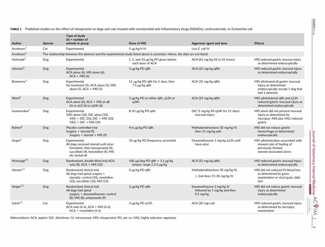
TABLE 1 Published studies on the effect of misoprostol on dogs and cats treated with nonsteroidal anti-inflammatory drugs (NSAIDs), corticosteroids, or Escherichia coli
Author Species
Type of study(n) = number ofanimals in group Dose of MIS Aggressor agent and dose Effects
Arvidsson1 Cat Experimental 5 μg/kg/h IV Live E. coli IV
Arvidsson2 The relationship between this abstract and the experimental study listed above is uncertain. Hence, the data are not listed.
Holroyde3 Dog Experimental 1, 5, and 10 μg/kg PO given before
each dose of ACA
ACA (65 mg/kg 4X in 24 hours) MIS reduced gastric mucosa injury
as determined endoscopically
Johnson4 Dog Experimental
ACA alone (6), MIS alone (6),
ACA + MIS (6)
3 μg/kg PO q8h ACA (35 mg/kg q8h) MIS reduced gastric mucosal injury
as determined endoscopically
Bowersox5 Dog Experimental
No treatment (5), ACA alone (5), MISalone (5), ACA + MIS (5)
15 μg/kg PO q8h for 5 days, then
7.5 μg/kg q8h
ACA (35 mg/kg q8h) MIS eliminated all gastric mucosal
injury as determinedendoscopically except 1 dog that
had 1 petechia
Ward6 Dog ExperimentalACA alone (6), ACA + MIS at q8
(6) or q12 (6) or q24h (6)
3 μg/kg PO at either q8h, q12h orq24h
ACA (25 mg/kg q8h) MIS administered q8h and q12hreduced gastric mucosal injury as
determined endoscopically
Guannoukas7 Dog Experimental
VAG alone (10), DIC alone (10),
VAG + DIC (10), DIC + MIS (10),
VAG + DIC + MIS (10)
8-20 μg/kg PO q6h DIC (1 mg/kg IM q24h for 12 days)
mucosal injury
MIS alone did not prevent mucosal
injury as determined by
necropsy; MIS plus VAG reduced
gastric
Rohrer8 Dog Placebo controlled trial
Surgery + steroid (9),Surgery + steroid + MIS (9)
4-6 μg/kg PO q8h Methylprednisolone 30 mg/kg IV,
then 15 mg/kg q6h
MIS did not reduce gastric
hemorrhage as determinedendoscopically
Dogra9 Dog ExperimentalAll dogs received steroid until ulcer
formation, then lansoprazole (4),
sucralfate (4), famotidine (4), MIS
(4), herbal (4)
10 μg/kg PO (frequency uncertain) Dexamethasone 1 mg/kg q12h untilhave ulcer
MIS administration associated withslowest rate of healing of
previously formed
steroid-associated ulcers
Murtaugh10 Dog Randomized, double-blind trial ACA
only (8), ACA + MIS (10)
100 μg/dog PO q8h = 3.1 μg/kg
(mean), range 2.3-5 μg/kg
ACA (25 mg/kg q8h) MIS reduced gastric mucosal injury
as determined endoscopically
Hansen11 Dog Randomized clinical trial
All dogs had spinal surgery +
steroids: control (10), cimetidine(10), sucralfate (10), MIS (10)
4 μg/kg PO q8h Methylprednisolone 30 mg/kg IV,
� 2nd dose 15-30 mg/kg IV
MIS did not reduced GI blood loss
as determined by gross
examination or stool guiac slidetest
Neiger12 Dog Randomized clinical trialAll dogs had spinal
surgery + dexamethasone: control
(8), MIS (8), omeprazole (9)
2 μg/kg PO q8h Dexamethasone 2 mg/kg IVfollowed by 1 mg/kg and then
0.5 mg/kg
MIS did not reduce gastric mucosalinjury as determined
endoscopically
Satoh13 Cat Experimental
ACA only (4-6), ACA + MIS (4-6),
ACA + cimetidine (4-6)
3 μg/kg PO q12h ACA (20 mg/cat) MIS reduced gastric mucosal injury
as determined by necropsy
examination
Abbreviations: ACA, aspirin; DIC, diclofenac; IV, intravenous; MIS, misoprostol; PO, per os; VAG, highly selective vagotomy.
MARKS
ETAL.
7
Sucralfate also may provide a barrier for bile salts. Sucralfate is known
to stimulate prostaglandin production in the gastric epithelium. This
may be a potential secondary effect of sucralfate in the esophagus,
although the importance and effectiveness of sucralfate as an agent
for the treatment of erosive esophagitis is not as established as it has
been for H2RAs or PPIs.
In rabbits, esophagitis induced by acid and pepsin was prevented
by administration of sucralfate.118 In another study, cats pretreated
with liquid sucralfate before acid infusion were protected against
esophagitis.119 Studies in humans have compared sucralfate to other
forms of treatment including alginic acid/antacid, cimetidine, and
ranitidine. Sucralfate was as effective as Gaviscon containing sodium
alginate with regard to healing of esophagitis and symptomatic
improvement.120,121 The H2RAs, ranitidine and cimetidine, and
sucralfate had equal efficacy for treating reflux esophagitis,121–125
although higher grade esophagitis did not heal as well compared to
lower grade esophagitis. In foals, sucralfate had a protective effect
on oral, esophageal, and gastric ulcers associated with IV administra-
tion of high-dose phenylbutazone.126 When sucralfate is compared
to placebo, conflicting data regarding therapeutic benefit have been
obtained in human patients with reflux esophagitis.127 Limited
esophageal retention time may decrease effectiveness. In a study of
technetium-labeled sucralfate, the drug was retained within the
esophagus for 3 hours in <50% of the patients with reflux esophagi-
tis.128 In addition, sucralfate deposited in a nonacidified esophagus
was rapidly cleared and poorly timed to provide protection against
reflux injury.129 Sucralfate decreased the frequency of stricture for-
mation in human patients with advanced corrosive esophagitis,130
and topical sucralfate was effective for post-tonsillectomy analgesia
in people.131 No controlled studies have been completed to assess
the analgesic effects of sucralfate in people or animals with severe
esophagitis, but anecdotal evidence indicates the drug's analgesic
properties in people with esophagitis. In addition, in controlled stud-
ies, no significant benefit of treatment was observed involving a
combination of sucralfate and H2RA, compared to either drug alone
in treating acute duodenal ulcer, in ulcer maintenance treatment, in
stress bleeding, or in reflux esophagitis.
4.5.4 | Adverse effects
Sucralfate is a relatively safe compound and has minimal adverse
effects. Aluminum absorption during sucralfate treatment is compa-
rable to that during treatment with aluminum hydroxide, and caution
should be exercised with long-term treatment in patients with renal
insufficiency to avoid aluminum intoxication.132,133 Constipation,
caused by aluminum hydroxide, is one of the most common adverse
effects, and typically occurs in 1%-3% of human patients taking the
drug.134 Other adverse effects in humans include xerostomia, nau-
sea, vomiting, headache, urticaria, and rashes in 0-5% of
patients.134,135
4.5.5 | Drug interactions with sucralfate
Coadministration of the following drugs with sucralfate results in a
substantially decreased bioavailability of single doses of the
drug: ciprofloxacin,136–138 theophylline,139 tetracycline, doxycycline,
minocycline,140,141 phenytoin,142 and digoxin.143 The bioavailability
of digoxin, tetracycline, doxycycline, and phenytoin was not
decreased when they were given 2 hours before sucralfate. Sucral-
fate impairs absorption of ciprofloxacin in humans and dogs when
administered concurrently,136,144 but the bioavailability of ciproflox-
acin is markedly increased when administration of sucralfate is
delayed by 2 hours. Interestingly, no significant difference in bio-
availability was documented for enrofloxacin coadministered with
sucralfate in dogs.136,144
In contrast to sucralfate suspension, administration of sucralfate
tablets had no effect on the absorption of doxycycline in dogs.140 This
lack of interaction with sucralfate tablets suggests sucralfate tablets
do not adequately disintegrate in dogs and should be administered as
a suspension rather than an intact tablet.
Consensus opinion on the effectiveness of sucralfate for
managing esophagitis or gastroduodenal ulceration
There is weak evidence in experimental animals and humans to support
the use of sucralfate for preventing or treating esophageal injury. There is
moderate evidence that sucralfate may have analgesic effects in people
post-tonsillectomy, but no studies have evaluated the analgesic properties
of sucralfate in people or animals with esophagitis. No evidence supports
either a benefit or interaction when sucralfate is administered concur-
rently with H2RAs or PPIs. When administered to dogs (and perhaps cats),
intact tablets may not fully disintegrate and may not be as effective as a
liquid suspension. No evidence indicates that combining sucralfate with
either a PPI or an H2RA for treatment of GUE is beneficial or indicated.
Proton pump inhibitors are superior to sucralfate for management
of GUE.
5 | INDICATIONS AND GUIDELINES FOR
GASTROPROTECTANTS IN HUMANS
In a meta-analysis involving >14 000 patients, healing of gastric and
duodenal ulcers and erosive esophagitis were directly related to the
extent and duration of gastric acid suppression over a 24-hour
period.70,71 Healing of esophagitis was significantly correlated with
maintaining gastric pH ≥ 4.0 for at least 16 hours per day, whereas
treatment of duodenal ulcers was optimized by maintaining gastric
pH ≥ 3 for 18-20 hours per day.71 Control of nocturnal acidity
(as opposed to 24-hour acidity) was directly proportional to healing of
duodenal ulcers.145 In a meta-analysis involving 56 published clinical
trials in people, healing of benign gastric ulcers was most strongly cor-
related with the duration of treatment, unlike with duodenal ulcers.146
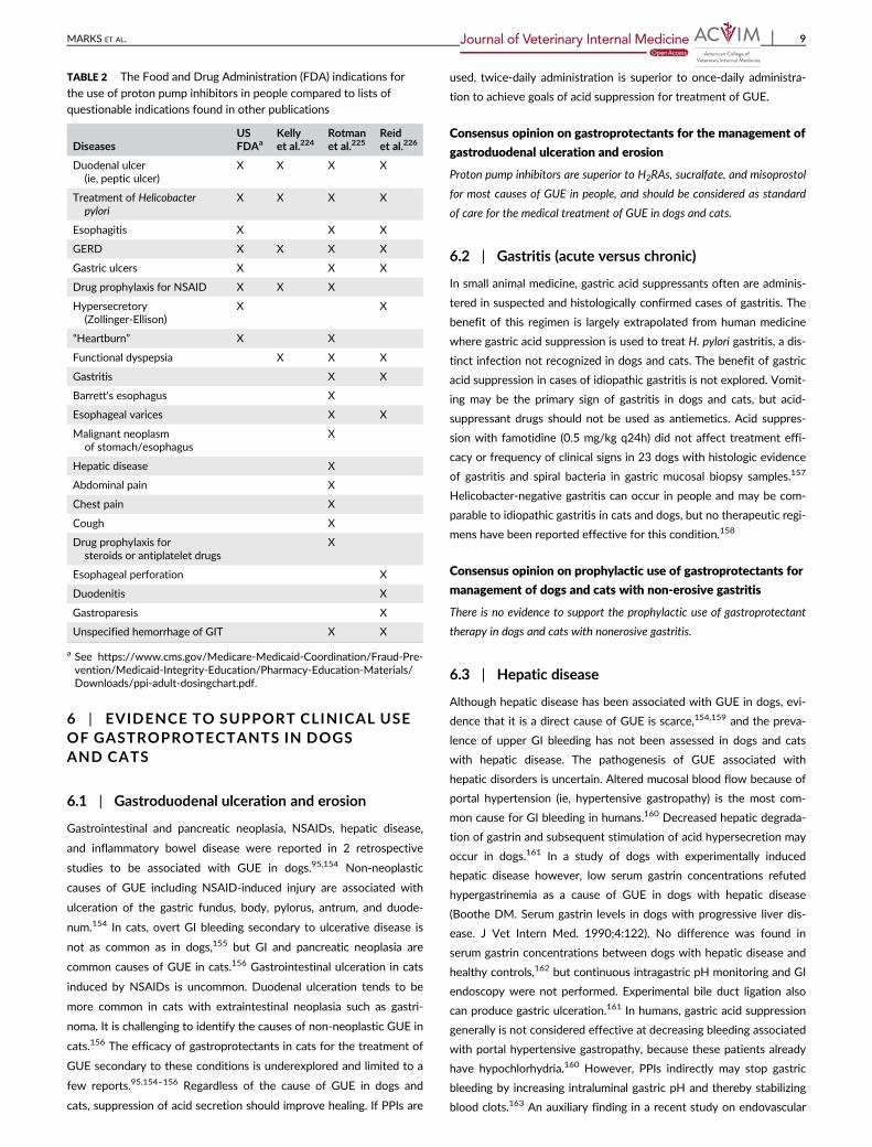
The FDA has listed indications for the use of PPIs in adult human
patients (Table 2), and treatment of Helicobacter pylori peptic ulcers147
and GERD148 are universally considered indications for PPIs. Treat-
ment of erosive esophagitis,149 benign gastric ulcers,150 dyspepsia,151
hypersecretory states (eg, Zollinger-Ellison syndrome),152 and prophy-
laxis for NSAID-associated ulcers153 also are listed as indications for
PPI treatment.
8 MARKS ET AL.
6 | EVIDENCE TO SUPPORT CLINICAL USE
OF GASTROPROTECTANTS IN DOGS
AND CATS
6.1 | Gastroduodenal ulceration and erosion
Gastrointestinal and pancreatic neoplasia, NSAIDs, hepatic disease,
and inflammatory bowel disease were reported in 2 retrospective
studies to be associated with GUE in dogs.95,154 Non-neoplastic
causes of GUE including NSAID-induced injury are associated with
ulceration of the gastric fundus, body, pylorus, antrum, and duode-
num.154 In cats, overt GI bleeding secondary to ulcerative disease is
not as common as in dogs,155 but GI and pancreatic neoplasia are
common causes of GUE in cats.156 Gastrointestinal ulceration in cats
induced by NSAIDs is uncommon. Duodenal ulceration tends to be
more common in cats with extraintestinal neoplasia such as gastri-
noma. It is challenging to identify the causes of non-neoplastic GUE in
cats.156 The efficacy of gastroprotectants in cats for the treatment of
GUE secondary to these conditions is underexplored and limited to a
few reports.95,154–156 Regardless of the cause of GUE in dogs and
cats, suppression of acid secretion should improve healing. If PPIs are
used, twice-daily administration is superior to once-daily administra-
tion to achieve goals of acid suppression for treatment of GUE.
Consensus opinion on gastroprotectants for the management of
gastroduodenal ulceration and erosion
Proton pump inhibitors are superior to H2RAs, sucralfate, and misoprostol
for most causes of GUE in people, and should be considered as standard
of care for the medical treatment of GUE in dogs and cats.
6.2 | Gastritis (acute versus chronic)
In small animal medicine, gastric acid suppressants often are adminis-
tered in suspected and histologically confirmed cases of gastritis. The
benefit of this regimen is largely extrapolated from human medicine
where gastric acid suppression is used to treat H. pylori gastritis, a dis-
tinct infection not recognized in dogs and cats. The benefit of gastric
acid suppression in cases of idiopathic gastritis is not explored. Vomit-
ing may be the primary sign of gastritis in dogs and cats, but acid-
suppressant drugs should not be used as antiemetics. Acid suppres-
sion with famotidine (0.5 mg/kg q24h) did not affect treatment effi-
cacy or frequency of clinical signs in 23 dogs with histologic evidence
of gastritis and spiral bacteria in gastric mucosal biopsy samples.157
Helicobacter-negative gastritis can occur in people and may be com-
parable to idiopathic gastritis in cats and dogs, but no therapeutic regi-
mens have been reported effective for this condition.158
Consensus opinion on prophylactic use of gastroprotectants for
management of dogs and cats with non-erosive gastritis
There is no evidence to support the prophylactic use of gastroprotectant
therapy in dogs and cats with nonerosive gastritis.
6.3 | Hepatic disease
Although hepatic disease has been associated with GUE in dogs, evi-
dence that it is a direct cause of GUE is scarce,154,159 and the preva-
lence of upper GI bleeding has not been assessed in dogs and cats
with hepatic disease. The pathogenesis of GUE associated with
hepatic disorders is uncertain. Altered mucosal blood flow because of
portal hypertension (ie, hypertensive gastropathy) is the most com-
mon cause for GI bleeding in humans.160 Decreased hepatic degrada-
tion of gastrin and subsequent stimulation of acid hypersecretion may
occur in dogs.161 In a study of dogs with experimentally induced
hepatic disease however, low serum gastrin concentrations refuted
hypergastrinemia as a cause of GUE in dogs with hepatic disease
(Boothe DM. Serum gastrin levels in dogs with progressive liver dis-
ease. J Vet Intern Med. 1990;4:122). No difference was found in
serum gastrin concentrations between dogs with hepatic disease and
healthy controls,162 but continuous intragastric pH monitoring and GI
endoscopy were not performed. Experimental bile duct ligation also
can produce gastric ulceration.161 In humans, gastric acid suppression
generally is not considered effective at decreasing bleeding associated
with portal hypertensive gastropathy, because these patients already
have hypochlorhydria.160 However, PPIs indirectly may stop gastric
bleeding by increasing intraluminal gastric pH and thereby stabilizing
blood clots.163 An auxiliary finding in a recent study on endovascular
TABLE 2 The Food and Drug Administration (FDA) indications for
the use of proton pump inhibitors in people compared to lists of
questionable indications found in other publications
DiseasesUSFDAa
Kellyet al.224
Rotmanet al.225
Reidet al.226
Duodenal ulcer
(ie, peptic ulcer)
X X X X
Treatment of Helicobacter
pylori
X X X X
Esophagitis X X X
GERD X X X X
Gastric ulcers X X X
Drug prophylaxis for NSAID X X X
Hypersecretory
(Zollinger-Ellison)
X X
“Heartburn” X X
Functional dyspepsia X X X
Gastritis X X
Barrett's esophagus X
Esophageal varices X X
Malignant neoplasmof stomach/esophagus
X
Hepatic disease X
Abdominal pain X
Chest pain X
Cough X
Drug prophylaxis for
steroids or antiplatelet drugs
X
Esophageal perforation X
Duodenitis X
Gastroparesis X
Unspecified hemorrhage of GIT X X
a See https://www.cms.gov/Medicare-Medicaid-Coordination/Fraud-Pre-
vention/Medicaid-Integrity-Education/Pharmacy-Education-Materials/Downloads/ppi-adult-dosingchart.pdf.
MARKS ET AL. 9
treatment of intrahepatic shunts in dogs was a substantial decrease in
deaths attributed to GI hemorrhage or ulceration after implementation
of peri- and postsurgical lifelong administration of PPIs.164 However,
the mechanism for GI bleeding in these dogs is uncertain and may not
be related to hepatic dysfunction or portal hypertension.
Consensus opinion on prophylactic use of acid suppressants in
dogs and cats with hepatic disease
There is weak evidence to support the prophylactic use of acid suppres-
sant therapy in dogs and cats with hepatic disease that is not associated
with GI bleeding.
6.4 | Stress-related mucosal damage (SRMD)
The benefits of stress ulcer prevention in intensive care unit (ICU)
patients currently are unresolved. Administration of H2RAs to people
receiving enteral nutrition was associated with an increased risk of
hospital acquired pneumonia and mortality.165 In a meta-analysis of
14 randomized, controlled parallel group trials involving 1720 people
in an ICU, PPIs were more effective than H2RAs in preventing clini-
cally important upper GI bleeding, but their use did not decrease mor-
tality in the hospital or the duration of ICU stay.166
Stress-related mucosal damage occurs in some critically ill dogs and
cats, but its prevalence, severity, and the efficacy of prophylactic gastro-
protection is unknown.167 In a retrospective study evaluating SRMD in
critically ill dogs from 3 ICUs in the United Kingdom, hemorrhagic gastric
disease was significantly associated with mortality. However, gastropro-
tectant drugs did not improve survival in this population of dogs.168
Although no evidence supports the routine use of H2RAs or PPIs
for prevention of SRMD in critically ill dogs and cats, they may prevent
GUE in performance animals. Most of the research on SRMD in dogs
has been performed on racing Alaskan sled dogs. Strenuous exercise in
increments of 100 miles/d for 5 days was associated with development
of SRMD.169 Gastric erosions, ulcers, and hemorrhage were observed
in 48% of 70 dogs after completing the Iditarod sled dog race.97 The
study was limited by not examining the dogs before and after the race,
precluding confirmation of when the lesions might have been present.
Omeprazole (0.85 mg/kg PO q24h) was significantly superior to high-
dose famotidine (1.7 mg/kg q12h) in decreasing SRMD in a randomized
positive control study of 52 dogs before and after a 300-mile race.44
Stress-related mucosal disease also has been documented in Labrador
retrievers undergoing explosive detection training in North Carolina
during the summer months.170 In an experimental study on 20 horses,
gastric ulceration was associated with decreases in physiologic indices
of performance.171 Therefore, in highly competitive events (eg, sled
dog racing) or working dogs in adverse environments (eg, military work-
ing dogs), there may be benefits to PPI administration.
Consensus opinion on the use of gastroprotectants in critically
ill dogs and cats
There is no compelling evidence that gastroprotectant therapy is benefi-
cial or indicated in critically ill human and animal patients unless definite
risk factors such as GI hemorrhage or concurrent NSAID administration
are present. Prophylactic PPI administration to animals competing in
strenuous, competitive events might decrease SRMD and improve overall
performance.
6.5 | Renal disease
Gastric acid secretion is variably affected by renal dysfunction in
humans and may partially depend on H. pylori infection status.172
However, gastritis and GUE can be complications of end-stage renal
disease in human patients.173,174 Acid suppression in people is often
recommended for renal disease patients with ulcer bleeding.175 There
is no recommendation for the use of prophylactic acid suppressant
treatment in human patients with renal disease, but acid suppressants
generally are recommended if other risk factors (eg, NSAID or cortico-
steroid treatment) for ulcer development are present. Dose adjust-
ments of H2RAs based on projected glomerular filtration rate are
recommended because of the renal elimination of these drugs.176
Gastroduodenal ulceration and erosion is not a typical finding in
dogs and cats with advanced renal disease.177–180 Moreover, in a
recent study of 10 cats with chronic renal disease and 9 healthy age-
matched control cats, no significant differences were observed in
serum gastrin concentrations and gastric pH between groups, suggest-
ing that cats with CKD may not have gastric hyperacidity compared to
healthy cats, and therefore, may not need acid suppression.181 How-
ever, despite this evidence, acid suppressants are commonly pre-
scribed to dogs and cats with CKD.182 Chronic administration of acid
suppressants to dogs and cats with CKD may not be benign. Pro-
longed administration of acid suppressants has been associated with
derangements in serum calcium and PTH concentrations, osteoporo-
sis, and pathologic fractures in at-risk human populations.183
Approximately 36%-80% of cats with moderate to severe CKD
have renal secondary hyperparathyroidism,184,185 with possible conse-
quences of decreased bone mineral density and increased bone
resorption cavities.186 Thus, the deleterious effects of chronic acid
suppressant administration on calcium metabolism and bone remodel-
ing in dogs and cats with CKD could lead to serious sequelae. Positive
fecal occult blood tests have been documented in dogs with CKD,187
but the mechanism of GI bleeding and benefit of acid suppressant
treatment have not been investigated. Until such studies are pub-
lished, acid suppression should be restricted to dogs and cats with
renal disease that have additional risk factors for ulceration or when
concern for severe GI bleeding (eg, melena, severe iron deficiency
anemia) or vomiting-induced esophagitis exists.
Consensus opinion on prophylactic use of gastroprotectant
treatment in dogs and cats with renal disease
There is no evidence to support the prophylactic use of gastroprotectants
in dogs and cats with International Renal Interest Society (IRIS) stages
1-3 renal disease. Additional studies are warranted to determine the ben-
efits of acid suppression in animals with IRIS stage 4 renal disease.
6.6 | Pancreatitis
Gastric acid suppressants frequently are used in the management of
acute pancreatitis in dogs and cats. The rationale for acid suppression
is the perceived increased risk of upper GI bleeding with pancreatitis
10 MARKS ET AL.

secondary to hypovolemia and local peritonitis, but the incidence of
upper GI bleeding in dogs and cats with pancreatitis currently is
unknown. Similarly, severe illness, hypoxemia, and use of NSAIDs for
pain relief, together with GI hypoperfusion, have been proposed as
potential causes of GI mucosal barrier failure, and contribute to acute
mucosal lesions or ulcers in people with severe acute pancreatitis.188 In
some reports, PPIs are anti-inflammatory and decrease pancreatic secre-
tions, whereas in others there is no effect or a pro-inflammatory
effect.189–191 Pantoprazole possesses reactivity toward hydroxyl radicals
and ameliorates inflammation in rodent models of pancreatitis,191 but a
recent placebo-controlled study failed to demonstrate a benefit of pan-
toprazole administration in human patients with acute pancreatitis.192
Consensus opinion on use of acid suppressants in dogs and cats
with pancreatitis
There is no evidence that acid suppression treatment is beneficial or indi-
cated in the management of dogs or cats with pancreatitis, unless the ani-
mal has concurrent evidence of GUE.
6.7 | Reflux esophagitis
Gastroesophageal reflux during anesthesia is associated with 46%-
65% of cases of benign esophageal stricture in dogs and represents
the most common cause of high-grade esophagitis and stricture for-
mation in dogs.193,194 Relaxation of the lower esophageal sphincter
(LES) is mediated by nonadrenergic noncholinergic pathways195 and
has been shown to occur with the administration of injectable prea-
nesthetic and inhalant anesthetic agents.196–198 The LES may be ren-
dered incompetent by a sliding hiatal hernia, which often is
accompanied by GER and can be exacerbated by increased inspiratory
effort typical of brachycephalic breeds.199
Esophagitis results from abnormal exposure to activated pepsin-
containing acid gastric contents because of distortion of the physio-
logical function of the LES. The prolonged exposure of the esophageal
mucosa to acid is an important cause of esophagitis and potential
stricture formation, particularly when pH is <4.0 because the proteo-
lytic pH range for the conversion of pepsinogen to pepsin is between
1.5 and 3.5.200,201 Preanesthetic administration of IV esomeprazole at
12-18 hours and 1-1.5 hours before anesthetic induction to 22 dogs
undergoing elective orthopedic procedures was associated with a sig-
nificant increase in gastric and esophageal pH throughout the surgery
procedure compared to a placebo group, but did not have an impact
on the number of reflux events.69 Similarly, preanesthetic administra-
tion of 2 PO doses of omeprazole in cats at 18-24 hours and 4 hours
before anesthetic induction, respectively, was associated with signifi-
cant increases in gastric and esophageal pH within 24 hours.202 Other
preventive measures for decreasing reflux esophagitis in dogs under-
going surgery are administration of cisapride or metoclopramide, with
cisapride being more effective.203–205
The superiority of PPIs in healing erosive esophagitis and decreas-
ing rate of relapse compared to that of H2RAs has been well established
in people.46,206,207 In addition, treatment with PPIs provided quicker and
more complete relief from heartburn clinical signs (11.5% per week)
compared with H2RAs (6.4% per week). Proton pump inhibitors were
found to provide greater clinical sign relief in patients with erosive reflux
disease (70%-80%) as compared to those with nonerosive reflux disease
(50%-60%).208 Additional studies are warranted to determine the bene-
fits of preanesthetic administration of H2RAs in dogs and cats in which
prolonged maintenance of esophageal pH > 4.0 is not necessary.
Consensus opinion on the use of acid suppressants for
prevention of reflux esophagitis
There is a lack of empirical evidence in dogs and cats, but compelling evi-
dence from studies in people, that acid-suppressing agents may be benefi-
cial for prevention of esophagitis secondary to GER, particularly in
animals when it is associated with an anesthetic procedure. Administra-
tion of PPIs does not decrease gastric reflux, but may prevent injury by
increasing the pH of the refluxate.
6.8 | Helicobacter
Treatment of H. pylori in people currently consists of multiple drugs,
either simultaneously or sequentially, and PPIs are almost always an
integral component of treatment.209,210 Infected dogs and cats almost
always have non-H. pylori Helicobacter (NHPH) that appears to have
different pathophysiologic effects and different responses to treatment
compared to H. pylori. The importance of triple or quadruple treatment
for effective management of H. pylori in people might not translate to
the same recommendation for dogs and cats with NHPH. Ten studies
report treatment of spontaneous NHPH in dogs and cats (Table 3). The
variety of therapies employed, the relatively small number of animals
studied, and the different means and times by which elimination of Heli-
cobacter were determined make it impossible to draw meaningful con-
clusions about the value of acid suppression treatment when treating
dogs or cats with NHPH. A single RCT compared antibiotics with and
without acid suppression and showed no benefit from adding famoti-
dine, but the study was relatively underpowered.157 Because PPIs are
superior to H2RAs in decreasing gastric acidity, a similar study with PPIs
might have different results. However, based upon currently available
information, acid suppression treatment with H2RAs is not indicated for
first-line treatment when treating NHPH in dogs or cats.
Consensus opinion on the use of acid suppression treatment for
the management of NHPH
There is no evidence that acid suppression treatment is beneficial or indi-
cated in dogs or cats undergoing treatment for NHPH.
6.9 | Thrombocytopenia-induced bleeding
In experimental studies, platelet aggregation at an ulcer is essentially
normal at a gastric pH 7.4, but progressively diminishes until absent at
pH ≤ 6.2.211 Increasing gastric pH may limit degradation of previously
formed platelet plugs and decrease proteolytic activity of pepsin on
existing thrombi. Proton pump inhibitors in combination with other
therapies have been successful for treatment of gastric bleeding in
human patients with myelodysplasia and thrombocytopenia or idio-
pathic thrombocytopenia purpura.212,213 Despite this phenomenon,
acid suppressants are not routinely administered in humans
with immune-mediated thrombocytopenia (ITP). Gastrointestinal hem-
orrhage is rarely observed in human patients with ITP.214 In contrast,
MARKS ET AL. 11

GI bleeding is relatively common in dogs with ITP.215 Platelet aggrega-
tion and clot formation in vitro are optimal at pH > 6.8.216,217 For this
reason, acid suppressants are commonly used as adjunctive treatment
of dogs with ITP-related GI bleeding. However, in a recent study, acid
suppressants did not influence the probability of survival to dis-
charge.176 In studies of healthy dogs and cats, twice-daily administra-
tion of acid suppressants did not achieve the target of pH > 6 for a
prolonged period.178–180,182 More studies are warranted to determine
the efficacy and optimal dosage of acid suppressants for the treat-
ment of thrombocytopenic-induced GI bleeding in dogs and cats.
Consensus opinion on use of acid suppression for the
prevention or management of thrombocytopenia-induced
bleeding
There is insufficient evidence to support the use of standard dosages of
acid suppressant treatment for prevention or management of
thrombocytopenia-induced bleeding.
6.10 | Spinal cord injury and intervertebral disc
surgery
Spinal cord injury and intervertebral disc surgery in dogs have been
associated with GI complications in dogs. Information for cats is not
available to make any conclusions.
A consistent finding in the published reports of GI complications
in dogs with spinal cord disease and spinal surgery is that high doses
of corticosteroids were administered (eg, 30 mg/kg methylpredniso-
lone, which is equivalent to 3.75 mg/kg dexamethasone). In these
reports, there were high rates of GI complications, including diarrhea,
melena, bleeding, and perforation.111,218–221 The same findings have
been reported in research dogs that had spinal surgery in combination
with high-dose corticosteroids.222
These reports concluded that the high doses of corticosteroids
contributed to GI complications rather than disc disease or surgery
alone. High doses of corticosteroids administered to otherwise
healthy dogs can produce gastric hemorrhage.223 Injury from gluco-
corticoids is not caused by hypersecretion of acid, but is more likely
TABLE 3 Results of treatment of Helicobacter spp. in dogs and cats
Author SpeciesType of study/design (n) = # animals/group
Antibacterialsused (days)
Acidsuppressionused Results
Means of evaluatingefficacy (time of testingpost treatment)
Kubota1 Dogs Uncontrolled experiment AMO, MET (14) OME 6/6 cleared HIST, RUT, PCR (uncertain)
All dogs treated (6)
Simpson2 Dogs Controlled experiment AMO, MET (14) FAM 6/8 cleared
at 4 days
HIST, RUT, IMP (4 days)
Treated (8), untreated (6) 0/8 cleared
at 29 days
HIST, RUT, IMP (29 days)
Mirzaeian3 Dogs Uncontrolled experiment AMO, CLA (7) LAN 20/20 cleared HIST, RUT (uncertain)
All dogs treated (20)
Costa4 Dogs Controlled experiment AMO, MET (15) OME 7/7 cleared HIST (uncertain)
No tx (7), antibiotics (7)
garlic oil (7)
Happonen5 Dogs Uncontrolled clinical trial (9) AMO, MET
(10-14) + BIS
None 7/9 cleared HIST (within 84 days)
Repeated tx for initial failures TET (10) OME 1/1 cleared HIST (within 84 days)
Jergens6 Dogs Uncontrolled clinical trial (3) AMO, MET, BIS (21) None 3/3 cleared HIST, FISH, PCR(4-14 weeks)
Cats Uncontrolled clinical trial (2) AMO, MET, BIS (21) None 2/2 cleared HIST, FISH, PCR(4-14 weeks)
Leib7 Dogs Randomized clinical trial AMO, MET, BIS (14)
Tx with antibiotics (10) None 7/10 cleared HIST (4 weeks)
Tx with antibiotics +
famotidine (14)
FAM 11/14 cleared HIST (4 weeks)
Khoshnegah8 Cats Uncontrolled experiment AMO, MET, CLA (14) OME 13/13 cleared HIST (uncertain)
All cats treated (13)
Perkins9 Cats Controlled experiment AMO, MET (21) OME 6/6 cleared CUL of mucosa/
fluid (2 weeks)
Treated (6), untreated (2) 1/6 cleared PCR (2 weeks)
Neiger10 Cats Controlled clinical trial
Untreated (4),Antibiotics-1 (6)
AZI, BIS, TIN (4) 4/6 cleared Urea breath test (10 days)
Antibiotics-2 (11) CLA, MET, BIS (4) RAN 11/11 cleared
Antibiotics-3 (8) CLA, MET, BIS (7) RAN 8/8 cleared
Abbreviations: AMO, amoxicillin; AZI, azithromycin; BIS, bismuth subcitrate or subsalicylate; CLA, clarithromycin; CUL, culture; FAM, famotidine; FISH,
fluorescent in situ hybridization; HIST, histology of gastric mucosa; IMP, impression smears of gastric mucosa; LAN, lansoprazole; MET, metronidazole;OME, omeprazole; PCR, polymerase chain reaction testing; RAN, ranitidine; RUT, rapid urea testing of gastric mucosa; TET, tetracycline; TIN, tinidazole.
12 MARKS ET AL.

the result of decreased defense mechanisms and altered gastric muco-
sal blood flow. Sympathetic-parasympathetic imbalance in spinal sur-
gery patients may contribute to the GI problems.109
No evidence from clinical reports of dogs or from research studies
shows that gastroprotectant drugs are beneficial for preventing or
decreasing GI complications from high doses of corticosteroids. The
gastroprotectants used in these reports were sucralfate, misoprostol,
and H2RAs. We acknowledge that some of these reports were from
the 1980s, before the use of PPIs, although a more recent study
showed no benefit in healing or preventing development of gastric
mucosal lesions in dogs using either misoprostol or omeprazole.109
Consensus opinion on gastroprotectants for prevention or
management of glucocorticoid-associated gastroduodenal
ulceration/erosion
Gastrointestinal complications associated with spinal cord disease and
spinal surgery are more likely caused by the administration of high doses
of glucocorticoids than other factors. In these cases, there is no convinc-
ing evidence that gastroprotectant drugs are beneficial.
7 | FUTURE CONSIDERATIONS
This consensus statement has highlighted the problems our panel
faced when attempting to provide recommendations for or against
the use of gastric protectants in dogs and cats. There is a need for the
use of selected GI protectants in some patients, but there is ample
evidence documenting overuse of these drugs for many disorders in
dogs and cats. Veterinarians need to better define the appropriate
clinical applications for the use of gastroprotectants in small animals.
Technological advances have become available with the use of GI
telemetric monitoring devices and capsule endoscopy that should
facilitate future research to more precisely identify the efficacy of
these drugs in dogs and cats. Well-defined endpoints and goals of
treatment should be established. The benefits of gastroprotectants
that we have defined in this article are largely based on extrapolation
from human medicine or from studies in healthy dogs and cats. More
information is needed to define optimal dosage, timing of doses, and
selection of products (eg, one PPI versus another), particularly in clini-
cal patients. Newer generation PPIs, such as delayed-release and
longer-acting PPIs, are commercially available and may improve dosing
and effectiveness in dogs and cats. We are cognizant that not a single
agent listed in this article is FDA-approved for use in dogs or cats.
Clearly, the pharmaceutical industry should become involved in evalu-
ating these drugs. Controlled studies evaluating the efficacy of gastro-
protectant drugs in clinical patients, as well as the judicious
application of these drugs, should further optimize the management
of reflux esophagitis and GUE in dogs and cats.
ACKNOWLEDGMENT
The authors thank Dr Frédéric Gaschen for his critical review and
feedback on their consensus statement.
CONFLICT OF INTEREST DECLARATION
Stanley L. Marks has consulted for Zoetis (Formerly Pfizer Animal
Health), Aratana Therapeutics, and Virbac; received research funding
from Aratana Therapeutics, Morris Animal Foundation, Winn Feline
Foundation; received speaking honoraria from Zoetis (Formerly Pfizer
Animal Health), Aratana Therapeutics, Virbac, Elanco, Nutramax Labo-
ratories. Michael D. Willard serves as Associate Editor for the Journal
of Veterinary Internal Medicine. He was not involved in review of this
manuscript. Mark G. Papich has consulted for Zoetis (Formerly Pfizer
Animal Health), Bayer Corporation, Dechra (formerly Putney), Merck
Animal Health, Elanco, Novartis, Pennfield (now Pharmgate),
e5pharma, and Kindred Biomed; authored veterinary drug books pub-
lished by Elsevier and Wiley; received research funding from Bayer
Corporation, Merck Animal Health, Zoetis (Formerly Pfizer Animal
Health), and Morris Animal Foundation; received speaking honoraria
from Zoetis (Formerly Pfizer Animal Health), Dechra (formerly Put-
ney), and Bayer Corporation. M. Katie Tolbert received research
grants from the Comparative Gastroenterology Society, Morris Animal
Foundation, Winn Feline Foundation, and The ACVIM Foundation to
study acid suppressants in cats.
OFF-LABEL ANTIMICROBIAL DECLARATION
Authors declare no off-label use of antimicrobials.
INSTITUTIONAL ANIMAL CARE AND USE COMMITTEE
(IACUC) OR OTHER APPROVAL DECLARATION
Authors declare no IACUC or other approval was needed.
ORCID
Stanley L. Marks https://orcid.org/0000-0001-7991-702X
Peter H. Kook https://orcid.org/0000-0002-9492-3484
Mark G. Papich https://orcid.org/0000-0002-7591-7898
M. Katherine Tolbert https://orcid.org/0000-0001-8725-9530
REFERENCES
1. Nehra AK, Alexander JA, Loftus CG, Nehra V. Proton pump inhibi-
tors: review of emerging concerns. Mayo Clin Proc. 2018;93:240-246.
2. Othman F, Card TR, Crooks CJ. Proton pump inhibitor prescribing
patterns in the UK: a primary care database study. Pharmacoepidemiol
Drug Saf. 2016;25:1079-1087.
3. Rakesh TP. Proton pump inhibitors: use, misuse and concerns about
long-term therapy. Clin J Gastroenterol. 2011;4:53-59.
4. Yadlapati R, Kahrilas PJ. When is proton pump inhibitor use appropri-
ate? BMC Med. 2017;15:36.
5. Gould E, Clements C, Reed A, et al. A prospective, placebo-controlled
pilot evaluation of the effect of omeprazole on serum calcium, mag-
nesium, cobalamin, gastrin concentrations, and bone in cats. J Vet
Intern Med. 2016;30:779-786.
6. Haastrup PF, Thompson W, Sondergaard J, et al. Side effects of
long-term proton pump inhibitor use: a review. Basic Clin Pharmacol
Toxicol. 2018;123:114-121.
7. Kook PH, Kempf J, Ruetten M, Reusch CE. Wireless ambulatory
esophageal pH monitoring in dogs with clinical signs interpreted as
gastroesophageal reflux. J Vet Intern Med. 2014;28:1716-1723.
MARKS ET AL. 13

8. Tolbert K, Bissett S, King A, et al. Efficacy of oral famotidine and
2 omeprazole formulations for the control of intragastric pH in dogs.
J Vet Intern Med. 2011;25:47-54.
9. Tolbert MK, Graham A, Odunayo A, et al. Repeated famotidine
administration results in a diminished effect on intragastric pH in
dogs. J Vet Intern Med. 2017;31:117-123.
10. Lane MB, Larson JC, Stokes JE, Tolbert MK. Continuous radiotele-
metric monitoring of intragastric pH in a dog with peptic ulceration. J
Am Vet Med Assoc. 2017;250:530-533.
11. Mahar KM, Portelli S, Coatney R, Chen EP. Gastric pH and gastric
residence time in fasted and fed conscious beagle dogs using the
Bravo pH system. J Pharm Sci. 2012;101:2439-2448.
12. Youngberg CA, Wlodyga J, Schmaltz S, Dressman JB. Radiotelemetric
determination of gastrointestinal pH in four healthy beagles.
Am J Vet Res. 1985;46:1516-1521.
13. Pandolfino JE, Richter JE, Ours T, Guardino JM, Chapman J, Kahrilas
PJ. Ambulatory esophageal pH monitoring using a wireless system.
Am J Gastroenterol. 2003;98:740-749.
14. Pandolfino JE, Zhang Q, Schreiner MA, Ghosh S, Roth MP, Kahrilas
PJ. Acid reflux event detection using the Bravo wireless versus the
Slimline catheter pH systems: why are the numbers so different?
Gut. 2005;54:1687-1692.
15. Hunt RH, Camilleri M, Crowe SE, et al. The stomach in health and
disease. Gut. 2015;64:1650-1668.
16. Waldum HL, Kleveland PM, Fossmark R. Upper gastrointestinal phys-
iology and diseases. Scand J Gastroenterol. 2015;50:649-656.
17. Betesh AL, Santa Ana CA, Cole JA, Fordtran JS. Is achlorhydria a
cause of iron deficiency anemia? Am J Clin Nutr. 2015;102:9-19.
18. Haffner-Luntzer M, Heilmann A, Heidler V,
et al. Hypochlorhydria-induced calcium malabsorption does not
affect fracture healing but increases post-traumatic bone loss in the
intact skeleton. J Orthop Res. 2016;34:1914-1921.
19. Chey WY, Chang TM. Neural control of the release and action of
secretin. J Physiol Pharmacol. 2003;54(Suppl 4):105-112.
20. Friis-Hansen L. Achlorhydria is associated with gastric microbial over-
growth and development of cancer: lessons learned from the gastrin
knockout mouse. Scand J Clin Lab Invest. 2006;66:607-621.
21. Dressman JB. Comparison of canine and human gastrointestinal
physiology. Pharm Res. 1986;3:123-131.
22. Kararli TT. Comparison of the gastrointestinal anatomy, physiology,
and biochemistry of humans and commonly used laboratory animals.
Biopharm Drug Dispos. 1995;16:351-380.
23. Emas S, Grossman MI. Comparison of gastric secretion in conscious
dogs and cats. Gastroenterology. 1967;52:29-34.
24. Sagawa K, Li F, Liese R, Sutton SC. Fed and fasted gastric pH and
gastric residence time in conscious beagle dogs. J Pharm Sci. 2009;
98:2494-2500.
25. Mojaverian P. Evaluation of gastrointestinal pH and gastric residence
time via the Heidelberg radiotelemetry capsule: pharmaceutical appli-
cation. Drug Dev Res. 1996;38:73-85.
26. Parkinson S, Tolbert K, Messenger K, et al. Evaluation of the effect of
orally administered acid suppressants on intragastric pH in cats. J Vet
Intern Med. 2015;29:104-112.
27. Sutalo S, Ruetten M, Hartnack S, et al. The effect of orally adminis-
tered ranitidine and once-daily or twice-daily orally administered
omeprazole on intragastric pH in cats. J Vet Intern Med. 2015;29:
840-846.
28. Fackler WK, Vaezi MF, Richter JE. Ambulatory gastric pH monitoring:
proper probe placement and normal values. Aliment Pharmacol Ther.
2001;15:1155-1162.
29. Maton PN, Burton ME. Antacids revisited: a review of their clinical
pharmacology and recommended therapeutic use. Drugs. 1999;57:
855-870.
30. Decktor DL, Robinson M, Gottlieb S. Comparative effects of liquid
antacids on esophageal and gastric pH in patients with heartburn.
Am J Ther. 1995;2:481-486.
31. Decktor DL, Robinson M, Maton PN, Lanza FL, Gottlieb S. Effects of
aluminum/magnesium hydroxide and calcium carbonate on esopha-
geal and gastric pH in subjects with heartburn. Am J Ther. 1995;2:
546-552.
32. Sweis R, Kaufman E, Anggiansah A, et al. Post-prandial reflux sup-
pression by a raft-forming alginate (Gaviscon Advance) compared to
a simple antacid documented by magnetic resonance imaging and
pH-impedance monitoring: mechanistic assessment in healthy volun-
teers and randomised, controlled, double-blind study in reflux
patients. Aliment Pharmacol Ther. 2013;37:1093-1102.
33. De Ruigh A, Roman S, Chen J, et al. Gaviscon double action liquid
(antacid & alginate) is more effective than antacid in controlling
post-prandial oesophageal acid exposure in GERD patients: a
double-blind crossover study. Aliment Pharmacol Ther. 2014;40:
531-537.
34. Segev G, Bandt C, Francey T, Cowgill LD. Aluminum toxicity follow-
ing administration of aluminum-based phosphate binders in 2 dogs
with renal failure. J Vet Intern Med. 2008;22:1432-1435.
35. Komazawa Y, Adachi K, Mihara T, et al. Tolerance to famotidine and
ranitidine treatment after 14 days of administration in healthy sub-
jects without Helicobacter pylori infection. J Gastroenterol Hepatol.
2003;18:678-682.
36. Lachman L, Howden CW. Twenty-four-hour intragastric pH: toler-
ance within 5 days of continuous ranitidine administration.
Am J Gastroenterol. 2000;95:57-61.
37. Tolbert MK, E Golly, A Odunayo, et al. Frequency of oral famotidine
administration determines the development of tolerance in cats. J
Vet Intern Med. 2018; In: 36th Annual Forum of the ACVIM. Seat-
tle, WA.
38. Labenz J, Peitz U, Leusing C, Tillenburg B, Blum AL, Borsch G.
Efficacy of primed infusions with high dose ranitidine and omepra-
zole to maintain high intragastric pH in patients with peptic ulcer
bleeding: a prospective randomised controlled study. Gut. 1997;40:
36-41.
39. Sandvik AK, Brenna E, Waldum HL. Review article: the pharmacologi-
cal inhibition of gastric acid secretion--tolerance and rebound. Ali-
ment Pharmacol Ther. 1997;11:1013-1018.
40. Smith JT, Gavey C, Nwokolo CU, et al. Tolerance during 8 days of
high-dose H2-blockade: placebo-controlled studies of 24-hour acid-
ity and gastrin. Aliment Pharmacol Ther. 1990;4(Suppl 1):47-63.
41. Bersenas AM, Mathews KA, Allen DG, et al. Effects of ranitidine,
famotidine, pantoprazole, and omeprazole on intragastric pH in dogs.
Am J Vet Res. 2005;66:425-431.
42. Diggs HE, Ogden BE, Haliburton JC. Vomiting and diarrhea in a cat
colony. Contemp Top Lab Anim Sci. 1995;34:75-76.
43. Williamson KK, Willard MD, McKenzie EC, et al. Efficacy of famoti-
dine for the prevention of exercise-induced gastritis in racing Alaskan
sled dogs. J Vet Intern Med. 2007;21:924-927.
44. Williamson KK, Willard MD, Payton ME, Davis MS. Efficacy of omep-
razole versus high-dose famotidine for prevention of
exercise-induced gastritis in racing Alaskan sled dogs. J Vet Intern
Med. 2010;24:285-288.
45. Barkun AN, Bardou M, Pham CQ, et al. Proton pump inhibitors
vs. histamine 2 receptor antagonists for stress-related mucosal
bleeding prophylaxis in critically ill patients: a meta-analysis.
Am J Gastroenterol. 2012;107:507-520. quiz 521.
46. Chiba N, De Gara CJ, Wilkinson JM, et al. Speed of healing and
symptom relief in grade II to IV gastroesophageal reflux disease: a
meta-analysis. Gastroenterology. 1997;112:1798-1810.
47. Fellenius E, Berglindh T, Sachs G, et al. Substituted benzimidazoles
inhibit gastric acid secretion by blocking (H+ + K+)ATPase. Nature.
1981;290:159-161.
48. Kromer W, Postius S, Riedel R. Animal pharmacology of reversible
antagonism of the gastric acid pump, compared to standard antise-
cretory principles. Pharmacology. 2000;60:179-187.
49. Abelo A, Holstein B, Eriksson UG, et al. Gastric acid secretion in the
dog: a mechanism-based pharmacodynamic model for histamine
stimulation and irreversible inhibition by omeprazole. J Pharmacoki-
net Pharmacodyn. 2002;29:365-382.
50. Larsson H, Carlsson E, Junggren U, et al. Inhibition of gastric acid
secretion by omeprazole in the dog and rat. Gastroenterology. 1983;
85:900-907.
51. Wallmark B, Larsson H, Humble L. The relationship between gastric
acid secretion and gastric H+,K+-ATPase activity. J Biol Chem. 1985;
260:13681-13684.
14 MARKS ET AL.
52. Andersson T, Rohss K, Bredberg E, et al. Pharmacokinetics and phar-
macodynamics of esomeprazole, the S-isomer of omeprazole. Aliment
Pharmacol Ther. 2001;15:1563-1569.
53. Prichard PJ, Yeomans ND, Mihaly GW, et al. Omeprazole: a study of
its inhibition of gastric pH and oral pharmacokinetics after morning
or evening dosage. Gastroenterology. 1985;88:64-69.
54. Mathew M, Gupta VD, Bailey RE. Stability of omeprazole solutions at
various pH values as determined by high-performance liquid chroma-
tography. Drug Dev Ind Pharm. 1995;21:965-971.
55. Merritt AM, Sanchez LC, Burrow JA, Church M, Ludzia S. Effect of
GastroGard and three compounded oral omeprazole preparations on
24 h intragastric pH in gastrically cannulated mature horses. Equine
Vet J. 2003;35:691-695.
56. Andersson T, Andren K, Cederberg C, Lagerstrom PO, Lundborg P,
Skanberg I. Pharmacokinetics and bioavailability of omeprazole after
single and repeated oral administration in healthy subjects. Br J Clin
Pharmacol. 1990;29:557-563.
57. Hatlebakk JG, Katz PO, Camacho-Lobato L, Castell DO. Proton pump
inhibitors: better acid suppression when taken before a meal than
without a meal. Aliment Pharmacol Ther. 2000;14:1267-1272.
58. De Graef J, Woussen-Colle MC. Influence of the stimulation state of
the parietal cells on the inhibitory effect of omeprazole on gastric
acid secretion in dogs. Gastroenterology. 1986;91:333-337.
59. Lee RD, Vakily M, Mulford D, et al. Clinical trial: the effect and timing
of food on the pharmacokinetics and pharmacodynamics of dexlan-
soprazole MR, a novel dual delayed release formulation of a proton
pump inhibitor--evidence for dosing flexibility. Aliment Pharmacol
Ther. 2009;29:824-833.
60. Furuta S, Kamada E, Suzuki T, et al. Inhibition of drug metabolism in
human liver microsomes by nizatidine, cimetidine and omeprazole.
Xenobiotica. 2001;31:1-10.
61. Gerson LB, Triadafilopoulos G. Proton pump inhibitors and their drug
interactions: an evidence-based approach. Eur J Gastroenterol Hepa-
tol. 2001;13:611-616.
62. Mullin JM, Gabello M, Murray LJ, et al. Proton pump inhibitors:
actions and reactions. Drug Discov Today. 2009;14:647-660.
63. Court MH. Canine cytochrome P-450 pharmacogenetics. Vet Clin
North Am Small Anim Pract. 2013;43:1027-1038.
64. Martinez MN, Antonovic L, Court M, et al. Challenges in exploring
the cytochrome P450 system as a source of variation in canine drug
pharmacokinetics. Drug Metab Rev. 2013;45:218-230.
65. Horn J. The proton-pump inhibitors: similarities and differences. Clin
Ther. 2000;22:266-280. discussion 265.
66. Miner P Jr, Katz PO, Chen Y, et al. Gastric acid control with esome-
prazole, lansoprazole, omeprazole, pantoprazole, and rabeprazole: a
five-way crossover study. Am J Gastroenterol. 2003;98:2616-2620.
67. Qi Q, Wang R, Liu L, Zhao F, Wang S. Comparative effectiveness and
tolerability of esomeprazole and omeprazole in gastro-esophageal
reflux disease: a systematic review and meta-analysis. Int J Clin Phar-
macol Ther. 2015;53:803-810.
68. Hwang JH, Jeong JW, Song GH, Koo TS, Seo KW. Pharmacokinetics
and acid suppressant efficacy of esomeprazole after intravenous,
oral, and subcutaneous administration to healthy beagle dogs. J Vet
Intern Med. 2017;31:743-750.
69. Zacuto AC, Marks SL, Osborn J, et al. The influence of esomeprazole
and cisapride on gastroesophageal reflux during anesthesia in dogs. J
Vet Intern Med. 2012;26:518-525.
70. Bell NJ, Burget D, Howden CW, et al. Appropriate acid suppression
for the management of gastro-oesophageal reflux disease. Digestion.
1992;51(Suppl 1):59-67.
71. Burget DW, Chiverton SG, Hunt RH. Is there an optimal degree of
acid suppression for healing of duodenal ulcers? A model of the rela-
tionship between ulcer healing and acid suppression. Gastroenterol-
ogy. 1990;99:345-351.
72. Jenkins CC, DeNovo RC, Patton CS, Bright RM, Rohrbach BW.
Comparison of effects of cimetidine and omeprazole on mechanically
created gastric ulceration and on aspirin-induced gastritis in dogs.
Am J Vet Res. 1991;52:658-661.
73. Tolbert MK, Odunayo A, Howell RS, Peters EE, Reed A. Efficacy of
intravenous administration of combined acid suppressants in healthy
dogs. J Vet Intern Med. 2015;29:556-560.
74. Zhou R, Moench P, Heran C, et al. pH-dependent dissolution in vitro
and absorption in vivo of weakly basic drugs: development of a
canine model. Pharm Res. 2005;22:188-192.
75. Gabardi S, Olyaei A. Evaluation of potential interactions between
mycophenolic acid derivatives and proton pump inhibitors. Ann Phar-
macother. 2012;46:1054-1064.
76. Laine L, Hennekens C. Proton pump inhibitor and clopidogrel interac-
tion: fact or fiction? Am J Gastroenterol. 2010;105:34-41.
77. Bonello L, Bonello N, Grosdidier C, Camoin-Jau L. Unveiling the mys-
teries of clopidogrel metabolism and efficacy. Clin Pharmacol Ther.
2011;90:774-776.
78. Mega JL, Close SL, Wiviott SD, et al. Cytochrome p-450 polymor-
phisms and response to clopidogrel. N Engl J Med. 2009;360:
354-362.
79. Thames BE, Lovvorn J, Papich MG, et al. The effects of clopidogrel
and omeprazole on platelet function in normal dogs. J Vet Pharmacol
Ther. 2017;40:130-139.
80. Lazarus B, Chen Y, Wilson FP, et al. Proton pump inhibitor use and
the risk of chronic kidney disease. JAMA Intern Med. 2016;176:
238-246.
81. Sierra F, Suarez M, Rey M, et al. Systematic review: proton pump
inhibitor-associated acute interstitial nephritis. Aliment Pharmacol
Ther. 2007;26:545-553.
82. Xie Y, Bowe B, Li T, Xian H, Balasubramanian S, al-Aly Z. Proton
pump inhibitors and risk of incident CKD and progression to ESRD. J
Am Soc Nephrol. 2016;27:3153-3163.
83. Gomm W, von Holt K, Thome F, et al. Association of proton pump
inhibitors with risk of dementia: a pharmacoepidemiological claims
data analysis. JAMA Neurol. 2016;73:410-416.
84. Lam JR, Schneider JL, Zhao W, Corley DA. Proton pump inhibitor
and histamine 2 receptor antagonist use and vitamin B12 deficiency.
JAMA. 2013;310:2435-2442.
85. Yang YX, Lewis JD, Epstein S, Metz DC. Long-term proton pump
inhibitor therapy and risk of hip fracture. JAMA. 2006;296:
2947-2953.
86. Lambert AA, Lam JO, Paik JJ, Ugarte-Gil C, Drummond MB, Crowell
TA. Risk of community-acquired pneumonia with outpatient proto-
n-pump inhibitor therapy: a systematic review and meta-analysis.
PLoS One. 2015;10:e0128004.
87. Cheungpasitporn W, Thongprayoon C, Kittanamongkolchai W,
et al. Proton pump inhibitors linked to hypomagnesemia: a system-
atic review and meta-analysis of observational studies. Ren Fail.
2015;37:1237-1241.
88. Freedberg DE, Lamouse-Smith ES, Lightdale JR, et al. Use of acid
suppression medication is associated with risk for C. difficile infection
in infants and children: a population-based study. Clin Infect Dis.
2015;61:912-917.
89. Freedberg DE, Toussaint NC, Chen SP, et al. Proton pump inhibitors
alter specific taxa in the human gastrointestinal microbiome: a cross-
over trial. Gastroenterology. 2015;149:883-885.e889.
90. Xu HB, Wang HD, Li CH, et al. Proton pump inhibitor use and risk of
spontaneous bacterial peritonitis in cirrhotic patients: a systematic
review and meta-analysis. Genet Mol Res. 2015;14:7490-7501.
91. Lamberts R, Creutzfeldt W, Struber HG, et al. Long-term omeprazole
therapy in peptic ulcer disease: gastrin, endocrine cell growth, and
gastritis. Gastroenterology. 1993;104:1356-1370.
92. Lo WK, Chan WW. Proton pump inhibitor use and the risk of small
intestinal bacterial overgrowth: a meta-analysis. Clin Gastroenterol
Hepatol. 2013;11:483-490.
93. Wallace JL, Syer S, Denou E, et al. Proton pump inhibitors exacerbate
NSAID-induced small intestinal injury by inducing dysbiosis. Gastro-
enterology. 2011;141:1314-1322. 1322.e1311-1315.
94. Marlicz W, Loniewski I, Grimes DS, et al. Nonsteroidal
anti-inflammatory drugs, proton pump inhibitors, and gastrointestinal
injury: contrasting interactions in the stomach and small intestine.
Mayo Clin Proc. 2014;89:1699-1709.
95. Cariou M, Lipscomb VJ, Brockman DJ, Gregory SP, Baines SJ.
Spontaneous gastroduodenal perforations in dogs: a retrospective
study of 15 cases. Vet Rec. 2009;165:436-441.
96. Kook PH, Reusch CE. Severe gastrointestinal bleeding secondary to
lornoxicam in the dog. Schweiz Arch Tierheilkd. 2011;153:223-229.
MARKS ET AL. 15

97. Davis MS, Willard MD, Nelson SL, et al. Efficacy of omeprazole for
the prevention of exercise-induced gastritis in racing Alaskan sled
dogs. J Vet Intern Med. 2003;17:163-166.
98. Schmid SM, Suchodolski JS, Price JM, Tolbert MK. Omeprazole mini-
mally alters the fecal microbial community in six cats: a pilot study.
Front Vet Sci. 2018;5:79.
99. Bauer RF. Misoprostol preclinical pharmacology. Dig Dis Sci. 1985;
30:118s-125s.
100. Robert A. An intestinal disease produced experimentally by a prosta-
glandin deficiency. Gastroenterology. 1975;69:1045-1047.
101. Fenn GC, Robinson GC. Misoprostol—a logical therapeutic approach
to gastroduodenal mucosal injury induced by non-steroidal
anti-inflammatory drugs? J Clin Pharm Ther. 1991;16:385-409.
102. Bowersox TS, Lipowitz AJ, Hardy RM, et al. The use of a synthetic
prostaglandin E1 analog as a gastric protectant against
aspirin-induced hemorrhage in the dog. J Am Anim Hosp Assoc. 1996;
32:401-407.
103. Johnston SA, Leib MS, Forrester SD, Marini M. The effect of miso-
prostol on aspirin-induced gastroduodenal lesions in dogs. J Vet
Intern Med. 1995;9:32-38.
104. Ward DM, Leib MS, Johnston SA, Marini M. The effect of dosing
interval on the efficacy of misoprostol in the prevention of
aspirin-induced gastric injury. J Vet Intern Med. 2003;17:282-290.
105. Murtaugh RJ, Matz ME, Labato MA, Boudrieau RJ. Use of synthetic
prostaglandin E1 (misoprostol) for prevention of aspirin-induced gas-
troduodenal ulceration in arthritic dogs. J Am Vet Med Assoc. 1993;
202:251-256.
106. Holroyde MJ, Pepple SC. An endoscopic study comparing gastropro-
tection by prostaglandin E2, misoprostol and cimetidine against mul-
tiple doses of aspirin in dogs. Gastroenterology. 1986;90:1463.
107. Giannoukas AD, Baltoyiannis G, Milonakis M, et al. Protection of the
gastroduodenal mucosa from the effects of diclofenac sodium: role
of highly selective vagotomy and misoprostol. World J Surg. 1996;20:
501-505. discussion 505-506.
108. Satoh H, Amagase K, Takeuchi K. The role of food for the formation
and prevention of gastrointestinal lesions induced by aspirin in cats.
Dig Dis Sci. 2013;58:2840-2849.
109. Neiger R, Gaschen F, Jaggy A. Gastric mucosal lesions in dogs with
acute intervertebral disc disease: characterization and effects of
omeprazole or misoprostol. J Vet Intern Med. 2000;14:33-36.
110. Rohrer CR, Hill RC, Fischer A, et al. Efficacy of misoprostol in preven-
tion of gastric hemorrhage in dogs treated with high doses of meth-
ylprednisolone sodium succinate. Am J Vet Res. 1999;60:982-985.
111. Hanson SM, Bostwick DR, Twedt DC, Smith MO. Clinical evaluation
of cimetidine, sucralfate, and misoprostol for prevention of gastroin-
testinal tract bleeding in dogs undergoing spinal surgery. Am J Vet
Res. 1997;58:1320-1323.
112. Arvidsson S, Fult K, Haglund U. Stress ulceration prevention in live
E. coli sepsis in cats. Gastroenterology. 1985;88:1310.
113. Rostom A, Muir K, Dube C, et al. Prevention of NSAID-related upper
gastrointestinal toxicity: a meta-analysis of traditional NSAIDs with
gastroprotection and COX-2 inhibitors. Drug Healthc Patient Saf.
2009;1:47-71.
114. Tang OS, Gemzell-Danielsson K, Ho PC. Misoprostol: pharmacoki-
netic profiles, effects on the uterus and side-effects. Int J Gynaecol
Obstet. 2007;99(Suppl 2):S160-S167.
115. Nagashima R. Mechanisms of action of sucralfate. J Clin Gastroen-
terol. 1981;3:117-127.
116. Orlando RC, Turjman NA, Tobey NA, Schreiner VJ, Powell DW.
Mucosal protection by sucralfate and its components in acid-ex-
posed rabbit esophagus. Gastroenterology. 1987;93:352-361.
117. Hill TL, Lascelles BDX, Blikslager AT. Effect of sucralfate on gastric
permeability in an ex vivo model of stress-related mucosal disease in
dogs. J Vet Intern Med. 2018;32:670-678.
118. Schweitzer EJ, Bass BL, Johnson LF, Harmon JW. Sucralfate prevents
experimental peptic esophagitis in rabbits. Gastroenterology. 1985;
88:611-619.
119. Clark S, Katz PO, Wu WC, Geisinger KR, Castell DO. Comparison of
potential cytoprotective action of sucralfate and cimetidine. Studies
with experimental feline esophagitis. Am J Med. 1987;83:56-60.
120. Evreux M. Sucralfate versus alginate/antacid in the treatment of
peptic esophagitis. Am J Med. 1987;83:48-50.
121. Laitinen S, Stahlberg M, Kairaluoma MI, et al. Sucralfate and algina-
te/antacid in reflux esophagitis. Scand J Gastroenterol. 1985;20:
229-232.
122. Bremner CG, Marks IN, Segal I, Simuee A. Reflux esophagitis therapy:
sucralfate versus ranitidine in a double blind multicenter trial.
Am J Med. 1991;91:119s-122s.
123. Ros E, Toledo-Pimentel V, Bordas JM, Grande L, Lacima G, Segú L.
Healing of erosive esophagitis with sucralfate and cimetidine: influ-
ence of pretreatment lower esophageal sphincter pressure and
serum pepsinogen I levels. Am J Med. 1991;91:107s-113s.
124. Simon B, Mueller P. Comparison of the effect of sucralfate and raniti-
dine in reflux esophagitis. Am J Med. 1987;83:43-47.
125. Tytgat GN. Clinical efficacy of sucralfate in reflux esophagitis. Com-
parison with cimetidine. Am J Med. 1987;83:38-42.
126. Geor RJ, Petrie L, Papich MG, Rousseaux C. The protective effects of
sucralfate and ranitidine in foals experimentally intoxicated with
phenylbutazone. Can J Vet Res. 1989;53:231-238.
127. Carling L, Cronstedt J, Engqvist A, et al. Sucralfate versus placebo in
reflux esophagitis. A double-blind multicenter study. Scand J Gastro-
enterol. 1988;23:1117-1124.
128. Goff JS, Adcock KA, Schmelter R. Detection of esophageal ulcera-
tions with technetium-99m albumin sucralfate. J Nucl Med. 1986;27:
1143-1146.
129. Orlando RC. Sucralfate therapy and reflux esophagitis: an overview.
Am J Med. 1991;91:123s-124s.
130. Gumurdulu Y, Karakoc E, Kara B, et al. The efficiency of sucralfate in
corrosive esophagitis: a randomized, prospective study. Turk J Gas-
troenterol. 2010;21:7-11.
131. Siupsinskiene N, Zekoniene J, Padervinskis E, et al. Efficacy of sucral-
fate for the treatment of post-tonsillectomy symptoms. Eur Arch
Otorhinolaryngol. 2015;272:271-278.
132. Leung AC, Henderson IS, Halls DJ, Dobbie JW. Aluminium hydroxide
versus sucralfate as a phosphate binder in uraemia. Br Med J (Clin Res
Ed). 1983;286:1379-1381.
133. Robertson JA, Salusky IB, Goodman WG, Norris KC, Coburn JW.
Sucralfate, intestinal aluminum absorption, and aluminum toxicity in
a patient on dialysis. Ann Intern Med. 1989;111:179-181.
134. Fisher RS. Sucralfate: a review of drug tolerance and safety. J Clin
Gastroenterol. 1981;3:181-184.
135. Ishimori A. Safety experience with sucralfate in Japan. J Clin Gastro-
enterol. 1981;3:169-173.
136. Garrelts JC, Godley PJ, Peterie JD, Gerlach EH, Yakshe CC. Sucral-
fate significantly reduces ciprofloxacin concentrations in serum. Anti-
microb Agents Chemother. 1990;34:931-933.
137. Nix DE, Watson WA, Handy L, Frost RW, Rescott DL, Goldstein HR.
The effect of sucralfate pretreatment on the pharmacokinetics of
ciprofloxacin. Pharmacotherapy. 1989;9:377-380.
138. Yuk JH, Nightingale CN, Quintiliani R. Ciprofloxacin levels when
receiving sucralfate. JAMA. 1989;262:901.
139. Cantral KA, Schaaf LJ, Jungnickel PW, Monsour HP. Effect of sucral-
fate on theophylline absorption in healthy volunteers. Clin Pharm.
1988;7:58-61.
140. KuKanich K, KuKanich B. The effect of sucralfate tablets
vs. suspension on oral doxycycline absorption in dogs. J Vet Pharma-
col Ther. 2015;38:169-173.
141. KuKanich K, KuKanich B, Harris A, Heinrich E. Effect of sucralfate on
oral minocycline absorption in healthy dogs. J Vet Pharmacol Ther.
2014;37:451-456.
142. Hall TG, Cuddy PG, Glass CJ, Melethil S. Effect of sucralfate on phe-
nytoin bioavailability. Drug Intell Clin Pharm. 1986;20:607-611.
143. Rey AM, Gums JG. Altered absorption of digoxin, sustained-release
quinidine, and warfarin with sucralfate administration. DICP. 1991;
25:745-746.
144. KuKanich K, KuKanich B, Guess S, Heinrich E. Effect of sucralfate on
the relative bioavailability of enrofloxacin and ciprofloxacin in
healthy fed dogs. J Vet Intern Med. 2016;30:108-115.
145. Wolfe MM, Sachs G. Acid suppression: optimizing therapy for gas-
troduodenal ulcer healing, gastroesophageal reflux disease, and
stress-related erosive syndrome. Gastroenterology. 2000;118:S9-S31.
16 MARKS ET AL.
146. Howden CW, Jones DB, Peace KE, Burget DW, Hunt RH. The treat-
ment of gastric ulcer with antisecretory drugs. Relationship of phar-
macological effect to healing rates. Dig Dis Sci. 1988;33:619-624.
147. Gisbert JP, Pajares R, Pajares JM. Evolution of Helicobacter pylori
therapy from a meta-analytical perspective. Helicobacter. 2007;12-
(Suppl 2):50-58.
148. Hunt RH. The relationship between the control of pH and healing
and symptom relief in gastro-oesophageal reflux disease. Aliment
Pharmacol Ther. 1995;9(Suppl 1):3-7.
149. Weijenborg PW, Cremonini F, Smout AJ, et al. PPI therapy is equally
effective in well-defined non-erosive reflux disease and in reflux
esophagitis: a meta-analysis. Neurogastroenterol Motil. 2012;24:
747-757. e350.
150. Salas M, Ward A, Caro J. Are proton pump inhibitors the first choice
for acute treatment of gastric ulcers? A meta analysis of randomized
clinical trials. BMC Gastroenterol. 2002;2:17.
151. Wang WH, Huang JQ, Zheng GF, et al. Effects of proton-pump inhib-
itors on functional dyspepsia: a meta-analysis of randomized
placebo-controlled trials. Clin Gastroenterol Hepatol. 2007;5:178-185.
quiz 140.
152. Krampitz GW, Norton JA. Current management of the
Zollinger-Ellison syndrome. Adv Surg. 2013;47:59-79.
153. Medlock S, Eslami S, Askari M, et al. Co-prescription of gastroprotec-
tive agents and their efficacy in elderly patients taking nonsteroidal
anti-inflammatory drugs: a systematic review of observational stud-
ies. Clin Gastroenterol Hepatol. 2013;11:1259-1269.e1210.
154. Stanton ME, Bright RM. Gastroduodenal ulceration in dogs. Retro-
spective study of 43 cases and literature review. J Vet Intern Med.
1989;3:238-244.
155. Hinton LE, McLoughlin MA, Johnson SE, et al. Spontaneous gastro-
duodenal perforation in 16 dogs and seven cats (1982-1999). J Am
Anim Hosp Assoc. 2002;38:176-187.
156. Liptak JM, Hunt GB, Barrs VR, et al. Gastroduodenal ulceration in
cats: eight cases and a review of the literature. J Feline Med Surg.
2002;4:27-42.
157. Leib MS, Duncan RB, Ward DL. Triple antimicrobial therapy and acid
suppression in dogs with chronic vomiting and gastric Helicobacter
spp. J Vet Intern Med. 2007;21:1185-1192.
158. Genta RM, Sonnenberg A. Helicobacter-negative gastritis: a distinct
entity unrelated to Helicobacter pylori infection. Aliment Pharmacol
Ther. 2015;41:218-226.
159. Murray M, Robinson PB, McKeating FJ, Baker G, Lauder I. Peptic
ulceration in the dog: a clinico-pathological study. Vet Rec. 1972;91:
441-447.
160. Gjeorgjievski M, Cappell MS. Portal hypertensive gastropathy: a sys-
tematic review of the pathophysiology, clinical presentation, natural
history and therapy. World J Hepatol. 2016;8:231-262.
161. Silen W, Hein MF, Albo RJ, et al. Influence of liver upon canine gas-
tric secretion. Surgery. 1963;54:29-36.
162. Mazaki-Tovi M, Segev G, Yas-Natan E, Lavy E. Serum gastrin concen-
trations in dogs with liver disorders. Vet Rec. 2012;171:19.
163. Zhou Y, Qiao L, Wu J, et al. Comparison of the efficacy of octreotide,
vasopressin, and omeprazole in the control of acute bleeding in
patients with portal hypertensive gastropathy: a controlled study. J
Gastroenterol Hepatol. 2002;17:973-979.
164. Weisse C, Berent AC, Todd K, Solomon JA, Cope C. Endovascular
evaluation and treatment of intrahepatic portosystemic shunts in
dogs: 100 cases (2001-2011). J Am Vet Med Assoc. 2014;244:78-94.
165. Marik PE, Vasu T, Hirani A, Pachinburavan M. Stress ulcer prophy-
laxis in the new millennium: a systematic review and meta-analysis.
Crit Care Med. 2010;38:2222-2228.
166. Alhazzani W, Alenezi F, Jaeschke RZ, Moayyedi P, Cook DJ. Proton
pump inhibitors versus histamine 2 receptor antagonists for stress
ulcer prophylaxis in critically ill patients: a systematic review and
meta-analysis. Crit Care Med. 2013;41:693-705.
167. Monnig AA, Prittie JE. A review of stress-related mucosal disease. J
Vet Emerg Crit Care (San Antonio). 2011;21:484-495.
168. Swann JW, Maunder CL, Roberts E, McLauchlan G, Adamantos S.
Prevalence and risk factors for development of hemorrhagic gastro-
intestinal disease in veterinary intensive care units in the United
Kingdom. J Vet Emerg Crit Care (San Antonio). 2016;26:419-427.
169. Davis M, Willard M, Williamson K, et al. Temporal relationship
between gastrointestinal protein loss, gastric ulceration or erosion,
and strenuous exercise in racing Alaskan sled dogs. J Vet Intern Med.
2006;20:835-839.
170. Davis MS, Willard MD, Bowers D, Payton ME. Effect of simulated
deployment patrols on gastric mucosa of explosive detection dogs.
Comp Exercise Physiol. 2014;10:99-103.
171. Nieto JE, Snyder JR, Vatistas NJ, Jones JH. Effect of gastric ulcera-
tion on physiologic responses to exercise in horses. Am J Vet Res.
2009;70:787-795.
172. Chang SS, Hu HY. Helicobacter pylori: effect of coexisting diseases
and update on treatment regimens. World J Gastroint Pharmacol Ther.
2015;6:127-136.
173. Chalasani N, Cotsonis G, Wilcox CM. Upper gastrointestinal bleeding
in patients with chronic renal failure: role of vascular ectasia.
Am J Gastroenterol. 1996;91:2329-2332.
174. Falcao HA, Wesdorp RI, Fischer JE. Gastrin levels and gastric acid
secretion in anephric patients and in patients with chronic and acute
renal failure. J Surg Res. 1975;18:107-111.
175. Kalman RS, Pedrosa MC. Evidence-based review of gastrointestinal
bleeding in the chronic kidney disease patient. Semin Dial. 2015;28:
68-74.
176. Maples HD, James LP, Stowe CD, et al. Famotidine disposition in
children and adolescents with chronic renal insufficiency. J Clin Phar-
macol. 2003;43:7-14.
177. Cianciolo RE, Bischoff K, Ebel JG, van Winkle TJ, Goldstein RE,
Serfilippi LM. Clinicopathologic, histologic, and toxicologic findings in
70 cats inadvertently exposed to pet food contaminated with mela-
mine and cyanuric acid. J Am Vet Med Assoc. 2008;233:729-737.
178. Goldstein RE, Marks SL, Kass PH, Cowgill LD. Gastrin concentrations
in plasma of cats with chronic renal failure. J Am Vet Med Assoc.
1998;213:826-828.
179. McLeland SM, Lunn KF, Duncan CG, et al. Relationship among serum
creatinine, serum gastrin, calcium-phosphorus product, and uremic
gastropathy in cats with chronic kidney disease. J Vet Intern Med.
2014;28:827-837.
180. Peters RM, Goldstein RE, Erb HN, Njaa BL. Histopathologic features
of canine uremic gastropathy: a retrospective study. J Vet Intern Med.
2005;19:315-320.
181. Tolbert MK, Olin S, MacLane S, et al. Evaluation of gastric pH and
serum gastrin concentrations in cats with chronic kidney disease. J
Vet Intern Med. 2017;31:1414-1419.
182. Markovich JE, Freeman LM, Labato MA, Heinze CR. Survey of die-
tary and medication practices of owners of cats with chronic kidney
disease. J Feline Med Surg. 2015;17:979-983.
183. Moberg LM, Nilsson PM, Samsioe G, et al. Use of proton pump inhib-
itors (PPI) and history of earlier fracture are independent risk factors
for fracture in postmenopausal women. The WHILA study. Maturitas.
2014;78:310-315.
184. Barber PJ, Elliott J. Feline chronic renal failure: calcium homeostasis
in 80 cases diagnosed between 1992 and 1995. J Small Anim Pract.
1998;39:108-116.
185. Finch NC, Syme HM, Elliott J. Parathyroid hormone concentration in
geriatric cats with various degrees of renal function. J Am Vet Med
Assoc. 2012;241:1326-1335.
186. Shipov A, Segev G, Meltzer H, et al. The effect of naturally occurring
chronic kidney disease on the micro-structural and mechanical prop-
erties of bone. PLoS One. 2014;9:e110057.
187. Crivellenti LZ, Borin-Crivellenti S, Fertal KL, Contin CM, Miranda
CMJ, Santana AE. Occult gastrointestinal bleeding is a common find-
ing in dogs with chronic kidney disease. Vet Clin Pathol. 2017;46:
132-137.
188. Zhan XB, Guo XR, Yang J, Li J, Li ZS. Prevalence and risk factors for
clinically significant upper gastrointestinal bleeding in patients with
severe acute pancreatitis. J Dig Dis. 2015;16:37-42.
189. Burdan F, Siezieniewska Z, Maciejewski R, Burski K, Wójtowicz Z.
Temporary elevation of pancreatic lysosomal enzymes, as a result of
the omeprazole-induced peripancreatic inflammation in male Wistar
rats. J Physiol Pharmacol. 2000;51:463-470.
MARKS ET AL. 17
190. Cai J, Zhou W, Luo HS, Peng LV. Effect of proton pump inhibitor on
amylase release from isolated pancreatic acini. In Vitro Cell Dev Biol
Anim. 2007;43:25-27.
191. Hackert T, Tudor S, Felix K, et al. Effects of pantoprazole in experi-
mental acute pancreatitis. Life Sci. 2010;87:551-557.
192. Yoo JH, Kwon CI, Yoo KH, et al. Effect of proton pump inhibitor in
patients with acute pancreatitis—pilot study. Korean J Gastroenterol.
2012;60:362-367.
193. Adamama-Moraitou KK, Rallis TS, Prassinos NN, Galatos AD. Benign
esophageal stricture in the dog and cat: a retrospective study of
20 cases. Can J Vet Res. 2002;66:55-59.
194. Wilson DV, Walshaw R. Postanesthetic esophageal dysfunction in
13 dogs. J Am Anim Hosp Assoc. 2004;40:455-460.
195. Kortezova N, Mizhorkova Z, Milusheva E, Varga G, Vizi ES, Papasova
M. Non-adrenergic non-cholinergic neuron stimulation in the cat
lower esophageal sphincter. Eur J Pharmacol. 1996;304:109-115.
196. Strombeck DR, Harrold D. Effects of atropine, acepromazine, meper-
idine, and xylazine on gastroesophageal sphincter pressure in the
dog. Am J Vet Res. 1985;46:963-965.
197. Wilson DV, Boruta DT, Evans AT. Influence of halothane, isoflurane,
and sevoflurane on gastroesophageal reflux during anesthesia in
dogs. Am J Vet Res. 2006;67:1821-1825.
198. Wilson DV, Evans AT, Miller R. Effects of preanesthetic administra-
tion of morphine on gastroesophageal reflux and regurgitation during
anesthesia in dogs. Am J Vet Res. 2005;66:386-390.
199. Martin CJ, Dodds WJ, Liem HH, Dantas RO, layman R, Dent J.
Diaphragmatic contribution to gastroesophageal competence and
reflux in dogs. Am J Physiol. 1992;263:G551-G557.
200. Bremner RM, Crookes PF, DeMeester TR, et al. Concentration of
refluxed acid and esophageal mucosal injury. Am J Surg. 1992;164:
522-526. discussion 526-527.
201. Hirschowitz BI. Pepsin and the esophagus. Yale J Biol Med. 1999;72:
133-143.
202. Garcia RS, Belafsky PC, Della Maggiore A, et al. Prevalence of gastro-
esophageal reflux in cats during anesthesia and effect of omeprazole
on gastric pH. J Vet Intern Med. 2017;31:734-742.
203. Kempf J, Lewis F, Reusch CE, Kook PH. High-resolution manometric
evaluation of the effects of cisapride and metoclopramide hydrochlo-
ride administered orally on lower esophageal sphincter pressure in
awake dogs. Am J Vet Res. 2014;75:361-366.
204. Ullal TV, Kass PH, Conklin JL, Belafsky PC, Marks SL. High-resolution
manometric evaluation of the effects of cisapride on the esophagus
during administration of solid and liquid boluses in awake healthy
dogs. Am J Vet Res. 2016;77:818-827.
205. Wilson DV, Evans AT, Mauer WA. Influence of metoclopramide on gas-
troesophageal reflux in anesthetized dogs. Am J Vet Res. 2006;67:26-31.
206. Robinson M, Sahba B, Avner D, Jhala N, Greski-Rose PA, Jennings
DE. A comparison of lansoprazole and ranitidine in the treatment of
erosive oesophagitis. Multicentre Investigational Group. Aliment
Pharmacol Ther. 1995;9:25-31.
207. Vantrappen G, Rutgeerts L, Schurmans P, Coenegrachts JL. Omepra-
zole (40 mg) is superior to ranitidine in short-term treatment of ulcer-
ative reflux esophagitis. Dig Dis Sci. 1988;33:523-529.
208. Vakil N, Fennerty MB. Direct comparative trials of the efficacy of
proton pump inhibitors in the management of gastro-oesophageal
reflux disease and peptic ulcer disease. Aliment Pharmacol Ther.
2003;18:559-568.
209. Yazbek PB, Trindade AB, Chin CM, dos Santos JL. Challenges to the
treatment and new perspectives for the eradication of Helicobacter
pylori. Dig Dis Sci. 2015;60:2901-2912.
210. Dos Santos AA, Carvalho AA. Pharmacological therapy used in the
elimination of Helicobacter pylori infection: a review. World J Gastro-
enterol. 2015;21:139-154.
211. Li Y, Sha W, Nie Y, et al. Effect of intragastric pH on control of peptic
ulcer bleeding. J Gastroenterol Hepatol. 2000;15:148-154.
212. Dang S, Atiq M, Julka R, Olden KW, Aduli F. Isolated gastric mucosal
hemorrhages in idiopathic thrombocytopenic purpura. Gastrointest
Endosc. 2009;70:167 discussion 167.
213. Gurzo M, Varga G, Dobos K, et al. Therapeutic possibilities in the
management of gastrointestinal bleeding in thrombocytopenic
patients. Orv Hetil. 1994;135:805-808.
214. Zhou F, Xu Y, Zhang Z, Wu X, Jin R. Severe hemorrhage in Chinese
children with immune thrombocytopenia. J Pediatr Hematol Oncol.
2015;37:e158-e161.
215. Waldrop JE, Rozanski EA, Freeman LM, Rush JE. Packed red blood
cell transfusions in dogs with gastrointestinal hemorrhage: 55 cases
(1999-2001). J Am Anim Hosp Assoc. 2003;39:523-527.
216. Green FW Jr, Kaplan MM, Curtis LE, et al. Effect of acid and pepsin
on blood coagulation and platelet aggregation. A possible contributor
prolonged gastroduodenal mucosal hemorrhage. Gastroenterology.
1978;74:38-43.
217. Patchett SE, Enright H, Afdhal N, O'Connell W, O'Donoghue DP.
Clot lysis by gastric juice: an in vitro study. Gut. 1989;30:1704-1707.
218. Boag AK, Otto CM, Drobatz KJ. Complications of methylpredniso-
lone sodium succinate therapy in Dachshunds with surgically treated
intervertebral disc disease. J Vet Emerg Crit Care. 2001;11:105-110.
219. Culbert LA, Marino DJ, Baule RM, Knox VW 3rd. Complications asso-
ciated with high-dose prednisolone sodium succinate therapy in dogs
with neurological injury. J Am Anim Hosp Assoc. 1998;34:129-134.
220. Moore RW, Withrow SJ. Gastrointestinal hemorrhage and pancreati-
tis associated with intervertebral disk diseases in the dog. J Am Vet
Med Assoc. 1982;180:1443-1447.
221. Toombs JP, Collins LG, Graves GM, Crowe DT, Caywood DD.
Colonic perforation in corticosteroid-treated dogs. J Am Vet Med
Assoc. 1986;188:145-150.
222. Sorjonen DC, Dillon AR, Powers RD, Spano JS. Effects of dexameth-
asone and surgical hypotension on the stomach of dogs: clinical,
endoscopic, and pathologic evaluations. Am J Vet Res. 1983;44:
1233-1237.
223. Rohrer CR, Hill RC, Fischer A, et al. Gastric hemorrhage in dogs given
high doses of methylprednisolone sodium succinate. Am J Vet Res.
1999;60:977-981.
224. Kelly OB, Dillane C, Patchett SE, et al. The inappropriate prescription
of oral proton pump inhibitors in the hospital setting: a prospective
cross-sectional study. Dig Dis Sci. 2015;60:2280-2286.
225. Rotman SR, Bishop TF. Proton pump inhibitor use in the
U.S. ambulatory setting, 2002-2009. PLoS One. 2013;8:d56060.
226. Reid M, Keniston A, Heller JC, et al. Inappropriate prescribing of pro-
ton pump inhibitors in hospitalized patients. J Hosp Med. 2012;7:
421-425.
SUPPORTING INFORMATION
Additional supporting information may be found online in the Sup-
porting Information section at the end of the article.
How to cite this article: Marks SL, Kook PH, Papich MG,
Tolbert MK, Willard MD. ACVIM consensus statement: Sup-
port for rational administration of gastrointestinal protectants
to dogs and cats. J Vet Intern Med. 2018;1–18. https://doi.org/
10.1111/jvim.15337
18 MARKS ET AL.

![[Herpes Zoster and its prevention in Italy. Scientific consensus statement]](https://static.fdokumen.com/doc/165x107/6332d5755f7e75f94e094855/herpes-zoster-and-its-prevention-in-italy-scientific-consensus-statement.jpg)