The rat Mist1 gene: structure and promoter characterization
10
Gene 242 (2000) 209–218 www.elsevier.com/locate/gene The rat Mist1 gene: structure and promoter characterization k C. Lemercier a, *,1, A. Brown b, M. Mamani a, J. Ripoche a, J. Rei ers a a Laboratoire de Gre e de Moelle, UMR5540, Universite ´ de Bordeaux 2, 33076 Bordeaux Cedex, France b Department of Biological Sciences, Purdue University, West Lafayette, IN, 47907USA Received 30 June 1999; received in revised form 15 November 1999; accepted 18 November 1999 Received by A. Sippel Abstract Transcription factors of the basic Helix–Loop–Helix (bHLH ) protein family play key roles in several developmental processes. Mist1 belongs to this group of proteins and shares several properties with the other family members. For example, Mist1 is capable of dimerization with the ubiquitously expressed E2A bHLH proteins and exhibits a strong DNA-binding activity to the core E-box sequence. Using in-situ hybridization and Northern blot hybridization, Mist1 mRNA has been detected in a variety of embryonic and adult rodent tissues. To understand the molecular mechanisms involved in the expression of the gene, we have cloned the rat Mist1 gene and analyzed 2.5 kb of its 5∞ flanking region. The Mist1 gene spans over 5 kilobases and is composed of two exons separated by a unique intron. The entire coding region is localized in the second exon. Sequence analysis of the promoter region indicated an absence of TATA-box or CAAT-box sequence, but several consensus Sp1-binding sites were present near the transcription start site. Deletion analysis of the promoter region identified a 272 bp proximal fragment to be su cient to drive expression of a reporter gene in NIH3T3 fibroblasts. Subsequent deletion of potential Sp1 sites results in a marked decrease in promoter activity. Electrophoretic mobility shift assays revealed that Sp1 binds to two di erent regions in the proximal promoter, a typical Sp1 site located at ( -38; -33) and a G/C-rich region between ( -67; -62). These data suggest that the basal expression of this TATA-less gene might be driven by general transcription factors, such as Sp1. © 2000 Elsevier Science B.V. All rights reserved. Keywords: bHLH factor; Promoter sequence; Regulatory elements; Sp1 1. Introduction two a-amphipathic helices separated by a variable loop (Murre et al., 1989a, Ma et al., 1994). Immediately The transcription machinery in eukaryotic cells is adjacent to the HLH is a basic region that is required tightly regulated by a number of key transcription for DNA-binding (Blackwell and Weintraub, 1990). The factors that control tissue-specific gene expression pat- HLH domain is involved in protein dimerization with terns. Among the di erent transcription factor families other bHLH factors, mediating the formation of bHLH that have been identified, the basic helix–loop–helix dimers that bind to the DNA consensus E-box site (bHLH ) family stands out as an essential regulator of (-CANNTG-) found in many structural genes (Murre many di erent developmental pathways. Members of et al., 1989b). A consequence of the binding of bHLH this family exhibit a common structural motif known as proteins to DNA targets is the transcriptional activation the helix–loop–helix (HLH ) region, which consists of or repression of specific genes. Based on their expression pattern, bHLH factors are classified in two major cate- Abbreviations: bHLH, basic Helix–Loop–Helix; EMSA, electropho- gories. The ‘tissue-specific’ bHLH proteins are only retic mobility shift assay. found in a particular tissue or cell-type, i.e. the MyoD k GenBank Accession No. AF049874 (rat Mist1 gene) and family in skeletal muscle (reviewed in Ludolph and AF049660 (mouse Mist1 cDNA). Konieczny, 1995; Molkentin and Olson, 1996), the * Corresponding author. Tel.: +33-4-76-54-95-97; fax: +33-4-76-54-95-95. neurogenic Mash1, NeuroD or HES family members in E-mail address: [email protected] neuronal cells (see Kageyama et al., 1997 for a review) (C. Lemercier) or the Tal-1 protein in hematopoietic cells (Shivadasani 1 Present address: Institut Albert Bonniot, INSERM U309, Equipe and Orkin, 1996). Other factors, such as the E2A gene Chromatine et Expression des Ge `nes, Domaine de la Merci, 38706 La Tronche, France. products, E12 and E47, are called ‘ubiquitous’ since 0378-1119/00/$ - see front matter © 2000 Elsevier Science B.V. All rights reserved. PII: S0378-1119(99)00523-5
-
Upload
independent -
Category
Documents
-
view
0 -
download
0
Transcript of The rat Mist1 gene: structure and promoter characterization

Gene 242 (2000) 209–218www.elsevier.com/locate/gene
The rat Mist1 gene: structure and promoter characterization k
C. Lemercier a,*,1, A. Brown b, M. Mamani a, J. Ripoche a, J. Reiffers aa Laboratoire de Greffe de Moelle, UMR5540, Universite de Bordeaux 2, 33076 Bordeaux Cedex, France
b Department of Biological Sciences, Purdue University, West Lafayette, IN, 47907USA
Received 30 June 1999; received in revised form 15 November 1999; accepted 18 November 1999Received by A. Sippel
Abstract
Transcription factors of the basic Helix–Loop–Helix (bHLH) protein family play key roles in several developmental processes.Mist1 belongs to this group of proteins and shares several properties with the other family members. For example, Mist1 iscapable of dimerization with the ubiquitously expressed E2A bHLH proteins and exhibits a strong DNA-binding activity to thecore E-box sequence. Using in-situ hybridization and Northern blot hybridization, Mist1 mRNA has been detected in a varietyof embryonic and adult rodent tissues. To understand the molecular mechanisms involved in the expression of the gene, we havecloned the rat Mist1 gene and analyzed 2.5 kb of its 5∞ flanking region. The Mist1 gene spans over 5 kilobases and is composedof two exons separated by a unique intron. The entire coding region is localized in the second exon. Sequence analysis of thepromoter region indicated an absence of TATA-box or CAAT-box sequence, but several consensus Sp1-binding sites were presentnear the transcription start site. Deletion analysis of the promoter region identified a 272 bp proximal fragment to be sufficient todrive expression of a reporter gene in NIH3T3 fibroblasts. Subsequent deletion of potential Sp1 sites results in a marked decreasein promoter activity. Electrophoretic mobility shift assays revealed that Sp1 binds to two different regions in the proximalpromoter, a typical Sp1 site located at (−38; −33) and a G/C-rich region between (−67; −62). These data suggest that the basalexpression of this TATA-less gene might be driven by general transcription factors, such as Sp1. © 2000 Elsevier Science B.V. Allrights reserved.
Keywords: bHLH factor; Promoter sequence; Regulatory elements; Sp1
1. Introduction two a-amphipathic helices separated by a variable loop(Murre et al., 1989a, Ma et al., 1994). Immediately
The transcription machinery in eukaryotic cells is adjacent to the HLH is a basic region that is requiredtightly regulated by a number of key transcription for DNA-binding (Blackwell and Weintraub, 1990). Thefactors that control tissue-specific gene expression pat- HLH domain is involved in protein dimerization withterns. Among the different transcription factor families other bHLH factors, mediating the formation of bHLHthat have been identified, the basic helix–loop–helix dimers that bind to the DNA consensus E-box site(bHLH ) family stands out as an essential regulator of (-CANNTG-) found in many structural genes (Murremany different developmental pathways. Members of et al., 1989b). A consequence of the binding of bHLHthis family exhibit a common structural motif known as proteins to DNA targets is the transcriptional activationthe helix–loop–helix (HLH) region, which consists of or repression of specific genes. Based on their expression
pattern, bHLH factors are classified in two major cate-Abbreviations: bHLH, basic Helix–Loop–Helix; EMSA, electropho- gories. The ‘tissue-specific’ bHLH proteins are only
retic mobility shift assay. found in a particular tissue or cell-type, i.e. the MyoDk GenBank Accession No. AF049874 (rat Mist1 gene) andfamily in skeletal muscle (reviewed in Ludolph andAF049660 (mouse Mist1 cDNA).Konieczny, 1995; Molkentin and Olson, 1996), the* Corresponding author. Tel.: +33-4-76-54-95-97;
fax: +33-4-76-54-95-95. neurogenic Mash1, NeuroD or HES family members inE-mail address: [email protected] neuronal cells (see Kageyama et al., 1997 for a review)
(C. Lemercier) or the Tal-1 protein in hematopoietic cells (Shivadasani1 Present address: Institut Albert Bonniot, INSERM U309, Equipeand Orkin, 1996). Other factors, such as the E2A geneChromatine et Expression des Genes, Domaine de la Merci, 38706 La
Tronche, France. products, E12 and E47, are called ‘ubiquitous’ since
0378-1119/00/$ - see front matter © 2000 Elsevier Science B.V. All rights reserved.PII: S0378-1119 ( 99 ) 00523-5

210 C. Lemercier et al. / Gene 242 (2000) 209–218
they are expressed in virtually every cell (Murre et al., -glutamine, penicillin (100 IU/ml ) and streptomycin(100 mg/ml ). C2C7 mouse myogenic cells were main-1989a). In general, tissue-specific bHLH factors form
heterodimers with a partner of the ubiquitous protein tained in DMEM:Ham F12 (1:1) supplemented asabove. Terminal differentiation into myotubes wasfamily, i.e. MyoD:E12 in skeletal muscle, and this high-
affinity, stable complex binds to an E-box sequence to induced by replacing the culture medium with a high-glucose DMEM containing 2% horse serum for 2 days.regulate gene transcription (Murre et al., 1989b;
Blackwell and Weintraub, 1990; Lassar et al., 1991).We recently identified a novel bHLH factor, Mist1
(Mak et al., 1996), that is capable of forming heterodim- 2.2. Northern blotsers with E12 and E47. In addition, Mist1 forms homodi-mers that also bind to E-box sequences (Lemercier et al., Total RNA was extracted from mouse cell lines using
RNAble solution (Eurobio). Fifteen micrograms of total1997). In skeletal muscle, the Mist1 protein accumulatesin undifferentiated myoblasts, but rapidly decreases as RNA were separated through a denaturing formalde-
hyde gel, transferred to nitrocellulose (BioBond-NC,the cells differentiate into myotubes. Our studies havealso shown that Mist1 associates with the myogenic Whatman) and hybridized with a 32P-labeled mouse
Mist 1 cDNA probe (GenBank Accession No.bHLH factor MyoD and represses MyoD from activa-ting gene transcription in myogenic cells (Lemercier AF049660). After high-stringency washes, the blots were
subjected to autoradiography. To normalize for RNAet al., 1998). Thus, Mist1 functions as a bHLH repressorin skeletal muscle. As detected by in-situ hybridization content, each blot was also probed with a mouse
GAPDH cDNA.in the mouse embryo, Mist1 mRNA also is found in theprimitive gut and organs of the gastro-intestinal tract(pancreas, intestine, submandibulary gland), as well asin the olfactory epithelium and lung (Lemercier et al., 2.3. Cloning of the rat Mist1 gene1997). In adult rat tissues, Northern blot analysis indi-cates that Mist1 mRNA is present in stomach, liver, Two rat Mist1 genomic clones were obtained by
screening an EMBL3 genomic library (Clontech) withspleen, skeletal muscle, testis and olfactory epithelium.However, the role(s) of Mist1 in these tissues remains a 32P-labeled Mist1 cDNA probe. Phage clone I (13 kb
insert) was isolated using a rat Mist1 cDNA probeto be elucidated.The molecular mechanisms that regulate transcription encoding amino acids 88–197. Subsequently, phage
clone II (14 kb insert) was identified using a 5∞ probeof the Mist1 gene are unknown. Previous studies ofeukaryotic class II promoters have shown that they can (containing the last 127 bp of intron 1 and part of the
coding region) derived from clone I. After restrictionbe dissected into a basal core promoter (frequentlycontaining a TATA box) and distal enhancer elements. digest analysis, a 7 kb SacI fragment derived from clone
I and a 3.3 kb SalI fragment obtained from clone IIThe TATA box is typically located at 25–30 bp upstreamfrom the transcription start site and is bound by TFIID, were subcloned in pBSKS+ plasmid (Stratagene).
Inserts were sequenced on both strands, and a DNAan essential transcription factor composed of the TATAbinding protein (TBP) and TBP-associated factors sequence analysis was performed using Mac Vector and
Blast software programs. Transcription factor consensus(TAFs) (reviewed in Burley and Roeder, 1996; Hahn,1998). A growing number of genes have been discovered binding sites were analyzed with the TFD database. The
gene sequence has been deposited in GenBankthat do not contain a TATA-box element, and it hasbeen found that the transcription initiation complex, (AF049874).including TBP and its associated factors, can still bindto this upstream region if appropriate tethering elementsare present. In order to investigate the molecular mecha- 2.4. 5∞ extension analysisnisms controlling the expression of the Mist1 gene, weisolated genomic clones and analyzed the 5∞ flanking The transcription start site in the rat Mist 1 gene was
determined by 5∞ extension analysis according to theregion for promoter activity.manufacturer’s instructions (Primer Extension Systemkit, Promega). The primer designed for this experiment(5∞-ACTGGGGATCCGAGGCCGTA-3∞) is positioned2. Materials and methods50 bp upstream of the initiation ATG. The extensionreaction was performed from 0.5 mg of rat spleen2.1. Cell culturepoly(A)+ RNA (Clontech). One tenth of the reactionwas loaded on to a 7% sequencing gel, along withNIH3T3 cells (mouse embryo fibroblasts) were cul-
tured in Dulbecco’s modified Eagle’s medium (DMEM) 32P-WX174 HinfI markers. The gel was exposed to X-rayfilm with an intensifying screen at −80°C for 15 h.supplemented with 10% fetal calf serum (FCS), 2 mM

211C. Lemercier et al. / Gene 242 (2000) 209–218
2.5. Luciferase reporter constructs (±SEM) of at least four experiments. Over-expressionof Mist1 in NIH3T3 cells was obtained by transfectingthe cells with 10 mg of pcDNA-Mist1 plasmid (LemercierA 2588 bp Mist1 promoter fragment was amplified
by PCR from clone II using T3 oligonucleotide and et al., 1997).Mist1 specific primer A (5∞-ggggtaccGAGGCCGT-ACGCGCAG-3∞). The PCR product was digested with
2.7. Western blotKpnI and ligated upstream of the luciferase reportergene in the pGL2-basic plasmid (Promega). All PCR
Western blot analyses were performed as describedamplifications were performed using a proofreading
in detail previously (Lemercier et al., 1998).polymerase mix of Taq/Pwo (Expand High FidelityPCR system, Boehringer Mannheim). Deletions in theWT fragment (−2492 to +66) were generated as above 2.8. Electrophoretic mobility shift assays (EMSA)by PCR using the following primers (5∞ to 3∞): Mist1(−2492 to −198): cgggtaccTTGGGGTCACGAC and Nuclear extracts from NIH3T3 cells were prepared
as described previously (Lassar et al., 1991). ProteinT3, Mist1 (−2492 to −464): cgggtaccTGACA-ATTGGGAAAAC and T3. For the deletions of the 5∞ concentration was determined using the BioRad protein
assay reagent. EMSA was performed as described pre-end: primer A (see above) and the following primers (5∞to 3∞): cgggtaccAATTGTCAGAC (−475 to +66), cggg- viously (Lemercier et al., 1998). Oligonucleotides were
end-labeled with c32P-ATP and T4 polynucleotidetaccCCAAGGCCAGGT (−206 to +66), cgggtaccAG-GTGCCGCCCTGG (−55 to +66), cgggtaccTGCCG- kinase. After removal of unincorporated nucleotides,
the sense and antisense strands were annealed and usedCCCTGGCCCCGC (−51 to +66), cgggtaccAGCG-AGCTCATTTAC (−34 to +66). The PCR products as a probe. The following oligonucleotides were tested
(sequence of the + strand, 5∞ to 3∞):were cloned into pGL2basic at the KpnI site. Mutationsof the Sp1 site b in the (−206, +66) construct were WT −54; −29: AGGTGCCGCCCTGGCCCCGCC-
CAGCGachieved by PCR using the overlapping mutated oligo-nucleotides (5∞ to 3∞): GCCCTGGCCgatCCCAGCTC mut a: AGGTGgatCCCTGGCCCCGCCCAGCG
mut b: AGGTGCCGCCCTGGCCgatCCCAGCG(+ strand) and CTCGCTGGGatcGGCCAGGCGG-CAC (− strand). Mutations were confirmed by DNA mut a/b: AGGTGgatCCCTGGCCgatCCCAGCG
−63; −38: CGTGGCCGCAGGTGCCGCCCTGGCCsequencing.−72; −48: AGCGAGGGGCGTGGCCGCAGGTGCCmut dist: AGCGAGtaaCGTGGCCGCAGGTGCC2.6. Cell transfectionsmut prox: AGCGAGGGGCGTGtaaGCAGGTGCC.
For competition experiments a 100-fold molar excessTransfections in NIH3T3 cells were performed with2 mg of a calcium phosphate DNA precipitate containing of unlabeled probe was included in the reaction. In
supershift experiments, 300 ng of polyclonal antibodyvarious Mist1 promoter constructs. After 2 days, cellextracts were prepared and assayed for luciferase activity directed against Sp1, E47 and AP2a (Santa Cruz, PEP-2,
N-649 and C-18 antiserum, respectively) were preincu-and protein content as described previously (Lemercieret al., 1997). Luciferase values were normalized to bated for 15 min at room temperature with the NIH3T3
nuclear extracts prior to the addition of the labeledprotein levels and are expressed as fold over control(pGL2 basic vector). Each value represents the average probe. DNA:protein complexes were resolved on a
Fig. 1. Intron/exon structure of the rat Mist1 gene. The two probes used in the screening procedures are indicated by thick bars. The relativepositions of the two isolated genomic clones are given (clone I and clone II ).

212 C. Lemercier et al. / Gene 242 (2000) 209–218
A

213C. Lemercier et al. / Gene 242 (2000) 209–218
of 5∞ untranslated sequence. The second exon spans over3.5 kb and contains the entire coding region plus the 3∞untranslated sequence. Taken together, the Mist1 geneis positioned within less than 5 kb of genomic DNA.
3.2. Promoter sequence of the Mist1 gene
The sequence of approximately 2.5 kb of the 5∞ regionflanking the Mist1 gene was established from clone II(Fig. 2A). The transcription start site was mapped by5∞ primer extension analysis using an oligonucleotidepositioned 50 bases upstream of the initiation ATG andpoly(A)+ RNA obtained from rat spleen. A majorproduct of 75 bases was obtained (Fig. 2B, lane E),which indicates that the 5∞ untranslated sequence is124 bp long. These data are in full agreement with ourprevious report on the isolation of the rat Mist1 cDNAby 5∞ RACE strategies (Lemercier et al., 1997).Therefore, the first nucleotide of the cDNA is referredto as +1, and the first base within the promoter as −1.Sequence analysis and database homology searchesrevealed that the Mist1 gene does not exhibit a typical
B
Fig. 2. (A) Nucleotide sequence of 2.5 kb of the Mist1 5∞-flanking TATA-box motif near the transcription start site butregion, exon I and intron I. The initiation ATG (in bold) is located in instead has a related TcATTTAcAT sequence located atexon II. The arrow denotes the transcription start site at +1. Potential −25 (Fig. 2A). Two consensus Sp1 binding sites (Briggstranscription factor binding consensus sequences are indicated:
et al., 1986) are present on the minus strand at positionsE-boxes: E1 to E21, Sp1, PU.1 and AP2 sites, as well as a C/A repeat(−38; −33) and (−49; −44) as well as an AP-2 binding(boxed) and a T/A-rich region (underlined). (B) Determination of the
transcription start site by 5∞ extension analysis. Rat spleen poly(A)+ site (GCCGCGGGC, Williams and Tjian, 1991) atRNA was used as the starting material. A sequencing ladder and −130 and a PU.1 site (GAGGGAAA, Klemsz et al.,molecular weight markers ( lane M, bp) were run on the left- and right- 1990) at −105. A striking feature in the Mist1 promoterhand sides, respectively. The extension product ( lane E) is indicated
region is the presence of 21 putative E-box sitesby an arrow.(CANNTG), named E1 to E21, raising the possibilitythat Mist1 gene expression is regulated through bindingof specific bHLH factors.4% 0.5× TBE acrylamide gel and visualized by
autoradiography.3.3. The Mist1 promoter is functional in NIH3T3 andC2C7 cells
3. Results and discussionIn order to confirm the presence of functional pro-
moter regulatory elements, the putative promoter region3.1. Isolation and structure of the rat Mist1 gene(about 2.5 kb) plus 66 bp of exon 1 was ligated 5∞ to aluciferase reporter gene (Mist WT-Luc), and the activityThe cloning of the entire Mist1 rat cDNA has been
previously reported (Lemercier et al., 1997). The next of the promoter was tested by transient DNA transfec-tions in NIH3T3 fibroblasts and in C2C7 myogenicstep led us to the isolation and the characterization of
the Mist1 gene. A phage EMBL3 rat genomic library cells. Because Mist1 expression was not detected in anumber of tissues (i.e. brain and heart; Lemercier et al.,was screened using a rat Mist1 cDNA probe encoding
for amino acids 88–197. A first positive phage (clone I, 1997), we first verified that endogenous Mist1 wasexpressed in these two cell lines by testing for the13 kb insert) was isolated (Fig. 1). Subsequently,
another screen was performed with the most 5∞ sequence presence of Mist1 mRNA and protein. Mist1 mRNA(3.6 kb) was detected in both cell types by Northernderived from clone I. A second overlapping phage (clone
II ) was obtained that contained additional 5∞ sequence blot, indicating that the endogenous Mist1 gene is active(Fig. 3A). Using a polyclonal rabbit serum raised(Fig. 1). Cloning and sequencing of the two clones
showed the Mist1 gene to be composed of two exons against a GST-Mist1 fusion protein, we also confirmedthat Mist1 protein was present in extracts from C2C7separated by a single 656 bp intron (Fig. 2). The
intron/exon borders follow the classical GT/AG splice and NIH3T3 cells (Fig. 3B). As reported before(Lemercier et al., 1998), Mist1 is expressed in C2C7rules. The first exon is 73 bp long and consists entirely

214 C. Lemercier et al. / Gene 242 (2000) 209–218
indicated that NIH3T3 and C2C7 cells can be used asa model system to study Mist1 promoter activity.
When the Mist WT-Luc plasmid was tested inNIH3T3 cells (Fig. 3C, white bar), 90-fold higher lucif-erase levels were obtained compared to the promoter-less plasmid (black bar). This result indicates that the2.5 kb Mist1 5∞-flanking region contains a functionalpromoter that allows the transcription of a reportergene. In C2C7 myogenic cells, we analyzed the expres-sion of Mist WT-luc in myoblasts kept in a proliferativestate (high serum growth medium) or in cells inducedto terminally differentiate into myotubes upon serumdeprivation. In this case, a muscle-specific gene such asthe troponin I gene (TnI-luc, Johnson et al., 1996)becomes highly expressed in differentiated myotubes(>250-fold over control, gray bar), whereas a low levelof TnI-luc activity is detected in myoblasts (Fig. 3C).When tested under similar conditions, a high luciferaseexpression was obtained with the Mist WT-luc reporterin proliferating myoblasts (215-fold above the control ),whereas the activity of the promoter decreases as thecells are induced to differentiate into myotubes (77-foldabove the control ). Thus, the Mist1 gene behaves in anopposite fashion to the TnI gene in myogenic cells.However, we do not know at present whether the loweractivity of Mist1 promoter in differentiated cells is dueto cell-cycle arrest upon serum deprivation or to aFig. 3. The Mist1 gene is active in NIH3T3 and in C2C7 cells. (A)specific inhibition mechanism.Fifteen micrograms of total RNA were fractionated through a denatur-
ing agarose gel, transferred to nitrocellulose membrane, and subse-quently hybridized with a mouse Mist1 probe and a GAPDH cDNA.C2C7: myoblasts, NIH3T3: embryo fibroblasts. The position of the
3.4. Deletion analysis of the Mist1 promoter28S and 18S ribosomal RNA is indicated. (B) Western blot analysisof Mist1 protein expression. Total cell extracts were prepared fromC2C7 cells [proliferating myoblasts (MB) or terminally differentiated To delineate the sequences responsible for the basalmyotubes (MT)] and from NIH3T3 cells [control (C ) or from cells promoter activity, several deletion constructs that lackedtranfected with 10 mg of pcDNA-Mist1 expression vector (Tx)]. After either the proximal (D1 and D2) or distal promoterseparation on a 12% SDS-PAGE gel, Mist expression was detected by
sequences (D3 to D7) were generated by PCR. The D1Western blot using an anti-Mist1 polyclonal serum. Molecular weightand D2 constructs, which lack 197 and 463 nucleotidesmarkers are given in kDa on the right-hand side. The lower band in
the transfected Mist1 lane is due to an internal translation initiation of proximal sequence, respectively, showed a greatlysite. (C ) A 2.5 kb fragment of Mist1 5∞ flanking sequence allows the reduced promoter activity when compared to the Mist1expression of a reporter gene in NIH3T3 and in C2C7 cells. NIH3T3
WT-Luc construct (Fig. 4). In contrast, D3 and D4fibroblats and C2C7 myoblasts (MB) were transfected with 2 mg ofconstructs, containing only 475 and 206 bp of proximalMist WT-Luc reporter (white bar), with 2 mg of TnI-luc reporter (tro-
ponin I enhancer linked to the luciferase gene, gray bar) or with a sequence, exhibited almost full promoter activity in thispromoter-less control plasmid (black bar). Cell extracts were prepared system. These results indicate that a 272 bp fragmentafter 48 h and assayed for luciferase activity. In some experiments, (−206 to +66) contains the regulatory element sufficientC2C7 were induced to differentiate into myotubes (MT) in low serum
to allow the transcription of the luciferase reporter gene.medium. Data are expressed as fold over the promoter-less controlFurther deletions of the two E-boxes E3 and E2 and a(pGL2 basic).
putative AP-2 site (D6) or the E1 E-box sequence (D5)lead to a marked decrease in luciferase activity.Constructs bearing an additional deletion of the twomyoblasts (MB lane) but not in terminally differentiatedconsensus Sp1 binding sites (D7) did not exhibit pro-myotubes (MT lane). Mist1 protein was also detectedmoter activity, suggesting that Sp1 binding at thesein NIH3T3 control cells (C lane, endogenous protein)positions is critical for Mist1 gene expression. The lackand in much larger amounts in cells tranfected with aof activity of this latter construct also indicates that theMist1 expression plasmid (Tx lane). No signal wasTATA-related sequence at position −25 is not sufficientobtained with the pre-immune serum or an irrelevant
normal rabbit serum (data not shown). Thus, these data to confer transcriptional activity on this gene.

215C. Lemercier et al. / Gene 242 (2000) 209–218
Fig. 4. Deletion analysis of the Mist1 5∞ flanking region. NIH3T3 cells were transfected with 2 mg of the various Mist1 promoter fragments linkedto a luciferase reporter gene. Forty-eight hours post-transfection, cell extracts were harvested and assayed for luciferase activity. The data areexpressed as fold over the promoter-less control (pGL2 basic).
3.5. Sp1 has two binding sites in the Mist1 proximal The formation of these complexes was efficiently inhib-ited by a 100-fold molar excess of the cold probe (100×promoterWT ), indicating a specific protein:DNA interaction.Since the oligonucleotide sequence contains two poten-To further examine the hypothesis that Sp1 binds to
the Mist1 promoter, EMSA was performed using the tial Sp1 binding sites, we next looked for the presenceof Sp1 protein using an antibody specific for Sp1. Whentwo consensus Sp1 binding sites as probes (−54 to −29)
and NIH3T3 nuclear extracts. The 32P-labeled Sp1 probe NIH3T3 extracts were incubated with anti-Sp1, complex1 was greatly reduced, while a supershifted complexwas incubated with NIH3T3 nuclear extracts and the
DNA:protein complexes were resolved on a non-dena- (‘1’) appeared. This new complex was not detected whenantibodies against unrelated factors (AP2 and E47 pro-turing gel and visualized by autoradiography. As shown
in Fig. 5A, three probe-bound complexes were detected. teins) were tested. This result indicates that endogenous
Fig. 5. Sp1 binds to the Sp1 site b of the −54 to −29 Mist1 DNA fragment. EMSA were performed using NIH3T3 nuclear extracts and 50 fmolof a 32P-labeled double-stranded oligonucleotide. (A) The DNA probe was incubated with NIH3T3 extract (+) alone or with an 100-fold molarexcess of the unlabeled oligonucleotide (100× WT) or with 300 ng of polyclonal antibodies raised against Sp1 (a-Sp1), AP2 (a-AP2) or E47(a-E47). The positions of the three detected complexes are indicated. Sp1 corresponds to complex 1. ‘1’ and F denote the supershifted Sp1 bandand the free probe, respectively. (B) Mutations disrupting the Sp1 site a (mut a), site b (mut b), or both sites simultaneously (mut a/b), wereintroduced in the −54 to −29 oligonucleotide. The mutated probes were then tested in competition experiments. NIH3T3 extracts were incubatedwith 32P-labelled −54; −29 probe, in the presence of a 100-fold excess of the cold wild type (WT) or the mutated competitors. The data indicatethat Sp1 only binds to site b. (C) To confirm the importance of the Sp1 site b for Mist1 promoter activity, this region was mutated by PCR togenerate the D4 mut Sp1b construct linked to the luciferase gene. The activity of this latter construct was then tested by transient DNA transfectionin NIH3T3 cells, along with the control counterpart (D4). Results are expressed as fold over the promoter-less plasmid (basic).
216 C. Lemercier et al. / Gene 242 (2000) 209–218
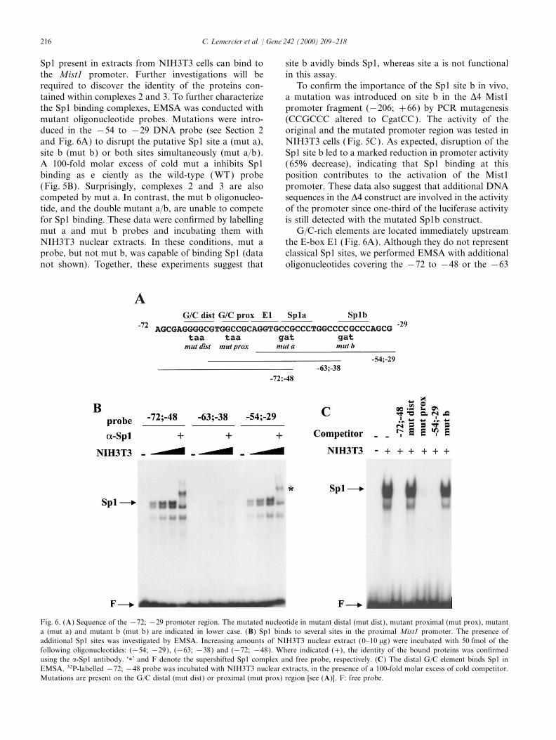
Sp1 present in extracts from NIH3T3 cells can bind to site b avidly binds Sp1, whereas site a is not functionalin this assay.the Mist1 promoter. Further investigations will be
required to discover the identity of the proteins con- To confirm the importance of the Sp1 site b in vivo,a mutation was introduced on site b in the D4 Mist1tained within complexes 2 and 3. To further characterize
the Sp1 binding complexes, EMSA was conducted with promoter fragment (−206; +66) by PCR mutagenesis(CCGCCC altered to CgatCC ). The activity of themutant oligonucleotide probes. Mutations were intro-
duced in the −54 to −29 DNA probe (see Section 2 original and the mutated promoter region was tested inNIH3T3 cells (Fig. 5C). As expected, disruption of theand Fig. 6A) to disrupt the putative Sp1 site a (mut a),
site b (mut b) or both sites simultaneously (mut a/b). Sp1 site b led to a marked reduction in promoter activity(65% decrease), indicating that Sp1 binding at thisA 100-fold molar excess of cold mut a inhibits Sp1
binding as efficiently as the wild-type ( WT) probe position contributes to the activation of the Mist1promoter. These data also suggest that additional DNA(Fig. 5B). Surprisingly, complexes 2 and 3 are also
competed by mut a. In contrast, the mut b oligonucleo- sequences in the D4 construct are involved in the activityof the promoter since one-third of the luciferase activitytide, and the double mutant a/b, are unable to compete
for Sp1 binding. These data were confirmed by labelling is still detected with the mutated Sp1b construct.G/C-rich elements are located immediately upstreammut a and mut b probes and incubating them with
NIH3T3 nuclear extracts. In these conditions, mut a the E-box E1 (Fig. 6A). Although they do not representclassical Sp1 sites, we performed EMSA with additionalprobe, but not mut b, was capable of binding Sp1 (data
not shown). Together, these experiments suggest that oligonucleotides covering the −72 to −48 or the −63
Fig. 6. (A) Sequence of the −72; −29 promoter region. The mutated nucleotide in mutant distal (mut dist), mutant proximal (mut prox), mutanta (mut a) and mutant b (mut b) are indicated in lower case. (B) Sp1 binds to several sites in the proximal Mist1 promoter. The presence ofadditional Sp1 sites was investigated by EMSA. Increasing amounts of NIH3T3 nuclear extract (0–10 mg) were incubated with 50 fmol of thefollowing oligonucleotides: (−54; −29), (−63; −38) and (−72; −48). Where indicated (+), the identity of the bound proteins was confirmedusing the a-Sp1 antibody. ‘1’ and F denote the supershifted Sp1 complex and free probe, respectively. (C ) The distal G/C element binds Sp1 inEMSA. 32P-labelled −72; −48 probe was incubated with NIH3T3 nuclear extracts, in the presence of a 100-fold molar excess of cold competitor.Mutations are present on the G/C distal (mut dist) or proximal (mut prox) region [see (A)]. F: free probe.

217C. Lemercier et al. / Gene 242 (2000) 209–218
to −38 regions. When increasing amounts of NIH3T3 transcription start site, indicating that the transcriptionis not initiated at multiple points but rather at a pre-nuclear extract (0–10 mg) were incubated with the −72
to −48 probe, a strong Sp1 binding complex could be cise site.Promoter deletion analysis suggests that Sp1 bindingdetected (Fig. 6B, left panel ). The signals obtained are
comparable to those obtained with the −54 to −29 sites are necessary for the transcriptional activation ofthe Mist1 gene. Pugh and Tjian (1990, 1991) showedSp1 probe (Fig. 6B, right panel ). In contrast, only a
weak binding activity is detected with the −63 to −38 that Sp1 transcription from a TATA-less promoterrequired a multisubunit TFIID complex that includesprobe, even in the presence of large amounts of nuclear
extract (Fig. 6B, middle panel ). Since this latter oligonu- TBP, TBP-associated factor (TAFs), coactivators andSp1. It is thought that Sp1 might be anchoring TBP tocleotide contains the Sp1 site a (not functional ) and the
most proximal G/C element (weak binding), it is likely the TATA-less promoter via tethering factors such asTAF110 (Hoey et al., 1993). Our study indicates thatthat the distal G/C site in the −72 to −48 probe
represents another strong Sp1 binding site. Sp1 binds to two different regions within the proximalpromoter, a typical Sp1 site located at (−38; −33) andThis issue was addressed in competition experiments
with an excess of cold mutant oligonucleotides for the a G/C-rich region between (−67; −62). In the contextof the TATA-less, Inr-less Mist1 promoter, it is possibleG/C distal or proximal regions (Fig. 6A). As expected,
competition for Sp1 binding was obtained with an that Sp1 serves as a bridging molecule to allow theassembly of the preinitiation complex. Moreover, two100-fold excess of the non-radioactive −72; −48 probe,
or with an oligonucleotide bearing a mutated proximal additional factors, that also bind to or near the Sp1sites, might also contribute to the basal expression ofG/C site (Fig. 6C, mut prox), indicating that this proxi-
mal G/C element is not necessary for competing Sp1. this gene. Our future studies will aim to identify thesetwo proteins as well as other transcription factors thatIn contrast, an intact distal G/C element is required,
since a mutant distal G/C oligonucleotide (mut dist) has might regulate the Mist1 gene during embryonic devel-opment and cell differentiation.no competitive effect on Sp1 binding to the probe.
Finally, we also tested whether cold Sp1 site b couldinterfere with Sp1 binding to the distal G/C element.Indeed, the incubation of nuclear extracts with an excess Acknowledgementsof cold −54; −29 DNA inhibited Sp1 binding to thelabeled probe, whereas mut b was inefficient (Fig. 6C). This work was supported by La Ligue NationaleThis result suggests that the G/C distal element and the Contre Le Cancer et L’Universite Victor SegalenSp1 site b might be functionally equivalent in the Mist1 Bordeaux 2. We thank Dr J. Gaffe, Dr S. Khochbin,promoter. Dr S.E. Johnson, Professor S. Nanchev and Dr M.
Callanan for helpful comments on the manuscript andDr S.F. Konieczny for reagents and communication
4. Conclusions of results.
The Mist1 transcription factor belongs to the basicHelix–Loop–Helix protein family, which is known to Referencesplay important roles during embryonic development andin cell fate determination. In this work, we have cloned Blackwell, T.K., Weintraub, H., 1990. Differences and similarities in
DNA-binding preferences of MyoD and E2A protein complexesthe rat Mist1 gene, determined its intron/exon structurerevealed by binding site selection. Science 250, 1104–1110.and analyzed its 5∞ flanking region for promoter activity.
Briggs, M.R., Kadonaga, J.T., Bell, S.P., Tjian, R., 1986. PurificationOur analysis showed that the Mist1 gene consists of twoand biochemical characterization of the promoter-specific tran-
exons separated by a single intron and that the whole scription factor, Sp1. Science 234, 47–52.gene is contained within less than 5 kb of genomic DNA. Burley, S.K., Roeder, R.G., 1996. Biochemistry and structural biology
of transcription factor IID (TFIID). Annu. Rev. Biochem. 65,Special features in the gene include the presence of an769–799.uninterrupted coding region in exon II and a very large
Hahn, S., 1998. The role of TAFs in RNA polymerase II transcription.3∞ untranslated region (2.8 kb).Cell 95, 579–582.
Sequence analysis of genomic clones containing the Hoey, T., Weinzierl, R.O.J., Gill, G., Chen, J.L., Dynlacht, B.D., Tjian,promoter region of the rat Mist1 gene reveals many R., 1993. Molecular cloning and functional analysis of Drosophila
TAF110 reveal properties expected of coactivators. Cell 72,features that are common to constitutively active eukary-247–260.otic promoters, including the absence of classical TATA
Johnson, S.E., Wang, X., Hardy, S., Taparowsky, E.J., Konieczny,and CAAT-box sequences and a high GC content.S.F., 1996. Casein kinase II increases the transcriptional activities
Although no typical initiator element (PyPyCA+1 of MRF4 and MyoD independently of their direct phosphoryla-NT/APyPy, Smale and Baltimore, 1989) was found, a tion. Mol. Cell. Biol. 16, 1604–1613.
Kageyama, R., Ishibashi, M., Takebayashi, K., Tomita, K., 1997.major product was obtained upon mapping of the

218 C. Lemercier et al. / Gene 242 (2000) 209–218
bHLH transcription factors and mammalian neuronal differentia- Konieczny, S.F., 1996. Examination of mammalian basic helix–loop–helix transcription factors using a yeast one-hybrid system.tion. Int. J. Biochem. Cell Biol. 29, 1389–1399.DNA Cell Biol. 15, 1–8.Klemsz, M.J., McKercher, S.R., Celada, A., Van Beveren, C., Maki,
Molkentin, J.D., Olson, E.N., 1996. Defining the regulatory networksR.A., 1990. The macrophage and B cell-specific transcription factorfor muscle development. Curr. Opin. Dev. 6, 445–453.PU.1 is related to the ets oncogene. Cell 61, 113–124.
Murre, C., McCaw, P.S., Baltimore, D., 1989a. A new DNA bindingLassar, A.B., Davis, R.L., Wright, W.E., Kadesh, T., Murre, C., Voro-and dimerization motif in immunoglobulin enhancer bindingnova, A., Baltimore, D., Weintraub, H., 1991. Functional activitydaughterless MyoD, and myc proteins. Cell 56, 777–783.of myogenic HLH proteins requires hetero-oligomerisation with
Murre, C., Schonleber-McCaw, P., Vaessin, H., Caudy, M., Jan, L.Y.,E12/E47-like proteins in vivo. Cell 66, 305–315.Jan, Y.N., Cabrera, C.V., Buskin, J.N., Hauschka, S.D., Lassar,Lemercier, C., To, R.Q., Swanson, B.J., Lyons, G.E., Konieczny, S.F.,A.B., Weintraub, H., Baltimore, D., 1989b. Interactions between1997. Mist1: A novel basic helix–loop–helix transcription factorheterologous helix–loop–helix proteins generate complexes thatexhibits a developmentally regulated expression pattern. Dev. Biol.bind specifically to a common DNA sequence. Cell 58, 537–544.
182, 101–113.Pugh, B.F., Tjian, R., 1990. Mechanism of transcriptional activation
Lemercier, C., To, R.Q, Carrasco, R.A., Konieczny, S.F., 1998. The by Sp1. Evidence for coactivators. Cell 61, 1187–1197.basic helix–loop–helix transcription factor Mist1 functions as a Pugh, B.F., Tjian, R., 1991. Transcription from a TATA-less promotertranscriptional repressor of MyoD. EMBO J. 17, 1412–1422. requires a multisubunit TFIID complex. Genes Dev. 5, 1935–1945.
Ludolph, D.C., Konieczny, S.F., 1995. Transcription factor families: Shivadasani, R.A., Orkin, S.H., 1996. The transcriptional control ofmuscling in on the myogenic program. FASEB J. 9, 1595–1604. hematopoiesis. Blood 87, 4025–4039.
Ma, P.C., Rould, M.A., Weintraub, H., Pabo, C.O., 1994. Crystal Smale, S.T., Baltimore, D., 1989. The initiator as a transcription con-structure of the MyoD bHLH domain–DNA complex: Perspectives trol element. Cell 57, 103–113.on DNA recognition and implications for transcriptional activa- Williams, T., Tjian, R., 1991. Analysis of the DNA-binding and activa-tion. Cell 77, 451–459. tion properties of the human transcription factor AP-2. Genes Dev.
5, 670–682.Mak, K.L., Longcor, L.C., Johnson, S.E., Lemercier, C., To, R.Q.,