Characterization of an unusual thyroid response unit in the promoter of the human placental lactogen...
8
THE JOURNAL (0 1991 by The American Society for Biochemistry OF BIOLOGICAL CHEMISTRY and Molecular Biology , Inc Vol. 266, No. 20, Issue of July 15, pp. 13397-13406,1991 Printed in U, S. A. Characterization of an Unusual Thyroid Response Unit in the Promoter of the Human Placental Lactogen Gene* (Received for publication, December 14, 1990) Marianne L. Vozs, Bernard Peerst, Alexandra Belayew, and Joseph A. Martial7 From the Laboratoire de Biologie Mokculaire et de Genie Gknetique, Universite de Liege, Institut de Chimie B6, B-4000 Sart-Tilman, Belgium The human placental lactogen B (hCS-B) promoter activity isstrongly stimulated by thyroid hormones in the rat pituitary GC cell line. The minimal DNA se- quence required for stimulation, as determined by transfection with 5‘ and 3’ deletion mutants, spans 67 base pairs, from coordinate -97 to -31. DNase I foot- printing experiments show that this thyroid response unit includes two adjacent binding sites: one for the thyroid receptor (-671-41), the other for the pituitary- specific factor GHFl (-95/-68). Neither region alone is sufficient to confer thyroid responsiveness. The thy- roid receptor binding element (TBE) does not contain any repeats or palindromes but is composedof two different domains, one of which is very similar to the half-palindromic motif described by Glass et al. (Glass, C. K., Holloway, J. M., Devary, 0. L., and Rosenfeld, M. G. (1988) Cell 54,313-323). The other is very rich in purine. The normal human growth hormone (hGH- N) promoter, which is 94% similar to the hCS-B pro- moter, differs from its hCS-B counterpart precisely in this TBE. This difference may explain the opposite 3,6,3‘-triiodothyronine (T3) regulation of these two genes. The thyroid hormones exert their effects mainly by regu- lating transcription of specific genes. They interact specifi- cally with nuclear receptors which then bind to regulatory DNA sequences of thetarget genes (reviewed in Ref. 2). Recent cloning of the genes encoding thyroid hormone recep- tors (3, 4) has revealed high homology to steroid, vitamin D, or retinoic acid receptors, thus placing these receptors in the nuclear hormone receptor superfamily (5, 6). However, as compared with the situation for steroid hor- mones, much less is known about the thyroid hormone-re- sponsive elements (TRE)’ that mediate thyroid hormone reg- * This work was supported by agrant from the Service de Program- mation de la Politique Scientifique (Belgium, Sciences de la Vie, BIO/ 15). The costs of publication of this article were defrayed in part by the payment of page charges. This article must therefore be hereby marked “advertisement” in accordance with 18 U.S.C. Section 1734 solely to indicate this fact. $ Held doctoral fellowship from Institut pour 1’Encouragement de la Recherche Scientifique dans1’Industrie et 1’Agriculture (Belgium). Held doctoral fellowship from Fonds National de la Recherche Scientifique (Belgium). 7 To whom correspondence should be addressed. ’ The abbreviations used are: TRE, thyroid hormone-responsive element; hCS, human chorionic somatomammotropin or placental lactogen; hGH-N, normal human growth hormone; rGH, rat growth hormone; TK, herpes simplex virus thymidine kinase; RSV, Rous sarcoma virus; CAT, chloramphenicol acetyltransferase; T3, 3,5,3’- triiodothyronine; TR, thyroid receptor; TRU, thyroid hormone-re- sponsive unit, TBE, thyroid receptor binding element; bp, base pairs; wt, wild type; DTT, dithiothreitol; Hepes, 4-(2-hydroxyethyl)-l-pi- perazineethanesulfonic acid. ulation. The few putative TREs defined to date, by transfec- tion with deletion mutants and/or in vitro binding analysis, often contain quite different sequences and make it difficult to define a general consensus sequence. Recently, Brent et al. (7) have proposed a T3-receptor-binding half-site, AGGTC/ AA, at least two copies of which are required for a T 3 response. In some reported TREs, however, this consensus is either absent or present only once (8-10). It is necessary to seek other TREs in order to define a general consensus sequence. Cloning of T3 receptor genes and their overexpression in eukaryoticsystems (11, 12) will probably greatly facilitate this research. Indeed, availability of nuclear extracts highly enriched in thyroid hormone receptor protein allows foot- printing experiments and thus identification of thyroid recep- tor binding sites. Such studies have been greatly hampered previously by difficulties in receptor purification. The human growth hormone (hGH) and placental lactogen (hCS) genes areanattractive model for studyingthyroid hormone regulation of geneexpression. Their 500-bp pro- moters, although presenting 94% similarity, are regulated in opposite ways by T3 in rat pituitary GC cells: hGH-N pro- moter activity is inhibited by T3 (13), whereas hCS-A pro- moter activity is stimulated (14). Although endogenous hCS (A andB) gene expression is restricted to the placenta, numerous studies have shown that the hCS-A and hCS-B promoters are active in GC cells (14-16). This can be ex- plained by the fact that these two promoters bind ubiquitous factors like SP1 but also the pituitary-specific GHFl factor (16, 17). In this study, we show that, like hCS-A, the highly (99%) similar hCS-B is strongly stimulated by T3 in GC cells. By transfecting with 5’ and 3‘ deletions of hCS-B promoter linked to a reporter gene, we have identified the minimal sequence required for T3 induction. Using the a thyroid receptor overexpressed in the vaccinia virus system (11, 12), we have precisely defined, by DNase I footprinting, the se- quences in the hCS-B and hGH-N promoters that interact with the thyroid receptor. EXPERIMENTAL PROCEDURES Plasmid Constructions-Plasmids rGH530CAT (18), TK105CAT (= pBLCAT2; (19)), and hGH496CAT (20) have been described elsewhere. The RSVamp plasmid contains the Rous sarcoma virus enhancer/promoter inserted upstream from the p-lactamase RTEM (Amp’) gene, followed by the SV40 splicing and polyadenylation signals (21). The hCS2289CAT plasmid, containing 2289 bp of the human hCS- B gene 5”flanking region and 6 bp of the transcribed sequence fused to the coding sequence of the bacterial chloramphenicol acetyltrans- ferase (CAT) gene, was constructed as follows. A 2.3-kilobase pair BamHI 5”flanking fragment of the hCS-B gene (from coordinates -2289 to +6) was cloned into the BamHI site of pAlOCAT3. pAlOCAT3 is identical to pAlOCAT2 (22) except that the Hind111 site has been replaced by a BglII site. A clone containing the insert 13397
-
Upload
independent -
Category
Documents
-
view
4 -
download
0
Transcript of Characterization of an unusual thyroid response unit in the promoter of the human placental lactogen...
THE JOURNAL (0 1991 by The American Society for Biochemistry
OF BIOLOGICAL CHEMISTRY and Molecular Biology , Inc
Vol. 266, No. 20, Issue of July 15, pp. 13397-13406,1991 Printed in U, S. A .
Characterization of an Unusual Thyroid Response Unit in the Promoter of the Human Placental Lactogen Gene*
(Received for publication, December 14, 1990)
Marianne L. Vozs, Bernard Peerst, Alexandra Belayew, and Joseph A. Martial7 From the Laboratoire de Biologie Mokculaire et de Genie Gknetique, Universite de Liege, Institut de Chimie B6, B-4000 Sart-Tilman, Belgium
The human placental lactogen B (hCS-B) promoter activity is strongly stimulated by thyroid hormones in the rat pituitary GC cell line. The minimal DNA se- quence required for stimulation, as determined by transfection with 5‘ and 3’ deletion mutants, spans 67 base pairs, from coordinate -97 to -31. DNase I foot- printing experiments show that this thyroid response unit includes two adjacent binding sites: one for the thyroid receptor (-671-41), the other for the pituitary- specific factor GHFl (-95/-68). Neither region alone is sufficient to confer thyroid responsiveness. The thy- roid receptor binding element (TBE) does not contain any repeats or palindromes but is composed of two different domains, one of which is very similar to the half-palindromic motif described by Glass et al. (Glass, C. K., Holloway, J. M., Devary, 0. L., and Rosenfeld, M. G. (1988) Cell 54,313-323). The other is very rich in purine. The normal human growth hormone (hGH- N) promoter, which is 94% similar to the hCS-B pro- moter, differs from its hCS-B counterpart precisely in this TBE. This difference may explain the opposite 3,6,3‘-triiodothyronine (T3) regulation of these two genes.
The thyroid hormones exert their effects mainly by regu- lating transcription of specific genes. They interact specifi- cally with nuclear receptors which then bind to regulatory DNA sequences of the target genes (reviewed in Ref. 2 ) . Recent cloning of the genes encoding thyroid hormone recep- tors (3, 4) has revealed high homology to steroid, vitamin D, or retinoic acid receptors, thus placing these receptors in the nuclear hormone receptor superfamily (5 , 6).
However, as compared with the situation for steroid hor- mones, much less is known about the thyroid hormone-re- sponsive elements (TRE)’ that mediate thyroid hormone reg-
* This work was supported by a grant from the Service de Program- mation de la Politique Scientifique (Belgium, Sciences de la Vie, BIO/ 15). The costs of publication of this article were defrayed in part by the payment of page charges. This article must therefore be hereby marked “advertisement” in accordance with 18 U.S.C. Section 1734 solely to indicate this fact.
$ Held doctoral fellowship from Institut pour 1’Encouragement de la Recherche Scientifique dans 1’Industrie et 1’Agriculture (Belgium).
Held doctoral fellowship from Fonds National de la Recherche Scientifique (Belgium).
7 T o whom correspondence should be addressed. ’ The abbreviations used are: TRE, thyroid hormone-responsive
element; hCS, human chorionic somatomammotropin or placental lactogen; hGH-N, normal human growth hormone; rGH, rat growth hormone; TK, herpes simplex virus thymidine kinase; RSV, Rous sarcoma virus; CAT, chloramphenicol acetyltransferase; T3, 3,5,3’- triiodothyronine; TR, thyroid receptor; TRU, thyroid hormone-re- sponsive unit, TBE, thyroid receptor binding element; bp, base pairs; wt, wild type; DTT, dithiothreitol; Hepes, 4-(2-hydroxyethyl)-l-pi- perazineethanesulfonic acid.
ulation. The few putative TREs defined to date, by transfec- tion with deletion mutants and/or in vitro binding analysis, often contain quite different sequences and make it difficult to define a general consensus sequence. Recently, Brent et al. (7) have proposed a T3-receptor-binding half-site, AGGTC/ AA, at least two copies of which are required for a T3 response. In some reported TREs, however, this consensus is either absent or present only once (8-10). I t is necessary to seek other TREs in order to define a general consensus sequence. Cloning of T 3 receptor genes and their overexpression in eukaryotic systems (11, 12) will probably greatly facilitate this research. Indeed, availability of nuclear extracts highly enriched in thyroid hormone receptor protein allows foot- printing experiments and thus identification of thyroid recep- tor binding sites. Such studies have been greatly hampered previously by difficulties in receptor purification.
The human growth hormone (hGH) and placental lactogen (hCS) genes are an attractive model for studying thyroid hormone regulation of gene expression. Their 500-bp pro- moters, although presenting 94% similarity, are regulated in opposite ways by T3 in rat pituitary GC cells: hGH-N pro- moter activity is inhibited by T3 (13), whereas hCS-A pro- moter activity is stimulated (14). Although endogenous hCS (A and B) gene expression is restricted to the placenta, numerous studies have shown that the hCS-A and hCS-B promoters are active in GC cells (14-16). This can be ex- plained by the fact that these two promoters bind ubiquitous factors like SP1 but also the pituitary-specific GHFl factor (16, 17).
In this study, we show that, like hCS-A, the highly (99%) similar hCS-B is strongly stimulated by T3 in GC cells. By transfecting with 5’ and 3‘ deletions of hCS-B promoter linked to a reporter gene, we have identified the minimal sequence required for T 3 induction. Using the a thyroid receptor overexpressed in the vaccinia virus system (11, 12), we have precisely defined, by DNase I footprinting, the se- quences in the hCS-B and hGH-N promoters that interact with the thyroid receptor.
EXPERIMENTAL PROCEDURES
Plasmid Constructions-Plasmids rGH530CAT (18), TK105CAT (= pBLCAT2; (19)), and hGH496CAT (20) have been described elsewhere. The RSVamp plasmid contains the Rous sarcoma virus enhancer/promoter inserted upstream from the p-lactamase RTEM (Amp’) gene, followed by the SV40 splicing and polyadenylation signals (21).
The hCS2289CAT plasmid, containing 2289 bp of the human hCS- B gene 5”flanking region and 6 bp of the transcribed sequence fused to the coding sequence of the bacterial chloramphenicol acetyltrans- ferase (CAT) gene, was constructed as follows. A 2.3-kilobase pair BamHI 5”flanking fragment of the hCS-B gene (from coordinates -2289 to +6) was cloned into the BamHI site of pAlOCAT3. pAlOCAT3 is identical to pAlOCAT2 (22) except that the Hind111 site has been replaced by a BglII site. A clone containing the insert
13397
13398 T3 Stimulation of hCS-B Promoter Activity TABLE I
Effect of T3 on various promoter activities Induction levels are expressed as the ratio of CAT activities in T3-
treated uersuS untreated cells. Each value is an average of n separate transfection experiments performed in triplicate f S.E. For each promoter, the values obtained from the T3-treated cells are signifi- cantly different from those obtained with the untreated cells ( p 0.001).
T3 induction n
hCS2289CAT 15.8 f 1.2 3 hCS493CAT 15.9 f 1.7 4 rGH530CAT 5.4 f 0.6 9 hGH496CAT 0.56 k 0.04 9 TK105CAT 1.4 f 0.1 5 RSVamp 1.4 f CO.1 31
in the correct orientation was selected by restriction analysis. All the following 5' deletion constructs were made in a modified
pBLCAT3 (19) plasmid (pBLNHCAT3) where a cryptic NF1 binding site located upstream from the polylinker' has been deleted. For that purpose, pBLCAT3 was digested by NdeI and HindIII, blunt-ended
by T4 DNA ligase. The HindIII site was thus restored. hCS493CAT with the Klenow fragment of DNA polymerase I, and recircularized
was constructed by subcloning the EcoRI(-493) (blunt-ended)- BamHI(+6) fragment of the hCS-B gene into the SphI (blunt-ended)/ BamHI site of pBLNHCAT3.
To construct the 5' deletion mutants hCS295CAT, hCS212CAT, hCS171CAT, hCSlGOCAT, hCS128CAT, and hCS83CAT, plasmid hCS493CAT was digested by HincII, DraIII, HphI, BstNI (partially), AluI, or NsiI, respectively, blunt-ended and digested by BamHI. The hCS promoter 5' deletion fragments were gel-purified and cloned into the HindIII (blunt-ended)/BamHI site of pBLNHCAT3.
hCS453CAT, hCS418CAT, hCS375CAT, hCS113CAT, and hCS97CAT were constructed with specific DNA fragments produced by the polymerase chain reaction. Polymerase chain reaction experi- ments were carried out using as 5' primer oligonucleotides corresponding to different portions of the hCS promoter (sense strand) bearing a HindIII site at their 5' end, and as 3' primer,
____
G. Schutz, personal communication.
-49s - 400 -800 - PO0 - 100
oligonucleotide B1 containing the sequence of the CAT structural gene located 80 bp downstream from the BamHI site (antisense strand) (sequences are shown below).
The DNA fragments were amplified between these two primers using hCS493CAT as a DNA template. The fragments obtained were digested by HindIII and BamHI, gel-purified, and cloned into the HindIII/BamHI site of pBLNHCAT3.
[CS-97/-31]TKCAT and [CS-97/-47]TKCAT were also con- structed with polymerase chain reaction-amplified fragments ob- tained with A5 as 5' primer and B2 or B3 as 3' primer, respectively. Amplified fragments were digested by Hind111 and BamHI and cloned into the HindIII/BamHI site of pBLCAT2 (19).
The oligonucleotides were purchased from Eurogentec (LiBge, Bel- gium) and their sequences are shown as follows.
Sense 5'cccaagcTTGGCTGTGCTTGGCCC3' A1 (-453/-436; hCs) 5'cccaagcTTCTGTCTGGTGGGTGG3' A2 (-418/-402; hCS) S'cccaagcttGGAATAGGATAGAGAGTGG3' A3 (-375/-357; hCS) 5'cccaagcTTAGCACAAGCCCGTCAG3' A4 (-113/-96; hCS) 5'cccaagcttAGTGGCCCCATGCATAAA3' A5 (-97/40; hCS) Antisense 3'AGTGACCTATATGGTGGCS' B1 (501/518;pBLCAT3) 3'CTCTCTTGACCGGTCCCtag&gc5' B2 (-47/-31; hCS) 3'CCCCAGTTCGTCCCTCctagggc5' B3 (-63/-47; hCS)
hCS97ANsiICAT, containing a 5-bp deletion (-87/43) was con- structed by cleaving hCS97CAT with NsiI and digestion of the protruding ends with Klenow fragment followed by ligation.
The structure of all mutants was confirmed by restriction mapping and/or sequencing. All plasmids were prepared by alkaline lysis, purified by centrifugation in CsC1-ethidium bromide gradients, treated with RNase, centrifuged through a cushion of 1 M NaCl, extracted with phenol-chloroform, and finally precipitated with ethanol. The concentration of all plasmid preparations was deter- mined by measuring the absorbance at 260 nm.
Cell Culture Conditions-GC cells were grown in Ham's F-12 medium supplemented with 15% fetal calf serum (Life Technologies, Belgium). One day before transfection, the cells received phenol red- free Ham's F-12 supplemented with calf serum pretreated with AG1-
.1
0 1 2 3 4 6 6 7 8 0 101112131416181718 -J
fold 13 induction
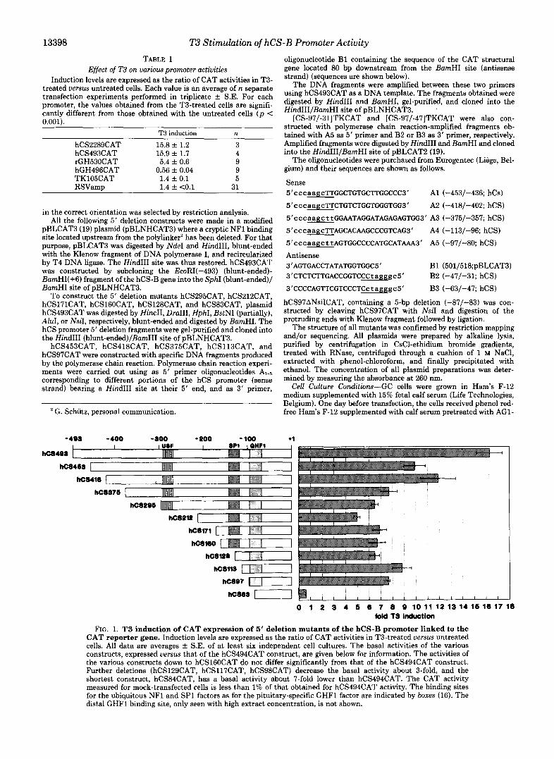
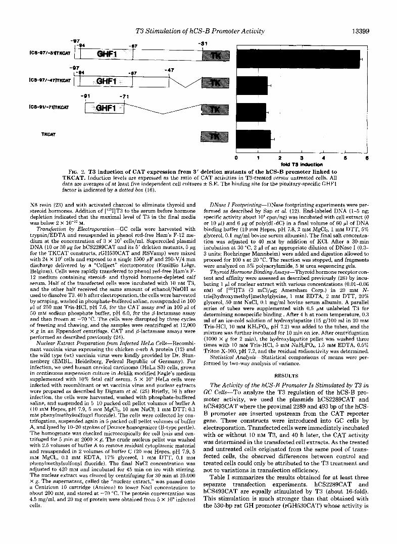
FIG. 1. T3 induction of CAT expression of 5' deletion mutants of the hCS-B promoter linked to the CAT reporter gene. Induction levels are expressed as the ratio of CAT activities in T3-treated versus untreated cells. All data are averages f S.E. of a t least six independent cell cultures. The basal activities of the various constructs, expressed versus that of the hCS494CAT construct, are given below for information. The activities of the various constructs down to hCS16OCAT do not differ significantly from that of the hCS494CAT construct. Further deletions (hCS129CAT, hCS117CAT, hCS98CAT) decrease the basal activity about 3-fold, and the shortest construct, hCS84CAT, has a basal activity about 7-fold lower than hCS494CAT. The CAT activity measured for mock-transfected cells is less than 1% of that obtained for hCS494CAT activity. The binding sites for the ubiquitous NF1 and SP1 factors as for the pituitary-specific GHFl factor are indicated by boxes (16). The distal GHFl binding site, only seen with high extract concentration, is not shown.
T3 Stimulation of hCS-B Promoter Activity 13399 -97
ICS-Q7/-SllTKcAt
ICS-Q71-47rTKCAl
-91 -71
0 1 2 9 4 6 6 told f 3 induction
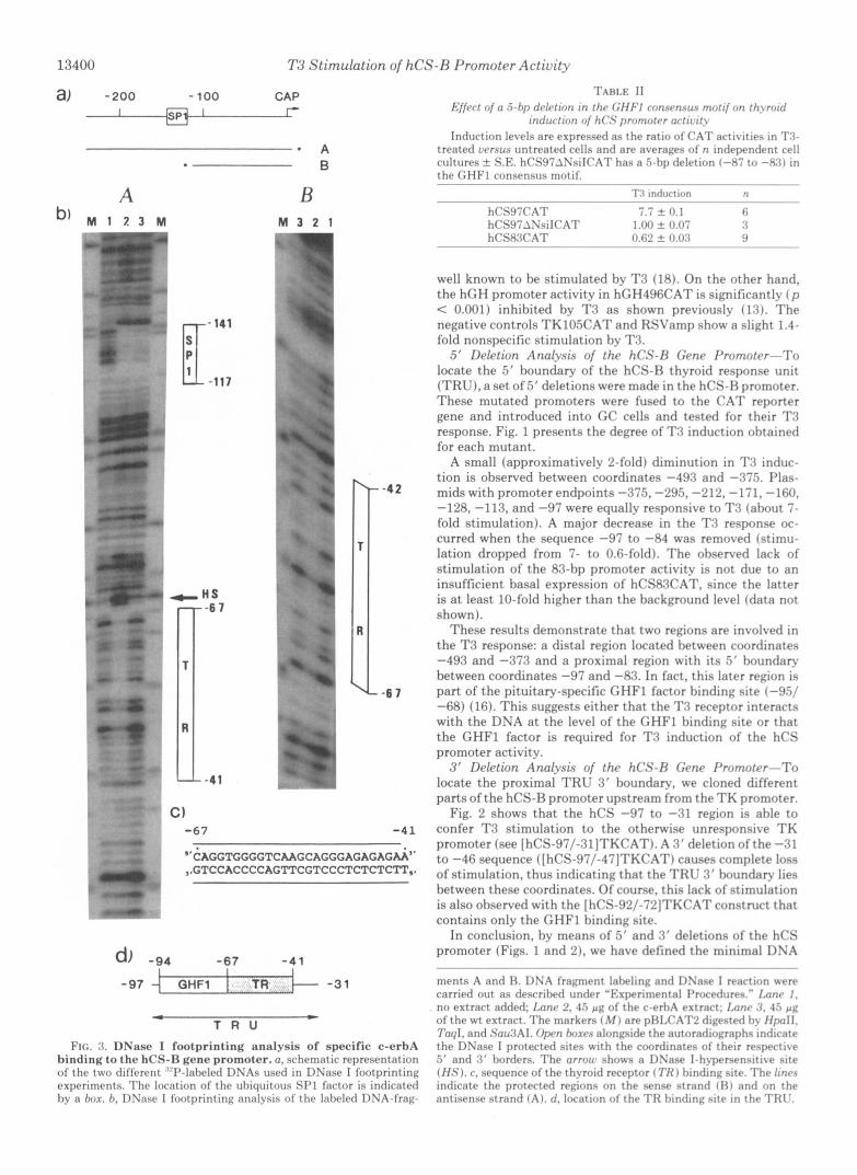
FIG. 2. T3 induction of CAT expression from 3’ deletion mutants of the hCS-B promoter linked to TKCAT. Induction levels are expressed as the ratio of CAT activities in T3-treated versus untreated cells. All data are averages of a t least five indeuendent cell cultures f S.E. The binding site for the pituitary-specific GHFl factor is indicited by a dotted box (16).
X8 resin (23) and with activated charcoal to eliminate thyroid and steroid hormones. Addition of [‘*‘II]T3 to the serum before hormone depletion indicated that the maximal level of T3 in the final media was below 2 X 10”’ M.
Transfection by Electroporation-GC cells were harvested with trypsin/EDTA and resuspended in phenol red-free Ham’s F-12 me- dium at the concentration of 3 X lo7 cells/ml. Supercoiled plasmid DNA (10 or 30 pg for hCS2289CAT and its 5’ deletion mutants, 5 pg for the TKCAT constructs, rGH530CAT and RSVamp) were mixed with 24 X 10‘ cells and exposed to a single 1500-pF and 250-V/4 mm discharge delivered by a “Cellject” electroporator (EquiBio LiGge, Belgium). Cells were rapidly transferred to phenol red-free Ham’s F- 12 medium containing steroid- and thyroid hormone-depleted calf serum. Half of the transfected cells were incubated with 10 nM T3, and the other half received the same amount of ethanol/NaOH as used to dissolve T3.40 h after electroporation, the cells were harvested by scraping, washed in phosphate-buffered saline, resuspended in 100 pl of 250 mM Tris-HC1, pH 7.6, for the CAT assay and in 100 pl of 50 mM sodium phosphate buffer, pH 6.0, for the p-lactamase assay and then frozen at -70 “C. The cells were disrupted by three cycles of freezing and thawing, and the samples were centrifuged at 12,000 X g in an Eppendorf centrifuge. CAT and 8-lactamase assays were performed as described previously (24).
Nuclear Extract Preparation from Infected HeLa Cells-Recombi- nant vaccinia virus expressing the chicken c-erb A protein (12) and the wild type (wt) vaccinia virus were kindly provided by Dr. Stun- nenberg (EMBL, Heidelberg, Federal Republic of Germany). For infection, we used human cervical carcinoma (HeLa S3) cells, grown in continuous suspension culture in Joklik modified Eagle’s medium supplemented with 10% fetal calf serum. 5 X 10’ HeLa cells were infected with recombinant or wt vaccinia virus and nuclear extracts were prepared as described by Dignam et al. (25) Briefly, 16 h after infection, the cells were harvested, washed with phosphate-buffered saline, and suspended in 5-10 packed cell pellet volumes of buffer A (10 mM Hepes, pH 7.9, 5 mM MgCl,, 10 mM NaCI; 1 mM D T T 0.1 mM phenylmethylsulfonyl fluoride). The cells were collected by cen- trifugation, suspended again in 5 packed cell pellet volumes of buffer A, and lysed by 10-20 strokes of Dounce homogenizer (B-type pestle). The homogenate was checked microscopically for cell lysis and cen- trifuged for 5 min at 2000 X g. The crude nucleus pellet was washed with 2.5 volumes of buffer A to remove residual cytoplasmic material and resuspended in 2 volumes of buffer C (20 mM Hepes, pH 7.9, 5 mM MgC12, 0.1 mM EDTA, 17% glycerol, 1 mM DTT, 0.1 mM phenylmethylsulfonyl fluoride). The final NaCl concentration was adjusted to 420 mM and incubated for 45 min on ice with stirring. The nuclear extract was cleared by centrifuging for 30 min at 25,000 X g. The supernatant, called the “nuclear extract,” was passed onto a Centricon 10 cartridge (Amicon) to lower Nacl concentration to about 200 mM, and stored at -70 ”C. The protein concentration was 4.5 mg/ml, and 20 mg of protein were obtained from 5 X IO8 infected cells.
DNase Z Footprinting-DNase footprinting experiments were per- formed as described by Sap et al. (12). End-labeled DNA (1-5 ng; specific activity about lo4 cpm/ng) was incubated with cell extract (0 or 10 pl) and 6 pg of poly(dI.dC) in a final volume of 60 pl of DNA binding buffer (10 mM Hepes, pH 7.8, 2 mM MgC12, 1 mM DTT, 5% glycerol, 0.1 mg/ml bovine serum albumin). The final salt concentra- tion was adjusted to 40 mM by addition of KCl. After a 30-min incubation at 30 “C, 2 pl of an appropriate dilution of DNase I (0.3- 3 units; Boehringer Mannheim) were added and digestion allowed to proceed for 100 s at 20 “C. The reaction was stopped, and fragments were analyzed on 5% polyacrylamide, 5 M urea sequencing gels.
Thyroid Hormone Binding Assays-Thyroid hormone receptor con- tent and affinity were assessed as described previously (26) by incu- bating 1 pl of nuclear extract with various concentrations (0.01-0.06 nM) of [‘*‘II]T3 (3 mCi/bg; Amersham Corp.) in 20 mM N- tris[hydroxymethyl]methylglycine, 1 mM EDTA, 2 mM DTT, 20% glycerol, 50 mM NaCl, 0.1 mg/ml bovine serum albumin. A parallel series of tubes were supplemented with 0.5 p~ unlabeled T 3 for determining nonspecific binding. After 4 h a t room temperature, 0.3 ml of an ice-cold solution of hydroxylapatite (15 g/lOO ml in 20 mM Tris-HCI, 10 mM KH,PO,, pH 7.2) was added to the tubes, and the mixture was further incubated for 10 min on ice. After centrifugation (1000 X g for 2 min), the hydroxylapatite pellet was washed three times with 10 mM Tris-HC1, 5 mM NaH2P04, 1.5 mM EDTA, 0.5% Triton X-100, pH 7.2, and the residual radioactivity was determined.
Statistical Analysis-Statistical comparisons of means were per- formed by two-way analysis of variance.
RESULTS
The Activity of the hCS-B Promoter Is Stimulated by T3 in GC Cells-To analyze the T 3 regulation of the hCS-B pro- moter activity, we used the plasmids hCS2289CAT and hCS493CAT where the proximal 2289 and 493 bp of the hCS- B promoter are inserted upstream from the CAT reporter gene. These constructs were introduced into GC cells by electroporation. Transfected cells were immediately incubated with or without 10 nM T3, and 40 h later, the CAT activity was determined in the transfected cell extracts. As the treated and untreated cells originated from the same pool of trans- fected cells, the observed differences between control and treated cells could only be attributed to the T3 treatment and not to variations in transfection efficiency.
Table I summarizes the results obtained for at least three separate transfection experiments. hCS2289CAT and hCS493CAT are equally stimulated by T 3 (about 16-fold). This stimulation is much stronger than that obtained with the 530-bp rat GH promoter (rGH530CAT) whose activity is
13400 T3 Stimulation of hCS-I? Promoter Activity
a, -200 - 100 CAP I r
* A B
A B
IT -117 14’
-42
-6 7
- 4 1
d, -94 -67 -4 1 I
-97 GHF1 I fR. , , -3 1
4 c T R U
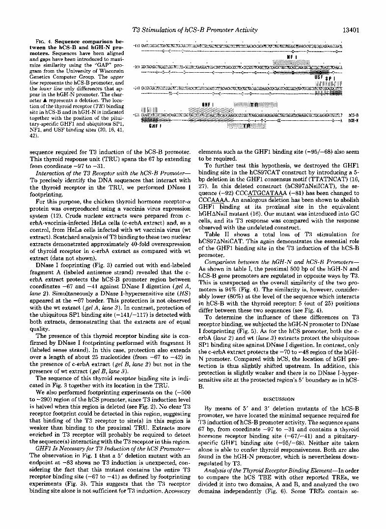
FIG. 3. DNase I footprinting analysis of specific c-erbA binding to t he hCS-R gene promoter. a, schematic representation o f the two different ~“I’-lal)eled DNAs used in DNase I footprinting experiments. The location of the uhiquitous SPl fartor is indicated hy a box. h, DNase I lootprinting analysis of the laheled DNA-frag-
TARI.E I I Kffrrt nfa 5 -hp drlcfinn in thr C H F I rnnwnsus motlf nn th.roid
induction of hCS prnmotrr arlivit? Induction levels are expressed as the ratio o f CAT activities in T 3 -
treated urrsus untreated cells and are averages o f n independent cell cultures f S.E. hCS97ANsilCA’I’ has a .i-hp deletion ( 4 - 7 to -84) in t h e GHFl consensu~ motif .
T3 indrlrtirm n
hCSS7CAT hCS9’iANsilCAT 1 .oo t 0.07 :< hCS83CAT 0.6’ t 0.0:1 9
” t 0.1 r,
well known to be stimulated by T.7 (18). On the other hand, the hGH promoter activity in hGH496CAT is significantly ( p < 0.001) inhibited by T.7 as shown previouslv (1.7). T h e negative controls TKlO5CAT and RSVamp show a slight 1.4- fold nonspecific stimulation by T.7.
Yj’ lleletion Analvsis of the hCS-R Gena Prornotcr-To locate the 5’ boundary of the hCS-R thyroid response unit (TRU), a set of 5’ deletions were made in the hCS-H promoter. These mutated promoters were fused to the CAT reporter gene and introduced into GC cells and tested for their T.7 response. Fig. 1 presents the degree of T.7 induction obtained for each mutant.
A small (approximatively 2-fold) diminution in T.7 induc- tion is observed between coordinates -49.7 and -375. Plas- mids with promoter endpoints -375, -295, -212, -171, -160, -128, -11.7, and -97 were equally responsive to T.7 (about 7- fold stimulation). A major decrease in the T.7 response oc- curred when the sequence -97 to -84 was removed (stimu- lation dropped from 7- to 0.6-fold). The ohserved lack of stimulation of the 8.7-bp promoter activitv is not due to an insufficient basal expression of hCSRXAT, since the latter is at least 10-fold higher than the background level (data not shown).
These results demonstrate that two regions are involved in the T3 response: a distal region located between coordinates -49.7 and -37.7 and a proximal region with its 5’ boundary between coordinates -97 and -8.7. In fact, this later region is part of the pituitary-specific GHFl factor binding site (-%/ -68) (16). This suggests either that the T.7 receptor interacts with the DNA at t h e level of the GHFl binding si te or that the GHFl factor is required for T.7 induction of the hCS promoter activity.
3 ’ Deletion Analysis of the hCS-R Gcne Promotcr-To locate the proximal TRU 3’ boundary, we cloned different parts of the hCS-R promoter upstream from the TK promoter.
Fig. 2 shows that the hCS -97 t o -31 region is able to confer T3 stimulation to the otherwise unresponsive TK promoter (see [ hCS-S7/-31]TKCAT). A 3’ deletion of the -31 to -46 sequence ([hCS-97/-47]TKCAT) causes complete loss of stimulation, thus indicating that the TRC‘ 3’ boundary lies between these coordinates. Of course, this lack of stimulation is also observed with the [hCS-92/-72]TKCAT construct that contains only the GHFl binding site.
In conclusion, bv means of 5‘ and 3’ deletions of the hCS promoter (Figs. 1 and 2 ) , we have defined the minimal DNA
ments A and H. DNA fragment labeling and DSase I reaction were carried out as desrrihed under ”Experimental I’rncedures.” Imnr 1. no extract added: Lnnr 2, 45 p g of the c-erhA extract; Imnc .‘I. 45 p g of the wt extract. The markers ( M ) are pHIX’AT2 digested hy HpoIl, 7’0q1, and S’nu3AI. Oprn h m r s alongside the autoradiographs intlirate the DNase I protected sites with the coordinates ol thelr respertive 5’ and 3 ‘ horders. The orrow shows a DSase l-h>-persensitive site (H?;) . r , sequence of the thyroid receptor ( 7’H 1 hindinz site. The linrs indicate the protected regions on the sense st rand 131 and on the antisense strand ( A ) . d, loration of the TH hinrling site in the TRt’.
T3 Stimulation of hCS-B Promoter Activity 13401
tween the hCS-B and hGH-N pro- FIG. 4. Sequence comparison be-
moters. Sequences have been aligned and gaps have been introduced to maxi- mize similarity using the “GAP pro- gram from the University of Wisconsin Genetics Computer Group. The upper line represents the hCS-B promoter, and the lower l ine only differences that ap- pear in the hGH-N promoter. The char- acter A represents a deletion. The loca- tion of the thyroid receptor (TR) binding site in hCS-B and in hGH-N is indicated together with the position of the pitui- tary-specific GHFl and ubiquitous SP1, NF1, and USF binding sites (20, 16, 41, 42).
GHF I
sequence required for T3 induction of the hCS-B promoter. This thyroid response unit (TRU) spans the 67 bp extending from coordinate -97 to -31.
Interaction of the T3 Receptor with the hCS-B Promoter- To precisely identify the DNA sequences that interact with the thyroid receptor in the TRU, we performed DNase I footprinting.
For this purpose, the chicken thyroid hormone receptor-a protein was overproduced using a vaccinia virus expression system (12). Crude nuclear extracts were prepared from c- erbA-vaccinia-infected HeLa cells (c-erbA extract) and, as a control, from HeLa cells infected with wt vaccinia virus (wt extract). Scatchard analysis of T3 binding to these two nuclear extracts demonstrated approximately 40-fold overexpression of thyroid receptor in c-erbA extract as compared with wt extract (data not shown).
DNase I fooprinting (Fig. 3) carried out with end-labeled fragment A (labeled antisense strand) revealed that the c- erbA extract protects the hCS-B promoter region between coordinates -67 and -41 against DNase I digestion (gel A, lane 2). Simultaneously a DNase I-hypersensitive site ( H S ) appeared at the -67 border. This protection is not observed with the wt extract (gel A, lane 3 ) . In contrast, protection of the ubiquitous SP1 binding site (-141/-117) is detected with both extracts, demonstrating that the extracts are of equal quality.
The presence of this thyroid receptor binding site is con- firmed by DNase I footprinting performed with fragment B (labeled sense strand). In this case, protection also extends over a length of about 25 nucleotides (from -67 to -42) in the presence of c-erbA extract (gel B, lane 2) but not in the presence of wt extract (gel B, lune 3 ) .
The sequence of this thyroid receptor binding site is indi- cated in Fig. 3 together with its location in the TRU.
We also performed footprinting experiments on the (-500 to -290) region of the hCS promoter, since T3 induction level is halved when this region is deleted (see Fig. 2). No clear T3 receptor footprint could be detected in this region, suggesting that binding of the T3 receptor to site(s) in this region is weaker than binding to the proximal TRU. Extracts more enriched in T3 receptor will probably be required to detect the sequence(s) interacting with the T3 receptor in this region.
GHFl Is Necessary for T3 Induction of the hCS Promoter- The observation in Fig. 1 that a 5’ deletion mutant with an endpoint at -83 shows no T3 induction is unexpected, con- sidering the fact that this mutant contains the entire T3 receptor binding site (-67 to -41) as defined by footprinting experiments (Fig. 3). This suggests that the T3 receptor binding site alone is not sufficient for T3 induction. Accessory
................ :::::::::::::::::.:.. ................. ....... ~iiiii.iiiiiliiiiijiiij ........................... WlHA ’
:.:.:.:.
A----A ha-N
elements such as the GHFl binding site (-95/-68) also seem to be required.
To further test this hypothesis, we destroyed the GHFl binding site in the hCS97CAT construct by introducing a 5- bp deletion in the GHFl consensus motif (TTATNCAT) (16, 27). In this deleted construct (hCS97ANsiICAT), the se- quence (-92) CCCATGCATAAA (-81) has been changed to CCC&AA. An analogous deletion has been shown to abolish GHFl binding at its proximal site in the equivalent hGHANsiI mutant (16). Our mutant was introduced into GC cells, and its T3 response was compared with the response observed with the undeleted construct.
Table I1 shows a total loss of T3 stimulation for hCS97ANsiCAT. This again demonstrates the essential role of the GHFl binding site in the T3 induction of the hCS-B promoter.
Comparison between the hGH-N and hCS-B Promoters- As shown in table I, the proximal 500 bp of the hGH-N and hCS-B gene promoters are regulated in opposite ways by T3. This is unexpected as the overall similarity of the two pro- moters is 94% (Fig. 4). The similarity is, however, consider- ably lower (80%) at the level of the sequence which interacts in hCS-B with the thyroid receptor: 5 (out of 25) positions differ between these two sequences (see Fig. 4).
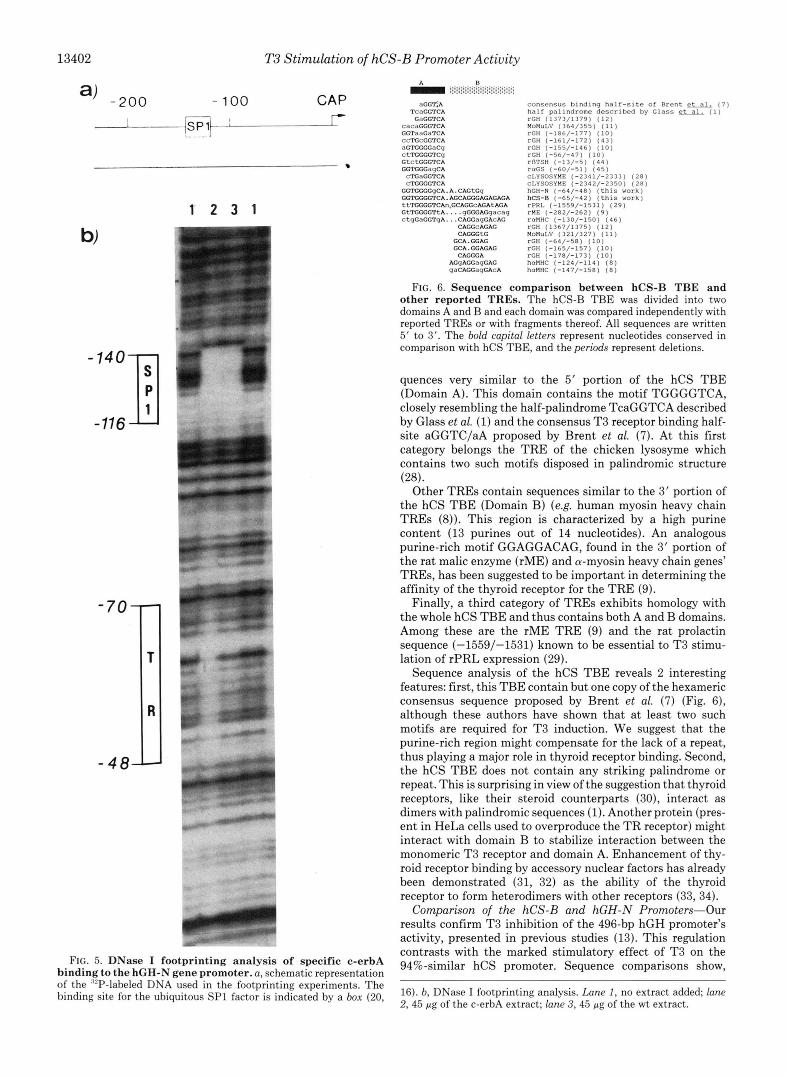
To determine the influence of these differences on T3 receptor binding, we subjected the hGH-N promoter to DNase I footprinting (Fig. 5). As for the hCS promoter, both the c- erbA (lane 2) and wt (lane 3) extracts protect the ubiquitous SP1 binding sites against DNase I digestion. In contrast, only the c-erbA extract protects the -70 to -48 region of the hGH- N promoter. Compared with hCS, the location of hGH pro- tection is thus slightly shifted upstream. In addition, this protection is slightly weaker and there is no DNase I-hyper- sensitive site at the protected region’s 5’ boundary as in hCS- B.
DISCUSSION
By means of 5’ and 3’ deletion mutants of the hCS-B promoter, we have located the minimal sequence required for T3 induction of hCS-B promoter activity. The sequence spans 67 bp, from coordinate -97 to -31 and contains a thyroid hormone receptor binding site (-67/-41) and a pituitary- specific GHFl binding site (-95/-68). Neither site taken alone is able to confer thyroid responsiveness. Both are also found in the hGH-N promoter, which is nevertheless down- regulated by T3.
Analysis of the Thyroid Receptor Binding Element-In order to compare the hCS TBE with other reported TREs, we divided it into two domains, A and B, and analyzed the two domains independently (Fig. 6). Some TREs contain se-
13402 T3 Stimulation of hCS-B Promoter Activity
a) -200 - 100 CAP I FP?”-: r-
1 2 3 1
- 140
-116
w
- 4 8 ll
a I
FIG. 5. DNase I footprinting analysis of specific c-erbA binding to the hGH-N gene promoter. a, schematic representation of the ‘“P-labeled DNA used in the footprinting experiments. The binding site for the ubiquitous SP1 factor is indicated by a box (20,
TcaGGTCA ‘nGGT,A
GaCCTCA
consensus binding half-site of Brent e- (7) half palindrome described by G l a s s et-al.. ( 1 1 rCH (1373/1379) (12) HoMuLV (364/355) ( 1 1 ) r C H (-186/-177) (10) rcH (-161/-172) (43) TGH (-155/-146) ( l o ) rCH (-56/-471 ( 1 0 ) rBTSH ( - 1 3 / - 5 ) (44) TUGS (-60/-51) (451 cLYSOSYHE (-2341/-2333) (28) cLYSOSYHE (-2342/-2350) (281 hCH-N ( - G 4 / - 4 8 ) (this Work) hCS-0 (-651-42) (this work) rPRL (-1559/-15311 (29) THE (-282/-262) (9) roHHC (-130/-1501 (46) rCH (1367/13751 (12)
FIG. 6. Sequence comparison between hCS-B TBE and other reported TREs. The hCS-B TBE was divided into two domains A and B and each domain was compared independently with reported TREs or with fragments thereof. All sequences are written 5’ to 3’. The bold capital letters represent nucleotides conserved in comparison with hCS TBE, and the periods represent deletions.
quences very similar to the 5‘ portion of the hCS TBE (Domain A). This domain contains the motif TGGGGTCA, closely resembling the half-palindrome TcaGGTCA described by Glass et al. (1) and the consensus T3 receptor binding half- site aGGTC/aA proposed by Brent et al. (7). At this first category belongs the TRE of the chicken lysosyme which contains two such motifs disposed in palindromic structure (28).
Other TREs contain sequences similar to the 3’ portion of the hCS TBE (Domain B) (e.g. human myosin heavy chain TREs (8)). This region is characterized by a high purine content (13 purines out of 14 nucleotides). An analogous purine-rich motif GGAGGACAG, found in the 3‘ portion of the rat malic enzyme (rME) and a-myosin heavy chain genes’ TREs, has been suggested to be important in determining the affinity of the thyroid receptor for the TRE (9).
Finally, a third category of TREs exhibits homology with the whole hCS TBE and thus contains both A and B domains. Among these are the rME TRE (9) and the rat prolactin sequence (-1559/-1531) known to be essential to T3 stimu- lation of rPRL expression (29).
Sequence analysis of the hCS TBE reveals 2 interesting features: first, this TBE contain but one copy of the hexameric consensus sequence proposed by Brent et al. (7) (Fig. 6), although these authors have shown that at least two such motifs are required for T3 induction. We suggest that the purine-rich region might compensate for the lack of a repeat, thus playing a major role in thyroid receptor binding. Second, the hCS TBE does not contain any striking palindrome or repeat. This is surprising in view of the suggestion that thyroid receptors, like their steroid counterparts (30), interact as dimers with palindromic sequences (1). Another protein (pres- ent in HeLa cells used to overproduce the TR receptor) might interact with domain B to stabilize interaction between the monomeric T3 receptor and domain A. Enhancement of thy- roid receptor binding by accessory nuclear factors has already been demonstrated (31, 32) as the ability of the thyroid receptor to form heterodimers with other receptors (33, 34).
Comparison of the hCS-B and hGH-N Promoters-Our results confirm T3 inhibition of the 496-bp hGH promoter’s activity, presented in previous studies (13). This regulation contrasts with the marked stimulatory effect of T3 on the 94%-similar hCS promoter. Sequence comparisons show,
16). b, DNase I footprinting analysis. Lane I , no extract added; lane 2,45 pg of the c-erbA extract; lane 3,45 pg of the wt extract.
T3 Stimulation of hCS-B Promoter Activity 13403
however, that similarity between the two promoters is clearly lower in the region of the hCS TBE, there being five differing positions in this 25-bp sequence (80% similarity). DNase I footprinting has shown that the thyroid receptor is able to bind the hGH promoter, but the footprint’s position is slightly shifted upstream as compared with the hCS promoter (-70/ -48 for hGH-N and -67 to -41 for hCS) (see Fig. 4). Furthermore for the same protein concentration, protection is less complete for hGH than for hCS, suggesting that the thyroid receptor’s affinity for the hGH sequence is lower. We thus propose that the mutations in hGH sequence, although not preclusive of thyroid receptor binding, reduce the affinity of the thyroid receptor for this site. Quantitative studies will be required to confirm this view.
From these observations, we can offer two possible expla- nations as to why the hGH-N promoter is not stimulated by T3. The first is that the T3 receptor’s affinity for the hGH site might not be sufficient to allow activation. The second is that the protected region’s shift in hGH leads to overlapping of the GHFl and thyroid receptor binding sites, whereas these sites are adjacent in hCS-B (Fig. 4). In this case, we suggest that synergism between the two factors is hindered, thus preventing stimulation of hGH-N promoter activity. To test this hypothesis, it will be interesting to perform footprinting experiments in the presence of both factors. A diminution of GHFl binding would explain the down-regulation of hGH promoter activity and lost binding of the T3 receptor would explain the lack of stimulation of hGH promoter activity. Unfortunately, our extract is insufficiently enriched in T3 receptor and does not allow such an experiment. We are currently attempting to increase the overexpression of the thyroid receptor to make such footprinting possible.
The GHFl Binding Site Is Necessary for the T3 Response- The GHFl factor’s essential role in the hCS promoter’s T3 response has been established here by means of two deletions: a 14-bp deletion of coordinates -97 through -84, removing a major part of the GHFl binding site, and a shorter, 5-bp deletion (-87/-83) at the center of the GHFl consensus motif. Both deletions, although leaving the thyroid receptor binding site intact, cause complete loss of T3 stimulation (see Fig. 1 and Table 11). GHFl alone is nevertheless insufficient to promote T3 stimulation, as proved by the lack of stimula- tion of the [hCS-94/-72]TKCAT construct, containing only the GHFl binding site, and of the longer [hCS-97/-471 TKCAT construct.
This study thus points to the importance of functional interactions between the thyroid receptor and other transcrip- tional factors in inducing an effective thyroid hormone re- sponse. Ye et al. (35) have also suggested such interactions. They have shown that both a distal thyroid response element and a cell-specific basal element are required for efficient T3- regulated expression of the rat growth hormone gene. To date, however, we know of no other data pointing to a functional interdependence of the thyroid receptor and unrelated tran- scriptional factors.
In contrast, functional interactions between steroid recep- tors and other essential factors are well documented. For instance, mutational analysis of the MMTV promoter has revealed that inducibility by glucocorticoids is strongly de- pendent on the integrity of an NF1 motif (36). Later experi- ments artificially combining glucocorticoid-responsive ele- ments with various transcription factor binding sites have shown that not only NF1 but a whole battery of other well characterized factors are able to act in combination with the glucocorticoid receptor to activate an adjacent promoter (37, 38). In this case, the observed cooperativity is mediated by
protein-protein interaction and does not depend on coopera- tive binding to DNA. Such interactions seem especially im- portant when the binding site for the receptor is not very strong (38) and/or is not in close proximity to the TATA box (39).
In the hCS-B promoter, the GHFl and thyroid receptor binding sites are in fact contiguous. Such proximity is also found for the glucocorticoid-responsive elements, which are often tightly clustered with other regulatory sequences (40).
As the placental cells do not contain GHFl factor, it will be interesting to investigate whether another trans-acting factor (eg. SP1) can interact with the thyroid receptor and thus allow T3 stimulation of the hCS-B promoter activity. Experiments are currently underway to examine this hypoth- esis.
Acknowledgments-We would like to thank B. Vennstrom and H. Stunnenberg for recombinant vaccinia virus expressing the c-erb-A protein, H. H. Samuels for rGH530CAT, M. Karin for hGH496CAT, G. Schiitz for pBLCAT2 and pBLCAT3, P. Gruss for pAlOCAT3, and G. Rousseau and S. Durviaux for [hCS-91/-71]TKCAT. We also thank F. Milican and A. Bollen for help with vaccinia infections.
REFERENCES 1. Glass, C. K., Holloway, J. M., Devary, 0. L., and Rosenfeld, M.
G. (1988) Cell 5 4 , 313-323 2. Samuels, H. H., Forman, B. M., Horowitz, Z. D., and Ye, Z-S.
(1989) Annu. Reu. Physiol. 5 1,623-639 3. Sap, J., Munoz, A., Damm, K., Goldberg, Y., Ghysdael, J., Leutz,
A., Beug, H., and Vennstrom, B. (1986) Nature 324,635-640 4. Weinberger, C., Thompson, C. C., Ong, E. S., Lebo, R., Gruol, D.
J., and Evans, R. M. (1986) Nature 324 , 641-646 5. Forman, B. M., and Samuels, H. H. (1990) Mol. Endocrinol. 4,
6. Evans, R. M. (1988) Science 240,889-895 7. Brent, G. A., Harney, J. W., Chen, Y., Warner. L., Moore, D. D.,
8. Flink, I. L., and Morkin, E. (1990) J. Biol. Chem. 265, 11233-
9. Petty, K. J., Desvergne, B., Mitsuhashi, T., and Nikodem, V. M.
10. Norman, M. F., Lavin, T. N., Baxter, J. D., and West, B. L.
11. Sap, J., Munoz, A,, Schmitt, J., Stunnenberg, H., and Vennstrom,
12. Sap, J., Magistris, L., Stunnenberg, H., and Vennstrom, B. (1990)
13. Morin, A., Louette, J., Voz, M. L., Tixier-Vidal, A,, Belayew, A.,
14. Cattini, P. A., and Eberhardt N. L. (1987) Nucleic Acids Res. 15,
15. Nachtigal, M. W., Nickel, B. E., Klassen, M. E., Zhang, W., Eberhardt, N. L., and Cattini, P. A. (1989) Nucleic Acids Res.
16. Lemaigre, F. P., Peers, B., Lafontaine, D. A., Mathy-Hartert, M., Rousseau, G. G., Belayew, A., and Martial, J. A. (1989) DNA
17. Nickel, B. E., Kardami, E., and Cattini, P. A. (1990) Biochem. J.
18. Flug, F., Copp, R. P., Casanova, J., Horowitz, Z. D., Janocko, L., Plotnick, M., and Samuels, H. H. (1987) J. Biol. Chem. 262 , 6373-6382
1293-1301
and Larsen, P. R. (1989) Mol. Endocrinol. 3 , 1996-2004
11237
(1990) J. Bwl. Chem. 2 6 5 , 7395-7400
(1989) J. Biol. Chem. 264, 12063-12073
B. (1989) Nature 340 , 242-244
EMBO J. 9,887-896
and Martial, J. A. (1990) Mol. Cell. Endocrinol. 7 1 , 261-267
1297-1309
17,4327-4337
(N . Y.) 8, 149-159
267,653-658
19. Luckow, B., and Schiitz, G. (1987) Nucleic Acids Res. 15, 5490 20. Lefevre, C., Imagawa, M., Dana, S., Grindlay, J., Bodner, M., and
Karin, M. (1987) EMBO J. 6,971-981 21. Muller, M., Bellefroid, E., Lambert, C., Belayew, A., and Martial,
J. A. (1986) Arch. Int. Physiol. Biochim. 94, B35 22. Laimins, L. A., Khoury, G., Gorman, C., Howard, B., and Gruss,
P. (1982) Proc. Natl. Acad. Sci. U. S. A. 79,6453-6457 23. Samuels, H. H., Stanley, F., and Casanova, J. (1979) Endocrinol-
24. Peers, B., Voz, M. L., Monget, P., Mathy-Hartert, M., Benvaer, M., Belayew, A., and Martial, J . A. (1990) Mol. Cell. Biol. 10 , 4690-4700
ogy 105,80-85
13404 T3 Stimulation of hCS-B Promoter Activity 25. Dignam, J. D., Lebovitz, R. M., and Roeder, R. G. (1983) Nucleic Casanova, J., and Samuels, H. H. (1988) J. Biol. Chem. 2 6 3 ,
Acids Res. 11, 1475-1489 7821-7829 26. Barlow, J. W., Voz, M. L. J., Eliard, P. H., Mathy-Hartert, M., 36. Miksicek, R., Borgmeyer, U., and Nowock, J. (1987) EMBO J. 6 ,
De Nayer, P., Economidis, I. V., Belayew, A., Martial, J. A., 1355-1360 and Rousseau, G. G. (1986) Proc. Natl. Acad. Sci. U. S. A. 8 3 , 37. Schule, R., Muller, M., Otsuka-Murakami, H., and Renkawitz,
27. Nelson, C., Albert, V. R., Elsholtz, H. P., Lu L. I.-W., and 38. Schule,R.,Muller,M., Kaltschmidt,C., andRenkawitz, R. (1988) Rosenfeld, M. G. (1988) Science 2 3 9 , 1400-1405 Science 2 4 2 , 1418-1420
28. Baniahmad, A., Steiner, C., Kohne, A. C., and Renkawitz, R. 39. Strahle, U., Schmid, W., and Schutz, G. (1988) EMBO J. 7,3389- (1990) Cell 61,505-514 3395
29. Day, R. N., and Maurer, R. A. (1989) Mol. Endocrinol. 3 , 931- 40. Steiner, C., Muller, M., Baniahmad, A., and Renkawitz, R. (1987) 938 Nucleic Acids Res. 15, 4163-4178
30. Scheidereit, C., Westphal, H. M., Carlson, C., Bosshard, H., and 41. Lemaigre, F. P., Courtois, S. J., Lafontaine, D. A., and Rousseau, Beato, M. (1986) DNA (N. Y . ) 5,383-391 G. G. (1989) Eur. J. Biochem. 181,555-561
31. Murray, M. B., andTowle, H. C. (1989) Mol. Endocr id . 3,1434- 42. Courtois, S. J., Lafontaine, D. A., Lemaigre, F. P., Durviaux, S. 1442 M., Rousseau, G. G. (1990) Nucleic Acids Res. 18,57-64
32. Burnside, J., Darling, D. S., and Chin, W. W. (1990) J. Biol. 43. Glass, C. K., Franco, R., Weinberger, C., Albert, V., Evans, R.
33. Forman, B. M., Yang, C.-R., Au, M., Casanova, J., Ghysdael, J., 44. Darling, D. S., Burnside, J., and Chin, W. W. (1989) Mol. Endo-
34. Glass, C. K., Lipkin, S. M., Devary, 0. V., and Rosenfeld, M. G. 45. Burnside, J., Darling, D. S., Carr, F. E., and Chin, W. W. (1989)
35. Ye, Z. S., Forman, B. M., Aranda, A., Pascual, A., Park, H. Y., 46. Izumo, S., and Mahdavi, V. (1988) Nature 334,539-542
9021-9025 R. (1988) Nature 332,87-90
Chem. 265,2500-2504 M., and Rosenfeld, M. G. (1987) Nature 3 2 9 , 738-741
and Samuels, H. H. (1989) Mol. Endocrinol. 3 , 1610-1626 crinol. 3, 1359-1368
(1989) Cell 59,697-708 J. Biol. Chem. 264,6886-6891