diseases of the thyroid gland
106
МІНІСТЕРСТВО ОХОРОНИ ЗДОРОВ’Я УКРАЇНИ Харківський національний медичний університет DISEASES OF THE THYROID GLAND Tutorial for students and interns ЗАХВОРЮВАННЯ ЩИТОПОДІБНОЇ ЗАЛОЗИ Навчальний посібник для студентів та лікарів-інтернів Харків ХНМУ 2020
-
Upload
khangminh22 -
Category
Documents
-
view
0 -
download
0
Transcript of diseases of the thyroid gland
МІНІСТЕРСТВО ОХОРОНИ ЗДОРОВ’Я УКРАЇНИ
Харківський національний медичний університет
DISEASES OF THE THYROID GLAND
Tutorial for students and interns
ЗАХВОРЮВАННЯ ЩИТОПОДІБНОЇ ЗАЛОЗИ
Навчальний посібник для студентів та лікарів-інтернів
Харків
ХНМУ
2020
2
УДК: 616.441(075.8)
Ж 91
Затверджено Вченою Радою ХНМУ
Протокол № 6 від 26.06.2020
Рецензенти:
В.І. Паньків – доктор медичних наук, професор, завідувач відділу
профілактики, лікування цукрового діабету та його ускладнень Українського
науково-практичного центру ендокринної хірургії, трансплантації
ендокринних органів та тканин МОЗ України,
М.В. Власенко - доктор медичних наук, професор, завідувачка кафедри
ендокринології з курсом післядипломної освіти Вінницького національного
медичного університету імені М.І. Пирогова.
L.V. Zhuravlyova, M.V. Filonenko
Ж 91 DISEASES OF THE THYROID GLAND: tutorial for students and interns. –
Kharkiv : KhNMU, 2020. – 105 p.
The textbook covers important issues of the course of internal medicine, including
endocrinology - the problems of diagnosis and treatment of thyroid diseases. The
textbook is intended for English-speaking students and interns.
Л.В. Журавльова, М.В. Філоненко
Ж 91 ЗАХВОРЮВАННЯ ЩИТОВИДНОЇ ЗАЛОЗИ: навч. посібник для
студентів та лікарів-інтернів. – Харків : ХНМУ, 2020. – 105 с.
У навчальному посібнику висвітлені важливі питання курсу внутрішньої
медицини, в тому числі ендокринології - проблеми діагностики та лікування
захворювань щитоподібної залози. Навчальний посібник призначений для
англомовних студентів та лікарів-інтернів.
Навчальний посібник представлено в авторській редакції.
УДК: 616.441(075.8)
© Харківський національний
медичний університет, 2020
© Журавльова Л.В., Філоненко М.В., 2020
3
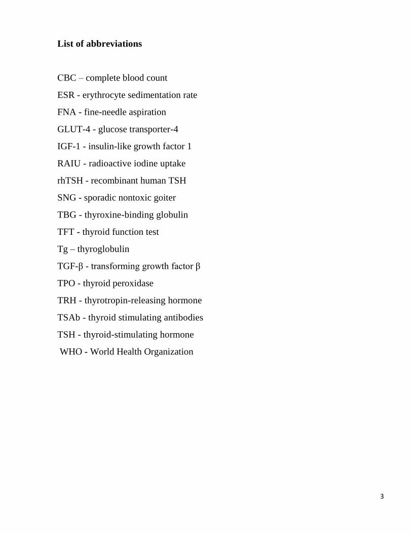
List of abbreviations
СBC – complete blood count
ESR - erythrocyte sedimentation rate
FNA - fine-needle aspiration
GLUT-4 - glucose transporter-4
IGF-1 - insulin-like growth factor 1
RAIU - radioactive iodine uptake
rhTSH - recombinant human TSH
SNG - sporadic nontoxic goiter
TBG - thyroxine-binding globulin
TFT - thyroid function test
Tg – thyroglobulin
TGF-β - transforming growth factor β
TPO - thyroid peroxidase
TRH - thyrotropin-releasing hormone
TSAb - thyroid stimulating antibodies
TSH - thyroid-stimulating hormone
WHO - World Health Organization
4
CONTENTS
Chapter1. Overview of thyroid anatomy and function……………………..4
Iodine deficiency……………………………………………………………..9
Non-toxic goiter……………………………………………………………..10
- Endemic goiter………………………………………………………..10
- Sporadic goiter………………………………………………………..13
Chapter 2. Hypothyroidism………………………………………………...20
Chapter 3. Thyroiditis……………………………………………………....33
Chapter 4. Hyperthyroidism………………………………………………..44
Chapter 5. Thyroid cancer………………………………………………….69
Case-based questions………………………………………………………...81
Answers to review questions………………………………………………...90
Answers to case-based questions with explanations…………………………91
References…………………………………………………………………....95
5
OVERVIEW OF THYROID ANATOMY AND FUNCTION
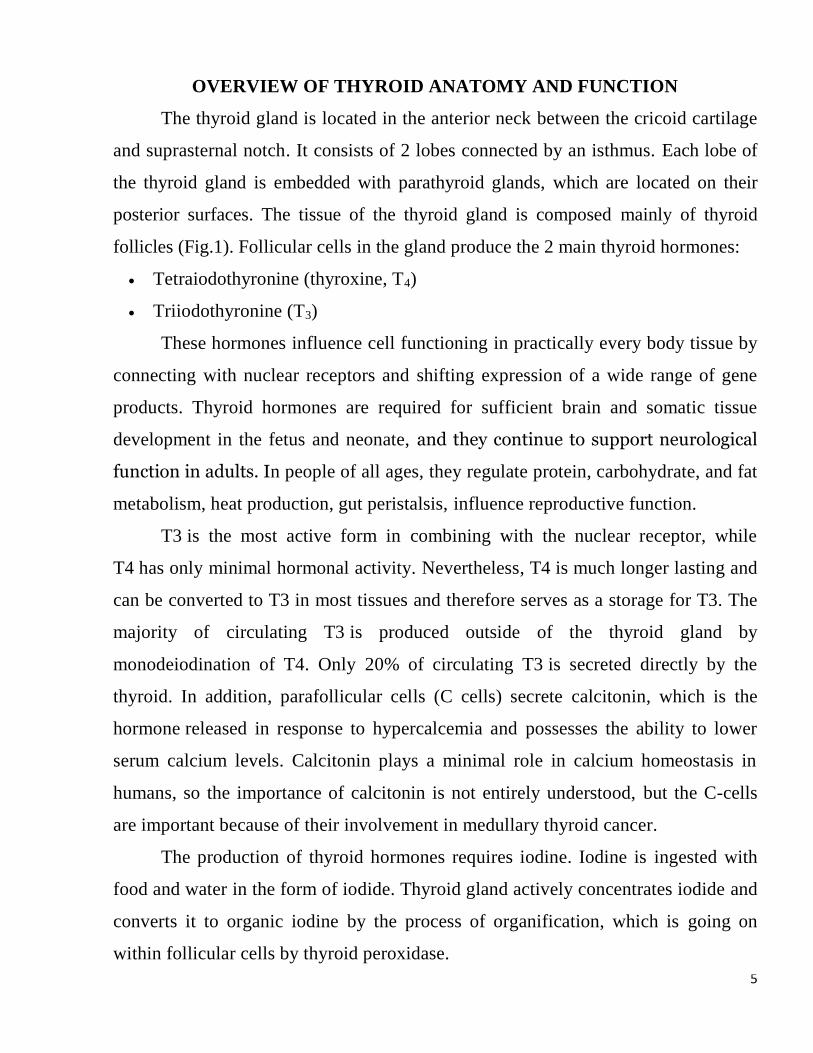
The thyroid gland is located in the anterior neck between the cricoid cartilage
and suprasternal notch. It consists of 2 lobes connected by an isthmus. Each lobe of
the thyroid gland is embedded with parathyroid glands, which are located on their
posterior surfaces. The tissue of the thyroid gland is composed mainly of thyroid
follicles (Fig.1). Follicular cells in the gland produce the 2 main thyroid hormones:
Tetraiodothyronine (thyroxine, T4)
Triiodothyronine (T3)
These hormones influence cell functioning in practically every body tissue by
connecting with nuclear receptors and shifting expression of a wide range of gene
products. Thyroid hormones are required for sufficient brain and somatic tissue
development in the fetus and neonate, and they continue to support neurological
function in adults. In people of all ages, they regulate protein, carbohydrate, and fat
metabolism, heat production, gut peristalsis, influence reproductive function.
T3 is the most active form in combining with the nuclear receptor, while
T4 has only minimal hormonal activity. Nevertheless, T4 is much longer lasting and
can be converted to T3 in most tissues and therefore serves as a storage for T3. The
majority of circulating T3 is produced outside of the thyroid gland by
monodeiodination of T4. Only 20% of circulating T3 is secreted directly by the
thyroid. In addition, parafollicular cells (C cells) secrete calcitonin, which is the
hormone released in response to hypercalcemia and possesses the ability to lower
serum calcium levels. Calcitonin plays a minimal role in calcium homeostasis in
humans, so the importance of calcitonin is not entirely understood, but the C-cells
are important because of their involvement in medullary thyroid cancer.
The production of thyroid hormones requires iodine. Iodine is ingested with
food and water in the form of iodide. Thyroid gland actively concentrates iodide and
converts it to organic iodine by the process of organification, which is going on
within follicular cells by thyroid peroxidase.
6
7
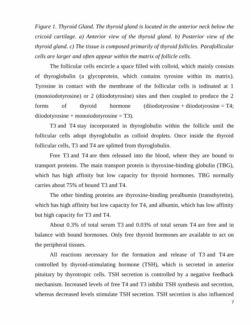
Figure 1. Thyroid Gland. The thyroid gland is located in the anterior neck below the
cricoid cartilage. a) Anterior view of the thyroid gland. b) Posterior view of the
thyroid gland. c) The tissue is composed primarily of thyroid follicles. Parafollicular
cells are larger and often appear within the matrix of follicle cells.
The follicular cells encircle a space filled with colloid, which mainly consists
of thyroglobulin (a glycoprotein, which contains tyrosine within its matrix).
Tyrosine in contact with the membrane of the follicular cells is iodinated at 1
(monoiodotyrosine) or 2 (diiodotyrosine) sites and then coupled to produce the 2
forms of thyroid hormone (diiodotyrosine + diiodotyrosine = T4;
diiodotyrosine + monoiodotyrosine = T3).
T3 and T4 stay incorporated in thyroglobulin within the follicle until the
follicular cells adopt thyroglobulin as colloid droplets. Once inside the thyroid
follicular cells, T3 and T4 are splitted from thyroglobulin.
Free T3 and T4 are then released into the blood, where they are bound to
transport proteins. The main transport protein is thyroxine-binding globulin (TBG),
which has high affinity but low capacity for thyroid hormones. TBG normally
carries about 75% of bound T3 and T4.
The other binding proteins are thyroxine-binding prealbumin (transthyretin),
which has high affinity but low capacity for T4, and albumin, which has low affinity
but high capacity for T3 and T4.
About 0.3% of total serum T3 and 0.03% of total serum T4 are free and in
balance with bound hormones. Only free thyroid hormones are available to act on
the peripheral tissues.
All reactions necessary for the formation and release of T3 and T4 are
controlled by thyroid-stimulating hormone (TSH), which is secreted in anterior
pituitary by thyrotropic cells. TSH secretion is controlled by a negative feedback
mechanism. Increased levels of free T4 and T3 inhibit TSH synthesis and secretion,
whereas decreased levels stimulate TSH secretion. TSH secretion is also influenced
8
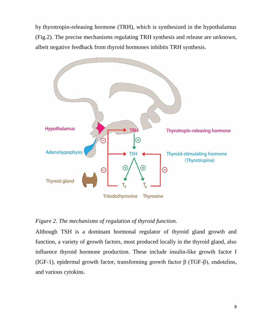
by thyrotropin-releasing hormone (TRH), which is synthesized in the hypothalamus
(Fig.2). The precise mechanisms regulating TRH synthesis and release are unknown,
albeit negative feedback from thyroid hormones inhibits TRH synthesis.
Figure 2. The mechanisms of regulation of thyroid function.
Although TSH is a dominant hormonal regulator of thyroid gland growth and
function, a variety of growth factors, most produced locally in the thyroid gland, also
influence thyroid hormone production. These include insulin-like growth factor I
(IGF-1), epidermal growth factor, transforming growth factor β (TGF-β), endotelins,
and various cytokins.
9
IODINE DEFICIENCY.
Dietary iodine is essential for the synthesis of T3 and T4. Most environmental
iodine occurs in seawater as iodide; a small amount enters the atmosphere and,
through rain, enters ground water and soil near the sea. However, for the large part of
the world’s population, foods do not provide proper levels of this trace mineral,
because the amount varies according to the level in the soil in which the food was
grown, as well as the irrigation and fertilizers used. Wild-grown seafood tends to have
high levels because it concentrates iodine from seawater, but many people in inland
regions lack access to marine fish and other sea products. That’s why iodine
deficiency is prevalent in many mountainous regions throughout the world and in
central Africa, central South America, and northern Asia. Europe remains slightly
iodine deficient and statistical data indicate that iodine intake has been decreasing in
the United States and Australia. The World Health Organization (WHO) estimates
that about 2 billion people in a world suffer from iodine deficiency, the data are based
on iodine urinary excretion screening. Thus, the main source of dietary iodine in
many countries is iodized salt, bread, vegetable oils. Consuming of iodine-enriched
products has markedly reduced the prevalence of cretinism in the world.
Dietary iodine deficiency can result in the abated ability to synthesize T3 and
T4, leading to a number of severe disorders. When T3 and T4 are lacking, TSH is
secreted in increasing amounts. As a result of overstimulation by TSH, thyroglobulin
is being accumulated in the follicles of thyroid gland, increasing their deposits of
colloid. The accumulation of colloid increases the overall size of the thyroid gland.
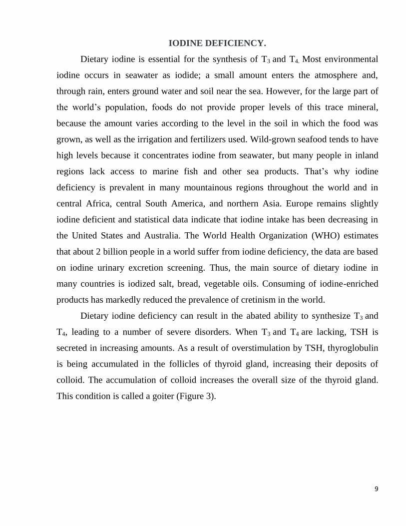
This condition is called a goiter (Figure 3).
10
Figure 3.Goiter.
NON-TOXIC GOITER
Non-toxic goiter (diffuse, endemic uninodular or multinodular) is a disease of
thyroid gland, characterized by the increase of its size; results from iodine deficiency
and is not associated with hyperthyroidism, hypothyroidism, thyroiditis or neoplasm.
Endemic goiter is defined as thyroid enlargement that occurs in more than 10%
of a population. A goiter is only a visible sign of the deficiency. If thyroid function is
preserved, patients are commonly asymptomatic. The deepening of iodine deficiency
or the increase of iodine demand due to puberty, pregnancy, severe somatic diseases
may provoke progressive increase of goiter size. Large goiters may cause obstructive
symptoms due to compression of the trachea and/or the esophagus. Nodules appear in
thyroid gland in 10-15 years of the disease and goiter eventually becomes
multinodular. In case of exhaustion of body’s compensatory abilities, hypothyrosis
might ultimately develop. When iodine deficiency is severe, hypothyroidism and
cretinism develop. Cretinism (neonatal hypothyroidism) is characterized by cognitive
underdevelopment, short body height, and occasionally deafness and muteness in
children and adults born to mothers who were lacking iodine during pregnancy and
who weren’t treated with iodine or thyroid hormone to restore normal thyroid
hormone levels during early childhood. Concomitant selenium, zinc, copper,
11
manganese deficiency may also contribute to the neurologic manifestations of
cretinism.
Other iodine deficiency disorders include stunted growth and development, low
fertility, and prenatal and infant death. Moreover, iodine deficiency is the main cause
of preventable mental retardation.
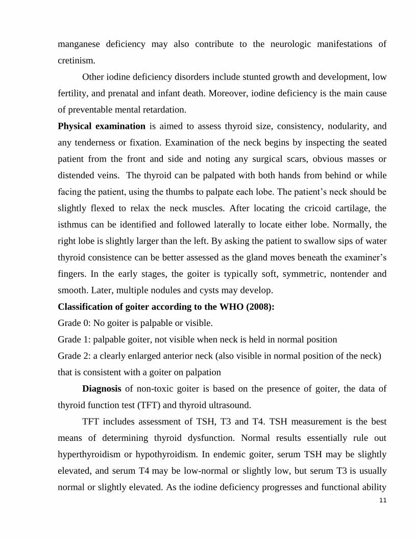
Physical examination is aimed to assess thyroid size, consistency, nodularity, and
any tenderness or fixation. Examination of the neck begins by inspecting the seated
patient from the front and side and noting any surgical scars, obvious masses or
distended veins. The thyroid can be palpated with both hands from behind or while
facing the patient, using the thumbs to palpate each lobe. The patient’s neck should be
slightly flexed to relax the neck muscles. After locating the cricoid cartilage, the
isthmus can be identified and followed laterally to locate either lobe. Normally, the
right lobe is slightly larger than the left. By asking the patient to swallow sips of water
thyroid consistence can be better assessed as the gland moves beneath the examiner’s
fingers. In the early stages, the goiter is typically soft, symmetric, nontender and
smooth. Later, multiple nodules and cysts may develop.
Classification of goiter according to the WHO (2008):
Grade 0: No goiter is palpable or visible.
Grade 1: palpable goiter, not visible when neck is held in normal position
Grade 2: a clearly enlarged anterior neck (also visible in normal position of the neck)
that is consistent with a goiter on palpation
Diagnosis of non-toxic goiter is based on the presence of goiter, the data of
thyroid function test (TFT) and thyroid ultrasound.
TFT includes assessment of TSH, T3 and T4. TSH measurement is the best
means of determining thyroid dysfunction. Normal results essentially rule out
hyperthyroidism or hypothyroidism. In endemic goiter, serum TSH may be slightly
elevated, and serum T4 may be low-normal or slightly low, but serum T3 is usually
normal or slightly elevated. As the iodine deficiency progresses and functional ability

12
of thyroid gland drops, TSH might get increased, levels of T3 and T4 decrease,
indicating the beginning of hypothyroidism development.
Thyroid antibodies are measured to rule out Hashimoto thyroiditis.
Thyroid ultrasoundis the method of choice when it is important to determine
thyroid size and structure accurately. It allows to detect nodules and cysts ≥2 mm and
to monitor their size.Ultrasound-guided fine-needle aspiration (FNA) biopsy of
thyroid lesions allows precise assessment of their histological structure and lowers the
rate of inadequate sampling.
In the early stages, thyroidal radioactive iodine uptake may be normal or high
with normal thyroid scans.
Urinary excretion of iodine allows assessing the level of iodine deficiency in
the population. The urinary excretion of iodine is determined in 50-100 people who
live at certain territory. Normal iodineuria in teenagers and adults is 100 mcg/L and
above.
Treatment and prevention
All patients who were diagnosed with hypothyroidism should be prescribedwith
L-thyroxine in the doses necessary to achieve euthyroidism. It is prescribed to patients
with 2nd degree goiter to reduce the TSH content in the blood. The dose of L-
thyroxine ranges from 100 to 150 micrograms per day. This requires careful
monitoring of the patient's condition. The appearance of sweating, palpitations,
irritability may indicate an overdose of L-thyroxine or the presence of autonomous
nodes.
Patients with large goiters are treated surgically, when the function of several
adjacent organs is deranged. Goiters with signs of malignancy also require surgical
treatment.
The elimination of iodine deficiency is achieved by iodine prevention: mass,
group, individual.
13
Mass prevention is performed to everyone living at iodine deficient territories.
The use of iodized salt is an effective method. Iodization is done with potassium
iodide and potassium iodate. The oil solution of iodine can be also used for mass
prevention. It provides the body with iodine for 6-12 months when taken orally, 1-3
years – when given in intramuscular injection.Iodine preparations (potassium iodide,
200 mcg 1/day) are used for individual and group prevention.
Group prevention is an additional provision of iodides against the background
of mass prevention for those categories of persons, who have increased need for
iodine: pregnant and lactating women, children, teenagers.
The recommended average daily intake of iodine for adults is 150-250 mcg/d,
for children – 90-120 mcg/d, for pregnant and lactating women – 250 mcg/d. The
oversupply of iodine is associated with an increased incidence of autoimmune thyroid
disease.
Individual prevention is performed for individuals with an increased personal
need for iodine. These include women a year before the planned pregnancy; patients
recovering from severe somatic or infectious diseases;patients finishing courses of
antibiotic therapy or treatment with sulfonamides; people with lesions of the digestive
system, when iodine absorption is affected.
SPORADIC NONTOXIC GOITER
Sporadic nontoxic goiter (SNG) is defined as a benign enlargement of the
thyroid gland in a euthyroid person, who lives in an iodine-sufficient area. This type
of goiter is 10 times more common in women than in men and it occurs around
puberty in both genders and during pregnancy and lactation in women suggesting its
relation to the physiological demand for iodine. The incidence decreases with age in
both genders, and there is significant geographic variation in nonendemic goiter areas
for the development of sporadic simple goiter. The cause of SNG is thought to be
relative iodine deficiency. In addition, other factors that play a significant role include
presence of dietary goiterogens (cassava, maize, bamboo shoots, sweet potatoes, lima
14
beans, broccoli, cabbage), certain chemicals that interfere with thyroid hormone
synthesis, and certain drugs, such as paramino-salicylic acid, sulphonylureas,
lithium,and excessive iodine. Also autoimmune and genetic factors play a role. A
large number of patients demonstrate no obvious cause of SNG.
SNG can be diffuse, uninodular, or multinodular and can vary greatly in size,
growth rate, and clinical presentation. SNG is a common essence in clinical practice,
because patients often present with a small, diffuse goiter or a solitary palpable
nodule.
The natural history of SNG is characterized by gradual increase of size, with
eventual development of multiple nodules, possible appearance of local compressive
symptoms, and/or cosmetic issues. The growth rate is changeable, and patients may
have stable goiter size for many years.
With the passage of time, there is a tendency for SNGs to form nodules, which
can become autonomous and eventually cause subclinical or overt hyperthyroidism. It
is found that hyperthyroidism develops in approximately 10% of patients with SNG
after 10 years of monitoring, however most of those patients had suppressed TSH
levels and subclinical hyperthyroidism on presentation.
The lab investigation of a patient with a SNG should begin with a determination
of TSH level in blood, because many patients who are clinically euthyroid have
biochemical evidence of hypo- or hyperthyroidism. The degree of thyroid malfunction
is often mild or subclinical, proven by an isolated TSH abnormality. Both subclinical
and overt hypothyroidism should be treated, to reverse or prevent symptoms as well
as prevent further gland enlargement. Apparent hyperthyroidism should also be
treated, especially because many patients with SNG and hyperthyroidism are elderlies
and have increased cardiac risks. It is more problematic to decide whether to treat
subclinical hyperthyroidism, which is the most common thyroid function disorder in
SNG. An increasing number of evidence suggests that subclinical hyperthyroidism is
15
harmful to the heart, bone, and cognitive function. Therefore treatment decisions
should take into account these risks in an individual patient.
Ultrasound should be done all patients, because approximately 50% have
multiple nodules that are not detected on physical examination. Also, periodical
ultrasound measurements are very sensitive in detecting nodule growth. Upon
discovery of nonpalpable nodules, a fine-needle aspiration biopsy should be done to
any nodule that is 1–1.5 cm in diameter and above, to exclude the presence of thyroid
cancer, as recommended by international endocrinology societies. The sonographic
signs suggestive of nodule malignancy are the following: the presence of
microcalcifications, hypoechogenicity, increased vascularity, rapid growth, uneven
borders.
Radioiodine uptake. The thyroid gland selectively transports radioisotopes of 123I, 125I,
131I, allowing thyroid imaging and quantitation of radioactive tracer uptake, so the
visualization of nodules and determination of their function can be done.
The treatment goals for a patient with a benign SNG include relief of local
compressive symptoms or cosmetic deformity, prevention of progressive thyroid
growth, and treatment of corresponding thyroid dysfunction. These symptoms vary
greatly among patients, from those with no symptoms and an accidentally discovered
goiter to those with compression of adjacent organs. Therefore, there is no optimal
treatment for SNG, and treatment decisions must be personalized.
As for SNG, four main treatment options exist:
follow-up without treatment;
thyroidectomy;
levothyroxine (L-thyroxine) suppression;
radioactive iodine.
Follow-up without treatment. A period of cautious waiting in patients with no
local symptoms or thyroid dysfunction is often the best option. Such patients should
16
be properly monitored by clinical examination and repeated ultrasound measurements
of overall thyroid and nodule size should be done.
Thyroidectomy is an appropriate option in SNG. Many factors should be
considered when making a pro-surgery decision, including patient’s general health
condition, size of goiter, symptomatology, and availability of skilled surgeon.
Indications to surgical treatment of nodular goiter:
• Nodes with signs of malignancy
• Autonomic adenomas
• Retrosternal nodes
• Nodes that rapidly increase in size, regardless of their cellular composition
• Nodes, squeezing the organs of the neck and mediastinum.
• Nodes in children, women up to 20 years of age, men of all ages.
The long-term recurrence rate after surgical treatment depends on the extent of
operation, ranging from 0% for total thyroidectomy to 60% for unilateral
thyroidectomy. The average time to recurrence can take many years, and most
patients with recurrence do not need reoperation. Postoperative treatment with L-
thyroxine does not affect the recurrence rates.
L-thyroxine suppression. L-thyroxine is used in doses meant to suppress TSH.
As we know, TSH is a growth factor for SNG and suppressing TSH levels removes
this growth stimulus and causes goiter stabilization. However, several placebo-
controlled studies have been disappointing concerning goiter shrinkage, although one
study registered a prevention of nodule growth with L-thyroxine over 5 year period.
SNGs typically regrow when L-thyroxine is discontinued, necessitating uncertain
treatment. In this case patient may have subclinical hyperthyroidism for many years.
Contemporary knowledge suggests that subclinical hyperthyroidism causes bone loss,
increased risk of atrial fibrillation and other heart disorders. Also, neuropsychiatric
and cognitive effects can be seen. The equivocal long-term efficacy of TSH
17
suppression, combined with these risks, has led to a descent of enthusiasm concerning
this treatment option.
Radioactive iodine. The first reports of the use of radioactive iodine for the
treatment of large multinodular goiters appeared in the 1960s, and a series of
uncontrolled studies followed. The size of the goiter decreased in all cases by 40% or
more, and most patients had significant relief of compressive symptoms. Iodine-131
(131I) was used for treatment. Side effects were mild, except the high rates of
hypothyroidism development. Another possible side effect includes the risk of
radioiodine-induced carcinogenesis: a slight increase in rates of kidney, stomach,
bladder, breast, and brain cancers were documented. The problem is that 131I doses for
SNG are typically higher than those proposed for treatment of Graves’ disease, and,
therefore, the extrathyroidal tissue exposure is much higher than that obtained when
treating Graves’ disease. Therefore, this might be a problem, especially for young
patients, and has to be discussed with them in the realms of whether to choose surgery
or radioactive iodine therapy for SNG.
If the administered dose of 131I is a problem, then efforts to improve treatment
effectiveness of SNG while minimizing 131I doses make sense. A reasonable option is
the use of recombinant human TSH (rhTSH) in SNGs. rhTSH promotes iodine uptake
into both normal and abnormal thyroid tissue. Very low doses of rhTSH (0.01 and
0.03 mg) significantly increase 24-h radioactive iodine uptake in patients with
multinodular goiters. This method is very promising and allows to minimize side
effects of radioiodine therapy in patients with SNG.
18
Review questions:
1. Which of the following statements about the thyroid gland is true?
A. It is located anterior to the trachea and inferior to the larynx.
B. The parathyroid glands are embedded within it.
C. It produces three hormones.
D. All of the above
2. The secretion of thyroid hormones is controlled by:
A. TSH from the hypothalamus
B. TSH from the anterior pituitary
C. Thyroxine from the anterior pituitary
D. Thyroglobulin from the thyroid’s parafollicular cells
3. The development of a goiter indicates that:
A. The anterior pituitary is abnormally enlarged
B. There is hypertrophy of the thyroid’s follicle cells
C. There is an excessive accumulation of colloid in the thyroid follicles
D. The anterior pituitary is secreting excessive growth hormone
4. Iodide ions cross from the bloodstream into follicle cells via:
A. Simple diffusion
B. Facilitated diffusion
C. Active transport
D. Osmosis
5. Which assay allows differentiating between endemic goiter and sporadic goiter?
A. Serum TSH
B. Ultrasound of the thyroid gland
C. Radioiodine uptake by the thyroid gland
D. Urinary excretion of iodine
6. The chronic oversupply of iodine is associated with an increased incidence of:
A. Autoimmune thyroid disease
19
B. Hypothyroidism
C. Thyrotoxicosis
D. Thyroid cancer
7. The types of iodine deficiency prevention in endemic region include:
A. Mass prevention
B. Group prevention
C. Individual prevention
D. All of the above
8. The treatment of endemic non-toxic diffuse goiter as a rule should be:
A. Surgical
B. Conservative
C. Radioiodine
D. Combined
9. What is the purpose of a radioiodine uptake study?
A. Determination of the size of the thyroid gland
B. Determination of the functional state of the thyroid gland
C. To detect the presence of nodes and determine their functional state
D. Determination of structure of the thyroid gland
10. What dosage of L-thyroxine should be prescribed to patients with 2nd degree
goiter and above who have an increased TSH content in the blood?
A. 1 mcg/kg
B. 2 mcg/kg
C. 0.5 mcg/kg
D. 3 mcg/kg

20
HYPOTHYROIDISM
Hypothyroidism is a syndrome, characterized by thyroid hormone deficiency.
Etiological classification of hypothyroidism:
Primary: caused by disease in the thyroid
Secondary: caused by disease in the hypothalamus or pituitary
Primary hypothyroidism develops because of the disease in the thyroid gland
itself. TSH in this case is increased. The most common cause is autoimmune lesion.
It usually occurs due to Hashimoto thyroiditis and the goiter is firm on palpation. As
disease progresses, a shrunken fibrotic thyroid can be palpated with little or no
function. The second most common cause is iatrogenic hypothyroidism, which is
typically caused by radioactive iodine therapy or thyroidectomy for hyperthyroidism
or goiter. Overtreatment with antithyroid medications like
propylthiouracil, methimazole, and iodide might cause hypothyroidism, however, it
abates after therapy is stopped.
Endemic goiter can be the reason for goitrous hypothyroidism due to iodine
deficiency. Iodine deficiency lowers the production of thyroid hormones. As the
result, TSH is released, which stimulates thyroid gland to grow and trap iodine
actively, therefore, goiter develops. Patient with endemic goiter becomes
hypothyroid if iodine deficiency is severe and prolonged.
Goitrous hypothyroidism can be also caused by rare inherited enzymatic
defects can modify the synthesis of thyroid hormone. Congenital goiters may be
caused by dyshormonogenesis (abnormal thyroid hormone production),
transplacental passage of maternal antibodies, or transplacental passage of
goitrogens. Some causes of congenital goiter are hereditary. Also, aplasia and
hypoplasia of thyroid gland can be the cause of hypothyroidism.
Infiltrative disorders that might cause primary hypothyroidism are the
following: amyloidosis, sarcoidosis, hemochromatosis, scleroderma.
21
Administration of lithium can be potential reason for hypothyroidism,
because lithium inhibits hormone release by the thyroid.
Some medications like amiodarone or other iodine-containing drugs;
interferon-alfa; checkpoint inhibitors or tyrosine kinase inhibitors for cancer can
also induce hypothyroidism. Hypothyroidism may result from radiation therapy for
laryngeal cancer or Hodgkin's lymphoma.
Secondary (central) hypothyroidism occurs when the hypothalamus does not
produce enough TRH or the pituitary gland does not produce enough TSH.
Sometimes insufficient secretion of TSH due to insufficient secretion of TSH is
called tertiary hypothyroidism. The causes for central hypothyroidism are the
following: pituitary adenomas, previous pituitary/hypothalamic surgery or
radiotherapy, history of head trauma, history of pituitary apoplexy,
hypothalamic/suprasellar tumors.
Subclinical hypothyroidism is characterized by increased serum TSH in
patients with minimal or no symptoms of hypothyroidism and normal serum levels
of free T4.
Subclinical hypothyroidism is relatively common. It occurs in more than 15%
of elderly women and 10% of elderly men, especially in those with
underlying Hashimoto thyroiditis.
Patients, whose plasma TSH is above 10 mU/L, have high probability of
progression to overt hypothyroidism with low plasma levels of free T4 in the next 10
years. Such patients are also more prone to hypercholesterolemia and
atherosclerosis. They should be prescribed L-thyroxine, even if they are
asymptomatic.
A trial of L-thyroxine makes sense for patients with TSH levels between 4.5
and 10 mU/L if symptoms of early hypothyroidism (for example fatigue,
forgetfulness, depression) are present.
22
A therapy with L-thyroxine is also indicated for pregnant women and those
women who plan to become pregnant in order to avoid the harmful effects of
hypothyroidism on the course of pregnancy and fetal development. Patients should
have their TSH and free T4 measured annually to evaluate the dynamics of condition
if untreated, or to adjust the dosage of L-thyroxine.
Peripheral hypothyroidism is a type of permanent congenital hypothyroidism
that results from peripheral defects in thyroid hormone metabolism. Peripheral
hypothyroidism may be caused by peripheral resistance to the action of thyroid
hormone due to dominantly inherited mutations in genes encoding for thyroid
hormone receptor beta. The majority of these individuals have normal thyroid
function. Peripheral hypothyroidism may also be caused by defects in thyroid
hormone transport, such as in Allan-Herndon-Dudley syndrome where X-linked
peripheral hypothyroidism is associated with mental retardation and neurologic
abnormalities including quadriplegia.
Transient hypothyroidism develops in case of certain diseases of thyroid
gland or might result from some treatment strategies of thyroid diseases. It is prone
to spontaneous disappearance after elimination of etiologic factors. It may occur in
patients with silent thyroiditis, including postpartum thyroiditis, subacute thyroiditis,
withdrawal of thyroxine treatment in patients with intact thyroid, after 131I treatment
or subtotal thyroidectomy for Grave’s disease.
Pathogenesis: the pathogenetic basis of clinical hypothyroidism is
derangement of all types of metabolism as a result of thyroid hormones deficiency,
which results in decline of basic metabolic rates. These changes are especially
manifested in the organs and tissues that are intensively renewed. Erythropoiesis
slows down and anemia develops. Due to the slowing down of lipid metabolism,
free fatty acids, triglycerides, and cholesterol accumulate in the body. The particular
role is played by the disorders of protein metabolism. As the result of slowing down
of protein metabolism, the accumulation of proteinaceous ground substance is going

23
on, which includes derivatives of proteins, as well as glucuronic and chondroitinic
acids. Proteinaceous ground substance accumulates in interstitia, causing mucinous
edema. Due to the high hydrophilicity of proteinaceous ground substance there is
accumulation of significant amount of sodium, chlorides and waterin the extra-
vascular structures (eg., skin, heart, muscles, body cavities). This leads to the
development of hydrothorax, hydropericardium, and ascites. In children, the
deficiency of thyroid hormones causes a slowing of growth, physical, mental, sexual
development, up to the growth retardation and cretinism. In the primary
hypothyroidism there is an absence of inhibitory effects towards the release of
thyrotropin and thyroliberin, so the latter is produced affluently. Due to the
immunological and structural proximity of thyroliberin and prolactoliberin, a
secretion of prolactin is promoted and galactorrhea develops.
Signs and symptoms:
Metabolic manifestations: Cold intolerance, moderate weight gain (due to fluid
retention and slowed metabolism), hypothermia;
Neurologic manifestations: Forgetfulness, impaired concentration, paresthesias
of the hands and feet (develop due to carpal tunnel syndrome caused by
deposition of proteinaceous ground substance in the ligaments around the wrist
and ankle); retardation of the relaxation phase of deep tendon reflexes;
Psychiatric manifestations: Dull facial expression, personality changes,
depression, dementia or frank psychosis (myxedema madness);
Dermatologic manifestations: Sparse, coarse and dry hair; facial puffiness;
myxedema; loss of lateral eyebrows, coarse, dry, scaly and thick skin;
macroglossia due to deposition of proteinaceous ground substance in the
tongue; carotenemia, particularly notable on the palms and soles (caused by
deposition of carotene in the lipid-rich epidermal layers);
24
Ocular manifestations: Dropping eyelids because of decreased adrenergic
drive; periorbital swelling due to infiltration with the mucopolysaccharides,
hyaluronic acid and chondroitin sulfate);
Gastrointestinal manifestations: Constipation;
Gynecologic manifestations: Menorrhagia or secondary amenorrhea, reduced
fertility, miscarriages;
Cardiovascular manifestations: Bradycardia (a decrease in both thyroid
hormone and adrenergic stimulation causes slow heart rate), enlarged heart on
examination and imaging (mainly because of pericardial effusion, but dilation
also contributes), hypertension (primarily diastolic);
Other manifestations: Hoarse voice, and slow speech; pleural and/or abdominal
effusions (pleural effusions develop slowly and only rarely cause respiratory or
hemodynamic distress).
The appearance of symptoms depends on the degree of hypothyroidism
severity.
Although secondary hypothyroidism is very rare, its etiological causes often
affect other endocrine organs controlled by the hypothalamic-pituitary system.
Secondary hypothyroidism is characterized by the fact that the skin and hair are dry,
but not very rough, there is skin depigmentation, minimal macroglossia, atrophic
breasts and low blood pressure. In addition, the heart is small, and serous pericardial
effusions do not occur. Hypoglycemia is common due to concomitant adrenal
insufficiency or growth hormone deficiency.
Myxedema coma is a life-threatening complication of hypothyroidism. It
usually occurs in older patients with a long history of hypothyroidism. Myxedema
coma almost always occurs in the elderly. Precipitating factors include infection,
illness, drugs that suppress the CNS (particularly sedatives, anesthetics,
antidepressants), trauma, and exposure to cold. There may be a history of treated
hypothyroidism with poor compliance, or the patient may be previously
25
undiagnosed. Hypoventilation, leading to hypoxia and hypercapnia, plays a major
role in pathogenesis; hypoglycemia and dilutional hyponatremia also contribute to
the development of myxedema coma. The coma is characterized by extreme
hypothermia (body temperature 24° to 32.2° C), seizures, areflexia, and inhibition of
respiratory functions with carbon dioxide retention. Assessment of severe
hypothermia requires usage of low-reading thermometers, otherwise it can be
missed. Prompt diagnosis based on history, physical examination and express lab
data is vitally important, because death is likely without quick treatment.
Diagnosis. The diagnosis of hypothyroidism is made from the history, the
clinical picture and the laboratory measurements.
TSH and free T4 measurement are the laboratory examinations necessary for
the diagnosis of hypothyroidism and the differential diagnosis between primary
(clinical or subclinical) and secondary one.
When TSH is increased and free T4 is decreased or normal hypothyroidism is
primary. In this case increased anti-TPO or anti-Tg antibodies point to the cause of
hypothyroidism, which is autoimmune thyroiditis. Primary hypothyroidism is
divided in clinical when TSH is increased and free T4 is decreased and in subclinical
when TSH is increased and free T4 is normal.
Many patients with primary hypothyroidism have normal circulating levels of
triiodothyronine (T3), which is probably due to prolonged TSH stimulation of the
failing thyroid gland, which leads to the predominant synthesis and secretion of
biologically active T3. Therefore, serum T3 is not sensitive for hypothyroidism.
When TSH is normal or decreased and free T4 is low hypothyroidism is
secondary (central). In order to discriminate whether the cause is in the pituitary or
the hypothalamus a test with the TSH releasing factor is performed (TRH test). In
the first case the response is normal, while in the second it is abnormal. In central
hypothyroidism imaging studies of the brain and the pituitary (MRI, CT) are
performed aiming at finding its cause.
26
Usually the reported normal limits of TSH are between 0.4-4.0 mU/l. When
TSH is found in the upper normal limits it may show mild hypothyroidism which
may progress to hypothyroidism, especially if antibodies are increased.
TSH may be increased in euthyroid individuals in certain situations. Increased
TSH (5-20 mU/l) is observed during convalescence from non-thyroidal illness
(euthyroid sick syndrome), as well in pituitary adenomas producing TSH or in
isolated resistance of the pituitary to thyroid hormones. Finally, TSH increase may
be observed in chronic renal failure and in primary adrenal insufficiency.
Complete blood count may demonstrate the presence of anemia, usually
normocytic-normochromic. In some cases, anemia may be hypochromic because of
menorrhagia and sometimes macrocytic because of associated vitamin B12 or B9
deficiency. Anemia is rarely severe and Hb level is usually > 90 g/L. Anemia
usually subsides as the hypometabolic state is corrected, which sometimes takes 6 to
9 months.
Serum cholesterol and triglycerides are usually elevated in primary
hypothyroidism but not so much in secondary hypothyroidism.
Hypothyroidism is associated with atherosclerosis, endothelial dysfunction,
increased carotid intima-media thickness and impaired cardiac contractility.
Treatment
Various thyroid hormone preparations can be used for replacement therapy, the
most common are: synthetic preparations of T4 (l-thyroxine), T3 (liothyronine),
combinations of two synthetic hormones, and desiccated animal thyroid extract. L-
Thyroxine is a drug of choice, the usual maintenance dose is 75 to 150 mcg per os
once a day, depending on age, body mass index, and the intensity of absorption. The
beginning dose in young patients or those of middle age, who are otherwise healthy,
can be 100 mcg or 1.7 mcg/kg per os once a day.
A careful approach should be applied to patients with heart disease, the therapy
should begin with low doses for them, which is usually 25 mcg once a day. The dose
27
is higher in individuals having been subjected to thyroidectomy than those with
chronic autoimmune thyroiditis, as in those there are remnants of functioning
thyroid tissue. In subclinical hypothyroidism the dose is low (0.5 µg/kg). In
pregnancy, a larger dose is required (2 µg/kg). During pregnancy the increase in
dose that may be required is 25-47% more than the one before pregnancy and it is
observed during the 4th to 6th week. In all cases the dose should be adjusted every 6
weeks until maintenance dose is reached. An increase of dose may be needed if
drugs that decrease T4 absorption or increase its biliary excretion are administered
concomitantly. The dose used should be the lowest that provides achievement of the
midnormal range of serum TSH or its lower normal limits (approximately 1.0 mU/l).
TSH measurement after the initiation of therapy is performed every 4-6 weeks. The
follow-up is performed by TSH measurement once every year. In pregnancy the first
TSH measurement should be performed when pregnancy is diagnosed and thereafter
every 3-4 weeks during the first half of the pregnancy and every 6 weeks thereafter.
The evaluation of TSH cannot be used to control the efficacy of treatment in patients
with secondary hypothyroidism. In secondary hypothyroidism the dose of L-
thyroxine should achieve a free T4 in the midnormal range.
The clinical effects of treatment with l-thyroxine appear slowly. Patients may
not have full relief from symptoms until 3-6 months after midnormal TSH levels are
restored.
Great caution is needed in substitution therapy with thyroxine as dose
overestimation has consequences. It has been observed that more than one fifth of
the patients have clinical or subclinical hyperthyroidism. These consequences are
atrial fibrillation, aggravation of coronary artery disease and a decrease in bone
mineral density, fractures of the spine and the hip being observed in women >65
years.
Liothyronine (L-triiodothyronine) has short half-life and produces the large
peaks in serum T3 levels, therefore it shouldn’t be used alone for long-term

28
replacement therapy. The use of standard replacement amounts (from 25 to 37.5
mcg twice a day) leads to a rapid increase in serum T3 levels to 300-1000 ng/dl
(4.62-15.4 nmol/L) within 4 hours because of its almost complete absorption. These
levels return to normal by 24 hours. In addition, patients receiving liothyronine
develop chemical hyperthyroid state for at least several hours a day, which
potentially increases the risk of heart disease.
Similar fluctuations of serum T3 occur when mixed preparations of T3 and
T4 are taken per os. Although the peak of T3 in this case is lower because less T3 is
given.
Replacement therapy with L-thyroxine produces different pattern of serum T3
response. Serum T3 levels increase gradually, and normal levels are maintained
when adequate doses of T4 are given. The use of desiccated animal thyroid extracts
demonstrates less controllable clinical effect because they contain different amounts
of T3 and T4, and should not be prescribed unless the patient is already taking the
preparation and has normal serum TSH.
In patients with secondary hypothyroidism, the administration of l-thyroxine
can potentially precipitate adrenal crisis, therefore l-thyroxine should not be given
until there is an evidence of adequate cortisol secretion, or cortisol therapy is given
concomitantly.
Treatment of myxedema coma. In myxedema coma, the danger of death was 60-
70% in 1985 but it has decreased to 20-25%, owing to the timely diagnosis and the
referral of patients to acute care units.
Levothyroxine can initially be administered as a single IV bolus of 500 mcg,
which serves as a loading dose. Although further levothyroxine is not strictly
necessary for several days, it is usually continued at a dose of 50-100 mcg/day. If
suitable IV preparation is not available, the same initial dose of levothyroxine can be
given by nasogastric tube (however, absorption can be altered in myxedema). An
alternative is to give liothyronine (T3) IV or via nasogastric tube in doses 10-25 mcg

29
every 8-12 hours. This treatment has been advocated because T4 to T3 conversion is
impaired in myxedema coma. The problem is that excess liothyronine can provoke
arrhythmias. An acceptable option is to combine levothyroxine (200 mcg) and
liothyronine (25 mcg) as a single, initial IV bolus, followed by daily treatment with
levothyroxine (50-100 mcg) and liothyronine (10 mcg every 8 hours).
Supportive therapy should be provided to correct any associated metabolic
disturbances. External warming is indicated only if the body temperature is below 30
degrees Celsius, as it can precipitate hypotension or arrhythmias. Otherwise patient
should be covered with blanket to conserve heat. Parenteral hydrocortisone (50 mg
every 6 hours) should be administered, because there is impaired adrenal reserve in
profound myxedema. The precipitating factors should be rapidly and appropriately
treated, including the early use of broad spectrum antibiotics, pending the exclusion of
infection. Hypoxemia is a common condition, so partial pressure of oxygen should be
monitored. If ventilation is jeopardized, immediate mechanical ventilatory assistance
is needed. Hypertonic saline or IV glucose may be needed if there is severe
hyponatremia or hypoglycemia. The fact that metabolism of most medications is
slowed down should be noted. Sedatives should be avoided if possible or used in low
doses.
Hypothyroidism and insulin resistance.
Several studies have shown overt and subclinical hypothyroidism being more
prevalent in patients with diabetes mellitus than in general population, and women
with subclinical hypothyroidism are believed to be at more risk to develop gestational
diabetes. The thyroid hormones are known to have a stimulating effect on maturation
of the insulin secreting beta cells, and thyroid hormone receptors have been detected
in these cells. Thyroid hormones enhance gluconeogenesis and glycogenolysis in an
opposing effect to insulin, whereas, they are known to facilitate the cellular glucose
uptake by expressing the glucose transporter-4 (GLUT-4) isozyme.
30
Clinical hypothyroidism is considered to be a risk factor for insulin resistance.
Patients with hypothyroidism are characterized by slowing of intestinal glucose
absorption and a decrease in the adrenergic activity, leading together to a reduction in
liver and muscle glycogenolysis, as well as a decrease in gluconeogenesis and
baseline insulin secretion. At the same time, a postprandial increase in insulin
secretion against the background of generalized peripheral insulin resistance has been
observed, associated with a higher concentration of free fatty acids, reduced glucose
uptake and increased glucose oxidation. Thus, hypothyroidism causes a drop in
insulin-dependent glucose utilization.
A strong influence of TSH on the fasting insulin levels and the insulin resistance
was revealed. Some studies have indicated the effect of TSH on insulin action and
that even a subtle increase in plasma TSH levels within the normal range can affect
insulin secretion and may cause insulin resistance and metabolic syndrome.
Moreover, hypothyroid patients are known to experience a decrease in glucose
transporters GLUT4 leading to a reduction of glucose uptake and promoting insulin
resistance. The relationship between thyroid hormonal status and insulin levels in the
pathogenesis of insulin resistance is complex. The higher fasting serum insulin
concentrations were believed to develop as a compensation result of the insulin
resistance. Many authors accept the concept that a patient suffering from an
autoimmune disorder is more prone to be affected by an autoimmune disorder of
insulin resistance. Other investigators relate the occurrence of insulin resistance
among hypothyroid patients to the high prevalence of obesity and the high fat deposits
in this population.
Also, a reduced ability of insulin to increase blood flow in tissues in
hypothyroidism was observed. This may be an alternative mechanism explaining the
effect of hypothyroidism on decreasing glucose utilization by peripheral cells.
31
Review questions:
1. One of the symptoms of hypothyroidism is:
A. Fatigue
B. Intolerance to cold
C. Hair loss
D. All of the above
2. Although the symptoms of hypothyroidism may be difficult to detect, if
hypothyroidism is suspected, the condition can best be diagnosed with:
A. MRI scan
B. Ultrasound
C. Thyroid stimulating hormone test (TSH)
D. Hemoglobin test or hematocrit test
3. In women, hypothyroidism can affect pregnancy by:
A. Reducing the chance of getting pregnant
B. Boosting the chance of getting pregnant
C. Making miscarriage more likely
D. Making labor and delivery more difficult
4. A person with untreated hypothyroidism may also suffer from:
A. High cholesterol
B. Low blood pressure
C. Low blood sugar
D. None of the above
5. How is hypothyroidism treated?
A. With radiation
B. With surgery
C. With a synthetic hormone
D. The condition can't be treated
6. What disease causes the most significant deceleration of Achilles tendon reflex?
32
A. Diabetes mellitus
B. Atherosclerosis
C. Hypokalemia
D. Primary hypothyroidism
7. What assay helps to perform the differential diagnosis between primary and
secondary hypothyroidism?
A. Determination of ТЗ and Т4 level in blood
B. Determination of ТSH in blood
C. Determination of thyrotropin-releasing hormone
D. Determination of prolactin
8. What assay helps to perform the differential diagnosis between secondary and
tertiary hypothyroidism?
A. Determination of ТSH in blood
B. Determination of thyrotropin-releasing hormone
C. Determination of prolactin
D. Determination of ТЗ and Т4 level in blood
9. The main reason for galactorrhea in patients with hypothyrosis is:
A. Deficiency of thyroid hormones
B. ExcessiveTSH
C. Hypersecretion of thyroliberine
D. All of the above
10. The major role in the pathogenesis of myxedema coma belongs to:
A. Hypoventilation, leading to hypoxia and hypercapnia
B. Progressive decline in cardiac output
C. Hypothermia and increasing hypocorticism
D. Hypoglycemia and dilutional hyponatremia
33
THYROIDITIS
Thyroiditis is a general term that refers to the presence of inflammatory process
in the thyroid gland. Thyroiditis includes a group of individual disorders causing
thyroidal inflammation but presenting in different ways.
Classification:
1. Acute thyroiditis (diffuse or local):
a) suppurative;
b) nonsuppurative.
2. Subacute thyroiditis:
a) diffuse;
b) local.
3. Chronic thyroiditis:
a) autoimmune thyroiditis (Hashimoto’s thyroiditis);
b) invasive fibrous (Riedel’s thyroiditis);
c) specific thyroiditis (tuberculosis, lues);
d) caused by physical or chemical agents;
e) parasitic.
Acute thyroiditis
Acute thyroiditis is rare and usually develops due to suppurative infection
(especially Staphylococcus, Streptococcus and Enterobacter) of the thyroid. The most
common cause in young patients is the presence of a piriform sinus, which is a
remainder of the fourth branchial pouch that connects the oropharynx with the
thyroid. A piriform sinus is usually left-sided. Acute thyroiditis can also develop in
the elderly and the risk factors are the long-existing goiter and degeneration in thyroid
malignancy.
Clinical presentation includes thyroid pain, which radiates to throat or ears, and
a small, tender goiter that may be asymmetric. Febrile fever and lymphadenopathy are
common, as well as the changes over the thyroid location: erythema, fever, dysphagia.
34
Lab data: complete blood count (CBC) - elevation of white cell count and
erythrocyte sedimentation rate (ESR). TFT – normal T3, T4, and TSH.
FNA biopsy shows infiltration by polymorphonuclear leukocytes; culture of the
sample and Gram stain can identify the microorganism.
Immunocompromised patients should be treated with a special attention as
fungal (Aspergillus, Candida, Histoplasma), mycobacterial, or Pneumocystis carinii
thyroiditis can occur in these cases.
Treatment: prompt administration of antibiotic therapy. Surgery might also be
needed to drain the abscess, which can be localized either by ultrasound or by CT
scan.
Complications: tracheal obstruction, septicemia, retropharyngeal abscess,
mediastinitis, jugular venous thrombosis. Complications may be successfully avoided
with prompt use of antibiotics.
Acute thyroiditis can be also non-suppurative. It can result from radiation
injury after 131I treatment or usage of amiodarone.
Subacute thyroiditis
(de Quervain Thyroiditis; Giant Cell Thyroiditis; Granulomatous Thyroiditis)
Subacute thyroiditis is an inflammatory disease of the thyroid probably caused
by a virus. History of an antecedent viral infection of upper respiratory tract is
common. Viruses like mumps, coxsackie, influenza, adenoviruses may play a role.
However, the identification of a virus in an individual patient doesn’t influence the
treatment. The peak incidence occurs at 30-50 years; women get sick 3 times more
often than men.
There is inflammation with characteristic giant cell infiltration,
polymorphonuclear lymphocytes, and follicular disruption. The follicular changes
progress to granulomas, accompanied by fibrosis. In several months thyroid gland
returns to normal.

35
Phases of the disease:
1. Hyperthyroid (3-4 weeks). Follicular destruction causes release of Tg and
thyroid hormones, leading to increase of circulating T3 and T4 and
suppression of TSH. At this phase, radioactive iodine uptake is low or
undetectable (Fig.4).
2. Hypothyroid (6-8 weeks). Thyroid gland is depleted of stored hormones,
there is low T3 and T4. TSH levels are moderately increased. Radioactive
iodine uptake returns to normal or might be even increased due to the rise
of TSH.
3. Recovery (3-4 weeks). Thyroid hormones and and TSH levels return to
normal as the disease subsides.
Figure 4. Time Course of Subacute Thyroiditis.
Clinical presentation includes pain in the anterior neck and fever of 37.5° to
38.5° C. Neck pain characteristically shifts from side to side and may settle in one
36
area, frequently radiating to the jaw and ears. It is often confused with dental pain,
pharyngitis, or otitis and is aggravated by swallowing or turning of the head.
There may be signs of thyrotoxicosis or hypothyroidism depending on the
phase of the disease. Clinical signs and symptoms of thyroiditis are often preceded
by symptoms of upper respiratory tract infection. Sometimes the onset of the disease
is sudden, acute and severe without typical foregoing symptoms. On physical
examination, the thyroid is asymmetrically enlarged, firm, and tender.
The usual outcome is complete resolution, but permanent hypothyroidism can
develop, especially in patients with coincidental thyroid autoimmunity or when
follicular destruction is extensive.
Diagnosis is primarily clinical, based on finding an enlarged, tender thyroid in
patients with the appropriate clinical history. Thyroid testing with TSH and at least a
free T4 measurement is usually also done. Radioactive iodine uptake should be
measured to confirm the diagnosis.
Laboratory findings early in the disease include an increase in free T4 and T3,
a marked decrease in TSH and thyroid radioactive iodine uptake (often 0), high ESR
and high white blood cell count. After several weeks, the thyroid is depleted of
T4 and T3 stores, and transient hypothyroidism develops accompanied by a decrease
in free T4 and T3, a rise in TSH, and recovery of thyroid radioactive iodine uptake.
Weakly positive thyroid antibodies may be present. Measurement of free T4, T3,
and TSH at 2-4-weeks intervals identifies the stages of the disease. When the
diagnosis is uncertain, fine-needle aspiration biopsy is useful. Thyroid
ultrasonography with color Doppler shows multiple irregular sonolucent areas and
reduced blood flow in contrast with the increased flow of Grave’s disease.
Treatment. Discomfort, pain in anterior neck is treated with high doses
of aspirin (600 mg every 4-6 hours) or other NSAIDs. In moderately and severely
symptomatic cases, corticosteroids (prednisone 40 to 60 mg per os once/day,
gradually decreasing the dose over 6 to 8 weeks) eradicate all symptoms within 48
37
hours. If a relapse occurs during prednisone withdrawal, treatment should be started
again and withdrawn more gradually. In these patients it is reasonable to wait until
the radioactive iodine uptake normalizes before stopping therapy.
Mild symptoms of thyrotoxicosis improve spontaneously and don’t require
specific treatment. Bothersome hyperthyroid symptoms may be treated with a short
course of a beta-blocker. Antithyroid drugs are useless in treatment of thyrotoxic
phase. If hypothyroidism is marked or persists, thyroid hormone replacement
therapy may be required, rarely permanently. The doses of L-thyroxine should be
low enough to allow TSH-mediated recovery.
Hashimoto’s thyroiditis
Hashimoto thyroiditis is chronic autoimmune inflammation of the thyroid
gland with lymphocytic infiltration.
Hashimoto thyroiditis is the most common cause of primary hypothyroidism
in the world. It is four times more prevalent among women. It is more common in
certain populations, such as the Japanese, probably because of genetic factors and
chronic overexposure to dietary iodine. Incidence increases with age and in patients
with chromosomal disorders, such as Down syndrome, Turner syndrome,
and Klinefelter syndrome. A family history of thyroid disorders is a common finding
upon collecting a history.
Hashimoto thyroiditis is sometimes associated with other autoimmune
disorders, including Addison disease (adrenal insufficiency), type 1 diabetes
mellitus, hypoparathyroidism, vitiligo, celiac disease, premature graying of hair,
connective tissue disorders (rheumatoid arthritis, systemic lupus
erythematosus, Sjögren syndrome), pernicious anemia, and type 2 polyglandular
deficiency syndrome (Schmidt syndrome - a combination of Addison disease with
hypothyroidism secondary to Hashimoto thyroiditis and/or type 1 diabetes mellitus).
There may be an increased incidence of thyroid tumors, rarely thyroid lymphoma.
38
As it happens in other autoimmune diseases, both genetic and environmental
factors work together to determine the susceptibility of a person to autoimmune
thyroiditis. The proven genetic risk factors are HLA-DR polymorphisms, especially
HLA-DR3, -DR4, -DR5 in whites. The female predisposition to thyroid
autoimmunity is associated with sex steroid effects on the immune response; a role
of X-linked genetic factor is also possible. Environmental factors include chronic
exposure to high-iodine diet and viral infections.
Pathologically, there is extensive infiltration of thyroid with lymphocytes
(CD4+ and CD8+ T cells, B cells), atrophy of the follicles, absence of colloid and
mild to moderate scarring. There is production of antibodies to TPO and Tg, which
are the important markers of thyroid autoimmunity, but possess a secondary
pathogenic role as they only reinforce the ongoing autoimmune response. About
20% of patients with autoimmune thyroiditis have inhibiting antibodies against TSH
receptors, which prevent the binding of TSH. These TSH receptor blocking
antibodies contribute to the development of hypothyroidism and, especially in Asian
patients, to thyroid atrophy.
Clinically, patients complain on painless enlargement of the thyroid or
fullness in the throat. Physical examination reveals a nontender goiter that is firm;
has smooth or nodular surface, and is rubberier than the normal thyroid. Many
patients present with symptoms of hypothyroidism, but some present
with hyperthyroidism that may be due to thyroiditis.
Hashimoto’s encephalopathy has been defined as a steroid-responsive
syndrome associated with TPO antibodies, myoclonus, and slow-wave activity on
electroencephalography, but the relationship with thyroid autoimmunity or
hypothyroidism is not established.
Lab diagnosis includes measuring T4, TSH, and thyroid autoantibodies. At the
early stages of the disease, T4 and TSH levels are normal and there are high titers of
thyroid peroxidase antibodies and, less commonly, of antithyroglobulin antibodies.
39
Later, as hypothyroidism progresses, T4 level decreases and TSH level goes up,
which is characteristic of primary hypothyroidism.
Low levels of circulating antibodies are common in other thyroid diseases,
such as multinodular goiter and thyroid malignancy. Antithyroid microsomal
antibodies in titers greater than 1:6,400 or antithyroid peroxidase antibodies in
excess of 200 IU per mL, however, are strongly suggestive of chronic autoimmune
thyroiditis.
Thyroid ultrasonography should be done if there are palpable nodules.
Ultrasonography often reveals that the thyroid tissue has a heterogeneous,
hypoechoic echotexture with septations that form hypoechoic micronodules.
Radioactive iodine uptake can be very variable and range between decreased,
normal or increased, reflecting the expanse of follicular destruction. Patchy pattern
of uptake is quite common, providing little diagnostic value.
If there is any doubt about the cause of a goiter associated with
hypothyroidism, fine needle aspiration biopsy can be used to confirm the presence
of autoimmune thyroiditis.
Testing for other autoimmune disorders is done when clinical manifestations
are present.
Many patients do not require treatment because thyroiditis is usually
asymptomatic and the size of the goiter is small. However, when hypothyroidism
develops, a treatment with thyroxine (T4) is indicated. Thyroid hormone replacement
therapy should also be administered in patients with a TSH level within the normal
range, to reduce goiter size and prevent progression to overt hypothyroidism in high-
risk patients. Lifetime replacement of L-thyroxine is recommended for hypothyroid
patients, at a starting dosage of 25 to 50 mcg per day, with gradual titration to an
average daily dosage of 75 to 150 mcg. A lower starting dosage (12.5 to 25 mcg per
day) and a more gradual titration are recommended in elderly patients and in patients
40
with cardiovascular disease. The dosage might be increased in these patients 25 to 50
mcg every 1-1,5 months until normal TSH level is achieved.
In patients with subclinical hypothyroidism (elevated TSH level and a normal T4
level), indications for treatment are less obvious. There is a high probability that the
patient will develop hypothyroidism if the TSH level is greater than 20 mU/L and
T4 level is normal. Hypothyroidism will develop in 80 percent of patients if the TSH
level is 10-20 mU/L and the antimicrosomal antibody titer is greater than 1:1,600.
Therefore, it is recommended to initiate treatment in patients, who are symptomatic;
in patients with a serum TSH level above 10 mU/L; in patients with high antibody
titers (because of an increased risk of progression to hypothyroidism). Patients with a
history of chronic lymphocytic thyroiditis need annual assessment of thyroid function,
due to the risk of developing hypothyroidism.
Riedel’s thyroiditis
Riedel’s thyroiditis is a rare disorder, first described by B. Riedel in 1896. It
typically occurs in middle-aged (30-60 years old) women. It presents with stealthy,
painless goiter. There are local symptoms due to compression of esophagus, trachea,
recurrent laryngeal nerves or neck veins. The fibrosis is very dense and can expand
outside the thyroid capsule. The functional ability of thyroid gland usually remains
intact. On palpation the goiter is firm, nontender, fixed and often asymmetric.
Diagnosis includes ultrasound, which reveals fibrosis of the thyroid gland,
often penetrating to the capsule. TFT shows normal levels of TSH, T3 and T4. The
decisive test is open biopsy (FNA biopsy is usually inadequate).
The differential diagnosis is usually made with thyroid cancer.
Treatment is surgical only and directed to relief of compressive symptoms.
Tamoxifen may also be applied.
Riedel’s thyroiditis is quite often associated with fibrosis of other sites –
retroperitoneum, mediastinum, orbit, tongue, biliary tree.

41
Figure 5. Algorithm for the evaluation of patients with suspected thyroiditis.
Review questions:
1. The Schmidt syndrome includes:
A. Addison disease +hypothyroidism
B. Addison disease + type 1 diabetes mellitus
C. Addison disease + hypogonadism
D. Galactorrhea + hypothyroidism
2. What criterion is the most adequate for the adjustment of L-thyroxine dose in
treatment of Hashimoto’s disease?
A. Normalization of Т4
B. Normalization of ТТH
C. Reduction of size and density of thyroid gland
D. Reduction of titers of antithyroglobulin antibodies
3. What is the most important feature of thyroid gland structure according to the
results of ultrasonography in case of autoimmune thyroiditis:
A. Echogenic density increased
42
B. Echogenic density decreased
C. Heterogeneous echogenic density
D. Homogeneous echogenic density
4. All listed below diseases have autoimmune origin, except of:
A. Hashimoto's thyroiditis
B. Аddison’s disease
C. Diabetes mellitus type 1
D. Diabetes mellitus type 2
5. Which type of therapy is preferred in the treatment of Riedel’s thyroiditis?
A. Radioiodine treatment
B. Surgery
C. Thyroid hormone replacement
D. Systemic glucocorticoids
6. Which one of the following symptoms would be expected in a patient with a
diagnosis of de Quervain thyroiditis?
A. Sudden onset of laryngitis
B. Recent lower respiratory infection
C. Dysphagia
D. Presence of large thyroid goiter
7. How can one differentiate between Graves’ disease and de Quervain thyroiditis
during the hyperthyroid state?
A. By doing a radioactive iodine uptake test
B. TSH in Graves’ disease is normal
C. T4 is the best marker of Graves’ disease
D. Neck ultrasound
8. A patient is suspected of having de Quervain thyroiditis. Which of the following
lab values are most likely?
A. Normal TSH, free T3, and free T4 levels

43
B. Elevated ESR and C-reactive protein levels
C. Elevated WBC count and positive blood cultures
D. Increased levels of anti-TPO antibodies and anti-Tg antibodies
9. What assay allows to confirm the autoimmune etiology of hypothyroidism in
Hashimoto thyroiditis?
A. TFT
B. CBC with differential
C. Radioactive iodine uptake
D. Determination of anti-TPO antibodies and anti-Tg antibodies in blood
10. A patient is suspected of having Riedel’s thyroiditis. Which of the following
diagnostic methods will allow to discern it from thyroid cancer?
A. Radioactive iodine uptake
B. FNA biopsy
C. Open biopsy
D. Neck ultrasound

44
HYPERTHYROIDISM
Hyperthyroidism is characterized by elevated plasma levels of free thyroid
hormones and hypermetabolism.
Hyperthyroidism is a syndrome, which results from increased synthesis and
secretion of thyroid hormones (T3 and T4) from the thyroid, caused by appearance of
thyroid stimulators in the blood or by autonomous thyroid hyperfunction. It can also
be caused by excessive release of thyroid hormones from the thyroid gland without
increased synthesis. Such release is commonly caused by the destructive changes that
occur in different types of thyroiditis. Different clinical syndromes also may cause
hyperthyroidism.
The most common causes of hyperthyroidism include:
Graves’ disease (toxic diffuse goiter)
Toxic multinodular goiter
Thyroiditis
Single, autonomous, hyperfunctioning "hot" nodule
Thyrotoxicosis factitia
Graves’ disease (toxic diffuse goiter), the most common cause of
hyperthyroidism, is characterized by hyperthyroidism, goiter, ophthalmopathy, and
pretibial myxedema. Usually thyroid enlargement, goiter, and the signs of excessive
thyroid hormone action are the features of the disease, but the presence of all
components together or any individual component alone fits a patient within the
syndrome, and patients don’t need to be hyperthyroid in order to have Graves' disease.
The syndrome typically consists of two major categories of phenomena. The first
category is specific to Graves' disease, and is caused by the autoimmune process
itself; it includes the exophthalmos, thyroid enlargement and thyroid stimulation; and
the skin changes. The second category of problems is caused by the excessive thyroid
hormone in blood. This thyrotoxicosis (or hyperthyroidism), does not differ from that
induced by any other cause of excessive thyroid hormone (see differential diagnosis
45
of hyperthyroidism). Excessive thyroid hormones causes a widespread disorders in
metabolism.
Pathophysiology. Graves' disease is an autoimmune disease, but the real cause
of autoimmune reaction remains unclear. A strong hereditary predisposition exists.
The presence of HLA antigens DR3, DQ 2, and DQA1*0501 predisposes to Graves'
disease. The abnormal immune response is characterized by the production of
antibodies aimed against thyroid tissue antigens, including antibodies that bind to
TSH receptor. These antibodies are characterized by stimulating influence on the
thyroid gland and are named the thyroid stimulating antibodies (TSAb). It was found
that T-lymphocyte suppressor cell function is diminished in active Graves' disease and
suppressor cell number is reduced. There is an idea that a malfunction in the control
of autoimmune responses is present in this disease, which leads to production of high
titers of autoantibodies that might stimulate the thyroid gland or even cause thyroid
damage and cell death. Graves’ disease might accompany other autoimmune
disorders, such as connective tissue disorders, type 1 diabetes mellitus, pernicious
anemia, premature graying of hair vitiligo, and polyglandular deficiency syndrome.
An abnormal immune response is characterized by the presence of antibodies
directed against thyroid tissue antigens, including antibodies that react with the TSH
receptor by binding to the receptor. Some of these antibodies act as agonists and
stimulate the thyroid gland. The best known antibodies are thyroid stimulating
antibodies (TSAb). With active Graves disease, it was reported that the function of T-
lymphocyte suppressor cells decreases, and the number of suppressor cells decreases.
It is believed that an abnormality in the control of autoimmune reactions is present in
this disease and leads to the generation of high levels of autoantibodies that can
stimulate the thyroid gland or ultimately cause thyroid damage and cell death.
Sometimes Graves' disease occurs with other autoimmune diseases, including type 1
diabetes, connective tissue diseases, pernicious anemia, vitiligo, premature graying of
hair, and polyglandular insufficiency syndrome.
46
Infiltrative ophthalmopathy is responsible for the development of exophthalmos
in Graves’ disease. Its pathogenesis is poorly understood, but may result from
production of immunoglobulins directed to the TSH receptors on the membrane of
orbital fibroblasts and adipocytes that leads to release of proinflammatory cytokines,
inflammation, and accumulation of glycosaminoglycans. Ophthalmopathy might
occur long before the onset of hyperthyroidism or late thereafter (several years or
even decades). It also possesses the ability to worsen or abate independently of the
clinical course of hyperthyroidism. The development of ophthalmopathy in patient
with Graves’ and normal thyroid function is called euthyroid Graves’ disease.
The clinical presentation may be obvious or subtle. A diffuse enlargement of
thyroid gland or palpable nodule may be present.
Many typical symptoms of hyperthyroidism are analogous to those of adrenergic
excess, such as irritability, nervousness, emotional lability, hyperactivity, palpitations,
increased perspiration, heat hypersensitivity, fatigue, insomnia or decreased sleep
requirement, weight loss (with increased appetite), weakness, tremor, and frequent
bowel movements (diarrhea might develop);
Cardiac symptoms are often mentioned as “thyrotoxic heart” and include heart
pounding or palpitations, ankle edema (without heart disease); less frequently, atrial
fibrillation, orthopnea, paroxysmal tachycardia, anginal pain, and chronic heart
failure.
Eye symptoms: prominence of eyes, pain or irritation of eyes, puffiness of lids,
decreasing acuity of vision, blurred or double vision, decreased eyeball motility.
Reproductive system disorders: decrease in menstrual flow; decreased fertility,
menstrual irregularity or amenorrhea.
Skin symptoms: change in texture of skin and nails, swelling over outer surface
of shin, vitiligo, thinning of hair.
Other symptoms include dyspnea, polyuria, occasional bursitis. Periodic
paralysis is possible, but rare.
47
Physical signs may include weight loss; restlessness; emotional lability;
hyperkinetic thought, speech and overall behavior; lymphadenopathy and occasional
splenomegaly; sometimes enlarged cervical nodes; goiter; tremor, thyroid thrill and
bruit; objective muscle wasting and weakness, quickened and hypermetric reflexes.
Cardiovascular signs: overactive heart, tachycardia, widened pulse pressure;
occasional cardiomegaly, signs of congestive heart failure, paroxysmal tachycardia or
atrial fibrillation, tachypnea on exertion.
Eye signs include stare, eyelid retraction, eyelid lag, globe lag, prominence of
eyes, and mild conjunctival injection. These signs occur because of excessive
adrenergic stimulation and usually subside with successful treatment. Infiltrative
ophthalmopathy is a more serious disorder. It is specific to Graves’ disease and can
occur long before or after hyperthyroidism. Ophthalmopathy is characterized by
lacrimation, orbital pain, irritation, increased retro-orbital tissue, exophthalmos,
photophobia, and lymphocytic infiltration of the extraocular muscles, which causes
their weakness and can result in double vision. Also scotomata, decreased visual
acuity, papilledema, retinal hemorrhage, and lid edema can be present.
Skin signs: fine, warm, moist skin, fine hair and often loss of curls, oncholysis
(Plummer’s nails), acropachy, pretibial myxedema, hyperpigmentation or vitiligo.
Infiltrative dermopathy (pretibial myxedema), presents with nonpitting
infiltration of the pretibial area by proteinaceous ground substance. It usually occurs
in the presence of infiltrative Graves ophthalmopathy. In the early stages, the shin
skin is often pruritic and erythematous and becomes hyperpigmented afterwards.
Infiltrative dermopathy may occur long before or after hyperthyroidism.
Thyroid storm is an acute form of hyperthyroidism, which occurs in patients
with untreated or inadequately treated severe hyperthyroidism. It is a rare condition,
which develops chiefly in patients with Graves’ disease or toxic multinodular goiter.
Toxic adenoma is a less common cause and is characterized by generally less severe
manifestations. The incidence of thyroid storm currently may be as low as 0.2 cases
48
per 100,000 of population. Thyroid storm may be precipitated by intercurrent illness,
particularly various infections (pneumonia, upper respiratory tract infection, enteric
infections, or any other infection), surgery, trauma, stroke, myocardial infarction,
embolism, preeclampsia, or diabetic ketoacidosis.
Thyroid storm presents with bright and drastic symptoms of hyperthyroidism
with one or more of the following: marked weakness and muscle wasting, fever (39-
41°C), tachycardia (usually above 130 beats per minute), tremor, extreme restlessness
with broad emotional swings, agitation/psychosis confusion, nausea, vomiting,
diarrhea and coma. Cardiovascular collapse and shock may also develop.
Rarely, thyroid storm can look dramatically different. This form is called
apathetic storm, which is characterized by emotional apathy, extreme weakness, and
confusion. Fever is low or absent.
Signs and symptoms of decompensation in organ systems may be present.
Congestive heart failure might develop, with congestive hepatomegaly, peripheral
edema, and respiratory distress. Heart rhythm disorders are common, such as marked
sinus tachycardia or some tachyarrhythmias (atrial fibrillation). Liver damage and
jaundice may result from congestive heart failure or the direct action of thyroid
hormone on the liver. Dehydration and prerenal azotemia may occur in patients whose
thyroid storm is accompanied by high fever and vomiting. Abdominal pain may be a
leading feature in some cases. The clinical presentation may be veiled by a secondary
infection.

49
Figure 6. Burch-Wartofsky Point Scale for the Diagnosis of Thyroid Storm.
Diagnosis of hyperthyroidism is made by collecting history, performing physical
examination, and obtaining thyroid function tests. Serum TSH measurement is the
most informative test, because TSH is suppressed in hyperthyroid patients. TSH may
be as low as 0 - 1 µU/ml in severe thyrotoxicosis, although values of 1 - 3 are possible
in patients with mild disease, especially in older patients with smoldering toxic
multinodular goiter. TSH can be elevated in the rare cases when hyperthyroidism is
caused by TSH-secreting pituitary adenoma or pituitary resistance to the inhibition by
thyroid hormone.
Free T4 level is elevated in hyperthyroidism. The degree of free T4 increase
above normal ranges provides an evaluation of the disease severity. However, in some
cases T4 can be falsely normal in patients with true hyperthyroidism. Such situation
50
may occur in patients with severe systemic illness; depressed serum levels of T4 -
binding protein; T3 toxicosis. If patient has subtle symptoms and signs of
hyperthyroidism, normal free T4 level and low TSH, then serum T3 should be
measured to reveal T3 toxicosis; an elevated level confirms the diagnosis.
The cause of hyperthyroidism can often be diagnosed clinically: for example, if
there is a history of exposure to a drug, or the presence of signs specific to Graves’
disease. Radioactive iodine uptake by the thyroid gland may be helpful for the
detection of a cause; it is measured by using 123I. Radioactive iodine uptake by the
thyroid is usually elevated when hyperthyroidism occurs due to hormone
overproduction. In contrary, radioactive iodine uptake is low when hyperthyroidism is
due to thyroiditis, ectopic hormone production, or iodine ingestion.
Antibodies to TSH receptor can be measured to reveal Graves' disease. The
measurement is performed in pregnant women with a history of Graves’ disease in the
3rd trimester of pregnancy to assess the risk of Graves’ disease in newborns.
Antibodies to the TSH receptor easily cross the placenta to stimulate the fetal thyroid.
Most patients with Graves’ disease are characterized by the presence of circulating
antithyroid peroxidase antibodies, and a small number of them have antithyroglobulin
antibodies. Positive Tg antibody tests are typical for thyroiditis. Positive tests confirm
autoimmunity. Negative tests are seen in patients with causes of thyrotoxicosis other
than Graves’ disease.
Antibodies to TSH-Receptor-Thyrotropin receptor antibody (TRAb) assays have
become readily available, and a positive result strongly supports the diagnosis of
Graves’ disease. Determination of TRAb is not required for the diagnosis, but the
implied specificity of a positive test provides security in diagnosis, and for this reason
the assay is now widely used. The assay is valuable as another supporting fact in
establishing the cause of exophthalmos, in the absence of thyrotoxicosis.
51
Inappropriate TSH secretion is uncommon. The diagnosis is confirmed when
hyperthyroidism occurs with elevated circulating free T4 and T3 concentrations and
normal or elevated serum TSH.
If thyrotoxicosis factitia is suspected, serum thyroglobulin can be measured; it is
usually low or low-normal—unlike in all other causes of hyperthyroidism.
Differential diagnosis for hyperthyroidism
Toxic multinodular goiter is usually distinguished by careful physical
examination and a history of goiter for many years before symptoms of
hyperthyroidism developed. The thyrotoxicosis comes on insidiously, and often, in
the older people usually afflicted, symptoms may be mild, or suggest another problem
such as heart disease. The thyroid scan may be diagnostic, showing areas of increased
and decreased isotope uptake. The results of assays for antithyroid antibodies,
including TRAb, are usually negative. TMNG is typically produced by activating
somatic mutations in TSH-R in one or more nodules, allowing them to enlarge and
become functional even in the absence of TSH stimulation.
Thyrotoxicosis factitia-thyrotoxicosis may be caused by taking T4 or its
analogs, most commonly due to administration of excessive replacement hormone by
the patient’s physician. Hormone may be taken surreptitiously by the patient for
weight loss or psychologic reasons. The typical findings are a normal or small thyroid
gland, a low131-I uptake, a low serum TG, and, of course, a striking lack of response
to antithyroid drug therapy. The problem can easily be confused with "painless
thyroiditis", but in thyrotoxicosis factitia, the gland is typically small.
Hyperfunctioning solitary adenoma is suggested on the physical finding of a
palpable nodule in an otherwise normal gland, and is proved by a scintiscan
demonstrating preferential radioisotope accumulation in the nodule. This type of
adenoma must be differentiated from congenital absence of one of the lobes of the
thyroid. Toxic nodules typically present in adults with gradually developing
hyperthyroidism and a nodule > 3 cm in size. These nodules are usually caused by
52
activating somatic mutations in the TSH-R, which endows them with mildly increased
function, compared to normal tissue, even in the absence of TSH. These nodules are
usually, but not always, monoclonal. In adults toxic nodules are very rarely malignant.
Rarely, functioning thyroid carcinomas produce thyrotoxicosis. The diagnosis is made
by the history, absence of the normal thyroid, and usually widespread functioning
metastasis in lung or bones. Invasion of the gland by lymphoma has produced
thyrotoxicosis, presumably due to thyroid destruction.
TSH secreting pituitary adenoma is rare and is usually missed unless one
measures the plasma TSH level, or until the enlargement is sufficient to produce
deficiencies in other pituitary hormones, optic chiasm pressure symptoms, or
expansion of the sella turcica. These patients have thyrotoxicosis with inappropriately
elevated TSH levels and may/or may not secrete more TSH after TRH stimulation.
The characteristic finding is a normal or elevated TSH, and an elevated TSH alpha
subunit level in blood, measured by special immunoassay. Thyrotropin receptor
antibodies are not present. Exophthalmos and antibodies of Graves’ disease are
absent. Family history is sometimes positive for a similar condition. Demonstration of
a suppressed TSH level excludes these rare cases.
Subacute thyroiditis - thyrotoxicosis is usually mild and transient, and the
patient lacks the physical findings of long-standing thyrotoxicosis. If thyrotoxicosis is
found in conjunction with a painful goiter and low or absent 131-I uptake, this
diagnosis is likely. Usually the erythrocyte sedimentation rate (ESR) and CRP are
greatly elevated, and the leukocyte count may also be increased. Occasionally the
goiter is non-tender. Antibody titers are low or negative.
Administration of large amounts of iodide in medicines, for X-ray
examinations, or in foods can occasionally precipitate thyrotoxicosis in patients with
multinodular goiter or functioning adenomas. This history is important to consider
since the illness may be self-limiting. Induction of thyrotoxicosis has also been
observed in apparently normal individuals following prolonged exposure to organic
53
iodide containing compounds such as antiseptic soaps and
amiodarone. Amiodarone is of special importance since the clinical problem often is
the presentation of thyrotoxicosis in a patient with serious cardiac disease including
arythmia. Amodarone can induce thyrotoxicosis in patients without known prior
thyroid disease, or with multinodular goiter. The illness appears to come in two
forms. In one the RAIU may be low or normal. In the second variety, which appears
to be more of a thyroiditis-like syndrome, the RAIU is very suppressed, and IL-6 may
be elevated. In either case TSH is suppressed, free thyroxine index may be normal or
elevated, but T3 is elevated if the patient is toxic. Antibodies are usually negative.
Metastatic thyroid cancer is a possible cause. Overproduction of thyroid
hormone occurs rarely from functioning metastatic follicular carcinoma, especially in
pulmonary metastases.
Nonautoimmune autosomal dominant hyperthyroidism manifests during
infancy. It results from mutations in the TSH receptor gene that produces continuous
thyroid stimulation.
Struma ovarii develops when ovarian teratomas contain enough thyroid tissue
to cause true hyperthyroidism. Radioactive iodine uptake occurs in the pelvis, and
uptake by the thyroid is usually suppressed.
Treatment
Treatment of hyperthyroidism depends on cause. No single approach is optimal
and patients may require multiple treatments to achieve remission. The possible
options to choose from include the following:
Antithyroid drugs
Beta-blockers
Iodine
Radioactive iodine
Surgery
54
The mechanism of action of antithyroid drugs involves blocking of thyroid
peroxidase, decreasing the organification of iodide, and impairment of the coupling
reaction. They also reduce thyroid antibody levels by mechanisms that remain
unclear. Antithyroid drugs are represented with propylthiouracil and methimazole.
About 20 to 50% of patients with Graves’ disease remain in remission after a 1-2 year
course of either drug. The return to normal or a marked decrease in gland size, the
restoration of a normal serum TSH level, and less severe hyperthyroidism before
therapy are good prognostic signs of long-term remission.
Toxic nodular goiter rarely goes into remission with pharmacological therapy
alone, therefore antithyroid drugsare given only in preparation for surgical treatment
or 131I therapy.
Propylthiouracil. In addition to mentioned above mechanism of action,
propylthiouracil in high doses also inhibits the peripheral conversion of T4 to
T3.Because of the increased risk of severe hepatic failure in some patients < 40,
especially teenagers and young adults, propylthiouracil is now recommended only in
special situations (in the 1st trimester of pregnancy, in thyroid storm).The usual
starting dosage of propylthiouracil is 100 to 150 mg per os every 8 hours and the
dosage is tapered down (as thyrotoxicosis improves) to 50 mg 2-3 times a
day.Usually,control is achieved in 2 to 3 months. More rapid control can be achieved
by increasing the dosage of propylthiouracil to 150 to 200 mg every 8 hours. Such
dosages or higher ones (up to 400 mg every 8 hours) are generally reserved for
severely ill patients, including those with thyroid storm, to block the conversion of
T4 to T3.The combination of high-dose propylthiouracil and dexamethasone, also a
potent inhibitor of T4 to T3 conversion, can relieve symptoms of severe
hyperthyroidism and restore the serum T3 level to normal within a week.
Currently, methimazole is the preferred drug. The usual starting dosage
of methimazole is 5 to 20 mg per os three times a day. When T4 and T3 levels
normalize, the dosage is decreased to the lowest effective amount,
55
usually methimazole 2.5 to 10 mg once/day. Maintenance doses of methimazole can
be continued for one or many years depending on the clinical circumstances. At this
point, methimazole need only be given once a day, which improves adherence.
Carbimazole, which is used widely in Europe, is rapidly converted to methimazole.
The usual starting dose is similar to that of methimazole; maintenance dosage is 2.5 to
10 mg per os once/day or 2.5 to 5 mg twice a day. Maximum remission rates (up to
30-50%) are achieved by 18-24 months. Patients with severe hyperthyroidism and
large goiters are most likely to relapse when treatment stops, but outcomes are
difficult to predict. All patients should be followed closely for relapse during the first
year after treatment and annually thereafter.
Side effects include rash, allergic reactions, arthralgia, abnormal liver function
(including hepatic failure with propylthiouracil), systemic lupus erythematosus-like
syndrome, and, in about 0.1% of patients, reversible agranulocytosis. Patients allergic
to one drug can be switched to the other, but cross-sensitivity may occur. If
agranulocytosis occurs, the patient cannot be switched to the other drug; other therapy
(radioiodine, surgery) should be used. Patients should be aware of agranulocytosis
symptoms (e.g., sore throat, fever, mouth ulcers). Complete blood count confirms the
diagnosis. There is no need to monitor blood counts prospectively, because the onset
of agranulocytosis is idiosyncratic and abrupt. When methimazole is used in dosages
less than 20 mg/day, agranulocytosis is less common; with propylthiouracil,
agranulocytosis may occur at any dosage.
In a very rare cases, methimazole has been associated with embryopathy, scalp
and gastrointestinal defects in neonates. Because of these
complications, propylthiouracil is used in the first trimester of pregnancy.
Beta-blockers may be helpful to control adrenergic symptoms, especially in the
early stages, before antithyroid drugs take effect. Manifestations typically responding
to beta-blockers: tachycardia, tremor, mental symptoms, eyelid lag; occasionally heat
intolerance and sweating, diarrhea, proximal myopathy. Propranolol is given 20-40
56
mg every 6 hours, but up to 200 mg every six hours may be needed. Longer-acting
beta-blockers such as atenolol and metoprolol can also be used.
Propranolol is indicated in thyroid storm. In this case it may be given
intravenously (1 - 3 mg, rarely up to 6 mg) over 3 - 10 minutes and repeated every
four to six hours under electrocardiographic control. It rapidly decreases heart rate,
usually within 2 to 3 hours when given orally and within minutes when given
IV. Esmolol should be used in the intensive care unit because it requires careful
titration and monitoring. Beta-blockers are also indicated for tachycardia with
hyperthyroidism, especially in elderly patients.
The drug must be used with caution when there is evidence of severe
thyrotoxicosis, or heart failure, but often control of tachycardia permits improved
circulation. Beta blockade can induce cardiovascular collapse in patients with or
without heart failure, and asystolic arrest. Calcium channel blockers may control
tachyarrhythmias in patients in whom beta-blockers are contraindicated.
Iodine in therapeutic doses inhibits the release of T3 and T4 within few hours. It also
inhibits the organification of iodine - a transitory effect lasting from a few days to one
week, after which inhibition usually ceases. Indications for the usage of iodine:
emergency management of thyroid storm; hyperthyroid patients undergoing
emergency nonthyroid surgery; preoperative preparation of hyperthyroid patients
undergoing thyroidectomy (because of its ability to decrease the vascularity of the
thyroid). Iodine generally is not used for routine treatment of hyperthyroidism. The
usual dosage is 2 to 3 drops (100 to 150 mg) of a saturated potassium iodide solution
per os 3-4 times a day or sodium iodide0.5 to 1 g in 1 liter of 0.9% saline solution IV
given slowly once a day. Combined dexamethasone, potassium iodide, and
propylthiouracil can lower the serum T3 level to normal in 24 hours, which is useful
in severe thyrotoxicosis. Complications of iodine therapy include inflammation of the
salivary glands, conjunctivitis, and rash.
57
Radioactive iodine(RAI) causes progressive destruction of thyroid cells. In
some countries, radioiodine is the most common treatment for hyperthyroidism,
because it is a quick, easy, moderately expensive method, which allows avoiding
surgery, and has no significant risk in adults. Therefore,131I is often recommended as
the treatment of choice for Graves’ disease and toxic nodular goiter in adult patients.
Indications and contraindications for RAI therapy.
Indications:
Age above 18 years old
Patients who fail to respond to antithyroid drugs
Prior thyroid or other neck surgery
Contraindications to surgery, such as severe heart, lung,or renal disease
Women intending to become pregnant (more than 6 months later)
Contraindications:
Pregnancy or lactation
Insufficient 131I uptake due to prior medication or disease
Suspicion of thyroid malignancy
Age below 18 years old
Patient concerns regarding radiation exposure
Unusually large glands
Active exophthalmos
Dosage of 131I is difficult to adjust because the response of the gland cannot be
predicted; some physicians give a standard dose of 8 to 15 microCuries (mCi). Others
calculate the dose based on estimated thyroid size and the 24-h uptake to give a dose
of 80 to 120 mCi per gramof thyroid tissue.
When sufficient 131I is given to cause euthyroidism, about 25 to 50% of patients
become hypothyroid 1 year later, and the incidence continues to increase yearly.
Thus, most patients eventually become hypothyroid. However, if smaller doses are
58
used, incidence of recurrence is higher. Larger doses, such as 10 to 20 mCi, often
cause hypothyroidism within 6 months, and thus ablative therapy with 131I has become
the preferred approach.
Radioactive iodine is not used during lactation because it can enter breast milk
and cause hypothyroidism in the infant. It is not used during pregnancy because it
crosses the placenta and can cause severe fetal hypothyroidism. There is no proof that
radioiodine increases the incidence of tumors, leukemia, thyroid cancer, or birth
defects in children born to previously hyperthyroid women who become pregnant
later in life.
The short-term side effects of 131I therapy are typically minimal. A transient
exacerbation of thyrotoxicosis can occur; also an apparent thyroid storm can be
induced within several days after 131I therapy. Few patients develop mild pain
and tenderness over the thyroid gland and, rarely, dysphagia. Some patients develop
temporary hair loss, which occurs two to three months after therapy.
Certain radiation safety precautions should be taken in the first few days after the
radioiodine treatment: patients should avoid close prolonged contact with children and
pregnant women because of possible transmission of residual isotope and excessive
exposure to radiation emanating from the gland.
Surgery. Subtotal thyroidectomy is an established and effective form of therapy
for Graves’ disease, if the patient has been suitably prepared for surgery.
The major advantage of surgery is that definitive management is often obtained
over an 8-12 -week period, including preoperative medical control, and many patients
are euthyroid after operation. Its well-known disadvantages include expense, surgery
itself, and the risks of recurrent nerve and parathyroid damage, hypothyroidism, and
recurrence. However, if a competent surgeon is available, surgical management may
be used as the primary or secondary therapy in many young adults. The indications
for surgery as the secondary therapy include: noresponse to prolonged antithyroid
drug therapy; development of toxic reactions to the drugs; patients who are unsuitable
59
for 131I therapy; patients with huge glands, which frequently do not regress adequately
after 131I therapy; children, poorly controlled on antithyroid drugs. The indications for
surgery as the primary therapyinclude: pregnant women requiring excessive doses of
antithyroid drugs; patients with significant exophthalmos; patients with coincident
thyroid nodules that are suspicious of carcinoma.
Early total thyroidectomy has been recommended for treating older, chronically
ill patients with thyrotoxic storm if high-dose propilthiouracil treatment, iodine, and
glucocorticoids fail to improve the patient’s condition within 12 – 24 hours. Total
thyroidectomy may also be preferred in patients with serious eye disease or high TSH
receptor antibody levels, in order to ease the eye disease and to decrease the incidence
of recurrence.
The contraindications to surgery include previous thyroid surgery, severe
coincident heart or lung disease, the lack of a qualified surgeon, and pregnancy in the
third trimester, since anesthesia and surgery may induce premature labor.
If subtotal throidectomy has been performed, thyroid hormone replacement may
not be needed. In 50-70% of patients, the residual gland is able to form enough
hormone to prevent even transient clinical hypothyroidism. Serum hormone levels
should be determined every two to four months until it is clear that the patient does
not need replacement. If total thyroidectomy has been performed, full replacement
doses of thyroxine (1.7 mcg/kg of body weight) should be instituted immediately, and
T4 levels checked in about 2 weeks for adjustment.
Immunosuppressive Therapy. The development of new targeted and relatively
safe immune suppressive treatments has allowed their usagein Graves’ disease.
Rituximab, an anti CD20 B-cell lymphocyte depleting monoclonal antibody, was
initially found to induce remission in Graves ophthalmopathy. It also mediates
decreases in anti-thyroid antibodies, and is currently employed in a Phase II trial for
therapy of mild, relapsing Graves’ disease. Significant adverse events during therapy
with rituximab have been reported: “serum sickness”, mild colitis, iridocyclitis,
60
polyarthritis, which will probably limit its usefulness. Use of agents of this type, that
work by increasing function of regulatory T cells, will probably become common in
the next few years.
Treatment of thyroid storm. Thyroid storm is a life-threatening emergency
requiring prompt treatment. Admission to an intensive care unit is usually
required. Treatment of underlying disorder, such as infection should be performed.
Supportive measures:
Rest
Mild sedation
Fluid and electrolyte replacement
Nutritional support
Oxygen therapy
Nonspecific therapy as indicated
Antibiotics
Cardio-support as indicated
Cooling, provided by cooling blankets and acetaminophen
Specific therapy:
Propranolol (20 to 200 mg orally every 6 hours, or 1 to 3 mg intravenously
every 4 to 6 hours. Treatment should be started with low doses, since
administration of beta-blockers to patients with severe thyrotoxicosis has been
associated with vascular collapse. Esmolol, a short-acting beta blocker, at a
loading dose of 250 mcg/kg to 500 mcg/kg followed by 50 mcg/kg to 100
mcg/kg/minute can be used in an intensive care unit setting. For patients with
reactive airway disease, a cardioselective beta blocker like atenolol or
metoprolol can be employed.
Antithyroid drugs (Propylthiouracil 500–1000mg load, then 250 mg every 4
hours or Methimazole 60-80mg/day), then taper as condition improves

61
Potassium iodide (one hour after first dose of antithyroid drugs): 250mg orally
every 6 hours
Dexamethasone 2 mg every 6 hours or hydrocortisone 300mg intravenous load,
then 100mg every 8 hours.
Second Line Therapy
Ipodate (Oragrafin) or other iodinated contrast agents (inhibit peripheral T4 to
T3 conversion)
Plasmapheresis (can remove circulating thyroid hormone and rapidly decrease
thyroid hormone levels)
Oral T4 and T3 binding resins- colestipol or cholestyramine (can trap thyroid
hormone in the intestine and prevent recirculation)
Dialysis
Lithium in patients who cannot take iodine
Definitive therapy after control of the crisis via ablation of the thyroid
with 131I or surgical treatment
Treatment of infiltrative ophthalmopathy and dermopathy.
Ophthalmopathy should be treated jointly by the endocrinologist/thyroidologist
and ophthalmologist. The treatment strategy includes application of selenium,
corticosteroids, orbital radiation, and surgery.
Radioiodine therapy may accelerate progression of ophthalmopathy, when
applied in active phase of the disease. 131I therapy causes an increase in titers of TSH
receptor antibody levels, and anti-Tg or TPO antibodies, which reflects an activation
of autoimmunity. It perhaps is due to release of thyroid antigens by cell damage, and
probably destruction of intrathyroidal T cells. Many endocrinologists are convinced
that 131I therapy can lead to exacerbation of infiltrative ophthalmopathy and
worsening of exophthalmos in nearly 25% of patients because of this immunologic
response. To compare, surgery is followed by worsening of ophthalmopathy in about
62
10-13%. Therefore, patients with significant ophthalmopathy may receive
corticosteroids along with131I. The recommended dose of prednisolone is 30 mg/day
for one month, tapering then over 2-3 months.
Pulse therapy with IV methylprednisolone can be used in case of severe
ophthalmopathy (500-1000 mg of methylprednisolone in 250 ml of saline infused
over 2 hours daily for 1 week, followed by oral regimen). When glucocorticoids are
ineffective, orbital decompression can be achieved by removing bone from any wall
of the orbit, thus allowing displacement of fat and swollen extraocular muscles. The
transantral route is used most often, because it requires no external incision. Proptosis
improves on average of 5 mm, but diplopia can stay or become even worse. After
stabilization of eye disease, surgery must be done to correct the appearance and to
cure the diplopia.
Orbital radiation has been used for many years, but the efficacy of this therapy
remains unclear. Therefore, it is reserved for patients, who don’t tolerate of have
failed glucocorticoid therapy.
Total thyroidectomy should be considered for patients with serious active
ophthalmopathy. Operative removal of the thyroid gland is followed by gradual
decreaseof TSH receptor antibodies.
Biological therapies (rituximab)were recently found to induce remission in
Graves’ ophthalmopathy.
In case of infiltrative dermopathy, caused by Graves’ disease, topical
corticosteroids or corticosteroid injections into the lesions may decrease the skin
lesions. Dermopathy sometimes remits spontaneously after months or years.
Octreotide may be beneficial in some cases.
Subclinical hyperthyroidism
Nowadays, thyrotoxicosis is not only a clinical but also a laboratory diagnosis.
Subclinical hyperthyroidism is low serum TSH in patients with normal serum free
T4 and T3 and absent or minimal symptoms of hyperthyroidism.
63
Subclinical hyperthyroidism is far less common than subclinical hypothyroidism.
Many patients with subclinical hyperthyroidism are taking L-thyroxine. The
other causes of subclinical hyperthyroidism are the same as those for clinically
apparent hyperthyroidism.
Patients with serum TSH < 0.1 mU/L have an increased incidence of atrial
fibrillation (particularly elderly patients), reduced bone mineral density, increased
fractures, and increased mortality. Patients with serum TSH that is only slightly below
normal are less likely to have these features.
In patients with subclinical hyperthyroidism who are taking L-thyroxine,
reduction of the dose is the most appropriate management unless therapy is aimed at
maintaining suppressed TSH levels in patients with thyroid cancer.
Therapy is indicated for patients with endogenous subclinical hyperthyroidism
(serum TSH < 0.1 mU/L), especially those with atrial fibrillation or reduced bone
mineral density. The usual treatment is 131I, but low doses of methimazole are also
effective.
Antithyroid drug treatment of patients with subclinical hyperthyroidism was
found to result in a decrease in heart rate, decrease in number of atrial and ventricular
premature beats, a reduction of the left ventricular mass index, and left ventricular
posterior wall thickness, as well as a reduction in diastolic peak flow velocity. These
changes are considered an argument for early treatment of subclinical
hyperthyroidism. Subclinical hyperthyroidism may disappear or evolve into Graves
hyperthyroidism, or when caused by multinodular goiter, persist for long periods
unchanged.
64
Hyperthyroidism and insulin resistance.
Hyperthyroidisminduces a hypermetabolic state in a human body. In order to
adapt to the greater energy loss, both baseline and insulin-stimulated rate of cellular
glucose depletion increases as a result of the more intense glucose oxidation and lactic
acid formation, the latter of which is subsequently used by the liver to accelerate
gluconeogenesis and the production of endogenous glucose.
Clinical hyperthyroidism is often accompanied by glucose intolerance and
insulin resistance. Glucose intolerance is observed approximately in 50% of patients
with hyperthyroidism, and 2–3% of patients have diabetes. In non-diabetic subjects,
normal or increased fasting insulin, peptide C and proinsulin concentrations are
observed, which is indicative of moderate peripheral insulin resistance. This is
associated with an increased insulin resistance in the liver, aggravation of general
peripheral insulin resistance, and increased glucose uptake in muscles.
Overt hyperthyroidism increases the demand for insulin. It is associated with
accelerated metabolism, tissue resistance to insulin and increased insulin degradation.
In thyrotoxicosis, increased glucose absorption occurs in the digestive tract thanks to
a higher rate of stomach emptying and increased blood flow in the portal vein, which
leads to postprandial hyperglycemia, characteristic of hyperthyroidism.
The effect of thyroid hormones on hepatocytes is antagonistic to insulin and
stimulates glucose production in the liver (increases gluconeogenesis and
glycogenolysis). Thyrotoxicosis increases the production of endogenous glucose in
the liver in baseline conditions (fasting state) and decreases the hepatic sensitivity to
insulin. Thyroid hormones may have both a direct and an indirect effect on hepatic
cells. The direct effect is achieved via alteration of transcription and translation of the
genes responsible for gluconeogenesis and glycogen metabolism. Another mechanism
by which thyroid hormones stimulate hepatic glucose production is based on the
increased expression of the GLUT2 glucose transporter on hepatocyte plasma
membranes. The indirect effect is achieved by increasing the activity of the

65
parasympathetic nervous system, modulated by the hypothalamus, and its influence
on hepatocytes.
Moreover, thyroid hormones promote lipolysis, thereby increasing the
concentration of free fatty acids in blood and accelerating insulin degradation. In
thyrotoxicosis, the high levels of free T3 directly stimulate gluconeogenesis as a result
of the increased activity of phosphoenolpyruvate carboxykinase.
In subjects with hyperthyroidism, lower peripheral tissue sensitivity to insulin
and abnormal insulin secretion are also responsible for glucose metabolism disorders.
Explaining the effect of hyperthyroidism on glucose utilisation in peripheral tissues is
difficult due to the complexity of the problem. On the one hand, it was observed that
the glucose uptake rate in peripheral tissues is increased by thyroid hormones, which
suggests that glucose utilisation is considerably higher, especially in skeletal muscles.
This increased glucose utilisation is caused mainly by the higher level of glucose
oxidation stimulated by insulin. On the other hand, it has also been observed that
anaerobic glucose metabolism stimulated by insulin is inhibited, as glycogenogenesis
decreases due to the ‘redirection’ of intracellular glucose to the process of glycolysis
and generation of lactic acid. The lactic acid released from peripheral cells returns to
the liver, where it becomes a substrate for the increased hepatic glucose production.
The occurrence of glucose intolerance associated with hyperthyroidism may be
explained simply by the hepatic type of insulin resistance.
Alternatively, peripheral insulin resistance in hyperthyroidism may be explained
by the increased secretion of bioactive mediators (adiponectins), such as interleukin-6
and TNF-alpha, by adipocytes. Higher levels of these adiponectins have been
observed in women with hyperthyroidism.
In case of hyperthyroidism, blood insulin levels may be variable: low, normal or
even high. However, majority of scientific data indicates an increased degradation of
insulin in hyperthyroidism. It is also suggested that in long-term severe
thyrotoxicosis, irreversible damage to the pancreas may occur.
66
Subclinical hyperthyroidism is defined as decreased TSH levels accompanied by
normal levels of free T3 and free T4. This state is characterized by significantly lower
insulin sensitivity. The increased HOMA index and the decreased Matsuda and
Belfiore indices in patients with clinical and subclinical hyperthyroidism, compared to
euthyroid patients, suggest that insulin resistance is present in either fasting or
postprandial state. It was also determined that insignificantly (slightly) lower levels of
thyroid hormones within the physiological range negatively correlate with the HOMA
index. This suggests that even small deviations from thyroid hormone balance may
lead to insulin resistance.
Review questions:
1. The hyperfunction of the thyroid gland in diffuse toxic goiter is associated with:
A. Hypersecretion of TSH
B. Hypersecretion of thyroliberin
C. Hyperproduction of thyroid stimulating antibodies
D. Tissue hypersensitivity to hormones
2. What should be the duration of antithyroid drug therapy in case of diffuse toxic
goiter in order to achieve and maintain euthyroid state?
A. 3 months
B. 6 months
C. 1 - 1,5 years
D. Lifetime
3. What are the complications of treatment with propilthiouracil?
A. Allergic reactions
B. Agranulocytosis
C. Hepatic failure
D. All of the above
67
4. Lesions of cardiovascular system in hyperthyroidism are characterized by:
A. Stable sinus tachycardia
B. Frequent episodes of atrial fibrillation
C. Development of heart failure
D. All of the above.
5. What is necessary to prescribe in case of active infiltrative ophthalmopathy:
A. Methimazole
B. Iodine preparations
C. Glucocorticosteroids
D. β-blockers
6. What are the common changes of complete blood count test in diffuse toxic
goiter?
A. Leukopenia;
B. Accelerated erythrocyte sedimentation rate;
C. Lymphocytosis;
D. all of the mentioned above
7. What determines the severity of thyrotoxicosis?
A. Enlargement of goiter
B. Degree of tachycardia
C. Degree of body mass loss
D. Degree of exophthalmos
8. What is the most common antithyroid drug that is used for the pharmacological
therapy of hyperthyroidism?
A. Methimazole
B. Propilthiouracil
C. Prednisolone
D. Potassium iodide
68
9. Which of the following diseases is the most common cause for
hyperthyroidism?
A. Toxic solitary adenoma
B. Subacute thyroiditis
C. Diffuse toxic goiter
D. Multinodular toxic goiter
10. What drugs may be helpful to control adrenergic symptoms, especially in the
early stages of hyperthyroidism, before antithyroid drugs take effect?
A. Dexamethasone
B. Beta-blocker
C. Potassium iodide
D. Antibiotic
69
THYROID CANCER
Thyroid carcinoma is the most common malignancy of the endocrine system.
The annual incidence of thyroid cancer varies considerably in different registries,
ranging from 1.2-2.6 per 100,000 individuals in men and from 2.0-3.8 per 100,000 in
women.
Differentiated tumors, such as papillary thyroid cancer or follicular thyroid
cancer histologically resemble normal thyroid tissue and possess differentiated
function (for example, thyroglobulin secretion is preserved). These cancers are often
curable and the prognosis is good for patients, identified at early stage disease.
Undifferentiated tumors, such as anaplastic and metastatic medullary carcinoma,
are aggressive, respond poorly to treatment, and are associated with bad prognosis.
The risk factors for thyroid carcinoma in patients with thyroid nodule include the
following:
History of head and neck irradiation
Increased nodule size (over 4 cm in diameter)
Age below 20 or above 45 years
New or quickly enlarging neck mass
Family history of thyroid cancer
Nodule fixed to adjacent tissues
Extrathyroidal extension
Vocal cord paralysis
Suspected lymph node involvement
Exposure to iodine deficiency (follicular cancer)
Thyroid carcinomas are monoclonal because of persistent growth of the progeny
of one cell which has somehow escaped the mechanisms which maintain normal cell
division. In addition to increased rates of proliferation, some thyroid cancers exhibit
impaired apoptosis and features that enhance invasion, angiogenesis and metastasis.
70
The process of oncogenesis is conceived to be a series of events induced by
genetic and environmental factors which alter growth control. External radiation may
predispose to chromosomal breaks, leading to genetic rearrangements and loss of
tumor-suppressor genes. Radiation exposure increases the risk of benign and
malignant thyroid nodules, and is associated with multicentric cancers. It also shifts
the incidence of thyroid cancer to earlier age group.
More than 30 "oncogenes" have been recognized in the human genome. These
genes are normally silent, but can become activated by chromosomal translocations,
deletions, or mutations, and then can "transform" normal cells into a condition of
uncontrolled growth. In general, these genes, when turned on, promote cell growth
and cell division and depress differentiation. Typically, activation of one such gene
may not be enough to produce malignancy, but if accompanied by expression of
another oncogene, or if gene mutation or reduplication occurs, the cell may progress
toward a malignant potential. Activation of 3 oncogenes (RET, RAS, BRAF) is seen
in majority of papillary thyroid cancers. RET and TRK1 oncogenes lead to activation
of tyrosine kinase and overexpression of its receptors. BRAF mutations activate the
kinase, which stimulates the mitogen-activated protein kinase (MAPK) cascade. RAS
mutations also stimulate the MAPK cascade, and are found in 30 % of thyroid
neoplasms, including both papillary and follicular thyroid cancers. RET mutation are
typical for familial medullary carcinomas. Anaplastic thyroid cancers (ATC) usually
contain BRAF mutations and CTNNB1 (specific for ATC). Mutations of tumor
suppressor p53 also play an important role in the development of ATC.
Thyroid neoplasms (adapted from WHO classification, 2017):
Follicular adenoma
Hyalinizing trabecular tumour
Other encapsulated follicular patterned thyroid tumours
o Follicular tumours of uncertain malignant potential
o Well differentiated tumour of uncertain malignant potential
71
o Noninvasive follicular thyroid neoplasm with papillary-like nuclear
features
Papillary thyroid carcinoma (and its variants: follicular, encapsulated,
microcarcinoma, columnar cell variant, oncocytic)
Follicular thyroid carcinoma
o minimally invasive
o encapsulated angioinvasive
o widely invasive
Hürthle (oncocytic) cell tumours (variants: adenoma, carcinoma)
Poorly differentiated thyroid carcinoma
Anaplastic thyroid carcinoma
Squamous cell carcinoma
Medullary thyroid carcinoma
Mixed medullary and follicular thyroid carcinoma
Mucoepidermoid carcinoma
Sclerosing mucoepidermoid carcinoma with eosinophilia
Mucinous carcinoma
Ectopic thymoma
Spindle epithelial tumour with thymus-like differentiation
Intrathyroid thymic carcinoma
Paraganglioma and mesenchymal / stromal tumours
Hematolymphoid tumours (Langerhans cell histiocytosis, Rosai-Dorfman disease,
follicular dendritic cell sarcoma, primary thyroid lymphoma)
Germ cell tumours (teratomas: benign, immature, malignant)
Secondary tumours
72
Clinical signs and symptoms.
Most frequently the tumor is discovered accidentally by the patient or physician
as a lump in the neck or may be a random finding at ultrasound of the neck. It also
may appear as a gradually enlarging, painful mass with associated symptoms of
hoarseness, dysphagia or dysphonia, or there may be difficulty in breathing.
Occasionally, thyroid cancer presents with enlargement of the cervical lymph
nodes, pulmonary symptoms from metastases, or a pathologic fracture of the spine or
hip. Usually there are no symptoms of hyper-or hypothyroidism, but in very rare cases
the tumor, can produce enough hormone to cause hyperthyroidism (usually metastatic
follicular carcinoma).
Upon examination of the neck, carcinoma of the thyroid characteristically
appears as an asymmetrical lump in the gland. If it is still located within the confines
of the gland, it will move with the gland when the patient swallows and may be
moveable within the gland. If it has invaded the trachea or neighboring structures, it
may be fixed and this sign is very useful. Lymph nodes containing metastases may be
found in the supraclavicular triangles, in the carotid chain, along the thyroid isthmus,
and rarely in the axillary nodes. Although thyroid cancers are typically firm or hard,
rapidly growing lesions, sometimes they might be soft or even fluctuant. When the
tumor is poorly differentiated or anaplastic, the lesions may undergo necrosis and
discharge through sinuses that developed on the skin surface of the neck.
Age at diagnosis has an important bearing on the patient’s subsequent course.
The adverse effect of age on the prognosis increases gradually with each decade. For
practical assessment purposes, it is clear that patients diagnosed before age 45 have a
much better prognosis than those detected later. Age is also directly related to the
incidence of undifferentiated tumors and to overall mortality. Pregnancy does not
seem to worsen the course of established or previously treated thyroid cancer. Overall,
women have a better prognosis than men with cancer. Other characteristics of the
73
tumor, including extrathyroid extension, gross invasion of the tumor capsule, and
increasing size also contain a bad prognosis.
Diagnosis. Most patients with thyroid carcinoma are recognized because of a
positive or suspicious FNA done for the clinical or ultrasonographical discovery of a
thyroid nodule, either single or in multinodular goiter. FNA and ultrasound are the
key diagnostic methods in case of suspected thyroid cancer. TSH and free T4 are
usually measured to verify metabolic status, and anti-TPO and TG antibodies may be
useful in helping differentiate thyroiditis.
Thyroglobulin assay. Although TG assay has been suggested as an important
marker for thyroid cancer, practice shows that elevated TG levels can be caused by
adenoma, multinodular goiter, and other diseases; thus the determination is of little
value before operation.
Chest Xray may be informative but is often omitted. In lesions which extend
outside the thyroid, or have metastasis, ultrasound of the neck, computer tomography
of the lungs, and MRI of the neck can provide useful information prior to surgery, and
especially when following disease progress.
FNA cytology is currently the most precise and reliable method of diagnosis of
thyroid neoplasms. FNA cytology of cervical nodes under ultrasound guidance is
useful to detect local metastases.
Ultrasound. Typical features of ultrasonic scanning can suggest malignancy with
quite good sensitivity. Microcalcifications, irregular margins, hypoechogeneicity,
intranodular blood vessels and round shape are all in support of malignancy and may
dictate the need for FNA. The ultrasound procedure has replaced other scanning
procedures such as angiography, thermography, and fluorescent scanning. Thyroid
elastosonography has been proposed recently as a very promising new tool to
distinguish benign and malignant nodules.
Isotope scanshave a limited role in initial diagnosis. Occasionally isotope
scanning is useful in demonstration of hyperfunction in a nodule, lack of a lobe,

74
extension below the sternum, or other factors. Demonstrate of failure of the involved
area to concentrate radioactive iodine used to be important, since malignant lesion are
invariably cold at scan.
Positron emission tomography(PET) scan has been introduced recently and
found informative for the imaging of metastatic disease devoid of 131-I uptake. 18-F
fluorodeoxyglucose PET can localize tumor and determine tumor volume. Large
deposits (>125ml) have a very adverse prognostic implication.
Papillary thyroid carcinoma accounts for 80 to 90% of all thyroid cancers. The
female: male ratio is 3:1. It may be familial in up to 5% of patients. The typical age is
30 – 60 years. The tumor is often more aggressive in older patients. About two-thirds
of papillary carcinomas contain follicular elements.
The tumor tends to remain localized in the thyroid gland and may exist for
decades without killing the patient. Patients < 55 years with small tumors confined to
the thyroid have an excellent prognosis.
In time, tumor spreads via lymphatic vessels to cervical or upper mediastinal
nodes and may metastasize hematogenously to the lungs and bones. Lung metastases
may appear as few nodules, or the lung fields may have a snowflake appearance
throughout. These tumors can be well tolerated and may allow relatively normal
physical activity for 10-30 years. At times, particularly in the follicular variant of
papillary thyroid cancer, the pulmonary metastases are active in forming thyroid
hormone, and may even function as the sole source of hormone supply after
thyroidectomy. The metastases may progress gradually to obstructive and restrictive
pulmonary disease. They also may develop arteriovenous shunts, with hypoxia or
cyanosis.
Tumors > 4 cm or that are diffusely spreading require total or near-total
thyroidectomy with postoperative radioiodine ablation of residual thyroid tissue with
appropriately large doses of iodine-131 administered when the patient is hypothyroid
75
or after recombinant human thyroid-stimulating hormone injections. Treatment may
be repeated after 6 to 12 months to ablate any remaining thyroid tissue.
TSH-suppressive doses of levothyroxine are given after treatment, and serum
thyroglobulin levels are measured to help detect recurrent or persistent disease. Neck
ultrasonography will detect recurrence in lymph nodes. About 20 to 30% of patients,
mainly older patients, have recurrent or persistent disease.
Treatment for encapsulated tumors < 4 cm localized to one lobe is usually near-
total thyroidectomy, although some experts recommend only lobectomy and
isthmusectomy; surgery is almost always curative. Thyroid hormone in thyroid-
stimulating hormone–suppressive doses is given to minimize chances of regrowth and
cause regression of any microscopic remnants of papillary carcinoma. Active
surveillance may be an alternative to surgery for papillary carcinomas < 1 to 1.5 cm
with no evidence of lymph node or distant metastases.
Follicular thyroid carcinoma, including the Hurthle cell variant, accounts for
about 10% of thyroid cancers and varies widely in different parts of the world. It is
more common among older patients and in regions of iodine deficiency. The tumor is
three times as common in women as in men. It is more malignant than papillary
carcinoma, spreading hematogenously with distant metastases to lungs, bones or
central nervous system. Bony metastases are usually osteolytic, rarely osteoblastic,
and the alkaline phosphatase level is rarely elevated.
Treatment requires near-total thyroidectomy with postoperative radioiodine
ablation of residual thyroid tissue as in treatment for papillary carcinoma. The tumor
and metastases often retain an ability to accumulate and hold iodide, and are therefore
sometimes susceptible to treatment with radioactive iodine. Indeed, some metastatic
tumors synthesize thyroid hormone in normal or even excessive amounts. Radioactive
iodine therapy, improves survival in these patients.
76
Thyroid-stimulating hormone-suppressive doses of l-thyroxine are given after
treatment. Serum thyroglobulin measurements and neck ultrasonography should be
done periodically to detect recurrent or persistent disease.
Medullary thyroid carcinoma constitutes about 4% of thyroid cancers and is
composed of parafollicular cells (C cells) that produce calcitonin. It may be sporadic
(usually unilateral); however, it is often familial, caused by a mutation of
the RET proto-oncogene. The familial form may occur in isolation or as a component
of multiple endocrine neoplasia (MEN) syndrome type 2A and MEN 2B.MEN 2A
includes patients with medullary thyroid cancers, pheochromocytomas, and
parathyroid hyperplasia or adenomas. MEN 2B includes medullary thyroid
carcinoma, mucosal neuromas, pheochromocytomas, which are usually bilateral and
often malignant, occasionally cafe-au-lait spots, and possibly Gardner’s syndrome
(mucocutaneous pigmented nevi and small intestinal polyps).
Familial medullary carcinoma is more aggressive than sporadic medullary
carcinoma. The female to male ratio is almost equal.
Although calcitonin can lower serum calcium and phosphate levels, serum
calcium is normal because the high level of calcitonin ultimately down-regulates its
receptors. Characteristic amyloid deposits that stain with Congo red are also present.
Metastases spread via the lymphatic system to cervical and mediastinal nodes
and sometimes to liver, lungs, and bones.
Patients typically present with an asymptomatic thyroid nodule. Gastrointestinal
symptoms including diarrhea, constipation, and rarely megacolon occur in these
patients and may occur before the thyroid tumor is detected. Many cases are now
diagnosed during routine screening of affected relatives with MEN 2A or MEN 2B
before a palpable tumor develops. Medullary carcinoma may have a various
biochemical presentation when associated with ectopic production of other hormones
or peptides (adrenocorticotropic hormone, vasoactive intestinal polypeptide,
prostaglandins, kallikreins, serotonin).

77
The best test is measurement of serum calcitonin, which is greatly elevated. A
challenge with calcium (15 mg/kg IV over 4 hours) provokes excessive secretion
of calcitonin.
The thyroid primary tumor and the metastases may show a dense, homogenous,
conglomerate calcification on X-ray film.
All patients with medullary thyroid carcinoma should have genetic testing;
relatives of those with mutations should have genetic testing and measurement of
basal and stimulated calcitonin levels. Every member of such family with either a
thyroid mass or elevated calcitonin levels should have a thyroidectomy. If no thyroid
nodule is detected and the serum calcitonin is normal according to the reference range
for that specific age group, prophylactic thyroidectomy should be considered in order
to remove the thyroid gland before the disease is initiated.
Total thyroidectomy is indicated for patients with confirmed medullary
carcinoma even if bilateral involvement is not obvious. Lymph nodes are also
dissected. If hyperparathyroidism is present, removal of hyperplastic or adenomatous
parathyroids is required.
External radiation treatment and chemotherapy may provide palliation in patients
with advanced disease.
Anaplastic thyroid carcinoma is an undifferentiated cancer that accounts for
about 1% of thyroid cancers. It occurs mostly in older patients and slightly more often
in women. The tumor is characterized by rapid, painful enlargement. Rapid
enlargement of the thyroid may also suggest thyroid lymphoma, particularly if found
in association with Hashimoto’s thyroiditis.
No effective therapy exists, and the disease is generally fatal. About 80% of
patients die within 1 year of diagnosis. In a few patients with smaller tumors,
thyroidectomy followed by external beam radiation therapy has been curative.
Chemotherapy is mainly experimental.
78
New potential therapies. Tyrosine kinase inhibitors are being explored as
potential drugs to target pathways known to be active in thyroid cancer, including the
RAS, BRAF, EGFR, VEGFR, and angiogenesis pathways. Positive results have been
seen in trials using motesanib, sorafenib, and other agents. Very recently, one of this
drugs, vandetanib, have been approved for the treatment of progressive, metastatic
medullary thyroid carcinoma, based in clear evidence of beneficial effects in clinical
trials.Therefore, target therapy might become the first line treatment of metastatic
refractory thyroid cancer patients in the near future.
Review questions:
1. What is the most common type of thyroid cancer?
A. Follicular
B. Papillary
C. Anaplastic
D. Medullary
2. Which method of diagnosis of thyroid cancer is the most adequate?
A. Palpation of the thyroid gland
B. Isotope scanning
C. FNA cytology under ultrasound guidance
D. Ultrasound
3. What hormone is getting increased in the blood in case of medullary thyroid
cancer?
A. Triiodothyronine
B. Thyroxine
C. Calcitonin
D. TSH
4. What type of thyroid cancer may be accompanied with hyperthyroidism?
A. Follicular
79
B. Papillary
C. Anaplastic
D. Medullary
5. What type of thyroid cancer is characterized by active absorption of 131I?
A. Papillary
B. Anaplastic
C. Medullary
D. Follicular
6. What type of thyroid cancer is the most susceptible to treatment with radioactive
iodine?
A. Papillary
B. Follicular
C. Anaplastic
D. Medullary
7. What type of thyroid cancer can occur as a component of multiple endocrine
neoplasia syndrome?
A. Papillary
B. Follicular
C. Anaplastic
D. Medullary
8. The most aggressive type of thyroid cancer is:
A. Papillary
B. Follicular
C. Anaplastic
D. Medullary
9. What is the main method of treatment for well-differentiated thyroid cancers?
A. Surgery
B. Radioactive iodine
80
C. L-thyroxine
D. Chemotherapy
10. What are the common sites for thyroid cancer metastases?
A. Lymph nodes
B. Lungs
C. Bones
D. All of theabove
81
Case-based questions on thyroid diseases.
1. A 45-year-old patient has had a 3-week history of upper respiratory tract
infection followed by general malaise. She now is complaining of pain in her neck
and sore throat. She denies palpitations, nervousness, or diaphoresis. Her thyroid is
diffusely enlarged and tender. Her erythrocyte sedimentation rate (ESR) and T4 levels
are elevated. What is the first-line treatment for this patient?
A. NSAIDs and rest
B. Corticosteroids
C. Levothyroxine
D. Atenolol
2. A 28-year-old woman has had difficulty concentrating at work for the past
month. She is constantly getting up and walking around to visit co-workers. She
complains that the work area is too hot. She seems nervous and often spills her coffee.
She has been eating more but has lost 5 kg in the past 2 months. On physical
examination her temperature is 37.5°C, pulse 101/minute, respiratory rate 22/minute,
and blood pressure 145/85 mm Hg. Which of the following laboratory findings is
most likely to be present in this woman?
A. Decreased plasma insulin
B. Decreased TSH
C. Increased ACTH
D. Increased calcitonin
3. A 40-year-old woman has noted enlargement of her anterior neck region over
the past 8 months. On physical examination her vital signs include T 36.8°C, P
64/minute, RR 16/minute, and BP 155/105 mm Hg. There is diffuse, symmetrical
thyroid enlargement without tenderness. A chest radiograph is normal. Fine needle
82
aspiration of the thyroid yields cells consistent with a neoplasm. Laboratory studies
show that she is euthyroid, but her serum ionized calcium is elevated. She is taken to
surgery and frozen sections of several thyroid masses show a malignant neoplasm
composed of polygonal cells in nests. A thyroidectomy is performed. Immunostaining
for calcitonin of the permanent sections is positive, and the neoplasm has an amyloid
stroma with Congo red staining. Which of the following neoplasms is she most likely
to have?
A. Anaplastic carcinoma
B. Medullary carcinoma
C. Papillary thyroid carcinoma
D. Follicular carcinoma
4. A 37-year-old man experiences abdominal pain, nausea, and constipation for
the past 3 days. On physical examination he has no palpable abdominal masses and
bowel sounds are present. His lungs are clear to auscultation. He has a heart rate of
80/min with an irregular rhythm. An electrocardiogram demonstrates a shortened
QT(corrected) interval and a prolonged PR interval. He has a stool positive for occult
blood. Upper GI endoscopy reveals multiple 1 cm diameter shallow ulcerations of the
gastric antrum. Which of the following laboratory test findings is most likely to be
present in this man?
A. Thyroid peroxidase antibody of 4 IU/mL
B. Serum calcium of 12.4 mg/dL
C. Blood glucose of 225 mg/dL
D. Total serum thyroxine of 21 ng/mL
E. Plasma cortisol of 45 microgm/dL at 8 am
5. A 40-year-old female complains of a sore throat, difficulty swallowing, and
intermittent fevers for about a week. She had an upper respiratory infection just a few
83
weeks ago. She has no significant past medical history. A physical exam reveals an
enlarged and tender thyroid. Her oropharynx is clear, and she has no cervical
lymphadenopathy. Laboratory evaluation shows a WBC count of 13,000
cells/microliter with a normal differential, thyroid stimulating hormone (TSH) 19
microIU/mL, and erythrocyte sedimentation rate (ESR) 45 mm/hr. What is the most
probable diagnosis?
A. Hashimoto thyroiditis
B. Postpartum thyroiditis
C. De Quervain (subacute) thyroiditis
D. Suppurative thyroiditis
6. A 58-year-old man with a history of diabetes mellitus has noted the presence
of bone pain, especially of his hands, for the past 6 months. On physical examination
there is no swelling or redness of his hands, no joint deformity, but the range of
motion is slightly decreased. Laboratory studies show sodium 139 mmol/L, potassium
4.0 mmol/L, chloride 98 mmol/L, C02 22 mmol/L, glucose 153 mg/dL, creatinine 7.8
mg/dL, calcium 7.8 mg/dL, phosphorus 5.7 mg/dL, total protein 6.2 g/dL, and
albumin 4.0 g/dL. Which of the following conditions is this man most likely to have?
A. Adrenal adenoma
B. Medullary thyroid carcinoma
C. Extra-adrenal pheochromocytoma
D. Parathyroid hyperplasia
7. A 49-year-old woman has had increasing cold intolerance, weight gain of 4
kg, and sluggishness over the past two years. A physical examination reveals dry,
coarse skin and alopecia of the scalp. Her thyroid is not palpably enlarged. Her serum
TSH is 11.7 mU/L with thyroxine of 2.1 micrograms/dL. A year ago, anti-
84
thyroglobulin and anti-microsomal autoantibodies were detected at high titer. Which
of the following thyroid diseases is she most likely to have?
A. DeQuervain disease
B. Papillary carcinoma
C. Hashimoto thyroiditis
D. Multinodular goiter
8. An 35-year-old woman has had insomnia for the past 4 months, as well as
episodes of diarrhea with up to 4 loose stools per day. On physical examination, she
exhibits bilateral proptosis. Her outstretched hands have a fine tremor. On palpation
of her neck, the thyroid gland does not appear to be enlarged and no masses are
palpable. Laboratory studies show a serum TSH of 8.8 microU/mL in association with
a serum total thyroxine of 15.1 microgram/dL. Which of the following is the most
likely diagnosis?
A. Graves disease
B. Pituitary adenoma
C. Chronic thyroiditis
D. Prior thyroidectomy
9. A 25-year-old woman has had a 7 kg weight loss over the past 6 months
without dieting or trying to lose weight. On physical examination she appears anxious
and worried. Her hands are warm and tremulous. Vital signs show her temperature to
be 37.4°C, pulse 105/minute, respirations 23/minute, and blood pressure 135/75 mm
Hg. Serum laboratory data include glucose 78 mg/dL and creatinine 0.8 mg/dL.
Which of the following additional laboratory test findings is most likely to be present
in this woman?
A. Plasma cortisol of 40 microgm/dL at 8 am
B. Serum antinuclear antibody of 1:256
85
C. Urinary free catecholamines of 500 microgm/24 hr
D. Serum total thyroxine of 14 microgm/dL
10. A 47-year-old woman feels a 'lump' in her neck that she didn't notice 5
months before. Her physician palpates a firm nodule about 2 cm in size to the left of
midline in the region of the thyroid gland. By scintigraphic scanning this nodule
appears 'cold' with normal activity in the surrounding normally sized thyroid gland.
Which of the following is the most likely diagnosis?
A. Papillary carcinoma
B. Follicular adenoma
C. Thyroglossal duct cyst
D. Toxic nodular goiter
11. A 40-year-old woman notes increasing enlargement and discomfort in her
neck over the past week. The nurse practitioner palpates diffuse, symmetrical
enlargement with tenderness in the region of the thyroid gland. Thyroid function tests
show serum TSH of 0.8 mU/L and thyroxine of 11.9 micrograms/dL. She is referred
to an endocrinologist, but the next available appointment is in 8 weeks. When
examined by the endocrinologist her thyroid is no longer palpable and there is no
pain. Repeat thyroid function tests reveal a serum TSH of 3.8 mU/L and thyroxine of
5.7 micrograms/dL. Which of the following thyroid diseases is most likely to produce
these findings?
A. Nodular goiter
B. DeQuervain thyroiditis
C. Hashimoto thyroiditis
D. Graves disease
86
12. A 53-year-old woman has pain in her neck for the past month. On physical
examination her vital signs include T 37°C, P 77/minute, RR 16/minute, and BP
130/80 mm Hg. There is an irregular firm mass palpable in her left neck. A CT scan
shows an infiltrative mass involving the left lobe of the thyroid and extending into
adjacent soft tissues. Laboratory studies show TSH 2.9 mU/L, total serum thyroxine
8.6 microgm/dL, calcium 8.7 mg/dL, and phosphorus 2.8 mg/dL. A fine needle
aspiration biopsy of the mass shows malignant spindle-shaped cells present that
demonstrate a p53 mutation immunohistochemically. Which of the following
neoplasms is this woman most likely to have?
A. Papillary carcinoma
B. Follicular adenoma
C. Anaplastic carcinoma
D. Medullary carcinoma
13. A 40-year-old woman has noted painless swelling of her neck for the past 3
weeks. On physical examination there is diffuse enlargement of her thyroid.
Laboratory studies show an increased titer of anti-thyroid peroxidase and anti-
thyroglobulin antibodies. Within a month, the swelling has diminished. Which of the
following complications is she most likely to develop?
A. Amyloidosis
B. Hypothyroidism
C. Papillary carcinoma
D. Riedel thyroiditis
14. A 38-year-old woman has had a feeling of fullness in her neck for the past
year. She is otherwise asymptomatic. Her physician's assistant palpates a
symmetrically enlarged but nontender thyroid gland. She has no difficulty
swallowing. There is no palpable lymphadenopathy. She is afebrile. Her serum TSH

87
is 3.5 mU/L with total thyroxine of 8.2 micrograms/dL. Thyroid peroxidase antibody
is not detected. Two years later, her thyroid has not appreciably changed in size.
Which of the following conditions is she most likely to have?
A. Graves disease
B. Nodular goiter
C. Hashimoto thyroiditis
D. Follicular adenoma
15. A 30-year-old woman from Barcelona has noted enlargement of her neck
over the past 4 months. On physical examination, she has a diffusely enlarged thyroid
that is not painful to palpation. Her TSH level is 0.2 mU/L. A subtotal thyroidectomy
is performed and histologically the tissue shows follicles with papillary infoldings
lined by tall columnar cells. Which of the following is the most likely diagnosis?
A. Papillary carcinoma
B. Multinodular goiter
C. Hashimoto thyroiditis
D. Graves disease
16. A 2-year-old child living in Odessa is small for its age and exhibits profound
intellectual disability. On physical examination he has dry, coarse skin. Which of the
following pathologic features involving the thyroid gland is this child most likely to
have?
A. Diffuse hyperplasia
B. Granulomatous inflammation
C. Metastatic carcinoma
D. Marked atrophy
88
17. A 35-year-old woman has increasing fullness in her neck for the past 5
months since delivery of her last infant. On physical examination her thyroid gland is
diffusely enlarged but not painful to palpation. Laboratory studies show an anti-
thyroglobulin titer of 1:256 and an anti-thyroid peroxidase titer of 1:512. Her TSH
and thyroxine are normal. Which of the following is the most likely diagnosis?
A. Diffuse goiter
B. Subacute granulomatous thyroiditis
C. Subacute lymphocytic thyroiditis
D. Adenovirus infection
18. A 39-year-old woman has noted discomfort with fullness to her neck over the
past year. On physical examination she is afebrile and normotensive. There is
irregular enlargement of her thyroid gland, but no tenderness on palpation. A
scintigraphic scan shows normal uptake except for increased uptake in a 1 cm area in
the left lower lobe. Her anti-thyroid peroxidase antibody is negative. Which of the
following complications is she most likely to develop?
A. Thyrotoxicosis
B. Subacute thyroiditis
C. Papillary carcinoma
D. Hypothyroidism
19. A 14-year-old boy has felt a 'bump' in his neck for the past year. On physical
examination just anterior to the trachea in the midline is a palpable non-tender 2 cm
mass. A fine needle aspirate of the mass yields only clear, mucoid fluid. Which of the
following is the most likely diagnosis?
A. Thyroglossal duct cyst
B. Follicular adenoma
C. Nodule of a multinodular goiter
D. Lymph node metastasis of follicular carcinoma
89
20. A 47-year-old woman has felt a 'lump' in her neck for the past 2 months. On
physical examination there is a firm nodule in the right lobe of her thyroid. Following
fine needle aspiration and cytologic diagnosis of a neoplasm, a thyroidectomy is
performed. Grossly, there is a 3 cm mass in the right lower pole that on sectioning is
cystic and has papillary excrescences. Which of the following microscopic pathologic
findings is most typical for this lesion?
A. Giant cells
B. Amyloid stroma
C. Small thyroid follicles
D. Clear nuclear chromatin
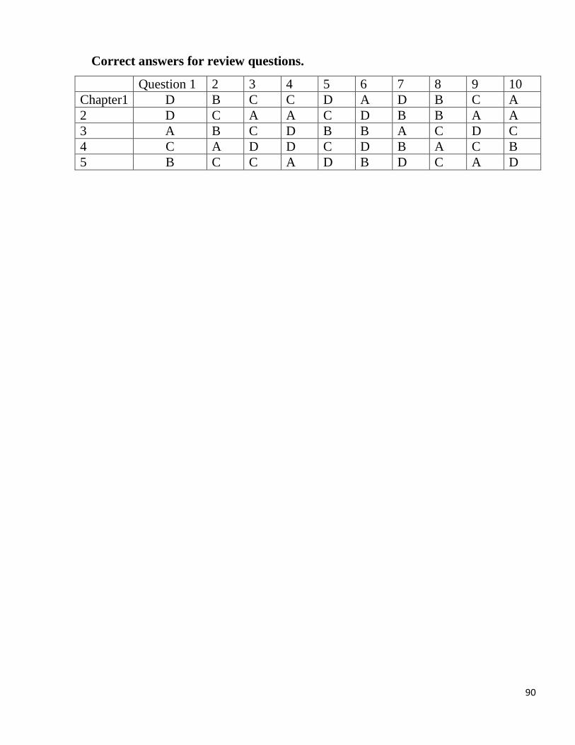
90
Correct answers for review questions.
Question 1 2 3 4 5 6 7 8 9 10
Chapter1 D B C C D A D B C A
2 D C A A C D B B A A
3 A B C D B B A C D C
4 C A D D C D B A C B
5 B C C A D B D C A D
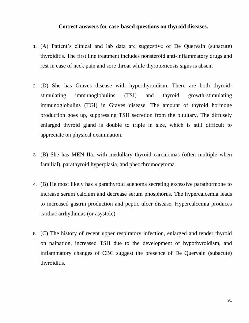
91
Correct answers for case-based questions on thyroid diseases.
1. (A) Patient’s clinical and lab data are suggestive of De Quervain (subacute)
thyroiditis. The first line treatment includes nonsteroid anti-inflammatory drugs and
rest in case of neck pain and sore throat while thyrotoxicosis signs is absent
2. (D) She has Graves disease with hyperthyroidism. There are both thyroid-
stimulating immunoglobulins (TSI) and thyroid growth-stimulating
immunoglobulins (TGI) in Graves disease. The amount of thyroid hormone
production goes up, suppressing TSH secretion from the pituitary. The diffusely
enlarged thyroid gland is double to triple in size, which is still difficult to
appreciate on physical examination.
3. (B) She has MEN IIa, with medullary thyroid carcinomas (often multiple when
familial), parathyroid hyperplasia, and pheochromocytoma.
4. (B) He most likely has a parathyroid adenoma secreting excessive parathormone to
increase serum calcium and decrease serum phosphorus. The hypercalcemia leads
to increased gastrin production and peptic ulcer disease. Hypercalcemia produces
cardiac arrhythmias (or asystole).
5. (C) The history of recent upper respiratory infection, enlarged and tender thyroid
on palpation, increased TSH due to the development of hypothyroidism, and
inflammatory changes of CBC suggest the presence of De Quervain (subacute)
thyroiditis.
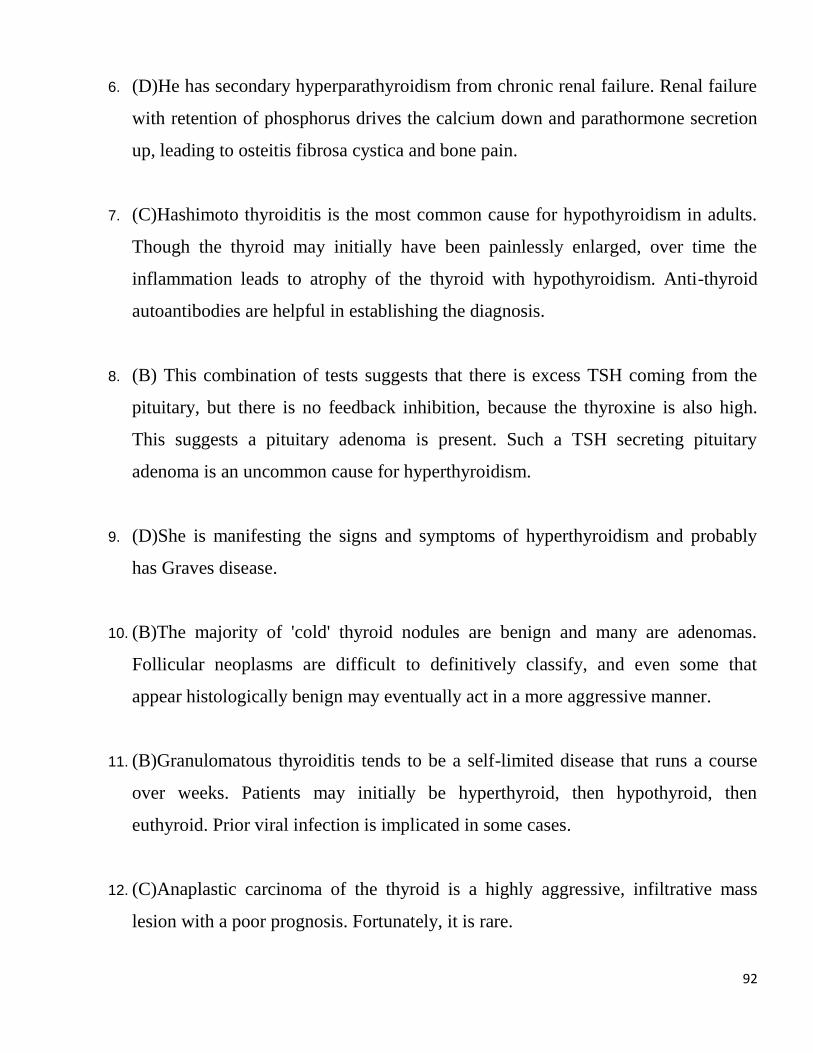
92
6. (D)He has secondary hyperparathyroidism from chronic renal failure. Renal failure
with retention of phosphorus drives the calcium down and parathormone secretion
up, leading to osteitis fibrosa cystica and bone pain.
7. (C)Hashimoto thyroiditis is the most common cause for hypothyroidism in adults.
Though the thyroid may initially have been painlessly enlarged, over time the
inflammation leads to atrophy of the thyroid with hypothyroidism. Anti-thyroid
autoantibodies are helpful in establishing the diagnosis.
8. (B) This combination of tests suggests that there is excess TSH coming from the
pituitary, but there is no feedback inhibition, because the thyroxine is also high.
This suggests a pituitary adenoma is present. Such a TSH secreting pituitary
adenoma is an uncommon cause for hyperthyroidism.
9. (D)She is manifesting the signs and symptoms of hyperthyroidism and probably
has Graves disease.
10. (B)The majority of 'cold' thyroid nodules are benign and many are adenomas.
Follicular neoplasms are difficult to definitively classify, and even some that
appear histologically benign may eventually act in a more aggressive manner.
11. (B)Granulomatous thyroiditis tends to be a self-limited disease that runs a course
over weeks. Patients may initially be hyperthyroid, then hypothyroid, then
euthyroid. Prior viral infection is implicated in some cases.
12. (C)Anaplastic carcinoma of the thyroid is a highly aggressive, infiltrative mass
lesion with a poor prognosis. Fortunately, it is rare.

93
13. (B) Hypothyroidism can occur years later in the course of Hashimoto thyroiditis,
even though there may initially be transient hyperthyroidism. Eventually the
inflammation leads to loss of thyroid follicles. This is the most common cause for
hypothyroidism in adults.
14. (B) The most common cause for thyroid enlargement is a simple, nodular goiter.
Most patients are euthyroid with this condition. Places far away from a seacoast (a
source for iodine) are where endemic goiter used to be seen. Use of iodized salt
eliminated the problem.
15. (D)Diffuse hyperplasia with active thyroid epithelial cells is typical for Graves
disease. The TSH is low from negative feedback from the increased thyroid
hormone production.
16. (D) The child is not living in a region of endemic goiter. His thyroid problem is
probably a developmental failure of thyroid gland formation. congenital
hypothyroidism, though rare, is one of the diseases screened for at birth, because
when recognized it can easily be treated with replacement thyroid hormone.
However, the health care system needs to function effectively to diagnose and treat
preventable diseases.
17. (C) Some postpartum women have thyroid autoantibodies. Microscopically,
extensive lymphoid infiltrates also suggest autoimmunity as an etiology for thyroid
disease. Early in the course of subacute lymphocytic thyroiditis, the thyroid may be
enlarged from extensive inflammation. Hyperthyroidism may be transient, and
most persons are euthyroid. A small number of affected persons develop
hypothyroidism.
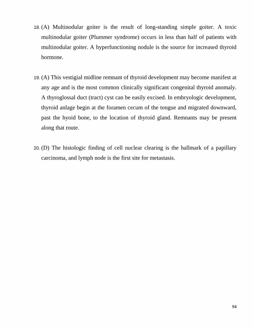
94
18. (A) Multinodular goiter is the result of long-standing simple goiter. A toxic
multinodular goiter (Plummer syndrome) occurs in less than half of patients with
multinodular goiter. A hyperfunctioning nodule is the source for increased thyroid
hormone.
19. (A) This vestigial midline remnant of thyroid development may become manifest at
any age and is the most common clinically significant congenital thyroid anomaly.
A thyroglossal duct (tract) cyst can be easily excised. In embryologic development,
thyroid anlage begin at the foramen cecum of the tongue and migrated downward,
past the hyoid bone, to the location of thyroid gland. Remnants may be present
along that route.
20. (D) The histologic finding of cell nuclear clearing is the hallmark of a papillary
carcinoma, and lymph node is the first site for metastasis.
95
References:
1. Abraham P., Avenell A., McGeoch S.C. et al. Antithyroid drug regimen for treating
Graves’ hyperthyroidism // Cochrane Database Syst Review. – 2010. CD003420.
2. Akamizu T. Japan Thyroid Association: Diagnostic criteria, clinical features, and
incidence of thyroid storm based on nationwide surveys /T. Akamizu, T. Satoh, O.
Isozaki, A. Suzuki // Thyroid. – 2012. – Vol. 22. – P. 661–679.
3. Alfadda A.A. Subacute thyroiditis: clinical presentation and long term outcome /
A.A. Alfadda, R.M. Sallam, G.E. Elawad, H. Aldhukair // International journal of
endocrinology. - 2014. – Vol. 2014. - Article ID 794943, 7 pages. DOI:
10.1155/2014/794943.
4. Andersen S.L. Antithyroid drug side effects in the population and in pregnancy /
S.L. Andersen, J. Olsen, P. Laurberg // Journal of Clinical Endocrinology and
Metabolism. – 2016. – Vol.101. – P. 1606–1614.
5. Azim S. Correction: Subclinical hypothyroidism / S. Azim, C. Nasr // Cleveland
Clinic Journal of Medicine. – 2019. – Vol.86. – P. 392.
6. Azizi F. Effect of long-term continuous methimazole treatment of hyperthyroidism:
comparison with radioiodine /F. Azizi, L. Ataie, M. Hedayati et al. // European
Journal of Endocrinology. – 2005. – Vol. 152. – P. 695.
7. Bartalena L. Diagnosis and management of Graves’ disease: a global overview / L.
Bartalena // Nature Reviews Endocrinology. – 2013. – Vol. 9. – P. 724–734.
8. Bartalena L. The dilemma of how to manage Graves’ hyperthyroidism in patients
with associated orbitopathy / L. Bartalena // Journal of Clinical Endocrinology and
Metabolism. – 2011. – Vol. 96. – P. 592–599.
9. Bartalena L., Burch H.B., Burman K.D. et al. A 2013 European survey of clinical
practice patterns in the management of Graves’ disease // Clinical Endocrinology. –
2016. – Vol. 84. – P. 115–120.
10. Bekkering G.E. Thyroid hormones treatment for subclinical hypothyroidism: a
clinical practice guideline / G.E. Bekkering, T. Agoritsas, I. Lytvyn, A.F. Heen et
al. // BMJ. – 2019. - May 14;365:l2006. doi: 10.1136/bmj.l2006.
11. Bonnema S.J. Radioiodine therapy in benign thyroid diseases: effects, side effects,
and factors affecting therapeutic outcome / S.J. Bonnema, L. Hegedus //
Endocrinology Review. – 2012. – Vol. 33. – P. 920–980.
96
12. Bonnema S.J. Management of the nontoxic multinodular goiter: a North American
survey / S.J. Bonnema, F.N. Bennedbaek, P.W. Ladenson, L.J. Hegedüs // Journal
of Clinical Endocrinology and Metabolism. – 2002. – Vol. 87. – P. 112.
13. Brent G.A. Mechanisms of thyroid hormone action / G.A. Brent // Journal of
Clinical Investigation. – 2012. – Vol.122. – P. 3035–3043.
14. Calo G. Differentiated thyroid cancer: feasibility of loboisthmectomy in
an endemic region. G. Calo, E. Erdas, F. Medas, L. Gordini et al. Giornale di
Chirurgia. – 2015. – Vol. 36(6). – P. 257-262.
15. Cappelli C. The predictive value of ultrasound findings in the management of
thyroid nodules / C. Cappelli, M. Castellano, I. Pirola, D. Cumetti // QJM. – 2007.
– Vol. 100. – P. 29–35.
16. Cappola A.R. Thyroid and Cardiovascular Disease: Research Agenda for
Enhancing Knowledge, Prevention, and Treatment / A.R. Cappola,A.S. Desai, M.
Medici, L.S. Cooper et al. // Thyroid. – 2019. – Vol.29(6). – P. 760-777. doi:
10.1089/thy.2018.0416.
17. Caturegli P. Hashimoto thyroiditis: clinical and diagnostic criteria / P. Caturegli,A.
De Remigis,N.R. Rose // Autoimmunity Reviews. – 2014. – Vol.13(4-5). – P. 391-
397. doi: 10.1016/j.autrev.2014.01.007.
18. Chen A.Y. American Thyroid Association statement on optimal surgical
management of goiter. A.Y. Chen,V.J. Bernet, S.E. Carty et al // Thyroid. – 2014. –
Vol. 24. – P. 181.
19. Chen R.H. Thyroid diseases increased the risk of type 2 diabetes mellitus: A
nation-wide cohort study / R.H. Chen, H.Y. Chen, K.M. Man, S.J. Chen et al. //
Medicine (Baltimore). – 2019. - Vol. 98(20). - e15631. doi:
10.1097/MD.0000000000015631.
20. Ciccone M.M. Increased carotid IMT in overweight and obese women affected by
Hashimoto’s thyroiditis: an adiposity and autoimmune linkage? /M.M. Ciccone, G.
De Pergola, M.T. Porcelli, P. Scicchitano et al.// BMC Cardiovascular Disorders. -
2010. – Vol. 10. – P. 22.
21. Cirocchi R. Total or near-total thyroidectomy versus subtotal thyroidectomy for
multinodular non-toxic goitre in adults /R. Cirocchi, S. Trastulli, J. Randolph et al.
// Cochrane Database of Systematic Reviews. - 2015; CD010370.
97
22. Cooper D.S. Antithyroid drugs in the management of patients with Graves’ disease:
an evidence-based approach to therapeutic controversies / D.S. Cooper // Journal of
Clinical Endocrinology and Metabolism. – 2003. – Vol. 88. – P. 3474–3481.
23. Cramon P. Quality of life in patients with benign nontoxic goiter: impact of disease
and treatment response, and comparison with the general population / P. Cramon,
S.J. Bonnema, J.B. Bjorner et al. // Thyroid. - 2015. – Vol. 25. – P. 284.
24. De Bellis A. Time course of Graves' ophthalmopathy after total thyroidectomy
alone or followed by radioiodine therapy: a 2-year longitudinal study / A. De
Bellis, G. Conzo, G. Cennamo, E. Pane et al. // Endocrine. – 2012. - Vol. 41(2). –
P. 320-326.
25. De Crea C. Actual incidence and clinical behaviour of follicular thyroid carcinoma:
an institutional experience / C. De Crea, M. Raffaelli, L. Sessa, S. Ronti et al. //
Scientific World Journal. – 2014. - 4;2014:952095. doi: 10.1155/2014/952095.
eCollection 2014.
26. De Groot L.J. Diagnosis and Treatment of Graves’ Disease. [Updated 2016 Nov 2].
In: Feingold KR, Anawalt B, Boyce A, et al., editors. Endotext [Internet]. Available
from: https://www.ncbi.nlm.nih.gov/books/NBK285548/
27. De Groot L.J., Bartalena L., Feingold K.R. Thyroid Storm. 2018 Dec 17. In:
Feingold KR, Anawalt B, Boyce A, et al., editors. Endotext [Internet]. Available
from: https://www.ncbi.nlm.nih.gov/books/NBK278927/
28. Doggui R. Iodine deficiency: Physiological, clinical and epidemiological features,
and pre-analytical considerations / R. Doggui, J. El Atia // Annales
d’Endocrinologie. – 2015. – Vol. 76(1). – P. 59-66. doi:
10.1016/j.ando.2014.12.002.
29. Donovan P.J. Cost-utility analysis comparing radioactive iodine, anti-thyroid drugs
and total thyroidectomy for primary treatment of Graves’ disease / P.J. Donovan,
D.S. McLeod, R. Little, L. Gordon // European Journal of Endocrinology. – 2016. –
Vol. 175. – P. 595–603.
30. Eisenberg M. TSH-based protocol, tablet instability, and absorption effects on LT4
bioequivalence / M. Eisenberg, J. Distefano // Thyroid. – 2009. – Vol.19. – P. 103–
110.

98
31. El-Eshmawy M.M. Thyroid and eye: Where they meet in clinical practice / M.M.
El-Eshmawy, M. Shahin // Endocrine, Metabolic and Immune Disorders - Drug
Targets. – 2019. - Jun 18. doi: 10.2174/1871530319666190618120107.
32. Elfenbein D.M. Clinical and socioeconomic factors influence treatment decisions in
Graves' disease /D.M. Elfenbein, D.F. Schneider, J. Havlena et al. // Annals of
Surgical Oncololy. – 2015. – Vol. 22/ - P. 1196.
33. Elisei R. Impact of routine measurement of serum calcitonin on the diagnosis and
outcome of medullary thyroid cancer: experience in 10,864 patients with nodular
thyroid disorders /R. Elisei, V. Bottici, F. Luchetti, G. Di Coscio // Journal of
Clinical Endocrinology and Metabolism. – 2004. – Vol. 89. – P. 163–168.
34. Fast S. Recombinant human thyrotropin-stimulated radioiodine therapy of nodular
goiter allows major reduction of the radiation burden with retained efficacy /S.
Fast, L. Hegedüs, P. Grupe P et al. // Journal of Clinical Endocrinology and
Metabolism. – 2010. - Vol. 95. – P. 3719.
35. Flemmer A. Iodized salt as a supplement - necessary or not? / A. Flemmer //
Kinderkrankenschwester. – 2016. – Vol.35(6). – P. 205-208.
36. Flemmer A. Do pregnant women need additional iodine? / A. Flemmer //
Kinderkrankenschwester. – 2015. – Vol.34(7). – P. 275-276.
37. Freitas J.E. Therapeutic options in the management of toxic
and nontoxic nodular goiter / J.E. Freitas // Seminars in Nuclear Medicine. – 2000.
– Vol.30(2). – P. 88-97.
38. Garber J.R. Clinical practice guidelines for hypothyroidism in adults: cosponsored
by the American Association of Clinical Endocrinologists and the American
Thyroid Association. / J.R. Garber, R.J. Cobin, H. Gharib H et al. // Thyroid. –
2012. – Vol. 22. – P. 1200-35.
39. Gharib H. Association of Clinical Endocrinologists, Associazione Medici
Endocrinologi, and European Thyroid Association Medical guidelines for clinical
practice for the diagnosis and management of thyroid nodules: executive summary
of recommendations. H. Gharib, E. Papini, R. Paschke, J. Duick et al. //
Endocrinology Practice. – 2010. – Vol.16. – P. 468–475.
40. Gharib H. Thyroid nodules: clinical importance, assessment, and treatment / H.
Gharib, E. Papini // Endocrinology and Metabolism Clinics of North America. -
2007. – Vol. 36. – P. 707–735.

99
41. Giovanella L. Management of endocrine disease: The Role of rhTSH in the
Management of Differentiated Thyroid Cancer: Pros and Cons / L. Giovanella,
L.H. Duntas // European Journal of Endocrinology. – 2019. - Jun 1. pii: EJE-19-
0149.R2. doi: 10.1530/EJE-19-0149.
42. Gourmelon R. Subclinical Hypothyroidism: is it Really Subclinical with Aging?/
R. Gourmelon, S. Donadio-Andréi, K. Chikh, M. Rabilloud et al. // Aging and
Disease. – 2019. - Vol. 10(3). P. 520-529. doi: 10.14336/AD.2018.0817.
43. Grebe S.K. Laboratory testing in hyperthyroidism / S.K. Grebe, G.J. Kahaly //
American Journal of Medicine. – 2012. – Vol. 125, Supplement2. – P.12-15.
44. Gronich N. Hypothyroidism Is a Risk Factor for New-Onset Diabetes: A Cohort
Study / N. Gronich, S.N. Deftereos, I. Lavi, A.S. Persidis et al. // Diabetes Care. –
2015. – Vol. 38. – P. 1657-1664.
45. Haugen A. 2015 American Thyroid Association Management Guidelines for Adult
Patients with Thyroid Nodules and Differentiated Thyroid Cancer: The American
Thyroid Association Guidelines Task Force on Thyroid Nodules and Differentiated
Thyroid Cancer / A. Haugen et al. // Thyroid. – 2016. – Vol.26(1). – P. 1-133.
46. Hegedus L. Clinical practice. The thyroid nodule / L. Hegedus // New England
Journal of Medicine. – 2004. – Vol.351. – P. 1764–1771.
47. Hegedus L. Thyroid ultrasound / L. Hegedus // Endocrinology and Metabolism
Clinics of North America. – 2001. – Vol. 30. - P. 339–360.
48. Hyperthyroidism. The Merck Manuals: The Merck Manual for Healthcare
Professionals.http://www.merckmanuals.com/professional/sec12/ch152/ch152e.ht
ml. Accessed May 10, 2019.
49. Hyporthyroidism. The Merck Manuals: The Merck Manual for Healthcare
Professionals.https://www.merckmanuals.com/professional/endocrine-and-
metabolic-disorders/thyroid-disorders/hypothyroidism. Accessed April 16, 2019.
50. Jonklaas J. Guidelines for the Treatment of Hypothyroidism: Prepared by the
American Thyroid Association Task Force on Thyroid Hormone Replacement / J.
Jonklaas, A.C. Bianco, A.J. Bauer et al. // Thyroid. – 2014. – Vol.24(12). – P.
1670-1751.
51. Kahaly G.J. Graves’ disease /G.J. Kahaly, P.D. Olivo // New England Journal of
Medicine. – 2017. – Vol. 376. – P. 184.
100
52. Kahaly G.2018 European Thyroid Association Guideline for the Management of
Graves’ Hyperthyroidism / G. Kahaly, L. Bartalena, L. Hegedüs, L. Leenhardt
// European Thyroid Journal. – 2018. - Vol.7. – P. 167–186.
https://doi.org/10.1159/000490384
53. Kalezic N.K. Risk factors for sporadic medullary thyroid carcinoma / N.K. Kalezic,
V.R.Zivaljevic, N.A. Slijepcevic, I.R. Paunovic et al. // European Journal of Cancer
Prevention. – 2013. – Vol.22(3). – P. 262-267. doi:
10.1097/CEJ.0b013e3283592c78.
54. Kaniuka S. Radioiodine - an attractive alternative to surgery in large non-toxic
multinodular goiters / S. Kaniuka, P. Lass, K. Sworczak // Nuclear medicine
review. Central & Eastern Europe. – 2009. – Vol.12(1). – P. 23-29.
55. Kondo T. Acute suppurative thyroiditis secondary to pyriform sinus fistula / T.
Kondo // The Lancet Infectious Diseases. – 2019. – Vol.19(4). P. 447. doi:
10.1016/S1473-3099(18)30657-1.
56. Kopp P. Thyroid hormone synthesis. In: The Thyroid: Fundamental and Clinical
Text, 9th, Braverman LE, Utiger RD (Eds), Lippincott Williams and Wilkins,
Philadelphia 2005. p.52.
57. Kostoglou A. Hypothyroidism - new aspects of an old disease / A. Kostoglou, K.I.
Ntalles // Hippokratia. – 2010. – Vol. 14(2). P. 82–87.
58. Langer J.E. Correlation of findings from iodine 123 scan and ultrasonography in
the recommendation for thyroid fine-needle aspiration biopsy / J.E. Langer, R.
Agarwal, H. Zhuang, S.S. Huang et al. // Endocrinology Practice. – 2011. – Vol.
17. P. 699–706.
59. Leenhardt L. Advances in diagnostic practices affect thyroid cancer incidence in
France /L. Leenhardt, M.O. Bernier, M.H. Boin-Pineau, D.B. Conte // European
Journal of Endocrinology. – 2004. – Vol.150. – P. 133–139.
60. Le Moli R. Type 2 diabetic patients with Graves' disease have more frequent and
severe Graves' orbitopathy / R. Le Moli, V. Muscia, A. Tumminia, L. Frittitta et al.
// Nutrition, Metabolism and Cardiovascular Diseases. – 2015. – Vol. 25(5). – P.
452-457. doi: 10.1016/j.numecd.2015.01.003.
61. Ma C. Radioiodine therapy versus antithyroid medications for Graves' disease.C.
Ma, J. Xie, H. Wang et al // Cochrane Database of Systematic Reviews. – 2016. –
Vol. 2. - CD010094.
101
62. Mansberg R. Riedel's Thyroiditis with Intense FDG Uptake Demonstrated on FDG
PET/CT / R. Mansberg, R. Bency, L. Shen, C. Bui // Molecular Imaging and
Radionuclide Therapy. – 2015. – 5;24(1):29-31. doi: 10.4274/mirt.98598.
63. Mahase E. Subclinical hypothyroidism: doctors shouldn't routinely prescribe
hormones / E. Mahase // BMJ. – 2019. - May 17;365:l2262. doi:
10.1136/bmj.l2262.
64. Makay O.Less than total thyroidectomy for goiter: when and how? / O. Makay //
Gland Surgery. – 2017. – Vol.6(1). – P. 49-58. doi: 10.21037/gs.2017.10.02.
65. Maratou E. Studies of insulin resistance in patients with clinical and subclinical
hypothyroidism / E. Maratou, D.J. Hadjidakis, A. Kollias A et al. // European
Journal of Endocrinology. - 2009. – Vol. 160. – P. 785–790.
66. Marqusee E. Usefulness of ultrasonography in the management of nodular thyroid
disease / E. Marqusee, C.B. Benson, M.C. Frates, P.M. Doubilet et al. // Annals of
Internal Medicine. – 2000. – Vol.133. – P. 696–700.
67. Martino E, Bartalena L, Pinchera. Central hypothyroidism. In: Braverman LE,
Utiger RD (eds): The Thyroid: A Fundamental and Clinical Text (8th ed.).
Philadelphia, JB Lippincott Williams & Wilkins, 2000, pp 762-773.
68. McLachlan S.M. The link between Graves' disease and Hashimoto's thyroiditis: a
role for regulatory T cells /S.M. McLachlan, Y. Nagayama, P.N. Pichurin, Y.
Mizutori // Endocrinology. – 2007. – Vol.148(12). – P. 5724-5733.
69. Milkau M. Thyroid Storm and Myxedema Coma / M. Milkau, F. Sayk //Deutsche
Medizinische Wochenschrift. – 2018. – Vol.143(6). – P. 397-405. doi: 10.1055/s-
0043-111728.
70. Moon H.J. Diagnostic performance of gray-scale US and elastography in solid
thyroid nodules / H.J. Moon, J.M. Sung, E.K. Kim, J.H. Yoon // Radiology. – 2012.
-Vol. 262. – P. 1002–1013.
71. Munir A. Myxedema Coma / A. Munir // Journal of Ayub Medical College
Abbottabad. – 2018. – Vol. 30(1). – P. 119-120.
72. Nakamura H. Analysis of 754 cases of antithyroid drug-induced agranulocytosis
over 30 years in Japan / H. Nakamura, A. Miyauchi, N. Miyawaki, J. Imagawa//
Journal of Clinical Endocrinology and Metabolism. – 2013. – Vol.98. – P. 4776–
4783.
102
73. National Center for Biotechnology Information, U.S. National Library of Medicine.
PubMed Health: How Does the Thyroid Gland Work? July 2011.
http://www.ncbi.nlm.nih.gov/pubmedhealth/PMH0010393/. Accessed March 12,
2019.
74. Nikiforov Y.E. Nomenclature revision for encapsulated follicular variant of
papillary thyroid carcinoma: A paradigm shifts to reduce overtreatment of indolent
tumors /R.R. Seethala, G. Tallini et al // JAMA Oncololy. Published online April
14, 2016. doi:10.1001/jamaoncol.2016.0386.
75. Ono Y. Clinical characteristics and outcomes of myxedema coma: Analysis of a
national inpatient database in Japan /Y. Ono, S. Ono, H. Yasunaga et al. // Journal
of Epidemiololy. – 2017. – Vol.27. – P. 117.
76. Pacini F, DeGroot LJ. Thyroid Cancer. [Updated 2013 Mar 27]. In: Feingold KR,
Anawalt B, Boyce A, et al., editors. Endotext [Internet]. Available from:
https://www.ncbi.nlm.nih.gov/books/NBK285559/
77. Parkin D. Cancer incidence in five continents. /C. Muir, S. Whelan, Y. Gao, J
Fenlay et al. // Volume 6. IARC Scientific Publication N120. International Agency
for Research on Cancer, Lyon.
78. Perros P. Guidelines for the management of thyroid cancer /P. Perros, K.
Boelaert, S. Colley, C. Evans // Clinical Endocrinology. – 2014. – Vol.81, Suppl. 1.
P. 1–122.
79. Pretell E.A. Elimination of iodine deficiency disorders from the Americas: a public
health triumph / E.A. Pretell, E.N. Pearce, S.A. Moreno, O. Dary et al. // The
Lancet Diabetes and Endocrinology. – 2017. – Vol.5(6). – P. 412-414. doi:
10.1016/S2213-8587(17)30034-7.
80. Razvi S. Levothyroxine treatment of subclinical hypothyroidism, fatal and nonfatal
cardiovascular events, and mortality / S. Razvi, J.U. Weaver, T.J. Butler et al. //
Archives of Internal Medicine. – 2012. – Vol.172. – P. 811–817.
81. Rhee C.M. Thyroid functional disease: an under-recognized cardiovascular risk
factor in kidney disease patients / C.M. Rhee, G.A. Brent, C.P. Kovesdy CP, O.P.
Soldin et al. // Nephrology Dialysis Transplantation. –2015. – Vol. 30. – P. 724-
737.
103
82. Richard-Eaglin A. Immunosuppressive/Autoimmune Disorders / A. Richard-
Eaglin, B.A. Smallheer // Nursing Clinics of North America. – 2018. – Vol.53(3). –
P. 319-334. doi: 10.1016/j.cnur.2018.04.002.
83. Richards M.L. Familial syndromes associated with thyroid cancer in the era of
personalized medicine / M.L. Richards // Thyroid. – 2010. – Vol.20. – P. 707–713.
84. Rizos C.V. Effects of thyroid dysfunction on lipid profile / C.V. Rizos, M.S. Elisaf,
E.N. Liberopoulos // Open Cardiovascular Medicine Journal. – 2011. – Vol. 5. – P.
76-84.
85. Ross D.S. American Thyroid Association Guidelines for Diagnosis and
Manangement of Hyperthyroidism and Other Causes of Thyrotoxicosis / D.S. Ross,
H.B.Burch, D.S. Cooper, M.C. Greenlee et al. // Thyroid. – 2016. – Vol.26(10). –
P. 1343–1421.
86. Ross D.S. Radioiodine therapy for hyperthyroidism / D.S. Ross // The New
England Journal of Medicine. – 2011. – Vol. 364. - P. 542.
87. Ruchala M. The role of sonoelastography in acute, subacute and chronic thyroiditis
- a novel application of the method / M Ruchala, E. Szczepanek-Parulska, A.
Zybek, J. Moczko // European Journal of Endocrinology. - 2012. – Vol.166(3). – P.
425-32. doi: 10.1530/EJE-11-0736
88. Samuels M.H. Editorial: Evaluation and treatment of sporadic nontoxic goiter—
some answers and more questions / M.H. Samuels // The Journal of Clinical
Endocrinology & Metabolism. – 2001. – Vol. 86 (3). – P. 994–997.
https://doi.org/10.1210/jcem.86.3.7384.
89. Sherman S. Thyroid carcinoma / S. Sherman // Lancet. – 2003. – Vol.361. – P.
501–511.
90. Shiber S. Glucocorticoid regimens for prevention of Graves’ ophthalmopathy
progression following radioiodine treatment: systematic review and meta-analysis /
S. Shiber, H. Stiebel-Kalish, I. Shimon, A. Grossman // Thyroid. – 2014. – Vol. 24.
P. 1515–1523.
91. Silberstein E.B. The SNMMI practice guideline for therapy of thyroid disease with 131I /E.B. Silberstein, A. Alavi, H.R. Balon, S.E. Clarke // Journal of Nuclear
Medicine. –2012. – Vol.53. – P. 1633–1651.
92. Singla M. Suppurative thyroiditis / M. Singla, S. Gaba, K. Bhinder // Clinical Case
Reports. – 2018. - 5;6(5):951-952. doi: 10.1002/ccr3.1463. eCollection 2018 May
104
93. Smith T.J. Graves’ disease /T.J. Smith, L. Hegedus// New England Journal of
Medicine. – 2016. – Vol. 375. – P. 1552–1565.
94. Stan M.N. Risk factors for development or deterioration of Graves' ophthalmopathy
/ M.N. Stan, R.S. Bahn // Thyroid. – 2010. – Vol. 20. – P. 777.
95. Stasiak M. Clinical characteristics of subacute thyroiditis is different than it used to
be - current state based on 15 years own material / M. Stasiak,R. Michalak, B.
Stasiak, A. Lewinski // Neuro Endocrinololy Letters. – 2019. – Vol. 39(7). – P.
489-495.
96. Sultan S. Concept of double salt fortification; a tool to curtail micronutrient
deficiencies and improve human health status / S. Sultan, F.M. Anjum, M.S. Butt,
N. Huma et al. // Journal of the Science of Food and Agriculture. – 2014. –
Vol.94(14). – P. 2830-2838. doi: 10.1002/jsfa.6634.
97. Sundaresh V. Comparative effectiveness of therapies for Graves’ hyperthyroidism:
a systematic review and network meta-analysis / V. Sundaresh, J.P. Brito, Z.
Wang,L.J. Prokop et al. // Journal of Clinical Endocrinology and Metabolism. –
2013. – Vol. 98. – P. 3671–3677.
98. Sweeney L.B. Thyroiditis: An Integrated Approach / L.B. Sweeney, C. Stewart,
D.Y. Gaitonde // American Family Physician. – 2014. – Vol. 90(6). – P. 389-396.
99. Tagami T. Short-term effects of β-adrenergic antagonists and methimazole in new-
onset thyrotoxicosis caused by Graves' disease /T. Tagami, Y. Yambe, T. Tanaka et
al // Internal Medicine. – 2012. – Vol. 51. – P. 2285.
100. Thyroid cancers. The Merck Manuals: The Merck Manual for Healthcare
Professionals.https://www.merckmanuals.com/professional/endocrine-and-
metabolic-disorders/thyroid-disorders/thyroid-cancers. Accessed June 20, 2019.
101. Thomas T. Clinical, biochemical & cytomorphologic study on Hashimoto's
thyroiditis / T. Thomas, S. Sreedharan, U.N. Khadilkar, D. Deviprasad et al. Indian
Journal of Medical Research. – 2014. – Vol. 140(6). – P. 729-735.
102. Tudela C.M. Relationship of subclinical thyroid disease to the incidence of
gestational diabetes / C.M. Tudela, B.M. Casey, D.D. McIntire, F.G. Cunningham
// Obstetrics and Gynecololy. – 2012. - Vol. 119. – P. 983-988.
103. Vanderpas J.B. Historical aspects of iodine deficiency control / J.B.
Vanderpas,R. Moreno-Reyes // Minerva Med. – 2017. – Vol. 108(2). – P. 124-135.
doi: 10.23736/S0026-4806.17.04884-4.
105
104. Wiersinga W. ETA Guidelines: the use of L-T4+L-T3 in the treatment of
hypothyroidism / W. Wiersinga, L.H. Duntas, V.V. Fadeyev, B. Nygaard et al. //
European Thyroid Journal. – 2012. – Vol.1. – P. 55–71.
105. Wiersinga W.M. Adult hypothyroidism and myxedema coma. In: DeGroot LJ,
Jameson JL (eds): Endocrinology (5th ed.). Philadelphia, WB Saunders Company,
2004, ch. 107.
106. Wu P. Thyroid disease and diabetes / P. Wu // Clinical Diabetes. – 2000. – Vol.
18. – P. 38-40.
107. Ylli D. Thyroid emergencies / D. Ylli, J. Klubo-Gwiezdzinska, L. Wartofsky L
// Polish Archives of Internal Medicine. – 2019. - Jun 25. doi:
10.20452/pamw.14876.
108. Zbigniew S. Role of Iodine in Metabolism / S. Zbigniew // Recent Patents on
Endocrine Metabolic and Immune Drug Discovery. – 2017. – Vol.10(2). P. 123-
126. doi: 10.2174/1872214811666170119110618.
109. Zinke A. Expression of thyroid hormone receptor isoform alpha1 in pancreatic
islets / A. Zinke, D. Schmoll, M. Zachmann, J. Schmoll et al. // Experimental and
Clinical Endocrinology and Diabetes. - 2003. – Vol. 111. – P. 198-202.
106
Навчальне видання
Журавльова Лариса Володимирівна
Філоненко Марина Вячеславівна
ЗАХВОРЮВАННЯ ЩИТОПОДІБНОЇ ЗАЛОЗИ
Навчальний посібник
для студентів та лікарів-інтернів
Відповідальна за випуск: Журавльова Л.В.
Комп’ютерна верстка О.Ю. Лавриненко
План 2020, поз. __.
Формат А4. Ризографія. Ум.друк.арк. __.
Тираж 100 прим. Зам. № ______.
Редакційно-видавничий відділ
ХНМУ, пр. Науки, 4, м. Харків, 61022
[email protected], [email protected]
Свідоцтво про внесення суб’єкта видавничої справи до Державного реєстру видавництв,
виготівників і розповсюджувачів видавничої продукції серії ДК № 3242 від 18.07.2008.