Pathology of the thyroid, and parathyroid gland(s)
120
Pathology of the thyroid, and parathyroid gland(s)
-
Upload
khangminh22 -
Category
Documents
-
view
0 -
download
0
Transcript of Pathology of the thyroid, and parathyroid gland(s)

Pathology of the thyroid, and parathyroid gland(s)


Development
Pharyngeal epithelial
pouch
(basis of the tongue)
( foramen cecum)
(Struma lingualis)
Ductus thyroglossus
Substernal
Thyroid tissue


Carbohydrate catabolism,
Lipid catabolism,
Protein synthesis
Metabolic
activity ^
Brain development…}

T3, T4 in the blood:
thyroxine-binding globulin (TBG), 70%
transthyretin or "thyroxine-binding prealbumin" (TTR or TBPA) 10-15%Albumin 15-20%
free T4 (fT4) 0.03%free T3 (fT3) 0.3%

Nomenclature
Struma diffusa Function (?!)nodosa
Normofunction
Hyperfunction
Hypofunction
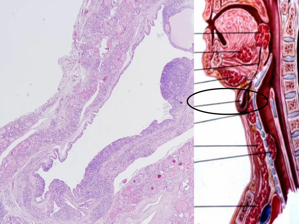
Examination of the thyroid
Physical
Laboratory TSH 0.3-3.6 mU /l
T49-19 pmol/l
T3 2.6-5.7 pmol/l
Scintigraphy
US
FNAB


Hyperthyreosis - effects
Sympathetic tone (ß-adrenerg tone) - basal metabolic activity- ˆˆ
Skin: warm, wet, heat intoleranceLoss of weight, diomyopatia
Heart: tachycardy, cardiomegaly, arrhytmia (atrial fibrillation), CHF- TDC (congestive heart failure, thyreotoxic dilatative cardiomyopaty)
Neuromuscular system:tremor, hyperactivity, insomny, emotional lability, anxiety, proximal muscle weakness, loss of muscle
Ocular changes: „eyes shut wide” - levator palpebrae sympathetic overdose
real exophtalm only in Graves disease
GI: hypermotility, malbsorption, diarrhea
Bones: osteoporosis due to enhanced resorption, brittleness ^ ^

Hyperthyreosis – laboratory testsTSH (low, even in subclinical stages!)
T4 levelT3 level FreeT4, T3 measurement
TRH testScintigraphy

Thyreotoxicosis - causesHyperthyreosis
Primary
Diffuse toxic hyperplasia (Graves- Basedow)
Toxic multinodular goiter
Toxic adenoma
Thyroid carcinoma
Neonatal hyperfunction (maternal Graves)
Secondary
Hypophysis adenoma

De Quervain thyreoiditis (Subacute granulomatous thyreoiditis)
Subacute lymphocytic thyreoiditis
Struma ovarii
Exogenous hormone overdose
Thyreotoxicosis – causesNon-Hyperthyreotic states

Hyperthyreosis - Therapy
Lowering of ß-adrenerg tone (ß blockers)
Propylthiouracil (hampers I oxidation, T4 synthesis, and the T4-T3 conversion in tissues)
Thiamazole (inhibits the enzyme thyroperoxidase, which normally acts in thyroid hormone synthesis by oxidizing the anion iodide (I−) to iodine (I2))
Jodine
hampers release of stored hormone
Radiojodine therapy
destroys thyroid tissue

Hypothyreosis - Cretenism
In case of maternal hypothyreosis in early pregnancy -severe
Later – less severe
Impaired development of the
skeleton, CNS
Short stature
Coarse facial features
Protruding tongue
Umbilical hernia

Hypothyreosis - causesPrimary
Developmental anomaly ( thyroid dysgenesis: PAX-8, TTF2, TSH-R mut.)
Thyroid hormon resistance (TRß mutation)
Congenital biosynthetic defect (dyshormonogenetic goiter)
Postablation
(operation, radiojodine th, irradiation)
Autoimmune thyroiditis
Iodine deficiency
Drugs (lithium, PAS)
Secondary
Pituitary failure
Tertiary
Hypothalamic failure

Hypothyreosis - MyxoedemaSlowing of physical and mental activity –similar to depression
Cold intolerance
Gain of weight
Obstipation
Decreased sweating
Reduced cardiac output
(Low output failure)
GAG, HA accumulation, oedema
Lab: TSH^^^, T3, T4ˇˇˇ,
Except for hypophysis,
hypothalamic origin

ThyreoiditisInfectiosus
Hashimoto ( chronic lymphocytic thyroiditis)
Subacute granulomatous thyroiditis – De Quervain
Subacute lymphocytic thyroiditis
Riedel goiter

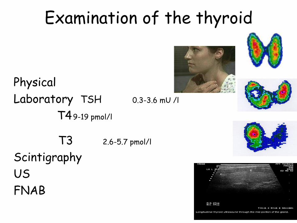
Hashimoto
Inheritance? (Monozigotic twins 30-60 % concordance)
HLA-Dr3, HLA-DR5, polymorphism,
6p, 12q – susceptibility locus
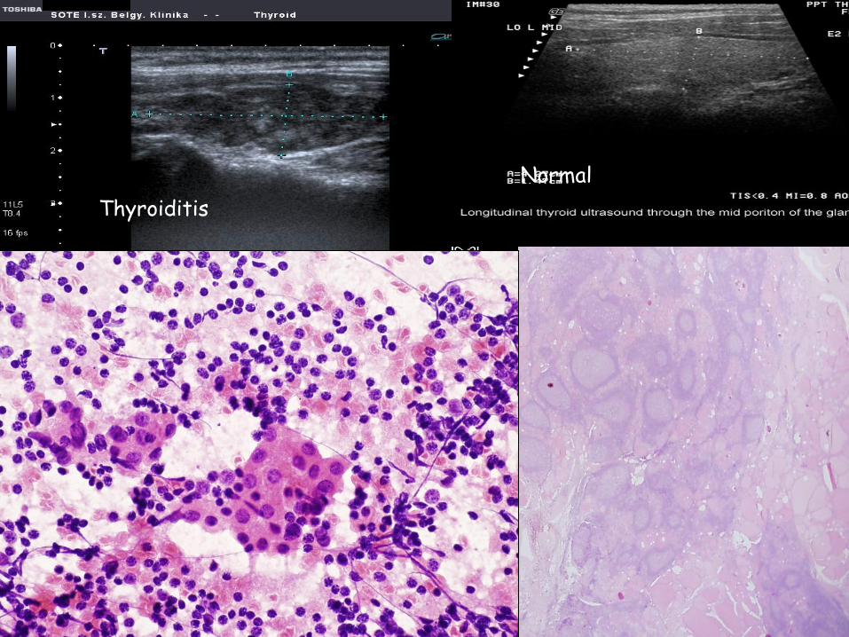
Normal
Thyroiditis


Clinical courseHyper ( in early stage), more frequently
Hypothyreosis
Painless diffuse thyroid enlargement
(may be localised, or nodular)
T3, T4 ˇ, TSH ^ ^ ^, anti TPO
May be associated with other autoimmune diseases:
Diabetes I., Autoimmune adrenalitis, SLE, myastenia gravis, Sjögren,
Possible consequence: NHL!
(not associated with epithelial tumors …(?))

Subacute lymphocytic thyroiditis
RarePathogenesis is not clear, but may have autoimmune origin (autoantibodies might occur, but not always!)
May be the precursor of Hashimoto ( not obviously!)
Frequently associated with pregnancy (postpartum thyreoiditis, may recur in repeated pregnancies)
Clin.: painless thyroid enlargement, thyreotoxicosis, T3, T4 ^, TSH ˇ, diminishing in 2-6 weeks,
After appr. 8 weeks, normal thyroid function returns
Some cases may evolve to chronic hypothyreosis

Subacute granulomatous thyroiditis – De Quervain
Postviral inflammation, -upper airway inflammation Coxsackie, mumps, measles, adenovirus
causes the release of(viral, or thyroid originated)
AB releaseCytotoxic T cellsAfter the cessation of the AB release, the process is ended
Clin.: severe cervical painHyper, - than hypothyreosis, TSH ˇ, T3, T4 ^, scintig: low uptakeSubsides in 6-8 weeks

Riedel thyroiditisStony hard, fixed thyroid mass,
Clinically mimicking thyroid malignancy
„Burnt out”, fibrotic thyroid mass
Etiology: (???), vs autoimmune
Palpation thyreoiditis???
Hashimoto????
Present concept: IgG4 disease

9297/07
48 y old female

Graves - BasedowGeneticsconcordance between monozygotic twins is: 60%
more frequent in certain HLA-DR 3, HLA-B8 types
CTLA-4 polymorphism ( ~ blocks the formation of autoantiboidies)
Autoantibodies:
Anti-TG, anti-T peroxisome, anti – TSH receptor
TSI (LATS), (this is Graves-specific)
TGI
TBII ( TSH-binding inhibitory immunoglobine) – this is blocking, or stimulating)
TRIGGER ?? ( loss of T cell tolerance)
Anti-TG, anti-T peroxisome

Graves - BasedowClinical course
HyperthyreosisExophtalmos (retroorbital ly, oedema, GAG, HA accumulation,)
Pretibial myxoedemaAssociated with other autoimmune diseases:
Diabetes I., Autoimmune adrenalitis, SLE, myastenia gravis, Sjögren, Anaemia perniciosa, + Hashimoto !!!!!
Lab: TSHˇˇˇT3, T4 ^^^Scinti: ^^^
Th.: propylthiouracyl, radioiodine ablation, surgical
Compared to


Graves Hashimoto

Diffuse/ nodular goiter

Diffuse goiter
Endemic( most frequent) (10 % of the population is involved)
Alps, Andes, Himalaya
Goitrogens: cabbage, cauliflower, Brussels sprouts, turnips, cassava
Iodine deficiencyHpl, htr – euthyreoid
hypothyreoidT3, T4 norm., TSH elevated, or upper range of normal
Sporadichereditary enzimatic defectsfrequently unknown etiology

Nodular goiter
All longstanding simple goiters convert into ~
One nodule might become autonomousHpl, atrophy, fibrosis, calcification, cyst
formationScintigr.: uneven uptake
toxic nodular goiter –when one nodule becomes autonomous

Strnod
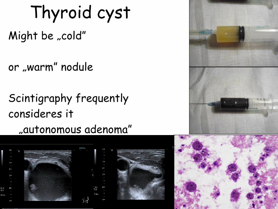
Thyroid cystMight be „cold”
or „warm” nodule
Scintigraphy frequently
consideres it
„autonomous adenoma”

Tumors
Suspicious, if:
Solitary nodule
Young patient
Male
Cold ( I, ! , Tc might show it to be hot ! )

TumorsAdenoma
Non-functioning
(frequently „cold”)
Hormon-producing
(„warm”, „hot nodule”
toxic adenoma)

Relative frequency of malignant thyroid tumors
– Papillary carcinoma
– Follicular carcinoma
– Medullarycarcinoma
– Anaplastic cc.
– Lymphoma
- Other, non-epithelial
- Metastatic
75%
15%
5%
2%
2%
0.8%
0.2%

Malignant thyroid tumors
Genetic background – mutations
ex.: ret/PTC
Ionizing irradiation
(therapeutic,
environmental)

Papillary cc.
Occurrence: middle aged women, any age, males can be affected
Signs
„Nodule”
Hoarseness
Cough
Dyspnoe
Metastasis: regional ln-s, rarely distant
Prognosis: relatively good
Th.: surgical + radioiodine th.


Papillary cc.
„Classic” (papillae, Orphan Annie, Psammoma)
Specific types
Encapsulted
Follicular
Tall cell
Diffuse sclerotizing (children)
Hyalinizing trabecular


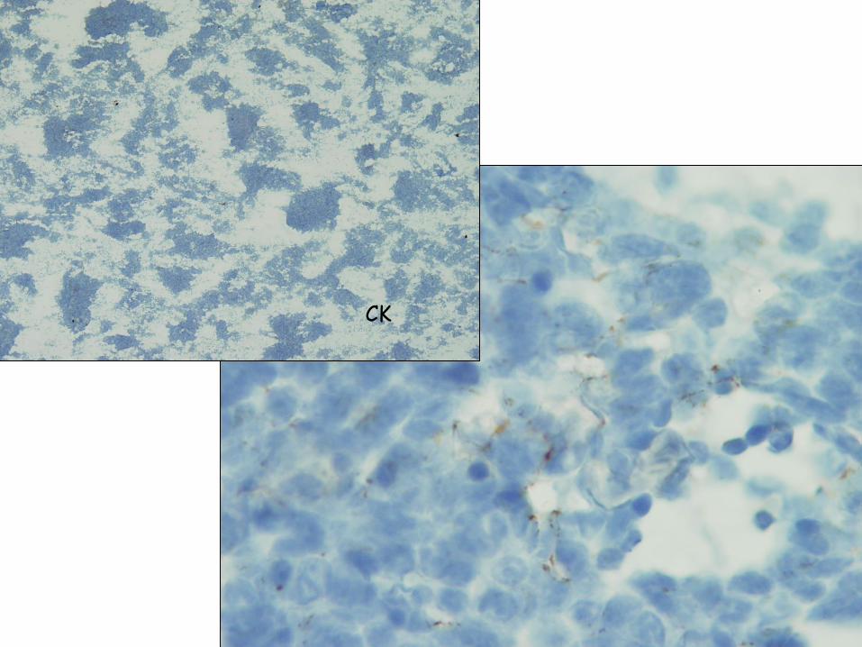
38 year old female. Round shadow. Npl? Met? TBC?
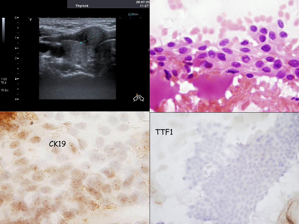
CK19
TTF1


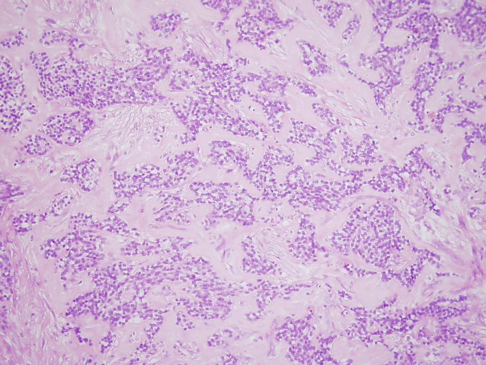
Follicular cc.Occurrence
Elder women
Cause: ras mutation (in foll. Adenomas also)Slowly growing nodule (usually cold, rarely warm)
Monotonous cellsCapsule / and/ or vascular invasionReg. Lymph nodes are rarely metastatic, but
liver, bones are frequently metastatic sites
Progn.: Depends on the metastatic capacityTh.: surgical + radioiodine th

12536/07 59 year old woman

11609/07
65 year old woman
?????

1996. Operation for left sided breast cc.
2003. rec.,
2005. Right sided breast tu.
2007.07. thyroid

TTF1


8042/07
65 year old woman
Cl.: Nod. goiter

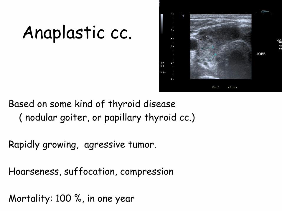
Anaplastic cc.
Based on some kind of thyroid disease
( nodular goiter, or papillary thyroid cc.)
Rapidly growing, agressive tumor.
Hoarseness, suffocation, compression
Mortality: 100 %, in one year


Medullary cc.
C cells
80 % sporadic
20 % a MEN sy 2A, 2B. or
FMTC
(Familiary medullary thyroid cc. FMTC,- spec. MEN2A)
Solitary nodule (sporadic), or multiple smaller familiary (on the basis of C cell hpl)

Medullary cc.
Symptoms: nodule, horseness, dysphagyparaneoplastic (?!) hormone production
Calcitonin ^^, but hypocalcaemia cannot be always shown
Familiary cases: RET-mutation is found in case of family screening. ( C cell hpl might be found in prophilactically resected thyroids )

A
Male, 53Examination for swelling of the neck

11109/07
55 y o female

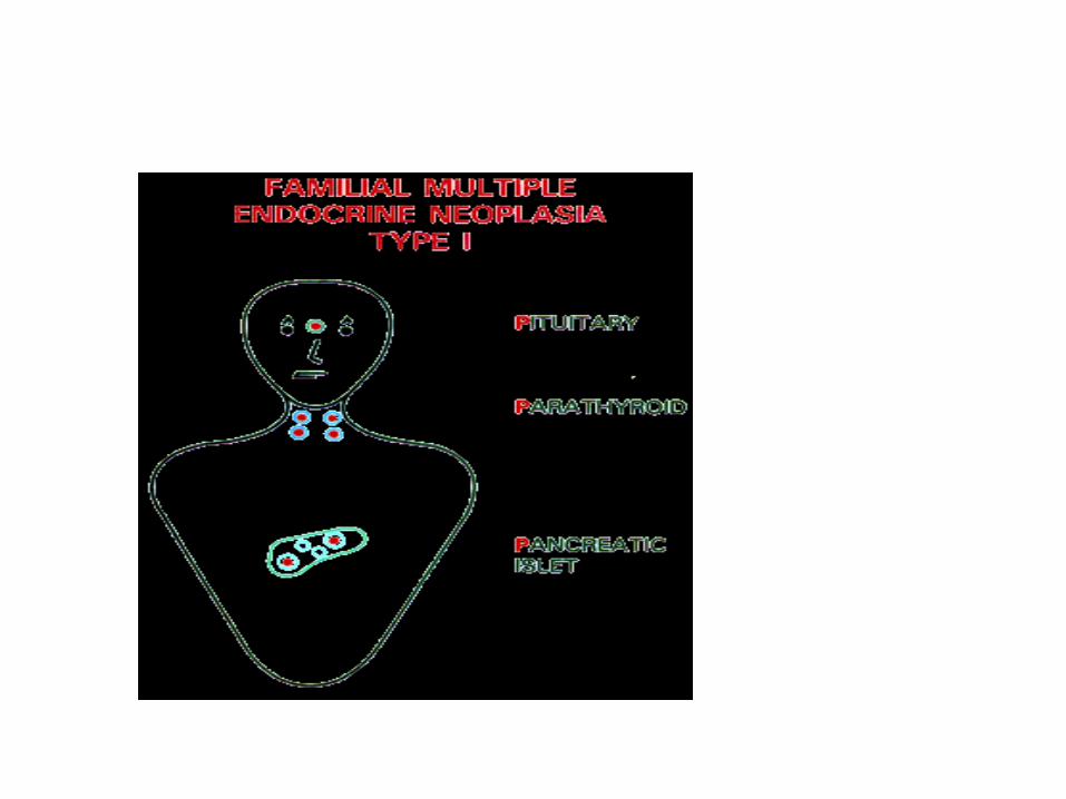
MEN1 Wermer sy MEN 2A Sipple sy MEN 2B
Hypophysis Adenomas
parathyroid HPL +++
Adenoma +
Hpl +
Langerhans islands
HPL ++
Adenoma ++
CC +++
Adrenal gland HPL Pheochromo-
cytoma ++
Pheochromo-
cytoma +++
Thyroid gland C cell hpl +++
Medull. Cc +++
C cell hpl +++
Medull. Cc +++
Extraendocrine organs
Mucocutan ganglioneuromas
Marfanoid stature
Genetic alteration
MEN1 11q13 RET 10q11.1 RET ?

Rakovecz-Kondré-Füki család
Rakovecz László1946.
MEN 2A
Kondré Attiláné1968. MEN 2A
Kun Zoltánné1970.
MEN 2A
?Füki Györgyné
1972.MEN 2A
Nikolett1986.
Zsuzsanna1988.
MEN 2A
Attila1989.
Norbert1993.
Zsanett1995.
MEN 2A?
Zsófia1998.
MEN 2A?
Henriett AlexandraBeatrix
94.MEN 2A
?
RET gén C634R mutáció-igazolt
nem igazolt
nem vizsgált
MEN2A-igazolt klinikai manifesztáció


14 year old female
MEN2 sy in the family
RET mutation
Prophylactic thyroidectomy


Other tumors
Mesenchymal tumors
Lymphomas
Metastatic (rare)

12685/07
70 year old male
Cl.: Nodular goiter

Metastatic
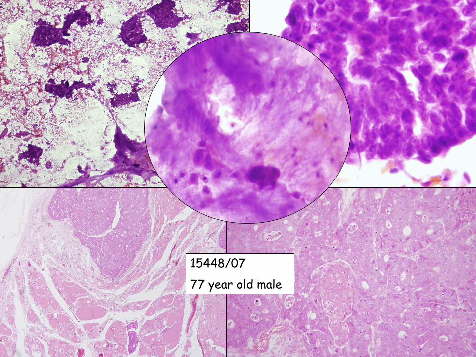
15448/07
77 year old male

Parathyroid gland
In 10 % of the cases, only 3 glands
Any localisation along the developmental pathway:
IV. Pharyngeal pouch


Norm.: 10-60 pg/ml or 1-6 pmol/l
Effects of the Parathyroid hormone …Osteoclast mobilization
Renal tubular Ca reabsorbtion
Renal vitamine D conversion (dihydroxy) Renal phosphate
excretion ^
GI.: Ca absorption^


35-40 mg/piece, yellow-brown
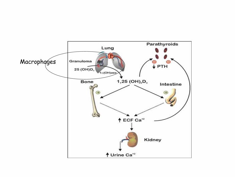
Macrophages

Primary hyperparathyreosis
parathyroid adenoma 75-80%parathyroid hyperplasia 10-15%parathyroid cc. <5%
Female/Male 3/1Middle aged or olderFrequently sporadic, rarely part of MEN
1, or MEN2

MEN1 : 11q13 (tumor supressor gene inactivation)
MEN2A : 10 Q - RET mutation(tyrosine kinase receptor)
Familiary hypocalciuric syndrome:3q (CASR) AD lowered sensitivity of the parathyroid
for Ca
Sporadic- PRAD1:PRAD1 gene - coding CyclinD1-(11q) overexpression due to inversion , > clonal proliferation

MEN1 Wermer sy MEN 2A Sipple sy MEN 2B
Hypophysis Adenomas
parathyroid HPL +++
Adenoma +
Hpl +
Langerhans ilands HPL ++
Adenoma ++
CC +++
Adrenal gland HPL Pheochromo-
cytoma ++
Pheochromo-
cytoma +++
Thyroid gland C cell hpl +++
Medull. Cc +++
C cell hpl +++
Medull. Cc +++
Extraendocrine organs
Mucocutan ganglioneuromas
Marfanoid stature
Genetic alteration
MEN1 11q13 RET 10q11.1 RET ?

HyperparathyreosisAsymptomatic
Blood test performed for unrelated conditions: Se Ca ^^^
associated with malignancy
Symptomatic,
Neuromuscular changes – weekness, fatigue
Cardial: aorta, mitral calcification
GI nausea, obstipation, ulcers, pancreatitis, gallstones
CNS depression, letargy, cramps
Bone diseases (osteitis fibrosa cystica
generalista secundum
Recklinghausen)

Sequales of Hyperparathyreosis

Osteitis fibrosa cystica generalisata secundum Recklinghausen

Causes of Hypercalcaemia
Elevated PTH
Hyperparathyreosis
primary
secondary
tertiary
Lowered PTH
Malignancy associated
Osteolytic met.
PTH-rP-mediated
D vitamine toxicity
Immobilization
Thiazids
Sarcoidosis ( other granulomatous diseases)

GM-CSF expands the osteoclast precursor pool and PTHrP increases RANK ligand and decreases osteoprotegerin (OPG) production by osteoblasts; OPG is a decoy receptor that blocks RANKL. RANKL then induces osteoclast precursor differentiation and increases osteoclast formation. The increase in bone resorption releases growth factors and calcium, which then enhances tumor growth.

parathyroid adenoma

Microscopy
Normal looking parathyroid tissue surrounded by a capsule
Endocrine atypia might occure
Adenoma:– 1 gland gets enlarged
Hyperplasia: – more glands get enlarged but not evidently all (??)
Carcinoma: diagnosis is based on vascular / capsular invasion, metastasis


Associated by renal insufficiencyLowered Calcium intakeSteatorrheaD vitamine deficiency
Low se Ca Symptoms are similar to primary ~, but less
severe
Tertiary hyperparathyreosis
Secondary hyperparathyreosis
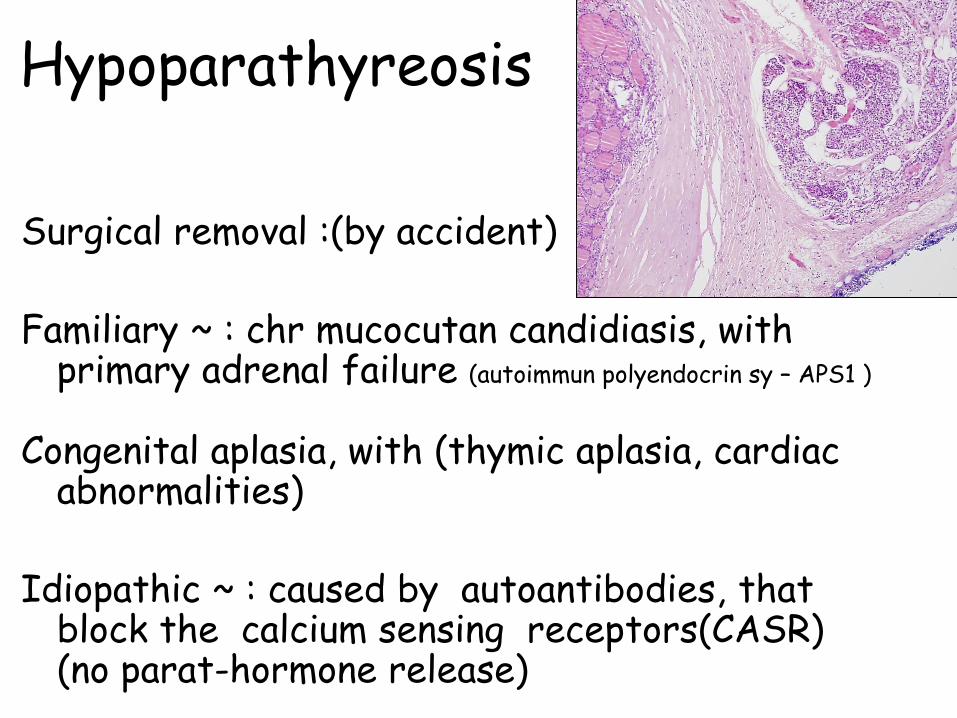
Hypoparathyreosis
Surgical removal :(by accident)
Familiary ~ : chr mucocutan candidiasis, with primary adrenal failure (autoimmun polyendocrin sy – APS1 )
Congenital aplasia, with (thymic aplasia, cardiac abnormalities)
Idiopathic ~ : caused by autoantibodies, that block the calcium sensing receptors(CASR) (no parat-hormone release)

HypoparathyreosisTetania- neuromuscular irritability
ChovstekTrousseau’s sign
Mental alterationsdepression, irritability, hallucinations, psychosis
CNSbasal ganglion calcificationparkinson like signspapilla oedema
Lense calcification – cataractaEKG – prolongation of QTintervalDental abnormalities

Pseudohypoparathyreosis
End-organ resistance to PTH


• Circulation• Most of the thyroid hormone circulating in
the blood is bound to transport proteins. Only a very small fraction of the circulating hormone is free (unbound) and biologically active, hence measuring concentrations of free thyroid hormones is of great diagnostic value.
• When thyroid hormone is bound, it is not active, so the amount of free T3/T4 is what is important. For this reason, measuring total thyroxine in the blood can be misleading.

• The thyroid hormones, thyroxine (T4) and triiodothyronine (T3), are tyrosine-based hormonesproduced by the thyroid gland. An important component in the synthesis is iodine. The major form of thyroid hormone in the blood is thyroxine (T4). The ratio of T4 to T3 released in the blood is roughly 20 to 1. Thyroxine is converted to the active T3 (three to four times more potent than T4) within cells by deiodinases (5'-iodinase). These are further processed by decarboxylation and deiodination to produce iodothyronamine (T1a) and thyronamine(T0a).

• T3 and T4 cross the cell membrane, probably via amino acid importins, and function via a well-studied set of nuclear receptors in the nucleus of the cell, the thyroid hormone receptors.
• T1a and T0a are positively charged and do not cross the membrane; they are believed to function via the trace amine-associated receptor TAAR1 (TAR1, TA1), a G-protein-coupled receptor located in the cell membrane.
• Another critical diagnostic tool is the amount of thyroid-stimulating hormone (TSH) that is present.

• Function• The thyronines act on the body to increase the basal metabolic
rate, affect protein synthesis and increase the body's sensitivity to catecholamines (such as adrenaline) by permissiveness. The thyroid hormones are essential to proper development and differentiation of all cells of the human body. These hormones also regulate protein, fat, and carbohydratemetabolism, affecting how human cells use energetic compounds. Numerous physiological and pathological stimuli influence thyroid hormone synthesis.
• The thyronamines function via some unknown mechanism to inhibit neuronal activity; this plays an important role in the hibernation cycles of mammals and the moulting behaviour of birds. One effect of administering the thyronamines is a severe drop in body temperature.

• Summary of the effects of hormones on skeletal metabolism• Increase Bone resorption• Parathyroid hormone
Glucocorticoids Thyroid Hormone Vitamin D metabolites in high doses Decrease Bone Resorption
• Calcitonin Gonadal steroids Increase Bone Formation
• Growth hormone Vitamin D metabolites Gonadal steroids Decrease Bone Formation
• Glucocorticoids
R

Female, 64 years old
Abruptly growing thyroid nodule.


NSE
Chromogranin
Synaptophysin
CK