THE ADRENAL GLAND
51
STEROIDOGENESIS AND ITS CONTROL DR SADIA HAROON DEPT OPOPOPOPF BIOCHEMISTRY SECOND YEAR MBBS UGS MODULE Date 4.11.2021
-
Upload
khangminh22 -
Category
Documents
-
view
3 -
download
0
Transcript of THE ADRENAL GLAND
STEROIDOGENESIS AND
ITS CONTROL
DR SADIA HAROON
DEPT OPOPOPOPF BIOCHEMISTRY
SECOND YEAR MBBS
UGS MODULE Date 4.11.2021
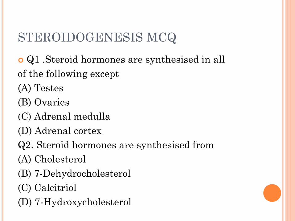
STEROIDOGENESIS MCQ
Q1 .Steroid hormones are synthesised in all
of the following except
(A) Testes
(B) Ovaries
(C) Adrenal medulla
(D) Adrenal cortex
Q2. Steroid hormones are synthesised from
(A) Cholesterol
(B) 7-Dehydrocholesterol
(C) Calcitriol
(D) 7-Hydroxycholesterol

STEROIDOGENESIS MCQQ3 .A common intermediate in the synthesis
of all the steroid hormones is
(A) Pregnenolone
(B) 17-Hydroxypregnenolone
(C) Corticosterone
(D) Progesterone
Q4. A common intermediate in the synthesis
of cortisol and aldosterone is
A)Progesterone
(B) Testosterone
(C) Estradiol
(D) None of these

STEROIDOGENESIS MCQ
Q5A common intermediate in the synthesis
of estrogens is
(A) Cortisol
(B) Andostenedione
(C) Corticosterone
(D) 11-Deoxycorticosterone
Q6In adrenogenital syndrome due to total
absence of 21-hydroxylase in adrenal
cortex, there is
(A) Deficient secretion of glucocorticoids
(B) Deficient secretion of mineralcorticoids
(C) Excessive secretion of androgens
(D) All of these

Q7Androgens are synthesised in
(A) Leydig cells in testes
(B) Sertoli cells in testes
(C) Seminiferous tubules
(D) Prostate gland
Q8Testosterone is transported in blood by
(A) Transcortin
(B) Testosterone binding globulin
(C) Testosterone estrogen binding globulin
(D) Albumin
STEROIDOGENESIS MCQ
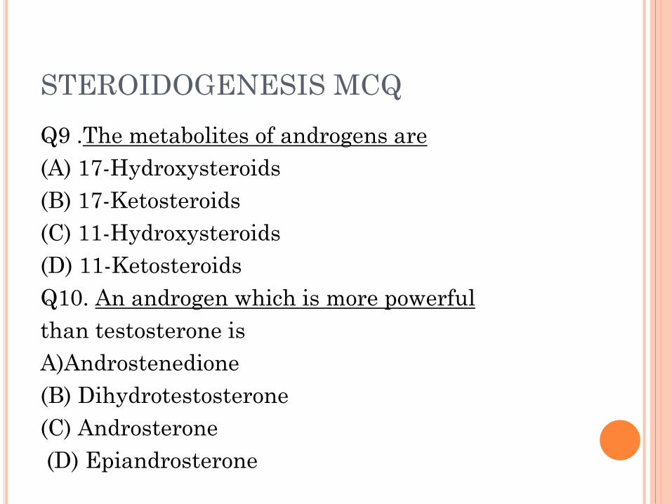
Q9 .The metabolites of androgens are
(A) 17-Hydroxysteroids
(B) 17-Ketosteroids
(C) 11-Hydroxysteroids
(D) 11-Ketosteroids
Q10. An androgen which is more powerful
than testosterone is
A)Androstenedione
(B) Dihydrotestosterone
(C) Androsterone
(D) Epiandrosterone

STEROIDOGENESIS MCQ
Q 11.Secretion of androgens is increased by
(A) LH
(B) FSH
(C) ACTH
(D) Growth hormone
Q12. During late pregnancy, the major source
of progesterone is
(A) Adrenal cortex (B) Placenta
(C) Corpus luteum (D) Graafian follicles

STEROIDOGENESIS MCQ
Q13. Progesterone is transported in blood by
(A) Transcortin
(B) Sex hormone binding globulin
(C) Albumin
(D) Testosterone estrogen binding globulin
Q 14 The major metabolite of progesterone is
(A) Pregnenolone
(B) Pregnanediol
(C) Estradiol
(D) Norethindrone

Q15. A hormone used for detection of pregnan-cy is
(A) Estrogen
(B) Progesterone
(C) Oxytocin
(D) Chorionic gonadotropin
Q16. Placenta secretes all of the following except
(A) FSH
(B) Progesterone
(C) Estrogen
(D) Chorionic gonadotropin

Q17.Women become susceptible to osteoporo-sis
after menopause due to decreased
(A) Secretion of Parathormone
(B) Conversion of vitamin D into calcitriol
(C) Secretion of estrogen
(D) Secretion of progesterone
Q18The major metabolite of progesterone is
(A) Pregnenolone
(B) Pregnanediol
(C) Estradiol
(D) Norethindrone

LEARNING OBJECTIVE
Introduction to male sex hormones or androgens
Functions of testicular cells
Synthesis and metabolism of testosterone

ADRENAL CORTEX
Is divided into 3 zones in the adult gland: Zona Glomerulosa, Zona Fasciculata, Zona Rericularis.
Is divided onto 4 zones in the fetal gland.
The three zones of the permanent cortex constitutes only 20% of the fetal gland’s size. The remaining zone (fetal cortex) comprises up to 80% of gland’s size during fetal life.
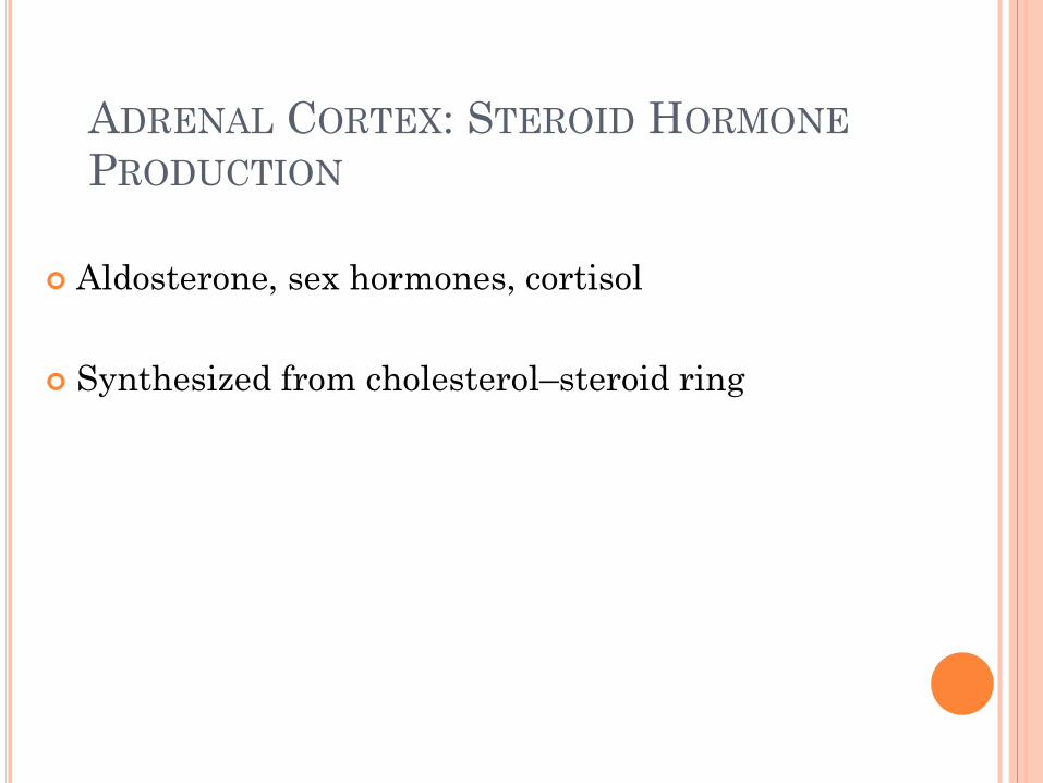
ADRENAL CORTEX: STEROID HORMONE
PRODUCTION
Aldosterone, sex hormones, cortisol
Synthesized from cholesterol–steroid ring

HORMONES OF ADRENAL CORTEX
Corticosteroids
Aldosterone (glomerulosa)
Mineralocorticoid
Cortisol (fasciculata)
Glucocorticoid
Sex hormones (reticularis)
Adrenal androgens

15
HORMONAL STIMULATION OF GLUCOCORTICOIDS
HPA AXIS (HYPOTHALAMIC/PITUITARY/ADRENAL AXIS)
With stress, hypothalamus sends CRH to anterior pituitary (adenohypophysis)
Pituitary secretes ACTH
ACTH goes to adrenal cortex where stimulates glucocorticoid secretion Sympathetic nervous system can also stimulate it
Adrenal cortex also secretes DHEA (dehydroepiandrosterone) Converted in peripheral tissues to testosterone and
estrogen (also steroid hormones)
Unclear function in relation to stress

Gonadotropic hormones
GnRH: pulsatile secretion
Cyclical secretion LH, FSH
Females: ovary
• LH: ovulation, corpus luteum
•FSH: dvpt follicle, oestradiol and progesterone
Males: testes
• LH: Leydig cells: testosterone
•FSH: Sertoli cells: spermatogenesis
FSH: inhibin: negative feedback

Gonadotropic hormones


SPERMATOGENESIS IN TESTIS
The development and maintenance of
spermatogenesis is dependent on
the pituitary gonadotropins; FSH, and
LH.
Both hormones are secreted and regulated
as a part of the HPG axis in response to
the hypothalamic gonadotropin-releasing
hormone (GnRH).
FSH stimulates the production of sperm.


INTERSTITIAL OR LEYDIG CELLS
Interstitial or Leydig cells are located in the
connective tissue surrounding the
seminiferous tubules.
They produce testosterone, the male sex
hormone responsible
for the growth and maintenance of the cells
of the germinal epithelium and
the development of secondary sex
characteristics.

INTERSTITIAL OR LEYDIG CELLS
They secrete testosterone, androstenedione
and dehydroepiandrosterone (DHEA), when
stimulated by the luteinizing hormone (LH)
which is released from the anterior pituitary in
response to gonadotrophin releasing hormone
which in turn is released by the hypothalamus.

INTERSTITIAL OR LEYDIG CELLS
The receptor allows the body to respond
appropriately to these hormones.
In males, chorionic gonadotropin stimulates
the development of cells in the testes called
Leydig cells, and luteinizing hormone
triggers these cells to produce androgens.
In the adult testis, estrogen is synthesized by
Leydig cells and the germ cells, producing a
relatively high concentration in rete testis fluid.
Estrogen receptors are present in the testis,
efferent ductules and epididymis of most species.
INTERSTITIAL OR LEYDIG CELLS
Leydig cells (LC) are present in the testicular
interstitial tissue, and their main function is to
produce testosterone (T) for the
maintenance of spermatogenesis and
extratesticular androgenic and anabolic
functions.

INTERSTITIAL OR LEYDIG CELLS
Testicular steroidogenesis initially involves
the production of T from cholesterol by the
interstitial Leydig cells of the testis. .
.. T undergoes conversion in the Leydig
cells, or in the prostate,
by 5α-reductase to 5α-dihydrotestosterone
(DHT), the most potent bioactive androgen.
Leydig cells stimulate the production of
sperm cells, and Sertoli cells nourish
developing sperm.
STEROIDOGENESIS
Steroidogenesis entails processes
by which cholesterol is converted
to biologically active steroid
hormones. ..
Qualitative regulation
determining the type of steroid to
be produced is mediated by many
enzymes and cofactors.

ADRENAL CORTEX: STEROID HORMONE
PRODUCTION
Figure 23-2: Synthesis pathways of steroid hormones

SYNTHESIS IN THE CORTEX


STEROID HORMONES: STRUCTURE
35
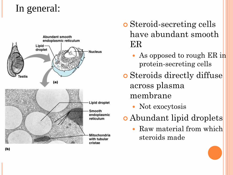
Steroid-secreting cells
have abundant smooth
ER
As opposed to rough ER in
protein-secreting cells
Steroids directly diffuse
across plasma
membrane
Not exocytosis
Abundant lipid droplets
Raw material from which
steroids made
In general:

TESTICULAR STEROIDOGENESIS
Testicular steroidogenesis is a
fine-regulated process, and its
main product, testosterone (T),
has a crucial role in fetal
development and maturation
and in adulthood for the
maintenance of secondary sexual
function and spermatogenesis
TESTICULAR STEROIDOGENESIS
The primary site for the synthesis of
testosterone within the mammalian
testis is the interstitial cells of
Leydig, which are specialized cells
located in the angular spaces
between seminiferous tubules

TESTICULAR STEROIDOGENESIS
When puberty begins,
usually between the ages of 9 and 15,
the pituitary gland — located near
the brain —
secretes hormones that stimulate the
testicles to produce testosterone.
The production of testosterone
brings about many physical changes.
EFFECT OF AGING .
Although the mitochondrial and smooth
endoplasmic reticulum steroidogenic enzymes are
reduced in aging Leydig cells, their levels
nonetheless are sufficient to support high levels
of steroid production if enough cholesterol is
translocated into the mitochondria and thus is
available to CYP11A1 of the inner .

ROLE OF GONADOTROPHINS
The hypothalamus produces
gonadotropin-releasing hormone,
which signals the pituitary gland to
make follicle-stimulating hormone (
FSH ) and luteinizing hormone (
LH ).
Luteinizing hormone then signals
the testes to produce testosterone.
LOW LEVELS OF TESTICULAR STEROIDS
Injury (trauma, interrupted blood supply to
the testes) or infection of the testes
(orchitis)
Chemotherapy for cancer.
Metabolic disorders such as
hemochromatosis (too much iron in the
body)
Dysfunction or tumors of the pituitary
gland
STEROID HORMONES SYNTHESIS
Steroids are derivatives of cholesterol
Cholesterol is from the lipid droplets in
cortical cells (cholesterol esters in
LDL)
Removed cholesterol is replenished by
cholesterol in LDL in blood or synthesized
from acetate
Steroid hormones are synthesized and secreted on
demand (not stored)
The first and rate-limiting step in the synthesis of all
steroid hormones is conversion of cholesterol to
pregnenolone by the enzyme cholesterol dismolase (aka
cholesterol side chain cleavage (SCC) enzyme
Newly synthesized steroid hormones are rapidly secreted
from the cell
Following secretion, all steroids bind to some extent to
plasma proteins: CBG and albumin
STEROID HORMONES SYNTHESIS (CONT.)

ENZYMES IN STEROID BIOSYNTHESIS
Side-chain cleavage enzyme; desmolase
(P450scc)
3 β-hydroxysteroid dehydrogenase (3 β HSD)
17 α-hydroxylase/17,20 lyase (P450 c17):
Adrenarche
* not present in glumerulosa cells
21-hydroxylase (P450c21)
11 beta-hydroxylase (P450c11)
Aldosterone synthase

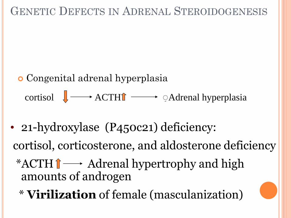
GENETIC DEFECTS IN ADRENAL STEROIDOGENESIS
Congenital adrenal hyperplasia
ACTH ِ Adrenal hyperplasia
• 21-hydroxylase (P450c21) deficiency:
cortisol, corticosterone, and aldosterone deficiency
*ACTH Adrenal hypertrophy and high amounts of androgen
* Virilization of female (masculanization)
cortisol

STEROID HORMONES: ACTION

ANDROGENS
are hormones that contribute to growth and
reproduction in both men and women.
Androgens are usually thought of as male
hormones, but the female body naturally
produces a small amount of androgens too.
Androgen deficiency in women is a controversial
concept.

CORTISOL: ROLE IN DISEASES AND
MEDICATION
Use as immunosuppressant
Hyperimmune reactions (bee stings)
Serious side effects
Hypercortisolism (Cushing's syndrome)
Tumors (pituitary or adrenal)
Iatrogenic (physician caused)
Hypocortisolism (Addison's disease)