Gastric Adenocarcinoma of the Fundic Gland Type Update ...
13
461 Am J Clin Pathol 2018;149:461-473 DOI: 10.1093/ajcp/aqy019 © American Society for Clinical Pathology, 2018. All rights reserved. For permissions, please e-mail: [email protected] AJCP / REVIEW ARTICLE Gastric Adenocarcinoma of the Fundic Gland Type Update and Literature Review Mark A. Benedict, DO, 1 Gregory Y. Lauwers, MD, 2 and Dhanpat Jain, MD 1 From the 1 Department of Pathology, Yale University School of Medicine, New Haven, CT; and 2 Department of Pathology, H. Lee Moffitt Cancer Center and Research Institute, Tampa, FL. Key Words: Gastrointestinal; Histology; Immunopathology Am J Clin Pathol June 2018;149:461-473 DOI: 10.1093/AJCP/AQY019 ABSTRACT Objectives: Gastric adenocarcinoma of the fundic gland type (GA-FG) is a newly described entity with a lack of awareness amongst general surgical pathologists and this review highlights the key features and controversies associated with this uncommon neoplasm. Methods: A literature search through PubMed using synonyms for GA-FG was conducted to obtain 111 cases. Results: GA-FG is a well-differentiated neoplasm of oxyntic mucosa, that is comprised of chief cells and parietal cells. Chief cell differentiation is highlighted with Muc-6, RUNX3, and pepsinogen. Parietal cells are highlighted with H+/K+ ATPase and PDGFRA-α. Association with Helicobacter infection, chronic gastritis, intestinal metaplasia, or gastric atrophy is not seen. Most GA-FGs are confined to the mucosa. Deeper invasion, lymphovascular invasion, nodal metastasis, and extragastric spread are uncommon. Conclusions: GA-FGs are rare lesions that typically follow a benign course. However, despite features of malignancy in some cases, complete surgical excision, sometimes with endoscopic mucosal resection, seems adequate treatment. Traditionally, gastric carcinoma has been grouped into two types: intestinal and diffuse (Lauren classifi- cation). 1 Evaluating the 54,099 stomach carcinomas reported during the 1978-2005 US National Cancer Institute’s Surveillance, Epidemiology, and End Results (SEER) data set, 74% were of the intestinal type, 16% of the diffuse, while 10% were other epithelial carcino- mas. 2 More relevant to our subject, a classification system devised by Nakamura et al 3 groups gastric carcinoma into differentiated and undifferentiated, based on the tumor’s ability to form glands. However, with time it has become evident that gastric carcinomas can differentiate along several different epithelial lineages: surface foveolar lin- eage, pyloric gland lineage, chief cell lineage, and parietal (oxyntic) cell lineage. Our understanding of these multi- ple phenotypic facets of gastric neoplasms is in its early stages. The specific predisposing factors, demographics, and prognosis of these neoplasms, and how they vary from the conventional carcinomas, is unclear. Gastric adenocarcinoma of the fundic gland type (GA-FG) is a novel entity first proposed in 2007 by Tsukamoto et al. 4 The characteristic oxyntic gland dif- ferentiation can be divided in three subcategories based on their composition: chief cell predominant (approxi- mately 99% of reported cases), parietal cell predominant, and mixed phenotype. To date, the nature of this lesion is debated and there is a lack of awareness of GA-FG in the pathology community. This review aims to highlight our current understanding of gastric neoplasms with differen- tiation along elements of fundic glands. Downloaded from https://academic.oup.com/ajcp/article/149/6/461/4965910 by guest on 09 January 2022
-
Upload
khangminh22 -
Category
Documents
-
view
0 -
download
0
Transcript of Gastric Adenocarcinoma of the Fundic Gland Type Update ...
461Am J Clin Pathol 2018;149:461-473DOI: 10.1093/ajcp/aqy019
© American Society for Clinical Pathology, 2018. All rights reserved. For permissions, please e-mail: [email protected]
AJCP / Review ARticle
Gastric Adenocarcinoma of the Fundic Gland Type
Update and Literature Review
Mark A. Benedict, DO,1 Gregory Y. Lauwers, MD,2 and Dhanpat Jain, MD1
From the 1Department of Pathology, Yale University School of Medicine, New Haven, CT; and 2Department of Pathology, H. Lee Moffitt Cancer Center and Research Institute, Tampa, FL.
Key Words: Gastrointestinal; Histology; Immunopathology
Am J Clin Pathol June 2018;149:461-473
DOI: 10.1093/AJCP/AQY019
ABSTRACT
Objectives: Gastric adenocarcinoma of the fundic gland type (GA-FG) is a newly described entity with a lack of awareness amongst general surgical pathologists and this review highlights the key features and controversies associated with this uncommon neoplasm.
Methods: A literature search through PubMed using synonyms for GA-FG was conducted to obtain 111 cases.
Results: GA-FG is a well-differentiated neoplasm of oxyntic mucosa, that is comprised of chief cells and parietal cells. Chief cell differentiation is highlighted with Muc-6, RUNX3, and pepsinogen. Parietal cells are highlighted with H+/K+ ATPase and PDGFRA-α. Association with Helicobacter infection, chronic gastritis, intestinal metaplasia, or gastric atrophy is not seen. Most GA-FGs are confined to the mucosa. Deeper invasion, lymphovascular invasion, nodal metastasis, and extragastric spread are uncommon.
Conclusions: GA-FGs are rare lesions that typically follow a benign course. However, despite features of malignancy in some cases, complete surgical excision, sometimes with endoscopic mucosal resection, seems adequate treatment.
Traditionally, gastric carcinoma has been grouped into two types: intestinal and diffuse (Lauren classifi-cation).1 Evaluating the 54,099 stomach carcinomas reported during the 1978-2005 US National Cancer Institute’s Surveillance, Epidemiology, and End Results (SEER) data set, 74% were of the intestinal type, 16% of the diffuse, while 10% were other epithelial carcino-mas.2 More relevant to our subject, a classification system devised by Nakamura et al3 groups gastric carcinoma into differentiated and undifferentiated, based on the tumor’s ability to form glands. However, with time it has become evident that gastric carcinomas can differentiate along several different epithelial lineages: surface foveolar lin-eage, pyloric gland lineage, chief cell lineage, and parietal (oxyntic) cell lineage. Our understanding of these multi-ple phenotypic facets of gastric neoplasms is in its early stages. The specific predisposing factors, demographics, and prognosis of these neoplasms, and how they vary from the conventional carcinomas, is unclear.
Gastric adenocarcinoma of the fundic gland type (GA-FG) is a novel entity first proposed in 2007 by Tsukamoto et al.4 The characteristic oxyntic gland dif-ferentiation can be divided in three subcategories based on their composition: chief cell predominant (approxi-mately 99% of reported cases), parietal cell predominant, and mixed phenotype. To date, the nature of this lesion is debated and there is a lack of awareness of GA-FG in the pathology community. This review aims to highlight our current understanding of gastric neoplasms with differen-tiation along elements of fundic glands.
Dow
nloaded from https://academ
ic.oup.com/ajcp/article/149/6/461/4965910 by guest on 09 January 2022

Benedict et al / Gastric adenocarcinoma Update and review
462 Am J Clin Pathol 2018;149:461-473DOI: 10.1093/ajcp/aqy019
© American Society for Clinical Pathology
Methodology
A literature search through PubMed using the phrases “gastric adenocarcinoma fundic gland type,” “gastric adenocarcinoma chief cell predominant type,” “gastric adenocarcinoma-mixed parietal and chief cell type,” “gastric adenocarcinoma-parietal cell type,” and “parietal cell carcinoma” revealed a total of 111 cases reported in the English language literature. A num-ber of reports in older literature have described cases of oncocytic gastric neoplasms as “parietal cell” carci-noma; however, the evidence of parietal cell differenti-ation in most of these papers is not conclusive and they appear different from the GA-FG as we currently recog-nize them.5,6 Hence, these cases were excluded from this review.
The clinical history and pathologic characteristics were examined for each case and summary data are tabu-lated in ❚Table 1❚.4-31
Demographics
The majority of reports of GA-FG originate from East Asia (Japan and Korea), where the incidence appears to be still low. In the study by Tohda et al7 only four (< 0.01%) patients with GA-FG were identified from the 30,182 individuals who underwent upper endoscopy (as part of a yearly check-up) between October 2010 and September 2014.
The patients are generally older adults and the aver-age patient age is 66 years (range, 39-85 years). There seems to be a slight male predilection, with the M:F ratio being 2.2:1.
Etiopathogenesis
The carcinogenesis of gastric adenocarcinoma of the conventional type is multifactorial. However, chronic Helicobacter pylori infection with subsequent intestinal metaplasia and mucosal atrophy plays a major role and is associated in 80% of cases.32 Autoimmune gastritis, which is also associated with increased risk for gastric adenocar-cinoma, also has chronic inflammation that is followed by intestinal or other types of metaplasia and atrophy.
Of the 111 cases of GA-FG published, H pylori data were available in only 43 (39%) cases, of which 17 (40%) were positive for infection Table 1. In only one series did the percentage of H pylori positivity reach a rate that mir-rored conventional gastric cancer, with 15 of 20 (75%)
patients being H pylori positive with atrophy.8 Yet, in the same study, the authors reported that the neoplasms developed in areas with no apparent atrophy observed endoscopically.8 Additionally, independent histologic assessment found that in nearly all cases of GA-FG no surrounding atrophy was detected.8
Interestingly, some cases of GA-FG show dilated fundic glands with admixed foveolar type cells resembling fundic gland polyps (FGPs), thus raising the question as to whether there is any relationship with proton pump inhib-itor (PPI) use and the development of these neoplasms.5 Of note, in a series of 12 cases, of the eight patients with available history of medication, seven had used acid sup-pressive therapy (six PPI and one H2 blocker).9 However, in most of the reports, the history of medication use is not thoroughly investigated, especially PPI use.
Endoscopy and Gross Appearance
The overwhelming majority of GA-FG, that is 89 of 111 cases (80%), have occurred in the upper third of the stomach. Twenty out of 111 (18%) tumors developed in the middle third and only one tumor was seen in the lower third of the stomach (1%). Typically, GA-FGs are small, with an average size of 10 mm and the largest reported case measured 85 mm.10 These lesions arise only in oxyntic mucosa. In one study, com-prising 20 cases examined by endoscopy, the lesions appeared elevated in 12 (60%), flat in five (25%), and depressed in three (15%) cases, as per the Japanese clas-sification of gastric carcinoma.8 In another endoscopic study, the tumors were classified as “submucosal” in six of 10 (60%) or mucosal with a flat/depressed surface in four of 10 (40%) patients.11 Some of the lesions mimic FGPs endoscopically.7 These tend to have a white hue (70%) or yellow discoloration, with frequent complex and often branching dilated mucosal vessels, in a nona-trophic adjacent mucosa (90%).8,11 Central depression noted by indigo carmine chromoendoscopy in one case is believed to be an evidence of submucosal involve-ment.12 Interestingly, several cases documented a black pigmentation in the lesion, which aided in the early detection.13,14 Narrow-band imaging, which enhances the visualization of the mucosal vessels, shows an irregular pattern, suggestive of the heterogeneity of the microvessels in GA-FG ❚Image 1A❚ and ❚Image 1B❚.15,22 The use of narrow-band imaging and endoscopic ultra-sound enhances the opportunity for complete excision via endoscopic mucosal resection (EMR), which suf-fices in superficial tumors.11
Dow
nloaded from https://academ
ic.oup.com/ajcp/article/149/6/461/4965910 by guest on 09 January 2022

AJCP / Review ARticle
463Am J Clin Pathol 2018;149:461-473DOI: 10.1093/ajcp/aqy019
© American Society for Clinical Pathology
❚Tab
le 1
❚ C
hara
cter
isti
cs o
f R
epor
ted
Gas
tric
Ade
noca
rcin
omas
-Fun
dic
Gla
nd T
ype
Aut
hor
(Yea
r)N
o. o
f C
ases
Age
or
Age
R
ange
(y)
Gen
der
Dis
trib
utio
nC
ell T
ype
(Pre
dom
inan
ce)
Loc
atio
n: N
o. o
f C
ases
Helicobacter pylori
Siz
e (m
m)
Inva
sion
Dep
thL
ymph
ovas
cula
r In
vasi
onO
utco
me
Cha
n et
al
(201
6)9
1239
-81
5 M
7 F
Chi
ef c
ell
Upp
er t
hird
—2-
20
Muc
osa:
10
Sub
muc
osa:
2N
oA
live,
no
recu
rren
ce
or m
etas
tasi
sS
ato
et a
l (2
016)
131
77F
Chi
ef c
ell
Upp
er t
hird
Neg
11S
ubm
ucos
aN
oA
live,
no
recu
rren
ce
or m
etas
tasi
sC
ha e
t al
(2
016)
231
69M
Chi
ef c
ell
Upp
er t
hird
—25
Mus
cula
ris
prop
riaN
oA
live,
no
recu
rren
ce
or m
etas
tasi
sC
hiba
et
al
(201
6)8
20 (9
ca
ses
with
E
MR
)
44-8
516
M4
FC
hief
cel
lU
pper
thi
rd: 1
4M
iddl
e th
ird: 6
Neg
: 5Po
s: 1
53-
20M
ucos
al: 2
Sub
muc
osa:
7N
o da
ta: 1
1
No:
9N
o da
ta: 1
1N
o m
orph
olog
ical
ch
ange
s by
en
dosc
opic
ex
amin
atio
nTo
hda
et a
l (2
016)
74
42-6
22
M2
FC
hief
cel
lU
pper
thi
rd: 3
Mid
dle
third
: 1N
eg: 2
Pos:
22-
5M
ucos
al: 2
Sub
muc
osa:
2N
oA
live
Kaw
asak
i et
al
(201
6)18
162
MC
hief
cel
lM
iddl
e th
irdN
eg—
Sub
muc
osa
Yes
Aliv
e
Kat
o et
al
(201
5)24
180
MC
hief
cel
lU
pper
thi
rdN
eg30
Sub
muc
osa
No
Aliv
e
Take
da e
t al
(2
015)
141
66M
Chi
ef c
ell
Upp
er t
hird
——
——
—
Miy
azaw
a et
al
(201
5)15
567
-78
3 M
2 F
Chi
ef c
ell
Upp
er t
hird
Neg
5-13
Sub
muc
osa
Yes:
1N
o: 4
Aliv
e, n
o re
curr
ence
or
met
asta
sis
Parik
h et
al
(201
5)22
166
MC
hief
cel
lU
pper
thi
rd—
7S
ubm
ucos
aN
oA
live,
no
recu
rren
ce
or m
etas
tasi
sH
ori e
t al
(2
015)
251
79M
Chi
ef c
ell
Mid
dle
third
——
Mus
cula
ris
muc
osae
No
Aliv
e, n
o re
curr
ence
or
met
asta
sis
Fujii
et
al
(201
5)11
164
FC
hief
cel
lU
pper
thi
rdN
eg—
Sub
muc
osa
No
—
Lew
in e
t al
(2
015)
261
49M
NS
Upp
er t
hird
—11
Sub
muc
osa
No
—
Uey
ama
et a
l (2
014)
1610
55-7
86
M4
FC
hief
cel
lU
pper
thi
rd: 6
Mid
dle
third
: 4N
eg: 7
No
data
: 33-
31M
ucos
al: 5
Sub
muc
osa:
5Ye
s: 1
No:
9A
live,
no
recu
rren
ce
or m
etas
tasi
sU
eo e
t al
(2
014)
191
62M
Chi
ef c
ell
Mid
dle
third
Neg
44 ×
35
Sub
sero
sa
(ven
ous
inva
sion
)
Yes
(mas
sive
)G
astr
ecto
my
with
ly
mph
nod
e m
etas
tasi
sFu
jimot
o et
al
(201
4)27
150
MC
hief
cel
l—
—5
——
—
Nom
ura
et a
l (2
014)
1026
49-7
922
M4
FN
SU
pper
thi
rd: 2
3M
iddl
e th
ird: 3
—3-
85S
ubm
ucos
aYe
s: 3
No:
23
Aliv
e, n
o re
curr
ence
or
met
asta
sis
Abe
et
al
(201
3)28
171
FC
hief
cel
lU
pper
thi
rdN
eg—
Sub
muc
osa
No
Aliv
e, n
o re
curr
ence
or
met
asta
sis
(con
t)
Dow
nloaded from https://academ
ic.oup.com/ajcp/article/149/6/461/4965910 by guest on 09 January 2022

Benedict et al / Gastric adenocarcinoma Update and review
464 Am J Clin Pathol 2018;149:461-473DOI: 10.1093/ajcp/aqy019
© American Society for Clinical Pathology
Aut
hor
(Yea
r)N
o. o
f C
ases
Age
or
Age
R
ange
(y)
Gen
der
Dis
trib
utio
nC
ell T
ype
(Pre
dom
inan
ce)
Loc
atio
n: N
o. o
f C
ases
Helicobacter pylori
Siz
e (m
m)
Inva
sion
Dep
thL
ymph
ovas
cula
r In
vasi
onO
utco
me
Kush
ima
et a
l (2
013)
173
56-7
81
M2
FC
hief
cel
lU
pper
thi
rd—
3-8
Sub
muc
osa
—A
live,
no
recu
rren
ce
or m
etas
tasi
sS
ingh
i et
al
(201
2)21
1044
-79
4 M
6 F
Chi
ef c
ell
Upp
er t
hird
—2-
8M
ucos
alN
oA
live,
no
recu
rren
ce
or m
etas
tasi
s: 8
Pers
iste
nce
of
dise
ase:
1N
o da
ta: 1
Che
n et
al
(201
2)29
179
MC
hief
cel
lU
pper
thi
rdN
eg—
Sub
muc
osa
No
Aliv
e, n
o re
curr
ence
or
met
asta
sis
Park
et
al
(201
2)5
347
-76
3 M
Chi
ef c
ell
Upp
er t
hird
: 1M
iddl
e th
ird: 1
Low
er t
hird
: 1
—12
-36
Muc
osal
: 1S
ubm
ucos
a: 2
No
Aliv
e, n
o re
curr
ence
or
met
asta
sis
Miy
aoka
et
al
(201
1)30
159
FC
hief
cel
lU
pper
thi
rd—
8S
ubm
ucos
aN
oA
live,
no
recu
rren
ce
or m
etas
tasi
sFu
kats
u et
al
(201
1)12
156
MC
hief
cel
lU
pper
thi
rd—
5S
ubm
ucos
aN
oA
live,
no
recu
rren
ce
or m
etas
tasi
sFu
jisaw
a et
al
(201
1)31
150
MC
hief
cel
lM
iddl
e th
ird—
42S
ubm
ucos
aN
o—
Tera
da (2
011)
201
78M
Chi
ef c
ell
Mid
dle
third
—15
(ulc
er)
——
Die
d of
ca
rcin
omat
osis
Tsuk
amot
o et
al (
2007
)41
82F
Chi
ef c
ell
Upp
er t
hird
—16
Muc
osal
No
—
Tota
l11
139
-85
75 M
36 F
Chi
ef c
ell:
84N
ot s
peci
fied:
27
Upp
er th
ird: 8
9M
iddl
e th
ird: 2
0Lo
wer
thi
rd: 1
No
data
: 1
Pos:
17
Neg
: 26
No
data
: 56
2-85
M
ucos
al: 3
1M
uscu
laris
m
ucos
ae: 1
Sub
muc
osa:
63
Sub
sero
sal:
1M
uscu
laris
pr
opria
: 1N
o da
ta: 1
4
Yes:
7N
o: 8
7N
o da
ta: 1
7
Aliv
e, n
o re
curr
ence
/m
etas
tasi
s: 8
1G
astr
ecto
my/
noda
l m
etas
tasi
s: 1
Car
cino
mat
osis
: 1D
isea
se p
ersi
sten
ce:
1N
o m
orph
olog
ical
ch
ange
s by
en
dosc
opic
ex
amin
atio
n: 2
0N
o da
ta: 7
EM
R, e
ndos
copi
c m
ucos
al r
esec
tion
; Neg
, neg
ativ
e; N
S, n
ot s
peci
fied
; Pos
, pos
itiv
e; —
, no
data
.
❚Tab
le 1
❚ (co
nt)
Dow
nloaded from https://academ
ic.oup.com/ajcp/article/149/6/461/4965910 by guest on 09 January 2022

AJCP / Review ARticle
465Am J Clin Pathol 2018;149:461-473DOI: 10.1093/ajcp/aqy019
© American Society for Clinical Pathology
Histology
The histologic appearance of GA-FG is often of a well-differentiated neoplasm, with the tumor bearing a resemblance to the fundic glands. Furthermore, at low mag-nification, GA-FG, especially of the mixed cell type, can mimic a fundic gland polyp or a pyloric gland neoplasm.7
The lesion is almost invariably lined on the surface by normal-appearing foveolar-type epithelium, while the deeper part shows a variable admixture of cell types nor-mally present in oxyntic glands7 ❚Image 2❚. The deeper glandular structures, instead of a tubular architecture seen in normal oxyntic mucosa, form anastomosing cords, producing a so-called “endless glands” pattern7 (Images 2C-2F). The glands may show a chief cell pre-dominant pattern, parietal cell predominate pattern, or an even admixture of both cell types, parietal and chief cells (Images 2C-2F) ❚Image 3❚. The distinction between the two cell types is usually easy because parietal cells
are oval or triangular, and have a characteristic eosin-ophilic finely granular cytoplasm with central nuclei, while chief cells are columnar with a slightly basophilic cytoplasm and basal nuclei. The parietal cells are more common towards the surface or periphery of the neo-plasm. Rarely, cribriforming and glandular infolding can be seen. Tumors with necrosis are rare. The neoplas-tic glands may show multilayering and nuclear stratifi-cation. Cytologic atypia is very mild in most cases, with the neoplastic chief and parietal cells showing slightly enlarged nuclei with opened up chromatin and small inconspicuous nucleoli. Mitotic figures are rare and the Ki67 index is low (< 5%).7 The background stroma may appear normal, or show edema, myxoid changes, or desmoplasia (Images 2E and 2F). Even in submucosally invasive tumors, desmoplasia can be minimal (Images 2A and 2B). The tumors commonly merge impercep-tibly with the adjacent mucosa (Image 2A) ❚Image 4❚. Typically, the adjacent oxyntic mucosa is normal with-out any intestinal metaplasia or atrophy.16
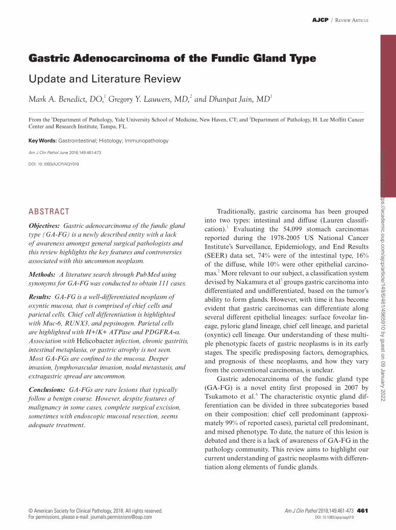
As noted earlier, once a pathologist is aware of the entity, recognition of the parietal and chief cells in the well-differ-entiated mixed phenotypes is easy on H&E stains; however, their identification in the less-differentiated cases may require immunohistochemical (IHC) markers to confirm lineage differentiation ❚Image 5❚.7 A variety of IHC markers can be helpful and include MUC5AC, MUC6, CD10, pepsino-gen-I, RUNX3, and H+/K+-ATPase ❚Table 2❚ (Images 5C and 5D). Of these, the markers for chief cell (pepsinogen-I) and parietal cell differentiation (RUNX3, H+/K+-ATPase, and PDFRA-α) are the most helpful, but are still not widely available.
❚Table 2❚ Cell Differentiation Markers
Marker Cell Type Identified
MUC2 Goblet cell MUC5AC Gastric foveolar epithelium MUC6 Mucous neck cell and pyloric
gland CD10 Brush border Pepsinogen-I Chief cell H+/K+-ATPase Parietal cell Human milk fat globule-2 (HMFG-2)
/platelet-derived growth factor receptor-α (PDGFRα)
Parietal cell
RUNX3 Chief cell
❚Image 1❚ A, Endoscopic narrow-band imaging is seen to enhance the visualization of the mucosal vessels. B, Endoscopic image revealing a small elevated lesion (from the same patient previously reported by Parikh et al22).
Dow
nloaded from https://academ
ic.oup.com/ajcp/article/149/6/461/4965910 by guest on 09 January 2022

Benedict et al / Gastric adenocarcinoma Update and review
466 Am J Clin Pathol 2018;149:461-473DOI: 10.1093/ajcp/aqy019
© American Society for Clinical Pathology
Most of the cases have been reported from Japan and Korea with only a few reports from the West, suggesting that either these tumors are uncommon in some part of the world or there is a lack of awareness. Almost all of the cases reported in the United States are of the well-differ-entiated mixed phenotype, as these are easy to recognize on routine histology. It is possible that less-differentiated neoplasms, especially along only one lineage, are under-recognized because of lack of appropriate immunohisto-chemical evaluation.
The superficial and the nonneoplastic foveolar cells tend to stain with Muc-5AC, while the chief and pari-etal cells are negative. In some cases, even foveolar type cells may also be seen within the tumor (Image 3C). The neoplastic cells are strongly positive for Muc 6. The chief cells express pepsinogen I and RUNX3, which seems to be a good marker for their identification. Pepsinogen II is less specific for identifying chief cells as the pyloric gland cells and mucus neck cells also tend to stain. The parietal cells stain positively for H+/K+-ATPase.
A B
C D
❚Image 2❚ A, Low magnification of gastric adenocarcinoma of the fundic gland type (GA-FG) (H&E, ×20) showing the tumor (within the circle) which blends imperceptibly with normal oxyntic glands (right portion of image). Note the submucosal invasion lacks any desmoplasia or myxoid change. Note occasional cystic glands in the lesion that may mimic a fundic gland polyp. The glands are visibly more complex than their normal oxyntic counterparts even at low magnification. B, Desmin immunostain (×40) highlights the breach of the muscularis mucosae by neoplastic glands invading into the superficial sub-mucosa. C, The complex glandular architecture seen at higher magnification producing anastomosing and so-called “endless glands” pattern (H&E, ×100). D, Higher power of GA-FG showing an admixture of parietal cells (arrows) and chief cells (H&E, ×400) in the invasive component.
Dow
nloaded from https://academ
ic.oup.com/ajcp/article/149/6/461/4965910 by guest on 09 January 2022

AJCP / Review ARticle
467Am J Clin Pathol 2018;149:461-473DOI: 10.1093/ajcp/aqy019
© American Society for Clinical Pathology
Endocrine cells are generally reported to be absent. In one study, staining for synaptophysin and CD56 showed diffuse positivity in the glands, while chromogranin was completely negative.12 As the foregut-derived endocrine cells are invariably positive for chromogranin, we have concluded that there is no good evidence of endocrine differentiation.5 Other studies also failed to show any endocrine cell component using chromogranin stain, although in our experience rare admixed endocrine cells can also be seen, especially at the periphery (Image 3D).
There are several reports of gastric carcinomas with parietal cell differentiation and oncocytic neo-plasms in the literature that predate the first report of GA-FG, and may represent GA-FG with predominant parietal cells, although many of these lack conclusive evidence of parietal cell differentiation.33,34 However, a couple of the recent examples have shown expres-sion of H+-K+ ATPase, suggesting that some of these lesions may represent true parietal cell neoplasms or “GA-FG with predominant parietal cell differentia-tion.”33-36 As these tumors are very poorly character-ized, at this time we have opted to keep them separate from GA-FG.
The differential diagnosis of GA-FG includes: pyloric gland adenoma and other well-differentiated GA, especially those with pyloric phenotypes, and less commonly dysplastic FGPs ❚Image 6❚. The pyloric gland adenomas (PGA) show low columnar to cuboidal cells containing finely vacuolated and lightly eosinophilic
or sometimes mildly granular eosinophilic cytoplasm (Images 6A and 6B).37,38 These stain strongly with MUC6 and variably with MUC-5AC. However, one study showed similarities in the IHC profile and molec-ular phenotype, suggesting that these lesions may be closely related.17 In this study, positivity for pepsinogen and MIST1 was present in 100% (3/3) of GA-FGT and 67% (8/12) of PGAs.17
Dysplastic fundic gland polyps are increasingly rec-ognized and less likely to be confused with GA-FG. The dysplastic changes involve only the superficial foveolar epithelium (Images 6C and 6D), while the deeper part of the lesion lacks the architectural complexity, epithelial multilayering of the glands, anastomosing cord pattern, and stromal changes.39 Rarely, large fundic gland polyps may undergo ischemia secondary to local trauma or tor-sion leading to intralesional hemorrhage, reactive epithe-lial atypia, and glandular cystic dilatation with sloughed epithelium, mimicking tumor necrosis (Images 6E and 6F). The very well-differentiated (“crawling”) gastric adenocarcinoma with foveolar and pyloric phenotypes are in the differential diagnosis but are extremely rare. These differ from GA-FG largely by the morphology of tumor cells, which differentiate towards lightly eosino-philic foveolar MUC-5AC-positive epithelium or gastric pyloric (antral) gland lineages. Cytologic atypia may be mild and similar to GA-FG, but the cells lack the admix-ture of chief or parietal cells and have a clearly invasive growth pattern.37,40
❚Image 2❚ (cont) E, Low-power view of another example of GA-FG with submucosal invasion with myxoid change in the stroma. In this example glands have more infiltrating architecture compared to previous example shown in D (H&E, ×40). F, The higher magnification showing the area of submucosal invasion (H&E, ×100). The admixture of parietal cells and chief cells in the infiltrating glands is also obvious at this magnification.
E F
Dow
nloaded from https://academ
ic.oup.com/ajcp/article/149/6/461/4965910 by guest on 09 January 2022

Benedict et al / Gastric adenocarcinoma Update and review
468 Am J Clin Pathol 2018;149:461-473DOI: 10.1093/ajcp/aqy019
© American Society for Clinical Pathology
Molecular Characteristics
The data on molecular alteration associated with GA-FG are limited. Activation of the WNT-β-catenin signaling pathway or the ERK1/2 MAPK pathway is believed to play a role in the tumorigenesis.10 In a study by Nomura et al,10 nuclear β-catenin positivity was found in 22 (80%) of 26 cases by IHC, and 13 cases (50%) har-bored mutations in at least the GNAS, CTNNB1, AXIN 1or 2, and APC genes.
In 11 of the 26 cases the mechanism of WNT signal-ing activation was unclear. In this study, five cases revealed
GNAS mutations, at exons 8 and 9, of which two cases (7.7%) also revealed KRAS mutations.10 Nuclear β-catenin expression coincided with GNAS mutations in four of five cases, three of which lacked mutations in CTNNBl, APC, or AXINs, suggesting a role for GNAS activation in WNT sig-naling.10 However, only membranous staining for β-catenin without any nuclear staining has been our own experience. Interestingly, sporadic fundic gland polyps also show acti-vating mutations in β-catenin.41
Of note, GNAS mutations seemed to be associ-ated with submucosal invasion and a larger tumor
A B
C D
❚Image 3❚ A, Gastric adenocarcinoma of the fundic gland type (GA-FG) with glands showing predominantly chief cells (H&E, ×400). B, Example of GA-FG mixed type showing admixture of parietal cells (arrows) and chief cells in the infiltrating glands (H&E, ×400). C, Another example of invasive GA-FG mixed type that shows even foveolar cells (arrowhead) admixed with chief and parietal cells in the deeper part of the tumor (H&E, ×200). Several foci of lymphovascular invasion are also identified (arrows). D, GA-FG mixed type with scattered chromogranin-positive endocrine cells, especially at the periphery of the lesion (×200). Also note foveolar cells admixed with other cells types in the tumor.
Dow
nloaded from https://academ
ic.oup.com/ajcp/article/149/6/461/4965910 by guest on 09 January 2022

AJCP / Review ARticle
469Am J Clin Pathol 2018;149:461-473DOI: 10.1093/ajcp/aqy019
© American Society for Clinical Pathology
size; however, the findings were not statistically signifi-cant.10 A second study comparing GA-FG with PGAs found frequent GNAS activating mutations in both lesions.17 In contrast, GNAS mutations are either ab-sent or infrequent in conventional gastric adenomas and adenocarcinomas.42,43
Behavior and Prognosis
At this time, of the total of 111 reported cases of GA-FG, 63 (57%) have shown submucosal invasion. Some have suggested that this may represent “pro-lapse-type” misplaced glands rather than true submucosal invasion. One case revealed subserosal invasion via lym-phovascular spread, although lymphovascular invasion is infrequent, being reported in only seven (6%) of these 111 cases.10,15,16,18,19 It should be noted that studies reporting lymphatic or vascular invasion did not report the use any special lymphatic/vascular markers of invasion, hence these are best classified as lymphovascular invasion. Of the cases with follow-up data, one patient died of car-cinomatosis (histologic type was not confirmed and no autopsy was performed), a second required gastrectomy, and a third had persistent disease (noted likely to be due to incomplete resection).19-21 In a rare example where a tumor was followed for 12 years, the tumor remained stable over a decade, suggesting that these are slow-grow-ing neoplasms. In this example, the tumor was eventually removed by EMR and did show submucosal invasion;
however, there was no nodal or distant metastasis and the patient did well during the limited follow-up.
The current evidence suggests that the majority of GA-FGs are limited to mucosa or superficial submucosa, are less aggressive than the conventional intestinal or dif-fuse types of gastric cancers, and follow a benign course. This appears to be especially true for well-differentiated cases with mixed phenotype. With that being said, there is evidence to suggest that rarely metastasis and death may ensue in deeply invasive tumors, especially with chief cell differentiation.19,21 In light of these characteristics, it appears that tumors with superficial submucosal invasion can be treated with limited gastric resection or endoscopic mucosal resection, while extended gastrectomy with lymph nodes should be reserved for more deeply invasive lesions or those with suspected nodal metastasis.
Evolving Issues
Nomenclature remains a topic of controversy. As highlighted above, GA-FG with invasion limited to the mucosa are typically well-differentiated slowly progres-sive lesions with a seemingly good prognosis. Hence, some have suggested that these should be called “oxyntic gland adenomas,” “fundic gland adenoma,” or “fundic gland polyps with chief or parietal cell differentiation,” and certainly for cases without any evidence of lymphovas-cular invasion these appellations appear appropriate.9,21 It is equally reasonable to accept that intramucosal fundic
❚Image 4❚ A, An example of gastric adenocarcinoma of the fundic gland type (GA-FG) that was an incidental finding in a gas-tric biopsy. The lesion consists of only a few complex glands in a stroma with myxoid change (H&E, ×200). B, Higher-power image of (A) (×400) showing the lesion consists predominantly of parietal cells with mild nuclear atypia. The nuclei have open chromatin, occasional nucleoli, and are slightly larger compared to adjacent benign glands.
Dow
nloaded from https://academ
ic.oup.com/ajcp/article/149/6/461/4965910 by guest on 09 January 2022
Benedict et al / Gastric adenocarcinoma Update and review
470 Am J Clin Pathol 2018;149:461-473DOI: 10.1093/ajcp/aqy019
© American Society for Clinical Pathology
gland adenocarcinoma has no metastatic potential. While both concepts appear justified, at this time there are insuf-ficient data to make a more definitive recommendation. For lesions showing submucosal invasion the designation of “fundic gland carcinoma” should be used, until more data become available, with an understanding that in the absence of deep invasion or lymphovascular invasion the outcome is still likely to be benign.
The risk factors and etiopathogenesis of GA-FG appear different from conventional carcinoma. The use of PPI therapy has increased greatly over the years. It causes histologic changes, notably fundic gland polyps and
parietal cell hyperplasia, and seems to coincide with the increasing recognition of GA-FG in practice. Concurrent detection of PPI-associated changes and use of acid sup-pressive therapy in some cases of GA-FG lend some sup-port to this hypothesis. Moreover, PPI-related changes in the gastric microbiota and possible facilitation of more carcinogenic organisms also need to be investigated.44,45
The other issue is identification of GA-FG that are poorly differentiated. Because the IHC markers that iden-tify parietal and chief cells are not widely used in practice, it is likely that some of the poorly differentiated adeno-carcinomas of the stomach belonging to this category go
❚Image 5❚ A, An example of fundic gland carcinoma where the differentiation along parietal and chief cell lineages is not obvious on H&E (×10). B, Higher magnification of A to show when the lesions are less differentiated, the diagnosis is diffi-cult solely based on H&E morphology (×40). C, Pepsinogen I stain is strongly positive within the tumor cells confirming chief cell differentiation (×10). D, H/K+-ATPase stain reveals patchy positivity within the tumor cells confirming parietal cell differ-entiation (×10).
Dow
nloaded from https://academ
ic.oup.com/ajcp/article/149/6/461/4965910 by guest on 09 January 2022

AJCP / Review ARticle
471Am J Clin Pathol 2018;149:461-473DOI: 10.1093/ajcp/aqy019
© American Society for Clinical Pathology
unrecognized. Examples of some of the oncocytic neo-plasms of the stomach may belong to the category of GA-FG with predominant parietal cell differentiation. It is also likely that some of the more advanced lesions may consist of only one cell phenotype. It is anticipated that as more cases are identified and gastric carcinoma classifica-tion evolves to include the various gastric phenotypes, our understanding of these lesions will also advance, leading to a better diagnostic criteria and nomenclature.
In summary, GA-FG is a well-differentiated neo-plasm composed of cells normally occupying the oxyntic
mucosa, that is, chief cells and parietal cells. The tumors may show predominance of either chief cells (over-whelming majority) or rarely parietal cells. The neoplasm appears very distinctive but with unclear etiopathogene-sis. Whether a subset has no ability to invade or metas-tasize and can be truly called a “fundic gland adenoma” needs further studies. Some cases do show submucosal invasion, nodal metastasis, and rarely peritoneal dissem-ination. Despite clear features of malignancy in some cases, complete surgical excision, sometimes with EMR, seems adequate treatment and will afford a cure.
❚Image 6❚ A, Low-power view of a pyloric gland adenoma showing tightly packed glands lined by cuboidal or columnar cells with anastomosing structures similar to gastric adenocarcinoma of the fundic gland type (GA-FG). Many cystically dilated glands are also visible (H&E, ×20). B, Higher power of the same lesion showing slightly amphophilic finely vacuolated cyto-plasm with round to oval nuclei and prominent nucleoli that can mimic chief cells (H&E, ×200). C, Fundic gland polyp with focal surface dysplasia (seen at the left of the image) (H&E, ×40). D, Dysplastic foveolar epithelium and normal-appearing chief and parietal cells (H&E, ×40).
Dow
nloaded from https://academ
ic.oup.com/ajcp/article/149/6/461/4965910 by guest on 09 January 2022

Benedict et al / Gastric adenocarcinoma Update and review
472 Am J Clin Pathol 2018;149:461-473DOI: 10.1093/ajcp/aqy019
© American Society for Clinical Pathology
Corresponding author: Dhanpat Jain, MD, Dept of Pathology, Yale University School of Medicine, 310 Cedar St, New Haven, CT 06520-8023; [email protected].
References 1. Lauren P. The two histological main types of gastric carci-
noma: diffuse and so-called intestinal-type carcinoma. an attempt at a histo-clinical classification. Acta Pathol Microbiol Scand. 1965;64:31-49.
2. Wu H, Rusiecki JA, Zhu K, et al. Stomach carcinoma incidence patterns in the United States by histologic type and anatomic site. Cancer Epidemiol Biomarkers Prev. 2009;18:1945-1952.
3. Nakamura K, Sugano H, Takagi K. Carcinoma of the stomach in incipient phase: its histogenesis and histological appear-ances. Gan. 1968;59:251-258.
4. Tsukamoto T, Yokoi T, Maruta S, et al. Gastric ade-nocarcinoma with chief cell differentiation. Pathol Int. 2007;57:517-522.
5. Park ES, Kim YE, Park CK, et al. Gastric adenocarcinoma of fundic gland type: report of three cases. Korean J Pathol. 2012;46:287-291.
6. Mardi K. Oncocytic adenocarcinoma of the stomach: parietal cell carcinoma. J Cancer Res Ther. 2013;9:162-163.
7. Tohda G, Osawa T, Asada Y, et al. Gastric adenocarcinoma of fundic gland type: endoscopic and clinicopathological fea-tures. World J Gastrointest Endosc. 2016;8:244-251.
8. Chiba T, Kato K, Masuda T, et al. Clinicopathological features of gastric adenocarcinoma of the fundic gland (chief cell pre-dominant type) by retrospective and prospective analyses of endoscopic findings. Dig Endosc. 2016;28:722-730.
9. Chan K, Brown IS, Kyle T, et al. Chief cell-predominant gastric polyps: a series of 12 cases with literature review. Histopathology. 2016;68:825-833.
10. Nomura R, Saito T, Mitomi H, et al. GNAS mutation as an alternative mechanism of activation of the Wnt/β-catenin sig-naling pathway in gastric adenocarcinoma of the fundic gland type. Hum Pathol. 2014;45:2488-2496.
11. Fujii M, Uedo N, Ishihara R, et al. Endoscopic features of early stage gastric adenocarcinoma of fundic gland type (chief cell predominant type): a case report. Case Rep Clin Pathol. 2015;2:17-22.
12. Fukatsu H, Miyoshi H, Ishiki K, et al. Gastric adenocarci-noma of fundic gland type (chief cell predominant type) treated with endoscopic aspiration mucosectomy. Dig Endosc. 2011;23:244-246.
13. Sato Y, Fujino T, Kasagawa A, et al. Twelve-year natural his-tory of a gastric adenocarcinoma of fundic gland type. Clin J Gastroenterol. 2016;9:345-351.
14. Takeda S, Mitoro A, Namisaki T, et al. Gastric adenocar-cinoma of fundic gland type (chief cell predominant type) with unique endoscopic appearance curatively treated by endoscopic submucosal resection. Acta Gastroenterol Belg. 2015;78:340-343.
15. Miyazawa M, Matsuda M, Yano M, et al. Gastric adenocarci-noma of fundic gland type: five cases treated with endoscopic resection. World J Gastroenterol. 2015;21:8208-8214.
16. Ueyama H, Matsumoto K, Nagahara A, et al. Gastric adeno-carcinoma of the fundic gland type (chief cell predominant type). Endoscopy. 2014;46:153-157.
17. Kushima R, Sekine S, Matsubara A, et al. Gastric adenocar-cinoma of the fundic gland type shares common genetic and phenotypic features with pyloric gland adenoma. Pathol Int. 2013;63:318-325.
18. Kawasaki K, Kurahara K, Oshiro Y, et al. Depressed gas-tric adenocarcinoma of the fundic gland type. Intern Med. 2016;55:543-544.
19. Ueo T, Yonemasu H, Ishida T. Gastric adenocarcinoma of fundic gland type with unusual behavior. Dig Endosc. 2014;26:293-294.
❚Image 6❚ (cont) E, Rare example of a fundic gland polyp with ischemic changes that shows somewhat distorted glands, but lacks the endless gland pattern of GA-FG (H&E, ×20). F, Higher magnification of the same lesion showing reactive atypia that may be confused with a GA-FG (H&E, ×40).
Dow
nloaded from https://academ
ic.oup.com/ajcp/article/149/6/461/4965910 by guest on 09 January 2022
AJCP / Review ARticle
473Am J Clin Pathol 2018;149:461-473DOI: 10.1093/ajcp/aqy019
© American Society for Clinical Pathology
20. Terada T. Well differentiated adenocarcinoma of the stomach composed of chief cell-like cells and parietal cells (gastric adenocarcinoma of fundic gland type). Int J Clin Exp Pathol. 2011;4:797-798.
21. Singhi AD, Lazenby AJ, Montgomery EA. Gastric adenocar-cinoma with chief cell differentiation: a proposal for reclas-sification as oxyntic gland polyp/adenoma. Am J Surg Pathol. 2012;36:1030-1035.
22. Parikh ND, Gibson J, Aslanian H. Gastric fundic gland ade-nocarcinoma with chief cell differentiation. Clin Gastroenterol Hepatol. 2015;13:A17-A18.
23. Cha HJ, Kim K, Kim M, et al. Concurrent gastric ade-nocarcinoma of fundic gland type and carcinoma with lymphoid stroma: a rare case report. Case Rep Gastroenterol. 2016;10:292-301.
24. Kato M, Uraoka T, Isobe Y, et al. A case of gastric adeno-carcinoma of fundic gland type resected by combination of laparoscopic and endoscopic approaches to neoplasia with non-exposure technique (CLEAN-NET). Clin J Gastroenterol. 2015;8:393-399.
25. Hori K, Ide YH, Hirota S, et al. Early gastric adenocarci-noma of the fundic gland type. Endoscopy. 2015;47(suppl 1 UCTN):E177-E178.
26. Lewin E, Daroca P, Sikka S, et al. Very well-differentiated gas-tric adenocarcinoma of the fundic gland type with intriguing morphologic features: a case report and review of the litera-ture. Am J Clin Pathol. 2015;144:A391.
27. Fujimoto A, Horii J, Goto O, et al. A case of gastric adenocar-cinoma of fundic gland type resected by ESD. Prog Dig Endosc. 2014;84:100-101.
28. Abe T, Nagai T, Fukunaga J, et al. Long-term follow-up of gastric adenocarcinoma with chief cell differentiation using upper gastrointestinal tract endoscopy. Intern Med. 2013;52:1585-1588.
29. Chen WC, Rodriguez-Waitkus PM, Barroso A, et al. A rare case of gastric fundic gland adenocarcinoma (chief cell predominant type). J Gastrointest Cancer. 2012;43(suppl 1):S262-S265.
30. Miyaoka Y, Izumi D, Mikami H, et al. A case report of an extremely well differentiated gastric adenocarcinoma of the fundic gland type successfully treated with ESD. Gastroenterol Endosc. 2011;53:1778-1785.
31. Fujisawa T, Ueyama S, Ouchi S, et al. Early gastric ade-nocarcinoma of the fundic gland type (chief cell predom-inant type) observed with magnifying endoscopy using narrow band imaging: report of a case. Gastroenterol Endosc. 2011;53:3769-3775.
32. Nagini S. Carcinoma of the stomach: a review of epidemiol-ogy, pathogenesis, molecular genetics and chemoprevention. World J Gastrointest Oncol. 2012;4:156-169.
33. Rychterova V, Hägerstrand I. Parietal cell carcinoma of the stomach. Apmis. 1991;99:1008-1012.
34. Gaffney EF. Favourable prognosis in gastric carcinoma with parietal cell differentiation. Histopathology. 1987;11:217-218.
35. Takubo K, Honma N, Sawabe M, et al. Oncocytic adenocarci-noma of the stomach: parietal cell carcinoma. Am J Surg Pathol. 2002;26:458-465.
36. Yang GY, Liao J, Cassai ND, et al. Parietal cell carcinoma of gastric cardia: immunophenotype and ultrastructure. Ultrastruct Pathol. 2003;27:87-94.
37. Joo M, Han SH. Gastric-type extremely well-differentiated adenocarcinoma of the stomach: a challenge for preoperative diagnosis. J Pathol Transl Med. 2016;50:71-74.
38. Mochizuki K, Kondo T, Tahara I, et al. Gastric adenocarci-noma of pyloric gland type with high-grade malignancy. Pathol Int. 2015;65:148-150.
39. Jalving M, Koornstra JJ, Boersma-van Ek W, et al. Dysplasia in fundic gland polyps is associated with nuclear beta-catenin expression and relatively high cell turnover rates. Scand J Gastroenterol. 2003;38:916-922.
40. Khor TS, Alfaro EE, Ooi EM, et al. Divergent expression of MUC5AC, MUC6, MUC2, CD10, and CDX-2 in dysplasia and intramucosal adenocarcinomas with intestinal and foveo-lar morphology: is this evidence of distinct gastric and intesti-nal pathways to carcinogenesis in Barrett esophagus? Am J Surg Pathol. 2012;36:331-342.
41. Torbenson M, Lee JH, Cruz-Correa M, et al. Sporadic fundic gland polyposis: a clinical, histological, and molecular analysis. Mod Pathol. 2002;15:718-723.
42. Matsubara A, Sekine S, Kushima R, et al. Frequent GNAS and KRAS mutations in pyloric gland adenoma of the stomach and duodenum. J Pathol. 2013;229:579-587.
43. Lee SH, Jeong EG, Soung YH, et al. Absence of GNAS and EGFL6 mutations in common human cancers. Pathology. 2008;40:95-97.
44. Paroni Sterbini F, Palladini A, Masucci L, et al. Effects of proton pump inhibitors on the gastric mucosa-associated microbiota in dyspeptic patients. Appl Environ Microbiol. 2016;82:6633-6644.
45. Zhang C, Powell SE, Betel D, et al. The gastric microbiome and its influence on gastric carcinogenesis: current knowl-edge and ongoing research. Hematol Oncol Clin North Am. 2017;31:389-408.
Dow
nloaded from https://academ
ic.oup.com/ajcp/article/149/6/461/4965910 by guest on 09 January 2022