The venom gland transcriptome of the Desert Massasauga ...
18
Virginia Commonwealth University VCU Scholars Compass Biochemistry and Molecular Biology Publications Dept. of Biochemistry and Molecular Biology 2007 e venom gland transcriptome of the Desert Massasauga Ralesnake (Sistrurus catenatus edwardsii): towards an understanding of venom composition among advanced snakes (Superfamily Colubroidea) Susanta Pahari Sri Bhagawan Mahaveer Jain College Stephen P. Mackessy University of Northern Colorado R. Manjunatha Kini National University of Singapore & Virginia Commonwealth University, [email protected] Follow this and additional works at: hp://scholarscompass.vcu.edu/bioc_pubs © 2007 Pahari et al; licensee BioMed Central Ltd. is is an Open Access article distributed under the terms of the Creative Commons Aribution License (hp://creativecommons.org/licenses/by/2.0), which permits unrestricted use, distribution, and reproduction in any medium, provided the original work is properly cited. is Article is brought to you for free and open access by the Dept. of Biochemistry and Molecular Biology at VCU Scholars Compass. It has been accepted for inclusion in Biochemistry and Molecular Biology Publications by an authorized administrator of VCU Scholars Compass. For more information, please contact [email protected]. Downloaded from hp://scholarscompass.vcu.edu/bioc_pubs/1
-
Upload
khangminh22 -
Category
Documents
-
view
1 -
download
0
Transcript of The venom gland transcriptome of the Desert Massasauga ...

Virginia Commonwealth UniversityVCU Scholars Compass
Biochemistry and Molecular Biology Publications Dept. of Biochemistry and Molecular Biology
2007
The venom gland transcriptome of the DesertMassasauga Rattlesnake (Sistrurus catenatusedwardsii): towards an understanding of venomcomposition among advanced snakes (SuperfamilyColubroidea)Susanta PahariSri Bhagawan Mahaveer Jain College
Stephen P. MackessyUniversity of Northern Colorado
R. Manjunatha KiniNational University of Singapore & Virginia Commonwealth University, [email protected]
Follow this and additional works at: http://scholarscompass.vcu.edu/bioc_pubs
© 2007 Pahari et al; licensee BioMed Central Ltd. This is an Open Access article distributed under the terms of theCreative Commons Attribution License (http://creativecommons.org/licenses/by/2.0), which permits unrestricted use,distribution, and reproduction in any medium, provided the original work is properly cited.
This Article is brought to you for free and open access by the Dept. of Biochemistry and Molecular Biology at VCU Scholars Compass. It has beenaccepted for inclusion in Biochemistry and Molecular Biology Publications by an authorized administrator of VCU Scholars Compass. For moreinformation, please contact [email protected].
Downloaded fromhttp://scholarscompass.vcu.edu/bioc_pubs/1

BioMed Central
Page 1 of 17(page number not for citation purposes)
BMC Molecular Biology
Open AccessResearch articleThe venom gland transcriptome of the Desert Massasauga Rattlesnake (Sistrurus catenatus edwardsii): towards an understanding of venom composition among advanced snakes (Superfamily Colubroidea)Susanta Pahari1, Stephen P Mackessy2 and R Manjunatha Kini*3
Address: 1Center for Post Graduate Studies, Sri Bhagawan Mahaveer Jain College, 18/3, 9th Main, Jayanagar 3rd Block, Bangalore, India, 2School of Biological Sciences, University of Northern Colorado, Greeley, CO 80639-0017, USA and 3Protein Science Laboratory, Department of Biological Sciences, National University of Singapore, Singapore 117543 and Deparment of Biochemistry, Virginia Commonwealth University, Medical college of Virginia, Richmond, VA 23298-0614, USA
Email: Susanta Pahari - [email protected]; Stephen P Mackessy - [email protected]; R Manjunatha Kini* - [email protected]
* Corresponding author
AbstractBackground: Snake venoms are complex mixtures of pharmacologically active proteins andpeptides which belong to a small number of superfamilies. Global cataloguing of the venomtranscriptome facilitates the identification of new families of toxins as well as helps in understandingthe evolution of venom proteomes.
Results: We have constructed a cDNA library of the venom gland of a threatened rattlesnake (apitviper), Sistrurus catenatus edwardsii (Desert Massasauga), and sequenced 576 ESTs. Our resultsdemonstrate a high abundance of serine proteinase and metalloproteinase transcripts, indicatingthat the disruption of hemostasis is a principle mechanism of action of the venom. In addition tothe transcripts encoding common venom proteins, we detected two varieties of low abundanceunique transcripts in the library; these encode for three-finger toxins and a novel toxin possiblygenerated from the fusion of two genes. We also observed polyadenylated ribosomal RNAs in thevenom gland library, an interesting preliminary obsevation of this unusual phenomenon in a reptiliansystem.
Conclusion: The three-finger toxins are characteristic of most elapid venoms but are rare inviperid venoms. We detected several ESTs encoding this group of toxins in this study. We alsoobserved the presence of a transcript encoding a fused protein of two well-characterized toxins(Kunitz/BPTI and Waprins), and this is the first report of this kind of fusion in a snake toxintranscriptome. We propose that these new venom proteins may have ancillary functions forenvenomation. The presence of a fused toxin indicates that in addition to gene duplication andaccelerated evolution, exon shuffling or transcriptional splicing may also contribute to generatingthe diversity of toxins and toxin isoforms observed among snake venoms. The detection of lowabundance toxins, as observed in this and other studies, indicates a greater compositional similarityof venoms (though potency will differ) among advanced snakes than has been previouslyrecognized.
Published: 20 December 2007
BMC Molecular Biology 2007, 8:115 doi:10.1186/1471-2199-8-115
Received: 28 May 2007Accepted: 20 December 2007
This article is available from: http://www.biomedcentral.com/1471-2199/8/115
© 2007 Pahari et al; licensee BioMed Central Ltd. This is an Open Access article distributed under the terms of the Creative Commons Attribution License (http://creativecommons.org/licenses/by/2.0), which permits unrestricted use, distribution, and reproduction in any medium, provided the original work is properly cited.
BMC Molecular Biology 2007, 8:115 http://www.biomedcentral.com/1471-2199/8/115
Page 2 of 17(page number not for citation purposes)
BackgroundThe advanced snakes (superfamily Colubroidea) consistof a monophyletic group of four families: Atractaspididae,"Colubridae", Elapidae and Viperidae [1]. These snakeshave evolved biochemical weapon (toxins), rather thanmechanical means of handling prey. Phylogenetic studiesshow that the venom gland (where toxins are produced)evolved once at the base of the Colubroidea about 60–80million years ago and has undergone extensive "evolu-tionary tinkering" of delivery systems and compositionsof venom [2,3]. Phylogenetic reconstruction betweentoxin genes and snake families showed that the recruit-ment of toxin families into the venom gland has occurredmultiple times by both basal (e.g. metalloproteinases,CRISP, Kunitz-type serine protease inhibitors, NGF) andindependent (e.g. PLA2, natriuretic peptides) recruitmentevents [4]. Approximately 26 families of toxins have beencatalogued in snake venom proteomes, and several fami-lies appear to be specific to a particular family of venom-ous snakes (Additional file 1). Sarafotoxins are foundonly in venoms of Atractaspididae; serine proteinasesrelated to blood coagulation factors Xa, cobra venom fac-tor, waprins and AVIT (prokineticin) family peptidesappear to be limited to the Elapidae; and vascularendothelial growth factor (VEGF), disintegrins, waglerins,dipeptidyl peptidase IV and crotamine occur primarily invenoms of the Viperidae (Additional file 1). The occur-rence, relative abundance and pharmacological potencyof various members of these toxin families in venommake envenomation remarkably complex. Envenomationby elapid snakes is usually characterized by rapid neuro-toxic complications due to presence of large amounts ofpostsynaptic neurotoxins [5], while envenomation byviperid snakes evokes complex hemorrhagic, hypotensiveand inflammatory effects caused by the actions of numer-ous serine proteinases, metalloproteinases and C-typelectins (CLP) [6-9]. Effects of envenomation by snakes inthe genus Atractaspis can include vasoconstriction, result-ing in cardiac arrest [10]. Despite overall similarity in clin-ical symptoms exhibited after envenomation by membersof a particular family of snakes, there exists considerablespecies-specific variation in absolute effects within eachgroup, contributing to the difficulty in assessing and treat-ing envenomated victims.
Previously, identification and characterization of venomcomponents relied primarily on various methods in pro-tein chemistry or on cloning of individual genes. How-ever, neither approach is well-suited to detect toxins thatare found in low abundance. Therefore, the apparentabsence of a particular family of toxins from venom couldbe due either to their very low abundance or to the lack ofexpression in the venom gland. The genes of low abun-dance toxins are best discovered by the construction of acDNA library and sequencing of a sizeable number of
ESTs. Using this approach, new toxin genes in knownfamilies as well as several completely new families of tox-ins have been discovered, and the spectrum of snake toxinproteome is gradually expanding [11-28]. To search fornovel and low abundance toxin genes or new families oftoxins, we constructed a cDNA library and sequenced ESTsfrom the venom gland of Sistrurus catenatus edwardsii(Desert Massasauga).
Sistrurus catenatus (Massasauga Rattlesnake) is a small pit-viper broadly distributed across the North American prai-ries from Ontario, Canada and New York to extremesoutheastern Arizona, with an apparently disjunct popu-lation in northern Chihuahua, Mexico [29,30]. One sub-species, S. c. edwardsii (Desert Massasauga), occursprimarily in arid and desert grasslands, occasionallyoccurring in dune formations and desert scrub [31-33].Populations of S. catenatus generally are threatened ordeclining rangewide, primarily as a result of habitat lossand human encroachment, and therefore endangered spe-cies status has been recommended [34,35]. In a systematicstudy, Holycross and Mackessy [33] showed that amongColorado, Arizona and New Mexico populations of S. c.edwardsii, lizards are the major prey, followed by smallmammals and centipedes. In the present work, the venomgland has been collected from snakes originating from theColorado population.
General symptoms of envenomation resulting from manyNorth American pitvipers bite are pain, local tissue effects(progressive edema, erythema and necrosis) with coagu-lopathy (hypofibrinogenemia and prolongation of pro-thrombin time) and thrombocytopenia as systemiceffects. However, there is no specific report to date in theliterature concerning envenomation by S. c. edwardsii.Profiling of toxin expression of this threatened snake spe-cies will give a global view for the expression of all genresof toxins, including variation in coding/noncodingsequences and their evolutionary trends. The results ofthis study will also help in the understanding of enveno-mation processes of rattlesnake bites, which in turn willbe important for more effective clinical treatment andantivenom management in cases of snakebite.
Results and DiscussionA total of 518 out of 576 ESTs produced readablesequences. The sizes of sequences showed a distributionbetween 300 and 2000 base pairs, with an average of 800base pairs (data not shown). A total of 232 clusters wereobtained and subsequently all clusters checked manually(Table 1; Figures 1a and 1b and Additional files 2 and 3).
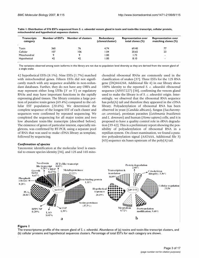
A large number of ESTs matched with snake toxins (360ESTs in 76 clusters; 69.4%). Others code for cellular (non-toxin) proteins (107 ESTs in 106 clusters; 20.65%), and
BMC Molecular Biology 2007, 8:115 http://www.biomedcentral.com/1471-2199/8/115
Page 3 of 17(page number not for citation purposes)
42 hypothetical ESTs (8.1%). Nine ESTs (1.7%) matchedwith mitochondrial genes. Fifteen ESTs did not signifi-cantly match with any sequence available in non-redun-dant databases. Further, they do not have any ORFs andmay represent either long UTRs (3' or 5') or regulatoryRNAs and may have important functions in the rapidlyexpressing gland tissues. The library contains a large por-tion of putative toxin genes (69.4%) compared to the cel-lular EST population (20.6%). We determined thecomplete sequence of the longest EST of each cluster andsequences were confirmed by repeated sequencing. Wecompleted the sequencing for all major toxins and twolow abundant toxin-like transcripts (described below).The existence of genes of particular interest, especially sin-gletons, was confirmed by RT-PCR, using a separate poolof RNA that was used to make cDNA library as template,followed by sequencing.
Confirmation of speciesTaxonomic identification at the molecular level is essen-tial to ensure species identity [36], and 12S and 16S mito-
chondrial ribosomal RNAs are commonly used in theclassification of snakes [37]. Three ESTs for the 12S RNAgene (DQ464268, Additional file 4) in our library show100% identity to the reported S. c. edwardsii ribosomalsequence (AF057227) [38], confirming the venom glandused to make the library is of S. c. edwardsii origin. Inter-estingly, we observed that the ribosomal RNA sequencehas poly(A) tail and therefore they appeared in the cDNAlibrary. Polyadenylation of ribosomal RNA has beenobserved in yeast (Candida albicans), fungus (Saccharomy-ces cerevisiae), protistan parasites (Leishmania braziliensisand L. donovani) and human (Homo sapiens) cells, and it isproposed to have a quality control role in rRNA degrada-tion [39-42]. This is a preliminary report showing the pos-sibility of polyadenylation of ribosomal RNA in areptilian system. On closer examination, we found a puta-tive polyadenylation signal (AATAAA, Additional file 3)[43] sequence six bases upstream of the poly(A) tail.
The transcriptome profile of the venom gland of S. c. edwardsiiFigure 1The transcriptome profile of the venom gland of S. c. edwardsii. Abundance of (a) toxins and toxin-like transcript clusters, and (b) cellular proteins and hypothetical sequences clusters. Percentage of total ESTs for each category are shown.
Table 1: Distribution of 576 ESTs sequenced from S. c. edwardsii venom gland in toxin and toxin-like transcript, cellular protein, mitochondrial and hypothetical sequence clusters.
Transcripts category
Number of ESTs Number of clusters Redundancy (clones/cluster)
Representation over total clones (%)
Representation over matching clones (%)
Toxin 360 76 4.74 69.40 77Cellular 107 106 1.04 20.65 23Mitochondrial 9 9 1.00 1.73 -Hypothetical 42 42 1.00 8.10 -
The variations observed among toxin isoforms in this library are not due to population level diversity as they are derived from the venom gland of a single snake

BMC Molecular Biology 2007, 8:115 http://www.biomedcentral.com/1471-2199/8/115
Page 4 of 17(page number not for citation purposes)
Identification of toxin familiesSerine proteinaseThe serine proteinases in the venom gland library of S. c.edwardsii are expressed with the highest transcript abun-dance (38% of 360 ESTs) (Figure 1a) and belong to 19clusters. Multiple clones appeared in 12 clusters, while 7were singletons (Additional file 2). One representativeEST from each cluster was completely sequenced(DQ464238–DQ464248, DQ439973). One of the clus-ters (DQ439973) contains only 3'UTR (2 ESTs). This clus-ter shows 90% similarity with the 3'UTR of a serineproteinase from Bothrops jararaca venom gland [44].
Most snake venom serine proteinases (SVSPs) to date aresingle polypeptide chains, except for two fibrinolyticenzymes from the venom of a Korean Viper, Agkistrodonblomhoffi brevicaudus (brevinase, AJ243757 and sal-monase, AF176679). In both cases, a single chain precur-sor is most likely cleaved by proteolysis [45]. In ourlibrary, one cluster (DQ464244) shows 90% and 83%sequence identity at the nucleotide and amino acid levelsrespectively to salmonase. It is not clear whether or not itis also processed to form a heterodimeric serine protein-ase in S. c. edwardsii venom.
The SVSPs are found in all families of snakes and in gen-eral, they perturb the hemostatic mechanisms of prey.They act on diverse protein substrates such as fibrinogen,kininogen and platelet receptors [46,47]. Some SVSPsexhibit more than one activity. For example, in additionto their thrombin-like activity, bothrombin, crotalase andLM-TL induce platelet aggregation, kinin release and gyra-tory activities, respectively [48-50]. We constructed aneighbor-joining (NJ) phylogenetic tree with 11 newlyidentified SVSP isoforms from S. c. edwardsii, to assignputative functions and to examine trends in the evolutionof new isoforms [47] (Figure 2). The phylogenetic treeshowed a scattered distribution of various isoforms withdifferent pharmacological activities from several speciesof pitvipers. This pattern indicates that SVSPs divergedafter snake lineages speciated. Many SVSPs are commonlyconsidered as thrombin-like enzymes (TLEs) because theymimic the fibrinogenolytic function of thrombin, pro-moting blood coagulation. Therefore, in most cases onlyfibrinogenolytic function of SVSPs is tested and the SVSPis categorized as a TLE. However, some thrombin-likeenzymes, in addition to releasing fibrinopeptide A and/orB from fibrinogen, also activate protein C [51], comple-ment C3 [52] and platelets [53]. Therefore, it would beinteresting to determine the specific pharmacologicalproperties of various SVSP isoforms within each groupand map these on their evolutionary relationships.
SVSP genes belong to a multigene family, and the protein-coding regions have been shown to be experiencing accel-
erated evolution within the venom glands of pitvipers[54]. Such accelerated evolution could lead to the changesin surface loops surrounding the substrate binding site,resulting in the variation of substrate recognition andhence, the function of the protein. The ratio between non-synonymous and synonymous substitution (dN/dS) of theprotein coding sequences of serine proteinase isoforms ofthis species was found to be 0.99, indicating a trendtoward accelerated evolution and therefore divergence inpharmacological function during envenomation.
Metalloproteinase and DisintegrinA total of 44 ESTs fall into 7 clusters and 7 singletons forthis family of proteins (12% transcript abundance) (Fig-ure 1a, Additional file 2). One representative EST fromeach cluster was sequenced (DQ464249–DQ464255).Snake venom metalloproteinase (SVMP) precursors areclassified into four groups according to size and domaincomposition: PI (metalloproteinase domain only); PII(metalloproteinase and disintegrin domains); PIII (metal-loproteinase, disintegrin and cysteine-rich domains); andPIV (PIII type domains linked to a lectin-like domain bydisulfide bonds) [55]. None of the clusters encode PI typeSVMPs.
The PII isoform from S. c. edwardsii (DQ464254) matches(83–85% identity at the protein level) with the precursorsof contortrostatin (Q9IAB0) and acostatin β chain(Q805F6) from A. contortrix contortrix venom. Contor-trostatin and acostatin are homodimeric and het-erodimeric disintegrins, respectively [56,57]. The α chainof acostatin is independently encoded and unlike otherdisintegrins, it is not derived by proteolytic processing[56]. However, we did not identify any ESTs matching theα chain of acostatin. Phylogenetic analysis shows thatDQ464254 is closer to dimeric than monomeric disin-tegrins (Figure 3a). In the dimeric disintegrins, C246 andC251 form disulfide bridges with the other subunit[58,59]. However, in the monomeric disintegrins, C233
and C235 form disulfide bridges with C246 and C251, respec-tively, making them unavailable for dimerization (Figure3b). Thus, characteristic Cys residues (C233 and C235) arepresent in all monomeric disintegrins but are absent fromdimeric disintegrins [60]. The PII SVMP of S. c. edwardsii(DQ464254) contains C233 and C235 in the disintegrindomain and is likely the precursor of a monomeric disin-tegrin. Other monomeric disintegrins, barbourin andtergeminin, were characterized previously from thevenom of S. miliarius barbouri and S. c. tergeminus, respec-tively [61].
The main integrin receptor binding motif of disintegrins,RGD, is found to be at the tip of a flexible hairpin loop.Variation of amino acid residues in this motif (R/K/M/W/VGD, MLD, MVD or K/RTS) on the flexible loop confers

BMC Molecular Biology 2007, 8:115 http://www.biomedcentral.com/1471-2199/8/115
Page 5 of 17(page number not for citation purposes)
Phylogenetic (NJ) tree of SVSPsFigure 2Phylogenetic (NJ) tree of SVSPs. Sequences (complete ORF) available from other pit vipers [47] and 11 isoforms (DQ464238–DQ464248) from this study (filled circle) were used. Tissue kallikrein-1 (P06870) is used as outgroup. The num-bers on the branches indicate the bootstrap support values for nodes, and the horizontal bar represents number of substitu-tions per site. TLE, thrombin-like enzymes; KN, kininogenase; PA, plasminogen activator; PAI, platelet aggregation inducer; BCD, blood clot dispersion; X, activity unknown. Experimentally verified activities are shaded.

BMC Molecular Biology 2007, 8:115 http://www.biomedcentral.com/1471-2199/8/115
Page 6 of 17(page number not for citation purposes)
specificity towards specific receptors, e.g., replacement ofR with a K in RGD motif of barbourin and ussuristatin 2significantly increases the selectivity for αIIbβ3 (fibrinogenreceptor) without affecting its binding to α5β1 (fibronec-tin receptor) or αvβ3 (vitronectin receptor) [62,63]. Addi-tionally, the residues immediately adjacent to the RGDloop also influence both selectivity and affinity forintegrin receptors [64,65]. For example, disintegrins withRGDW and RGDNP have selectively higher affinity forαIIbβ3 and αVβ3, respectively [63]. The RGDNP-containingdisintegrins are 10-fold more potent than RGDW-contain-
ing disintegrins in blocking the adhesion of cells medi-ated by α5β1. The putative disintegrin from S. c. edwardsiihas RGDNP, compared to RGDW and KGDW in tergemi-nin and barbourin, respectively. Therefore, further studiesof the physiological relevance of variation in receptorselectivity among disintegrins from the same genus will bevery informative.
The PIII class of SVMPs are functionally more diverse: theyexhibit hemorrhagic activity, inflammatory effects, inhibi-tion of platelet aggregation, apoptosis and prothrombin
(a) Phylogenetic (NJ) tree for class PII metalloproteinases of viperid venomsFigure 3(a) Phylogenetic (NJ) tree for class PII metalloproteinases of viperid venoms. Dataset (complete ORF) used from [60] in addi-tion to one isoform (DQ464254) obtained in this study (filled circle). ADAM8 from Danio rerio (Q6PFT3) and ADAM7 (from Mus musculus) were used as outgroup. (b) Alignment of the disintegrin domain of class PII SVMPs showing C233, C235, C246 and C251 (marked in grey) which are proposed to be involved in the formation of both monomeric [M] and dimeric [D] disintegrins in the venom. Only relevant portions of the sequences are shown.

BMC Molecular Biology 2007, 8:115 http://www.biomedcentral.com/1471-2199/8/115
Page 7 of 17(page number not for citation purposes)
activation [66-73]. All members of the PIII class of SVMPshave six conserved Cys residues at positions 126, 166,168, 173, 190 and 206 in their metalloproteinasedomain, and some isoforms have a seventh Cys residue atthree variable positions (195, 181 or 100) [60,74]. Thepresence of the seventh Cys residue at position 195 (sub-group PIIIa) results in proteolysis/autolysis, producing aproduct comprised of the disintegrin-like and cysteine-rich domains (DC domain), whereas when it is present atposition 181 (subgroup PIIIb), the formation of ahomodimeric structure results [60]). We have not foundany isoform having a Cys residue at position 100 (103 inour alignment, Additional file 5) in our library. Two iso-forms (DQ464249 and DQ464255) from S. c. edwardsiivenom possess a seventh Cys residue in positions 195 and181, and they can be grouped as PIIIa and PIIIb SVMPs,respectively (Additional file 5). Two other isoforms(DQ464250 and DQ464251) do not possess a seventhCys residue and hence cannot be grouped with any sub-groups. Some other isoforms, such as HR1a [75,76] andHF3 [68,77], also do not have the seventh Cys residue inthe metalloproteinase domain. We propose that thesemetalloproteinases be grouped under PIII0 (suffix '0' toindicate the absence of the seventh Cys residue) (Addi-tional data file 5). The isoform DQ464253 is a partial seg-ment and it cannot be assigned to any subgroup.However, it shows identity with the A chain of a het-erodimeric metalloproteinase identified in the venom ofVipera lebetina which induces apoptosis in endothelial celllines [78]. Overall, the venom of S. c. edwardsii appears tohave significant molecular variation among metallopro-teinases and their derived components.
Phospholipase A2Interestingly, in our cDNA library only one cluster of PLA2(DQ464264) was found, despite having the second high-est transcript abundance (28%) (Additional file 2, Figure1a). It matches with an acidic PLA2(AAS79430) of S. c.tergeminus, with only one amino acid residue (nucleotide)difference at position 80, P(CCG) → Q(CAG), in themature form. Thus there is no diversity of PLA2 in S. c.edwardsii venom, though snake venom PLA2 is one of themost rapidly evolving enzyme families. In most species,several isoforms of PLA2 are observed in cDNA librariesand venoms [79-81], and these have acquired diversephysiological functions [82-84]. This observation is alsosupported by proteomic analysis of S. c. edwardsii venom,while venoms from individuals of other species of Sistru-rus contain multiple PLA2 isoforms [85].
PhosphodiesteraseSequence of a partial singleton EST (transcript abundance0.28%; Additional file 2, Figure 1a) (DQ464266) shows60% identity to the C-terminal region of the phosphodi-esterase gene from chimpanzee (XP_001168685). This is
the first cDNA sequence for a phosphodiesterase fromsnake venom. Phosphodiesterase activity has beenobserved in venoms of Elapidae, Viperidae and Colubri-dae snakes [86-88]; however, the role of this enzyme inenvenomation is not yet clear. Venom phosphodieste-rases hydrolyze 5'-phosphodiester and pyrophosphatebonds in nucleotides and nucleic acids and release 5'-diphosphates, 5'-monophosphates and purines [89]. Freepurines are also present in snake venoms, and they maycontribute to envenomation sequelae [90].
L-amino acid oxidaseWe obtained one cluster having 13 ESTs (transcript abun-dance 3.5%) (Additional file 2, Figure 1a). The completesequence (DQ464267) shows high sequence identity(96%) with LAO of Crotalus adamanteus venom. LAOs arewidely found in snake venoms and in addition to catalyz-ing the oxidative deamination of amino acids, they affectplatelets, induce apoptosis and have hemorrhagic effects[91].
C-type lectinIn our library, CLP account for approximately 1.4% abun-dance and have one cluster (DQ464256) and two single-tons (DQ464257 and DQ464258) (Additional file 2,Figure 1a). On BLASTP search, they match with the β sub-unit of mamushigin (Q9YI92; 80% identity), CHH-B(P81509; 83% identity), and the A chain of Factor IX/Fac-tor X binding protein (IX/X-bp) (2124381A; 86% iden-tity) respectively. Mamushigin, CHH-B and IX/X-bp areheterodimeric; however, in our library, we did not findany match to ESTs encoding the corresponding comple-mentary subunits. Therefore, it may be interesting toexamine the CLP-related proteins in this venom anddetermine their biological properties.
Growth factorsWe obtained one cluster (transcript abundance 7%)encoding vascular endothelial growth factor (VEGF)(Additional file 2, Figure 1a). Sequencing of 8 clones fromthis cluster showed there are two isoforms (DQ464259and DQ464260) with only two amino acid residue(nucleotide) differences at positions 105, Q(CAG) →E(GAG), and 114, K(AAG) → E(GAG). We also sequenceda singleton (DQ464261) encoding nerve growth factor(NGF). Another singleton (DQ464277) matched with theC-terminus of connective tissue growth factor-related pro-tein (CTGF). This is the first report of CTGF-related pro-tein in a venom cDNA library. Its origin in the venomgland, instead of other surrounding tissues, needs to beverified.
Cysteine-rich secretory proteinWe obtained one cluster (transcript abundance 7%)(Additional file 2, Figure 1a) for a CRISP (DQ464263)

BMC Molecular Biology 2007, 8:115 http://www.biomedcentral.com/1471-2199/8/115
Page 8 of 17(page number not for citation purposes)
which matches with Catrin (AAO62995, 87% identity)from C. atrox venom. CRISPs are widely distributed inmammals, reptiles, amphibians, arthropods, nematodes,cone snails and plants, and they exhibit diverse biologicalfunctions [92]. They are single chain (MW of ~20–30kDa), highly conserved proteins organized in threedomains: a PR-1 (Pathogenesis Related proteins of group1) domain, a hinge domain and a cysteine-rich domain(CRD). They contain 16 Cys residues forming eight con-served disulfide bonds. A few snake venom CRISPs havebeen shown to act upon various ion channels through theCRD domain [93-96]. However, the function of themajority of CRISPs from snake venom is unknown [97].Therefore, it may be interesting to examine the biologicalproperties of the CRISP found in S. c. edwardsii venom.
Bradykinin-potentiating peptide and C-type natriuretic peptideWe found a singleton (transcript abundance 0.28%; Addi-tional file 2, Figure 1a) encoding a BPP-CNP (DQ464265)which showed 80% identity with a BPP-CNP precursorfrom Lachesis muta [98]. The BPP-CNP family of proteinslowers the blood pressure of prey during envenomation.Its low abundance in our library indicates that BPP-CNPmay not have a significant role in envenomation by Sistru-rus, unlike bites by other pitvipers (Bothrops and Lachesis)in Southern America [15,98].
Three-finger toxin like transcriptsWe obtained three individual singletons (Additional file2, Figure 1a) in the library (transcript abundance 0.83%)which belong to the 3FTx family of proteins. As 3FTxs arevery uncommon in viperid venoms, using targetedapproach we performed RT-PCR using a separate pool ofRNA as template and sequenced 96 clones. We found atotal of five isoforms of 3FTx-like trancripts (DQ464281,DQ464282, DQ464283, DQ464284 and DQ464285)(Figure 4). They have a signal peptide followed by amature protein consisting of 64–68 residues. They appearto belong to the non-conventional 3FTxs [99], with five
disulfide bridges, and the fifth disulfide bridge is in loop1 (Figure 4). All isoforms have the potential N-glycosyla-tion motif, N-X-T/S (Figure 4).
3FTxs were thought to be found only in elapid/hydrophiid venoms, though the origin of recruitment tothe elapid/hydrophid venom proteome is not clear [100].A polypeptide toxin (8 kDa) which crossreacts with α-bungarotoxin and binds with high affinity to nicotinicacetylcholine receptor (Kd of 7.3 × 10⁻¹⁰ M in competitionwith α-bungarotoxin) was isolated from the venom of A.halys (a pitviper) [101]. However, no sequence informa-tion of this protein is available. Recently, three clones(DY403363, DY403848 and DY403174) were obtainedfrom a cDNA library of L. muta venom gland which poten-tially encode polypeptides similar to 3FTx fold proteins[98]. However, only one clone (DY403363) has the startand stop codons (complete ORF); the other two do not.These sequences do not have any homology, at either thenucleotide or protein levels, to those obtained from S. c.edwardsii (this study).
Phylogenetic analysis of 3FTXs from three families ofsnakes (Elapidae, Colubridae and Viperidae) wasachieved using PAUP 4.10b [102]. Trees obtained usingNeighbor Joining (bootstrapping) or Parsimony Analysis(strict consensus, tree is not shown) were somewhat dif-ferent, but major topological features were retained (Fig-ure 5). One transcript from S. c. edwardsii (DQ464283)does not cluster with the other four but falls within a sep-arate clade containing Naja and Bungarus (both elapids)3FTxs. Four other transcripts of S. c. edwardsii form amonophyletic clade within an exclusively elapid clade.Interestingly, both methods place L. muta (a viperid)clones (DY403363 and DY403174) and Coelognathusradiatus (a colubrid) 3FTx as basal to all other 3FTxs, sug-gesting a common origin followed by diversification of3FTxs among all advanced snakes. Very similar trees, withthe same topology of family groups, were obtained using
Alignment of amino acid sequences of the putative precursors of 3FTxsFigure 4Alignment of amino acid sequences of the putative precursors of 3FTxs. Cys residues which are shaded in grey are commonly present in short chain 3FTx and form 4 disulfide bridges (solid black lines). Cys residues shaded in black possibly form the addi-tional disulfide bridge (dotted black line) present in the non-conventional 3FTx family. Potential N-glycosylation sites are underlined.

BMC Molecular Biology 2007, 8:115 http://www.biomedcentral.com/1471-2199/8/115
Page 9 of 17(page number not for citation purposes)
Bayesian analyses and hence support the above conclu-sions (Additional file 6).
This family of proteins was not observed in a detailed pro-teomic characterization of S. catenatus and S. miliarius bar-bouri venoms [14]. cDNA libraries of other viper venomglands, including B. jararacussu, B. insularis, A. acutus andDeinagkistrodon acutus, do not show their presence[15,16,22,26]. This could be due to either low abundancetranscripts and proteins or non-uniform recruitment of3FTx into the venom proteome within Viperidae. In S. c.edwardsii, the low transcript abundance (0.83%) suggeststhat 3FTx are minor components of the mature venom.
In snake venoms, 3FTXs exhibit diverse pharmacologicaleffects due to their ability to target various receptors andion channels [103]. It is important to note that the β-sheeted loops play crucial roles in binding to various tar-gets, and these regions are the most variable among S. c.edwardsii 3FTXs. Further, the dN/dS ratio of 0.98 (close to1) for their coding sequences indicates that a trendtowards accelerated evolution is present, as with the serineproteinases. If the variations in the β sheet loop regionsare the result of positive selection (accelerated evolution),they may exhibit distinct and novel biological activities.
Neighbor-joining cladogram of 3FTx sequencesFigure 5Neighbor-joining cladogram of 3FTx sequences. Numbers preceeding each species name refer to Genbank accession numbers, and numbers before most nodes indicate bootstrap values (1000 replicates).

BMC Molecular Biology 2007, 8:115 http://www.biomedcentral.com/1471-2199/8/115
Page 10 of 17(page number not for citation purposes)
Novel toxin-like transcriptIn our library, we obtained one singleton (Additional file2, Figure 1a) (DQ464286, transcript abundance 0.28%)with an ORF encoding a signal peptide (24 residues) anda mature protein (128 residues). The putative mature pro-tein is rich in Cys residues, similar to many other snakevenom toxins. Its N-terminal domain matches withKunitz/BPTI toxins (53–68% identity) and the middledomain matches with waprins (45–58% identity), andthe novel transcript has an extended C-terminus (Figure6). Both Kunitz/BPTI [104] and waprins [105,106] arefound separately as single domain proteins in snake ven-oms. Two of the Cys residues, which form one of the fourdisulfide bonds in waprins, are missing in the new tran-script (Figure 6). RT-PCR using a fresh RNA (other thanused to make cDNA library) as template and sequencingexperiments show the presence of this fused transcript inthe venom gland and hence it is not an artifact due to tem-plate switching by the Reverse Transcriptase used for mak-ing the cDNA library [107-109]. Although a number ofcDNA sequences of Kunitz/BPTI from snake venoms havebeen completed, none of them have the waprin domainand the C-terminal extension. Currently, cDNA sequencesof waprins are not known. However, this is the first exper-imental evidence for the presence of a waprin domain(though fused with another toxin) in viperid venom.
The longer ORF having Kunitz/BPTI and waprin domainstogether could be due to the fusion of two individualgenes encoding Kunitz/BPTI and waprin. Gene fusionmediated by exon shuffling (intron mediated recombina-tion or retrotransposition) has been established as anessential genetic mechanism for the origin of new genes ininvertebrates, vertebrates and plants [110,111]. Recently,a new genetic process, transcription-induced chimerism(TIC), in cases of tandemly located gene pairs has beenshown to be responsible for gene fusion in the humangenome, producing chimeric proteins [112,113]. It is not
clear at this stage how this novel fused gene has originatedin the snake venom gland. This fused transcript may codeeither for a precursor which is processed to form two indi-vidual classes of venom proteins (Kunitz/BPTI andwaprin) or a novel toxin with two distinct domains andhaving a new biological function. It has been observedthat new genes often give rise to new biological functionsdriven by adaptive Darwinian selection [114-116]. Themechanism of fusion of these apparently independentgenes, the evolutionary trajectory of this fused gene andthe potential new toxic function of the chimeric proteinare all areas for future investigation.
Iron-binding proteinFour ESTs (Additional file 2, Figure 1a) (dbEST:SCEHYPO1, transcript abundance 1.11%) showedhomology with an iron-binding protein with a potentialsignal peptide. Although most iron-binding proteins aregenerally categorized as storage protein, some of them,such as ovotransferrin and lactoferrin, have antimicrobialactivities [117-119]. It is not clear whether or not this pro-tein is found in the venom. However, omwaprin, a mem-ber of the waprin protein family, and the C-terminalregion of a myotoxic PLA2 were both shown to have anti-microbial activity [105,120].
Identification of cellular transcriptsWe obtained 106 clusters (transcript abundance 21%, 107sequences) which are involved in various cellular func-tions, including transcription and translation, secretion,post-translational modification, general metabolism andother functions (Additional file 3, Figure 1b). Similarhouse-keeping protein products have been observed inother snake venom glands [13,15,22]. One of the ESTs(SCE438) matches (74%) a calcium- and integrin-bindingprotein which assists platelet spreading [121]. Althoughmodulation of platelet and integrin functions is a keyactivity of several snake venom components, we do not
Alignment of the novel toxin-like transcript with snake venom Kunitz/BPTI proteins and waprinsFigure 6Alignment of the novel toxin-like transcript with snake venom Kunitz/BPTI proteins and waprins. ABD24043 (Daboia russellii russellii); Q90W98 (Textilinin, Pseudonaja textilis textilis); P81658 (Calcicludin, Dendroaspis angusticeps); P00981 (Dendrotoxin-K, Dendroaspis polylepis polylepis) and BPTI from Bovine. P83952 (Omwaprin, Oxyuranus microlepidotus); P60589 (Nawaprin, Naja nigricollis). The presence of conserved disulfide bonds are indicated by solid black lines. The disulfide bond which is missing in the novel toxin but conserved in waprins is indicated by dotted lines. The extended C-terminus of the novel toxin is under-lined.

BMC Molecular Biology 2007, 8:115 http://www.biomedcentral.com/1471-2199/8/115
Page 11 of 17(page number not for citation purposes)
believe that this protein is present in venom, as it lacks thesignal peptide.
Overall, results from our cDNA library demonstrate exten-sive molecular diversity in the venom composition of S. c.edwardsii. Serine proteinase and metalloproteinase iso-forms are the most abundant components and in thevenom, they exert diverse pharmacological activities, par-ticularly disrupting hemostasis. The numerous minorcomponents likely play an ancillary role in envenoma-tion. These diverse toxin isoforms, together with minorcomponents, may be characteristic of venoms from spe-cies utilizing different prey types, such as lizards (ecto-therms) and birds and mammals (endotherms) [122].
Venom composition and genetics of their originSnake venoms consist of a diverse range of pharmacolog-ically active protein and peptide toxins which are prima-rily used in prey capture and secondarily as defenseweapons. To date, the majority of the work on toxin iden-tification and characterization has been concentrated onsnakes of the families Elapidae and Viperidae becausethey are often abundant, produce high yields of venomand represent a significant risk to human health world-wide. Recent studies of venom transcriptomes and pro-teomes indicate that our knowledge of venomcomposition is partly limited by experimental detectabil-ity. For example, 3FTxs, which were thought to be foundexclusively in elapid venoms, were detected in viperidvenom gland transcriptomes only recently [[98] and inthis study]. Similarly, CLP, thought to be limited toviperid venoms, have been detected recently in the venomgland of Philodryas olfersii (Colubridae) and Bungarus spe-cies (Elapidae) [27,106,123,124]. Further, a new family oflow abundance toxin (vespryns) was identified in bothelapid and viperid venoms [98,123], Therefore, with theapplication of advanced techniques like EST sequencing,"compositional specificities" between families of venom-ous snakes may become less distinct (Additional file 1).Multiple recruitment events may lead to an increase in thespectrum of known and unknown toxin families, decreas-ing the compositional specificities among venomoussnakes. However, differential contribution of specific tox-ins to the overall expressed proteome of venomous snakesdoes lead to significant differences in venom compositionbetween species.
A central theme in the evolution of venom systems is com-plete duplication of toxin genes, followed by acceleratedevolution which favors nonsynonymous amino acid sub-stitution towards neofunctionalization. Modification ofselected surface areas of toxins [82] is responsible for pro-ducing the functional diversity in animal (invertebrates:snails and scorpions; vertebrates: snakes) toxin multigenefamilies [125]. However, one important observation in
the present report is the occurrence of a novel toxin-liketranscript generated by fusion of two individual toxingenes, Kunitz/BPTI and waprin, in a snake venom gland.Though the mechanism for creation of this fused geneneeds to be studied further, it clearly indicates that othergenetic processes (gene shuffling or TIC) are also operat-ing in the venom gland to create novel toxin genes. Genesoriginating by other genetic processes such as exon shuf-fling are recent [111], and therefore the addition of thisfused toxin-like transcript to the venom proteome is per-haps new. At this stage, it is tempting to speculate that theorigin of modular organization of different classes ofSVMPs, which appears to be the result of gene fusionevents, may be due to a genetic process other than geneduplication. SVMPs are very abundant toxins and carryout a principal role in envenomation by viperid snakes,and therefore studies of their genetic origin and organiza-tion will be of great interest. Circumstantial evidence oftrans-splicing for the generation of serine proteinase iso-forms in the venom gland of V. lebetina has been pre-sented [126]. Kopelman et al.[127] have shown thatalternative splicing and gene duplication are inversely cor-related evolutionary mechanisms. According to Parra et al.[113], only 4–5% of the tandem gene pairs in the humangenome can produce chimeric proteins. It is obvious thatthese alternative genetic processes responsible for expand-ing functional proteomes are uncommon among biologi-cal systems, and it is therefore not surprising in our case tohave just a singleton of the fused transcript out of 576ESTs (transcript abundance 0.28%). This also demon-strates that to detect rare genetic processes operating in thevenom gland, the library generated must be of high qual-ity and that subsequent analyses must be performed verycarefully. In turn, these analyses help elucidate in detailthe principles of evolution of snake venom transcriptomewhich have led to the evolutionary success of theadvanced snakes [128].
ConclusionThe composition of snake venoms has been shown to bedependent on numerous factors, including phylogeny,diet, age, geography and even sex [129-132]. In general,greater similarity of venoms will be observed along broadphylogenetic lines (e.g., within-family than between-fam-ily). However, as this study has demonstrated, some tox-ins classically considered to occur in only one family, suchas the 3FTxs, are actually broadly distributed among theadvanced snakes (Colubroidea). The present capacity todetect low abundance toxins indicates a greater composi-tional similarity of venoms among advanced snakes thanhas been previously recognized. Further, we have demon-strated that in addition to gene duplication, exon shuf-fling or transcriptional splicing may also contribute togenerating the diversity of toxins and toxin isoformsobserved among snake venoms. Overall, the elucidation

BMC Molecular Biology 2007, 8:115 http://www.biomedcentral.com/1471-2199/8/115
Page 12 of 17(page number not for citation purposes)
of the venom gland transcriptome of S. c. edwardsii con-tributes to a broader picture of toxin expression whichcomplements and extends proteomic analysis of thisvenom [85]. These approaches can lead to the identifica-tion of new toxins and provides mechanistic explanationsfor their evolution and diversification. An unresolvedquestion involves the relationship between the venomgland transcriptome and how this is ultimately translatedto the final proteome. This variable proteomic composi-tion in turn determines the complex and often difficult toresolve sequelae which frequently develop followingenvenomation by the different species of venomoussnakes.
MethodsVenom extraction and collection of venom glandsSpecimens of Sistrurus c. edwardsii (Desert Massasauga)were collected in Lincoln County, Colorado, USA underpermits granted by the Colorado Division of Wildlife toSPM (permits #0456, 06HP456). Venom was extractedfrom adult snakes using standard manual methods [133];venoms were then centrifuged to remove particulates, fro-zen and lyophilized. Prior to gland removal, snakes wereextracted of venom. Four days later, when mRNA levelsare presumed maximal [134], two snakes were anesthe-tized with isofluorane and then sacrificed by decapitation.Glands were then rapidly dissected from the snakes, cutinto small pieces and placed in approximately 0.5 mLRNAlater (Qiagen) and frozen at -80°C until used.
cDNA library construction and sequencingTotal RNA was extracted from a single venom gland usingthe RNeasy Mini Kit (Qiagen, Hilden, Germany). Theintegrity of total RNA was confirmed using agarose gelelectrophoresis. The mRNA was purified using an mRNAisolation kit (Roche Applied Science, Mannheim, Ger-many). The purified total mRNA was used to make thecDNA library following the instructions of the SMARTcDNA library construction kit (Vector used: λ TriplEx2)(Clontech, Mountain view, California, USA). Small sizeand incomplete cDNAs were removed by passing thelibrary through CHROMA SPIN-400column. The librarywas packaged using Gigapack gold packaging extract(Stratagene, Cedar Creek, Texas, USA). Individual cloneswere rescued from randomly selected white plaques andgrown in Luria broth + ampicillin medium. Plasmids werepurified using the QIAprep spin miniprep kit (Qiagen,Hilden, Germany). Purified plasmids were sequenced bycycle sequencing reactions using the BigDye Terminatorv3.1 kit (Applied Biosystem, Foster City, California, USA)and an automated DNA sequencer (Model 3100A,Applied Biosystem, Foster City, California, USA).
RT-PCRRT-PCR was performed in order to search for isoforms of3FTx sequences in the venom gland. In brief, total RNAwas isolated from venom glands as above and was used astemplate. The following primers were used for amplifica-tion: forward primer, 5' ATGAAAACTCTGCTGNTGATC-CTGGNG 3' (N = A/C/G/T); reverse primer, 5'GGTTTATGGACCATCCTGTGGTAAAGGC 3'. Reversetranscription and subsequent amplification reactionswere done using the one step RT-PCR protocol of Qiagen(Hilden, Germany). The amplified product was clonedinto pDrive vector (Qiagen, Hilden, Germany) and 96random clones were sequenced. RT-PCR was also per-formed to confirm the presence of fused toxin transcriptin the venom using same procedure with the followingprimers: forward primer, 5' ATGTCTTCTGGAGGTCTTCT-GCTG 3'; reverse primer, 5' TCCAG GACAGAAGAAG-GCTCTGAT 3'.
Bioinformatic analysisClustering of the ESTs was performed using the CAP3 pro-gram [135] after removing poor quality sequences andvector sequences using VecScreen from NCBI. We lookedfor Sfi I (A & B) recognition sequences in the ESTs andmanually removed upstream and downstream sequencesof these sites as well as poly(A) tails (at least 10 A's in arow) from the 3' ends. A minimum overlap of 50 bp and100% identity in the overlap region were selected as crite-ria for the clustering. All clusters and singletons were sub-jected to BLAST searches (BLASTN and BLASTX asrequired) against the non-redundant database of NCBI (e-values cutoff < 10-5 and having a good coverage of mini-mum 100 base pairs and >98% identity) for the putativeidentification of the genes [136]. Presence of signal pep-tides was predicted individually by submission of thesequences to the SignalP server as available in the Expasywebsite. Gene and protein alignments were done usingthe programs ClustalW and DNAMAN (Lynnon Corpora-tion, Vaudreuil-Dorion, Quebec, Canada). The ratiobetween nonsynonymous (dN) and synonymous substitu-tion (dS) were calculated using the SNAP program [137].The program SNAP has been developed based on themethod of [138] with incorporation of statistical analysisdeveloped by [139].
Phylogenetic analysisPhylogenetic analysis was carried out using the programMEGA version 3.1[140], using Poisson-corrected dis-tances, and trees were constructed applying bootstraps of1000 replicates. PAUP 4.0b10 [102] was also used forBootstrap, Neighbor Joining and Parsimony analyses. Forthe Bayesian inferences of phylogeny (based upon theposterior probability distribution of the trees: Markovchain Monte Carlo methods), MrBayes v3.1.2 [141] wasused. The analysis was run for 5 × 10⁶ generations in four
BMC Molecular Biology 2007, 8:115 http://www.biomedcentral.com/1471-2199/8/115
Page 13 of 17(page number not for citation purposes)
chains and sampled every 100 generations, resulting in50,000 sample trees. The log-likelihood score of eachsaved tree was plotted against the number of generationsto determine the point at which the log-likelihood scoresof the analysis reached the asymptote. The posterior prob-abilities for the clades were established by constructing aconsensus tree of all trees generated after the completionof the burn-in phase.
We included the following sequences from three familiesof snakes in our analyses: the newly identified 3FTxsequences out of this study from S. c. edwardsii (Viperidae)[GenBank: DQ464281, DQ464282, DQ464283,DQ464284 and DQ464285]; L. muta (Viperidae) [Gen-Bank: DY403363, DY403174], α-colubritoxin [Swiss-Prot: P83490] from Coelognathus radiatus (Colubridae),and non-conventional 3FTx sequences [Swiss-Prot:P81783, O42255 and P82935], 3FTx-like sequences[Swiss-Prot: Q02454, P62375, P24778, P24777, P24776,P01471, P62390, P01473, 229475, P01448, P01474,Q8UUK0 and Z54231] [142] and 3TFx sequences [Swiss-Prot: P10808, P01433, P01427, Q9YGC7, P10456,Q9YHV0, P80958, P60772, P34076, P01419 andP60237] from Elapidae. All these sequences belong to theshort chain 3FTx family. A BLASTP search using [Gen-Bank: DY403174] from L. muta venom found that a ratpeptide sequence [GenBank: AAH63176] showed thehighest homology to Ly6 antigen, which has been pro-posed to be a potential ancestor of snake venom 3FTxs[100,143], and this peptide was used as outgroup in ouranalysis.
Accession numbersNucleotide sequence data reported here have been depos-ited in GenBank under accession numbers [GenBank:DQ464238–DQ464286]. ESTs are deposited in dbESTwith accession numbers [dbEST: DY587747–DY588245and DY625701–DY625710].
List of abbreviationsEST, expressed sequence tag; ORF, open reading frame;PLA2, phospholipase A2; 3FTx, three-finger toxin; CRISP,cysteine-rich secretory protein; CLP, C-type lectins; BPP,bradykinin-potentiating peptides; CNP, C-type natriureticpeptides; LAO, L-amino acid oxidase; BPTI, bovine pan-creatic trypsin inhibitor; VEGF, vascular endothelialgrowth factors; NGF, nerve growth factor; SVSP, snakevenom serine proteinases; TLE, thrombin-like enzymes;CRD, cysteine-rich domain; NJ, neighbor-joining; TIC,transcription-induced chimerism.
Authors' contributionsSP has designed and carried out experiments, analyseddata, developed the concept and wrote the manuscript.SPM has collected the venom gland sample. RMK and
SPM have edited the manuscript to improve its quality.SPM has helped in the phylogenetic analysis of 3FTxssequences. All the authors have approved the final form ofthe manuscript.
Additional material
Additional file 1It is a table showing the distribution of snake venom toxin families among Superfamily Colubroidae.Click here for file[http://www.biomedcentral.com/content/supplementary/1471-2199-8-115-S1.pdf]
Additional file 2It is a table showing the clusters of ESTs, number of clones in each cluster and their putative identity.Click here for file[http://www.biomedcentral.com/content/supplementary/1471-2199-8-115-S2.pdf]
Additional file 3It is a table showing the clusters of ESTs encoding cellular proteins.Click here for file[http://www.biomedcentral.com/content/supplementary/1471-2199-8-115-S3.pdf]
Additional file 4ClustalW alignment between 12S ribosomal RNA sequence DQ464268 (from this study) and AF057227 (used for taxonomic identification of S. c. edwardsii). Polyadenylation signal sequence is underlined.Click here for file[http://www.biomedcentral.com/content/supplementary/1471-2199-8-115-S4.pdf]
Additional file 5ClustalW alignment of PIII metalloproteinases (only proteinase domain is shown). Cysteine residues which are conserved are marked in grey and variable in black. Accession numbers of the used sequences are as follows: VAP1 [GenBank: BAB18307], HV1 [GenBank: BAB60682], Halysase [GenBank: 27465044], VLAIP-A [GenBank: 61104775], VLAIP-B [GenBank: 61104777], Kaouthiagin [Swiss-Prot: P82942], Berythracti-vase [Swiss-Prot: Q8UVG0], Ecarin [Swiss-Prot: Q90495], Jararhagin [Swiss-Prot: P30431], Bothropasin [Swiss-Prot: O93523], Acurhagin [Swiss-Prot: Q6Q274], Catrocollastatin [Swiss-Prot: Q90282], Atro-lysin [Swiss-Prot: Q92043], Stejnihagin-A [Swiss-Prot: Q3HTN1], Ste-jnihagin-B [Swiss-Prot: Q3HTN2], HR1A [Swiss-Prot: Q8JIR2], HR1B [Swiss-Prot: P20164], HF3 [GenBank: 31742525]. P, signal peptide domain; PRO, pro-domain; S, spacer; DISIN, disintegrin domain; CRD, cysteine-rich domainClick here for file[http://www.biomedcentral.com/content/supplementary/1471-2199-8-115-S5.pdf]
BMC Molecular Biology 2007, 8:115 http://www.biomedcentral.com/1471-2199/8/115
Page 14 of 17(page number not for citation purposes)
AcknowledgementsThis work was supported from the grants from Biomedical Research Coun-cil, Agency for Science and Technology Research, Singapore (RMK). Per-mits and support for collection of snakes was provided by the Colorado Division of Wildlife (06HP456, SPM). The assistance of W.H. Heyborne and Susan J Moore with some aspects of phylogenetic analyses was greatly appreciated.
References1. Vidal N: Colubroid systematics: Evidence for an early appear-
ance of the venom apparatus followed by extensive evolu-tionary tinkering. J Toxicol Toxin Rev 2002, 21:21-41.
2. Mackessy SP: Biochemistry and pharmacology of colubridsnake venoms. J Toxicol Toxin Rev 2002, 21:43-83.
3. Vidal N, Hedges SB: Higher-level relationships of snakesinferred from four nuclear and mitochondrial genes. C R Biol2002, 325:977-985.
4. Fry BG, Wuster W: Assembling an arsenal: origin and evolu-tion of the snake venom proteome inferred from phyloge-netic analysis of toxin sequences. Mol Biol Evol 2004, 21:870-883.
5. Hodgson WC, Wickramaratna JC: In vitro neuromuscular activ-ity of snake venoms. Clin Exp Pharmacol Physiol 2002, 29:807-814.
6. Meier J, Stocker K: Effects of snake venoms on hemostasis. CritRev Toxicol 1991, 21:171-182.
7. Braud S, Bon C, Wisner A: Snake venom proteins acting onhemostasis. Biochimie 2000, 82:851-859.
8. Matsui T, Fujimura Y, Titani K: Snake venom proteases affectinghemostasis and thrombosis. Biochim Biophys Acta 2000,1477:146-156.
9. Morita T: C-type lectin-related proteins from snake venoms.Curr Drug Targets Cardiovasc Haematol Disord 2004, 4:357-373.
10. Kloog Y, Ambar I, Sokolovsky M, Kochva E, Wollberg Z, Bdolah A:Sarafotoxin, a novel vasoconstrictor peptide: phosphoi-nositide hydrolysis in rat heart and brain. Science 1988,242:268-270.
11. Bazaa A, Marrakchi N, El Ayeb M, Sanz L, Calvete JJ: Snake venom-ics: comparative analysis of the venom proteomes of theTunisian snakes Cerastes cerastes, Cerastes vipera and Mac-rovipera lebetina. Proteomics 2005, 5:4223-4235.
12. Birrell GW, Earl S, Masci PP, de Jersey J, Wallis TP, Gorman JJ, LavinMF: Molecular diversity in venom from the Australian BrownSnake, Pseudonaja textilis. Mol Cell Proteomics 2006, 5:379-389.
13. Francischetti IM, My-Pham V, Harrison J, Garfield MK, Ribeiro JM:Bitis gabonica (Gaboon viper) snake venom gland: toward acatalog for the full-length transcripts (cDNA) and proteins.Gene 2004, 337:55-69.
14. Juarez P, Sanz L, Calvete JJ: Snake venomics: characterization ofprotein families in Sistrurus barbouri venom by cysteinemapping, N-terminal sequencing, and tandem mass spec-trometry analysis. Proteomics 2004, 4:327-338.
15. Junqueira-de-Azevedo IL, Ho PL: A survey of gene expressionand diversity in the venom glands of the pitviper snake Both-rops insularis through the generation of expressed sequencetags (ESTs). Gene 2002, 299:279-291.
16. Kashima S, Roberto PG, Soares AM, Astolfi-Filho S, Pereira JO, Giuli-ati S, Faria M Jr., Xavier MA, Fontes MR, Giglio JR, Franca SC: Anal-ysis of Bothrops jararacussu venomous gland transcriptomefocusing on structural and functional aspects: I--gene expres-
sion profile of highly expressed phospholipases A2. Biochimie2004, 86:211-219.
17. Li M, Fry BG, Kini RM: Eggs-only diet: its implications for thetoxin profile changes and ecology of the marbled sea snake(Aipysurus eydouxii). J Mol Evol 2005, 60:81-89.
18. Li M, Fry BG, Kini RM: Putting the brakes on snake venom evo-lution: the unique molecular evolutionary patterns ofAipysurus eydouxii (Marbled sea snake) phospholipase A2toxins. Mol Biol Evol 2005, 22:934-941.
19. Li S, Wang J, Zhang X, Ren Y, Wang N, Zhao K, Chen X, Zhao C, LiX, Shao J, Yin J, West MB, Xu N, Liu S: Proteomic characteriza-tion of two snake venoms: Naja naja atra and Agkistrodonhalys. Biochem J 2004, 384:119-127.
20. Nawarak J, Sinchaikul S, Wu CY, Liau MY, Phutrakul S, Chen ST: Pro-teomics of snake venoms from Elapidae and Viperidae fami-lies by multidimensional chromatographic methods.Electrophoresis 2003, 24:2838-2854.
21. Pierre LS, Woods R, Earl S, Masci PP, Lavin MF: Identification andanalysis of venom gland-specific genes from the coastaltaipan (Oxyuranus scutellatus) and related species. Cell MolLife Sci 2005, 62:2679-2693.
22. Qinghua L, Xiaowei Z, Wei Y, Chenji L, Yijun H, Pengxin Q, XingwenS, Songnian H, Guangmei Y: A catalog for transcripts in thevenom gland of the Agkistrodon acutus: identification of thetoxins potentially involved in coagulopathy. Biochem BiophysRes Commun 2006, 341:522-531.
23. Serrano SM, Shannon JD, Wang D, Camargo AC, Fox JW: A multi-faceted analysis of viperid snake venoms by two-dimensionalgel electrophoresis: an approach to understanding venomproteomics. Proteomics 2005, 5:501-510.
24. Tsai IH, Chen YH, Wang YM: Comparative proteomics and sub-typing of venom phospholipases A2 and disintegrins of Pro-tobothrops pit vipers. Biochim Biophys Acta 2004, 1702:111-119.
25. Wagstaff SC, Harrison RA: Venom gland EST analysis of thesaw-scaled viper, Echis ocellatus, reveals novel alpha9beta1integrin-binding motifs in venom metalloproteinases and anew group of putative toxins, renin-like aspartic proteases.Gene 2006, 377:21-32.
26. Zhang B, Liu Q, Yin W, Zhang X, Huang Y, Luo Y, Qiu P, Su X, Yu J,Hu S, Yan G: Transcriptome analysis of Deinagkistrodon acu-tus venomous gland focusing on cellular structure and func-tional aspects using expressed sequence tags. BMC Genomics2006, 7:152.
27. Ching AT, Rocha MM, Paes Leme AF, Pimenta DC, de Fatima DF, Ser-rano SM, Ho PL, Junqueira-de-Azevedo IL: Some aspects of thevenom proteome of the Colubridae snake Philodryas olfersiirevealed from a Duvernoy's (venom) gland transcriptome.FEBS Lett 2006, 580:4417-4422.
28. Cidade DA, Simao TA, Davila AM, Wagner G, de LMJ, Lee HP, BonC, Zingali RB, Albano RM: Bothrops jararaca venom gland tran-scriptome: Analysis of the gene expression pattern. Toxicon2006, 48:437-461.
29. Mackessy SP: Desert Massasauga Rattlesnake (Sistrurus cate-natus edwardsii): a technical conservation assessment.2005:57 [http://www.fs.fed.us/r2/projects/scp/assessments/massasauga.pdf].
30. Stebbins RC: A field guide to western reptiles and amphibians 2nd edition.Edited by: Mifflin H. New York; 1985.
31. Degenhardt WG, Painter CW, Price AH: The Amphibians and Reptilesof New Mexico University of New Mexico Press, Albuquerque, NM;1996.
32. Hobert JP, Montgomery CE, Mackessy SP: Natural History of theMassasauga Sistrurus catenatus edwardsii, in SoutheasternColorado. The Southwestern Naturalist 2004, 49:321-326.
33. Holycross AT, Mackessy SP: Variation in the diet of Sistruruscatenatus (Massasauga), with emphasis on Sistrurus catena-tus edwardsii (Desert Massasauga). Journal of Herpetology 2002,36:454-464.
34. Allen WB JR.: State lists of endangered and threatened species of reptilesand amphibians and laws and regulations covering collecting of reptiles andamphibians in each state Chicago, Illinois, Chicago Herpetological Soci-ety; 1988.
35. Seigel AR: Ecology and conservation of an endangered rattle-snake, Sistrurus catenatus, in Missouri, USA. Biological Conser-vation 1986, 35:333-346.
Additional file 6Bayesian tree generated from 39 aligned 3FTx sequences as described in Materials and Methods. Numbers on branches indicate percentage of pos-terior clade probability. 3FTx sequences from S. c. edwardsii and L. muta libraries are marked with a filled circle and triangle respectively.Click here for file[http://www.biomedcentral.com/content/supplementary/1471-2199-8-115-S6.pdf]
http://www.ncbi.nlm.nih.gov/entrez/query.fcgi?cmd=Retrieve&db=PubMed&dopt=Abstract&list_uids=2039593
http://www.ncbi.nlm.nih.gov/entrez/query.fcgi?cmd=Retrieve&db=PubMed&dopt=Abstract&list_uids=2845579
http://www.ncbi.nlm.nih.gov/entrez/query.fcgi?cmd=Retrieve&db=PubMed&dopt=Abstract&list_uids=2845579
BMC Molecular Biology 2007, 8:115 http://www.biomedcentral.com/1471-2199/8/115
Page 15 of 17(page number not for citation purposes)
36. Wuster W, Golay P, Warrell DA: Synopsis of recent develop-ments in venomous snake systematics, No. 3. Toxicon 1999,37:1123-1129.
37. Pook CE, McEwing R: Mitochondrial DNA sequences fromdried snake venom: a DNA barcoding approach to the iden-tification of venom samples. Toxicon 2005, 46:711-715.
38. Parkinson CL: Molecular systematics and biogeographical his-tory of pitvipers as determined by mitochondrial ribosomalDNA sequences. Copeia 1999, 3:576-586.
39. Decuypere S, Vandesompele J, Yardley V, De Donckeri S, Laurent T,Rijal S, Llanos-Cuentas A, Chappuis F, Arevalo J, Dujardin JC: Differ-ential polyadenylation of ribosomal RNA during post-tran-scriptional processing in Leishmania. Parasitology 2005,131:321-329.
40. Fleischmann J, Liu H, Wu CP: Polyadenylation of ribosomal RNAby Candida albicans also involves the small subunit. BMC MolBiol 2004, 5:17.
41. Kuai L, Fang F, Butler JS, Sherman F: Polyadenylation of rRNA inSaccharomyces cerevisiae. Proc Natl Acad Sci U S A 2004,101:8581-8586.
42. Slomovic S, Laufer D, Geiger D, Schuster G: Polyadenylation anddegradation of human mitochondrial RNA: the prokaryoticpast leaves its mark. Mol Cell Biol 2005, 25:6427-6435.
43. Wahle E, Kuhn U: The mechanism of 3' cleavage and polyade-nylation of eukaryotic pre-mRNA. Prog Nucleic Acid Res Mol Biol1997, 57:41-71.
44. Saguchi K, Hagiwara-Saguchi Y, Murayama N, Ohi H, Fujita Y,Camargo AC, Serrano SM, Higuchi S: Molecular cloning of serineproteinases from Bothrops jararaca venom gland. Toxicon2005, 46:72-83.
45. Lee JW, Seu JH, Rhee IK, Jin I, Kawamura Y, Park W: Purificationand characterization of brevinase, a heterogeneous two-chain fibrinolytic enzyme from the venom of Korean snake,Agkistrodon blomhoffii brevicaudus. Biochem Biophys Res Com-mun 1999, 260:665-670.
46. Serrano SM, Maroun RC: Snake venom serine proteinases:sequence homology vs. substrate specificity, a paradox to besolved. Toxicon 2005, 45:1115-1132.
47. Wang YM, Wang SR, Tsai IH: Serine protease isoforms of Dein-agkistrodon acutus venom: cloning, sequencing and phyloge-netic analysis. Biochem J 2001, 354:161-168.
48. Markland FS, Kettner C, Schiffman S, Shaw E, Bajwa SS, Reddy KN,Kirakossian H, Patkos GB, Theodor I, Pirkle H: Kallikrein-likeactivity of crotalase, a snake venom enzyme that clots fibrin-ogen. Proc Natl Acad Sci U S A 1982, 79:1688-1692.
49. Nishida S, Fujimura Y, Miura S, Ozaki Y, Usami Y, Suzuki M, Titani K,Yoshida E, Sugimoto M, Yoshioka A, .: Purification and character-ization of bothrombin, a fibrinogen-clotting serine proteasefrom the venom of Bothrops jararaca. Biochemistry 1994,33:1843-1849.
50. Silveira AM, Magalhaes A, Diniz CR, de Oliveira EB: Purification andproperties of the thrombin-like enzyme from the venom ofLachesis muta muta. Int J Biochem 1989, 21:863-871.
51. Kisiel W, Kondo S, Smith KJ, McMullen BA, Smith LF: Characteriza-tion of a protein C activator from Agkistrodon contortrixcontortrix venom. J Biol Chem 1987, 262:12607-12613.
52. Yamamoto C, Tsuru D, Oda-Ueda N, Ohno M, Hattori S, Kim ST:Flavoxobin, a serine protease from Trimeresurus flavoviridis(habu snake) venom, independently cleaves Arg726-Ser727of human C3 and acts as a novel, heterologous C3 conver-tase. Immunology 2002, 107:111-117.
53. Niewiarowski S, Kirby EP, Brudzynski TM, Stocker K: Thrombocy-tin, a serine protease from Bothrops atrox venom. 2. Inter-action with platelets and plasma-clotting factors. Biochemistry1979, 18:3570-3577.
54. Deshimaru M, Ogawa T, Nakashima K, Nobuhisa I, Chijiwa T, Shimo-higashi Y, Fukumaki Y, Niwa M, Yamashina I, Hattori S, Ohno M:Accelerated evolution of crotalinae snake venom gland ser-ine proteases. FEBS Lett 1996, 397:83-88.
55. Hite LA, Jia LG, Bjarnason JB, Fox JW: cDNA sequences for foursnake venom metalloproteinases: structure, classification,and their relationship to mammalian reproductive proteins.Arch Biochem Biophys 1994, 308:182-191.
56. Okuda D, Koike H, Morita T: A new gene structure of the disin-tegrin family: a subunit of dimeric disintegrin has a shortcoding region. Biochemistry 2002, 41:14248-14254.
57. Zhou Q, Nakada MT, Brooks PC, Swenson SD, Ritter MR, ArgounovaS, Arnold C, Markland FS: Contortrostatin, a homodimeric dis-integrin, binds to integrin alphavbeta5. Biochem Biophys ResCommun 2000, 267:350-355.
58. Calvete JJ, Jurgens M, Marcinkiewicz C, Romero A, Schrader M,Niewiarowski S: Disulphide-bond pattern and molecular mod-elling of the dimeric disintegrin EMF-10, a potent and selec-tive integrin alpha5beta1 antagonist from Eristocophismacmahoni venom. Biochem J 2000, 345 Pt 3:573-581.
59. Calvete JJ, Moreno-Murciano MP, Theakston RD, Kisiel DG, Marcink-iewicz C: Snake venom disintegrins: novel dimeric disin-tegrins and structural diversification by disulphide bondengineering. Biochem J 2003, 372:725-734.
60. Fox JW, Serrano SM: Structural considerations of the snakevenom metalloproteinases, key members of the M12 repro-lysin family of metalloproteinases. Toxicon 2005, 45:969-985.
61. Scarborough RM, Rose JW, Hsu MA, Phillips DR, Fried VA, CampbellAM, Nannizzi L, Charo IF: Barbourin. A GPIIb-IIIa-specificintegrin antagonist from the venom of Sistrurus m. barbouri.J Biol Chem 1991, 266:9359-9362.
62. Oshikawa K, Terada S: Ussuristatin 2, a novel KGD-bearing dis-integrin from Agkistrodon ussuriensis venom. J Biochem 1999,125(1):31-35.
63. Scarborough RM, Rose JW, Naughton MA, Phillips DR, Nannizzi L,Arfsten A, Campbell AM, Charo IF: Characterization of theintegrin specificities of disintegrins isolated from Americanpit viper venoms. J Biol Chem 1993, 268:1058-1065.
64. Aota S, Nagai T, Yamada KM: Characterization of regions offibronectin besides the arginine-glycine-aspartic acidsequence required for adhesive function of the cell-bindingdomain using site-directed mutagenesis. J Biol Chem 1991,266:15938-15943.
65. Ruoslahti E, Pierschbacher MD: New perspectives in cell adhe-sion: RGD and integrins. Science 1987, 238:491-497.
66. Gao R, Kini RM, Gopalakrishnakone P: A novel prothrombin acti-vator from the venom of Micropechis ikaheka: isolation andcharacterization. Arch Biochem Biophys 2002, 408:87-92.
67. Bjarnason JB, Fox JW: Hemorrhagic metalloproteinases fromsnake venoms. Pharmacol Ther 1994, 62:325-372.
68. Costa EP, Clissa PB, Teixeira CF, Moura-da-Silva AM: Importanceof metalloproteinases and macrophages in viper snakeenvenomation-induced local inflammation. Inflammation 2002,26:13-17.
69. Laing GD, Moura-da-Silva AM: Jararhagin and its multiple effectson hemostasis. Toxicon 2005, 45:987-996.
70. Wang WJ, Shih CH, Huang TF: Primary structure and antiplate-let mechanism of a snake venom metalloproteinase, acur-hagin, from Agkistrodon acutus venom. Biochimie 2005,87:1065-1077.
71. Masuda S, Ohta T, Kaji K, Fox JW, Hayashi H, Araki S: cDNA clon-ing and characterization of vascular apoptosis-inducing pro-tein 1. Biochem Biophys Res Commun 2000, 278:197-204.
72. You WK, Seo HJ, Chung KH, Kim DS: A novel metalloproteasefrom Gloydius halys venom induces endothelial cell apopto-sis through its protease and disintegrin-like domains. J Bio-chem 2003, 134(5):739-749.
73. Loria GD, Rucavado A, Kamiguti AS, Theakston RD, Fox JW, AlapeA, Gutierrez JM: Characterization of 'basparin A,' a pro-thrombin-activating metalloproteinase, from the venom ofthe snake Bothrops asper that inhibits platelet aggregationand induces defibrination and thrombosis. Arch Biochem Biophys2003, 418:13-24.
74. Wan SG, Jin Y, Lee WH, Zhang Y: Cloning of two novel P-III classmetalloproteinases from Trimeresurus stejnegeri venomgland. Toxicon 2006, 47:465-472.
75. Omori-Satoh T, Ohsaka A: Purification and some properties ofhemorrhagic principle I in the venom of Trimeresurus flavo-viridis. Biochim Biophys Acta 1970, 207:432-444.
76. Omori-Satoh T, Sadahiro S: Resolution of the major hemor-rhagic component of Trimeresurus flavoviridis venom intotwo parts. Biochim Biophys Acta 1979, 580:392-404.
77. Silva CA, Zuliani JP, Assakura MT, Mentele R, Camargo AC, TeixeiraCF, Serrano SM: Activation of alpha(M)beta(2)-mediatedphagocytosis by HF3, a P-III class metalloproteinase isolatedfrom the venom of Bothrops jararaca. Biochem Biophys Res Com-mun 2004, 322:950-956.
http://www.ncbi.nlm.nih.gov/entrez/query.fcgi?cmd=Retrieve&db=PubMed&dopt=Abstract&list_uids=9175430
http://www.ncbi.nlm.nih.gov/entrez/query.fcgi?cmd=Retrieve&db=PubMed&dopt=Abstract&list_uids=9175430
http://www.ncbi.nlm.nih.gov/entrez/query.fcgi?cmd=Retrieve&db=PubMed&dopt=Abstract&list_uids=7043462
http://www.ncbi.nlm.nih.gov/entrez/query.fcgi?cmd=Retrieve&db=PubMed&dopt=Abstract&list_uids=7043462
http://www.ncbi.nlm.nih.gov/entrez/query.fcgi?cmd=Retrieve&db=PubMed&dopt=Abstract&list_uids=7043462
http://www.ncbi.nlm.nih.gov/entrez/query.fcgi?cmd=Retrieve&db=PubMed&dopt=Abstract&list_uids=8110787
http://www.ncbi.nlm.nih.gov/entrez/query.fcgi?cmd=Retrieve&db=PubMed&dopt=Abstract&list_uids=8110787
http://www.ncbi.nlm.nih.gov/entrez/query.fcgi?cmd=Retrieve&db=PubMed&dopt=Abstract&list_uids=8110787
http://www.ncbi.nlm.nih.gov/entrez/query.fcgi?cmd=Retrieve&db=PubMed&dopt=Abstract&list_uids=2583354
http://www.ncbi.nlm.nih.gov/entrez/query.fcgi?cmd=Retrieve&db=PubMed&dopt=Abstract&list_uids=2583354
http://www.ncbi.nlm.nih.gov/entrez/query.fcgi?cmd=Retrieve&db=PubMed&dopt=Abstract&list_uids=2583354
http://www.ncbi.nlm.nih.gov/entrez/query.fcgi?cmd=Retrieve&db=PubMed&dopt=Abstract&list_uids=3624272
http://www.ncbi.nlm.nih.gov/entrez/query.fcgi?cmd=Retrieve&db=PubMed&dopt=Abstract&list_uids=3624272
http://www.ncbi.nlm.nih.gov/entrez/query.fcgi?cmd=Retrieve&db=PubMed&dopt=Abstract&list_uids=3624272
http://www.ncbi.nlm.nih.gov/entrez/query.fcgi?cmd=Retrieve&db=PubMed&dopt=Abstract&list_uids=8941719
http://www.ncbi.nlm.nih.gov/entrez/query.fcgi?cmd=Retrieve&db=PubMed&dopt=Abstract&list_uids=8941719
http://www.ncbi.nlm.nih.gov/entrez/query.fcgi?cmd=Retrieve&db=PubMed&dopt=Abstract&list_uids=8941719
http://www.ncbi.nlm.nih.gov/entrez/query.fcgi?cmd=Retrieve&db=PubMed&dopt=Abstract&list_uids=8311451
http://www.ncbi.nlm.nih.gov/entrez/query.fcgi?cmd=Retrieve&db=PubMed&dopt=Abstract&list_uids=8311451
http://www.ncbi.nlm.nih.gov/entrez/query.fcgi?cmd=Retrieve&db=PubMed&dopt=Abstract&list_uids=2033037
http://www.ncbi.nlm.nih.gov/entrez/query.fcgi?cmd=Retrieve&db=PubMed&dopt=Abstract&list_uids=2033037
http://www.ncbi.nlm.nih.gov/entrez/query.fcgi?cmd=Retrieve&db=PubMed&dopt=Abstract&list_uids=9880793
http://www.ncbi.nlm.nih.gov/entrez/query.fcgi?cmd=Retrieve&db=PubMed&dopt=Abstract&list_uids=9880793
http://www.ncbi.nlm.nih.gov/entrez/query.fcgi?cmd=Retrieve&db=PubMed&dopt=Abstract&list_uids=8419314
http://www.ncbi.nlm.nih.gov/entrez/query.fcgi?cmd=Retrieve&db=PubMed&dopt=Abstract&list_uids=8419314
http://www.ncbi.nlm.nih.gov/entrez/query.fcgi?cmd=Retrieve&db=PubMed&dopt=Abstract&list_uids=8419314
http://www.ncbi.nlm.nih.gov/entrez/query.fcgi?cmd=Retrieve&db=PubMed&dopt=Abstract&list_uids=1874740
http://www.ncbi.nlm.nih.gov/entrez/query.fcgi?cmd=Retrieve&db=PubMed&dopt=Abstract&list_uids=1874740
http://www.ncbi.nlm.nih.gov/entrez/query.fcgi?cmd=Retrieve&db=PubMed&dopt=Abstract&list_uids=1874740
http://www.ncbi.nlm.nih.gov/entrez/query.fcgi?cmd=Retrieve&db=PubMed&dopt=Abstract&list_uids=2821619
http://www.ncbi.nlm.nih.gov/entrez/query.fcgi?cmd=Retrieve&db=PubMed&dopt=Abstract&list_uids=2821619
http://www.ncbi.nlm.nih.gov/entrez/query.fcgi?cmd=Retrieve&db=PubMed&dopt=Abstract&list_uids=7972338
http://www.ncbi.nlm.nih.gov/entrez/query.fcgi?cmd=Retrieve&db=PubMed&dopt=Abstract&list_uids=7972338
http://www.ncbi.nlm.nih.gov/entrez/query.fcgi?cmd=Retrieve&db=PubMed&dopt=Abstract&list_uids=4989125
http://www.ncbi.nlm.nih.gov/entrez/query.fcgi?cmd=Retrieve&db=PubMed&dopt=Abstract&list_uids=4989125
BMC Molecular Biology 2007, 8:115 http://www.biomedcentral.com/1471-2199/8/115
Page 16 of 17(page number not for citation purposes)
78. Trummal K, Tonismagi K, Siigur E, Aaspollu A, Lopp A, Sillat T, SaatR, Kasak L, Tammiste I, Kogerman P, Kalkkinen N, Siigur J: A novelmetalloprotease from Vipera lebetina venom induceshuman endothelial cell apoptosis. Toxicon 2005, 46:46-61.
79. Singh SB, Armugam A, Kini RM, Jeyaseelan K: Phospholipase A(2)with platelet aggregation inhibitor activity from Austrelapssuperbus venom: protein purification and cDNA cloning.Arch Biochem Biophys 2000, 375:289-303.
80. Tsai IH, Wang YM, Chen YH, Tsai TS, Tu MC: Venom phospholi-pases A2 of bamboo viper (Trimeresurus stejnegeri): molec-ular characterization, geographic variations and evidence ofmultiple ancestries. Biochem J 2004, 377:215-223.
81. Vishwanath BS, Kini RM, Gowda TV: Characterization of threeedema-inducing phospholipase A2 enzymes from habu(Trimeresurus flavoviridis) venom and their interaction withthe alkaloid aristolochic acid. Toxicon 1987, 25:501-515.
82. Kini RM, Chan YM: Accelerated evolution and molecular sur-face of venom phospholipase A2 enzymes. J Mol Evol 1999,48:125-132.
83. Lynch VJ: Inventing an arsenal: adaptive evolution and neo-functionalization of snake venom phospholipase A2 genes.BMC Evol Biol 2007, 7:2.
84. Ogawa T, Nakashima K, Nobuhisa I, Deshimaru M, Shimohigashi Y,Fukumaki Y, Sakaki Y, Hattori S, Ohno M: Accelerated evolutionof snake venom phospholipase A2 isozymes for acquisition ofdiverse physiological functions. Toxicon 1996, 34:1229-1236.
85. Sanz L, Gibbs HL, Mackessy SP, Calvete JJ: Venom proteomes ofclosely related Sistrurus rattlesnakes with divergent diets. JProteome Res 2006, 5:2098-2112.
86. Boman HG, Kaletta U: Identity of the phosphodiesterase anddeoxyribonuclease in rattle-snake venom. Nature 1956,178:1394-1395.
87. Mackessy SP: Phosphodiesterases, DNases and RNases. Bailey,G.S. (Ed.) edition. Ft. Collins, CO, USA., Alaken Press, Inc.,;1998:361-404.
88. Russell FE: Phosphodiesterase of some snake and arthropodvenoms. Toxicon 1966, 4:153-154.
89. Furstenau CR, Trentin DS, Barreto-Chaves ML, Sarkis JJ: Ecto-nucleotide pyrophosphatase/phosphodiesterase as part of amultiple system for nucleotide hydrolysis by platelets fromrats: kinetic characterization and biochemical properties.Platelets 2006, 17:84-91.
90. Aird SD: Ophidian envenomation strategies and the role ofpurines. Toxicon 2002, 40:335-393.
91. Tan NH: L-amino acid oxidases and lactate dehydrogenases.In Enzymes from Snake Venoms Volume 12. In: Bailey, G.S. (Ed.), edition.Ft. Collins, CO, USA,, Alaken Inc.,; 1998:579-598.
92. Yamazaki Y, Morita T: Structure and function of snake venomcysteine-rich secretory proteins. Toxicon 2004, 44:227-231.
93. Guo M, Teng M, Niu L, Liu Q, Huang Q, Hao Q: Crystal structureof the cysteine-rich secretory protein stecrisp reveals thatthe cysteine-rich domain has a K+ channel inhibitor-like fold.J Biol Chem 2005, 280:12405-12412.
94. Wang J, Shen B, Guo M, Lou X, Duan Y, Cheng XP, Teng M, Niu L,Liu Q, Huang Q, Hao Q: Blocking effect and crystal structure ofnatrin toxin, a cysteine-rich secretory protein from Najaatra venom that targets the BKCa channel. Biochemistry 2005,44:10145-10152.
95. Yamazaki Y, Koike H, Sugiyama Y, Motoyoshi K, Wada T, HishinumaS, Mita M, Morita T: Cloning and characterization of novelsnake venom proteins that block smooth muscle contrac-tion. Eur J Biochem 2002, 269:2708-2715.
96. Yamazaki Y, Brown RL, Morita T: Purification and cloning of tox-ins from elapid venoms that target cyclic nucleotide-gatedion channels. Biochemistry 2002, 41:11331-11337.
97. Osipov AV, Levashov MY, Tsetlin VI, Utkin YN: Cobra venom con-tains a pool of cysteine-rich secretory proteins. Biochem Bio-phys Res Commun 2005, 328:177-182.
98. Junqueira-de-Azevedo IL, Ching AT, Carvalho E, Faria F, NishiyamaMY Jr., Ho PL, Diniz MR: Lachesis muta (Viperidae) cDNAsReveal Diverging Pit Viper Molecules and Scaffolds Typicalof Cobra (Elapidae) Venoms: Implications for Snake ToxinRepertoire Evolution. Genetics 2006, 173:877-889.
99. Nirthanan S, Gopalakrishnakone P, Gwee MC, Khoo HE, Kini RM:Non-conventional toxins from Elapid venoms. Toxicon 2003,41:397-407.
100. Fry BG: From genome to "venome": molecular origin andevolution of the snake venom proteome inferred from phyl-ogenetic analysis of toxin sequences and related body pro-teins. Genome Res 2005, 15:403-420.
101. Jiang M, Haggblad J, Heilbronn E: Isolation and pharmacologicalcharacterization of a new alpha-neurotoxin (alpha-AgTx)from venom of the viper Agkistrodon halys (Pallas). Toxicon1987, 25:1019-1022.
102. Swofford DL: PAUP*. Phylogenetic Analysis Using Parsimony(*and Other Methods). Sinauer Associates; 2000.
103. Kini RM: Molecular moulds with multiple missions: functionalsites in three-finger toxins. Clin Exp Pharmacol Physiol 2002,29:815-822.
104. Strydom DJ, Joubert FJ: The amino acid sequence of a weaktrypsin inhibitor B from Dendroaspis Polylepis polylepis(black mamba) venom. Hoppe Seylers Z Physiol Chem 1981,362:1377-1384.
105. Nair DG, Fry BG, Alewood P, Kumar PP, Kini RM: Antimicrobialactivity of omwaprin, a new member of the waprin family ofsnake venom proteins. Biochem J 2007, 402:93-104.
106. Torres AM, Wong HY, Desai M, Moochhala S, Kuchel PW, Kini RM:Identification of a novel family of proteins in snake venoms.Purification and structural characterization of nawaprinfrom Naja nigricollis snake venom. J Biol Chem 2003,278:40097-40104.
107. Ouhammouch M, Brody EN: Temperature-dependent templateswitching during in vitro cDNA synthesis by the AMV-reverse transcriptase. Nucleic Acids Res 1992, 20:5443-5450.
108. Zeng XC, Wang SX: Evidence that BmTXK beta-BmKCTcDNA from Chinese scorpion Buthus martensii Karsch is anartifact generated in the reverse transcription process. FEBSLett 2002, 520:183-184.
109. Zaphiropoulos PG: Template switching generated duringreverse transcription? FEBS Lett 2002, 527:326.
110. Long M: A new function evolved from gene fusion. Genome Res2000, 10:1655-1657.
111. Long M, Betran E, Thornton K, Wang W: The origin of new genes:glimpses from the young and old. Nat Rev Genet 2003,4:865-875.
112. Akiva P, Toporik A, Edelheit S, Peretz Y, Diber A, Shemesh R, NovikA, Sorek R: Transcription-mediated gene fusion in the humangenome. Genome Res 2006, 16:30-36.
113. Parra G, Reymond A, Dabbouseh N, Dermitzakis ET, Castelo R,Thomson TM, Antonarakis SE, Guigo R: Tandem chimerism as ameans to increase protein complexity in the humangenome. Genome Res 2006, 16:37-44.
114. Nixon AE, Warren MS, Benkovic SJ: Assembly of an activeenzyme by the linkage of two protein modules. Proc Natl AcadSci U S A 1997, 94:1069-1073.
115. Patthy L: Modular assembly of genes and the evolution of newfunctions. Genetica 2003, 118:217-231.
116. Zhang J, Dean AM, Brunet F, Long M: Evolving protein functionaldiversity in new genes of Drosophila. Proc Natl Acad Sci U S A2004, 101:16246-16250.
117. Orsi N: The antimicrobial activity of lactoferrin: current sta-tus and perspectives. Biometals 2004, 17:189-196.
118. Giansanti F, Massucci MT, Giardi MF, Nozza F, Pulsinelli E, Nicolini C,Botti D, Antonini G: Antiviral activity of ovotransferrin derivedpeptides. Biochem Biophys Res Commun 2005, 331:69-73.
119. Ibrahim HR, Iwamori E, Sugimoto Y, Aoki T: Identification of a dis-tinct antibacterial domain within the N-lobe of ovotransfer-rin. Biochim Biophys Acta 1998, 1401:289-303.
120. Santamaria C, Larios S, Quiros S, Pizarro-Cerda J, Gorvel JP, LomonteB, Moreno E: Bactericidal and antiendotoxic properties ofshort cationic peptides derived from a snake venom Lys49phospholipase A2. Antimicrob Agents Chemother 2005,49:1340-1345.
121. Naik UP, Naik MU: Association of CIB with GPIIb/IIIa duringoutside-in signaling is required for platelet spreading onfibrinogen. Blood 2003, 102:1355-1362.
122. Mackessy SP, Sixberry NM, Heyborne WH, Fritts T: Venom of theBrown Treesnake, Boiga irregularis: ontogenetic shifts andtaxa-specific toxicity. Toxicon 2006, 47:537-548.
123. Pung YF, Wong PT, Kumar PP, Hodgson WC, Kini RM: Ohanin, anovel protein from king cobra venom, induces hypolocomo-
http://www.ncbi.nlm.nih.gov/entrez/query.fcgi?cmd=Retrieve&db=PubMed&dopt=Abstract&list_uids=3617087
http://www.ncbi.nlm.nih.gov/entrez/query.fcgi?cmd=Retrieve&db=PubMed&dopt=Abstract&list_uids=3617087
http://www.ncbi.nlm.nih.gov/entrez/query.fcgi?cmd=Retrieve&db=PubMed&dopt=Abstract&list_uids=3617087
http://www.ncbi.nlm.nih.gov/entrez/query.fcgi?cmd=Retrieve&db=PubMed&dopt=Abstract&list_uids=9929380
http://www.ncbi.nlm.nih.gov/entrez/query.fcgi?cmd=Retrieve&db=PubMed&dopt=Abstract&list_uids=9929380
http://www.ncbi.nlm.nih.gov/entrez/query.fcgi?cmd=Retrieve&db=PubMed&dopt=Abstract&list_uids=9027978
http://www.ncbi.nlm.nih.gov/entrez/query.fcgi?cmd=Retrieve&db=PubMed&dopt=Abstract&list_uids=9027978
http://www.ncbi.nlm.nih.gov/entrez/query.fcgi?cmd=Retrieve&db=PubMed&dopt=Abstract&list_uids=9027978
http://www.ncbi.nlm.nih.gov/entrez/query.fcgi?cmd=Retrieve&db=PubMed&dopt=Abstract&list_uids=4309653
http://www.ncbi.nlm.nih.gov/entrez/query.fcgi?cmd=Retrieve&db=PubMed&dopt=Abstract&list_uids=4309653
http://www.ncbi.nlm.nih.gov/entrez/query.fcgi?cmd=Retrieve&db=PubMed&dopt=Abstract&list_uids=3433300
http://www.ncbi.nlm.nih.gov/entrez/query.fcgi?cmd=Retrieve&db=PubMed&dopt=Abstract&list_uids=3433300
http://www.ncbi.nlm.nih.gov/entrez/query.fcgi?cmd=Retrieve&db=PubMed&dopt=Abstract&list_uids=3433300
http://www.ncbi.nlm.nih.gov/entrez/query.fcgi?cmd=Retrieve&db=PubMed&dopt=Abstract&list_uids=7309000
http://www.ncbi.nlm.nih.gov/entrez/query.fcgi?cmd=Retrieve&db=PubMed&dopt=Abstract&list_uids=7309000
http://www.ncbi.nlm.nih.gov/entrez/query.fcgi?cmd=Retrieve&db=PubMed&dopt=Abstract&list_uids=7309000
http://www.ncbi.nlm.nih.gov/entrez/query.fcgi?cmd=Retrieve&db=PubMed&dopt=Abstract&list_uids=1279521
http://www.ncbi.nlm.nih.gov/entrez/query.fcgi?cmd=Retrieve&db=PubMed&dopt=Abstract&list_uids=1279521
http://www.ncbi.nlm.nih.gov/entrez/query.fcgi?cmd=Retrieve&db=PubMed&dopt=Abstract&list_uids=1279521
http://www.ncbi.nlm.nih.gov/entrez/query.fcgi?cmd=Retrieve&db=PubMed&dopt=Abstract&list_uids=9037007
http://www.ncbi.nlm.nih.gov/entrez/query.fcgi?cmd=Retrieve&db=PubMed&dopt=Abstract&list_uids=9037007
http://www.ncbi.nlm.nih.gov/entrez/query.fcgi?cmd=Retrieve&db=PubMed&dopt=Abstract&list_uids=9540819
http://www.ncbi.nlm.nih.gov/entrez/query.fcgi?cmd=Retrieve&db=PubMed&dopt=Abstract&list_uids=9540819

Publish with BioMed Central and every scientist can read your work free of charge
"BioMed Central will be the most significant development for disseminating the results of biomedical research in our lifetime."
Sir Paul Nurse, Cancer Research UK
Your research papers will be:
available free of charge to the entire biomedical community
peer reviewed and published immediately upon acceptance
cited in PubMed and archived on PubMed Central
yours — you keep the copyright
Submit your manuscript here:http://www.biomedcentral.com/info/publishing_adv.asp
BioMedcentral
BMC Molecular Biology 2007, 8:115 http://www.biomedcentral.com/1471-2199/8/115
Page 17 of 17(page number not for citation purposes)
tion and hyperalgesia in mice. J Biol Chem 2005,280:13137-13147.
124. Zha HG, Lee WH, Zhang Y: Cloning of cDNAs encoding C-typelectins from Elapidae snakes Bungarus fasciatus and Bunga-rus multicinctus. Toxicon 2001, 39:1887-1892.
125. Kordis D, Gubensek F: Adaptive evolution of animal toxin mul-tigene families. Gene 2000, 261:43-52.
126. Siigur E, Aaspollu A, Siigur J: Sequence diversity of Vipera lebet-ina snake venom gland serine proteinase homologs--result ofalternative-splicing or genome alteration. Gene 2001,263:199-203.
127. Kopelman NM, Lancet D, Yanai I: Alternative splicing and geneduplication are inversely correlated evolutionary mecha-nisms. Nat Genet 2005, 37:588-589.
128. Mackessy SP, Baxter LM: Bioweapons synthesis and storage: thevenom gland of front-fanged snakes. Zoologischer Anzeiger 2006,245:147-159.
129. Chippaux JP, Boche J, Courtois B: Electrophoretic patterns of thevenoms from a litter of Bitis gabonica snakes. Toxicon 1982,20:521-523.
130. Daltry JC, Wuster W, Thorpe RS: Diet and snake venom evolu-tion. Nature 1996, 379:537-540.
131. Jayanthi GP, Gowda TV: Geographical variation in India in thecomposition and lethal potency of Russell's viper (Viperarusselli) venom. Toxicon 1988, 26:257-264.
132. Sasa M: Diet and snake venom evolution: can local selectionalone explain intraspecific venom variation? Toxicon 1999,37:249-252.
133. Mackessy SP: Venom ontogeny in the Pacific rattlesnakes Cro-talus viridis helleri and Crotalus viridis oreganus. Copeia1988:92-101.
134. Rotenberg D, Bamberger ES, Kochva E: Studies on ribonucleicacid synthesis in the venom glands of Vipera palaestinae(Ophidia, Reptilia). Biochem J 1971, 121:609-612.
135. Huang X, Madan A: CAP3: A DNA sequence assembly pro-gram. Genome Res 1999, 9:868-877.
136. Altschul SF, Madden TL, Schaffer AA, Zhang J, Zhang Z, Miller W, Lip-man DJ: Gapped BLAST and PSI-BLAST: a new generation ofprotein database search programs. Nucleic Acids Res 1997,25:3389-3402.
137. Korber B: HIV signature and sequence variation analysis. InComputational and evolutionary analysis of HIV molecular sequences Rod-rigo GA Learn HG edition. Dordrecht, The Netherlands, Kluwer Aca-demic Publishers; 2000:55-72.
138. Nei M, Gojobori T: Simple methods for estimating the num-bers of synonymous and nonsynonymous nucleotide substi-tutions. Mol Biol Evol 1986, 3:418-426.
139. Ota T, Nei M: Variance and covariances of the numbers of syn-onymous and nonsynonymous substitutions per site. Mol BiolEvol 1994, 11:613-619.
140. Kumar S, Tamura K, Nei M: MEGA3: Integrated software forMolecular Evolutionary Genetics Analysis and sequencealignment. Brief Bioinform 2004, 5:150-163.
141. Ronquist F, Huelsenbeck JP: MrBayes 3: Bayesian phylogeneticinference under mixed models. Bioinformatics 2003,19:1572-1574.
142. Chang LS, Lin SK, Chung C: Molecular cloning and evolution ofthe genes encoding the precursors of taiwan cobra cardio-toxin and cardiotoxin-like basic protein. Biochem Genet 2004,42:429-440.
143. Miwa JM, Ibanez-Tallon I, Crabtree GW, Sanchez R, Sali A, Role LW,Heintz N: lynx1, an endogenous toxin-like modulator of nico-tinic acetylcholine receptors in the mammalian CNS. Neuron1999, 23:105-114.
http://www.ncbi.nlm.nih.gov/entrez/query.fcgi?cmd=Retrieve&db=PubMed&dopt=Abstract&list_uids=7080059
http://www.ncbi.nlm.nih.gov/entrez/query.fcgi?cmd=Retrieve&db=PubMed&dopt=Abstract&list_uids=7080059
http://www.ncbi.nlm.nih.gov/entrez/query.fcgi?cmd=Retrieve&db=PubMed&dopt=Abstract&list_uids=8596631
http://www.ncbi.nlm.nih.gov/entrez/query.fcgi?cmd=Retrieve&db=PubMed&dopt=Abstract&list_uids=8596631
http://www.ncbi.nlm.nih.gov/entrez/query.fcgi?cmd=Retrieve&db=PubMed&dopt=Abstract&list_uids=3293265
http://www.ncbi.nlm.nih.gov/entrez/query.fcgi?cmd=Retrieve&db=PubMed&dopt=Abstract&list_uids=3293265
http://www.ncbi.nlm.nih.gov/entrez/query.fcgi?cmd=Retrieve&db=PubMed&dopt=Abstract&list_uids=3293265
http://www.ncbi.nlm.nih.gov/entrez/query.fcgi?cmd=Retrieve&db=PubMed&dopt=Abstract&list_uids=4940047
http://www.ncbi.nlm.nih.gov/entrez/query.fcgi?cmd=Retrieve&db=PubMed&dopt=Abstract&list_uids=4940047
http://www.ncbi.nlm.nih.gov/entrez/query.fcgi?cmd=Retrieve&db=PubMed&dopt=Abstract&list_uids=4940047
http://www.ncbi.nlm.nih.gov/entrez/query.fcgi?cmd=Retrieve&db=PubMed&dopt=Abstract&list_uids=9254694
http://www.ncbi.nlm.nih.gov/entrez/query.fcgi?cmd=Retrieve&db=PubMed&dopt=Abstract&list_uids=9254694
http://www.ncbi.nlm.nih.gov/entrez/query.fcgi?cmd=Retrieve&db=PubMed&dopt=Abstract&list_uids=3444411
http://www.ncbi.nlm.nih.gov/entrez/query.fcgi?cmd=Retrieve&db=PubMed&dopt=Abstract&list_uids=3444411
http://www.ncbi.nlm.nih.gov/entrez/query.fcgi?cmd=Retrieve&db=PubMed&dopt=Abstract&list_uids=3444411
http://www.ncbi.nlm.nih.gov/entrez/query.fcgi?cmd=Retrieve&db=PubMed&dopt=Abstract&list_uids=8078400