Fetal programming of adrenal PNMT and hypertension ... - PLOS
16
RESEARCH ARTICLE Fetal programming of adrenal PNMT and hypertension by glucocorticoids in WKY rats is dose and sex-dependent Sandhya Khurana 1☯ , Julie Grandbois 2☯ , Sujeenthar Tharmalingam 1 , Alyssa Murray 3 , Kelly Graff 1 , Phong Nguyen 2 , T. C. Tai ID 1,2,3,4 * 1 Medical Sciences Division, Northern Ontario School of Medicine, Sudbury, Ontario, Canada, 2 Department of Biology, Laurentian University, Sudbury, Ontario, Canada, 3 Department of Chemistry and Biochemistry, Laurentian University, Sudbury, Ontario, Canada, 4 Biomolecular Sciences Program, Laurentian University, Sudbury, Ontario, Canada ☯ These authors contributed equally to this work. * [email protected] Abstract Biochemical changes in utero may alter normal fetal development, resulting in disease later in life, a phenomenon known as fetal programming. Recent epidemiological studies link fetal programming to negative health outcomes, such as low birth weight and hypertension in adulthood. Here, we used a WKY rat model and studied the molecular changes triggered by prenatal glucocorticoid (GC) exposure on the development of hypertension, and on the reg- ulation of phenylethanolamine N-methyl transferase (PNMT), the enzyme responsible for biosynthesis of epinephrine, and a candidate gene linked to hypertension. Clinically, high doses of the synthetic GC dexamethasone (DEX) are used to treat infant respiratory dis- tress syndrome. Elevated maternal GCs have been correlated with fetal programming of hypertension. The aim of this study was to determine if lower doses of DEX would not lead to detrimental fetal programming effects such as hypertension. Our data suggests that pre- natal stress programs for increased expression of PNMT and altered regulation of PNMT in males and females. Importantly, we identified that DEX mediated programming was more apparent in the male rats, and the lower dose 10μg/kg/day of DEX did not lead to changes in blood pressure (BP) in female rats suggesting that this dose is below the threshold for pro- gramming of hypertension. Furthermore, sex-specific differences were observed in regards to programming mechanisms that may account for hypertension in males. Introduction Emerging evidence suggests that in-utero insults experienced during critical stages of fetal development lead to an unfavourable biochemical environment causing compensatory fetal adaptations. These adaptive mechanisms occur via alterations in structure and/or function of organs that may be permanent, leading to an increased risk of developing disease later in life, a phenomenon known as fetal programming of adult disease [1]. Maternal undernutrition, PLOS ONE | https://doi.org/10.1371/journal.pone.0221719 September 4, 2019 1 / 16 a1111111111 a1111111111 a1111111111 a1111111111 a1111111111 OPEN ACCESS Citation: Khurana S, Grandbois J, Tharmalingam S, Murray A, Graff K, Nguyen P, et al. (2019) Fetal programming of adrenal PNMT and hypertension by glucocorticoids in WKY rats is dose and sex- dependent. PLoS ONE 14(9): e0221719. https:// doi.org/10.1371/journal.pone.0221719 Editor: Michael Bader, Max Delbruck Centrum fur Molekulare Medizin Berlin Buch, GERMANY Received: May 7, 2019 Accepted: August 13, 2019 Published: September 4, 2019 Copyright: © 2019 Khurana et al. This is an open access article distributed under the terms of the Creative Commons Attribution License, which permits unrestricted use, distribution, and reproduction in any medium, provided the original author and source are credited. Data Availability Statement: All relevant data are within the manuscript files. Funding: This research was funded by the Canadian Institutes for Health Research (Grant IHD98766 to TCT) and the NOSMFA Research Development Fund (2016-RDF to TCT). The funders played no role in the study design, data collection and analysis, decision to publish, nor the preparation of the manuscript. Competing interests: The authors have declared that no competing interests exist.
-
Upload
khangminh22 -
Category
Documents
-
view
0 -
download
0
Transcript of Fetal programming of adrenal PNMT and hypertension ... - PLOS

RESEARCH ARTICLE
Fetal programming of adrenal PNMT and
hypertension by glucocorticoids in WKY rats is
dose and sex-dependent
Sandhya Khurana1☯, Julie Grandbois2☯, Sujeenthar Tharmalingam1, Alyssa Murray3,
Kelly Graff1, Phong Nguyen2, T. C. TaiID1,2,3,4*
1 Medical Sciences Division, Northern Ontario School of Medicine, Sudbury, Ontario, Canada, 2 Department
of Biology, Laurentian University, Sudbury, Ontario, Canada, 3 Department of Chemistry and Biochemistry,
Laurentian University, Sudbury, Ontario, Canada, 4 Biomolecular Sciences Program, Laurentian University,
Sudbury, Ontario, Canada
☯ These authors contributed equally to this work.
Abstract
Biochemical changes in utero may alter normal fetal development, resulting in disease later
in life, a phenomenon known as fetal programming. Recent epidemiological studies link fetal
programming to negative health outcomes, such as low birth weight and hypertension in
adulthood. Here, we used a WKY rat model and studied the molecular changes triggered by
prenatal glucocorticoid (GC) exposure on the development of hypertension, and on the reg-
ulation of phenylethanolamine N-methyl transferase (PNMT), the enzyme responsible for
biosynthesis of epinephrine, and a candidate gene linked to hypertension. Clinically, high
doses of the synthetic GC dexamethasone (DEX) are used to treat infant respiratory dis-
tress syndrome. Elevated maternal GCs have been correlated with fetal programming of
hypertension. The aim of this study was to determine if lower doses of DEX would not lead
to detrimental fetal programming effects such as hypertension. Our data suggests that pre-
natal stress programs for increased expression of PNMT and altered regulation of PNMT in
males and females. Importantly, we identified that DEX mediated programming was more
apparent in the male rats, and the lower dose 10μg/kg/day of DEX did not lead to changes in
blood pressure (BP) in female rats suggesting that this dose is below the threshold for pro-
gramming of hypertension. Furthermore, sex-specific differences were observed in regards
to programming mechanisms that may account for hypertension in males.
Introduction
Emerging evidence suggests that in-utero insults experienced during critical stages of fetal
development lead to an unfavourable biochemical environment causing compensatory fetal
adaptations. These adaptive mechanisms occur via alterations in structure and/or function of
organs that may be permanent, leading to an increased risk of developing disease later in life, a
phenomenon known as fetal programming of adult disease [1]. Maternal undernutrition,
PLOS ONE | https://doi.org/10.1371/journal.pone.0221719 September 4, 2019 1 / 16
a1111111111
a1111111111
a1111111111
a1111111111
a1111111111
OPEN ACCESS
Citation: Khurana S, Grandbois J, Tharmalingam S,
Murray A, Graff K, Nguyen P, et al. (2019) Fetal
programming of adrenal PNMT and hypertension
by glucocorticoids in WKY rats is dose and sex-
dependent. PLoS ONE 14(9): e0221719. https://
doi.org/10.1371/journal.pone.0221719
Editor: Michael Bader, Max Delbruck Centrum fur
Molekulare Medizin Berlin Buch, GERMANY
Received: May 7, 2019
Accepted: August 13, 2019
Published: September 4, 2019
Copyright: © 2019 Khurana et al. This is an open
access article distributed under the terms of the
Creative Commons Attribution License, which
permits unrestricted use, distribution, and
reproduction in any medium, provided the original
author and source are credited.
Data Availability Statement: All relevant data are
within the manuscript files.
Funding: This research was funded by the
Canadian Institutes for Health Research (Grant
IHD98766 to TCT) and the NOSMFA Research
Development Fund (2016-RDF to TCT). The
funders played no role in the study design, data
collection and analysis, decision to publish, nor the
preparation of the manuscript.
Competing interests: The authors have declared
that no competing interests exist.

hypoxia, exposure to alcohol, and high levels of glucocorticoids are examples of stressors that
can impact fetal development, resulting in fetal programming. Exposure to prenatal stress has
been strongly associated with the development of the “thrifty phenotype”, leading to increased
risk of cardiovascular disease, kidney disease, metabolic syndrome and hypertension amongst
others, exemplifying the crucial role of early development in-utero in determining the onset of
disease [2,3].
Numerous researchers have identified that stress hormones such as glucocorticoids (GCs)
contribute to fetal programming of hypertension; however, the molecular mechanisms are not
fully understood [4,5]. GCs are hormones that are lipophilic molecules that easily cross the pla-
centa, and can induce a stress-like state upon the developing fetus [2]. Placental GC concentra-
tions are substantially low in comparison to maternal levels due to 11β-dehydroxysteroid
dehydrogenase 2 (11β-HSD2), which converts bioactive GCs into their inactive form [2]. How-
ever, synthetic GCs such as dexamethasone (DEX) are weak substrates for 11β-HSD2, and are
able to cross the placenta and exert their effects onto the fetus [6].
Exposure of the fetus to GCs may occur endogenously (maternal stress) or exogenously (to
treat neonatal respiratory distress syndrome). Clinically, DEX is administered to pregnant
women at risk for preterm birth to promote fetal lung maturation. In humans, and animal
studies, exposure to DEX and other GCs during pregnancy has been linked to intrauterine
growth impairment, development of hypertension and cardiovascular diseases (CVD) in
adulthood [1,5,7]. Although the mechanisms are not completely understood, altered expres-
sion of receptors, enzymes, transporters, and ion channels leads to deviations in organ devel-
opment, and collectively these physiological changes result in hypertension, diabetes, and
CVD [4].
PNMT is the terminal enzyme in the catecholamine biosynthetic pathway, converting nor-
epinephrine to epinephrine. Genetic mapping and microarray studies have linked PNMT to
hypertension, making it a candidate gene for hypertension [8,9]. We, and others, have shown
that the expression of PNMT is higher in spontaneously hypertensive rats (SHRs), compared
to their normotensive counterparts, the Wistar Kyoto (WKY) rats; however no polymorphisms
were detected in the PNMT gene which could account for the overexpression [9–11]. In
humans, genotyping studies identified polymorphisms in the PNMT gene promoter that cor-
related with the development of hypertension in African-Americans, but not in white Ameri-
cans or in individuals of European descent suggesting that other mechanisms could account
for overexpression of PNMT [12,13].
The PNMT gene promoter is regulated by stress sensitive transcription factors including
early growth response-1 (Egr-1), activating enhancer binding protein 2 (AP-2), specificity pro-
tein 1 (Sp1), and glucocorticoid receptor (GR) [14–17]. Hormonal regulation is achieved via
the hypothalamic-pituitary-adrenal (HPA) axis; GCs secreted under stress bind to GR. The
GC-GR complex then binds to GC response elements (GRE) on the PNMT promoter [16,18].
The developmental factor AP-2 requires the presence of GCs for significant activation of the
PNMT promoter [19,20]. Neural regulation of the PNMT promoter is triggered through the
sympatho-adrenal (SA) axis, where the splanchnic nerve releases acetylcholine and pituitary
adenylate cyclase-activating polypeptide (PACAP), activating Egr-1 and Sp1 in adrenal chro-
maffin cells via cAMP/PKA, PKC and MAPK signalling pathways [21,22]. All 4 transcription
factors can independently activate PNMT transcription, and/or synergistically amplify PNMT
promoter activity beyond their individual effects [16,18].
Recent evidence supports that overexpression of the PNMT gene in adrenal glands, heart
and brainstem of SHRs is caused by altered transcriptional regulation, and contributes to their
hypertensive phenotype [11,23,24]. Studies from our lab have also previously demonstrated
that the expression of adrenal PNMT is elevated when offspring are exposed to high doses of
Prenatal GC exposure programs PNMT in a dose and sex-dependent manner
PLOS ONE | https://doi.org/10.1371/journal.pone.0221719 September 4, 2019 2 / 16
DEX prenatally, caused by altered transcriptional mechanisms [25]. A lower prenatal DEX
dose that does not cause elevated BP in rat offspring could show that a lower, safer alternative
might exist in humans. The objective of this study was to identify doses of prenatal DEX that
do not result in fetal programming of hypertension, and the dysregulation of catecholamine
biosynthetic enzymes, and other stress sensitive genes.
Methods
Animal protocols and housing
The study was carried out in accordance with guidelines from the Canadian Council on Ani-
mal Care. The protocol was approved by the Animal Research Laurentian University Animal
Care Committee (AUP6013736). Food and water were available ad libitum. Rats were kept in a
12-hour light-dark cycle, with the light phase being 6:00 am to 6:00 pm.
WKY male and female rats (aged 8 weeks; Charles Rivers Laboratory; Montreal, QC, CA)
were acclimatized for 2 weeks. One male was introduced to three female rats for five days.
Females were monitored daily for vaginal plugs, and subsequently were housed individually.
Pregnant females were administered subcutaneous injections of DEX (prepared in 0.9% NaCl,
4% ethanol) at concentrations of 10, 50 or 100 μg/Kg/day (denoted as 10-DEX, 50-DEX, or
100-DEX, respectively), or a vehicle solution throughout the third trimester; days 15–21 [26].
The naïve group did not receive any injections. Pups were weaned at 3 weeks of age, separated
by sex, and housed 2–3 rats per cage. For all groups, a minimum of n = 3 mothers were used;
n = 6–12 animals were analyzed per sex, per group with care to take at least 2 animals per sex,
per litter for analyses. In cases where this wasn’t possible (for example only 1 male in the litter)
remainder pups were randomly chosen from other litters for making up n values.
Physiological measurements
Offspring were weighed at 5, 10, and 14 days of age, and subsequently once a week. At three
weeks of age, offspring were acclimated to a non-invasive tail-cuff plethysmography (CODA 6,
Kent Scientific, Torrington, CT, USA); BP was monitored 3-times /week from 4–19 weeks of
age [11,25,27].
Tissue collection and qRT-PCR
At 19 weeks, rats were anaesthetized by an intraperitoneal administration of 75 mg of keta-
mine (Ketalean, CDMV Inc., Montreal, QC, CA) and 5 mg xylazine (Sigma, St. Louis, MO,
USA) per Kg of body weight. Animals were sacrificed and trunk blood collected in Vacutainer
vials (BD, Franklin lakes, NJ, USA). Plasma aliquots were stored at -80˚C until further process-
ing. Adrenal glands were immediately frozen on dry ice and stored at -80˚C until further pro-
cessing. RNA extraction and qRT-PCRs were performed as previously described, and primers
for PNMT, TH, DBH, PNMT and the transcription factors were as previous [24,25]. Primers
for PAH, SLC9A3 and CYP2E1 were designed as follows: PAH: F: 5’-GCTGCTAAGCTAGACACCTCA-3’, R: 5’-CTTGTTTCCTGCCCAAAGTCT-3’; SLC9A3 F: 5’-CCGCCTCAGCAACAAATCAG-3’, R: 5’-GAGCCTGTATCACATGTGTGTGG-3’, CYP2E1 F:5’TTCACCAAGTTGGCAAAGCG-3’, R: 5’- CCTTGACAGCCTTGTAGCCA-3’.
ELISA
Epinephrine (CAT ELISA, Rocky Mountain Diagnostics, CO, USA), and Corticosterone (Cor-
ticosterone ELISA kit, R&D Diagnostics, MN, USA) were measured in the plasma as per the
manufacturer’s instructions.
Prenatal GC exposure programs PNMT in a dose and sex-dependent manner
PLOS ONE | https://doi.org/10.1371/journal.pone.0221719 September 4, 2019 3 / 16
Statistical analyses
The physiological measurements are presented as mean ± SEM with individual pups being
used as a unit of comparison. Fold-changes in gene expression were calculated by relative
quantification (ΔΔCt) of qRT-PCR threshold cycles (Ct) as per Livak and Schmittgen using Ct
values of housekeeping gene, GAPDH, and is presented as mean ± SD [28]. Statistical signifi-
cance between groups was determined by ANOVA followed by Dunnetts or Tukey’s post-hoc
test (GraphPad Prism, La Jolla, CA, USA). Results with p� 0.05 were considered statistically
significant.
Results
Physiological measurements
Body weight was measured in the offspring of naïve, saline, and prenatally DEX-exposed ani-
mals (Fig 1). In both sexes, no statistical differences were detected between the saline and the
Fig 1. Body weight of DEX exposed offspring. Body weight (grams; g) of male (left) and female (right) offspring at 5 weeks (A, B), and 19 weeks of age
(C, D), of naive, saline, 10-DEX, 50-DEX, and 100-DEX groups. Data are displayed as a box plot with average, minimal, and maximal weights ± SEM.
ANOVA: Statistical significance between naive and DEX-treated groups is shown by: � p< 0.05, �� p< 0.01, and ��� p< 0.001.
https://doi.org/10.1371/journal.pone.0221719.g001
Prenatal GC exposure programs PNMT in a dose and sex-dependent manner
PLOS ONE | https://doi.org/10.1371/journal.pone.0221719 September 4, 2019 4 / 16
naive groups, at any age. At 5 weeks of age, the average body weight of prenatally DEX-exposed
animals was lower than either naïve or saline counterparts, in all doses in the males. In the
females, the 10-DEX dose was least affected and weights were comparable to the naïve and
saline groups. By 19 weeks, the growth of the animals in both sexes showed higher weight com-
pared to saline in the 10-DEX group, while with the 50-DEX and 100-DEX groups, animals
weighed almost as much as controls, possibly due to catch-up growth in the latter.
BP was measured from 4–19 weeks of age. For both sexes, no significant differences were
observed between the naïve and saline groups. At 19 weeks (Fig 2), male offspring showed
increased systolic BP in 10-DEX (149.5 ± 2.35 mmHg), 50-DEX (160.4 ± 2.46 mmHg), and
100-DEX (165.1 ± 2.04 mmHg) groups, compared to naïve (134.5 ± 1.86 mmHg), and saline
(134.43 ± 2.77 mmHg). In females, systolic BP was higher in 50-DEX (141.2 ± 1.87 mmHg),
and 100-DEX (17%; 153.4 ± 2.32 mmHg), compared to naïve (130 ± 1.97 mmHg) and saline
controls (128.1 ± 2.88 mmHg). Similar results were found for mean arterial pressure for both
sexes.
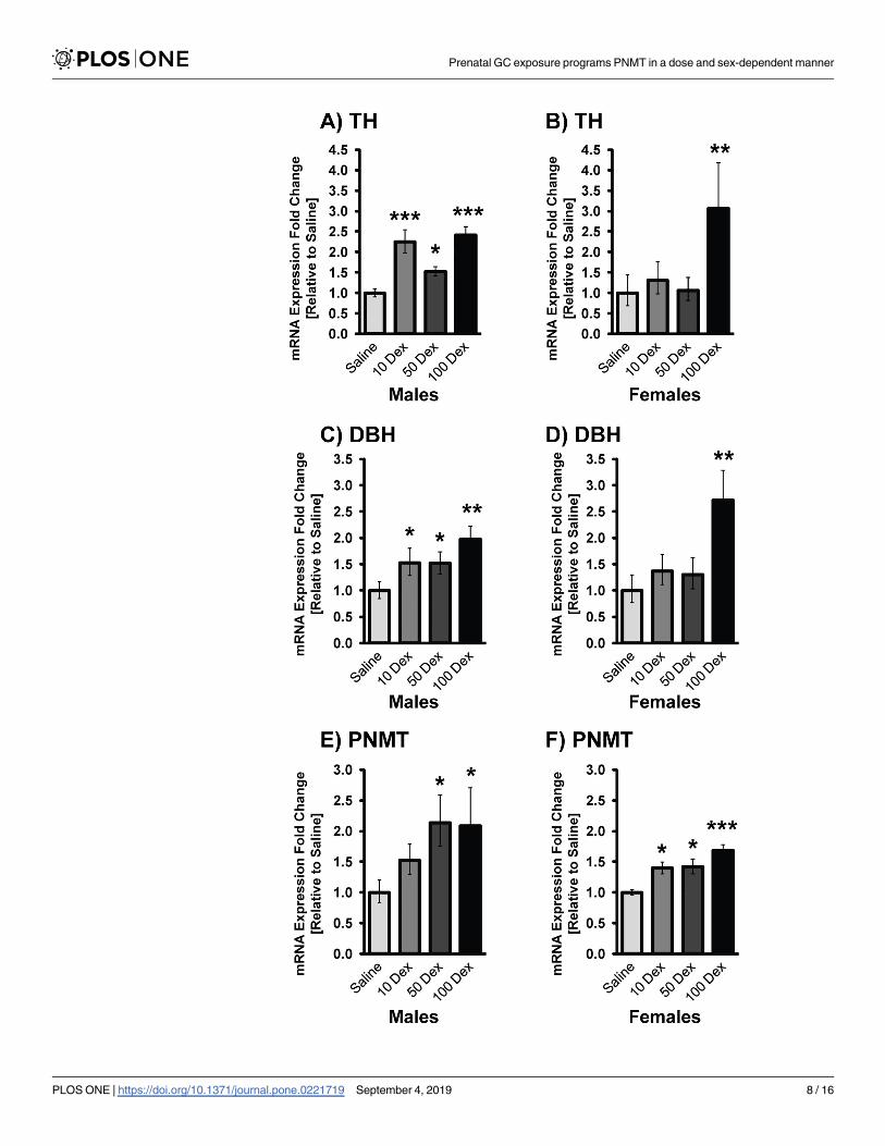
Adrenal PNMT and transcriptional regulators
Transcripts of TH, DBH, and PNMT were analyzed by qRT-PCR in the adrenal glands of
DEX-treated animals (Fig 3). Since the body weight and BPs of the naïve and saline groups
were indistinguishable from each other, all further comparisons of treatment groups were
made to the saline. Predominantly, a dose response was seen in transcripts of TH (10-DEX:
2.25 fold; 50-DEX: 1.53 fold and 100-DEX: 2.41 fold), DBH (10-DEX: 1.53 fold; 50-DEX: 1.51
fold and 100-DEX: 1.91 fold) and PNMT (50-DEX: 2.13 fold and 100-DEX: 2.09 fold) in the
males compared to saline. Similarly, females also showed changes in TH (100-DEX: 3.06 fold),
DBH (100-DEX: 2.72 fold), and PNMT (10-DEX: 1.40 fold; 50-DEX: 1.42 fold and 100-DEX:
1.68 fold). In the females, the majority of significant changes were seen in the higher doses of
DEX exposure, while the lower doses were comparable to the saline group.
To verify if altered PNMT was a consequence of changes in the mRNA levels of its key tran-
scriptional regulatory factors, expression of Egr-1, AP-2, Sp1, and GR were determined (Fig
4). Overall, in both sexes a clear dose response was observed with minimal changes in the
10-DEX group, and significantly higher alterations observed in the 100-DEX group. In the
males, changes in Sp1 and AP2 were not significant, even in the 100-DEX group, while Egr-1
and GR were elevated 2.47 fold and 1.73 fold respectively. Interestingly, Egr-1 was upregulated
even at the lowest DEX dose suggesting lower stress sensitivity for the activation of this tran-
scription factor in males. In the females, all 4-transcription factors were significantly elevated
compared to the saline group at 100-DEX (Egr-1: 3.35 fold; GR: 1.97 fold; Sp1: 2.14 fold, and
AP-2: 2.01 fold). Although some of these transcription factors were elevated at the lower doses
(10DEX), PNMT transcript was not increased, likely because promoter activation requires
cooperative interaction of all factors as previously shown [20].
Plasma corticosterone and epinephrine
Plasma corticosterone and epinephrine were quantified by ELISA (Fig 5). Corticosterone levels
remained unchanged in all three DEX-doses regardless of sex. Epinephrine showed trends
towards increased levels in the 50 and 100-DEX males, but no changes were observed in the
females.
Adrenal stress-sensitive genes
To further comprehend the genetic alterations in prenatally DEX-exposed animals that could
predispose them to hypertension and cardiovascular complications, we chose to analyze the
Prenatal GC exposure programs PNMT in a dose and sex-dependent manner
PLOS ONE | https://doi.org/10.1371/journal.pone.0221719 September 4, 2019 5 / 16

Prenatal GC exposure programs PNMT in a dose and sex-dependent manner
PLOS ONE | https://doi.org/10.1371/journal.pone.0221719 September 4, 2019 6 / 16

expression of phenylalanine hydroxylase (PAH), a member of the solute carrier family of Na
+/H exchangers SLC9A3, and the cytochrome P450 oxidases E class CYP2E1 genes (Fig 6) due
to their role in catecholamine biosynthesis and regulation. These genes were analyzed only in
the 100-DEX dosage group. When compared to saline, PAH expression was 3.59 fold higher in
males, and 8.13 fold higher in females. On the other hand, while SLC9A3 expression was
increased by 5.40 fold in males, its expression was reduced 2.17 fold in the females compared
to the saline group. The expression of CYP2E1 in the males was decreased 4.16 fold, while in
the females the expression of this gene remained unchanged compared to saline.
Discussion
Previous studies have shown that prenatal exposure to GCs contributes to fetal programming
of hypertension [25,29–31]. Increased PNMT expression, and its altered transcriptional regu-
lation in brainstem adrenergic neurons, and in the adrenal gland have been proposed to be
responsible for the overproduction of epinephrine and may conceivably be a genetic mecha-
nism for the pathogenesis of hypertension [9,11,24,25].
Fetal programming by DEX exposure is dose and sex dependent
Prenatal exposure to DEX in the third trimester is a paradigm for fetal programming of low
birth weight offspring resulting in adulthood hypertension [7,25,26,30]. Low birth weight was
observed in offspring prenatally exposed to DEX, and negatively correlated to the dose, where
the highest dose resulted in the lowest birth weight compared to controls. These findings paral-
lel a study on Sprague-Dawley rats prenatally DEX-exposed at concentrations of 25 and 50 μg/
kg/day [32].
The lowest DEX showed higher weights compared to the saline group in both males and
females possibly because at lower concentrations, DEX did not significantly inhibit fetal cellu-
lar growth and may be a consequence of excess adiposity. In non-human primates and in
humans, low dose DEX resulted in heavier or equal weighted offspring, compared to controls
[33]. Higher DEX doses however affected the birth weight more significantly, and catch-up
growth trends were observed until the end of the study period, albeit at a lower rate. Individu-
als born with low birth weight may undergo a compensatory growth where either body weight
is fully caught-up to the average at a young age, or grows slowly and never catches up to the
average body weight, the latter may be the case in the present study [34].
Results from this study show that BP is positively correlated with the prenatal dose of DEX.
Furthermore, males had significantly higher increases in BP compared to female offspring, a
finding also reported in the literature [30,35]. Studies suggest that males are more vulnerable,
while females are relatively protected from prenatal insults [36,37].
We have previously shown increases in transcripts of adrenal PNMT in male offspring of
pregnant dams that received Dex 100 μg/Kg/day [25]. To the best of our knowledge, this is the
first report showing a dose dependent response of prenatal exposure to DEX altering adrenal
catecholaminergic gene expression, with sex-specific responses. While the males showed clear
dose dependent increases in TH, DBH and PNMT, the females were resistant to alterations in
these genes at the lower doses, suggesting a higher threshold for stress mediated alterations,
and more resilience. Clinically, this finding is very relevant because it establishes that lower
DEX doses administered during pregnancy will likely not have long-term consequences on
Fig 2. Blood pressure of 19-week-old DEX exposed offspring. Systolic (A, B) and mean arterial blood pressure (C, D) of male (left) and
female (right) naive, saline, 10-DEX, 50-DEX, and 100-DEX offspring are expressed in mmHg. Data are expressed in mean ± SEM. ANOVA:
Statistical significance between naive and DEX-treated groups is shown by: � p< 0.05, �� p< 0.01, ��� p< 0.001, and ���� p< 0.0001.
https://doi.org/10.1371/journal.pone.0221719.g002
Prenatal GC exposure programs PNMT in a dose and sex-dependent manner
PLOS ONE | https://doi.org/10.1371/journal.pone.0221719 September 4, 2019 7 / 16
Prenatal GC exposure programs PNMT in a dose and sex-dependent manner
PLOS ONE | https://doi.org/10.1371/journal.pone.0221719 September 4, 2019 8 / 16

CAs and BP in female offspring. Clinically, treatment with DEX remains a controversial issue
due to its adverse side effects, and much more still needs to be considered such as duration of
DEX injections, timing of injection during gestation, and also the sex of the offspring.
Published studies have demonstrated sex-specificity in the catecholamine biosynthesis, with
cell-type specific effects of estrogen on the expression of TH and DBH, with dependency on
estrogen receptor subtype [38]. Other researchers have shown that the Y chromosome linked
SRY gene exerts multiple effects on male physiology, including effects on catecholamines,
renin-angiotensin system, and blood pressure homeostasis [39,40]. Numerous studies suggest
sexual dimorphism in the interaction of the HPA with the HPG (hypothalamic pituitary
gonadal) axes, thus rendering females more prone to stress-related anxiety, depression and
psychiatric disorders, while males are more prone to metabolic alterations [41]. These changes
may arise from the differential activation and feedback inhibition of the HPA axis under situa-
tions of stress; numerous studies show sex based differences in CRF levels, activation of CRF
response, ACTH levels and other factors such as functioning of GR [42,43]. Thus, inherent
sex-specific differences in the sensitivity and functioning of the HPA may confer protection
Fig 3. Gene expression of catecholamine biosynthetic enzymes in adrenal glands of 19-week-old prenatally DEX-
exposed offspring. mRNA levels of TH (A, B), DBH (C, D), and PNMT (E, F) in male (left) and female (right)
offspring of saline, 10-DEX, 50-DEX, and 100-DEX treatment groups. Data is expressed as mean fold change ± SEM.
ANOVA: Statistical significance between saline and DEX-treated groups is shown by: � p< 0.05, �� p< 0.01, and ���
p< 0.001.
https://doi.org/10.1371/journal.pone.0221719.g003
Fig 4. mRNA levels of PNMT transcriptional regulators in adrenal glands of 19-week-old prenatally DEX-exposed offspring.
mRNA levels of Egr-1 (A,B), GR (C,D), Sp1 (E, F), and AP-2 (G, H) in male (left) and female (right) offspring of saline, 10-DEX,
50-DEX, and 100-DEX treatment groups. Data is expressed as mean fold change ± SD. ANOVA: Statistical significance between
saline and DEX-treated groups is shown by �, p< 0.05, �� p< 0.01, and ��� p< 0.001.
https://doi.org/10.1371/journal.pone.0221719.g004
Prenatal GC exposure programs PNMT in a dose and sex-dependent manner
PLOS ONE | https://doi.org/10.1371/journal.pone.0221719 September 4, 2019 9 / 16

from stress mediated programing of hypertension in females. This is an area of research that
needs further investigation to tease out sex-specific differences in CA biosynthesis and com-
prehend their role in the sex difference of HPA activation.
Stress-activated transcriptional regulators that modulate the PNMT promoter include Egr-
1, AP-2, GR, and Sp1 [16,17,19,44]. Here, these transcription factors were increased in a dose
and sex dependent manner. While, all 4 factors were elevated in the females at the highest
dose, in the males, Egr-1 and GR were markedly increased while AP2 and Sp1 were only
slightly altered even at the highest DEX dose, a finding comparable to our previous study on
100-DEX males [25]. Our data suggests that sex specific alterations in transcription factors
might be crucial in the regulation of CA biosynthesis in prenatally stressed animals, and that
Fig 5. Effect of prenatal DEX exposure on plasma corticosterone and epinephrine levels. Plasma corticosterone (A, B), and epinephrine
levels (C, D), in male and female offspring from saline or DEX-exposed animals. Fold changes between saline controls versus DEX-treated
animals are presented as mean ± SEM. Significant difference between groups designated as �, p� 0.05.
https://doi.org/10.1371/journal.pone.0221719.g005
Prenatal GC exposure programs PNMT in a dose and sex-dependent manner
PLOS ONE | https://doi.org/10.1371/journal.pone.0221719 September 4, 2019 10 / 16

Prenatal GC exposure programs PNMT in a dose and sex-dependent manner
PLOS ONE | https://doi.org/10.1371/journal.pone.0221719 September 4, 2019 11 / 16

Egr-1 may play a prominent role in PNMT regulation in adrenals of males given that it is sig-
nificantly increased even at the lowest dose.
Our study showed no significant changes in Corticosterone; studies in the literature have
shown variable results for prenatal stress mediated alterations in Corticosterone as this is con-
founded by the stressor, circadian rhythm changes associated with the stress paradigm, and
age of the animals [45]. The estrus cycle also affects the levels in females. Epi levels also weren’t
significantly altered in the prenatally stressed animals, with only trends toward increases seen
in the males with the higher doses, as we have shown previously [25]. Despite this observation,
the increases in PNMT and other catecholamines’ gene expression are relevant and important
as one of the factors that can affect hypertension. Moreover, other studies have shown that the
variability in baseline levels of these hormones is not uncommon, but rapid elevations in
response to stress as well as behavioural changes observed in animals that are prenatally
stressed by glucocorticoids are reflective of overall SA/HPA defects [46,47].
Adrenal PAH expression was significantly elevated in both males and females of 100-DEX
offspring compared to the sex-matched saline group. Interestingly, of all the genes analyzed in
our study, PAH showed significant baseline differences between males and females, with
females expressing much lower PAH. Predominantly located in the liver, PAH biosynthesizes
tyrosine from phenylalanine; tyrosine is further utilized for CA biosynthesis in the adrenal
gland [48,49]. Thus PAH is critical in CA biosynthesis as it generates the precursor molecule
that feeds into the pathway. The role of PAH in the adrenal gland, and in the stress response is
just beginning to be discovered; it has been highlighted by Jacobson (2017) as a stress sensitive
gene, and was elevated in adrenal glands of male rats exposed to chronic stress [50]. In a recent
metabolomics study, serum from DEX-treated animals showed decreases in Phe and increases
in Tyr, which were suggested as predictors for DEX-mediated effects on metabolism [51].
The SLC9A3 gene showed a more sex specific alteration in that males were more promi-
nently affected with prenatal DEX, and showed marked elevations, while females showed a
slight, but significant decrease. SLC9A3, a member of the solute carrier family, is a known Na
+/H+ exchanger that functions in Na+ absorption in the intestine and kidney [52]. Studies
show that disruption of genes in this family, and ensuing disturbances in electrolyte homeosta-
sis can lead to alterations in BP [53]. Although studies in the adrenal expression of this gene
are limited, SLC9A3 has been shown to be elevated in the adrenals of males in other models of
stress sensitive hypertension [54].
Finally, cytochrome P450 enzymes are crucial in the metabolism and clearance of xenobiot-
ics from the body. In our model, CYP2E was significantly reduced in the DEX-exposed males,
however no significant changes were seen in the females. There are sex based and tissue based
differences for the expression of CYP genes reported in the literature [55]. The endocrine sys-
tem and sex hormones can complicate the regulation of CYPs, and our finding could be useful
in understanding the differences in pharmacokinetic handling of drugs in prenatally stressed
males versus females. Overall, the sex-specific effects seen in PAH, SLC9A3 and CYP2E1 genes
likely play an important role in sex-specific responses to prenatal stress, and a comprehensive
study needs to be undertaken to examine their role in the programming of hypertension.
In summary, this study provides possible molecular mechanisms elucidating increased pro-
duction of PNMT mRNA in adrenal glands of prenatally stressed rats (Tables 1 and 2). Expres-
sion of catecholamine biosynthetic enzymes, PNMT transcriptional regulators and other stress
Fig 6. mRNA levels of stress related genes in adrenal glands of 19 week old prenatally DEX-exposed offspring.
mRNA levels of PAH (A,B), SLC9A3 (C,D), and CYP2E1 (E, F) in male (left) and female (right) offspring of saline and
100-DEX treatment groups. Data is expressed as mean fold change ± SEM. ANOVA: Statistical significance between
saline and DEX-treated groups is shown by: � p< 0.05, �� p< 0.01, and ��� p< 0.001, and ���� p< 0.001.
https://doi.org/10.1371/journal.pone.0221719.g006
Prenatal GC exposure programs PNMT in a dose and sex-dependent manner
PLOS ONE | https://doi.org/10.1371/journal.pone.0221719 September 4, 2019 12 / 16

sensitive genes was both dose and sex dependent in the offspring. Although, the low dose was
sufficient to increase adrenal PNMT expression, it did not significantly increase BP in the off-
spring. Therefore, if feasible, a lower DEX dose may be a safer alternative in a clinical setting,
although extrapolation to a corresponding dose in humans is difficult. The sex differences
might be a mechanism by which males express higher PNMT than females, consequently
resulting in the elevated BP of males. This research has implications for differences in sex-
based physiology that would be of significance in evaluating the effects of prenatal stress on
catecholaminergic systems, and to further comprehend predisposition to inheritance and epi-
genetic alterations in future generations.
Author Contributions
Conceptualization: Sandhya Khurana, T. C. Tai.
Data curation: Sandhya Khurana, Julie Grandbois, Sujeenthar Tharmalingam, Alyssa Murray,
Kelly Graff, Phong Nguyen, T. C. Tai.
Formal analysis: Sandhya Khurana, Julie Grandbois, Sujeenthar Tharmalingam, Alyssa Mur-
ray, Kelly Graff, Phong Nguyen, T. C. Tai.
Funding acquisition: T. C. Tai.
Investigation: Sandhya Khurana, Julie Grandbois, Sujeenthar Tharmalingam, Alyssa Murray,
Kelly Graff, Phong Nguyen, T. C. Tai.
Methodology: Sandhya Khurana, Julie Grandbois, Sujeenthar Tharmalingam, Alyssa Murray,
Kelly Graff, Phong Nguyen, T. C. Tai.
Table 1. Summary revealing dose dependent differences in the expression of stress sensitive genes in male
offspring.
MALES 10 DEX 50 DEX 100 DEX
EGR-1 ➔ ➔ ➔
GR − − ➔
SP1 − − −AP2 − − −TH ➔ ➔ ➔
DBH ➔ ➔ ➔
PNMT ➔ ➔ ➔
Blood Pressure ➔ ➔ ➔
https://doi.org/10.1371/journal.pone.0221719.t001
Table 2. Summary revealing dose dependent differences in the expression of stress sensitive genes in female
offspring.
FEMALES 10 DEX 50 DEX 100 DEX
EGR-1 − − ➔
GR − − ➔
SP1 − − ➔
AP2 − − ➔
TH − − ➔
DBH − − ➔
PNMT ➔ ➔ ➔
Blood Pressure − ➔ ➔
https://doi.org/10.1371/journal.pone.0221719.t002
Prenatal GC exposure programs PNMT in a dose and sex-dependent manner
PLOS ONE | https://doi.org/10.1371/journal.pone.0221719 September 4, 2019 13 / 16

Project administration: Sandhya Khurana, T. C. Tai.
Resources: T. C. Tai.
Supervision: Sandhya Khurana, Sujeenthar Tharmalingam, T. C. Tai.
Validation: T. C. Tai.
Writing – original draft: Sandhya Khurana, Julie Grandbois.
Writing – review & editing: Sujeenthar Tharmalingam, T. C. Tai.
References1. Barker DJP. In utero programming of chronic disease. Clin Sci. 1998; 95: 115–128. https://doi.org/10.
1042/cs0950115 PMID: 9680492
2. Welberg LA, Seckl JR. Prenatal stress, glucocorticoids and the programming of the brain. J Neuroendo-
crinol. 2001; 13: 113–28. https://doi.org/10.3389/neuro.08.019.2009 PMID: 11168837
3. Hales CN, Barker DJP. The thrifty phenotype hypothesis: Type 2 diabetes. Br Med Bull. 2001; 60: 5–20.
https://doi.org/10.1093/bmb/60.1.5 PMID: 11809615
4. Moisiadis VG, Matthews SG. Glucocorticoids and fetal programming part 2: Mechanisms. Nat Rev
Endocrinol. Nature Publishing Group, a division of Macmillan Publishers Limited. All Rights Reserved.;
2014; 10: 403–411. https://doi.org/10.1038/nrendo.2014.74 PMID: 24863383
5. Moisiadis VG, Matthews SG. Glucocorticoids and fetal programming part 1: outcomes. Nat Rev Endo-
crinol. Nature Publishing Group; 2014; 10: 1–12. https://doi.org/10.1038/nrendo.2013.228
6. Benediktsson R, Lindsay RS, Noble J, Seckl JR, Edwards CR. Glucocorticoid exposure in utero: new
model for adult hypertension. Lancet. 1993; 341: 339–341. https://doi.org/10.1016/0140-6736(93)
90138-7 PMID: 8094115
7. Bloom SL, Sheffield JS, McIntire DD, Leveno KJ. Antenatal dexamethasone and dereased birth weight.
Obstet Gynecol. 2001; 97: 485–490. https://doi.org/10.1016/s0029-7844(00)01206-0 PMID: 11275014
8. Jacob HJ, Lindpaintner K, Lincoln SE, Kusumi K, Bunker RK, Mao Y-P, et al. Genetic mapping of a
gene causing hypertension in the stroke-prone spontaneously hypertensive rat. Cell. Cell Press; 1991;
67: 213–224. https://doi.org/10.1016/0092-8674(91)90584-L
9. Koike G, Jacob H, Krieger J. Investigation of the Phenylethanolamine N-Methyltransferase Gene as a
Candidate Gene for Hypertension. Hypertension. 1995; 595–601. Available: http://hyper.ahajournals.
org/content/26/4/595.short
10. Rumantir MS, Jennings GL, Lambert GW, Kaye DM, Seals DR, Esler MD. The “adrenaline hypothesis”
of hypertension revisited: evidence for adrenaline release from the heart of patients with essential
hypertension. J Hypertens. 2000; 18: 717–723. https://doi.org/10.1097/00004872-200018060-00009
PMID: 10872556
11. Nguyen P, Peltsch H, de Wit J, Crispo J, Ubriaco G, Eibl J, et al. Regulation of the phenylethanolamine
N-methyltransferase gene in the adrenal gland of the spontaneous hypertensive rat. Neurosci Lett.
Elsevier Ireland Ltd; 2009; 461: 280–4. https://doi.org/10.1016/j.neulet.2009.06.022 PMID: 19539715
12. Cui J, Zhou X, Chazaro I, DeStefano AL, Manolis AJ, Baldwin CT, et al. Association of polymorphisms
in the promoter region of the PNMT gene with essential hypertension in African Americans but not in
whites. Am J Hypertens. Elsevier; 2003; 16: 859–863. https://doi.org/10.1016/s0895-7061(03)01026-4
PMID: 14553966
13. Kepp K, Juhanson P, Kozich V, Ots M, Viigimaa M, Laan M. Resequencing PNMT in European hyper-
tensive and normotensive individuals: no common susceptibilily variants for hypertension and purifying
selection on intron 1. BMC Med Genet. BioMed Central Ltd; 2007; 8: 47. https://doi.org/10.1186/1471-
2350-8-47 PMID: 17645789
14. Ebert SN, Wong DL. Differential activation of the rat phenylethanolamine N-methyltransferase gene by
Sp1 and Egr-1. J Biol Chem. 1995; 270: 17299–305. https://doi.org/10.1074/jbc.270.29.17299 PMID:
7615530
15. Tai TC, Morita K, Wong DL. Role of Egr-1 in cAMP-dependent protein kinase regulation of the pheny-
lethanolamine N-methyltransferase gene. J Neurochem. 2001; 76: 1851–1859. https://doi.org/10.1046/
j.1471-4159.2001.00189.x PMID: 11259503
16. Tai TC, Claycomb R, Her S, Bloom AK, Wong DL. Glucocorticoid responsiveness of the rat phenyletha-
nolamine N-methyltransferase gene. Mol Pharmacol. 2002; 61: 1385–92. Available: http://www.ncbi.
nlm.nih.gov/pubmed/12021400 https://doi.org/10.1124/mol.61.6.1385 PMID: 12021400
Prenatal GC exposure programs PNMT in a dose and sex-dependent manner
PLOS ONE | https://doi.org/10.1371/journal.pone.0221719 September 4, 2019 14 / 16

17. Ebert SN, Balt SL, Hunter JP, Gashler A, Sukhatme V, Wong DL. Egr-1 activation of rat adrenal pheny-
lethanolamine N-methyltransferase gene. J Biol Chem. 1994; 269: 20885–98. PMID: 8063705
18. Wong DL, Siddall B, Wang W. Hormonal control of rat adrenal phenylethanolamine N-methyltransfer-
ase: Enzyme activity, the final critical pathway. Neuropsychopharmacology. 1995. pp. 223–234. https://
doi.org/10.1016/0893-133X(95)00066-M PMID: 8602895
19. Ebert SN, Ficklin MB, Her S, Siddall BJ, Bell RA, Ganguly K, et al. Glucocorticoid-dependent action of
neural crest factor AP-2: stimulation of phenylethanolamine N-methyltransferase gene expression. J
Neurochem. 1998; 70: 2286–95. Available: http://www.ncbi.nlm.nih.gov/pubmed/9603193 https://doi.
org/10.1046/j.1471-4159.1998.70062286.x PMID: 9603193
20. Tai T-C, Wong DL. Phenylethanolamine N-methyltransferase gene regulation by cAMP-dependent pro-
tein kinase A and protein kinase C signaling pathways. Ann N Y Acad Sci. 2002; 971: 83–5. Available:
http://www.ncbi.nlm.nih.gov/pubmed/12438094 https://doi.org/10.1111/j.1749-6632.2002.tb04438.x
PMID: 12438094
21. Wong DL, Anderson LJ, Tai TC. Cholinergic and peptidergic regulation of phenylethanolamine N-
methyltransferase gene expression. Ann N Y Acad Sci. 2002; 971: 19–26. https://doi.org/10.1111/j.
1749-6632.2002.tb04428.x PMID: 12438084
22. Tai TC, Claycomb R, Siddall BJ, Bell RA, Kvetnansky R, Wong DL. Stress-induced changes in epineph-
rine expression in the adrenal medulla in vivo. J Neurochem. 2007; 101: 1108–18. https://doi.org/10.
1111/j.1471-4159.2007.04484.x PMID: 17394532
23. Peltsch H, Khurana S, Byrne CJ, Nguyen P, Khaper N, Kumar A, et al. Cardiac phenylethanolamine N-
methyltransferase: localization and regulation of gene expression in the spontaneously hypertensive
rat. Can J Physiol Pharmacol. 2016; 94: 363–72. https://doi.org/10.1139/cjpp-2015-0303 PMID:
26761434
24. Grandbois J, Khurana S, Graff K, Nguyen P, Meltz L, Tai TC. Phenylethanolamine N-methyltransferase
gene expression in adrenergic neurons of spontaneously hypertensive rats. Neurosci Lett. Elsevier Ire-
land Ltd; 2016; 635: 103–110. https://doi.org/10.1016/j.neulet.2016.10.028 PMID: 27769893
25. Nguyen P, Khurana S, Peltsch H, Grandbois J, Eibl J, Crispo J, et al. Prenatal glucocorticoid exposure
programs adrenal PNMT expression and adult hypertension. J Endocrinol. 2015;227. https://doi.org/10.
1530/JOE-15-0244 PMID: 26475702
26. Levitt NS, Lindsay RS, Holmes MC, Seckl JR. Dexamethasone in the Last Week of Pregnancy Attenu-
ates Hippocampal Glucocorticoid Receptor Gene Expression and Elevates Blood Pressure in the Adult
Offspring in the Rat. Neuroendocrinology. 1996; 412–418. https://doi.org/10.1159/000127146 PMID:
8990073
27. Feng M, Whitesall S, Zhang Y, Beibel M, D’Alecy L, DiPetrillo K. Validation of volume-pressure record-
ing tail-cuff blood pressure measurements. Am J Hypertens. Oxford University Press; 2008; 21: 1288–
91. https://doi.org/10.1038/ajh.2008.301 PMID: 18846043
28. Livak KJ, Schmittgen TD. Analysis of Relative Gene Expression Data Using Real-Time Quantitative
PCR and the 2(-DDCt) Method. Methods. Elsevier; 2001; 25: 402–408. https://doi.org/10.1006/meth.
2001.1262 PMID: 11846609
29. Seckl JR. Prenatal glucocorticoids and long-term programming. Eur J Endocrinol. 2004; 151 Suppl:
U49–62.
30. Ortiz LA, Quan A, Zarzar F, Weinberg A, Baum M. Prenatal Dexamethasone Programs Hypertension
and Renal Injury in the Rat. Hypertension. 2002; 41: 328–334. https://doi.org/10.1161/01.HYP.
0000049763.51269.51 PMID: 12574103
31. Wintour EM, Moritz KM, Johnson K, Ricardo S, Samuel CS, Dodic M. Reduced nephron number in
adult sheep, hypertensive as a result of prenatal glucocorticoid treatment. J Physiol. 2003; 549: 929–
35. https://doi.org/10.1113/jphysiol.2003.042408 PMID: 12730337
32. Woods LL. Maternal glucocorticoids and prenatal programming of hypertension. Am J Physiol Regul
Integr Comp Physiol. 2006; 291: R1069–75. https://doi.org/10.1152/ajpregu.00753.2005 PMID:
16644906
33. de Vries A, Holmes MC, Heijnis A, Seier J V, Heerden J, Louw J, et al. Prenatal dexamethasone expo-
sure induces changes in nonhuman primate offspring cardiometabolic and hypothalamic pituitary-adre-
nal axis function. J Clin Invest. 2007; 117: 1058–1067. https://doi.org/10.1172/JCI30982 PMID:
17380204
34. Barker DJP, Bagby SP, Hanson MA. Mechanisms of disease: in utero programming in the pathogenesis
of hypertension. Nat Clin Pract Nephrol. 2006; 2: 700–7. https://doi.org/10.1038/ncpneph0344 PMID:
17124527
35. O’Regan D, Kenyon C. Glucocorticoid exposure in late gestation in the rat permanently programs gen-
der-specific differences in adult cardiovascular and metabolic physiology. Am J Physiol Endocrinol
Metab. 2004; 287: 863–870. https://doi.org/10.1152/ajpendo.00137.2004 PMID: 15238353
Prenatal GC exposure programs PNMT in a dose and sex-dependent manner
PLOS ONE | https://doi.org/10.1371/journal.pone.0221719 September 4, 2019 15 / 16

36. Ojeda NB, Grigore D, Alexander BT. Intrauterine growth restriction: fetal programming of hypertension
and kidney disease. Adv Chronic Kidney Dis. 2008; 15: 101–106. S1548-5595(08)00002-5 [pii]\r https://
doi.org/10.1053/j.ackd.2008.01.001 PMID: 18334233
37. Grigore D, Ojeda NB, Alexander BT. Sex differences in the fetal programming of hypertension. Gend
Med. 2008; 5 Suppl A: S121–32. https://doi.org/10.1016/j.genm.2008.03.012 PMID: 18395678
38. Sabban EL, Maharjan S, Nostramo R, Serova LI. Divergent effects of estradiol on gene expression of
catecholamine biosynthetic enzymes. Physiol Behav. Elsevier B.V.; 2010; 99: 163–8. https://doi.org/10.
1016/j.physbeh.2009.07.011 PMID: 19638280
39. Milsted A, Serova L, Sabban EL, Dunphy G, Turner ME, Ely DL. Regulation of tyrosine hydroxylase
gene transcription by Sry. Neurosci Lett. 2004; 369: 203–7. https://doi.org/10.1016/j.neulet.2004.07.
052 PMID: 15464265
40. Ely D, Underwood A, Dunphy G, Boehme S, Turner M, Milsted A. Review of the Y chromosome, Sry
and hypertension. Steroids. 2010; 75: 747–53. https://doi.org/10.1016/j.steroids.2009.10.015 PMID:
19914267
41. Bourke CH, Harrell CS, Neigh GN. Stress-induced sex differences: adaptations mediated by the gluco-
corticoid receptor. Horm Behav. 2012; 62: 210–8. https://doi.org/10.1016/j.yhbeh.2012.02.024 PMID:
22426413
42. Panagiotakopoulos L, Neigh GN. Development of the HPA axis: where and when do sex differences
manifest? Front Neuroendocrinol. 2014; 35: 285–302. https://doi.org/10.1016/j.yfrne.2014.03.002
PMID: 24631756
43. Bangasser DA. Sex differences in stress-related receptors: “Micro” differences with “macro” implica-
tions for mood and anxiety disorders. Biol Sex Differ. 2013; 4: 1–13. https://doi.org/10.1186/2042-6410-
4-1
44. Her S, Claycomb R, Tai TC, Wong DL. Regulation of the rat phenylethanolamine N-methyltransferase
gene by transcription factors Sp1 and MAZ. Mol Pharmacol. 2003; 64: 1180–8. https://doi.org/10.1124/
mol.64.5.1180 PMID: 14573768
45. Welberg LAM, Seckl JR. Prenatal Stress, Glucocorticoids and the Programming of the Brain. 2001; 13:
113–128. PMID: 11168837
46. Kapoor A, Petropoulos S, Matthews SG. Fetal programming of hypothalamic-pituitary-adrenal (HPA)
axis function and behavior by synthetic glucocorticoids. Brain Res Rev. 2008; 57: 586–595. https://doi.
org/10.1016/j.brainresrev.2007.06.013 PMID: 17716742
47. Viltart O, Vanbesien-Mailliot CCA. Impact of prenatal stress on neuroendocrine programming. Scienti-
ficWorldJournal. 2007; 7: 1493–1537. https://doi.org/10.1100/tsw.2007.204 PMID: 17767365
48. McGee MM, Greengard O, Knox WE. The quantitative determination of phenylalanine hydroxylase in
rat tissues. Its developmental formation in liver. Biochem J. 1972; 127: 669–74. https://doi.org/10.1042/
bj1270669 PMID: 4651134
49. Joh TH, Hwang O, Abate C. Phenylalanine Hydroxylase,Tyrosine Hydroxylase,Tryptophan Hydroxy-
lase. Neurotransmitter Enzymes. New Jersey: Humana Press; pp. 1–32. https://doi.org/10.1385/0-
89603-079-2:1
50. Jacobson ML, Kim LA, Rosati B, Mckinnon D, Mckinnon D. Universal Transcriptional Responses to Dif-
ferent Modalities of Traumatic Stress as a Measure of Stress Exposure. 2017;
51. Malkawi AK, Alzoubi KH, Jacob M, Matic G, Ali A, Al Faraj A, et al. Metabolomics based profiling of
Dexamethasone side effects in rats. Front Pharmacol. 2018; 9: 1–14. https://doi.org/10.3389/fphar.
2018.00001
52. Donowitz M, Ming Tse C, Fuster D. SLC9/NHE gene family, a plasma membrane and organellar family
of Na+/H+ exchangers. Mol Aspects Med. 2009; 34: 236–51. https://doi.org/10.1016/j.mam.2012.05.
001 PMID: 23506868
53. Boedtkjer E, Aalkjaer C. Disturbed acid-base transport: An emerging cause of hypertension. Front Phy-
siol. 2013; 4 DEC: 1–9. https://doi.org/10.3389/fphys.2013.00001
54. Fedoseeva LA, Klimov LO, Ershov NI, Alexandrovich Y V., Efimov VM, Markel AL, et al. Molecular
determinants of the adrenal gland functioning related to stress-sensitive hypertension in ISIAH rats.
BMC Genomics. 2016;17. https://doi.org/10.1186/s12864-015-2333-3
55. Renaud HJ, Cui JY, Khan M, Klaassen CD. Tissue distribution and gender-divergent expression of 78
cytochrome p450 mRNAs in mice. Toxicol Sci. 2011; 124: 261–277. https://doi.org/10.1093/toxsci/
kfr240 PMID: 21920951
Prenatal GC exposure programs PNMT in a dose and sex-dependent manner
PLOS ONE | https://doi.org/10.1371/journal.pone.0221719 September 4, 2019 16 / 16