6. Adrenal Diseases (Final Draft).pdf - KSUMSC
11
Objectives: Not Given. Resources: ● Davidson’s (Chapter 20 pg 364). ● 436 doctors slides. ● 435’s teamwork. ● Surgical Recall. Done by: Shoag Alahmari, Ghadah Almazrou, Abdullah AlMana Leaders: Heba Alnasser, Jawaher Abanumy, Mohammed Habib, Mohammad Al-Mutlaq Revised by: Abdulaziz Aljasser Color index: Notes , Important , Extra , Davidson’s Editing file Feedback
-
Upload
khangminh22 -
Category
Documents
-
view
1 -
download
0
Transcript of 6. Adrenal Diseases (Final Draft).pdf - KSUMSC

Objectives:
Not Given.
Resources:
● Davidson’s (Chapter 20 pg 364).
● 436 doctors slides.
● 435’s teamwork.
● Surgical Recall.
Done by: Shoag Alahmari, Ghadah Almazrou, Abdullah AlMana
Leaders: Heba Alnasser, Jawaher Abanumy, Mohammed Habib, Mohammad Al-Mutlaq
Revised by: Abdulaziz Aljasser
Color index: Notes , Important , Extra , Davidson’s
Editing file Feedback

Basics Review Video(10:42)
● The adrenal glands lie immediately above and medial to the kidneys and they receive their blood supply from 3 arteries originating from: the inferior phrenic artery, abdominal aorta, and the renal artery.
● The right adrenal vein drains into the vena cava. The left adrenal vein drains into the left renal vein.
● It is divided into two parts; each with separate functions: ○ Adrenal Medulla (inner layer) ○ Adrenal Cortex (outer layer):
● The cortex has three specific zones and each produces a specific class of steroid hormone:
I. Zona Glomerulosa – mineralocorticoids (Aldosterone) II. Zona Fasciculata – glucocorticoids (Cortisol)
III. Zona Reticularis - androgens (to remember the layers from outer to inner → “GFR”)
Explanation of the picture: What you need to know is that the hormones or secretions from the adrenal cortex can be stimulated directly or indirectly: ● Directly by decreased Na or
increased K in the blood. ● Indirectly by:
(1) hypotension or hypovolemia → kidney releases renin → angiotensin will give indirect effect. (2) by ACTH
The function of the cortex can be remembered by: salt, sugar, and sex. I. Aldosterone (SALT) ● Mineralocorticoid. ● Function: fluid & electrolyte balance, and maintains blood volume & BP:
○ Na+ retention ○ Water retention ○ K+ excretion → (serum K+ will ↓ and urine K+ will ↑)
● Renin from kidneys controls adrenal cortex production of aldosterone. ● Questions: (you will get case scenarios like these in the MCQs)
If your Na level is low, will aldosterone secretion ↑ or ↓ ? increase If your serum K+ level is high, will aldosterone secretion ↑ or ↓ ? increase
II. Cortisol (SUGAR) ● Glucocorticoid ● Function:
○ Regulate metabolism & are critical in stress response. Stress might cause pseudo-cushing syndrome (like in exams: eating + studying = weight gain)
○ Cortisol is responsible for control and & metabolism of:
1

Carbohydrates Fats Proteins
↑ amount of glucose formed ↑ amount of glucose released
causing hyperglycemia
stimulates fatty acid mobilization from adipose
tissue
stimulates protein synthesis in liver protein breakdown in tissues
○ Other functions: ↓ inflammatory and allergic response & ↓ immune response, therefore prone to infection
● Release of glucocorticoids is controlled by → ACTH (Adrenocorticotropic hormone) produced in anterior pituitary gland*. ○ decreased levels of circulating cortisol causes stimulation of ACTH ○ increased levels of circulating cortisol causes decreased release of
ACTH ○ What type of feedback mechanism is this? Negative feedback
*Ectopic ACTH might come from tumors in lung or pancreas.
● Also affected by: ○ Individual biorhythms → ACTH levels are highest 2 hours before
and just after awakening (usually 5AM - 7AM). These gradually decrease rest of day
○ Stress increases cortisol production and secretion III. Androgens (SEX)
● Hormones which ↑ male characteristics (release of testosterone). ● Androgen is seen more in women than men.
IV. Medulla
● What is released by the adrenal medulla? ○ Catecholamines: Epinephrine and Norepinephrine .
● These hormones are responsible for Fight or flight response.
This picture is extra and summarizes what’s mentioned above:
The diseases of the adrenal gland can be divided into: hypofunction, hyperfunction, & masses. You have to understand each disease: how it presents, and how it will affect the electrolyte levels.
2

CORTEX 1. Cushing’s Syndrome Video(13:42)
● TOO MUCH CORTISOL! Increases secretion of cortisol from adrenal cortex, or it can be from another source (endogenous or exogenous).
● 4 times more frequent in females, and usually occurs at 35-50 years of age.
ETIOLOGY in exam it doesn’t come as “what is the most common cause” instead, “Sx and signs”
Primary (20%) Secondary (80%) Ectopic Iatrogenic
Tumor of the adrenal cortex: ● Adenoma (more
common, unilateral) ● Carcinoma (rare,
present late)
Tumor of the anterior pituitary gland. (when the syndrome is caused by a pituitary tumor it is referred to as cushing’s disease).
ACTH secreting tumor: ● Lung (small cell
carcinoma) ● Pancreas ● Thyroid
Therapeutic steroid administration (exogenous source).
SIGNS & SYMPTOMS: ● Cushing’s original description was a “tomato head, potato body, and four matches as legs.” ● To understand the presentation remember the functions of cortisol and exaggerate them:
I. Increase protein catabolism: - Increase muscle wasting - Loss of collagen support (thin, fragile
skin, bruises easily) - Poor wound healing II. Increase carbohydrate metabolism: - Hyperglycemia → can get diabetes
because insulin can’t keep up - Polyuria III. Increase fat metabolism: - Truncal obesity + buffalo hump - Moon face - ↑ weight but ↓ strength IV. Decrease immune response: - More prone to infection 1
- Decrease resistance to stress - Death usually occurs from infection V. Increase androgen secretion: - Excessive hair growth - Acne - Change in voice - Receding hairline VI. Increase mineralocorticoid activity (aldosterone) → increase sodium and water retention → elevated BP.
Investigation (the test must be repeated at least twice to confirm the diagnosis) ● Exclude exogenous steroid use then order: 24 hr urinary free cortisol and/or dexamethasone
suppression test. Imaging can be done → MRI (pituitary adenoma) or CT (adrenal adenoma).
1 keep in mind that surgical pt. With cushing syndrome must be kept on antibiotic to avoid infections. 3

MANAGEMENT (depends on the cause)
● Adrenal adenoma: are rarely bilateral → unilateral adrenalectomy is most commonly indicated. ● Adrenal carcinoma: should be completely removed whenever possible +/- chemotherapy. ● Pituitary disease: → bilateral adrenalectomy, but at the price of lifelong steroid therapy. Pituitary
irradiation or surgery avoids the side-effects of adrenalectomy, and microsurgical removal of the adenoma is now the treatment of choice.
2. Conn’s Syndrome Video(06:11)
● HYPERALDOSTERONISM → too much aldosterone secretion. ● Usually is caused by adrenal tumor most common in young or middle-aged women. ● What does aldosterone do? Sodium and water retention, and potassium excretion. ● Types of hyperaldosteronism:
Primary (Conn’s syndrome) Secondary
Usually due to a benign adenoma (small, single). The high circulating levels of aldosterone suppress renin secretion – a helpful biochemical diagnostic
observation.
Most commonly secondary to excessive renin secretion in chronic liver, renal or cardiac
disease.
(High aldosterone & Low renin). (High aldosterone & High renin).
Case scenario: A patient came to the ER, she is diagnosed with HTN & is on 3 medications but her BP is still high (not responding to medication). Upon Investigation, her serum K+ is low . She has been admitted to the ICU with hypokalemia 3 times before. 1- what further investigation do you want to? After history and examination, we check electrolyte (including serum Na+ and K+) and imaging to rule out tumors. 2- what is the most likely DDx she has? Conn’s syndrome
SIGNS & SYMPTOMS: 1. Na and water retention (hypernatremia) → high blood pressure & visual disturbance +/- headache 2. Decreased K+ (hypokalemia → Worsening hypokalemia, episodes of muscle weakness and
nocturnal polyuria). What is the normal serum K+ level? 3.5-5 (mEq/L) 3. Usually no edema. (but if it’s Secondary PT could have Edema)
DIAGNOSIS ● Increased plasma aldosterone levels with low plasma renin levels (in case of primary) ● Plasma (or serum): increased Na+ and decreased K+ . 2
● Increased Urinary K+ (remember serum K+ is low and urine K+ is high because of excretion!). ● EKG changes (to check for signs of hypokalemia → flattening of T waves, U waves). ● CT scan to rule out tumors .
MANAGEMENT: ● Surgery (ADRENALECTOMY) but before the surgery you need to prep the pt and after you need
to follow up (it’s enough to know that you don’t have to know the details).
2 Giving the aldosterone antagonist, Spironolactone, should ↓ blood pressure and reverse hypokalaemia. 4

Pre-op Post-op
● Stabilize hormonally ● Correct fluid and electrolytes ● Cortisol PM before surgery, AM of
surgery and during OR.
Preop: make sure serum electrolytes (Na+ & K+) are normal , no ECG changes
● We observe the pt post-op and sometime we put them in the ICU (What type of problems to expect??)
● IV cortisol for 24 hours ● IM cortisol 2nd day ● PO cortisol 3rd day ● Poor wound healing ● If unilateral adrenalectomy the steroids are
weaned (other adrenal takes over 6-12 months)
3. Addison’s Disease Video(16:27)
● Adrenocortical Insufficiency: Hypofunction of adrenal cortex. So which hormones will be decreased? ALL OF THEM (so the treatment is hormones replacement).
● Types of adrenocortical insufficiency:
PRIMARY (ADDISON’S DISEASE) SECONDARY
All hormones will be decreased: from all 3 cortical layers. Only glucocorticoids will be decreased.
autoimmune , idiopathic, infection (TB) , iatrogenic inadequate pituitary ACTH secretion.
ACTH normal or high ACTH is low
- Hyperpigmentation - High K and low Na - Metabolic Acidosis
- No pigmentation - Normal K and normal Na or low - No metabolic Acidosis
ETIOLOGY OF ADDISON'S ● Idiopathic atrophy → autoimmune condition where antibodies attack against own adrenal cortex,
and 90% of tissue destroyed. ● TB/fungal infections (histoplasmosis) ● Iatrogenic causes → adrenalectomy, chemotherapy, anticoagulant tx
SIGNS & SYMPTOMS:
● Fluid & electrolyte imbalances: ○ Hypotension. ○ Hyponatremia. ○ Hyperkalemia (serum is high but urine is low). ○ Hypoglycemia.
● Salt craving. ● Fatigue, weight loss, anorexia; due to low cortisol. ● Changes in skin pigmentation: ↓cortisol - ↑ACTH - ↑MSH ● Muscular weakness . 3
● Androgens are low causing: pubic hair loss and decreased sexual drive for women. (men are not affected)
● Mental disturbances: anxiety, irritability, etc.
3 cortisol helps muscles maintain contraction and avoid fatigue 5
DIAGNOSIS: ● 24 hours urinary 17-OHCS2 and 17 KS3 is low. ● Serum cortisol is low → serum glucose is low ● Serum K is high, and Na is low .
INTERVENTION: ● Lifelong hormone replacement (no need to remember the details):
○ Primary need oral cortisone 20-25 mg in AM and 20-12 mg in PM. ○ Change dose PRN for stress. ○ Also need mineralocorticoid (florinef)
● General advice we give the patients: ○ Salt food liberally (add a lot of salt to their food helps increase their blood pressure) ○ Do not fast or omit meals. ○ Eat between meals and snack. ○ Eat high in carbs and proteins. ○ Wear medical alert bracelet. ○ Always carry a kit of 100 mg hydrocortisone IM. (stress dose ) 4
○ Keep parenteral glucocorticoids at home for injection during illness. ○ Avoid infections/stress.
COMPLICATION: ● Adrenal crisis (severe hypotension like in addison’s or severe hypertension like pheochromocytoma) ● Electrolyte imbalance (hyperkalemia and hyponatremia) ● Hypoglycemia
MEDULLA 1. Pheochromocytoma Video(05:46)
● Rare, benign tumor of the adrenal medulla → 10% could be malignant (Remember the rule of 10%) . 5
● Hypersecretion of? Epinephrine and norepinephrine → released sporadically. ● The median age for presentation of pheochromocytomas is 40 years. ● Associated conditions are neurofibromatosis, medullary carcinoma of the thyroid (as part of MEN
type II), duodenal ulcer and renal artery stenosis.
SIGNS & SYMPTOMS ● Not specific, they may have similar presentation as conn’s syndrome → headache and hypertension
BUT here potassium and sodium levels are normal! So we check electrolyte to differentiate. ● Hallmark is hypertension: 200/150 or greater (high blood pressure not responding to treatment) ● Paroxysmal attacks “spells” provoked by bladder distension, emotional distress, exposure to cold. ● Deep breathing, pounding heart (tachycardia), headache, moist cool hands and feet, visual
disturbance. ● Classic triad (not found in most patients): Palpitations, Headache, Episodic diaphoresis. ● A few patients present with predominantly metabolic effects, such as those found in thyrotoxicosis.
4 If you find them collapsed (and they have a medical bracelet saying they have addison’s) look in their bag and you’ll find this syringe → give it to them to stimulate their adrenal until them until they get to a hospital. 5 10% familial, 10% malignant, 10% extra-adrenal
6

DIAGNOSIS By history and examination then tests:
● Urine: 24h urine- VMA (VanillylMandelic Acid → metabolite of epinephrine) or urinary metanephrine. Also glycosuria is common.
● Blood: Plasma catecholamines (metanephrine). ● Imaging: CT or MRI (to locate tumor), or scintigraphy (MIBG scan).
INTERVENTION
● Surgical removal of the tumor is the treatment of choice. ● The patient should come to operation with blood pressure and pulse rate controlled to
reduce the risk of adrenal crisis! How do we do that?
PRE-OP (admit the pt. 3
days before surgery to
control HR & BP)
What to give the patient? ➔ Adrenergic blocking agents: Minipress → lower BP ➔ Beta blocking agents: Inderal → lower HR, BP & force of contraction ➔ Diet: high in vitamin, mineral,calorie, no caffeine, low salt ➔ Sedatives What to do to the patient? ➔ Monitor BP ➔ Eliminate attacks ➔ If attack → complete bed rest and head of bed 45 degrees
DURING SURGERY
Give regitine and nipride to prevent hypertensive crisis. Make sure you have a stress dose ready because we worry about hypertension during and hypotension after the intervention. Anesthesia should have Hydralazine (for hypertension) and phenylephrine (for hypotension)
POST-OP BP may be high initially, BUT CAN BOTTOM OUT so we prepare: - Volume expanders - Vasopressors - Hourly Input and Output - Observe for hemorrhage
2. Incidentaloma
● Mass lesion greater than 1 cm. ● Serendipitously (مصادفة) discovered by radiologic examinations done for other reasons such as:
○ Computed tomography (CT) ○ Magnetic resonance imaging (MRI)
● If you discover any mass you have to answer two questions: ○ Is it functioning (producing hormones)? You have to rule out pheochromocytoma by labs. ○ Is it malignancy? (may be primary tumor or metastasis* → check if they have hx of cancer)
*the most common tumors that metastasize to the adrenal gland are renal cell carcinoma and breast cancer (sometimes the lung will metastasize in rare cases). MANAGEMENT
● First make sure it is not functioning (by history or by investigations), then check the size. ● If it is more than 4 cm then take it out immediately. ● Less than 4 cm observe and repeat the imaging (CT or MRI) in 6 months:
○ No increase in size → repeat after one year and etc.. → continue to follow up the pt. ○ If it increased (>1 cm in 1 year) → you can take it out because you can’t rule out cancer.
7
Case scenarios given by the Dr. (understand them to know how to answer the exam): Case 1: OB/GYN patient was referred to you with 1 cm mass on the adrenal found by US (done for fibroid). Patient has NO complaint. What is the most appropriate workup you should ask for? Is the mass functional or not? We check labs to rule out pheochromocytoma: 24h urinary VMA, plasma and urinary metanephrines, and CT scan. If they find it to be normal→ It is most likely incidentaloma (also because pt has no complaint). For the management see above. Case 2: A patient diagnosed with breast cancer 10 years ago, and she is presenting with a mass on the adrenal. → You have to rule out cancer (metastasis). So we do PET scan or biopsy to rule it out. Case 3: A patient is referred from nephrology with headache and uncontrolled BP not responding to medications (despite being on two antihypertensives). It could be Conn’s syndrome or pheochromocytoma: ● Check her electrolytes → if her serum Na and K are normal it’s unlikely to be conn. ● If they did imaging and found a mass → pheochromocytoma. ● How to confirm that’s pheochromocytoma? by serum and urine metanephrine level (their accuracy is
around 97%)
Recall: (EXTRA)
What is the most common cause of Cushing’s Syndrome? Iatrogenic (i.e, prescribed prednisone) What is the second most common cause of Cushing’s Syndrome? Cushing’s disease (most common non iatrogenic cause) What is Cushing’s disease? Cushing’s syndrome caused by excess production of ACTH by anterior pituitary How can cortisol levels be indirectly measured over a short duration? By measuring urine cortisol or the breakdown product of cortisol, 17 hydroxycorticosteroid (17-OHCS), in the urine What is a direct test of serum cortisol? Serum cortisol level (highest in the morning and lowest at night in healthy patients) How are the following tumors treated: Adrenal adenoma? Adrenalectomy (almost always unilateral) Adrenal carcinoma? Surgical excision (only 33% of cases are operable) Ectopic ACTH-producing tumor? Surgical excision, if feasible Cushing’s disease? transphenoidal adenectomy What are two classic clues of Conn’s syndrome? 1. Hypertension 2. Hypokalemia What are the causes of Conn’s syndrome? Adrenal adenoma (66%), Bilateral idiopathic adrenal hyperplasia (30%), Adrenal cancer ( < 1%) What diagnostic tests should be ordered? 1. Plasma aldosterone concentration 2. Plasma renin activity What is the treatment of the following conditions: Adenoma Unilateral adrenalectomy (laparoscopic) Unilateral hyperplasia Unilateral adrenalectomy (laparoscopic) Bilateral hyperplasia Spironolactone (usually no surgery) What are the renin levels in patients with PRIMARY hyperaldosteronism? Normal or low (key point!) How do you remember what ADDISON’s disease is? Think: ADDison’s disease = ADrenal Down What are the electrolyte findings? HYPERkalemia, hyponatremia. Which age group is most likely affected by pheochromocytoma? Any age (children and adults); average age is 40 to 60 years. How can the pheochromocytoma SYMPTOMS triad be remembered? Think of the first three letters in the word Pheochromocytoma: Palpitations, Headache and Episodic diaphoresis What are the other common lab findings? Hyperglycemia (epinephrine increases glucose, norepinephrine decreases insulin) Polycythemia (resulting from intravascular volume depletion). What is the classic pheochromocytoma “rule of 10’s”? 10% malignant, 10% bilateral, 10% in children, 10% multiple tumors, 10% extra-adrenal and 10% familial What is the surgical treatment? Tumor resection with early ligation of venous drainage (lower possibility of catecholamine release/crisis by tying off drainage) and minimal manipulation In the patient with pheochromocytoma, what must be ruled out? MEN type II (almost all cases are bilateral) What is the most common cause of incidentaloma? Nonfunctioning adenoma ( >75% of cases)
8
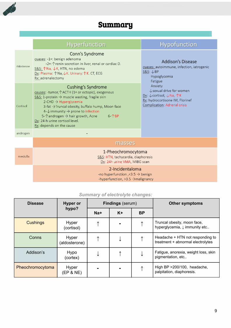
Summary
Summary of electrolyte changes:
Disease Hyper or hypo?
Findings (serum) Other symptoms
Na+ K+ BP
Cushings Hyper (cortisol)
↑ - ↑ Truncal obesity, moon face, hyperglycemia, ↓ immunity etc..
Conns Hyper (aldosterone)
↑ ↓ ↑ Headache + HTN not responding to treatment + abnormal electrolytes
Addison’s Hypo (cortex)
↓ ↑ ↓ Fatigue, anorexia, weight loss, skin pigmentation, etc..
Pheochromocytoma Hyper (EP & NE)
- - ↑ High BP >200/100, headache, palpitation, diaphoresis.
9

Question 1- Which of the following is not associated with cushing’s syndrome ?
A) Increase carbohydrate metabolism B) Decreased fat metabolism C) Increase protein catabolism D) Increase androgen secretion
2- Which of the following scenarios is most commonly associated with conn’s syndrome ?
A) Young female ( 25 year old ) B) Old male ( 65 year old ) C) Children ( under the age of 5 ) D) Patients with TB
3- A 45 year old female patients presents at the clinic with a 2 month history of weakness, dizziness when standing, anorexia, weight loss and has also noticed pubic hair loss. What’s the most common cause of her presentation ?
A) TB B) Adrenalectomy C) Adrenal adenoma D) Autoimmune
4- A 38 year old male patient present to the clinic complaining of headache, palpitation and sweating. You found out that his BP is 195/121 and you diagnosed him with pheochromocytoma, which of the following is the best choice for management ?
A) Hormonal replacement B) ACEi + CCB + thiazide diuretics C) Surgical removal of the tumour D) Removing the pituitary
5- A 40 year old female patient came to the clinic complaining of visual disturbance, muscle weakness and nocturnal polyuria, you found that she has a BP of 175/109. Which of the following results would you expect to see in this patient ?
A) High Na, high K B) High Na, normal K C) High Na, low K D) Low Na, High K
Answers: 1:B 2:A 3:D 4:C 5:C
10