Pyrexia and pancytopenia with unusual host immune response
18
SELF ASSESSMENT QUESTIONS Fever and a rash in a 61 year old man Daniel G Federman, Robert S Kirsner A 61 year old white homosexual man com- plained of profound fatigue, proximal lower extremity weakness, myalgias, and fever of two days’ duration. He also complained of a wors- ening rash over his proximal upper extremities, trunk, and back for the past month. Two months earlier he had been diagnosed with seronegative rheumatoid arthritis when he pre- sented with bilateral hand and wrist pain. He denied cough, shortness of breath, photosensi- tivity, Raynaud’s phenomenon, dry mouth or eyes, recent travel, pets, morning stiVness, penile discharge, or genital ulcers. He tested HIV negative three months before admission. His past medical history was significant for hypertension, gastro-oesophageal reflux dis- ease, and obstructive sleep apnoea. He had been taking prednisone 15 mg and cimetidine 400 mg daily. On examination, the patient looked acutely ill and uncomfortable. He was febrile at 102.4°F, his blood pressure was 148/80 mm Hg, and his pulse was 90 beats/min. He had confluent erythematous and purpuric plaques on his back, anterior trunk, and proximal upper extremities (see fig 1); some were in a polycyclic arrangement but without scale. No muscle tenderness or weakness was detected. There was no appreciable arthritis or synovial thickening. He had a II/VI systolic murmur throughout his precordium without radiation. The remainder of his physical examination was without abnormalities. The initial evaluation is listed in box 1. A skin biopsy showed superficial and deep perivascular infiltrates with extensive interface damage. Immunofluorescence revealed strong granular C3 and trace granular IgG and IgM at the dermal-epidermal junction. Questions (1) What diagnoses would be consistent with the results of the skin biopsy? (2) What are the cutaneous lesions of lupus erythematosus? (3) Does he meet criteria for the diagnosis of systemic lupus erythematosus (SLE)? (4) How common is SLE in patients of this age, race, and gender? (5) As the patient was at risk for HIV infection, what dermatological manifestations of HIV should be considered? Answers on p243. Figure 1 Purpuric plaques. Box 1: Initial evaluation White blood count = 2.6 × 10 9 /l Haemoglobin = 145 g/l Platelet count = 143 × 10 9 /l Electrolytes, blood urea nitrogen, creatinine, urinalysis, calcium, albumin = normal Alkaline phosphatase, bilirubin normal Aspartate aminotransferase = 60 U/L Alanine aminotransferase = 56 U/L Aldolase, creatine phosphokinase normal Erythrocyte sedimentation rate = 50 mm/hour Antinuclear antibody positive at 1:160 IgM anticardiolipin antibody positive Rheumatoid factor positive at 1:1280 Venereal Disease Research Laboratory non-reactive Anti-Ro, anti-La, antiribonuclear protein, anti-Sm, antidouble stranded DNA, anticentromere antibodies negative C3, C4, CH50 concentrations normal Quantitative serum HIV RNA by polymerase chain reaction negative Blood cultures negative Electromyography with nerve conduction studies normal Postgrad Med J 2000;76:237–253 237 Department of Internal Medicine, Yale University School of Medicine, New Haven and West Haven VA Medical Center, West Haven, Connecticut, USA D G Federman Department of Dermatology and Cutaneous Surgery, Department of Epidemiology and Public Health, University of Miami School of Medicine, Miami, Florida, USA R S Kirsner Correspondence to: Dr D G Federman, VA Connecticut Health Care System (111-GIM), 950 Campbell Ave, West Haven, CT 06516, USA Submitted 31 November 1998 Accepted 26 July 1999 group.bmj.com on September 24, 2016 - Published by http://pmj.bmj.com/ Downloaded from
-
Upload
umaryland-med -
Category
Documents
-
view
3 -
download
0
Transcript of Pyrexia and pancytopenia with unusual host immune response

SELF ASSESSMENT QUESTIONS
Fever and a rash in a 61 year old man
Daniel G Federman, Robert S Kirsner
A 61 year old white homosexual man com-plained of profound fatigue, proximal lowerextremity weakness, myalgias, and fever of twodays’ duration. He also complained of a wors-ening rash over his proximal upper extremities,trunk, and back for the past month. Twomonths earlier he had been diagnosed withseronegative rheumatoid arthritis when he pre-sented with bilateral hand and wrist pain. Hedenied cough, shortness of breath, photosensi-tivity, Raynaud’s phenomenon, dry mouth oreyes, recent travel, pets, morning stiVness,penile discharge, or genital ulcers. He testedHIV negative three months before admission.His past medical history was significant forhypertension, gastro-oesophageal reflux dis-ease, and obstructive sleep apnoea. He hadbeen taking prednisone 15 mg and cimetidine400 mg daily.
On examination, the patient looked acutelyill and uncomfortable. He was febrile at102.4°F, his blood pressure was 148/80 mmHg, and his pulse was 90 beats/min. He hadconfluent erythematous and purpuric plaqueson his back, anterior trunk, and proximal upperextremities (see fig 1); some were in apolycyclic arrangement but without scale. Nomuscle tenderness or weakness was detected.There was no appreciable arthritis or synovialthickening. He had a II/VI systolic murmurthroughout his precordium without radiation.The remainder of his physical examination waswithout abnormalities.
The initial evaluation is listed in box 1. Askin biopsy showed superficial and deepperivascular infiltrates with extensive interfacedamage. Immunofluorescence revealed strong
granular C3 and trace granular IgG and IgM atthe dermal-epidermal junction.
Questions(1) What diagnoses would be consistent with
the results of the skin biopsy?(2) What are the cutaneous lesions of lupus
erythematosus?(3) Does he meet criteria for the diagnosis of
systemic lupus erythematosus (SLE)?(4) How common is SLE in patients of this
age, race, and gender?(5) As the patient was at risk for HIV infection,
what dermatological manifestations ofHIV should be considered?
Answers on p243.
Figure 1 Purpuric plaques.
Box 1: Initial evaluationWhite blood count = 2.6 × 10⁹/l
Haemoglobin = 145 g/l
Platelet count = 143 × 109/l
Electrolytes, blood urea nitrogen,creatinine, urinalysis, calcium, albumin =
normal
Alkaline phosphatase, bilirubin normal
Aspartate aminotransferase = 60 U/L
Alanine aminotransferase = 56 U/L
Aldolase, creatine phosphokinase normal
Erythrocyte sedimentation rate = 50mm/hour
Antinuclear antibody positive at 1:160
IgM anticardiolipin antibody positive
Rheumatoid factor positive at 1:1280
Venereal Disease Research Laboratorynon-reactive
Anti-Ro, anti-La, antiribonuclear protein,anti-Sm, antidouble stranded DNA,anticentromere antibodies negative
C3, C4, CH50 concentrations normal
Quantitative serum HIV RNA bypolymerase chain reaction negative
Blood cultures negative
Electromyography with nerve conductionstudies normal
Postgrad Med J 2000;76:237–253 237
Department ofInternal Medicine,Yale University Schoolof Medicine, NewHaven and West HavenVA Medical Center,West Haven,Connecticut, USAD G Federman
Department ofDermatology andCutaneous Surgery,Department ofEpidemiology andPublic Health,University of MiamiSchool of Medicine,Miami, Florida, USAR S Kirsner
Correspondence to:Dr D G Federman, VAConnecticut Health CareSystem (111-GIM), 950Campbell Ave, West Haven,CT 06516, USA
Submitted 31 November1998Accepted 26 July 1999
group.bmj.com on September 24, 2016 - Published by http://pmj.bmj.com/Downloaded from

Painful knee: a dilemma
A A Syed
A 55 year old postmenopausal female, previ-ously in good health, had an abrupt onset ofright knee pain. This was managed by her gen-eral practitioner for three months, with analge-sics and non-steroidal anti-inflammatory drugs(NSAIDs). She had no recollection of anytrauma. As the symptoms failed to resolve, shewas referred to the orthopaedic service. Onhistory, the knee pain was deep seated, sharp,and intermittent. It was made worse onexertion and relieved by rest. There was noassociated musculoskeletal or neurologicalabnormality. On physical examination, shelooked well. The knee joint appeared normalwithout any restriction of motion. All move-ments of her lumbar spine were normal. Exam-ination of the hip joint was negative except forpainful restriction of internal and externalrotation. Blood tests including full bloodcount, erythrocyte sedimentation rate, andbone profile were normal. Specific tests forrheumatoid factor, antinuclear antibodies, bru-cella antigen, HLA B-27, and thyroid functiontests were all negative. Radiographic examina-tion of the lumbar spine, knee, and the hip (fig1) revealed no abnormality. An isotope bonescan of the pelvis is shown (fig 2). Arthrocente-sis of the hip joint yielded few millilitres of clearyellow fluid with no bacteria seen on acid-fast,fungal, or Gram stains. Culture for aerobic,anaerobic, acid-fast, and fungal organisms wasalso negative. A bone biopsy under generalanaesthesia gave unremarkable results. Finally,
a magnetic resonance imaging (MRI) scan wasconducted (fig 3).
Questions(1) What are the findings on the bone scan and
the MRI scans?(2) What is the diagnosis?
Answers on p244.
Figure 1 Radiograph of the pelvis at three months sinceonset of symptoms.
Figure 2 Radionuclide bone scan of the pelvis.
Figure 3 (A) Coronal T1 weighted MRI image and (B)coronal T2 weighted MRI image.
238 Self assessment questions
Cappagh OrthopaedicHospital, Dublin 11,Republic of Ireland
Correspondence to:Mr A A Syed, Department ofOrthopaedics, BradfordRoyal Infirmary, BradfordBD9 6RJ, UK
Submitted 15 January 1999Accepted 5 July 1999
group.bmj.com on September 24, 2016 - Published by http://pmj.bmj.com/Downloaded from

Back pain and dyspnoea in a middle aged diabeticmale
J Kelly, G Thorning, A Ozzard, K Kelleher
A 59 year old insulin dependent diabetic malepresented with an eight week history of lowerthoracic back pain and a two to three week his-tory of progressive exertional dyspnoea, limit-ing his exercise tolerance to around 100 yards,associated with mild ankle oedema. He did notcomplain of cough or chest pain. He had beendischarged from another hospital four weeksearlier after a prolonged admission with pneu-monia and renal failure.
There was no other past medical history ofnote. In particular, there was no history of car-diac disease. He was taking insulin andenalapril.
On examination, he was comfortable at restthough appeared chronically unwell. Tempera-ture was 38.5°C. there were no cutaneous stig-mata of endocarditis. Cardiovascular examina-tion revealed a resting tachycardia of 120beats/min, a gallop rhythm, systolic and diasto-lic murmur over the pulmonary area, and mildbilateral ankle oedema. Chest auscultationrevealed bilateral basal rales. Abdominal andneurological examinations were unremarkable.There was tenderness over the lower thoracicspine.
Blood results are shown in box 1.
Chest radiography showed cardiomegalywith patchy shadowing at both bases (fig 1).The electrocardiogram showed sinus tachycar-dia with right bundle branch block.
Answers on p245.
Box 1: Blood results (normal range inparentheses)Sodium, potassium normal
Urea = 11.4 mmol/l (2.5–6.5)
Creatinine = 158 µmol/l (40–110)
Glucose = 9.1 mmol/l (3–6.7)
Albumin = 30 g/l (30–52)
Globulins = 54 g/l (18–35)
Calcium, other liver function tests andthyroid function tests normal
Haemoglobin = 91 g/l (130–180)
Mean corpuscular volume = 83 fl (80–100)
White blood cells = 19.4 109/l (4–11)
Neutrophils = 16.2 109/l (2–7.5)
Clotting screen normal
Erythrocyte sedimentation rate = 144mm/hour
Arterial blood gases (room air):
pH = 7.49 (7.35–7.45)
Carbon dioxide pressure = 3.3 kPa(4.7–6.0)
Oxygen pressure = 8.9 kPa (>10.6)
Oxygen saturation = 94%
Bicarbonate = 19 mmol/l (24–30)
Figure 1 Chest radiograph.
Figure 2 Gadolinium enhanced magnetic resonanceimaging scan T1 and T2 weighted sagittal acquisitions oflower thoracic spine.
Self assessment questions 239
Queen Mary’sHospital, Sidcup,Kent, UKJ KellyG ThorningA OzzardK Kelleher
Correspondence to:Dr James Kelly, Northbrook,Fairmile Lane, Cobham,Surrey KT11 2DQ, UK
Submitted 8 July 1999Accepted 13 August 1999
group.bmj.com on September 24, 2016 - Published by http://pmj.bmj.com/Downloaded from

Given the history of back pain with hyper-globulinaemia and very high erythrocyte sedi-mentation rate, multiple myeloma was consid-ered likely complicated either by a communityacquired bibasal pneumonia or multiple pul-
monary emboli. Two sets of blood cultureswere taken and the patient started on sub-cutaneous low molecular weight heparin andantibiotics while awaiting urgent echocardio-graphy and VQ scanning.
The next day, Staphylococcus aureus was cul-tured from all four blood culture bottles. Theappearance of a magnetic resonance imagingscan of the thoracic spine and transthoracicechocardiogram are shown in figs 2 and 3.
Questions(1) What is the unifying diagnosis?(2) How does right sided endocarditis usually
present?(3) What risk factors are recognised for right
sided endocarditis in non-intravenousdrug users?
(4) What is the likelihood of endocarditis incases of S aureus bacteraemia?
(5) What is the relationship between back painand endocarditis?
A man with abdominal pain
K H Siddiqui, J G H Hubbard
A 44 year old man previously in good healthpresented to the accident and emergencydepartment with a 3-day history of graduallyworsening abdominal pain. The pain startedas a dull ache in the periumbilical region andlater became diVuse and more severe. Hedeveloped diarrhoea 24 hours after the painstarted, opening his bowels up to 10 timesa day. This was initially brown waterymotion but later became mixed with darkblood. There was associated vomiting andanuria.
On examination he was mildly jaundicedand dehydrated. His temperature was 39°C.His pulse was irregular at a rate of 120beats/min, blood pressure 95/40 mm Hg.His abdomen was distended and diVusely ten-der, rectal examination was unremarkable.Initial investigations showed a haemoglobin of17.9 g/dl, white cell count 24 ×109/l, urea 22µmol/l, creatinine 200 µmol/l, bilirubin 29µmol/l and alkaline phosphatase 308 IU/l; uri-nalysis, electrolytes and amylase were other-wise normal. Blood gases showed a mildmetabolic acidosis and electrocardiogram con-firmed the presence of atrial fibrillation. Chestx-ray was normal; abdominal x-ray is shown infig 1.
Questions(1) What is the unusual feature on the plain
abdominal x-ray?(2) What are the causes of this feature?(3) Is laparotomy indicated?
Sunderland RoyalHospital, Kayll Road,Sunderland, UKK H SiddiquiJ G H Hubbard
Correspondence to:Mr JGH Hubbard, NorthTees General Hospital,Hardwick, Stockton-on-TeesTS19 8PE, UK
Submitted 10 June 1998Accepted 23 June 1999
Figure 3 Appearance of pulmonary valve on atransthoracic echocardiogram.
Answers on p248.
Figure 1 Abdominal x-ray.
240 Self assessment questions
group.bmj.com on September 24, 2016 - Published by http://pmj.bmj.com/Downloaded from

A shot in the dark
N R Bees, J V Cook
A 9 year old boy of short stature presented toaccident and emergency with trauma to his leftwrist following an accident with an air gun (fig 1).Questions(1) What abnormalities are demonstrated on
the plain radiographs of the wrist (fig 1)?
(2) What biochemical tests would aid thediagnosis?
(3) What additional radiological investigationshould be performed and what abnormali-ties might be expected?
Pyrexia and pancytopenia with unusual hostimmune response
Zeba Niazi Singh, Shyama Jain, S K Jain, Sudha Rani
A 50 year old man presented to the emergencydepartment in a febrile drowsy state. His rela-tives gave a history of continuous moderategrade fever up to 38oC for the last 2 months.Occasional chills, cough with scanty mucoidexpectoration, anorexia and weight loss werealso present. He was a farmer by profession,non-smoker, non-alcoholic. There was nohistory of drug abuse, intravenous injections,tuberculosis or diabetes mellitus. Sexual his-tory could not be elicited. Physical examinationrevealed a conscious but drowsy patient, with
marked pallor and moderate hepatospleno-megaly. Cyanosis, petechiae and peripherallymphadenopathy were absent. Respiratoryand cardiovascular system were normal onexamination. No focal deficit was elicited in thecentral nervous system. Blood examinationrevealed a haemoglobin of 6.5 g/dl, total leuco-cyte count of 1.4 × 10⁹ /l and platelet count of40 × 109/l. The red cells were normocytic nor-mochromic, no abnormal cells were identifiedon the peripheral smear. Biochemical profilewas within normal limits. Bedside chest x-ray
Maulana Azad MedicalCollege and Lok NayakHospital, New Delhi,India: Department ofPathologyZ N SinghS RaniS Jain
Department ofMedicineS K Jain
Correspondence to:Dr Zeba Niazi Singh, D-2/2276 Vasant Kunj, New Delhi110 070, India
Submitted 22 March 1999Accepted 23 June 1999
Answers on p249.
Figure 1 Dorsopalmar and lateral radiographs of the left wrist.
Answers on p250.
Self assessment questions 241
Department ofRadiology, QueenMary’s Hospital forChildren, WrytheLane, Carshalton,Surrey SM5 1AA, UKN R BeesJ V Cook
Correspondence to:Dr Cook
Submitted 23 April 1999Accepted 23 June 1999
group.bmj.com on September 24, 2016 - Published by http://pmj.bmj.com/Downloaded from

did not show any lesion. A bone marrow aspi-ration was performed (fig 1 and 2A).
The patient deteriorated during the 12 hoursof hospital stay. Despite supportive therapy, hedeveloped petechial skin rashes all over thebody and started bleeding from the site ofarterial puncture. He also had upper and lowergastrointestinal bleed, became hypotensive andexpired. A postmortem splenic aspiration wasalso performed (fig 2B).
Questions(1) What cytomorphological phenomenon is
shown in fig 1?(2) List the conditions with which this cyto-
morphological phenomenon may be asso-ciated.
(3) What is the diagnosis in this case?
Abdominal colic after vigorous exercise in amiddle aged man
A G Speers, S K Das
A 39 year old Army recruit and a long distancerunner was referred to the vascular clinic witha two year history of abdominal colic after vig-orous exercise (typically running for greaterthan 40 minutes). It was described as cramplike with a heavy feeling in his guts. The painresolved with rest but was associated withincreased flatulence, nausea, and fatigue. Thepain was not demonstrated when he ate a heavymeal; there was no history of diarrhoea orweight loss.
Three years before the patient hadundergone coronary artery bypass grafting forangina, New York Heart Association class II.There was a positive history of familial hyper-cholesterolaemia and ischaemic heart disease.His serum cholesterol was initially 11.1 mg/l atpresentation but was brought down by medical
Figure 1 Photomicrograph of the bone marrow aspirate(Giemsa orig × 40).
Figure 2 Photomicrograph of (A) bone marrow aspiratesmear; (B) splenic aspirate (Giemsa orig × 100).
Department ofVascular Surgery,Frimley Park Hospital,Camberley, UKA G SpeersS K Das
Correspondence to:Mr S K Das, HillingdonHospital, Pield Heath Road,Uxbridge, MiddlesexUB8 3NN, UK
Submitted 9 March 1999Accepted 13 August 1999
Answers on p252.
Figure 1 Computed tomogram of the abdominal aortaand coeliac axis demonstrating a tight stenosis at the originof the coeliac artery with post-stenotic dilatation. Thesuperior mesenteric artery is seen to be of normal calibre.
Figure 2 Aortogram showing aortic atheroma with a tightstenosis at the origin of the coeliac artery and post-stenoticdilatation. The superior mesenteric artery is of normalcalibre.
242 Self assessment questions
group.bmj.com on September 24, 2016 - Published by http://pmj.bmj.com/Downloaded from

and dietary measures. He was a non-smoker.On examination the patient looked extremelyhealthy; blood examination revealed a mildlyraised cholesterol concentration (6.2 mg/l). Anultrasound examination for urinary and biliarytract was normal. A barium meal study did notreveal any abnormality in the upper gastro-intestinal tract. A duplex scan of the arterialtree with particular emphasis on the coeliacand mesenteric vessels did not reveal anyabnormality. This was followed by contrast
computed tomography (fig 1) and mesentericangiogram (fig 2).
Questions(1) What is the diagnosis?(2) What are the diVerential diagnoses in this
patient?(3) How would you establish the diagnosis?(4) What do the investigations illustrate?(5) What are the treatment options available?
SELF ASSESSMENT ANSWERS
Fever and rash in a 61 year old man
Q1: What diagnoses would be consistentwith the results of the skin biopsy?The biopsy findings in this patient are not spe-cific for one disease entity, but may be found inlupus erythematosus, dermatomyositis, as wellas some cutaneous drug reactions, and in acutegraft versus host disease. Given his constella-tion of features, as well as his normalneuromuscular examination, muscle enzymesand electromyography results, and the uncom-mon frequency of cutaneous reactions to pred-nisone or cimetidine, a clinical diagnosis oflupus erythematosus was made.
Q2: What are the cutaneous lesions oflupus erythematous?Lupus erythematosus may be limited to theskin or may exist as part of a multisystemdisease, systemic lupus erythematosus (SLE).Chronic cutaneous lupus erythematosus le-sions include discoid lesions, lupus panniculi-tis, or hypertrophic (verrucous) lupus ery-thematosus. The two most common types ofsubacute cutaneous lupus erythematosus arethe psoriasiform (papulosquamous) and annu-lar variants. Approximately 70% of these indi-viduals have the anti-Ro (SS-A) antibody.1
Cutaneous lesions seen in acute lupus includemalar erythema, photosensitivity, papular pal-mar erythema, periungual erythema, Ray-naud’s phenomenon, diVuse hair loss (telogeneZuvium), urticarial vasculitis, leukocytoclas-tic vasculitis, and bullous lesions. The fre-quency of systemic involvement is related tothe subtype of cutaneous lupus erythematosusand is more common in acute lupus erythema-tosus (100% SLE), than in subacute lupus ery-thematosus (50%), and chronic lupus ery-thematosus (5%–10%)
(3) Does he meet criteria for thediagnosis of SLE?According to the 1997 criteria by the AmericanCollege of Rheumatology,2 a diagnosis of SLEcan be made when four of the 11 criteria arefulfilled (box 2). Our patient initially met fourof the criteria.
Q4: How common is SLE in patients ofthis age, race, and gender?
SLE is more common in females, who areaZicted almost four times as often as men.Black women are three times more likely todevelop SLE than white women, while blackmen develop SLE 1.5 times more often thanwhite men.3 Although SLE is much more com-mon in younger individuals, approximately10% of patients are diagnosed when older than60.4
Box 2: 1997 American College ofRheumatology criteria for theclassification of SLE(1) Malar rash: fixed erythem, flat orraised, over the malar eminences.
(2) Discoid rash: erythematous raisedpatches with adherent keratotic scaling andfollicular plugging.
(3) Photosensitivity.
(4) Oral ulcers: oral or nasopharyngeal.
(5) Arthritis: non-erosive arthritis involvingtwo or more peripheral joints.
(6) Serositis: pleuritis or pericarditis.
(7) Renal disorder: proteinuria >0.5 g/dayor greater than 3+, or cellular casts.
(8) Neurological disorder: seizures orpsychosis without other cause.
(9) Haematological disorder: haemolyticanaemia or leukopenia (<4.0 × 10⁹/l) orlymphopenia (<1500 × 106/l) orthrombocytopenia (<100 × 109/l).
(10) Immunologic disorder: anti-ds DNAor anti-Sm or a positive finding ofanitphospholipid antibodies based on (I)an abnormal serum level of IgG or IgManticardiolipin antibodies, (ii) a positivetest result for lupus anticoagulant, or (iii) afalse positive serologic test for syphilis.
(11) Antinuclear antibodies: an abnormaltitre of antinuclear antibodies byimmunofluorscence or an equivalent assayat any point in time in the absence of drugsknown to induce antinuclear antibodies.
Self assessment questions 243
group.bmj.com on September 24, 2016 - Published by http://pmj.bmj.com/Downloaded from

Q5: As the patient was at risk for HIVinfection, what dermatologicalmanifestations of HIV should beconsidered?As the patient had been recently tested for HIVand was negative, it would be unlikely that hisfindings were caused by advanced HIV disease.Patients with acute HIV infection mightexperience an acute retroviral syndrome, whichconsists of fever, sweats, arthralgias, myalgias,lymphadenopathy, headache, and a maculopa-pular eruption on the trunk, face, and upperextremities. An enanthema is also frequentlypresent. The clinical and histological appear-ance of these lesions are not easily confusedwith the patient’s lesions.
DiscussionThere are a variety of cutaneous manifestationsof lupus erythematosus. The type of lesion mayhelp predict a patient’s course. Our patient’scutaneous lesions were most consistent withsubacute cutaneous lupus erythematosus de-spite the negative anti-Ro antibodies. Origi-nally described in a cohort of 27 patients bySontheimer and Gilliam in 1979,5 subacutecutaneous lupus erythematosus was found tobe associated with alopecia (59%), photosensi-tivity (52%), mucus membrane ulcers (37%),and facial telangiectasias (30%). Livedo reticu-laris, periungual telangiectasias, discoid le-sions, vasculitis, and sclerosis were each foundin less than 25% of their patients. Arthritis orarthralgia (74%), fever or malaise (37%), andmyalgia (22%) were the most common sys-temic manifestations of subacute cutaneouslupus erythematosus. Although central nervoussystem and renal disease were seen in 19% and11% respectively, the frequency and severity ofinvolvement is less than in acute lupus. Sixtythree per cent of their patients had anantinuclear antibody titre of greater than orequal to 1:160, 59% had an erythrocytesedimentation rate of greater than 30 mm/hour, 20% had a positive rheumatoid factor,while only 37% were found to have a positiveantidouble stranded DNA antibody test. Thiscontrasts to acute SLE, where the antinuclearantibody is positive in greater than 95% ofcases, and the antidouble stranded DNA anti-body is positive in 60% to 80% of cases.6
Although approximately 70% of subacutecutaneous lupus erythematosus patients willbe found to possess the anti-Ro antibody, ourpatient was found to be anti-Ro negative. Ourpatient could be part of a subset of patientswith subacute cutaneous lupus erythematosusthat do not posses the anti-Ro antibody. Alter-natively, titres may fluctuate and perhapswith time his anti-Ro antibody will becomedetectable.
Our patient was initially treated with 60 mgof prednisone daily and 200 mg of hydroxy-chloroquine twice daily. Within 72 hours, hissystemic manifestations were greatly improved.Over the ensuing three months, his skin lesionscompletely resolved. Interestingly, as the pred-nisone dose was slowly tapered, he developedoral ulcers approximately three weeks after hisdischarge from the hospital.
Final diagnosisSubacute cutaneous lupus erythematosus.
1 Callen JP, TuVanelli DL, Provost TT. Collagen-vasculardisease: an update. J Am Acad Dermatol 1993;28:477–84.
2 Hochberg MC. Updating the American College of Rheuma-tolgy revised criteria for the classification of systemic lupuserythematosus. Arthritis Rheum 1997;40:1725.
3 Kaslow RA, Masi AT. Age, sex, and race eVects on mortalityfrom systemic lupus erythematosus in the United States.Arthritis Rheum 1978;21:473.
4 Von Feldt JM. Systemic lupus erythematosus. Recognizingits various presentations. Postgrad Med 1995;97:79–94.
5 Sontheimer RD, Thomas JR, Gilliam JN. Subacute cutane-ous lupus erythematosus. A cutaneous marker for a distinctlupus erythematosus subset. Arch Dermatol 1979;115:1409–15.
6 Gilliam JN, Sontheimer RD. Distinctive cutaneous subsetsin the spectrum of lupus erythematosus. J Am Acad Derma-tol 1981;4:471–5.
Painful knee: a dilemma
Q1: What are the findings on the bonescan and the MRI scans?The isotope bone scan after injection of thetechnetium-99 methylene diphosphonateshows significantly increased uptake of theradiopharmaceutical by the right femoral headand the proximal femur (fig 2). Coronal T1and fat saturated T2, MRI images of both hipswere obtained. On T1 weighted image there isreduced signal intensity noted in relation to thehead, neck, and proximal right femur (fig 3A).On T2 weighted image, there is patchyincreased signal intensity in the same areas. Asmall joint eVusion is also noticeable on theright side on T2 weighted image (fig 3B). Theacetabulum appears normal in both films.
Q2: What is the diagnosis?Physical examination, MRI images, and thepattern of radionuclide uptake are highlysuggestive of transient osteoporosis of the righthip. In majority of the cases suVering from thisdisorder, osteopenic changes are evident onplain film in the proximal femur few weeksfrom onset. In rare cases, no loss of bone den-sity may be detected throughout the durationof the disease. This variant of transientosteoporosis has been called “transient bonemarrow oedema syndrome”1 The diVerentialdiagnosis includes avascular necrosis, rheuma-toid arthritis, osteomyelitis, septic arthritis, andreflex sympathetic dystrophy. However in viewof the clinical details and laboratory investiga-tions these can easily be out ruled. Our patientwas treated with NSAIDs and non-weightbearing on the aVected hip. She was able topartially weight bear after nine months and hercondition fully resolved at 12 months withoutsurgical intervention.
DiscussionTransient osteoporosis was initially describedas a clinical syndrome, which aVected womenin the third trimester of pregnancy. It is charac-terised by pain and focal loss of radiodensity inthe aVected hip. Contralateral hip may beinvolved in 30% of the cases. This is a rare, selflimiting disorder where function and bonedensity of the hip are restored in six to 12months. Absence of any underlying medicalcondition or drug therapy separates it from
244 Self assessment answers
group.bmj.com on September 24, 2016 - Published by http://pmj.bmj.com/Downloaded from
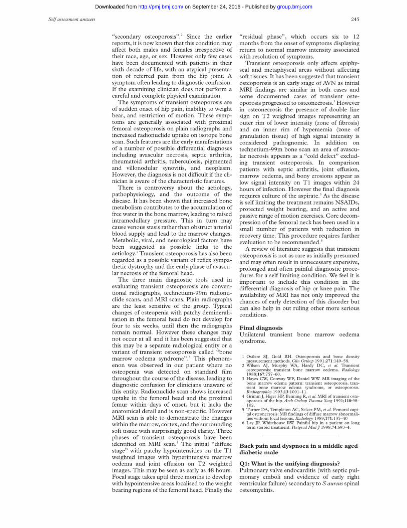
“secondary osteoporosis”.2 Since the earlierreports, it is now known that this condition mayaVect both males and females irrespective oftheir race, age, or sex. However only few caseshave been documented with patients in theirsixth decade of life, with an atypical presenta-tion of referred pain from the hip joint. Asymptom often leading to diagnostic confusion.If the examining clinician does not perform acareful and complete physical examination.
The symptoms of transient osteoporosis areof sudden onset of hip pain, inability to weightbear, and restriction of motion. These symp-toms are generally associated with proximalfemoral osteoporosis on plain radiographs andincreased radionuclide uptake on isotope bonescan. Such features are the early manifestationsof a number of possible diVerential diagnosesincluding avascular necrosis, septic arthritis,rheumatoid arthritis, tuberculosis, pigmentedand villonodular synovitis, and neoplasm.However, the diagnosis is not diYcult if the cli-nician is aware of the characteristic features.
There is controversy about the aetiology,pathophysiology, and the outcome of thedisease. It has been shown that increased bonemetabolism contributes to the accumulation offree water in the bone marrow, leading to raisedintramedullary pressure. This in turn maycause venous stasis rather than obstruct arterialblood supply and lead to the marrow changes.Metabolic, viral, and neurological factors havebeen suggested as possible links to theaetiology.1 Transient osteoporosis has also beenregarded as a possible variant of reflex sympa-thetic dystrophy and the early phase of avascu-lar necrosis of the femoral head.
The three main diagnostic tools used inevaluating transient osteoporosis are conven-tional radiographs, technetium-99m radionu-clide scans, and MRI scans. Plain radiographsare the least sensitive of the group. Typicalchanges of osteopenia with patchy deminerali-sation in the femoral head do not develop forfour to six weeks, until then the radiographsremain normal. However these changes maynot occur at all and it has been suggested thatthis may be a separate radiological entity or avariant of transient osteoporosis called “bonemarrow oedema syndrome”.3 This phenom-enon was observed in our patient where noosteopenia was detected on standard filmthroughout the course of the disease, leading todiagnostic confusion for clinicians unaware ofthis entity. Radionuclide scan shows increaseduptake in the femoral head and the proximalfemur within days of onset, but it lacks theanatomical detail and is non-specific. HoweverMRI scan is able to demonstrate the changeswithin the marrow, cortex, and the surroundingsoft tissue with surprisingly good clarity. Threephases of transient osteoporosis have beenidentified on MRI scan.4 The initial “diVusestage” with patchy hypointensities on the T1weighted images with hyperintensive marrowoedema and joint eVusion on T2 weightedimages. This may be seen as early as 48 hours.Focal stage takes uptil three months to developwith hypointensive areas localised to the weightbearing regions of the femoral head. Finally the
“residual phase”, which occurs six to 12months from the onset of symptoms displayingreturn to normal marrow intensity associatedwith resolution of symptoms.
Transient osteoporosis only aVects epiphy-seal and metaphyseal areas without aVectingsoft tissues. It has been suggested that transientosteoporosis is an early stage of AVN as initialMRI findings are similar in both cases andsome documented cases of transient oste-oporosis progressed to osteonecrosis.5 Howeverin osteonecrosis the presence of double linesign on T2 weighted images representing anouter rim of lower intensity (zone of fibrosis)and an inner rim of hyperaemia (zone ofgranulation tissue) of high signal intensity isconsidered pathognomic. In addition ontechnetium-99m bone scan an area of avascu-lar necrosis appears as a “cold defect” exclud-ing transient osteoporosis. In comparisonpatients with septic arthritis, joint eVusion,marrow oedema, and bony erosions appear aslow signal intensity on T1 images within 24hours of infection. However the final diagnosisrequires culture of the aspirate.6 As the diseaseis self limiting the treatment remains NSAIDs,protected weight bearing, and an active andpassive range of motion exercises. Core decom-pression of the femoral neck has been used in asmall number of patients with reduction inrecovery time. This procedure requires furtherevaluation to be recommended.5
A review of literature suggests that transientosteoporosis is not as rare as initially presumedand may often result in unnecessary expensive,prolonged and often painful diagnostic proce-dures for a self limiting condition. We feel it isimportant to include this condition in thediVerential diagnosis of hip or knee pain. Theavailability of MRI has not only improved thechances of early detection of this disorder butcan also help in out ruling other more seriousconditions.
Final diagnosisUnilateral transient bone marrow oedemasyndrome.
1 Ostlere SJ, Gold RH. Osteoporosis and bone densitymeasurement methods. Clin Orthop 1991;271:149–58.
2 Wilson AJ, Murphy WA, Hardy DC, et al. Transientosteoporosis: transient bone marrow oedema. Radiology1988;167:757–60.
3 Hayes CW, Conway WF, Daniel WW. MR imaging of thebone marrow edema pattern: transient osteoporosis, tran-sient bone marrow edema syndrome, or osteoporosis.Radiographics 1993;13:1001–11.
4 Grimm J, Higer HP, Benning R, et al. MRI of transient oste-oporosis of the hip. Arch Orthop Trauma Surg 1991;110:98–102.
5 Turner DA, Templeton AC, Selzer PM, et al. Femoral capi-tal osteonecrosis: MR findings of diVuse marrow abnormali-ties without focal lesions. Radiology 1989;171:135–40
6 Lay JP, Whitehouse RW. Painful hip in a patient on longterm steroid treatment. Postgrad Med J 1998;74:693–4.
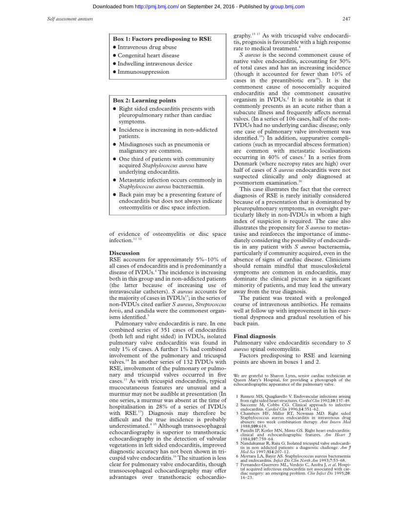
Back pain and dyspnoea in a middle ageddiabetic male
Q1: What is the unifying diagnosis?Pulmonary valve endocarditis (with septic pul-monary emboli and evidence of early rightventricular failure) secondary to S aureus spinalosteomyelitis.
Self assessment answers 245
group.bmj.com on September 24, 2016 - Published by http://pmj.bmj.com/Downloaded from

The spinal osteomyelitis is the likely sub-strate for the endocarditis rather than vice versaas symptoms of back pain preceded cardiopul-monary symptoms by several weeks.
It is unclear whether the initial presentationelsewhere was with septic pulmonary embolisecondary to pulmonary valve endocarditismimicking pneumonia, which was then par-tially treated, or whether he acquired S aureusbacteraemia secondary to an intravascularcatheter with subsequent development ofosteomyelitis and endocarditis: a murmur wasnoted at the initial presentation but one set ofblood cultures was negative.
The chest radiograph shows bilateral basalinfiltrates due to septic pulmonary emboli.Magnetic resonance imaging of the thoracicspine showed evidence of disc infection at theD9/D10 level, with abnormal enhancementextending into the paravertebral spaces anteri-orly and laterally. A transthoracic echocardio-gram shows a large vegetation attached to thepulmonary valve by a stalk. In addition,pulmonary artery pressure was raised at 45 mmHg, there was moderate tricuspid regurgitationand paradoxical septal motion.
Q2: How does right sided endocarditis(RSE) usually present?Presentation is usually with fever and symp-toms caused by septic pulmonary emboli,which occur in 60%–100% of patients.1 Theseinclude pleuritic chest pain (present in 60% ofintravenous drug users (IVDUs) with RSE2),cough, dyspnoea, and haemoptysis. The chestradiograph is abnormal in three quarters ofpatients at presentation.3 Abnormalities in-clude infiltrates, often multiple and appearingsequentially with a predilection for the lowerlobes, cavitation, pleural eVusion, andempyema.4 Tricuspid and pulmonary valvedysfunction is usually well tolerated haemody-namically so that pleuropulmonary rather thancardiac features usually dominate the presenta-tion.
While these features are likely to promptconsideration of the diagnosis in IVDUs, thediagnosis may not be considered in non-addicted patients. In one series of 29 non-IVDUs eventually proved to have RSE, anaverage of nine months elapsed between onsetof symptoms and diagnosis. Multiple admis-sions were common, positive blood culturestended to be either disregarded as contami-nants or ascribed to pulmonary events, andmisdiagnoses such as pneumonia, malignancy,and vasculitis were frequent.5
Q3: What risk factors are recognised forright sided endocarditis in non-IVDUs?The majority of these patients have chronicunderlying medical conditions.5 Predisposingfactors include congenital heart disease(whereas the majority of IVDUs with RSE havea structurally normal heart), inadequatelytreated sepsis, and indwelling vascular cath-eters (central lines, pacemakers, pulmonaryartery catheters, etc).
Q4: What is the likelihood of endocarditisin cases of S aureus bacteraemia?Eighty per cent of cases of S aureus bacterae-mia are community acquired. With no otherobvious primary source of bacteraemia, onethird of these patients have underlyingendocarditis.6 Nosocomially acquired casesare on the increase and usually occur in thecontext of intravenous cannulae/indwellingvascular devices.7 Factors known to increasethe chance of underlying endocarditis inpatients with S aureus bacteraemia includeabsence of an alternative primary site of infec-tion, community acquisition, and metastaticsequelae.8 The incidence of endocarditisresulting in intravenous catheter related Saureus bacteraemia is thought to be<5%–10%.6 However, many cases may beoverlooked: a recent study examining the util-ity of early transoesophageal echocardio-graphy in patients with S aureus bacteraemia(the majority of whom acquired it nosocomi-ally) has demonstrated that vegetations arefrequently identified in cases where endocardi-tis is not suspected clinically.9 These casesoften aVect the right side of the heart at sites ofendocardial injury induced by the catheters,commonly in the absence of pre-existingvalvular disease.7 In one series of 142 consecu-tively necropsied patients, 55 of whom hadundergone pulmonary artery catheterisationduring their final hospitalisation, right sidedendocardial lesions (subendocardial haemor-rhages and thrombi) were identified in overhalf of the 55 cases, with evidence of endo-carditis in 7%. Endocardial lesions werefound in only 3% of the non-catheterisedpatients.10
Q5: What is the relationship between backpain and endocarditis?Musculoskeletal symptoms are a common butunder-appreciated feature of endocarditis: inone series of 192 cases, 44% of patients hadmusculoskeletal manifestations (includingback pain, arthralgias, arthritis, and myalgias),and were the sole presenting feature in 15%. Inthe majority, musculoskeletal symptoms werepresent for some time before diagnosis andresponded rapidly to treatment of the endocar-ditis. The incidence of back pain in this serieswas 12.5%, with disc space infection identifiedin just under a quarter of these.11 In anotherseries of 108 patients with endocarditis, 30%had musculoskeletal manifestations withback pain an important feature in 13 of the108 (in whom four cases of vertebral osteomy-elitis were identified). In this study, patientspresenting with musculoskeletal symptomswere diagnosed significantly later and hada significantly higher mortality, presumablybecause of delays in initiation of treat-ment.12
Severe back pain may therefore be thepredominant presenting complaint in asignificant minority of cases of endocarditisand factors such as localised myositis, emboli,and immunological mechanisms have beencited to explain its presence in the absence
246 Self assessment answers
group.bmj.com on September 24, 2016 - Published by http://pmj.bmj.com/Downloaded from
of evidence of osteomyelitis or disc spaceinfection.11 12
DiscussionRSE accounts for approximately 5%–10% ofall cases of endocarditis and is predominantly adisease of IVDUs.4 The incidence is increasingboth in this group and in non-addicted patients(the latter because of increasing use ofintravascular catheters). S aureus accounts forthe majority of cases in IVDUs13; in the series ofnon-IVDUs cited earlier S aureus, Streptococcusbovis, and candida were the commonest organ-isms identified.5
Pulmonary valve endocarditis is rare. In onecombined series of 351 cases of endocarditis(both left and right sided) in IVDUs, isolatedpulmonary valve endocarditis was found inonly 1% of cases. A further 1% had combinedinvolvement of the pulmonary and tricuspidvalves.14 In another series of 132 IVDUs withRSE, involvement of the pulmonary or pulmo-nary and tricuspid valves occurred in fivecases.13 As with tricuspid endocarditis, typicalmucocutaneous features are unusual and amurmur may not be audible at presentation (Inone series, a murmur was absent at the time ofhospitalisation in 28% of a series of IVDUswith RSE.13) Diagnosis may therefore bediYcult and the true incidence is probablyunderestimated.4 15 Although transoesophagealechocardiography is superior to transthoracicechocardiography in the detection of valvularvegetations in left sided endocarditis, improveddiagnostic accuracy has not been shown in tri-cuspid valve endocarditis.16 The situation is lessclear for pulmonary valve endocarditis, thoughtransoesophageal echocardiography may oVeradvantages over transthoracic echocardio-
graphy.15 17 As with tricuspid valve endocardi-tis, prognosis is favourable with a high responserate to medical treatment.4
S aureus is the second commonest cause ofnative valve endocarditis, accounting for 30%of total cases and has an increasing incidence(though it accounted for fewer than 10% ofcases in the preantibiotic era18). It is thecommonest cause of nosocomially acquiredendocarditis and the commonest causativeorganism in IVDUs.2 It is notable in that itcommonly presents as an acute rather than asubacute illness and frequently aVects normalvalves. (In a series of 106 cases, half of the non-IVDUs had no underlying cardiac disease; onlyone case of pulmonary valve involvement wasidentified.19) In addition, suppurative compli-cations (such as myocardial abscess formation)are common with metastatic localisationsoccurring in 40% of cases.2 In a series fromDenmark (where necropsy rates are high) overhalf of cases of S aureus endocarditis were notsuspected clinically and only diagnosed atpostmortem examination.20
This case illustrates the fact that the correctdiagnosis of RSE is rarely initially consideredbecause of a presentation that is dominated bypleuropulmonary symptoms, an oversight par-ticularly likely in non-IVDUs in whom a highindex of suspicion is required. The case alsoillustrates the propensity for S aureus to metas-tasise and reinforces the importance of imme-diately considering the possibility of endocardi-tis in any patient with S aureus bacteraemia,particularly if community acquired, even in theabsence of signs of cardiac disease. Cliniciansshould remain mindful that musculoskeletalsymptoms are common in endocarditis, maydominate the clinical picture in a significantminority of patients, and may lead the unwaryaway from the true diagnosis.
The patient was treated with a prolongedcourse of intravenous antibiotics. He remainswell at follow up with improvement in his exer-tional dyspnoea and gradual resolution of hisback pain.
Final diagnosisPulmonary valve endocarditis secondary to Saureus spinal osteomyelitis.
Factors predisposing to RSE and learningpoints are shown in boxes 1 and 2.
We are grateful to Sharon Lynn, senior cardiac technician atQueen Mary’s Hospital, for providing a photograph of theechocardiographic appearance of the pulmonary valve.
1 Remetz MS, Quagliarello V. Endovascular infections arisingfrom right sided heart structures. Cardiol Clin 1992;10:137–49.
2 Saccente M, Cobbs CG. Clinical approach to infectiveendocarditis. Cardiol Clin 1996;14:351–62.
3 Chambers HF, Miller RT, Newman MD. Right sidedStaphylococcus aureus endocarditis in intravenous drugabusers: two week combination therapy. Ann Intern Med1988;109:619.
4 Panidis IP, Kotler MN, Mintz GS. Right heart endocarditis:clinical and echocardiographic features. Am Heart J1984;107:759–64.
5 Nandakumar R, Raju G. Isolated tricuspid valve endocardi-tis in non addicted patients: a diagnostic challenge. Am JMed Sci 1997;314:207–12.
6 Mortara LA, Bayer AS. Staphylococcus aureus bacteraemiaand endocarditis. Infect Dis Clin North Am 1993;7:53–68.
7 Fernandez-Guerrero ML, Verdejo C, Azofra J, et al. Hospi-tal acquired infectious endocarditis not associated with car-diac surgery: an emerging problem. Clin Infect Dis 1995;20:16–23.
Box 1: Factors predisposing to RSEx Intravenous drug abuse
x Congenital heart disease
x Indwelling intravenous device
x Immunosuppression
Box 2: Learning pointsx Right sided endocarditis presents with
pleuropulmonary rather than cardiacsymptoms.
x Incidence is increasing in non-addictedpatients.
x Misdiagnoses such as pneumonia ormalignancy are common.
x One third of patients with communityacquired Staphylococcus aureus haveunderlying endocarditis.
x Metastatic infection occurs commonly inStaphylococcus aureus bacteraemia.
x Back pain may be a presenting feature ofendocarditis but does not always indicateosteomyelitis or disc space infection.
Self assessment answers 247
group.bmj.com on September 24, 2016 - Published by http://pmj.bmj.com/Downloaded from

8 Bayer AS, Lam K, Ginzton L, et al. Staphylococcus aureusbacteraemia. Clinical, serological and echocradiographicfindings in patients with and without endocarditis. ArchIntern Med 1987;147:457–62.
9 Fowler VG Jr, Li J, Corey GR, et al. Role of echocardio-graphy in evaluation of patients with Staphylococcus aureusbacteraemia: experience in 103 patients. J Am Coll Cardiol1997;30:1072–8.
10 Rowley KM, Clubb KS, Walker Smith GJ, et al. Right sidedinfective endocarditis as a consequence of flow directed pulmo-nary valve catheterisation. N Engl J Med 1984;311:1152–6.
11 Churchill MA, Geraci JE, Hunder GG. Musculoskeletalmanifestations of bacterial endocarditis. Ann Intern Med1977;87:754–9.
12 Thomas P, Allal J, Bontoux D, et al. Rheumatological mani-festations of infective endocarditis. Ann Rheum Dis 1984;43:716–20.
13 Hecht SR, Berger M. Right sided endocarditis in intra-venous drug users. Ann Intern Med 1992;117:560–6.
14 Reisberg BE. Infective endocarditis in the narcotic addict.Prog Cardiovasc Dis 1979;22:193–204.
15 Shapiro SM, Young E, Ginzton LE, et al. Pulmonic valveendocarditis as an underdiagnosed disease: role of trans-oesophageal echocardiography. J Am Soc Echocardiogr 1992;5:48–51.
16 San Roman JA, Vilacosta I, Zamorano JL, et al. Tran-soesophageal echocardiography in right sided endocarditis.J Am Coll Cardiol 1993;21:1226–30.
17 Winslow T, Foster E, Adams JR, et al. Pulmonary valveendocarditis: improved diagnosis with biplane tran-soesophageal echocardiography. J Am Soc Echocardiogr1992;5:206–10.
18 Sanabria TJ, Alpert JS, Goldberg R, et al. Increasingfrequency of staphylococcal infective endocarditis. ArchIntern Med 1990;150:1305–9.
19 Watanakunakorn C. Staphylococcus aureus endocarditis ata community teaching hospital, 1980–1991. An analysis of106 cases. Arch Intern Med 1994;154:2330–5.
20 Espersen F, Frimodt B, Moller N. Staphylococcus aureusendocarditis: a review of 119 cases. Arch Intern Med1986;146:1118–21.
A man with abdominal pain
Q1: What is the unusual feature on theplain abdominal x-ray?The plain abdominal x-ray shows the presenceof hepatic-portal venous gas (HPVG) (fig 2).The distribution of gas is towards the peripheryof the liver, as opposed to gas in the biliary treewhich is central in distribution. Distendedloops of small bowel are also present.
Q2: What are the causes of this feature?The conditions in which HPVG has beenreported are listed in box 1.1 2
Q3: Is laparotomy indicatedThe patient underwent laparotomy, whichrevealed ischaemic bowel in the distribution ofthe superior mesenteric artery. Attempts torevascularise the bowel failed and the patientdied in the early postoperative period.
HPVG is not a specific disease entity and ismerely another diagnostic clue in patients withacute abdominal pathology. HPVG is associ-ated with various diverse conditions and itsimportance must be derived from the clinicalcontext of the patient.
Treatment is therefore directed at the under-lying cause. The majority of patients will haveischaemic bowel (72%) and will requireexploratory laparotomy. However, patientswith ulcerative colitis without other indicationfor laparotomy will not require exploration.3
DiscussionHPVG is an uncommon x-ray finding. It wasfirst described in infants in 19551 and in adultsin 1960.4 It appears as a branching radiolu-cency extending to within 2 cm of the livercapsule and x-rays are most revealing when
taken in the left lateral decubitus position. ThediVerence in distribution of gas in the biliarytree and portal venous gas is due to the fact thatgas in the biliary tract is carried towards theporta hepatis by the centripetal flow of the bileand therefore appears central in the liver whilethe HPVG travels peripherally enhanced by thecentrifugal flow of the blood.4
The factors that are thought to predispose toHPVG are5: mucosal damage, as seen in intes-tinal infarction, inflammatory bowel disease,and peptic ulcer disease; bowel distension as insmall bowel obstruction; and sepsis due tobowel necrosis, spreading cellulitis or intra-abdominal abscess, although these factors arenot identified in every patient.
With the increased use of computed tomog-raphy and ultrasound, HPVG has also beenreported from iatrogenic causes, most of whichwill not require laparotomy,2 eg, recent contrastenema, endoscopy, ERCP, and post-liver trans-plantation.
Final diagnosisHepatic-portal vein gas due to ischaemicbowel.
Figure 2 Abdominal x-ray.
Box 1: Reported causes of HPVGNon-iatrogenicx Necrotic bowel (72%)x Ulcerative colitis/Crohn’s disease (8%)x Intra-abdominal abscess (6%)x Bowel obstruction (3%)x Peptic ulcerx Acute haemorrhagic pancreatitisx Diabetic ketoacidosisx Necrotising enterocolitisx Unknown
Iatrogenicx Recent contrast enemax Endoscopy/ERCPx Recent liver transplantation
248 Self assessment answers
group.bmj.com on September 24, 2016 - Published by http://pmj.bmj.com/Downloaded from
1 Wolfe JN, Evans WA. Gas in the portal veins of infants: aroentgenographic demonstration with post-mortem ana-tomical correlation. AJR 1955;74:486–9.
2 Hong JJ, Gadaleta D, Rossi P, Esquivel J, Davis JM. Portalvein gas, a changing clinical entity. Arch Surg 1997;132:1071–5.
3 Traverso LW. Is hepatic portal venous gas an indication forexploratory laparotomy? Arch Surg 1981;116:936–8.
4 Susman N, Senturia HR. Gas embolisation of portal venoussystem. AJR 1960;83:847–50.
5 Liebman PR, Patten MT, Manny R, Benfield JR, HechtmanHB. Hepatic-portal venous gas in adults: etiology, patho-physiology and clinical significance. Ann Surg 1978;187:281–7.
A shot in the dark
Q1: What abnormalities aredemonstrated on the plain radiographs ofthe wrist?Radiograph of the left wrist (fig 1) shows an airgun pellet in the soft tissues of the forearm but,in addition, there is widening of the distal radialgrowth plate and irregularity, lucency and cup-ping of the radial and ulnar metaphysesconsistent with rickets. The carpal bone age isdelayed, at approximately 4 years, but a radio-graph of the left hand showed a dissociationbetween the carpal bone age and that of theremainder of the hand. This can occur due tointercurrent illness and, in isolation, is notindicative of globally delayed skeletal matura-tion. The diVerential diagnosis of the wristradiograph includes rickets secondary to vita-min D deficiency, an abnormality of vitamin Dmetabolism, target cell abnormalities, includ-ing renal tubular disorders, or tumour-inducedrickets, and metaphyseal chondrodysplasias(box 1).
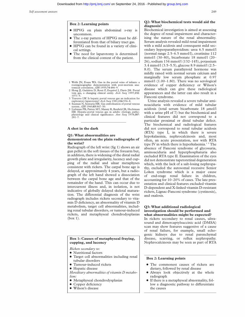
Q2: What biochemical tests would aid thediagnosis?Biochemical investigation is aimed at assessingthe degree of renal impairment and character-ising the nature of the renal abnormality.Serum analysis revealed mild renal impairmentwith a mild acidosis and consequent mild sec-ondary hyperparathyroidism: urea 6.5 mmol/l(normal range 2.5–6.5 mmol/l), creatinine 112mmol/l (30–80), bicarbonate 18 mmol/l (20–26), sodium 134 mmol/l (132–145), potassium3.4 mmol/l (3.5–5.5), glucose 8.9 mmol/l (2.5–8.0). The serum parathyroid hormone wasmildly raised with normal serum calcium andmarginally low serum phosphate at 0.97mmol/l (1.00–1.80). There was no serologicalevidence of copper deficiency or Wilson’sdisease which can give these radiologicalappearances and the latter can also result in aFanconi syndrome.
Urine analysis revealed a severe tubular ami-noaciduria with evidence of mild tubularacidosis (total serum bicarbonate 18 µmol/lwith a urine pH of 7) but the biochemical andclinical features did not correspond to aparticular proximal or distal tubular defect.The biochemical and radiological featuresdid not correspond to renal tubular acidosis(RTA) type I, in which there is severehypokalaemia, nephrocalcinosis and, mostoften, an acute presentation, nor with RTAtype IV in which there is hyperkalaemia.1 2 Theabsence of Fanconi syndrome of glycosuria,aminoaciduria and hyperphosphaturia alsoexcluded RTA type II. Examination of the eyesdid not demonstrate tapetoretinal degenerationwhich, with the lack of a salt-losing nephropa-thy, excluded the autosomal recessive SeniorLoken syndrome which is a major causeof end-stage renal failure in children,accounting for 10–20% of cases. The late pres-entation and clinical features excluded vitaminD-dependent and X-linked vitamin D-resistantrickets, Lignac-Fanconi syndrome (cystinosis),and oxalosis.
Q3: What additional radiologicalinvestigation should be performed andwhat abnormalities might be expected?In rickets secondary to renal causes, ultra-sound and dimercaptylsuccinic acid (DMSA)scan may show features suggestive of a causeof renal failure, for example, small echo-genic kidneys due to renal parenchymaldisease, scarring, or reflux nephropathy.Nephrocalcinosis may be seen as part of RTA
Box 2: Learning points
x HPVG on plain abdominal x-ray isuncommon.
x The x-ray pattern of HPVG must be dif-ferentiated from that of biliary tract gas.
x HPVG can be found in a variety of clini-cal settings.
x The need for laparotomy is determinedfrom the clinical context of the patient.
Box 1: Causes of metaphyseal fraying,cupping, and lucency
Rickets secondary to:x Nutritional factorsx Target cell abnormalities including renal
tubular disordersx Tumour-induced ricketsx Hepatic diseaseHereditary abnormalities of vitamin D metabo-lism:x Metaphyseal chondrodysplasiasx Copper deficiencyx Wilson’s disease
Box 2: Learning points
x The commonest causes of rickets aredietary, followed by renal disease
x Always look objectively at the wholeradiograph
x If there is a metaphyseal abnormality, fol-low a diagnostic pathway to diVerentiatethe causes
Self assessment answers 249
group.bmj.com on September 24, 2016 - Published by http://pmj.bmj.com/Downloaded from

type I. In our patient, ultrasound showed thatthe kidneys were small for age (below the 5thcentile), and mildly echogenic, with poorcortico/medullary diVerentiation but no defi-nite evidence of nephrocalcinosis. The findingof a renal abnormality in conjunction with theresults of serum and urine biochemistryexcluded the diagnosis of rickets due tohepatic or nutritional disorders of vitamin Dmetabolism and also excluded primary meta-physeal chondrodysplasias.
A DMSA scan was performed to see if thecause of the small echogenic kidneys wasreflux nephropathy. The scan showed almostno uptake of DMSA by the kidneys (fig 2).This can be due to inability to bind DMSAwithin the kidney or occasionally due to poorlyfunctioning kidneys where the serum creati-nine is over 300 mmol/l, ie, much higher thanin this case. DMSA binds to plasma proteinand, intracellularly, to cytosol proteins andmitochondria. Uptake occurs principally inthe cells of the proximal tubules. Non-visualisation of the kidneys with DMSA is,therefore, an indication of the functioningstate of the tubules. It was therefore concludedthat these findings were indicative of mildchronic renal failure due to renal dysplasia inwhich there was a tubular defect leading tosevere aminoaciduria and therefore, poorDMSA binding. It was not possible to excludescarring on a DMSA scan with such poorDMSA uptake.
DiscussionIn chronic renal disease, metaphyseal abnor-mality results from failure of the kidneys tohydroxylate 25-hydroxy-vitamin D3 and di-minishing capacity to excrete phosphate. Thisstimulates production of parathyroid hor-
mone, osteoclastic activity and bone resorp-tion, resulting in temporary correction ofserum calcium and phosphate.3 The serumcalcium and phosphorus may be maintainedwithin the normal range until the glomerularfiltration rate goes below 25–30 ml/min whenpersistent hyperphosphataemia occurs. Thegrowth plates, whilst appearing widened onplain radiographs, are actually narrowed whenexamined histologically with a region of ostei-tis fibrosa generalisata adjacent to the growthplate. This gives the widened growth plateappearance that simulates rickets. The growthplates are weak and susceptible to fractureresulting in a slipped epiphysis, although this isnot usually seen until 11–16 years of age.Unlike renal osteodystrophy in adults, it isunusual in children to see phalangeal subpe-riosteal erosion and soft tissue and arterialcalcification.
Management is aimed at reducing thedegree of hyperparathyroidism, improvingcalorie intake and long-term monitoring ofrenal impairment. This case illustrates theimportance of keeping an open mind whenreporting trauma radiographs and a diagnosticpathway for evaluating metaphyseal abnormal-ity.
Final diagnosisRenal rickets secondary to non-specific renalprotein-losing tubular abnormality.
1 Thomas PS. Metabolic bone disorders. In: Carty H, ShawD, Brunelle F, Kendall B, eds. Imaging children. New York:Churchill Livingstone, 1994:992–1011.
2 Taybi H. Radiology of syndromes and metabolic disorders.Chicago: Year Book Medical Publishers, 1983; pp 338–339.
3 Sundaram M. Renal osteodystrophy. Skeletal Radiol 1989;18:363–7.
Pyrexia and pancytopenia with unusualhost immune response
Q1: What cytomorphologicalphenomenon is shown in fig 1?Figure 1 shows a proliferation of benign histio-cytes in the bone marrow smears. They haveabundant foamy to granular cytoplasm andmany of them contain phagocytosed bloodcells and their precursors. This cytomorpho-logical phenomenon is called haemophagocytichistiocytosis and can be seen in the bone mar-row, lymph nodes, spleen and liver in a varietyof clinical settings.
Q2: List the conditions with which thiscytomorphological phenomenon may beassociatedThe conditions associated with haemophago-cytic histiocytosis are listed in box 1.
Q3: What is the diagnosis in this case?Figure 2 shows greater detail of the histiocyte.Numerous 2–4 µm round to oval bodies witheccentric to central nuclei and a perinuclearvacuole are identified within the cytoplasm.These were morphologically identified, onsilver methenamine and periodic acid SchiV
Figure 2 DMSA scan of the kidneys.
250 Self assessment answers
group.bmj.com on September 24, 2016 - Published by http://pmj.bmj.com/Downloaded from

stain, as yeast forms of histoplasma. Figure2(A) also shows phagocytosed red cell precur-sors.
DiscussionHaemophagocytic syndrome is a clinicopatho-logical state characterised by fever, blood cyto-penias, coagulopathy, hepatosplenomegaly anda systemic proliferation of mature histiocytesshowing phagocytosis of blood cells.1 Thediagnostic guidelines as laid down by theHistiocytic Society1 are shown in box 2.Haemophagocytosis (HP) is a disorder of thecellular arm of immune response occurring as aconsequence of unrestricted monocyte activa-tion that results from marked elevation ofinflammatory cytokines produced by dysregu-lated reactive or neoplastic T lymphocytes.2
Profound cytopenias, which may result fromhaemophagocytosis or associated coagulopa-thy, may seriously compromise the alreadymorbid patient.
First described as a familial disease byFarquhar and Claireaux in 1952,3 haemo-phagocytic syndrome has subsequently beenreported to occur in a host of other clinical set-tings (box 2)5–9; it is also called secondary orreactive haemophagocytosis.5 6 Haematologicalmalignancies, especially T cell lymphomas,have been reported as the most frequent causeof reactive HP in most studies.7 Infections arean important association in tropical countries,in the oriental population6 and, with increasingincidence of AIDS, in the West as well.8 9
Histoplasmosis has been sporadically re-ported from diVerent parts of India. The com-monest manifestation in the Indian populationis involvement of skin and mucous mem-branes, with or without systemicinvolvement.10 The organism has been identi-fied in various sites, including skin, mucousmembranes, blood, bone marrow, lymphnodes, and sputum.10 However, HP in a settingof disseminated histoplasmosis has, to the bestof our knowledge, not been described previ-ously from India. The few reported cases inEnglish literature have occurred mostly in
patients with AIDS.8 9 In the wake of analarming increase in the number of AIDScases in the sub-continent, the importance ofcareful laboratory examination in the diagno-sis of histoplasmosis cannot be over-emphasised, particularly because, given thehigh prevalence of tuberculosis in India, casesof pulmonary or disseminated histoplasmosismay be misdiagnosed as tuberculosis. Pres-ence of severe cytopenias in a patient withprolonged pyrexia should alert the physician tothe possibility of HP and a meticulous searchin the bone marrow and other reticulo-endothelial sites should be made for theunderlying cause of HP. An early diagnosis ofinfection-associated reactive HP is crucial inthe management of this potentially reversiblecondition.
Final diagnosisDisseminated histoplasmosis with reactive hae-mophagocytosis.
1 Henter JI, Elinder G, Ost A, et al. Diagnostic guidelines forthe hemophagocytic lymphohistiocytosis. Semin Oncol 1991;18:29–33.
2 Favara BE. Hemophagocytic lymphohistiocytosis: a hemo-phagocytic syndrome. Semin Diagn Pathol 1992;9:63–74.
3 Farquhar JW, Claireaux AX. Familial haemophagocyticreticulosis. Arch Dis Child 1952;27:519–25.
4 Risdall RJ, McKenna RW, Nesbit ME, et al. Virus associatedhemophagocytic syndrome. Cancer 1979:44:993–1002.
5 Arya S, Hong R, Gilbert EF. Reactive hemophagocticsyndrome. Pediatr Pathol 1985;3:129–41.
6 Wong KF, Chan JKC. Reactive hemophagocytic syndrome.A clinicopathological study of 40 patients in an orientalpopulation. Am J Med 1992;93:177–80.
7 Linn YC, Tien SL, Lim LC, et al. Hemophagocytosis inbone marrow aspirate. A review of the clinical course of 10cases. Acta Haematol 1955;94:182–91.
8 Koduri PR, Chundi V, De Marais P, Mizock BA, Patel AR,Weinstein RA. Reactive hemophagocytic syndrome: a newpresentation of disseminated histoplasmosis in patients withAIDS. Clin Infect Dis 1995;21:1463–5.
9 Kurtin PJ, McKinsey DS, Gupta MR, Dirks M. Histoplas-mosis in patients with acquired immunodeficiencysyndrome—hematologic and bone marrow manifestations.Am J Clin Pathol 1990;93:367–72.
10 Mukherjee AK, Mukherjee D, Mukopadhyay M. Histo-plasmosis in India. A clinicopathological review with reportof a case in a child. Indian J Pathol Microbiol 1986;29:263–70.
Box 1: Clinical states associated withhaemophagocytosis
Primaryx Familial haemophagocytic lymphohistio-
cytosisx Virus-associated haemophagocytosis
(with no prior immunodeficiency)
Secondary/reactivex Malignancies, especially T cell lympho-
masx Acquired and hereditary immuno-
deficiencyx Parenteral fat overloadx Panniculitisx Bacterial, parasitic and fungal infections
(tuberculosis, typhoid, brucellosis, ma-laria, leishmaniasis, histoplasma, etc)
Box 2: Diagnostic guidelines forhaemophagocytic lymphohistiocytosis1
Clinical and laboratory criteriax Fever (>7 days; peaks >38.5oC)x Splenomegaly (>3 cm below costal mar-
gin)x Cytopenia (aVecting >2 of three lineages
in the peripheral blood not caused byhypocellular or dysplastic bone marrow)
x Haemoglobin < 9 g/dlx Platelets < 100 × 109/lx Neutrophils < 1.0 × 10⁹/lx Hypertriglyceridaemia and/or hypofi-
brinogenaemia*
Morphological criteriax Haemophagocytosis in bone marrow or
lymph nodes or spleen
*Some of these guidelines may not apply toa secondary haemophagocytic state2
Self assessment answers 251
group.bmj.com on September 24, 2016 - Published by http://pmj.bmj.com/Downloaded from

Abdominal colic after vigorous exercisein a middle aged man
Q1: What is the diagnosis?The diagnosis is coeliac band with coeliacartery stenosis causing chronic intestinal is-chaemia. This is such a rare clinical conditionthat even institutions with an enormousexperience in peripheral vascular surgery haveaccumulated some experience over severalyears. It is only by reflex consideration of thiscondition after excluding other common diag-nosis that the alert surgeon will reach this diag-nosis.
Q2: What are the diVerential diagnoses inthe patient?The main diVerential diagnoses include me-senteric angina that may be secondary toatherosclerosis, Buerger’s disease, fibromuscu-lar dysplasia, and arteritis such as Takayasu’sdisease. Other possibilities include renal andbiliary colic.
Q3: How would you establish thediagnosis?This patient’s presentation was atypicalof common abdominal conditions and theroutine investigations were normal. With ahistory such as this, atherosclerotic disease ofthe coeliac/mesenteric arteries causing chronicmesenteric ischaemia was a possibility. Meas-uring the erythrocyte sedimentation ratewould help to rule out an inflammatory cause.A duplex scan would be performed as the firststep of vascular imaging as this is non-invasive.1 However because of their anatomicallocation, assessment of visceral arterial lesionsby duplex scan is not as reliable as it is in othersituations. Contrast computed tomography(fig 1) or magnetic resonance angiographyof the coeliac/mesenteric arteries, providebetter information including detection of anycoexisting extrinsic pathology. Finally thedefinitive investigation is an angiogram,which in this case clearly demonstratedpathology at the origin of the coeliac artery.This was left until last due to its invasivenature and the risks, although rare, associatedwith it.
Q4: What do the investigations illustrate?The angiogram demonstrates a tight stenosis(lateral film) (fig 2) at the origin of the coeliacartery. This may be due to extrinsic or intrinsicfactors or a combination of both; the superiormesenteric artery (SMA) is of normal calibre.The inferior mesenteric artery (IMA) is notseen.
Q5: What are the treatment optionsavailable?Treatment options should be based on riskversus benefit for each procedure. In thispatient median arcuate ligament (coeliacband) syndrome had to be ruled out as an
extrinsic cause of compression. The risk ofreleasing the band is much less than arterialreconstruction. At operation he was foundto have a coeliac band. This was divided,however a thrill over the coeliac arterypersisted. This indicated the presence of dualpathology, with stenosis of the coeliac arterybeing also present. In this case the stenosis waslocalised to the proximal portion of the coeliacartery; arterial reconstruction between theaorta and hepatic artery was seen as the besttreatment option. The SMA was of normalcalibre. The IMA was found to be small andcould possibly have contributed to thepatient’s symptoms as the collateral circulationhad failed to develop. A reversed longsaphenous vein graft was anastomosed be-tween the infra renal aorta and the hepaticartery and the graft was tunnelled behind thepancreas.
Aortocoeliac grafting is an option, howeverdue to the short course of the coeliac artery, theprocedure is often diYcult. Another option isto reimplant the coeliac artery into the aorta atanother site; this is particularly suitable if thereis a tight proximal stenosis with an aortic com-ponent.2 Angioplasty with or without stentingof the mesenteric vessels is a further option, therisk being distal embolisation and dissectionwith acute thrombosis causing ischaemia orinfarction.
When reviewed in the clinic at three monthsand six months the patient was completely freefrom symptoms and has returned back to hisnormal exercise programme. Contrast com-puted tomography performed at three monthsconfirmed the patency of the aortohepaticgraft.
DiscussionIt has been reported in the literature thatup to 33% of the population have ananatomical variation in the structure of thecoeliac axis.3 Such anomaly seldom givesrise to symptoms of chronic intestinalischaemia. The coeliac axis, SMA, and IMAalong with the iliacs provide the main bloodsupply to the gastrointestinal tract. Due to thepresence of a good collateral circulation,stenosis usually has to be of a severe degreebefore symptoms occur. This can be graded bymeasuring the pressure gradient across thestenotic lesion. Stenoses likely to compromisethe collateral circulation may becomeclinically manifested during the postprandialphase or during muscular exertion. It hasbeen also demonstrated that mesentericvascular resistance increases during exerciseand produces a marked decrease in arterialinflow.4
Chronic gastrointestinal ischaemia isclassically described as presenting with post-prandial abdominal pain (termed visceralangina), weight loss, and an epigastric bruit(quoted in over 80% of cases). Lord et al, intheir series of 12 patients with coeliac axiscompression, noted that 75% (9) of theirpatients had atypical abdominal pain, 58% (7)of the patients had slight weight loss,
252 Self assessment answers
group.bmj.com on September 24, 2016 - Published by http://pmj.bmj.com/Downloaded from

and only 42% (5) had history ofdiarrhoea.5 The presentation in this patientwas unusual in that he had no postprandialsymptoms, but pain was induced byheavy exercise perhaps due to redistributionof blood flow. In the presence of coeliacartery stenosis and a small IMA the collateralblood flow to the gut through the internal iliacarteries was possibly diverted to the lowerlimbs during exercise. Since the SMA was pat-ent, the patient had very little symptoms ofweight loss, diarrhoea, or postprandial symp-toms.
Final diagnosisCoeliac band and coeliac artery stenosis.
1 Perko MJ. Importance of diastolic velocities in detection ofceliac and mesenteric artery disease by duplex ultrasound. JVasc Surg 1997;26:288–93.
2 Takach TJ. Celiac compression syndrome: tailored therapybased on intraoperative findings. J Am Coll Surg 1996;183:606–10.
3 Lindner HH, Kemprud E. A clinicoanatomical study of thearcuate ligament of the diaphragm. Arch Surg 1971;103:600–5.
4 White S, Patrick T, Higgins CB. EVects of altering ventricu-lar rate on blood flow distribution in conscious dogs. Am JPhysiol 1971;221:1402.
5 Lord RSA, Sydney MD, Stoney RJ, et al. Coeliac-axis com-pression. Lancet 1968;ii:795–8.
International Postgraduate Diary
23rd European Conference on Psychoso-matic Research
17–21 June 2000: Oslo, NorwayDetails: Congress-Conference AS - CON-GREX, Thomas Heftyes gt. 2, PO Box 2694Solli, N-0204 Oslo, Norway. Tel + 47 (0)2256 1930; fax + 47 (0) 2256 0541; email:[email protected]
Falk Symposia4–6 May 2000: Hepatology 2000 (Munich,Germany)9/10 June 2000: Cholestasis and gallstones(Cluj Napoca, Romania)
1/2 October 2000: Non-neoplastic diseases ofthe anorectum—an interdisciplinary ap-proach (Freiburg, Germany)3/4 October 2000: Immunosuppression ininflammatory bowel diseases—standards,news, and future trends (Freiburg, Germany)
12/13 October 2000: Biology of bile acids inhealth and disease (Den Haag, The Nether-lands)
4 November 2000: Chronic inflammatory boweldiseases—progress and controversies at theturn of the century (Bucharest, Romania)
Details: Falk Foundation eV—Congress Divi-sion, Leinenweberstr 5, PO Box 6529,D-79041 Freiburg, Germany. Tel +49 (0) 761130340; fax +49 (0) 761 1303459; email:[email protected]
Columbia University College of Physi-cians and Surgeons, New York
5/6 May 2000: 12th Annual orthopaedictrauma course. Current techniques in upper& lower extremity trauma
22–25 May 2000: 4th Annual conference.Botanical medicine in modern clinical prac-tice
28–31 July 2000: 10th Annual corse. A com-prehensive review of movement disorders forthe clinical practitioner
30 July–5 August 2000: 5th Annual course.Update and intensive review in internalmedicine
Details: Center for Continuing Education,Columbia University College of Physiciansand Surgeons, 630 West 168th Street, Unit39, New York, NY10032, USA. Tel + 1 212781 5990; fax + 1 212 781 6047; email:[email protected]
Ninth International Symposium on ce-liac disease
10–13 August 2000: Hunt Valley, MD, USA
Details:Althea Pusateri, Program Coordinator,University of Maryland School of Medicine,655 W Baltimore Street, Baltomore, MD21201, USA Tel +1 410 706 3957; fax +1 410706 3103; http://www.celiaccenter.org
Self assessment answers 253
group.bmj.com on September 24, 2016 - Published by http://pmj.bmj.com/Downloaded from

immune responsePyrexia and pancytopenia with unusual host
Zeba Niazi Singh, Shyama Jain, S K Jain and Sudha Rani
doi: 10.1136/pmj.76.894.241a2000 76: 241-242 Postgrad Med J
http://pmj.bmj.com/content/76/894/241.2Updated information and services can be found at:
These include:
serviceEmail alerting
box at the top right corner of the online article. Receive free email alerts when new articles cite this article. Sign up in the
Notes
http://group.bmj.com/group/rights-licensing/permissionsTo request permissions go to:
http://journals.bmj.com/cgi/reprintformTo order reprints go to:
http://group.bmj.com/subscribe/To subscribe to BMJ go to:
group.bmj.com on September 24, 2016 - Published by http://pmj.bmj.com/Downloaded from