Animal cells as host
22
27 Animal cells as host Vijay Kumar of the production processes. Biopharmaceutical indus- tries are currently facing the challenge of producing biological recombinant medical products in a quick and cost-effective manner, while complying with the safety requirements set by the regulatory agencies. Mamma- lian cell lines, particularly the Chinese Hamster Ovary (CHO) cell line, are the eukaryotic systems of choice for many biopharmaceutical industries for large-scale pro- duction of recombinant proteins and mAbs due to their ability to grow in high-density suspension cultures and to produce proteins with human-likePTMs. However, the lengthy, expensive and tortuous path for approval of a new therapeutic candidate has a significant bearing on the timeline and economics of process development. Now-a-days, animal cell biotechnology has advanced to the stage where it is used in a variety of manufacturing processes. In fact, products derived from animal cell cultures are reported to generate nearly half of the rev- enues from the sale of modern biotechnology products. This chapter provides a historical perspective of ani- mal cell culture and describes the commonly used meth- ods, equipment, supplies and reagents in this process. The key steps required to maintain adherent cell lines and suspension-grown cells, their long-term cryogenic storage and revival from frozen stocks are outlined. More important, different methods of gene transfer in animal cells, their selection and scale-up culture for the production of recombinant proteins and methods used in their quality control are elaborated. 27.2 Some milestones in the development of tissue and cell cultures and their use in recombinant DNA technology The concept of maintaining live animal cell lines sepa- rated from their original tissue source was discovered in the early 20th century and animal cell culture became a 27.1 Introduction Recombinant DNA technology or genetic engineer- ing was developed in the 1970s to express mammalian genes in bacteria. However, it soon became apparent that large complex proteins, especially those of thera- peutic value, could not be produced in bacteria, as they do not have the appropriate cellular machinery to make appropriate post-translational modifications (PTMs) in these proteins. Therefore, genetically engineered ani- mal cells were developed for large-scale commercial production of such important proteins. Now-a-days, animal cells are used such asbacteria or yeast to pro- duce a variety of recombinant proteins of human ori- gin, including secreted proteins such as monoclonal antibodies (mAb) and growth factors, intracellular pro- teins such as enzymes, and membrane proteins such asgrowth factor receptors. The mammalian cells offer a distinct advantage of making recombinant proteins with fully humanized PTMs which drastically improve their biological activity and minimize their immuno- genicity. Such proteins can remain in body circulation for an extended period to produce desirable pharmaco- logical effects. Animal cell culture is a process of growing animal cells in vitro in a flask, dish or fermenter under a strictly con- trolled environment. Because maintaining the purity of cell cultures and preventing their contamination con- stitute the greatest challenge in animal cell culture, the principles of aseptic technique followed in cell culture are of paramount importance. Optimized animal cell culture has the ability to deliver sufficient quantities of recombinant products, and thus, it constitutes an important activity of the present-day biopharmaceu- tical industries. The range of commercially available recombinant pharmaceuticals produced by cell culture technologies has increased rapidly over the past few years. This is particularly so for therapeutic proteins syn- thesized from selected or genetically engineered mam- malian cells. They are needed in large quantities and hence require careful study of the underlying principles Chapter 27.indd 1 6/23/2014 2:39:07 PM
Transcript of Animal cells as host

27Animal cells as host Vijay Kumar
of the production processes. Biopharmaceutical indus-tries are currently facing the challenge of producing biological recombinant medical products in a quick and cost-effective manner, while complying with the safety requirements set by the regulatory agencies. Mamma-lian cell lines, particularly the Chinese Hamster Ovary (CHO) cell line, are the eukaryotic systems of choice for many biopharmaceutical industries for large-scale pro-duction of recombinant proteins and mAbs due to their ability to grow in high-density suspension cultures and to produce proteins with human-likePTMs. However, the lengthy, expensive and tortuous path for approval of a new therapeutic candidate has a significant bearing on the timeline and economics of process development. Now-a-days, animal cell biotechnology has advanced to the stage where it is used in a variety of manufacturing processes. In fact, products derived from animal cell cultures are reported to generate nearly half of the rev-enues from the sale of modern biotechnology products.
This chapter provides a historical perspective of ani-mal cell culture and describes the commonly used meth-ods, equipment, supplies and reagents in this process. The key steps required to maintain adherent cell lines and suspension-grown cells, their long-term cryogenic storage and revival from frozen stocks are outlined. More important, different methods of gene transfer in animal cells, their selection and scale-up culture for the production of recombinant proteins and methods used in their quality control are elaborated.
27.2 Some milestones in the development of tissue and cell cultures and their use in recombinant DNA technology
The concept of maintaining live animal cell lines sepa-rated from their original tissue source was discovered in the early 20th century and animal cell culture became a
27.1 Introduction
Recombinant DNA technology or genetic engineer-ing was developed in the 1970s to express mammalian genes in bacteria. However, it soon became apparent that large complex proteins, especially those of thera-peutic value, could not be produced in bacteria, as they do not have the appropriate cellular machinery to make appropriate post-translational modifications (PTMs) in these proteins. Therefore, genetically engineered ani-mal cells were developed for large-scale commercial production of such important proteins. Now-a-days, animal cells are used such asbacteria or yeast to pro-duce a variety of recombinant proteins of human ori-gin, including secreted proteins such as monoclonal antibodies (mAb) and growth factors, intracellular pro-teins such as enzymes, and membrane proteins such asgrowth factor receptors. The mammalian cells offer a distinct advantage of making recombinant proteins with fully humanized PTMs which drastically improve their biological activity and minimize their immuno-genicity. Such proteins can remain in body circulation for an extended period to produce desirable pharmaco-logical effects.
Animal cell culture is a process of growing animal cells in vitro in a flask, dish or fermenter under a strictly con-trolled environment. Because maintaining the purity of cell cultures and preventing their contamination con-stitute the greatest challenge in animal cell culture, the principles of aseptic technique followed in cell culture are of paramount importance. Optimized animal cell culture has the ability to deliver sufficient quantities of recombinant products, and thus, it constitutes an important activity of the present-day biopharmaceu-tical industries. The range of commercially available recombinant pharmaceuticals produced by cell culture technologies has increased rapidly over the past few years. This is particularly so for therapeutic proteins syn-thesized from selected or genetically engineered mam-malian cells. They are needed in large quantities and hence require careful study of the underlying principles
Chapter 27.indd 1 6/23/2014 2:39:07 PM

2 CHAPTER 27 Animal cells as host
cofactors, carbohydrates, and horse serum. By elimi-nating one component at a time, he then determined which nutrients were essential for cell growth. His minimum essential medium (MEM) contains 13 amino acids (human tissue in vivo requires only eight), eight-vitamins and cofactors, glucose as an energy source and a physiological salt solution that is isotonic to the cell. The pH is maintained at 7.2–7.4 by NaHCO
3 in equi-
librium with CO2. The pH indicator phenol red is usu-
ally incorporated into the medium; it turns red-purple if the medium is basic, yellow if the medium is acidic, and remains red-orange if the pH is in the right range. Serum in concentrations of 1–10% must be added to the medium to provide the cells with additional poorly defined factors, without which most cells will not grow. Most mammalian cells are incubated at 37oC. However, the avian, reptilian and arthropod cells may grow opti-mally at higher or lower temperatures. CMRL-1066 – a chemically defined medium – was developed in the late 1950s by Connaught Medical Research Laboratories. Later, Richard Ham (1965) introduced a defined, serum-free medium able to support the clonal growth of certain mammalian cells. Cherry and Hull (1956) used a mag-netic stir bar suspended from a fishing swivel to keep cells in suspension in a round bottom flask so that they could study the effects of varying the medium, speed and seeding densities on cell growth. McLimans and his group (1957) took the magnetic stir bar and attached it to a sliding wire so that it could be raised or lowered to match the medium volume. They added a side port for sampling and called it a ‘spinner flask’, which was used to grow 400 mL suspension cultures of HeLa and L cells. They adapted this approach to work in commercially available 5L microbial fermenter with baffles and sparg-ers driven by an overhead impeller and showed that sus-pension animal cell culture could be successfully scaled up to grow products at commercial level. As this method was not suitable for anchorage-dependent cell cultures, AL van Wezel (1967) resolved this issue by using small porous beads called microcarriers (200–250-µm diam-eter) as substances for cell attachment. These particles are made from a variety of materials, including polysty-rene, dextran, glass and cross-linked collagen. The use of these beads in culture required the use of more gentle stirring techniques, and by the 1980s, spinner vessels of different designs were available to minimize the dam-age caused by bead–bead collisions and sheer effects. Today, a number of therapeutic protein drugs and vac-cines are produced from cells grown in suspension on microcarriers.
Frank Graham and Alex van der Eb (1973) devel-opedthe famous calcium–phosphate method of DNA transfection into mammalian cells. The viral method of gene delivery was introduced by Paul Berg (1976), who used a modified SV40 virus containing DNA from the bacteriophage lambda to infect monkey kidney cells in culture. Manual microinjection of DNA into nuclei of
routine laboratory technique by 1950s. The earliest tis-sue culture experiment dates back to 19th century when Sydney Ringer – an English physiologist – developed ‘salt solutions’ containing chlorides of sodium, potassium, calcium and magnesium for maintaining isolated ani-mal hearts outside the body in culture. In 1885, Wilhelm Roux – a German embryologist – introduced ‘saline cul-ture’ by successfully cultivating the medullary plate of a chick embryo in a warm saline solution for several days and, thus, opened the roads for tissue culture. Between 1907 and 1910, Ross Harrison from Johns Hopkins Med-ical School published a series of papers describing the methods of tissue culture. He used frog embryonic tis-sue to grow nerve fibers in the presence of frog lymph by the hanging-drop method. In 1911, Alexis Carrel and Montrose Burrows used horse plasma to grow explants of chick heart tissue and introduced strict aseptic tech-niques so that cells could be cultured for long periods. The development of antibiotics during World War II simplified long-term animal cell culture by minimizing the problems of microbial contamination in cell culture. Peyton Rous and FS Jones (1916) introduced the pro-teolytic enzyme trypsin for the subculture of adherent cells. In 1950s, Wilton Earle and his colleagues stand-ardized the technique to dissociate cells from growing chick embryo with the help of trypsin. He observed that when a single-cell suspension was mixed with plasma and embryo extract and transferred in a sterile glass container, the cells adhered to the glass and divided to form a monolayer of primary culture that covered the entire bottom surface of the vessel and then stopped dividing. The primary culture that contained a variety of cell types could then be re-dispersed with trypsin and planted in new culture vessels containing fresh medium. Often, these secondary cultures were composed entirely of spindle-shaped cells called fibroblasts because of their resemblance to cultured connective tissue. Earle also isolated mouse L fibroblasts, which formed clones from single cells. In 1952, George Gey and colleagues established a cell line derived from a human cervical carcinoma, which later became the well-known HeLa cell line, while the first successful suspension cultures, lymphoblastic MBIII cells, were grown by Owens, Gey and Gey(1953). In 1981, the teams led by Martin Evans and Gail Martin first derived embryonic stem (ES) cells from mouse embryos, which laid the foundation of ‘gene knock-out technology’. Subsequently, the teams led by James Thomson and John Gearhart established human ES cells in 1998 that are extremely useful in regenerative medicine.
In 1955,Harry Eagle made the first systematic inves-tigation of the essential nutritional requirements of cultured cells and reported that animal cells can prop-agate in a defined mixture of small molecules supple-mented with a small proportion of serum protein. He began by showing that HeLa and mouse L cells would grow in a mixture of salts, amino acids, vitamins and
Chapter 27.indd 2 6/23/2014 2:39:07 PM

27.3 Characteristic features of animal cell culture 3
hGH), haemophilia (coagulation factors VIII and IX), anaemia (erythropoietin; EPO), cancer and viral infec-tions (interferons). The list also includes many mAbs used in diagnostics. These examples illustrate the tre-mendous impact of animal cell biotechnology on mod-ern medicine.
27.3 Characteristic features of animal cell culture
Animal cells, just as plant cells, when removed from tissues will continue to grow in a favourable artificial environment having appropriate nutrients and condi-tions. Therefore, cell culture is a technique of growing and maintaining the cells outside of the multicellular organism in specially designed vessels and which are incubated under conditions to mimic precise environ-mental conditions such as temperature, humidity and nutrition and contamination-free conditions that were present in that organism. The cells may be removed from the tissue directly and disaggregated by mechani-cal or enzymatic means before culturing, or they may be derived from an established cell line or cell strain. The culture process allows single cells to act as inde-pendent units, much like a microorganism. These cells are capable of dividing as they increase in size or in a batch culture and can continue to grow until limited by nutrient depletion or other culture variables. Fur-ther, the cells in culture may be genetically identical (homogenous population) or may exhibit some genetic variation (heterogeneous population). A homogenous population of cells is derived from a single parental cell and are therefore genetically identical and referred to as ‘clone’.
mammalian cells was successfully tried by Graessman and coworkers (1978) to introduce genes into mam-malian cells and study their expression and functions. The method of electroporation to introduce DNA into fibroblasts was established by Tai-Kin Wong and Eber-hard Neumann (1982), while the cationic liposome-based DNA transfection was initiated by Philip Felgner and coworkers (1987). Today, cationic liposome method is one of the most popular methods of mammalian cell transfection.
In the early days, the vaccine industry used animals such as artificially infected rabbits for rabies vaccine or cows for vaccines against smallpox as a source of some of the required viruses for producing viral vaccines. Bacteria or their toxins were also used as the basis for bacterial vaccines. Between 1920 and 1950, virus and bacterial vaccines against a number of diseases such as typhoid, diphtheria, tuberculosis, tetanus, cholera, per-tussis, influenza and yellow fever were developed and marketed. However, the major advancement made in animal cell culture during 1940s and 1950s due to major epidemics of polio during these years prompted a lot of effort to develop polio vaccine through cell culture. Growing the virus in cell culture paved the way for man-ufacturing effective viral vaccines. The polio vaccine was made possible by the incessant efforts of John Enders, Thomas Weller and Frederick Robbins, who shared the Nobel Prize in 1954 for their discovery to grow poliomy-elitis viruses in tissue culture. In fact, the polio vaccine developed by Jonas Salk was the first product to be man-ufactured in mass scale using animal cell culture tech-niques. The Salk vaccine changed the medical history by preventing many thousands of cases of crippling illness and saving thousands of lives. Another important land-mark in the animal cell culture was the development of ‘hybridoma technology’ by Ceaser Milstein and George Kohler in 1975, which allowed the continuous produc-tion of a single type of antibody or mAb. The diagnos-tic and therapeutic potential of mAbs is evident from their production in kilogram quantities from large-scale cultures of hybridomas.
In short, the development of science of animal culture had a tremendous impact on both basic and applied research as well as contributed immensely to the area of biotechnology (Box 27.1). Its application ranges from basic studies such as the cell cycle control mecha-nisms, molecular biology of cancer, discovery of new viruses, creation of new disciplines such as genomics and transcriptomics, etc. to anticancer drug discovery, production of recombinant pharmaceuticals and stem cell therapy. The recombinant DNA and hybridoma techniques boosted the use of animal cell technology for the production of vaccines and natural therapeu-tic proteins. Today, the list of products resulting from these methods is extensive and includes drugs for the treatment of cardiovascular diseases (tissue plasmino-gen activator; tPA), dwarfism (human growth hormone;
Box 27.1 Major discoveries from cell culture
• Identification and isolation of almost all animal/human viruses
• Discovery and study of bacteria associated with cells such as Chlamydia
• Growth and attenuation of viruses in culture for vaccines, e.g., rubella, polio, chicken pox, measles, mumps, rabies, yellow fever, etc.
• Studies of the genetic basis of cancer, immunology, neuro-science, embryology and in vitro fertilization
• Genomic revolution: Human genomic DNA sequencing made possible
• Characterization of expressed genes by DNA microarrays (transcriptomics) and pattern of protein expression under a defined condition (proteomics)
• Production of mAbs from hybridomas
• Screening/evaluation of drugs against viruses and cancer
• Development of gene therapy/stem cell therapy
Chapter 27.indd 3 6/23/2014 2:39:07 PM

4 CHAPTER 27 Animal cells as host
27.3.3 Limitations of cell culture
Cell culture is referred to as an ex vivo study of the cellular milieu. This is a serious limitation because the cell is not in its normal physiological and original environment. Cell culture is simply an attempt to pro-vide a simulated environment. Thus, the study of cells in cell culture is akin to studying animal behaviour in captivity. With the exception of some tumour-derived cells, most primary cell cultures have a limited lifes-pan. After a certain number of doubling, cells undergo the process of senescence and stop dividing, but may retain their viability. This phenomenon is known as the Hayflick limit (Hayflick and Moorhead, 1961). Also a problem in cell culture is the usage of cells which are transformed. For example, HepG2 cells are derived from human hepatoma or liver cancer and, therefore, do not represent normal human hepatocytes. These cells differ markedly from normal cells as they have altered or lost specific cell functiona due to mutations. Alternatives to using transformed cells are the cultures of primary cells or immortalized cells which are untransformed. Immor-talized cells can grow continuously in culture, but they areunique in the sense that they do not form tumours in xenograft models (see Section 27.4).
27.3.4 Problems with animal cell culture
Cell culture has many problems, including cell culture contamination by bacteria, mycoplasma and fungus. Cells must be subcultured frequently to prevent over-crowding of cells, leading to a change in their properties. Further, the culture media must be changed regularly to prevent the build-up of contaminants, metabolites and toxins and to provide fresh nutrients as the nutri-tional requirements of fast-growing transformed cells are higher.
27.4 Origin of cell culture, cell types and cell culture systems
The term ‘tissue culture’ was originally used to denote the explants of tissue grown in the plasma. The term subsequently became associated with the culture of cells and is now obsolete in its original sense. Cell cul-ture refers to tissue dissociated into a single-cell sus-pension, usually with the help of trypsin. After a brief wash, cells are counted, diluted in a growth medium and allowed to settle onto the flat-bottom culture ves-sels. Freshly isolated cultures from mammalian tis-sues are known as primary culture until subcultured (Fig. 27.1). At this stage, cells are usually heterogeneous but still closely represent the parent cell types as well
27.3.1 Purpose of cell culture
Animal cell culture has proven to be of immense value in biomedical research and biotechnology industry (Box 27.2). It permits the study of cells under controlled conditions and allows researchers to investigate the nor-mal physiology and biochemistry of cells, cell metabo-lism and the cell cycle. It also allows us to examine the effects of specific conditions and mutations on cell physiology and evaluate the effect such as the toxicity of chemical compounds or drugs on a given cell type. Most important, it allows us to do high-throughput screening of anticancer agents.
27.3.2 Missing features in cell culture
Cells are often but not always in contact with other cells because in cultures, cells are never left to complete or 100% confluency. This is a major problem as nor-mal cells (primary cells) need cell-to-cell contacts and show ‘contact inhibition’. This means that when cells grow and reach the walls of the container (i.e. reach confluency), they stop growing further. Transformed cells, on the other hand, can grow and divide quite well in the absence of cell-to-cell contacts and form multi-layers. The following important features are missed in animal cell culture:
• No original tissue organization and three-dimen-sional (3D) structure
• Poor or no cell–cell and cell–matrix interaction • No original blood circulation • Altered hormonal and nutritional environment. Fur-ther, hormones are usually added at high concentra-tions which may not be physiological
Box 27.2 Importance of animal cell culture
Advantages
• Hasahomogenouspopulationofcells
• Isgrownincontrolledphysicochemicalenvironment
• Hastheflexibilitytoaddgenes(transfection)orregulateprotein levels (RNAi)
• Generatessufficientnumberofcellsfordoing biochemistry
• Usedinproductionofbiopharmaceuticals
• Isethicaltouseandwithfewerregulatorycontrols
• Involveslow-costscreening/assays
Disadvantages
• Requiressensitivetechniquestodetectchangesintinycells
• Hasachallengingscale-up
• Doesnotnecessarilyrepresenttheoriginallivingsystems
Chapter 27.indd 4 6/23/2014 2:39:07 PM

27.4 Origin of cell culture, cell types and cell culture systems 5
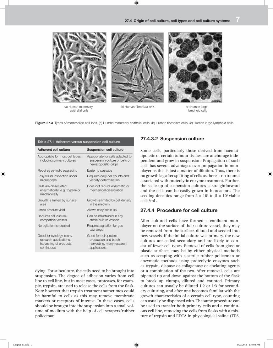
• Fibroblast like: cells which are attached to a substrate and appear bipolar and elongated, frequently forming swirls in heavy cultures
• Lymphoblast like: cells which do not attach normally to a substrate but remain in suspension and have a spherical shape.
27.4.2 Types of cultured cells
There are three main types of cultured cells depending on the number of times the cells can divide:
• Primary cell cultures: When cells are taken freshly from animal tissue and placed in culture, the culture consists of a wide variety of cell types, most of which are capable of very limited growth in vitro, usually fewer than 10 divisions. These cells retain their diploid karyotype, that is, they have the chromosome number and morphology of their tissues of origin. They also retain some of the differentiated characteristics that they possessed in vivo. For this reason, these cells sup-port the replication of a wide range of viruses. Primary cultures derived from monkey kidneys, mouse foe-tuses and chick embryos are commonly used for many laboratory experiments and for diagnostic purposes.
• Diploid cell strains: Some primary cells can be passed through secondary and several subsequent subcul-tures while retaining their original morphological characteristics and karyotype. Subcultures will have fewer cell types than primary cultures. After 20–50 passages in vitro, these diploid cell strains usually undergo a crisis in which their growth rate slows and they eventually die out. Diploid strains of fibroblasts derived from human foetal tissue are widely used in diagnostic virology and vaccine production.
• Continuous cell lines: Certain cultured cells, notably mouse foetal fibroblasts, kidney cells from various mammalian species and human carcinoma cells, are able to survive the growth crisis and undergo indefinite propagation in vitro. After several passages, the growth rate of the culture slows down; then isolated colonies of cells begin to grow more rapidly than diploid cells, their karyotype becomes abnormal (aneuploid), their morphology changes and other poorly understood changes take place that make the cells immortal. The cells are now ‘dedifferentiated’, having lost the special-ized morphology and biochemical abilities they pos-sessed as differentiated cells in vivo. Continuous cell lines such as KB and HeLa, both derived from human carcinomas, support the growth of many viruses.
27.4.3 Cell culture systems
For growing cells, two culture systems are used. They are mainly based on the ability of the cells to either
as the expression of tissue-specific properties. When these cells are subcultured in new containers with fresh medium, this is called a secondary culture. After several subcultures onto fresh media, the cell line will either die out or ‘transform’ to become a continuous cell line. Cells can also be virally transformed Such cell lines show many alterations from primary cultures, includ-ing changes in morphology, chromosomal variations and an increase in the capacity to form colonies in soft agar (colony formation assay) or give rise to tumours when implanted in immune-deficient hosts (xenograft models; Fig. 27.2).
27.4.1 Types of cells
Cultured cells are usually described based on their mor-phology or their functional characteristics. Animal cells exhibit three basic morphologies (Fig. 27.3):
• Epithelial like: cells which are attached to a substrate and appear flattened and polygonal in shape
Tissues or slice of Organ
Dissociated cells/Organ explants
Primary culture
Cell line
Clonal line
Single cell isolation
Collagenase
Culture medium
Subculture/passage
Immortalization
Continuous Cell line (e.g.NIH3T3, IHH)
(Immortalized)
Transformation/Loss of growth co ntrol
Transformed cell line
(e.g., COS-1)
Tumor cell lines
HepG2, MCF7
Figure 27.1 Scheme showing the possible mode of origin of cell lines [modified from Ian Freshney (2010).
Chapter 27.indd 5 6/23/2014 2:39:07 PM

6 CHAPTER 27 Animal cells as host
flasks, where the cells are keep it in a fully suspended medium. The advantages and limitations of using monolayer and suspension cultures are summarized in Table 27.1.
27.4.3.1 Monolayer culture
Adherent cell lines will grow in vitro until they have covered the surface area available or the medium is depleted of nutrients. Before this point, the cell lines should be subcultured to prevent the culture from
attach to a glass or treated plastic substrate, known as monolayer culture or ‘anchorage-dependent system’, and the floating free in the culture medium is known as suspension culture or ‘anchorage-independent sys-tem’. Monolayer cultures are usually grown in tissue culture–treated dishes, T-flasks, roller bottles, petri plates or multiple well plates; the choice should be based on the number of cells needed, the nature of the culture, personal preference and cost. While the suspension cultures are usually grown either in mag-netically rotated spinners or in shaken Erlenmeyer
Monolayer culture
Stacking of transformed cells
Virus
(a) Viral transformation of cells
Control HBx
c-MYC HBx+c-Myc
(b) Colony formation assay
Control siRNA Specific siRNA
(c) Xenograft model
Courtsey:Dr.Anil Suri NII, New Delhi
Figure 27.2 Animal cell transformation and its assays. (a) Viral transformation of cells. (b) Colony formation assay. (c) Xenograft model (Court-sey: Dr Anil Suri, National Institute of Immunology, New Delhi).
Chapter 27.indd 6 6/23/2014 2:39:08 PM
27.4 Origin of cell culture, cell types and cell culture systems 7
27.4.3.2 Suspension culture
Some cells, particularly those derived from haemat-opoietic or certain tumour tissues, are anchorage inde-pendent and grow in suspension. Propagation of such cells has several advantages over propagation in mon-olayer as this is just a matter of dilution. Thus, there is no growth lag after splitting of cells as there is no trauma associated with proteolytic enzyme treatment. Further, the scale-up of suspension cultures is straightforward and the cells can be easily grown in bioreactors. The seeding densities range from 2 × 10⁴ to 5 × 10⁵ viable cells/mL.
27.4.4 Procedure for cell culture
After cultured cells have formed a confluent mon-olayer on the surface of their culture vessel, they may be removed from the surface, diluted and seeded into new vessels. If the initial culture was primary, the new cultures are called secondary and are likely to con-sist of fewer cell types. Removal of cells from glass or plastic surfaces may be by either physical methods such as scraping with a sterile rubber policeman or enzymatic methods using proteolytic enzymes such as trypsin, dispase or collagenase or chelating agents or a combination of the two. After removal, cells are pipetted up and down against the bottom of the flask to break up clumps, diluted and counted. Primary cultures can usually be diluted 1:2 or 1:3 for second-ary culturing, and after one becomes familiar with the growth characteristics of a certain cell type, counting can usually be dispensed with. The same procedure can be used to transfer both primary cells and a continu-ous cell line, removing the cells from flasks with a mix-ture of trypsin and EDTA in physiological saline (TES,
dying. For subculture, the cells need to be brought into suspension. The degree of adhesion varies from cell line to cell line, but in most cases, proteases, for exam-ple, trypsin, are used to release the cells from the flask. Note however that trypsin treatment sometimes could be harmful to cells as this may remove membrane markers or receptors of interest. In these cases, cells should be brought into the suspension into a small vol-ume of medium with the help of cell scrapers/rubber policeman.
(a) Human mammary epithelial cells
(b) Human fibroblast cells (c) Human large lymphoid cells
Figure 27.3 Types of mammalian cell lines. (a) Human mammary epithelial cells. (b) Human fibroblast cells. (c) Human large lymphoid cells.
Table 27.1 Adherent versus suspension cell culture
Adherent cell culture Suspension cell culture
Appropriate for most cell types, including primary cultures
Appropriate for cells adapted to suspension culture or cells of hematopoietic origin
Requires periodic passaging
Easy visual inspection under microscope
Easier to passage
Requires daily cell counts and viability determination
Cells are dissociated enzymatically (e.g. trypsin) or mechanically
Does not require enzymatic or mechanical dissociation
Growth is limited by surface area
Limits product yield
Growth is limited by cell density in the medium
Allows easy scale-up
Requires cell culture–compatible vessels
No agitation is required
Can be maintained in any sterile culture vessels
Requires agitation for gas exchange
Good for cytology, many research applications, harvesting of products continuous
Good for bulk protein production and batch harvesting, many research applications
Chapter 27.indd 7 6/23/2014 2:39:08 PM

8 CHAPTER 27 Animal cells as host
determined by the media formulation. Salt and glu-cose are the major contributors to the osmolality of the medium, but amino acids are equally important. Most commercial media are formulated to have a final osmolality of ~300 mOsm, which can be checked with the help of an osmometer.
• Culture medium: Apart from temperature and CO2,
the most common variable in culture systems is the growth medium. Recipes for growth media can vary in pH, glucose concentration, growth factors and the presence of other nutrients. Glucose (0.8g to >5g/L) is commonly used as an energy source, which helps in maintaining osmolarity of the medium. Many of the media contain phenol red as a pH indicator, which is very helpful in monitoring the pH of the culture medium in a CO
2 incubator. Highly acidic
conditions turn the phenol red into yellow, while highly alkaline conditions turns the phenol red into pink. The growth factors used to supplement media are often derived from animal blood, such as foe-tal bovine serum (FBS). One complication of these blood-derived ingredients is the potential for con-tamination of the culture with viruses, mycoplasmas or prions and shows variation in growth factor and hormone contents. Therefore, current practice is to minimize or eliminate the use of these ingredients wherever possible and use chemically defined media. Commercially available medium are highly sterile and ready-to-use liquids in a concentrated liquid or powdered form. Instead of providing nutrients for growing cells, the medium is generally supplemented with fungicides and antibiotics, or both, to inhibit contamination.
• Dulbecco’s Modified Eagle Medium (DMEM) is a basal medium consisting of amino acids, vitamins, glucose, salts and a pH indicator which contains no proteins or growth-promoting agents. Therefore, it needs sup-plementation to be a ‘complete’ medium. It is com-monly supplemented with 5–10% FBS. DMEM used a sodium bicarbonate buffer system (3.7 g/L) and therefore requires artificial levels of CO
2 to maintain
the required pH 7 and 10% CO2 is optimal, but many
researchers successfully use it in 5% CO2.
• Serum and antibiotics: Serum is one of the most important components of animal cell culture as it supports cell proliferation. It is the source of peptide hormones and hormone-like growth factors that pro-mote a healthy growth of cells. Serum is also a source of various amino acids, hormones, lipids, vitamins, polyamines and salts of calcium, potassium and iron, etc. However, one of the key limitations of FBS usage is the variation in its growth factor contents and chances of its contamination with viruses or prions that may adversely affect the culture. Therefore, current prac-tice is to minimize the use of FBS in cell culture and use serum-free medium instead. Although not essen-tial for cell growth, antibiotics such as penicillin and
i.e.Tris- EDTA-Saline with 10 mM Tris chloride, pH 7.4, 150 mM NaCl, 1 mM EDTA).
27.4.5 Culture conditions
Culture conditions vary widely for each cell type, but the artificial environment in which the cells are cul-tured invariably consists of a suitable vessel containing the following:
• A substrate or medium that supplies the essential nutrients (amino acids, carbohydrates, vitamins, min-erals)
• Growth factors • Hormones • Gases (O
2, CO
2)
• A regulated physicochemical environment (pH, osmotic pressure, temperature)
Cells can also be adapted to different culture environ-ments (e.g., different nutrients, temperatures, salt concentrations, etc.) by varying the activities of their enzymes. Most cells are anchorage dependent and must be cultured while attached to a solid or semi-solid sub-strate (adherent or monolayer culture), while others can be grown floating in the culture medium (suspension culture):
• Incubation conditions: Most mammalian cells are grown and maintained at an appropriate tempera-ture and gas mixture (typically at 37°C with 5% CO
2
for mammalian cells) in a CO2 incubator. This tem-
perature is selected because it is the core temperature of the human body. Besides, most cells derived from other warm-blooded animals grow well at 37oC.
• pH: The extracellular and intracellular pHs are criti-cal for the survival of mammalian cells. They help in maintaining the appropriate ion balance and opti-mal function of cellular enzymes, hormones and growth factors. Fluctuations in pH adversely affect cell metabolism, which can lead to cell death. Most media strive to achieve and maintain the pH between 7 and 7.4. Most media use the bicarbonate–CO
2 buffering
system. The interaction of CO2 derived from cells or
the atmosphere with water leads to a drop in the pH, while the bicarbonate content of the medium neu-tralizes the effect of increased CO
2 until equilibrium
is reached at pH 7.4. This kind of system is called an open system and can be described by the following equation:
H2O + CO
2 H
2CO
3 H+ + HCO
3–
• Osmolality: The osmolality of the culture medium also plays a crucial role in cell growth and function by maintaining the outside and inside osmotic pres-sure of the cell. Osmolality of the medium used is
Chapter 27.indd 8 6/23/2014 2:39:08 PM

27.4 Origin of cell culture, cell types and cell culture systems 9
5. Seed an appropriate volume of cell suspension for the size of the flask to get the desired cell density.
27.4.7 Cell counting
Cell counting is done in the following steps with the help of a haemacytometer (Fig. 27.4):
1. Clean the chamber and cover slip with alcohol. Dry and fix the coverslip in position.
2. Harvest the cells. Add 10 µL of the cells to the haemacy-tometer.
3. Place the chamber under the inverted microscope under a 10× objective. May use phase contrast to distinguish the cells.
4. Count the cells in four 16-square grids (marked 1 to 4) and take their average.
The following example illustrates how to convert your cell count to the number of cells in the original suspen-sion. Assume a 1:10 dilution is being used to count cell numbers; if you count an average of 12 cells in the four 16-square grid (1 mm2), then the cell numbers in a given suspension can be derived as follows:
12 × 104× 10 = 1.2 × 10⁶ cells/mL
streptomycin are often used in the culture medium to control microbial growth.
• Seeding density: Plating density (the number of cells per volume of the culture medium) plays a critical role for some cell types. For example, HepG2 cells when seeded at lower density do not grow optimally in cul-ture vessels.
• Mode of culturing: Cells can be grown either in sus-pension or adherent cultures. Some cells naturally live in suspension, without being attached to a surface, such as cells that exist in the bloodstream. There are also cell lines that have been modified to be able to survive in suspension cultures so they can be grown to a higher density than adherent conditions would allow. Adherent cells require a surface, such as tissue culture plastic or microcarrier, which may be coated with extracellular matrix components to increase adhesion properties and provide other signals needed for growth and differentiation. Most cells derived from solid tissues are adherent. Another type of adherent culture is organotypic culture, which involves grow-ing cells in a 3D environment as opposed to two-dimensional culture dishes. This 3D culture system is biochemically and physiologically more similar to in vivo tissue, but is technically challenging to main-tain because of many factors (e.g. diffusion). Cultures should be examined daily for their morphology, col-our of the medium and density of cells.
27.4.6 Typical experimental protocol for cell culture
A typical experimental protocol for cell culture is as follows:
1. Examine the cells growing in a 25-cm2 flask with an inverted microscope to see if they have formed a confluent (~80%) cell monolayer. If there are sufficient cells, pour off the medium.
2. Wash the monolayer with 2 mL of phosphate-buffered saline. Rinse well without shaking the flask (shaking pro-duces bubbles) and pour off. Repeat.
3. Add 1.0 mL of TES to the flask and incubate at 37oC for 2–10 minutes with TES covering the cells. Observe periodi-cally to determine when cells are loosened from the plas-tic. (Note: TES should contain a pH indicator. Below pH 7, trypsin is inactive. However, above pH 8, trypsin is damag-ing to cells.)
4. When cells are seen to detach from the surface upon shak-ing or jarring against the heel of one’s hand (this can be checked under the microscope), add 4 mL of fresh growth medium with 10% serum and suspend cells by pipetting up and down a few times. Count cells in a haemacytom-eter, calculate the volume of additional medium needed to bring the cell concentration to ~5 × 10⁵ cells/mL and add this volume to the cell suspension.
1 2
3 4
5
Loading groove
Mirrored surface
Grid
Figure 27.4 Haemacytometer along with an enlarged view of the counting chamber or grids.
Chapter 27.indd 9 6/23/2014 2:39:09 PM

10 CHAPTER 27 Animal cells as host
should be characterized and checked for contamina-tion. There are several common media used to freeze cells.
For serum-containing medium, the constituents may be as follows:
• Complete medium containing 10% glycerol • Complete medium containing 10% DMSO (dimethyl-sulphoxide)
• 50% cell-conditioned medium with 50% fresh medium containing10% glycerol or 10% DMSO.
Cells are stored in sterile cryogenic storage vials or cryovials that have a screw-top and a rubber seal to keep the nitrogen out. Cryoprotective agents such asDMSO help in reducing the freezing point and facili-tate a slow cooling rate. Gradual freezing decreases the risk of cell damage and ice crystal formation. Ideally, a cooling rate of 1°C/min is recommended. Afterwards, cells must be stored at –70oC or lower (ideally in liquid nitrogen at –196oC). One must ensure that the liquid nitrogen containers carrying the canisters of cryovi-als are relatively full of nitrogen during the period of storage.
27.4.9.2 Resuscitation of frozen cell lines
When cryopreserved cells are needed for study, it is vital to thaw cells correctly to maintain the viability of cells and enable the culture to recover quickly. Some cryo-protectants, such as DMSO, are toxic above 4°C; there-fore, it is essential that cultures are thawed quickly and diluted in culture medium to reduce their toxic effects. With careful handling, 50–80% of the cells of a healthy culture will survive freezing.
The following steps are recommended for getting best results:
1. Thaw frozen cells rapidly (<1 min) in a 37°C water bath. 2. Dilute the thawed cells slowly using a pre-warmed growth
medium. 3. Plate thawed cells at a high density to optimize recovery.
27.4.10 Commonly used equipment in animal cell culture
A typical cell culture laboratory should have the follow-ing main equipment for doing animal cell culture work: cell culture hood, CO
2 incubator, inverted microscope,
centrifuge, etc.
27.4.10.1 Cell culture hood
All cell culture manipulations must be performed asep-tically in laminar air flow hoods to keep the work area free any bacterial or fungal contamination. Therefore, the cell culture hoods should never be used for bacterial
where 12 is the average number of cells in four grids, × 104 converts to cells per millilitre, × 10 is the dilution fac-tor and 1.2 × 10⁶ cells/mL is the number of cells in the original suspension.
27.4.8 Cell viability
Viability as says measure the number of viable cells in a population and provide an accurate indication of the health of cells in culture. These as says rely upon the integrity of the cell membrane as an indicator of cell via-bility. Stains such as trypan blue are actively excluded by viable cells, while they are taken up and retained by dead cells that lack an intact membrane.
27.4.9 Cryopreservation (freezing, storage and revival) of cells
Cell culture has many problems, including culture con-tamination by bacteria, mycoplasma, and fungus. Also, cells must be subcultured frequently to prevent over-crowding of cells. Furthermore, media must often be change to prevent the build-up of contaminants and toxins and to provide fresh nutrients as nutrients are used up quickly by rapidly proliferating cells. There-fore, as soon as a small surplus of cells becomes avail-able from subculturing, they should be frozen as a seed stock, protected and should not be made available for general laboratory use. Working stocks can be pre-pared and replenished from frozen seed stocks. If the seed stocks become depleted, cryo preserved working stocks can then serve as a source for preparing a fresh seed stock with a minimum increase in the generation number from the initial freezing. Cell lines in continu-ous culture are also prone to genetic drift and are fated for senescence, and even the best-run laboratories can experience equipment failure. As an established cell line is a valuable resource and its replacement is expen-sive and time-consuming, it is vitally important that it is cryopreserved for long-term storage.
27.4.9.1 Storage by freezing
Viability of viruses and bacteria is well preserved dur-ing freezing, but original attempts to preserve animal cells by freezing resulted in cell death. This was first thought to be due to laceration of cell plasma mem-branes by ice crystals, but more recent evidence sug-gests that the cause may be osmotic changes during freezing which give rise to irreversible changes in lipoprotein complexes in intracellular membranes. In any case, mammalian cells are cryopreserved to avoid loss by contamination, to minimize genetic change in continuous cell lines and to avoid aging and trans-formation in finite cell lines. Before cryopreservation, one need to ensure that cells are healthy (>90% viabil-ity) and growing in the log phase. Further, the culture
Chapter 27.indd 10 6/23/2014 2:39:09 PM

27.4 Origin of cell culture, cell types and cell culture systems 11
hazardous agents as it blows air directly at users, but are good for cultures. Both vertical and horizontal hoods have continuous air flow that passes through a high effi-ciency particle air (HEPA) filter to remove particulate matter from the air. The hoods are also equipped with a short-wave ultra violet (UV) light source which needs be turned on for a few minutes before use to sterilize the work surfaces of the hood. Also, the hood should be turned on about 10–20 min before being used. It is important to keep the hood free of all clutter as they interfere with the laminar flow air pattern. Wiping of all work surfaces with 70% ethanol inside the hood before and after each use is a good practice.
27.4.10.2 CO2 incubator
The CO2 incubator is designed to maintain sterility
inside the chamber and keep a constant temperature, high relative humidity and an atmosphere with a fixed level of CO
2. Thus, it reproduces an environmental
or fungal work. A laminar hood has a dual role in cell culture:
• It protects the tissue culture from the users (by pro-viding a sterile environment).
• It protects the user from the tissue culture (from pos-sible infection risk).
There are two types of laminar air flow hoods – vertical and horizontal (Fig. 27.5). In a vertical hood, the filtered air blows down from the top of the cabinet; in a hori-zontal hood, the filtered air blows out at the user in a horizontal fashion. The vertical hood is also known as a biology safety cabinet (BSC) and is appropriate for work-ing with infectious agents. Here, the infectious aerosols generated in the hood are filtered out before they are released into the surrounding environment. Depend-ing on the nature of the infective agent being used in cell culture, the BSCs are designated as class I, II or III. The horizontal hoods are unsuitable for working with
Hepa filterGlass screen
Work area
Prefilter
Blower
Stand
(c) Horizontal Laminar Flow
Hepa exhaust filter
Hepa filter
Prefilter
Recirculating fan
Glass screen
Work area
Stand
(d) Vertical Laminar Flow(b) Vertical Laminar Hood
(a) Horizontal Laminar Hood
Figure 27.5 Cell culture laminar hoods. (a) Horizontal laminar hood. (b) Vertical laminar hood. (c) Horizontal laminar flow. (d) Vertical laminar flow.
Chapter 27.indd 11 6/23/2014 2:39:09 PM

12 CHAPTER 27 Animal cells as host
The inverted microscope allows viewing of cells from the bottom because its optical system is at the bottom, with the light source on top [Fig. 27.6(b)]. Observation of cultures in this way gives an immediate idea of the health and growth of cells. The lights of microscope should be turned off when not in use and should be kept covered to protect the optical parts from dust.
27.4.10.4 Centrifuge
A low-speed centrifuge is required for animal cell pel-leting during thes ubculturing step. In most cases, cells are centrifuged at ambient temperature. Nev-ertheless, a low operation temperature is desirable to minimize exposure of cells to uncontrolled higher temperatures.
27.4.11 Commonly used culture wares
The need for sterility also applies to the glassware or plastic ware used to prepare media and reagents and to grow cells. Metal and glass devices can be steri-lized by autoclaving while disposable plastic culture flasks and other plastic ware are sterilized by gamma radiation. Sterilization methods have the goal of kill-ing contaminating organisms without affecting the surface of the flask or vessel. The animal cells are usually grown and maintained in petri dishes, cul-ture flasks or multi-well plates of various shapes and sizes (Fig. 27.7) at an appropriate temperature and gas mixture (typically, 37°C, 5% CO
2 for mammalian
cells) in an incubator. There are numerous suppliers of cell culture plastic ware such as BD Biosciences, Corning, Falcon and Nunc International. Note that
condition very close to that of living cells. The incuba-tor chamber has an airtight silicone gasket on the inner door which keeps it isolated from the external environ-ment [Fig. 27.6(a)]. The animal cells are maintained in an atmosphere of 5–10% CO
2, which keeps the medium
buffered with sodium bicarbonate/carbonic acid. Culture vessels have vents to allow for sufficient gas exchange. A pan of water is kept at all times in the incu-bator chamber to maintain a high relative humidity, prevent desiccation and maintain the correct osmolar-ity of the culture medium. The incubator doors should not be left open for very long during use.
27.4.10.3 Inverted Microscope
In tissue culture vessels such as petri dish, the cells are present at the bottom, with the culture medium above.
CO2 inculator Inverted microscope
Figure 27.6 Cell culture equipment. (a) CO2 incubator. (b) Inverted microscope.
Figure 27.7 Cell culture plas-tic wares and accessories.
Chapter 27.indd 12 6/23/2014 2:39:10 PM

27.5 Gene delivery in cells 13
For long-term gene expression, the transfected gene is integrated into the host cell genome to make permanent cell lines. A selection marker is used to identify cells that have successfully integrated the gene sequence of inter-est. The selection agent enriches and enables the growth of a subpopulation of cells where the exogenous genetic material has been incorporated into the genome.
Transfection of DNA or RNA molecules into cultured mammalian cells can be accomplished using a variety of methods and reagents. These methods include physi-cal (electroporation, microinjection), chemical (cal-cium phosphate method, lipofection) and virus-based (adenovirus, retrovirus) delivery systems. The impor-tant parameters that affect the outcome of cell transfec-tion include: (i) cell type, (ii) cell density, (iii) amount of DNA to be transfected, (iv) ratio between the transfec-tion reagent and the DNA, (v) incubation period of cells with the DNA complex, (vi) post-transfection incuba-tion time of cells, etc.
27.5.2 Methods of transient transfection
The various methods of transient transfection are as follows.
27.5.2.1 Electroporation
The use of high-voltage pulses or electroporation to introduce DNA into cultured cells was first estab-lished by Wong and Neumann (1982) for fibroblasts. It is a highly efficient technique for delivering exogenous nucleic acids to suspension cells and non-adherent primary cells. This technique uses electricity to create transient pores (electropores) in the cellular membrane to enable the uptake of charged nucleic acid molecules into the target cells [Fig. 27.8(a)]. A highcell mortality in this case is a major drawback.
27.5.2.2 Calcium phosphate method
This method was first used by Graham and van der Eb in 1973 to introduce adenovirus DNA into mammalian cells. Here HEPES-buffered saline solution contain-ing phosphate ions is mixed with a calcium chloride solution containing the DNA to be transfected. When the two solutions are combined, a fine precipitate of the positively charged calcium and the negatively charged phosphate is formed, binding the DNA to be transfected on its surface [Fig. 27.8(b)]. The suspen-sion of the precipitate is then overlaid on the cells to be transfected. The resulting calciumphosphate–DNA complexes adhere to the cell membrane and enter the cytoplasm by endocytosis. This method is extremely simple to handle but not good for all cell types. Tran-sient hypertonic shock of the transfected cells with 10% glycerol or DMSO improves the transfection efficiency.
the most expensive cell culture plastic ware does not always mean the best choice for your cells. A plastic ware check is always worth doing with primary cells as their requirements are more specific. However, for cell lines, one may start with the brand currently used in the laboratory.
27.5 Gene delivery in cells
Animal cell transfection is a commonly used method to deliver exogenous DNA or RNA into cells to express them in cell culture. It offers an important mechanism for advancing the knowledge about the structure and functions of animal genomes and understanding their new traits through cell culture. There are several differ-ent ways to transfect mammalian cells, depending on the cell line characteristics, desired effect and down-stream applications. These methods can be broadly divided into two categories: those used to generate tran-sient transfection and those used to generate stable cell lines. Table 27.2 summarizes the important aspects of stable and transient transfection methods.
27.5.1 Transient versus stable transfection
Transient transfections are most commonly used to investigate the short-term impact of gene and/or pro-tein expression on the cell cycle, cell physiology and metabolism or even reporter genes. Plasmid DNA, mes-senger RNA (mRNA), short-interfering RNA (siRNA), short-hairpin RNA (shRNA) and microRNA (miRNA) are commonly used in transfection experiments. Dur-ing transient expression, however, the nucleic acid sequence is not integrated into the host cell genome. Therefore, the effect on target gene expression is tem-porary (24–72 h for RNA probes, 48–96 h for DNA probes).
Table 27.2 Transient versus stable expression of genes
Transient expression Stable expression
Short-term expression (usually 48 h)
Transfection by chemical methods or electroporation
Useful for evaluating gene activity or their regulation
No genomic integration of transfected DNA
Sustained expression on a long-term basis
Transfection by chemical methods or viral vectors followed by selection for antibiotic or drug resistance
Useful for continuous source of a gene product or gain/loss of a gene function
Non-specific integration of transfected DNA in host cell genome
Chapter 27.indd 13 6/23/2014 2:39:10 PM

14 CHAPTER 27 Animal cells as host
27.5.3 Methods of stable transfection
The various methods of stable tranfection are as follows.
27.5.3.1 Microinjection
Microinjection delivers the genetic material directly into the cell nucleus (Fig. 27.9). Using a light micro-scope and a fine needle (micromanipulator) guided into the nucleus, a small amount of DNA or RNA is injected. However, this method is labor intensive and requires trained personnel. Nevertheless, this method is extremely useful in the case of animal transgenesis and gene knock-in and knock-out technology.
27.5.3.2 Virus-mediated gene delivery (transduction)
Viral vector is the most effective means of gene trans-fer to modify specific cell type or tissue and can be manipulated to express heterologous genes. Several virus types are currently being investigated for use to deliver genes to cells to provide either transient or per-manent transgene expression. The viral vectors in cur-rent use are based on different RNA and DNA viruses
27.5.2.3 Liposome-mediated transfection
Liposomes are synthetic analogues of the phospholipid bilayer, the building block of the cellular membrane. These transfection compounds share a number of char-acteristics with their natural counterparts, including the presence of hydrophobic and hydrophilic regions of each molecule, which allow for the formation of spheroid liposomes under aqueous conditions. In the presence of free DNA or RNA, liposomes encapsulate the nucleic acids to create an efficient delivery system. The charge, composition and structure of the liposome define the affinity of the complex for the cellular mem-brane. The cationic liposomes were first introduced in 1987 by Felgner and coworkers. The liposomes cur-rently in use typically contain a mixture of cationic and neutral lipids organized into lipid bilayer structures. Transfection complex formation is based on the inter-action of the positively charged liposome with the nega-tively charged phosphate groups of the nucleic acid. Liposomes fuse with the cell membrane and deliver their cargo inside [Fig. 27.8(c)]. The method is quite simple as well as reproducible, and the transfection effi-ciency is reasonably high. However, cell toxicity could be a problem sometimes.
− +
Lid
Cuvette
Cell suspension
Electrode
Cationicliposome
DNA Mammalian cell
(c) Lipofection
(b) Calcium phosphate method(a) Electroporation
Ca++
Ca++
Ca++
Ca++
Ca++
Ca++
− −−
−−
−−
−−
−
−−−
−−−−
++
+
+
++
+
+
N C
N C
N C
N
DNA - Calciumphosphate precipitate
Figure 27.8 Methods of transient transfection. (a) Electroporation. (b) Calcium phosphate method; lipofection.
Chapter 27.indd 14 6/23/2014 2:39:11 PM

27.5 Gene delivery in cells 15
vectors are shown in Fig. 27.10. Very often, a selection marker (either antibioticbased or fluorescent protein-based) is used to select for cells that have been suc-cessfully transduced with the virus. Once the genetic material is incorporated into the host genome, it relies on the host transcriptional machinery for expression, while in the case of shRNA/miRNAprobes, the host RNAi machinery is required. However, viral vectors have limited laboratory uses because the viral vector integrates randomly into the genome and could yield different results.
27.5.4 Development of stable cell line
The episomal DNA stability is often limited, resulting in a gradual loss of transfected DNA vector from the cells. As DNA integration into host chromosomes is a rare event, stablytransfected cells need to be selected and cultured in different ways. A variety of systems for selecting transfected cells exist, including resistance to antibiotics or expression of metabolic enzymes. After gene transfer, cells are cultivated in a medium contain-ing the selective agent. Only those cells which have
that possess very different genomic structures and host ranges. Among viral vectors developed so far, retrovi-ral vectors represent the most prominent delivery sys-tem, asthese vectors have high gene transfer efficiency and mediate high expression of heterologous genes. Members of the DNA virus family such as adenovi-rus, adeno-associated virus or herpesvirus have also become attractive for efficient gene delivery. The viral genomes are modified to act as viral vectors to intro-duce foreign DNA into mammalian cells. The viruses have their own mechanism for moving nucleic acids into cells and transduction by this route is usually very efficient. The viral vectors are selected as gene deliv-ery vehicles based on their capacities to carry foreign genes and their ability to efficiently deliver these genes combined with their efficient gene expression and inte-gration into the target cell genome with the help of the viral machinery. While in adeniviral vectors, the early E1 and E3 genes can be replaced, the entire gag, pol and env genes of the retroviral genome can be replaced with foreign DNA. The lost viral gene functions can be provided in trans using appropriate packaging cell lines. The simplified maps of adenoviral and retroviral
Positivepressure
Egg pronucleus
DNA injecting needle
Cell holding pipet
Negativepressure
Inverted microscope
Cell holding pipet
Cellmanipulator
Control knob
DNAmicromanipulator
DNA injecting needle
Micromanipulator
e.g., microinjection of egg cell
Figure 27.9 Method of microinjection. Micromanipulator and microinjectionof egg cell.
Chapter 27.indd 15 6/23/2014 2:39:11 PM

16 CHAPTER 27 Animal cells as host
potent translational inhibitor in both prokaryotic and eukaryotic cells. Resistance to puromycin is conferred by the puromycin N-acetyl-transferase gene (pac) from Streptomyces. Puromycin has a fast mode of action, causing rapid cell death at low anti-biotic concentrations. Adherent mammalian cells are sensitive to concentrations of 2–5 µg/mL and stable mammalian cell lines can be generated within 1week
• Recessive selection markers: A number of mamma-lian cells that are deficient for enzymes of key meta-bolic pathways are suitable for developing stable cell lines. Thecells are transiently transfected with plas-mids carrying genes for deficient enzymes and then selected using appropriate substrate analogues.For example, dihydrofolate reductase (DHFR) is a ubiq-uitous cytosolic enzyme that catalyses the reduc-tion of 5,6-dihydrofolate to 5,6,7,8-tetrahydrofolate (THF). THF is an essential co-factor in several meta-bolic pathways, including purine and thymidylate biosynthesis. Methotrexate is a folate analogue that acts as a slow, tight-binding competitive inhibitor of DHFR. It acts by inhibiting cellular THF synthesis and, in turn, cellular proliferation. Therefore, only those cells that express the DHFR gene survive, while other cells die.
Popular hosts for stable expression are Chinese hamster ovary (CHO) cells, baby hamster kidney (BHK-21) cells, myeloma cells and human embryonic kidney (HEK) 293 cells.
integrated the plasmid survive, containing the selec-tion marker gene. Both dominant and recessive selec-tion marker genes are used for establishing permanent cell lines:
• Dominant selection markers: These marker genes are usually of microbial origin and confer resistance to the toxic effects of a drug or other pharmacologi-cally active compounds used for selection. Such markers are used to confer an advantage tomam-malian cells that have been transfected with exog-enous DNA constructs. Dominance refers to the fact that a single copy of the marker is sufficient to confer the resistant phenotype. For example, G418 is an aminoglycoside antibiotic produced by Micromonosporarhodorangea that blocks polypep-tide synthesis by inhibiting the elongation step in both prokaryotic and eukaryotic cells. Resistance to G418 is conferred by the neomycin resistance gene (neo) from Tn5, encoding an aminoglyco-side 3’- phosphotransferase (Fig. 27.11). Selection in mammalian cells is usually with concentrations ranging from 0.2 to 2 mg/mL.
Puromycin is another commonly used antibiotic for making permanent cell lines. It produced by the bacterium Streptomyces alboniger. Aspuromycin is an aminonucleoside and resembles an aminoacyl-tRNA, it can inhibit protein synthesis by disrupting peptide transfer on ribosomes, causing premature chain termination during translation. It is also a
E1Adenovirus5
E3
Deletion in E1and/or E3 regions
Therapeutic gene in transfer plasmid
Cotransfection in 293 cells (E1 in trans)
Recombinant Ad5 genome
1012 particles/mlNon-integrativeAll cell types
Recombinant virus
(a) Adenoviral Vector
LTR
LTR
ψ
ψ
gag pol envLTR
LTRTherapeutic gene(s)
Recombinant retrovirus
Transfection into pakaging cells
Cell with helper provirus
gag pol env
Transcription + Translation
Capsid proteins Genomic RNA
AAAAAA
106 particales/mlIntegrativeCell specific
Empty particles
Recombinant virus
(b) Retroviral Vector
Figure 27.10 Scheme of viral vectors for gene delivery. (a) Adenoviral vector.(b) Retroviral vector.
Chapter 27.indd 16 6/23/2014 2:39:11 PM

27.6 Scale-up of animal cell culture process 17
flasks (for suspension cultures) are used in scale-up of animal cell culture process:
• Roller bottles: Roller bottles provide total curved sur-face area of the micro-carrier beads for growth. This system offers the following advantages over the static monolayer culture: (i) provides an increase in the sur-face area, (ii) provides constant gentle agitation of the medium and (iii) provides an increased ratio of sur-face area of medium to its volume, which allows better gaseous exchange. Roller bottles are cylindrical ves-sels that revolve slowly (between 5 and 60 revolutions per hour). These tissue culture bottles can be used in specialized CO
2 incubators with attachments that
rotate the bottles along the long axis [Fig. 27.12(a)]. After each complete rotation of the bottle, the entire cell monolayer has transiently been exposed to the medium. The volume of medium need only be suf-ficient to provide a shallow covering over the mon-olayer. Typically, a surface area of 750–1500 cm2 with 200–500 mL medium will yield 1–2 × 10⁸ cells.
• Micro-carrier beads: Micro-carrier beads (~90–300 µm diameter) can be used in any application where anchorage-dependent cells are to be cultured. These are either dextran or glass based, and are available in a range of densities and sizes. They are primarily used to increase the surface area for cell growth, and thus, the number of adherent cells in a culture vessel is very high. For example, micro-carrier beads usually pro-vide 0.24 m2 area for every 100 mL of a culture flask. As the beads are buoyant, these can be easily used with a spinner culture vessel [Figure 27.12(b)]. Further, adherent cells can be grown to high densities with the help of micro-carrier beads.
• Spinner cultures: Spinner-type vessels are used for scal-ing up the production of suspension cells or anchor-age-dependent cells attached to micro-carrier beads. Spinner is a type of bioreactor having a suspended Teflon impeller, stirrer or similar device to agitate the medium when placed on a magnetic stirrer. The ves-sel is usually made of glass or stainless steel, with port holes to accommodate sensors, medium input and gas flow [Fig. 27.12(b)]. Commercial versions incorporate one or more side arms for sampling and/or decanta-tion. As cells are not allowed to settle to the bottom of the flask, they can be grown to very high densities. Further, stirring of the medium improves gas exchange.
27.7 Quality control of recombinant products
It is extremely important to do analytical and pharma-cokinetic characterization of protein biopharmaceuti-cals produced through rDNA technology before these could be marketed. Specific assays have been designed
27.6 Scale-up of animal cell culture process Mammalian cells are cultured by a variety of methods at a range of volumes for the production of therapeutic and diagnostic proteins. For commercial exploitation, the cells are grown in a bioreactor or fermentation ves-sels where physicochemical and biological factors are maintained at optimum levels. The most suitable bio-reactor used is a compact-loop bioreactor with marine impellers. Here, glucose and glutamine are the main car-bon and energy source and the metabolic products that affect cell growth are monitored by gas chromatography or high-performance liquid chromatography (HPLC). However, for batch cultures, mainly roller bottles with micro-carrier beads (for adherent cells) and spinner
CMV-Neo vector
SV40 origin
Neor
Ampr
SV40 poly ABaterial origin
f1 origin
CMVpromoter
Gene of interest
Cell transfection
G418 selection
Expansion of culture
Gene integration and expression analysis
Cells
Colonies
Figure 27.11 Development of stable cell lines by neomycin selection.
Chapter 27.indd 17 6/23/2014 2:39:12 PM

18 CHAPTER 27 Animal cells as host
• C-terminal sequencing by MS • Molecular weightdetermination of intact protein iso-
forms • Western blot analysis by specific antibodies • Quantitative amino acid analysis • HPLC profiling • Quantification by LC-MS/MS with isotope dilution
The analytical characterization of antigens is even more relevant in the case of subunit or complex protein-based vaccines. Further, it is important to do MS and proteomics-based analysis for the identity, potency and stability of each preparation, combined with documen-tation of antigen concentrations and comparison of dif-ferent batch preparations.
27.7.2 Pharmacokinetic analysis
Just for any other drug, pharmacokinetic studies are an integral part of human clinical studies to evaluate the effi-cacy and safety of recombinant protein biopharmaceuti-cals. This is required to ascertain the relationship between administered dose, the observed biological fluid concen-tration of the drug and time. These methods are used to measure the bio-product concentration in biological fluids (serum, plasma, urine) or target tissue collected at different timepoints. These studies are performed in com-pliance with good laboratory practice (GLP).
27.7.3 Toxicokinetic analysis
Toxicokinetic studies are performed to understand the relationship between administered drug dose and toxicity. It is a regulatory requirement done in accordance with OECD (Organization for Economic
to establish the identity, quality, purity and quantity of recombinant proteins, mAbs, vaccines, antigens, aller-gens and biosimilars and perform pharmacokinetic and toxicokinetic studies.
27.7.1 Analytical procedures
Combinations of specific analytical procedures are per-formed to ascertain different properties of bioproducts. For example, the antibody characterization involves:
• Heavy- and light-chain molecular weightdetermina-tion by mass spectrometry (MS)
• Peptide mapping by MS of heavy and light chains • Sequencing of variable complementary-determining regions
• N- and C-terminal sequencing of heavy and light chains bymatrix-assisted laser desorption ionization–in-source decay(MALDI-ISD)
• Edman N-terminal sequencing • Electrophoretic analysis by 1D and 2D polyacryla-mide gel electrophoresis(PAGE)
• Analysis of common translational modification– deamidation, oxidation, pyroglutamate, N- glycosylation
• Quantification by liquid chromatography–mass spec-trometry (LC-MS)/MS
• Host cell protein identification assay
Likewise, antigen and vaccine characterization involves establishing the identity and purity of the antigen in question using the following methods:
• Mass spectrometric peptide mapping with high sequence coverage
• N-terminal Edman sequencing
Gas vent
Teflon stirrer
Inoculationport
Roller bottle
Microcarrier culture
(a) Roller Inculbator (b) Spinner flaskFigure 27.12 Scale-up culture of animal cells. (a) Roller incubator. (b)Spinner flask.
Chapter 27.indd 18 6/23/2014 2:39:12 PM

27.8 Applications of animal cell technology 19
companies, followed by its marketing. This is the pri-mary means by which the developer of the drug can recover the investment cost for development of the biopharmaceutical. The first therapeutic recombi-nant biopharmaceutical for human use was ‘human’ insulin. The recombinant human insulin (rHI), also known as Humulin, was developed by the US phar-maceutical giant Genentech and licensed to Eli Lilly for manufacturing and marketing in 1982. The global biopharmaceutical industry has come a long way since the development of Humulin. Today, more than 300 biopharmaceuticals have already been approved and many more are in the late-stage clinical development, including blood factors, colony-stimulating factors, enzymes, growth factors, growth hormones, immuno-globulins, insulin, interferons, interleukins and mAbs. Some important milestones in the therapeutic use of recombinant protein biopharmaceuticals aregiven in Box 27.3, while those produced using animal cell cul-ture and rDNA technology are listed in Table 27.3.These drugs have not only advanced the prevention and treat-ment of a number of life-threatening diseases, but have also provided the thrust for the continued success of the pharmaceutical industry. The industry’s US$ 92 billion figures and double-digit growth rate in recent years is a testimony of the higher approval rate for investors. According to a global biopharmaceutical market report (International Market Analysis Research and Consult-ing’s group report, 2010–2015), the sale of recombinant protein pharmaceuticals is likely to touch US$ 167 bil-lion by 2015, including a share of $79 billion for mAbs. Next are some examples of recombinant therapeutics that are currently available in the market.
27.8.1 Blood coagulation factors
Haemophilia A is a common heritable genetic disor-der where the body lacks the ability to produce factor VIII, required for blood clotting, while haemophilia B or Christmas disease is the second most common type of bleeding disorder due to the deficiency of factor IX – a
Co-operation and Development) guidelines to assess systemic exposure in toxicology studies, that is, in pre-clinical animal studies for setting human plasma con-centration limits and safety margins. Quantification of systemic exposure is often represented by plasma, serum or blood concentration of the compound. Toxi-cokinetic studies also need to be performed in consist-ence with GLP.
27.7.4 Purity analysis
When human proteins are expressed recombinantly in another organism, the purified protein always contains trace amounts of contaminating host proteins from the expression organism. Such process-related impurities are identified and evaluated qualitatively and quantita-tively using procedures such as1D sodium dodecyl sul-phate polyacrylamide gel electrophoresis(SDS-PAGE), combined with silver staining of the gel, followed by protein identification using mass spectrometric peptide mapping. The twin procedures offer an attractive sup-plement to the enzyme-linked immunosorbent assay (ELISA)-based protein assay as these are less time-consuming and do not involve development of specific antibodies. The 1D SDS-PAGE is helpful in visualizing the individual host protein bands and allows batch-to-batch comparison of the relative amounts. The pro-tein bands are then cut out from the gel, digested with trypsin and the peptide mixture is analysed by MS. Then, the Mascot database is used to establish the iden-tity of the individual host protein bands. This database includes protein/peptide sequences from different host systems, such asyeast, E. coli, sf9 insect cells, rice and CHO cells.
27.8 Applications of animal cell technology
Cell culture is one of the major tools used in cellular and molecular biology, providing excellent model systems to study the normal physiology and biochemistry of cells (e.g. cell cycle, metabolic studies), the effects of drugs and toxic compounds on the cells, and mutagenesis and carcinogenesis (e.g. apoptosis, genetoxicity). It is also used in drug screening and development, and large-scale manufacturing of biological compounds (e.g. therapeutic proteins and vaccines). The main advan-tage of using animal cell culture in the above applica-tions is the consistency and reproducibility of results obtained through batch culture of clonal cells. However, some changes in the characteristics of these cells after a period of continuous growth are an important concern.
When a biopharmaceutical is developed, the institu-tion or company will apply for a patent. Then exclusive or non-exclusive rights are granted to manufacturing
Box 27.3 History of therapeutic use of recombinant biopharmaceuticals
1982: Human insulin became the first recombinant protein to be licensed.
1985: Human growth hormone wasproduced from recombi-nant bacteria.
1986: Lymphoblastoid gamma-interferon (IFN)waslicensed.
1987: Tissue-type plasminogen activator (tPA) from recom-binant animal cells became commercially available.
1989: Recombinant erythropoietin came intoclincial trial.
1990: Recombinant products came into clinical trial (HBsAg, factor VIII, HIVgp120, CD4, GM-CSF, EGF, mAbs, IL-2).
Chapter 27.indd 19 6/23/2014 2:39:12 PM

20 CHAPTER 27 Animal cells as host
of patients with anaemia associated with renal fail-ure. The recombinant human EPO (r-HuEPO) is now produced using CHO cell lines. The use of r-HuEPO is advantageous over blood transfusion as it does not require donors or blood transfusion facilities and is free of risk for transfusion-associated disease.
27.8.3 Follicle-stimulating hormone
Follicle-stimulating hormone (FSH) is a glycoprotein hormone produced by the anterior pituitary gland. It regulates the development, growth, pubertal matura-tion and reproductive processes of the human body along with luteinizing hormone. Low levels of FSH may result in failure of gonadal functions. Recombinant FSH produced in CHO cell lines is used commonly in infer-tility therapy to stimulate follicular development, nota-bly in in vitro fertilization (IVF) therapy, as well as with interuterine insemination.
27.8.4 Growth hormone
Human growth hormone (hGH) is a peptide hormone produced by the anterior pituitary gland and respon-sible for regulating normal body growth by stimulating cell division and regeneration in the body. GH defi-ciency is linked with growth failure and short stature in children and delayed sexual maturity. hGH is used for the treatment of dwarfism. Initially, hGH was obtained from the pituitary glands of deceased humans, before it could be produced from genetically modified animal cells. The quantity of hormone produced by human pituitaries was only sufficient to treat some, but not all, patients. In addition, this production method involved a risk of transferring other infectious agents. The current hGH production method using genetically modified animal cells enables the treatment of all patients and is a much safer product.
27.8.5 Interleukin-2
Interleukin-2 (IL-2) is a type of cytokine produced by lymphocytes as a natural immune response in the body. It is secreted in response to microbial infections to increase growth and activity of other T- and B-cells and activate maturation of the immune response. The recombinant form of IL-2 is used in the case of cancers, chronic viral infections and adjuvant for vaccines.
27.8.6 Tissue plasminogen activator
Tissue plasminogen activator (tPA) is a thrombolytic (clot-dissolving) agent produced by the endothelial lin-ing of blood vessels. It is a serine protease that cataly-ses the conversion of plasminogen to plasmin, which triggers the dissolving blood clots. It is quite effective in patients of pulmonary embolism and myocardial
serine protease of the blood coagulation system. Many patients with haemophilia are treated with factor VIII or IX. Traditionally, factors VIII and IX were isolated from the plasma. However, due to the short supply of plasma, increased blood testing and quality demands on donor’s blood in the light of the potential danger from HIV, hepatitis and some unknown viruses, it increasingly became difficult to produce these factors from human blood. Further, some of the patients treated with con-ventional factor VIII or IX products developed inhibitors against these drugs. The production of factors VIII and IX using animal cell technology will ensure sufficient virus-free supplies.
In principle, another coagulation factor, factor VII, could also be used for treating haemophilia patients. As factor VII is available only in minute quantities in our body fluids, it is a non-viable proposition to isolate it from human blood. However, with the help of modern recombinantDNA technology, it has become possibleto produce this factor in mammalian cells. The recombi-nant factor VII is expected to reach the market soon for human use.
27.8.2 Erythropoietin
Erythropoietin (EPO) is a glycoprotein hormone essen-tial for RBC production and wound healing. It stimulates the bone marrow to produce more red cells and, thus, increase the oxygen-carrying capacity of the blood. Its requirement increases in the case of anaemia. There-fore, EPO is useful in the treatment of anaemia caused due to cancer, chronic renal failure and even AIDS. Ear-lier, 2500 Lof urine from patients with aplastic anaemia had to be processed to obtain small quantities of EPO. Now, with the aid of geneticallymodified cells, it is pos-sible to produce sufficient EPO to treat many thousands
Table 27.3 Protein pharmaceuticals produced from animal cell culture
Protein Animal cell line used
Therapeutic use
Erythropoietin (EPO)
Factor VIII
Factor IX
Follicle stimulating hormone (FSH)
Human growth hormone (hGH)
Interleukin 2 (IL2)
Tissue plasminogenactivator (tPA)
Monoclonal antibodies (mAbs)
CHO cells
CHO cells
CHO cells
CHO cells
CHO cells
CHO cells
CHO cells
Hybridoma cells
Anaemia
Haemophilia A
Haemophilia B
Infertility
GH deficiency
Cancer therapy
Stroke
Cancer therapy and autoimmune diseases
Chapter 27.indd 20 6/23/2014 2:39:12 PM

Concluding remarks 21
for treating cancer using a toxin or an enzyme pro-drug coupled to a specific antibody. The specific antibody binds to specific antigens on cell surface, leading to the accumulation of toxin or pro-drug in tumour cells and their eventual death.
Concluding remarks
Animal cell culture is a powerful tool in the hands of biologists. It has not only allowed us to do fundamen-tal studies such asunderstanding of cell metabolism and cell cycle control but is also very useful in the test-ing effects of drugs and toxins, and production of many important diagnostics and therapeutics. The animal cells can be cultured and scaledup to provide large quantities of the product of interest. Some of the thera-peutics would never have been available for clinical use because they could not be extracted from the natural sources in good quantities. Besides, mammalian cells offer special advantage of producing biopharmaceuti-cals with correct post-translational modifications, mak-ing them most suitable to get desired pharmacological effects without being immunogenic. Biopharmaceuti-cal industries are currently facing the challenge of pro-ducing biological recombinant medical products in a cost-effective manner, while meeting the safety stand-ards formulated by the regulatory agencies. Recent new developments in the area such as better methods of gene delivery into cells andbetter protocols for estab-lishing stable cell lines combined with new tools for scale-up culture have given a major thrust to animal cell biotechnology. The protein biopharmaceutical industry has been growing at an amazing pace in recent years, suggesting that it holds a bright future.
infarction, provided it is given within a few hours after symptoms begin. In different multicentric clinical trials, it has been shown that the impact of such thrombolytic therapy is significant. tPA is another product that could not be previously made in sufficient quantities to treat these patients. However, through biotechnological pro-duction of recombinant tPA in animal cell cultures, it has been made possible to produce sufficient quanti-ties of tPA for clinical treatment of patients. So far, more than 5,00,000 patients worldwide have been treated with recombinant tPA. The benefits for the patients are higher survival rates and higher reperfusion. There are fewer side effects of tPA as compared withother throm-bolytic agents or invasive methods.
27.8.7 Monoclonal antibodies
mAbs antibodies are antibodies with a single speci-ficity that can be produced in vitro by culturing the hybridoma cells or in vivo as mouse ascites tumours. Because of their specificity, mAbs are most suited as diagnostic agents for laboratory tests and were among the first products of the modern biotechnology to enter the market. At present, hundreds of such products are available. Their application ranges from detect-ing pregnancy to blood group typing prior to blood transfusion as well as detection of hormone deficien-cies. The therapeutic applications of mAbs are just starting, and many applications are being evaluated in the clinic. Developments with respect to therapeu-tic mAbs have already made significant advancements in the treatment of cancer, AIDS and other diseases. Some products are already on the market, foe example, mAbs for the treatment of transplant rejection and for the diagnosis of some cancers. Another example of the therapeutic use of mAbs is the ‘magic bullet’ concept
Web resourceshttp://www.atcc.org/
http://flashserver.sac.ac.uk/biology_sac/cellbiol_index.htm
http://www.scribd.com/doc/427679/Introduction-to-Cell-and-Tissue-Culture
References Carrel A, Burrows MT. Cultivation of tissues in vitro and its technique. J Exp Med 1911;13:387–396.
Earle WR, Schilling EL, Bryant JC, Evans VJ. The growth of pure strain L cells in fluid-suspension cultures. J Nat Cancer Inst 1954;14:1159–1171.
Eagle, H. Nutritional needs of mammalian cells in tissue culture. Science 1955;122:501–514.
Evans MJ, Kaufman MH. Establishment in culture of pluripotential cells from mouse embryos. Nature 1981;292:154–156.
Felgner PL, Gadek TR, Holm M, Roman R, Chan HW, Wenz M, et al.Lipofection: A highly efficient, lipid-mediated DNA-transfection procedure. ProcNatlAcadSci USA 1987;84:7413–7417.
Goff S, Berg P. Construction of hybrid viruses containing SV40 and lambda phage DNA segments and their propagation in cultured monkey cells. Cell 1976;9:695–705.
Graham FL, van der Eb AJ.A new technique for the assay of infectivity of human adenovirus 5 DNA. Virology 1973;52: 288–293.
Chapter 27.indd 21 6/23/2014 2:39:12 PM

22 CHAPTER 27 Animal cells as host
McLimans WF, Giardinello FE, Davis EV, Kuchera CJ, Rake GW. The submerged culture of mammalian cells: The five-liter fermentor. J Bacteriol 1957;74:768–774.
Mueller G, Graessmann A, Graessmann G. Mapping of early SV40 specific functions by microinjection of different early viral DNA fragments. Cell 1978;15:579–585.
Rous P, Jones FS. A method for obtaining suspensions of living cells from the fixed tissues, and for the plating out of individual cells. J Exp Med 1916;23:549–555.
Shamblott MJ, Axelman J, Wang S, Bugg EM, Littlefield JW, Donovan PF, et al. Derivation of pluripotent stem cells from cultured human primordial germ cells. Proc Natl Acad Sci USA 1998;95:13726–13731.
Thomson JA, Iskovitz-Eldor J, Shapiro SS, Waknitz MA, Swiergiel JJ, Marshall VS, et al. Embryonic stem cell lines derived from human blastocysts. Science 1998;282:1445–1447.
van Wezel AL. Growth of cell-strains and primary cells on micro-carriers in homogeneous culture. Nature 1967;216:64–65.
Wong TK, Neumann E. Electric field mediated gene transfer. Biochem Biophys Res Commun 1982;107:584–587.
Ham RG.Clonal growth of mammalian cells in a chemically defined synthetic medium. Proc Natl Acad Sci USA 1965;53:7413–7417.
Harrison RG.Observations on the living developing nerve fiber. Proc Soc Exp Biol 1907;4:140–143.
Harrison RG.The outgrowth of the nerve fiber as a mode of protoplasmic extension. J Exp Zool 1910;9:787–846.
Hayflick L, Moorhead PS.The serial cultivation of human diploid cell strains. Exp Cell Res 1961;25:585–621.
Ian Freshney R.Culture of Animal Cells: A Manual of Basic Technique and Specialized Applications. Hoboken, NJ: Wiley-Blackwell; 2010
Lewis MR, Lewis WH.The cultivation of tissues from chick embryos in solutions of NaCl, CaCl2, KCl and NaHCO3.Anat Rec 1911;5: 277–293.
Martin GR. Isolation of a pluripotent cell line from early mouse embryos cultured in medium conditioned by teratocarcinoma stem cells. Proc Natl Acad Sci USA 1981;78:7634–7638.
McLimans WF, Davis EV, Glover FL, Rake GW. The submerged culture of mammalian cells: The spinner culture. J Immunol 1957;79:428–433.
Chapter 27.indd 22 6/23/2014 2:39:12 PM