ANIMAL CELLS AND TISSUES
27
ANIMAL CELLS AND TISSUES by Michael J. Farabee, Ph.D., Estrella Mountain Community College, updated 12/06 Organization of the Animal Body | Animals are multicellular heterotrophs whose cells lack cell walls . At some point during their lives, all animals are capable of movement, although not all animals have muscles they use for this. In the most commonly encountered animals, the mobile stage is the adult, although some animals (such as corals and sponges) have sessile (or non-mobile) adult phases and mobile juvenile forms. Both animal and plant evolutionary history show the development of multi-cellularity and the move from water to land (as well as a secondary adaptation back to water, for example dolphins, whales, duckweed, and elodea). Animals developed external or internal skeletons to provide support, skin to prevent or lessen water loss, muscles that allowed them to move in search of food, brains and nervous systems for integration of stimuli, and internal digestive systems .
-
Upload
independent -
Category
Documents
-
view
0 -
download
0
Transcript of ANIMAL CELLS AND TISSUES
ANIMAL CELLS AND TISSUESby Michael J. Farabee, Ph.D., Estrella Mountain Community
College, updated 12/06
Organization of the Animal Body | Animals are multicellular heterotrophs whose cells
lack cell walls. At some point during their lives, all
animals are capable of movement, although not all animals
have muscles they use for this. In the most commonly
encountered animals, the mobile stage is the adult,
although some animals (such as corals and sponges) have
sessile (or non-mobile) adult phases and mobile juvenile
forms. Both animal and plant evolutionary history show
the development of multi-cellularity and the move from
water to land (as well as a secondary adaptation back to
water, for example dolphins, whales, duckweed, and
elodea).
Animals developed external or internal skeletons to
provide support, skin to prevent or lessen water loss,
muscles that allowed them to move in search of food,
brains and nervous systems for integration of stimuli,
and internal digestive systems.
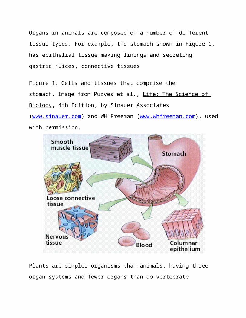
Organs in animals are composed of a number of different
tissue types. For example, the stomach shown in Figure 1,
has epithelial tissue making linings and secreting
gastric juices, connective tissues
Figure 1. Cells and tissues that comprise the
stomach. Image from Purves et al., Life: The Science of
Biology, 4th Edition, by Sinauer Associates
(www.sinauer.com) and WH Freeman (www.whfreeman.com), used
with permission.
Plants are simpler organisms than animals, having three
organ systems and fewer organs than do vertebrate
animals. Organs are composed of tissues, which are in
turn composed of cells. Plants have three tissue types:
ground, dermal, and vascular. Animals have four:
epithelial, connective, muscle, and bone.
Epithelial Tissue | Back to Top
Epithelial tissue covers body surfaces and lines body
cavities. Functions include lining, protecting, and
forming glands. Three types of epithelium occur:
Squamous epithelium is flattened cells.
Cuboidal epithelium is cube-shaped cells.
Columnar epithelium consists of elongated cells.
Any epithelium can be simple or stratified. Simple
epithelium has only a single cell layer. Stratified
epithelium has more than one layer of cells. Pseudo
stratified epithelium is a single layer of cells so
shaped that they appear at first glance to form two
layers.
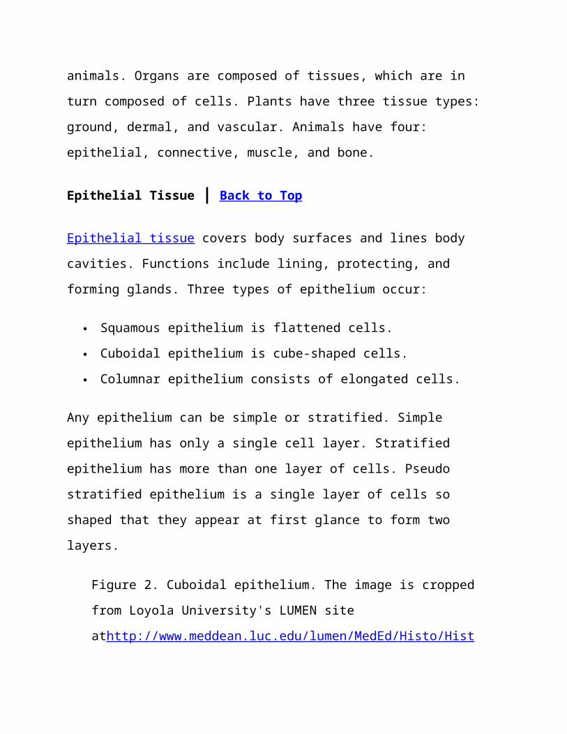
Figure 2. Cuboidal epithelium. The image is cropped
from Loyola University's LUMEN site
athttp://www.meddean.luc.edu/lumen/MedEd/Histo/Hist
oImages/hl1-04.jpg. Note the single layer of simple
cuboidal epithelium lining either side of a tubule.
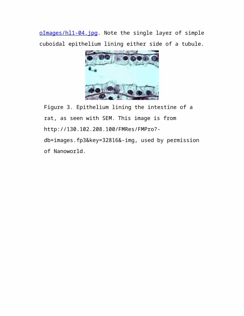
Figure 3. Epithelium lining the intestine of a
rat, as seen with SEM. This image is from
http://130.102.208.100/FMRes/FMPro?-
db=images.fp3&key=32816&-img, used by permission
of Nanoworld.
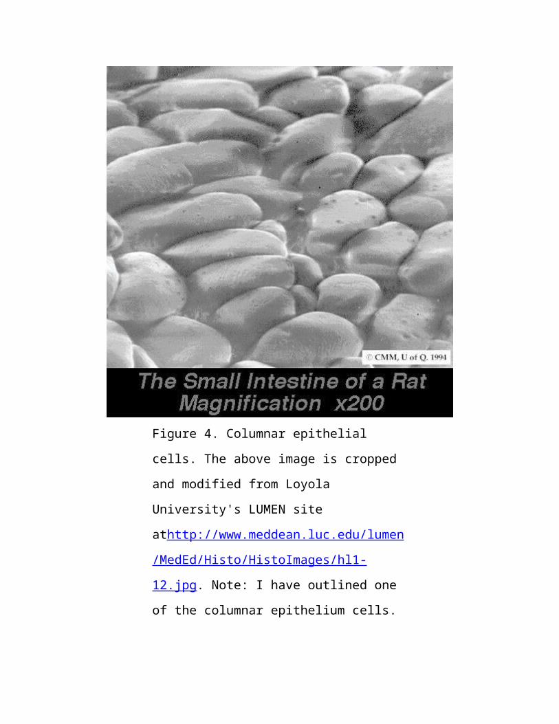
Figure 4. Columnar epithelial
cells. The above image is cropped
and modified from Loyola
University's LUMEN site
athttp://www.meddean.luc.edu/lumen
/MedEd/Histo/HistoImages/hl1-
12.jpg. Note: I have outlined one
of the columnar epithelium cells.
Functions of epithelial cells include:
movement materials in, out, or around the body.
protection of the internal environment against the
external environment.
Secretion of a product.
Glands can be single epithelial cells, such as the goblet
cells that line the intestine. Multicellular glands
include the endocrine glands. Many animals have their
skin composed of epithelium. Vertebrates have keratin in
their skin cells to reduce water loss. Many other animals
secrete mucus or other materials from their skin, such as
earthworms do.
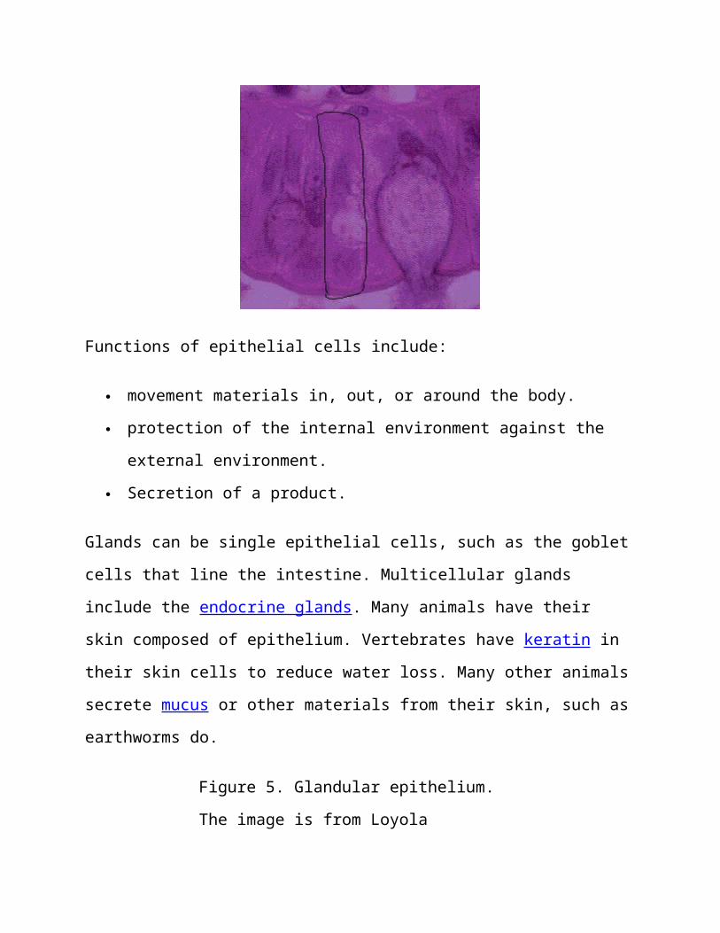
Figure 5. Glandular epithelium.
The image is from Loyola
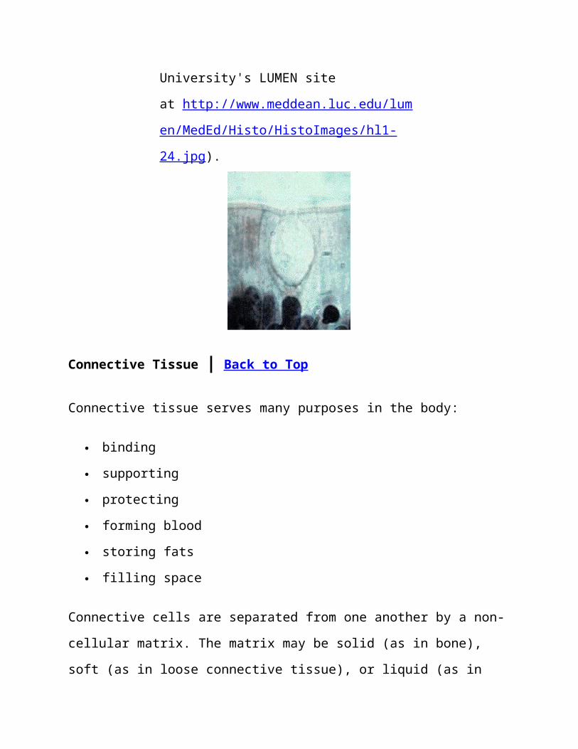
University's LUMEN site
at http://www.meddean.luc.edu/lum
en/MedEd/Histo/HistoImages/hl1-
24.jpg).
Connective Tissue | Back to Top
Connective tissue serves many purposes in the body:
binding
supporting
protecting
forming blood
storing fats
filling space
Connective cells are separated from one another by a non-
cellular matrix. The matrix may be solid (as in bone),
soft (as in loose connective tissue), or liquid (as in
blood). Two types of connective tissue are Loose
Connective Tissue (LCT) and Fibrous Connective Tissue
(FCT). Fibroblasts (LCT) are separated by a collagen
fiber-containing matrix. Collagen fibers provide
elasticity and flexibility. LCT occurs beneath epithelium
in skin and many internal organs, such as lungs, arteries
and the urinary bladder. This tissue type also forms a
protective layer over muscle, nerves, and blood vessels.
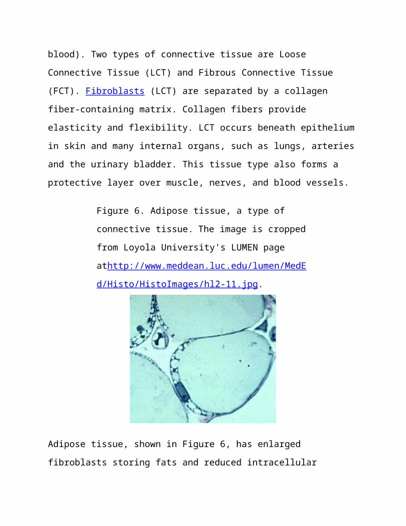
Figure 6. Adipose tissue, a type of
connective tissue. The image is cropped
from Loyola University's LUMEN page
athttp://www.meddean.luc.edu/lumen/MedE
d/Histo/HistoImages/hl2-11.jpg.
Adipose tissue, shown in Figure 6, has enlarged
fibroblasts storing fats and reduced intracellular
matrix. Adipose tissue facilitates energy storage and
insulation.
Fibrous Connective Tissue has many fibers of collagen
closely packed together. FCT occurs in tendons, which
connect muscle to bone.Ligaments are also composed of FCT
and connect bone to bone at a joint.
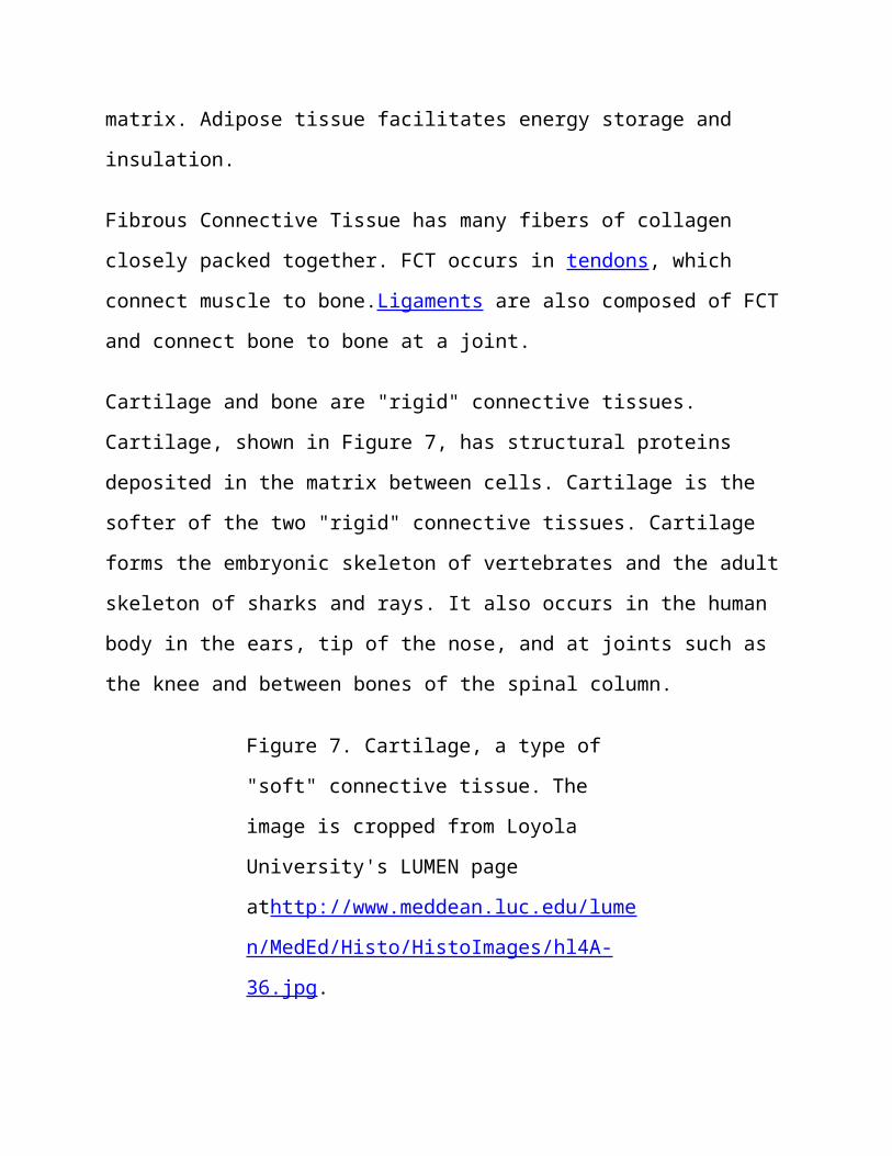
Cartilage and bone are "rigid" connective tissues.
Cartilage, shown in Figure 7, has structural proteins
deposited in the matrix between cells. Cartilage is the
softer of the two "rigid" connective tissues. Cartilage
forms the embryonic skeleton of vertebrates and the adult
skeleton of sharks and rays. It also occurs in the human
body in the ears, tip of the nose, and at joints such as
the knee and between bones of the spinal column.
Figure 7. Cartilage, a type of
"soft" connective tissue. The
image is cropped from Loyola
University's LUMEN page
athttp://www.meddean.luc.edu/lume
n/MedEd/Histo/HistoImages/hl4A-
36.jpg.
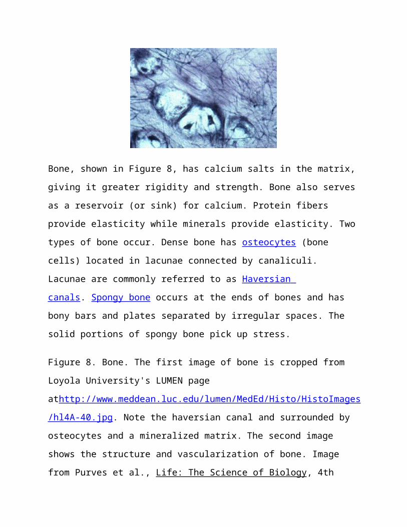
Bone, shown in Figure 8, has calcium salts in the matrix,
giving it greater rigidity and strength. Bone also serves
as a reservoir (or sink) for calcium. Protein fibers
provide elasticity while minerals provide elasticity. Two
types of bone occur. Dense bone has osteocytes (bone
cells) located in lacunae connected by canaliculi.
Lacunae are commonly referred to as Haversian
canals. Spongy bone occurs at the ends of bones and has
bony bars and plates separated by irregular spaces. The
solid portions of spongy bone pick up stress.
Figure 8. Bone. The first image of bone is cropped from
Loyola University's LUMEN page
athttp://www.meddean.luc.edu/lumen/MedEd/Histo/HistoImages
/hl4A-40.jpg. Note the haversian canal and surrounded by
osteocytes and a mineralized matrix. The second image
shows the structure and vascularization of bone. Image
from Purves et al., Life: The Science of Biology, 4th
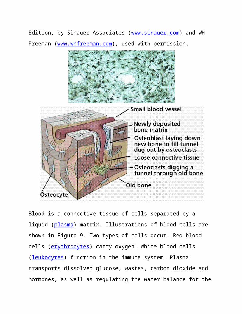
Edition, by Sinauer Associates (www.sinauer.com) and WH
Freeman (www.whfreeman.com), used with permission.
Blood is a connective tissue of cells separated by a
liquid (plasma) matrix. Illustrations of blood cells are
shown in Figure 9. Two types of cells occur. Red blood
cells (erythrocytes) carry oxygen. White blood cells
(leukocytes) function in the immune system. Plasma
transports dissolved glucose, wastes, carbon dioxide and
hormones, as well as regulating the water balance for the
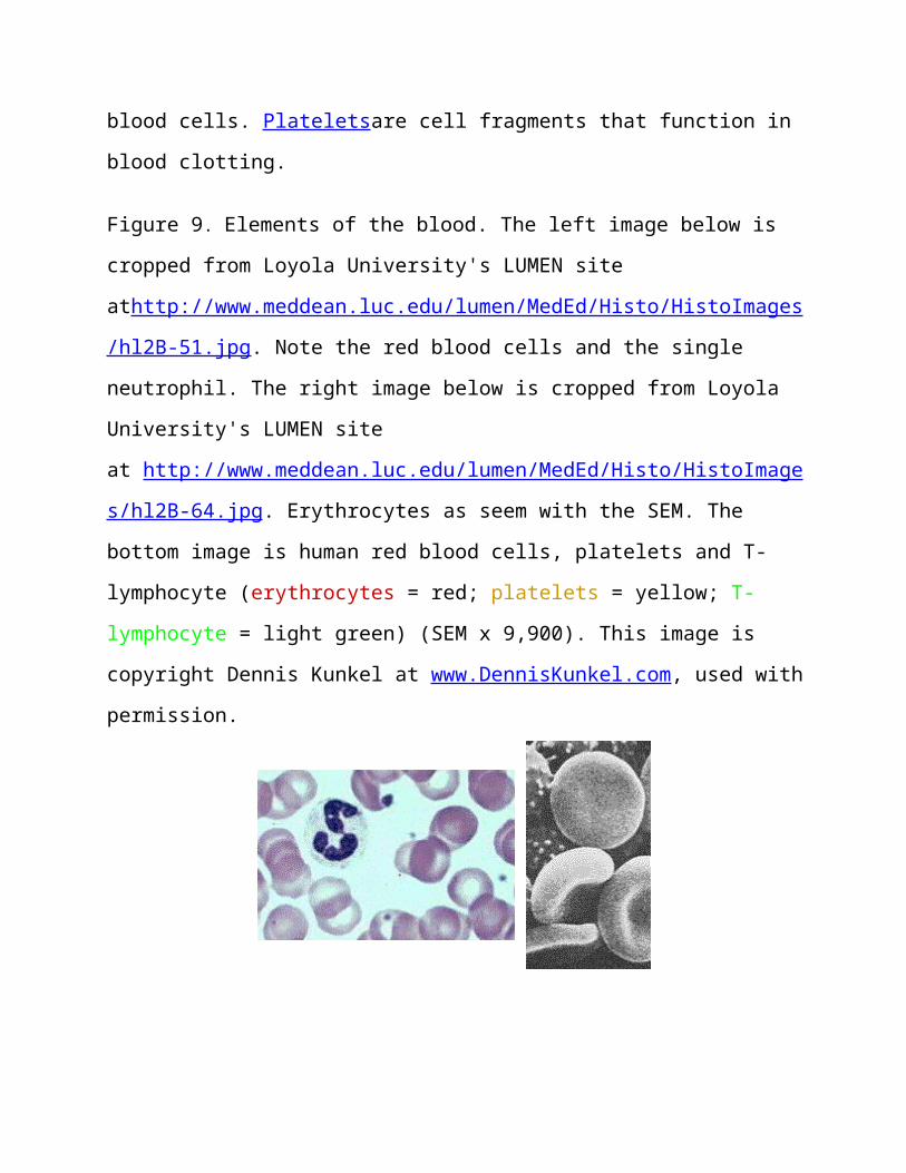
blood cells. Plateletsare cell fragments that function in
blood clotting.
Figure 9. Elements of the blood. The left image below is
cropped from Loyola University's LUMEN site
athttp://www.meddean.luc.edu/lumen/MedEd/Histo/HistoImages
/hl2B-51.jpg. Note the red blood cells and the single
neutrophil. The right image below is cropped from Loyola
University's LUMEN site
at http://www.meddean.luc.edu/lumen/MedEd/Histo/HistoImage
s/hl2B-64.jpg. Erythrocytes as seem with the SEM. The
bottom image is human red blood cells, platelets and T-
lymphocyte (erythrocytes = red; platelets = yellow; T-
lymphocyte = light green) (SEM x 9,900). This image is
copyright Dennis Kunkel at www.DennisKunkel.com, used with
permission.
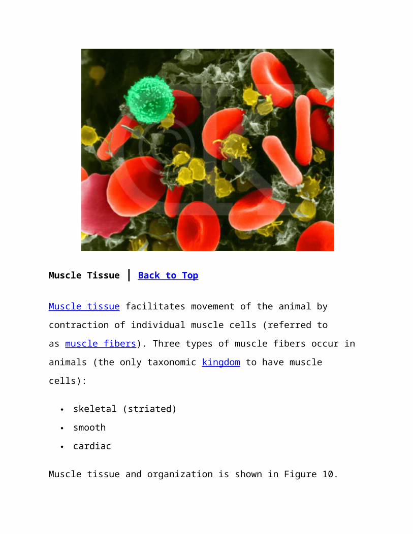
Muscle Tissue | Back to Top
Muscle tissue facilitates movement of the animal by
contraction of individual muscle cells (referred to
as muscle fibers). Three types of muscle fibers occur in
animals (the only taxonomic kingdom to have muscle
cells):
skeletal (striated)
smooth
cardiac
Muscle tissue and organization is shown in Figure 10.

Figure 10. Organization of muscle tissue. Images from
Purves et al., Life: The Science of Biology, 4th Edition,
by Sinauer Associates (www.sinauer.com) and WH Freeman
(www.whfreeman.com), used with permission.
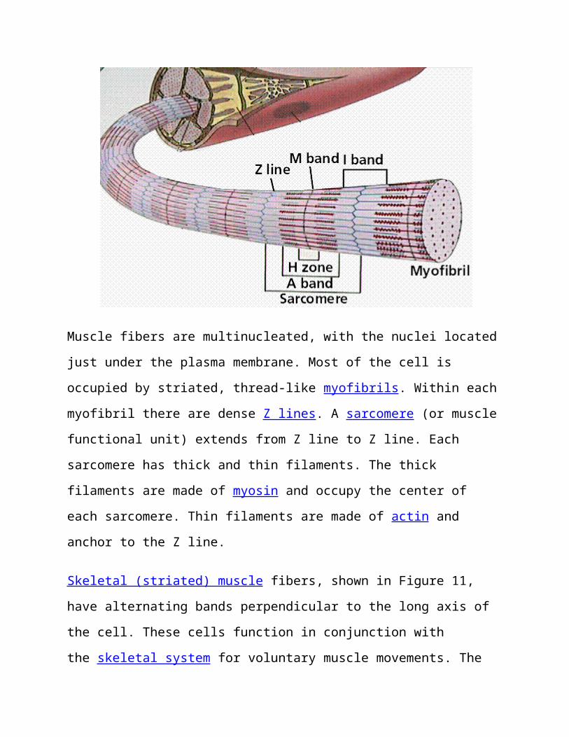
Muscle fibers are multinucleated, with the nuclei located
just under the plasma membrane. Most of the cell is
occupied by striated, thread-like myofibrils. Within each
myofibril there are dense Z lines. A sarcomere (or muscle
functional unit) extends from Z line to Z line. Each
sarcomere has thick and thin filaments. The thick
filaments are made of myosin and occupy the center of
each sarcomere. Thin filaments are made of actin and
anchor to the Z line.
Skeletal (striated) muscle fibers, shown in Figure 11,
have alternating bands perpendicular to the long axis of
the cell. These cells function in conjunction with
the skeletal system for voluntary muscle movements. The
bands are areas of actin and myosin deposition in the
cells.
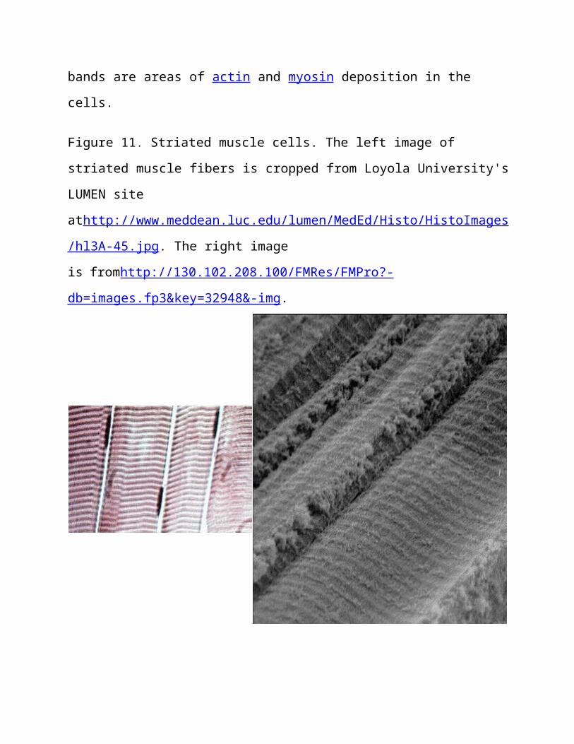
Figure 11. Striated muscle cells. The left image of
striated muscle fibers is cropped from Loyola University's
LUMEN site
athttp://www.meddean.luc.edu/lumen/MedEd/Histo/HistoImages
/hl3A-45.jpg. The right image
is fromhttp://130.102.208.100/FMRes/FMPro?-
db=images.fp3&key=32948&-img.
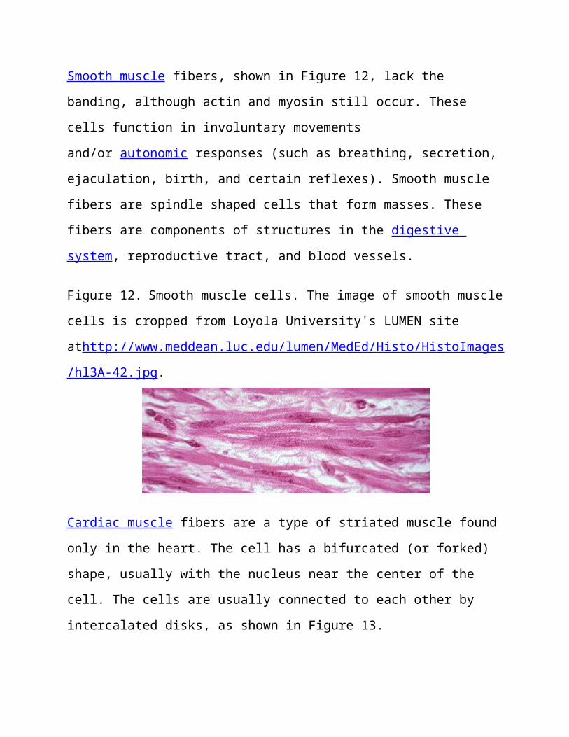
Smooth muscle fibers, shown in Figure 12, lack the
banding, although actin and myosin still occur. These
cells function in involuntary movements
and/or autonomic responses (such as breathing, secretion,
ejaculation, birth, and certain reflexes). Smooth muscle
fibers are spindle shaped cells that form masses. These
fibers are components of structures in the digestive
system, reproductive tract, and blood vessels.
Figure 12. Smooth muscle cells. The image of smooth muscle
cells is cropped from Loyola University's LUMEN site
athttp://www.meddean.luc.edu/lumen/MedEd/Histo/HistoImages
/hl3A-42.jpg.
Cardiac muscle fibers are a type of striated muscle found
only in the heart. The cell has a bifurcated (or forked)
shape, usually with the nucleus near the center of the
cell. The cells are usually connected to each other by
intercalated disks, as shown in Figure 13.
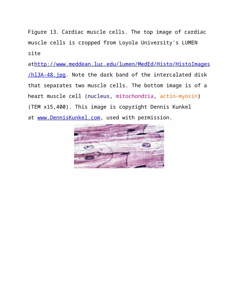
Figure 13. Cardiac muscle cells. The top image of cardiac
muscle cells is cropped from Loyola University's LUMEN
site
athttp://www.meddean.luc.edu/lumen/MedEd/Histo/HistoImages
/hl3A-48.jpg. Note the dark band of the intercalated disk
that separates two muscle cells. The bottom image is of a
heart muscle cell (nucleus, mitochondria, actin-myosin)
(TEM x15,400). This image is copyright Dennis Kunkel
at www.DennisKunkel.com, used with permission.
Nervous Tissue | Back to Top
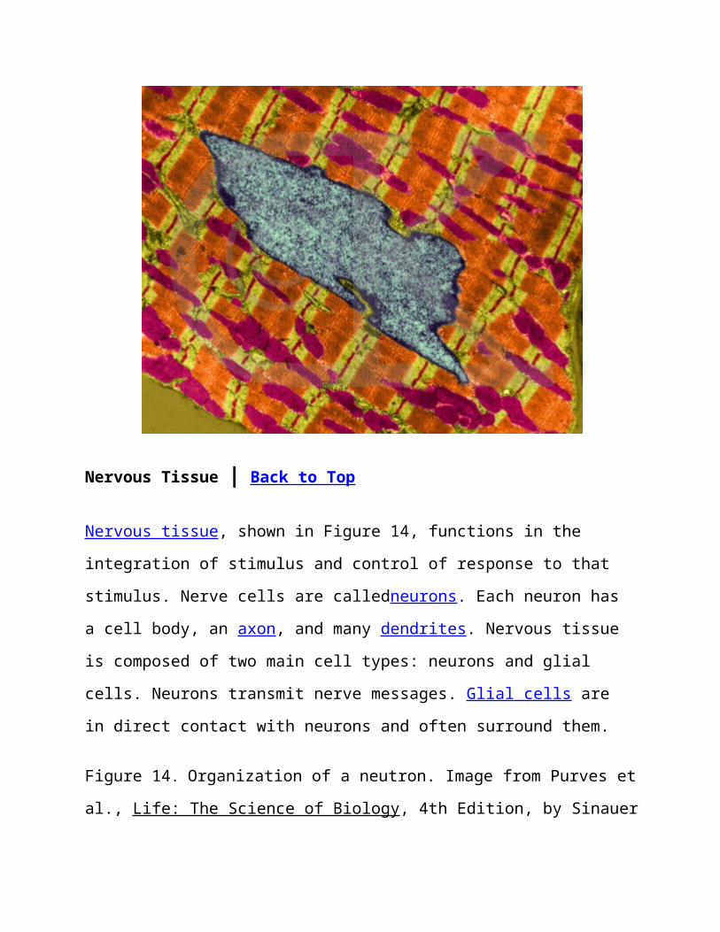
Nervous tissue, shown in Figure 14, functions in the
integration of stimulus and control of response to that
stimulus. Nerve cells are calledneurons. Each neuron has
a cell body, an axon, and many dendrites. Nervous tissue
is composed of two main cell types: neurons and glial
cells. Neurons transmit nerve messages. Glial cells are
in direct contact with neurons and often surround them.
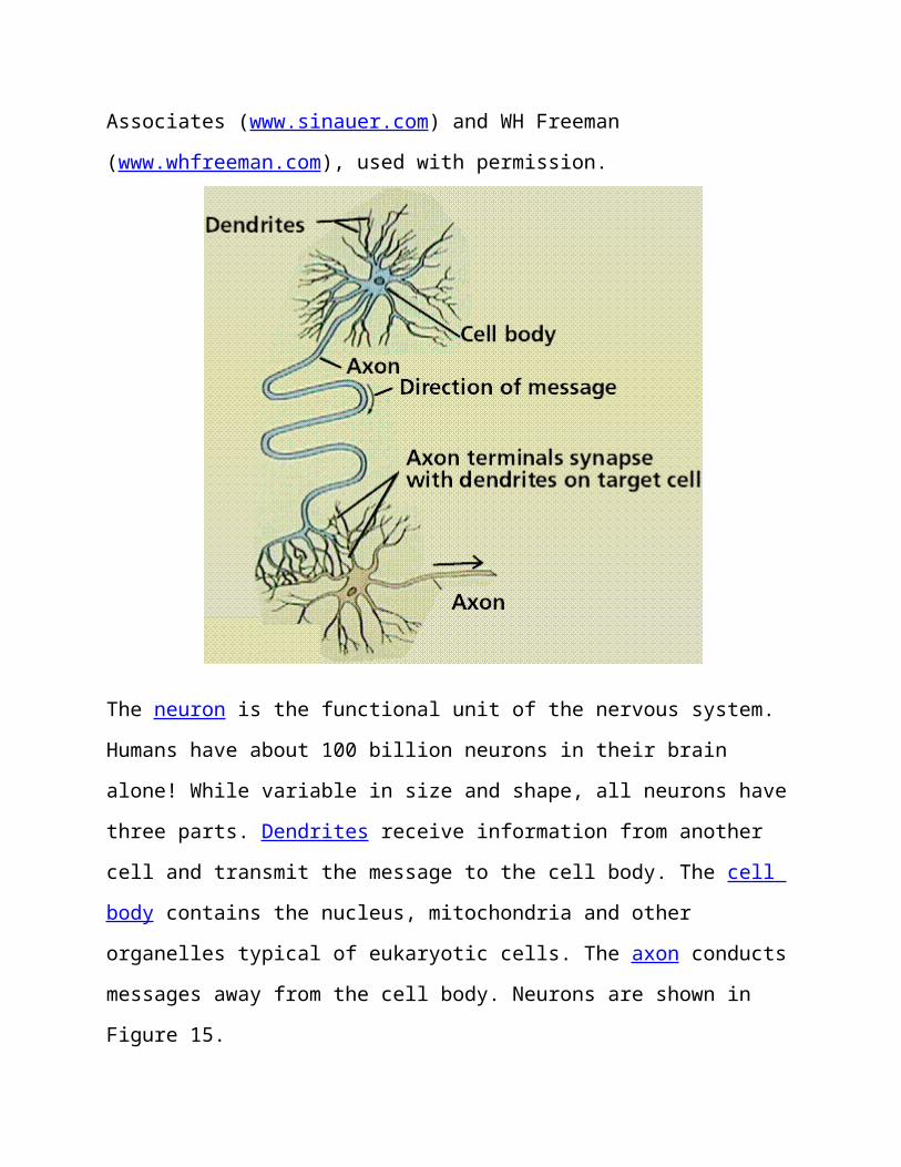
Figure 14. Organization of a neutron. Image from Purves et
al., Life: The Science of Biology, 4th Edition, by Sinauer
Associates (www.sinauer.com) and WH Freeman
(www.whfreeman.com), used with permission.
The neuron is the functional unit of the nervous system.
Humans have about 100 billion neurons in their brain
alone! While variable in size and shape, all neurons have
three parts. Dendrites receive information from another
cell and transmit the message to the cell body. The cell
body contains the nucleus, mitochondria and other
organelles typical of eukaryotic cells. The axon conducts
messages away from the cell body. Neurons are shown in
Figure 15.
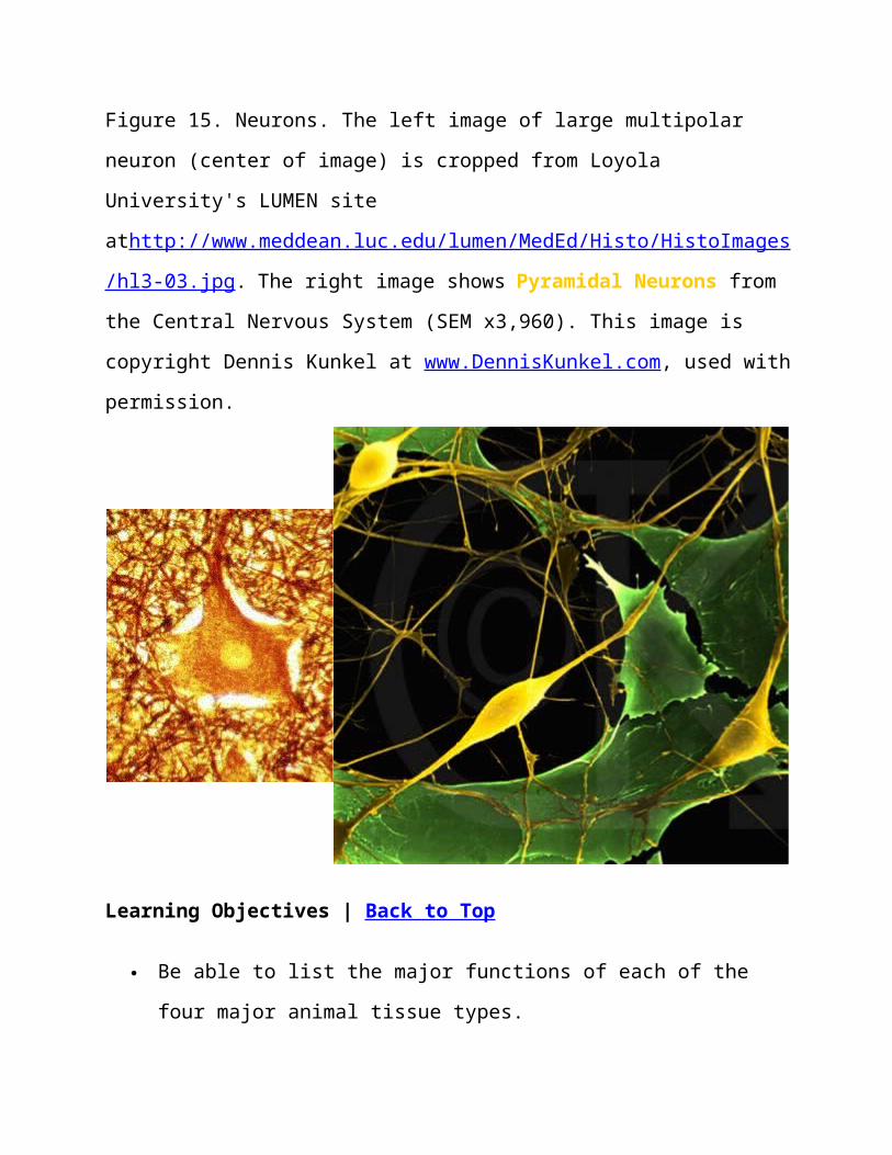
Figure 15. Neurons. The left image of large multipolar
neuron (center of image) is cropped from Loyola
University's LUMEN site
athttp://www.meddean.luc.edu/lumen/MedEd/Histo/HistoImages
/hl3-03.jpg. The right image shows Pyramidal Neurons from
the Central Nervous System (SEM x3,960). This image is
copyright Dennis Kunkel at www.DennisKunkel.com, used with
permission.
Learning Objectives | Back to Top
Be able to list the major functions of each of the
four major animal tissue types.
Distinguish between simple and stratified epithelial
tissue.
Compare and contrast the different types of
connective tissues: loose, dense, fibrous, cartilage,
bone, blood, adipose. Be able to list the function of
each type.
Know the three types of muscle and be able to
differentiate them visually and according to their
functions.
Be able to diagram a typical neuron and its three
areas: dendrite, axon, and cell body.
Know the characteristics of the various types of
animal tissues. Learn the types of cells that compose
each tissue type and be able to give some examples of
organs that contain significant amounts of each
tissue type.
Detail the functions carried out by epithelial tissue
and state the general location of each type.
Be able to discuss the meaning of the term gland,
cite three examples of glands, and state the
extracellular products secreted by each.
Describe the basic features of connective tissue, and
explain how the cells of this tissue type enable
connective tissue to carry out its various tasks.
List three of the functions of blood.
List two functions of bone and/or cartilage.
Distinguish among skeletal, cardiac, and smooth
muscle tissues in terms of location, structure, and
function.
Muscle tissues contain specialized cells that can
contract.
Neurons are organized as lines of communicaiton.
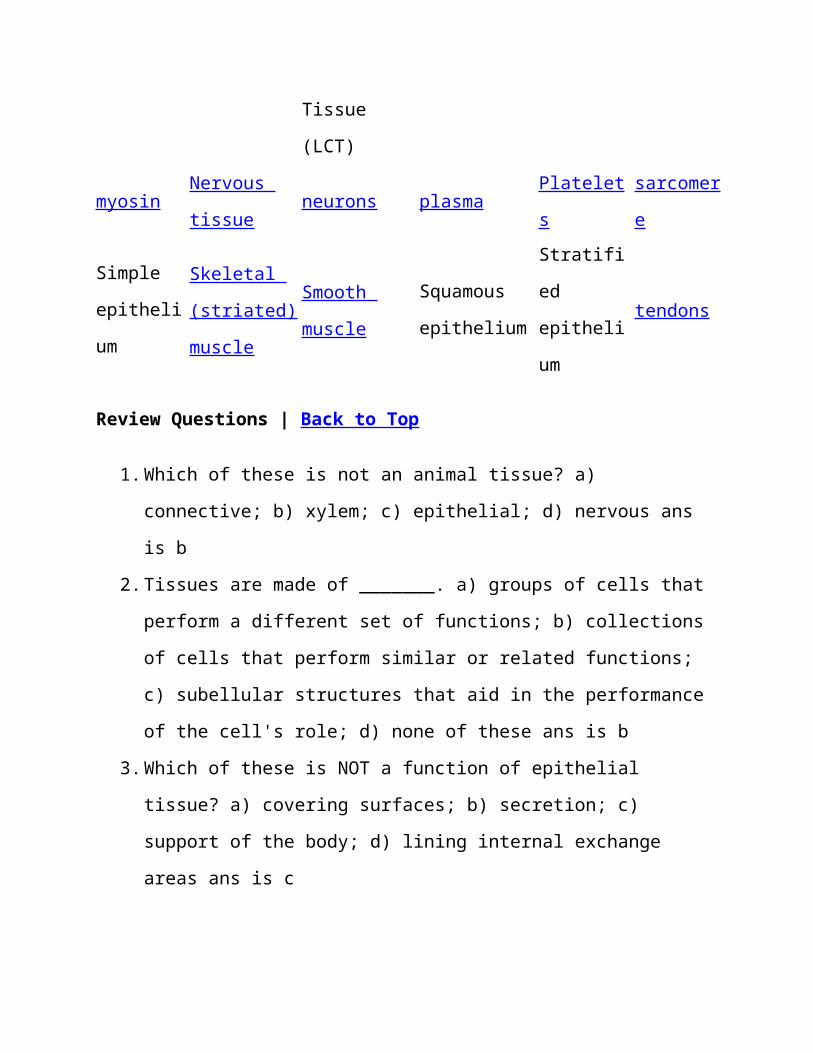
Terms | Back to Top
Adipose
tissueactin axon blood bone
Cardiac
muscleCartilag
e
Columnar
epithelium
Connective
tissue
Cuboidal
epitheliumdendrite
s
endocrin
e glands
Epitheli
al
tissue
erythrocyt
es
Fibroblast
s
Fibrous
Connective
Tissue
(FCT)
Glial
cellskeratin
leukocyt
es
Ligaments Loose
Connective
mucus Muscle
tissue
myofibri
ls
Tissue
(LCT)
myosinNervous
tissueneurons plasma
Platelet
s
sarcomer
e
Simple
epitheli
um
Skeletal
(striated)
muscle
Smooth
muscle
Squamous
epithelium
Stratifi
ed
epitheli
um
tendons
Review Questions | Back to Top
1.Which of these is not an animal tissue? a)
connective; b) xylem; c) epithelial; d) nervous ans
is b
2.Tissues are made of _______. a) groups of cells that
perform a different set of functions; b) collections
of cells that perform similar or related functions;
c) subellular structures that aid in the performance
of the cell's role; d) none of these ans is b
3.Which of these is NOT a function of epithelial
tissue? a) covering surfaces; b) secretion; c)
support of the body; d) lining internal exchange
areas ans is c
4.Layered epithelial tissue is referred to as which of
these? a) squamous; b) stratified; c) voluntary; d)
pseudostratified ans is d
5.Which of these cell types covers the inside of the
mouth? a) squamous epithelium; b) cartilage; c)
blood; d) cuboidal epithelium ANS is a
6.Protection of the body from infectious organisms is
accomplished by which of these tissues? a) bone; b)
muscle; c) nerve; d) blood ANS is d
7.Linking of bone to bone in a skeletal system is
accomplished by which of these tissues? a)
epithelial; b) connective; c) muscle; d) nervous ANS
is b
8.Cells that line the tubules in the kidney make up
which of these tissues? a) adipose; b) squamous
epithelium; c) cuboidal epithelium; d) stratified
epithelium ANS is c
9.The storage of fat is accomplished by which of these
cell types? a) adipose; b) squamous epithelium; c)
cuboidal epithelium; d) stratified epithelium ANS is
a
10. Glands are composed of which of these tissue
types? a) epithelium; b) connective; c) muscle; d)
nervous ANS is a
11. Hard parts of the body would be made of which of
these cell/tissue types? a) blood; b) bone; c)
muscle; d) nerves ANS is b
12. Bone acts as a reservoir for which of these
elements? a) carbon; b) nitrogen; c) calcium; d)
hydrogen ANS is c
13. The major function of bone is ___. a) covering
body surfaces; b) support; c) movement; d)
integration of stimulus ANS is b
14. New blood cells are formed in the ___. a)
matrix; b) bone marrow; c) liver; d) adipose cells
AMS is b
15. The blood cells that transport oxygen within the
body are the ___. a) macrophages; b) erythrocytes; c)
platelets; d) leukocytes ANS is b
16. The liquid part of the blood is ___. a) plasma;
b) adipose; c) cartilage; d) platelets ANS is a
17. When you move your arm to use your computer
mouse, which of these muscle cell types is involved?
a) cardiac; b) skeletal; c) smooth ANS is b
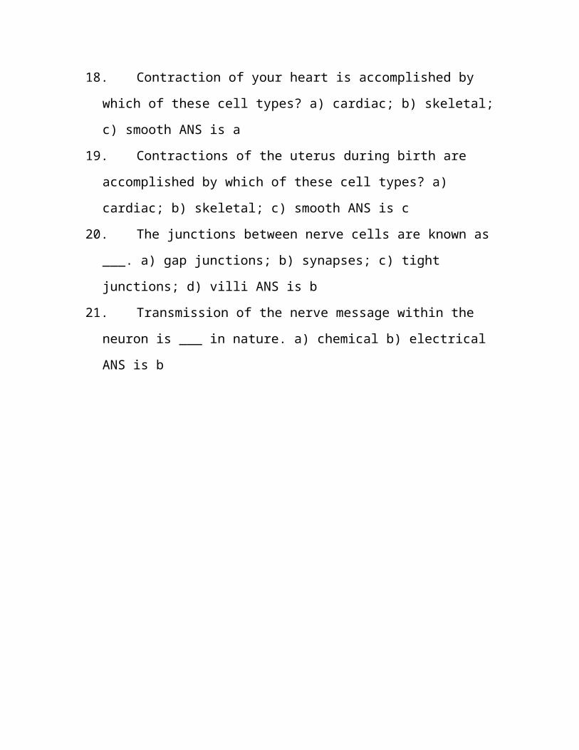
18. Contraction of your heart is accomplished by
which of these cell types? a) cardiac; b) skeletal;
c) smooth ANS is a
19. Contractions of the uterus during birth are
accomplished by which of these cell types? a)
cardiac; b) skeletal; c) smooth ANS is c
20. The junctions between nerve cells are known as
___. a) gap junctions; b) synapses; c) tight
junctions; d) villi ANS is b
21. Transmission of the nerve message within the
neuron is ___ in nature. a) chemical b) electrical
ANS is b