Differential gene expression associated with postnatal equine articular cartilage maturation
Upload
independentCategory
view
0download
0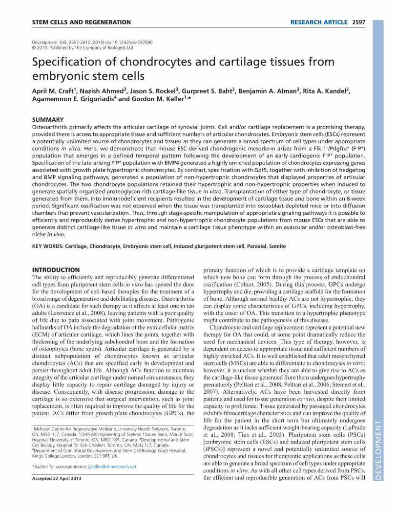
STEM CELLS AND REGENERATION RESEARCH ARTICLE 2597
Development 140, 2597-2610 (2013) doi:10.1242/dev.087890© 2013. Published by The Company of Biologists Ltd
INTRODUCTIONThe ability to efficiently and reproducibly generate differentiatedcell types from pluripotent stem cells in vitro has opened the doorfor the development of cell-based therapies for the treatment of abroad range of degenerative and debilitating diseases. Osteoarthritis(OA) is a candidate for such therapy as it affects at least one in tenadults (Lawrence et al., 2008), leaving patients with a poor qualityof life due to pain associated with joint movement. Pathogenichallmarks of OA include the degradation of the extracellular matrix(ECM) of articular cartilage, which lines the joints, together withthickening of the underlying subchondral bone and the formationof osteophytes (bone spurs). Articular cartilage is generated by adistinct subpopulation of chondrocytes known as articularchondrocytes (ACs) that are specified early in development andpersist throughout adult life. Although ACs function to maintainintegrity of the articular cartilage under normal circumstances, theydisplay little capacity to repair cartilage damaged by injury ordisease. Consequently, with disease progression, damage to thecartilage is so extensive that surgical intervention, such as jointreplacement, is often required to improve the quality of life for thepatient. ACs differ from growth plate chondrocytes (GPCs), the
primary function of which is to provide a cartilage template onwhich new bone can form through the process of endochondralossification (Colnot, 2005). During this process, GPCs undergohypertrophy and die, providing a cartilage scaffold for the formationof bone. Although normal healthy ACs are not hypertrophic, theycan display some characteristics of GPCs, including hypertrophy,with the onset of OA. This transition to a hypertrophic phenotypemight contribute to the pathogenesis of this disease.
Chondrocyte and cartilage replacement represent a potential newtherapy for OA that could, at some point dramatically reduce theneed for mechanical devices. This type of therapy, however, isdependent on access to appropriate tissue and sufficient numbers ofhighly enriched ACs. It is well established that adult mesenchymalstem cells (MSCs) are able to differentiate to chondrocytes in vitro;however, it is unclear whether they are able to give rise to ACs asthe cartilage-like tissue generated from them undergoes hypertrophyprematurely (Pelttari et al., 2008; Pelttari et al., 2006; Steinert et al.,2007). Alternatively, ACs have been harvested directly frompatients and used for tissue generation ex vivo, despite their limitedcapacity to proliferate. Tissue generated by passaged chondrocytesexhibits fibrocartilage characteristics and can improve the quality oflife for the patient in the short term but ultimately undergoesdegradation as it lacks sufficient weight-bearing capacity (LaPradeet al., 2008; Tins et al., 2005). Pluripotent stem cells (PSCs)[embryonic stem cells (ESCs) and induced pluripotent stem cells(iPSCs)] represent a novel and potentially unlimited source ofchondrocytes and tissues for therapeutic applications as these cellsare able to generate a broad spectrum of cell types under appropriateconditions in vitro. As with all other cell types derived from PSCs,the efficient and reproducible generation of ACs from PSCs will
1McEwen Centre for Regenerative Medicine, University Health Network, Toronto,ON, M5G 1L7, Canada. 2CIHR-BioEngineering of Skeletal Tissues Team, Mount SinaiHospital, University of Toronto, ON, M5G 1X5, Canada. 3Developmental and StemCell Biology, Hospital for Sick Children, Toronto, ON, M5G 1L7, Canada.4Department of Craniofacial Development and Stem Cell Biology, Guy’s Hospital,King’s College London, London, SE1 9RT, UK.
*Author for correspondence ([email protected])
Accepted 22 April 2013
SUMMARYOsteoarthritis primarily affects the articular cartilage of synovial joints. Cell and/or cartilage replacement is a promising therapy,provided there is access to appropriate tissue and sufficient numbers of articular chondrocytes. Embryonic stem cells (ESCs) representa potentially unlimited source of chondrocytes and tissues as they can generate a broad spectrum of cell types under appropriateconditions in vitro. Here, we demonstrate that mouse ESC-derived chondrogenic mesoderm arises from a Flk-1–/Pdgfrα+ (F–P+)population that emerges in a defined temporal pattern following the development of an early cardiogenic F–P+ population.Specification of the late-arising F–P+ population with BMP4 generated a highly enriched population of chondrocytes expressing genesassociated with growth plate hypertrophic chondrocytes. By contrast, specification with Gdf5, together with inhibition of hedgehogand BMP signaling pathways, generated a population of non-hypertrophic chondrocytes that displayed properties of articularchondrocytes. The two chondrocyte populations retained their hypertrophic and non-hypertrophic properties when induced togenerate spatially organized proteoglycan-rich cartilage-like tissue in vitro. Transplantation of either type of chondrocyte, or tissuegenerated from them, into immunodeficient recipients resulted in the development of cartilage tissue and bone within an 8-weekperiod. Significant ossification was not observed when the tissue was transplanted into osteoblast-depleted mice or into diffusionchambers that prevent vascularization. Thus, through stage-specific manipulation of appropriate signaling pathways it is possible toefficiently and reproducibly derive hypertrophic and non-hypertrophic chondrocyte populations from mouse ESCs that are able togenerate distinct cartilage-like tissue in vitro and maintain a cartilage tissue phenotype within an avascular and/or osteoblast-freeniche in vivo.
KEY WORDS: Cartilage, Chondrocyte, Embryonic stem cell, Induced pluripotent stem cell, Paraxial, Somite
Specification of chondrocytes and cartilage tissues fromembryonic stem cellsApril M. Craft1, Nazish Ahmed2, Jason S. Rockel3, Gurpreet S. Baht3, Benjamin A. Alman3, Rita A. Kandel2,Agamemnon E. Grigoriadis4 and Gordon M. Keller1,*
DEVELO
PMENT
2598
depend on our ability to recapitulate key aspects of embryonicdevelopment in vitro.
Chondrocytes in the vertebrae and ribs develop from paraxialmesoderm, whereas chondrocytes in the long bones and most of thegirdles are derived from lateral plate mesoderm (LPM), which alsogives rise to hematopoietic and cardiovascular lineages (Kinder etal., 1999; Lawson et al., 1991). Following induction, strips ofparaxial mesoderm are segmented into somites (Kulesa and Fraser,2002; Tam and Tan, 1992). Somite development is regulated, in part,by the transcription factors paraxis (Tcf15) and Tbx18, theexpression of which coincides with the induction of paraxialmesoderm (Burgess et al., 1996; Bussen et al., 2004; Singh et al.,2005). Individual somites are then patterned into the ventralsclerotome, which forms the axial skeleton, including cartilage andthe vertebral column, and the dorsal dermomyotome whichdevelops into skeletal muscles and the dermis of the back (Hirsingeret al., 2000). Specification of the sclerotome is marked by theexpression of two transcription factors, Meox1 (Mankoo et al.,2003) and Nkx3.2 (also known as Bapx1). A population of collagen2 (Col2a1)-positive mesenchymal cells with chondrogenic potentialdevelops from sclerotome-derived cells at embryonic day (E) 12.5of mouse development (Akiyama et al., 2002; Dao et al., 2012).
Whereas methods for differentiating progenitor cells to thechondrogenic lineage are well established, the ability to specifyACs, and ultimately articular cartilage tissue, remains poorlyunderstood. ACs are derived from interzone cells, a fibroticpopulation of cells that forms at future sites of synovial joints,marked by the upregulation of Wnt9a (previously known as Wnt14)and growth and differentiation factor 5 (Gdf5; previously known asBMP14 in human), a member of the TGFβ superfamily (Archer etal., 2003; Pacifici et al., 2006). Lineage-tracing studies have shownthat Gdf5-expressing interzone cells give rise to several joint tissues,including ACs, but do not contribute to the GPC population(Koyama et al., 2008). GPCs, by contrast, develop from thecondensing chondrogenic mesenchyme and express Bmp2, Bmp4and Bmp7, as well as hypertrophy-related genes, including collagen10. Distinct regions of ACs and GPCs are observed as early aspostnatal day 7-8 when the secondary ossification center begins toform (Blumer et al., 2007; Murakami et al., 2004). Theseobservations suggest that ACs and GPCs are generated fromseparate progenitor populations during development and, as such,might represent distinct lineages.
A number of studies have demonstrated that it is possible toderive chondrocytes from mouse (m) and human ESCs and iPSCsin vitro. Most, however, used serum-based media to support theearly stages of differentiation, resulting in the generation of mixedlineage end-stage cultures (Hwang et al., 2006; Hwang et al., 2008;Jukes et al., 2008; Kramer et al., 2000; Yamashita et al., 2008; zurNieden et al., 2005). Recent studies have reported the use of definedculture media with specific pathway agonists and antagonists todirect differentiation and in doing so have provided insights into theregulation of paraxial mesoderm and chondrocyte development(Darabi et al., 2008; Nakayama et al., 2003; Tanaka et al., 2009). Inone of the most detailed studies with mESCs, Tanaka et al. (Tanakaet al., 2009) showed that the combination of Wnt signaling withbone morphogenetic protein (BMP) inhibition resulted in thegeneration of paraxial mesoderm with chondrogenic potential,identified by the expression of Pdgfrα and a lack of expression ofFlk-1 (Kdr – Mouse Genome Informatics). This mesoderm alsodisplayed some cardiac potential but showed no capacity to generatehematopoietic cells, indicating that dependency on BMP signalingdistinguishes different types of mesoderm.
In this study, we have traced the origin of the chondrogeniclineage from mESCs and demonstrate that it arises from a Flk-1–/Pdgfrα+ (F–P+) mesoderm population that emerges following thedevelopment of an early cardiogenic F–P+ population. Specificationof this chondrogenic mesoderm with either BMP4 or thecombination of Gdf5, cyclopamine and soluble Bmpr1α (sBmpr1α)resulted in the development of populations with hypertrophic andnon-hypertrophic chondrocyte characteristics, respectively. Thesechondrocyte populations formed spatially organized, proteoglycan-rich tissue in vitro and retained their hypertrophic and non-hypertrophic phenotype during this process. Transplantation ofeither type of chondrocyte or tissues generated from them intoimmunodeficient recipients resulted in the development of cartilagetissue and bone within an 8-week period. Ossification wasdramatically reduced or completely absent in tissue transplantedinto osteoblast-depleted mice or in diffusion chambers thatprevented host vascularization. Together, these findings demonstratethat through stage-specific manipulation of appropriate signalingpathways it is possible to efficiently and reproducibly generatechondrogenic mesoderm that can be specified into populations withhypertrophic and non-hypertrophic chondrocyte properties.
MATERIALS AND METHODSESC maintenance and differentiationGFP-Bry (Fehling et al., 2003) mESCs, T-EGFP/Rosa26-tdRFP (RFP.bry)(Luche et al., 2007) mESCs and Oct4-GFP miPSCs (Stadtfeld et al., 2008)were maintained in a modified serum-free (SF), feeder-free culture systemas described previously (Gadue et al., 2006; Ying et al., 2003). Fordifferentiation, ESCs were dissociated and cultured in suspension inserum-free differentiation medium (SFD) (Gouon-Evans et al., 2006)without additional growth factors for 48 hours. Embryoid bodies (EBs)were then dissociated and re-aggregated in SFD with the addition ofgrowth factors or inhibitors as indicated [9 ng/ml activin A (inhibin, betaA), 25 ng/ml Wnt3a, 5 ng/ml vascular endothelial growth factor (VEGF),150 ng/ml noggin, 0.5 ng/ml BMP4]. EBs were harvested 24 hours later,the cells dissociated and the appropriate populations isolated by cellsorting. For re-aggregation, sorted cells were cultured at 250,000 cells/mlin 24-well ULA dishes (Costar) for 48 hours in SFD containing 10 ng/mlbFGF (FGF2) and 10 μM Y-27632. Aggregates were harvested anddissociated to single-cell suspension, and appropriate populations wereisolated by cell sorting, if applicable. Cells were cultured in monolayer on0.1% gelatin-coated 96-well tissue culture dishes (Falcon, BectonDickinson) at 30,000-120,000 cells/ml in SFD containing 2 mM L-glutamine, 10 ng/ml bFGF, and the following, as indicated: 30-100 ng/mlBMP4, 30 ng/ml Gdf5, 0.25 uM KAAD-cyclopamine, 500 ng/mlsBmpr1α, 2-4 μM dorsomorphin (Sigma). Human activin A, BMP4,VEGF, bFGF and noggin, mouse Wnt3a and Gdf5, and sBmpr1α werepurchased from R&D Systems; Y-27632 and KAAD-cyclopamine wereobtained from Toronto Research Chemicals.
Flow cytometry and cell sortingEBs generated from mESC differentiation experiments were dissociated byincubation with TrypLE (Invitrogen) and stained with following antibodies:anti-mouse Flk-1-biotin, anti-mouse Pdgfrα (CD140a)-allophycocyanin(APC; clone APA5, eBioscience, San Diego, CA, USA), streptavidin-phycoerythrin (PE) or streptavidin-PE-Cy7 (BD Pharmingen). Mostantibody stains were performed at 4°C in PBS containing 5% (v/v) fetalcalf serum (FCS). For cell sorting, antibody stains were performed inIscove’s modified Dulbecco’s medium (IMDM) containing 0.2% BSA(Sigma). Cells were acquired using a LSR II flow cytometer (BectonDickinson) or sorted using a FACS ARIA II (Becton Dickinson). Analysiswas performed using FlowJo (Tree Star).
Quantitative real-time PCRTotal RNA was prepared with the RNAqueous-Micro Kit (Ambion) withDNase treatment. RNA (0.1-1 μg) was reverse transcribed using random
RESEARCH ARTICLE Development 140 (12)
DEVELO
PMENT
hexamers and oligo(dT) with Superscript III reverse transcriptase(Invitrogen). Real-time quantitative (Q)-PCR was performed on aMasterCycler EP RealPlex (Eppendorf) using Quantifast SYBR Green(Qiagen). Genomic DNA standards were used to evaluate the efficiency ofthe PCR and to calculate the copy number of each gene relative to thehousekeeping gene Actb. Mean and standard errors of three to sixindependent experiments were calculated. Student’s t-test was used toevaluate statistical significance. Oligonucleotide sequences are listed insupplementary material Table S1.
Alcian Blue staining and fluorescence microscopyMonolayer cultures were fixed in 4% paraformaldehyde. For Alcian Bluestaining, monolayers were washed in 0.5 N HCl, then stained with 0.25%(w/v) Alcian Blue 8GX (Sigma) in 0.5 N HCl overnight. Cultures werevisualized by microscopy or imaged using a Scanmaker i900 Microtekscanner. Alcian Blue dye was solubilized overnight in 8 M guanidinehydrochloride (Sigma) and quantified by absorbance at 595 nm using aspectrophotometer. For immunofluorescence, monolayer cultures werestained using an antibody against the cardiac isoform of troponin T (cTnT)(clone 13-11, NeoMarkers, Fremont, CA, USA) and a donkey anti-mouseAlexa488-conjugated secondary antibody (Invitrogen). Cultures werecounterstained with DAPI (Dako).
In vitro tissue formationMonolayer cultures derived in indicated conditions for 12 days were treatedwith 0.2% (w/v) collagenase (Sigma), followed by brief trypsinization toobtain a single-cell suspension. Cells (1.5×10⁶) were seeded onto type IIcollagen-coated Millicell culture plate inserts (60 mm2; Millipore, Bedford,MA, USA). Cultures were maintained in Dulbecco’s modified Eagle’smedium (DMEM) (containing 20% FBS, high glucose) for 2-5 weeks(Ahmed et al., 2009).
Cell injections and tissue transplantationsMonolayer cultures were dissociated and cells were resuspended in growthfactor-reduced matrigel (Becton Dickinson). Cells (500,000) were injectedin a volume of 30 μl subcutaneously near the mammary fat pad of B10;B6-Rag2tm1Fwa II2rgtm1Wjl female mice aged 4-6 weeks. Alternatively, biopsypunches of cartilage-like tissues (4 mm diameter; derived from either day5 unsorted GFP-Bry mESCs or Pdgfrα+ cells sorted from RFP.bry mESCs)were transplanted subcutaneously between the shoulder blades. Cartilage-like tissues were placed within diffusion chambers (Millipore) andtransplanted subcutaneously in B10;B6-Rag2tm1Fwa II2rgtm1Wjl female miceaged 4-6 weeks. Grafts were harvested as indicated.
DTK mice carry a herpes simplex virus thymidine kinase gene undercontrol of the osteoblast-specific 2.3-kb fragment of the rat collagen α1 typeI promoter (Dacquin et al., 2002; Visnjic et al., 2001). Upon treatment withgancyclovir (6.25 mg/kg/day) endogenous osteoblasts of DTK miceundergo ablation (Ng et al., 2011). Mice either lacking (DTK–) or carrying(DTK+) the DTK transgene were injected with mESC-derived chondrocytessubcutaneously at the mammary fat pad site as described above at 12-16weeks of age. All mice were treated with gancyclovir (Sigma) for an 8-week period, at which point the engraftments were harvested.
Histology and ImmunohistochemistryIn vitro-derived cartilage-like tissues, and engraftments from in vivo studieswere fixed in 10% formalin and embedded in paraffin. Five-micron-thicksections were stained with Toluidine Blue, Hematoxylin and Eosin (H&E),Safranin O, and von Kossa, as indicated. Immunohistochemical analysiswas performed using antibodies recognizing collagen type 2 (MS306-P,Labvision, Fremont, CA, USA), collagen 10 (X53; Quartett, Berlin,Germany) and red fluorescent protein (Abcam). Sections werecounterstained with Mayer’s Hematoxylin.
RESULTSInduction of chondrogenic mesodermIn order to generate chondrogenic mesoderm efficiently frommESCs, we used a protocol in which a primitive streak (PS)-likepopulation is formed and mesoderm is induced within a 24-hour
period between days 2 and 3 of differentiation by a combination ofactivin/nodal, Wnt and BMP signaling (Nostro et al., 2008). In thesestudies, activin/nodal signaling is initiated by the addition of activinA (activin) whereas BMP signaling is manipulated by the presenceor absence of the agonist BMP4 or the inhibitor noggin. We used theGFP-Bry mESC reporter cell line in order to monitor PS/earlymesoderm formation and to isolate populations by flow cytometrybased on GFP expression (Fehling et al., 2003) (Fig. 1A). The Oct4-GFP mouse iPSC line (Stadtfeld et al., 2008) was included todemonstrate the applicability of this protocol for miPSCdifferentiation (supplementary material Fig. S1).
Day 3 embryoid bodies (EBs) induced in the absence of BMPsignaling (no BMP4 or noggin) in the presence of activin, Wnt andVEGF contained 80-85% GFP-Bry+ cells, demonstrating efficientPS/mesoderm formation under these conditions. The addition ofBMP4 during the induction stage led to a modest increase in theproportion of GFP-Bry+ cells to >90%. Although GFP-Bry PS-likepopulations were efficiently generated with the three conditions, thesize of the specific mesoderm subsets, defined by the expressionprofiles of the cell surface receptors Flk-1 (F) and Pdgfrα (P),differed dramatically between the BMP4-induced and noggin-induced EBs in both mESC and miPSC cultures (Fig. 1B;supplementary material Fig. S1C). With this combination ofmarkers, the F+P– population represents hemangioblast/hematopoietic mesoderm, the F+P+ population cardiogenicmesoderm, and the F–P+ population chondrogenic mesoderm(Kattman et al., 2011; Takakura et al., 1997). BMP4-induced EBscontained a large F+P+ population, and substantially smaller F–P+
and F–P– populations. These EBs also contained a small, butdetectable F+P– population. By contrast, EBs differentiated in theabsence of BMP signaling had no detectable F+P– population, adramatically smaller F+P+ population and larger F–P+ and F–P–
populations than those induced with the agonist (Fig. 1B).Following induction, EBs were dissociated and cells were re-aggregated in the presence of bFGF for 2 days (day 5 ofdifferentiation) to specify a chondrogenic fate (Fletcher andHarland, 2008) (Fig. 1A).
Gene expression analyses of day 2 and day 3 EBs, and the day 5aggregates revealed distinct differences in the populations inducedin the presence or absence of BMP4 (Fig. 1C,D). Cells induced withBMP4 expressed significantly higher levels of genes associated withLPM [Mesp1, Foxf1a (Foxf1 – Mouse Genome Informatics)](Ormestad et al., 2004) hematopoietic (Gata1) and cardiac (Nkx2.5)development compared with those generated in the absence of BMPsignaling (Fig. 1C). Expression of Mesp1 was restricted to the day3 BMP4-induced EBs whereas Foxf1a was detected in both the day3 EBs and day 5 aggregates. Gata1 was expressed only in the day5 aggregates derived from BMP4-induced EBs, suggesting thathematopoietic potential was restricted to this population.Methylcellulose-based colony assays confirmed the molecularstudies and showed that primitive erythroid and macrophageprogenitors were detected only in BMP4-induced EBs(supplementary material Fig. S2). Nkx2.5 was expressed in BMP4-induced cultures on day 3 and on day 5, and at levels that weresignificantly higher than those in comparable EBs and aggregatesgenerated in the absence of BMP signaling.
In contrast to the expression of LPM-associated transcriptionfactors, genes indicative of paraxial mesoderm and somitedevelopment, including Tcf15, Meox1, Nkx3.2 and Tbx18 wereexpressed at significantly higher levels in the day 5 aggregatesderived from EBs induced in the absence of BMP signaling(Fig. 1D). Gene expression analyses of day 5 aggregates from
2599RESEARCH ARTICLEMesoderm and cartilage from ESCs
DEVELO
PMENT
2600
miPSC differentiations showed very similar patterns(supplementary material Fig. S1D,E). Taken together, these findingsdemonstrate that BMP is required for the generation ofhematopoietic and cardiac mesoderm, whereas chondrogenicmesoderm that expresses markers associated with paraxialmesoderm and somites develops in the absence of the BMPsignaling pathway. These results confirm previous results in ourlaboratory (Kattman et al., 2011; Nostro et al., 2008) and those ofTanaka et al. (Tanaka et al., 2009).
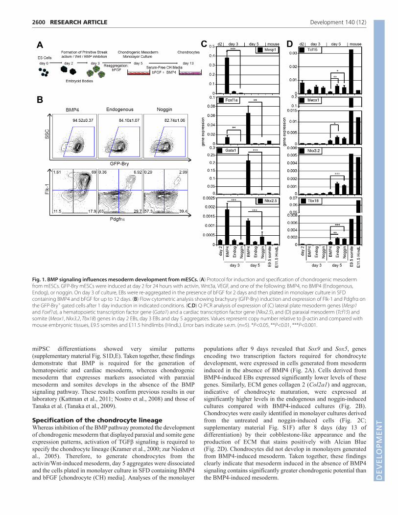
Specification of the chondrocyte lineageWhereas inhibition of the BMP pathway promoted the developmentof chondrogenic mesoderm that displayed paraxial and somite geneexpression patterns, activation of TGFβ signaling is required tospecify the chondrocyte lineage (Kramer et al., 2000; zur Nieden etal., 2005). Therefore, to generate chondrocytes from theactivin/Wnt-induced mesoderm, day 5 aggregates were dissociatedand the cells plated in monolayer culture in SFD containing BMP4and bFGF [chondrocyte (CH) media]. Analyses of the monolayer
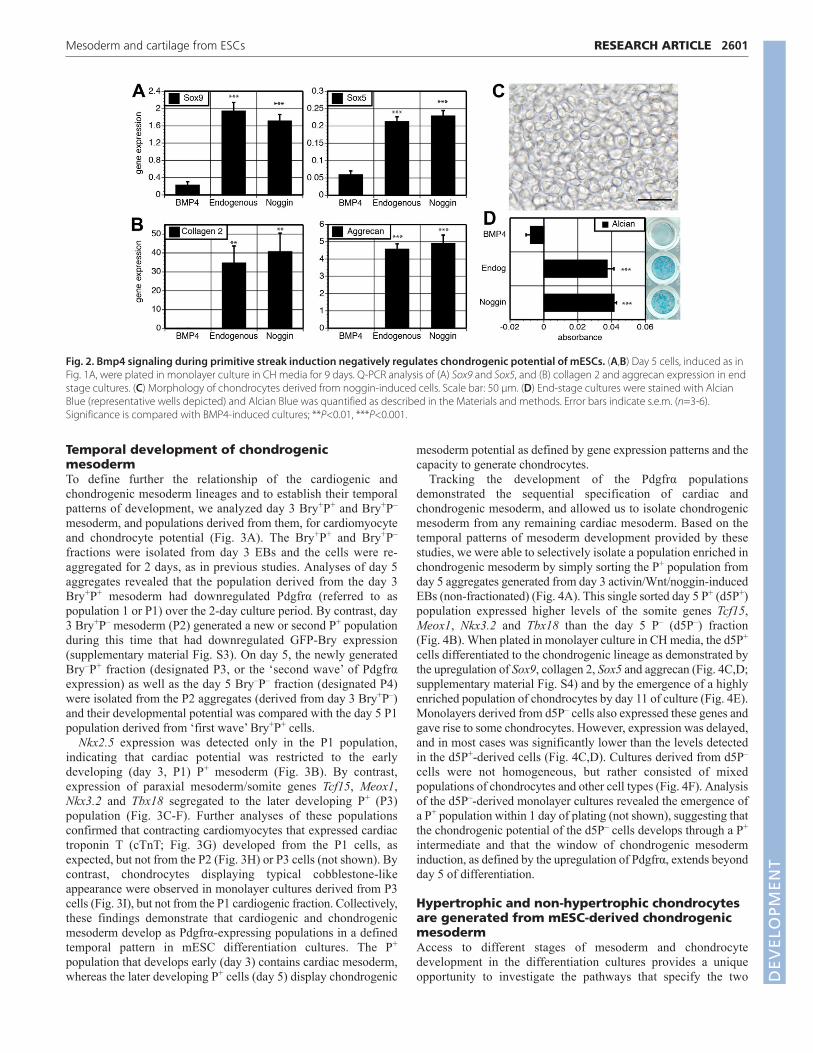
populations after 9 days revealed that Sox9 and Sox5, genesencoding two transcription factors required for chondrocytedevelopment, were expressed in cells generated from mesoderminduced in the absence of BMP4 (Fig. 2A). Cells derived fromBMP4-induced EBs expressed significantly lower levels of thesegenes. Similarly, ECM genes collagen 2 (Col2a1) and aggrecan,indicative of chondrocyte maturation, were expressed atsignificantly higher levels in the endogenous and noggin-inducedcultures compared with BMP4-induced cultures (Fig. 2B).Chondrocytes were easily identified in monolayer cultures derivedfrom the untreated and noggin-induced cells (Fig. 2C;supplementary material Fig. S1F) after 8 days (day 13 ofdifferentiation) by their cobblestone-like appearance and theproduction of ECM that stains positively with Alcian Blue(Fig. 2D). Chondrocytes did not develop in monolayers generatedfrom BMP4-induced mesoderm. Taken together, these findingsclearly indicate that mesoderm induced in the absence of BMP4signaling contains significantly greater chondrogenic potential thanthe BMP4-induced mesoderm.
RESEARCH ARTICLE Development 140 (12)
Fig. 1. BMP signaling influences mesoderm development from mESCs. (A) Protocol for induction and specification of chondrogenic mesodermfrom mESCs. GFP-Bry mESCs were induced at day 2 for 24 hours with activin, Wnt3a, VEGF, and one of the following: BMP4, no BMP4 (Endogenous,Endog), or noggin. On day 3 of culture, EBs were re-aggregated in the presence of bFGF for 2 days and then plated in monolayer culture in SFDcontaining BMP4 and bFGF for up to 12 days. (B) Flow cytometric analysis showing brachyury (GFP-Bry) induction and expression of Flk-1 and Pdgfrα onthe GFP-Bry+ gated cells after 1 day induction in indicated conditions. (C,D) Q-PCR analysis of expression of (C) lateral plate mesoderm genes (Mesp1and Foxf1a), a hematopoietic transcription factor gene (Gata1) and a cardiac transcription factor gene (Nkx2.5), and (D) paraxial mesoderm (Tcf15) andsomite (Meox1, Nkx3.2, Tbx18) genes in day 2 EBs, day 3 EBs and day 5 aggregates. Values represent copy number relative to β-actin and compared withmouse embryonic tissues, E9.5 somites and E11.5 hindlimbs (HindL). Error bars indicate s.e.m. (n=5). *P<0.05, **P<0.01, ***P<0.001.
DEVELO
PMENT
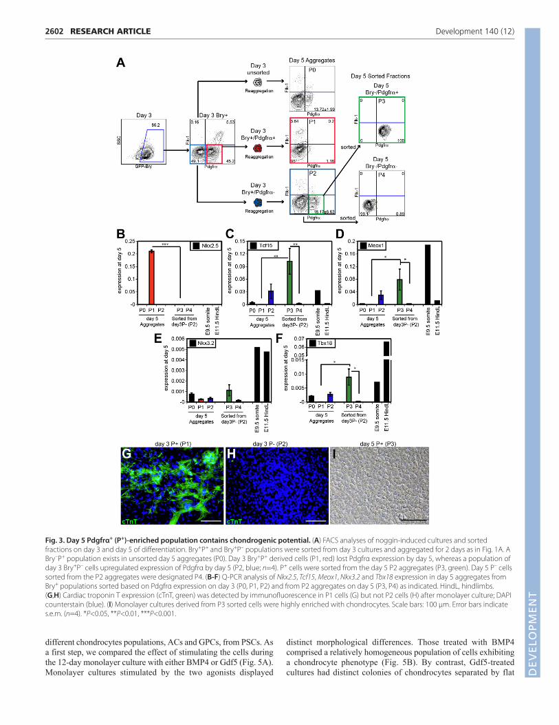
Temporal development of chondrogenicmesodermTo define further the relationship of the cardiogenic andchondrogenic mesoderm lineages and to establish their temporalpatterns of development, we analyzed day 3 Bry+P+ and Bry+P–
mesoderm, and populations derived from them, for cardiomyocyteand chondrocyte potential (Fig. 3A). The Bry+P+ and Bry+P–
fractions were isolated from day 3 EBs and the cells were re-aggregated for 2 days, as in previous studies. Analyses of day 5aggregates revealed that the population derived from the day 3Bry+P+ mesoderm had downregulated Pdgfrα (referred to aspopulation 1 or P1) over the 2-day culture period. By contrast, day3 Bry+P– mesoderm (P2) generated a new or second P+ populationduring this time that had downregulated GFP-Bry expression(supplementary material Fig. S3). On day 5, the newly generatedBry–P+ fraction (designated P3, or the ‘second wave’ of Pdgfrαexpression) as well as the day 5 Bry–P– fraction (designated P4)were isolated from the P2 aggregates (derived from day 3 Bry+P–)and their developmental potential was compared with the day 5 P1population derived from ‘first wave’ Bry+P+ cells.
Nkx2.5 expression was detected only in the P1 population,indicating that cardiac potential was restricted to the earlydeveloping (day 3, P1) P+ mesoderm (Fig. 3B). By contrast,expression of paraxial mesoderm/somite genes Tcf15, Meox1,Nkx3.2 and Tbx18 segregated to the later developing P+ (P3)population (Fig. 3C-F). Further analyses of these populationsconfirmed that contracting cardiomyocytes that expressed cardiactroponin T (cTnT; Fig. 3G) developed from the P1 cells, asexpected, but not from the P2 (Fig. 3H) or P3 cells (not shown). Bycontrast, chondrocytes displaying typical cobblestone-likeappearance were observed in monolayer cultures derived from P3cells (Fig. 3I), but not from the P1 cardiogenic fraction. Collectively,these findings demonstrate that cardiogenic and chondrogenicmesoderm develop as Pdgfrα-expressing populations in a definedtemporal pattern in mESC differentiation cultures. The P+
population that develops early (day 3) contains cardiac mesoderm,whereas the later developing P+ cells (day 5) display chondrogenic
mesoderm potential as defined by gene expression patterns and thecapacity to generate chondrocytes.
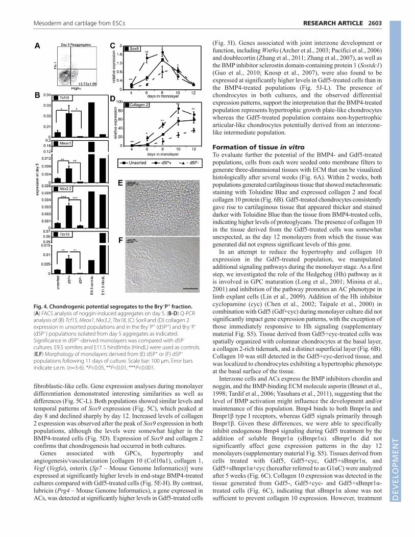
Tracking the development of the Pdgfrα populationsdemonstrated the sequential specification of cardiac andchondrogenic mesoderm, and allowed us to isolate chondrogenicmesoderm from any remaining cardiac mesoderm. Based on thetemporal patterns of mesoderm development provided by thesestudies, we were able to selectively isolate a population enriched inchondrogenic mesoderm by simply sorting the P+ population fromday 5 aggregates generated from day 3 activin/Wnt/noggin-inducedEBs (non-fractionated) (Fig. 4A). This single sorted day 5 P+ (d5P+)population expressed higher levels of the somite genes Tcf15,Meox1, Nkx3.2 and Tbx18 than the day 5 P– (d5P–) fraction(Fig. 4B). When plated in monolayer culture in CH media, the d5P+
cells differentiated to the chondrogenic lineage as demonstrated bythe upregulation of Sox9, collagen 2, Sox5 and aggrecan (Fig. 4C,D;supplementary material Fig. S4) and by the emergence of a highlyenriched population of chondrocytes by day 11 of culture (Fig. 4E).Monolayers derived from d5P– cells also expressed these genes andgave rise to some chondrocytes. However, expression was delayed,and in most cases was significantly lower than the levels detectedin the d5P+-derived cells (Fig. 4C,D). Cultures derived from d5P–
cells were not homogeneous, but rather consisted of mixedpopulations of chondrocytes and other cell types (Fig. 4F). Analysisof the d5P–-derived monolayer cultures revealed the emergence ofa P+ population within 1 day of plating (not shown), suggesting thatthe chondrogenic potential of the d5P– cells develops through a P+
intermediate and that the window of chondrogenic mesoderminduction, as defined by the upregulation of Pdgfrα, extends beyondday 5 of differentiation.
Hypertrophic and non-hypertrophic chondrocytesare generated from mESC-derived chondrogenicmesodermAccess to different stages of mesoderm and chondrocytedevelopment in the differentiation cultures provides a uniqueopportunity to investigate the pathways that specify the two
2601RESEARCH ARTICLEMesoderm and cartilage from ESCs
Fig. 2. Bmp4 signaling during primitive streak induction negatively regulates chondrogenic potential of mESCs. (A,B) Day 5 cells, induced as inFig. 1A, were plated in monolayer culture in CH media for 9 days. Q-PCR analysis of (A) Sox9 and Sox5, and (B) collagen 2 and aggrecan expression in endstage cultures. (C) Morphology of chondrocytes derived from noggin-induced cells. Scale bar: 50 μm. (D) End-stage cultures were stained with AlcianBlue (representative wells depicted) and Alcian Blue was quantified as described in the Materials and methods. Error bars indicate s.e.m. (n=3-6).Significance is compared with BMP4-induced cultures; **P<0.01, ***P<0.001.
DEVELO
PMENT
2602
different chondrocytes populations, ACs and GPCs, from PSCs. Asa first step, we compared the effect of stimulating the cells duringthe 12-day monolayer culture with either BMP4 or Gdf5 (Fig. 5A).Monolayer cultures stimulated by the two agonists displayed
distinct morphological differences. Those treated with BMP4comprised a relatively homogeneous population of cells exhibitinga chondrocyte phenotype (Fig. 5B). By contrast, Gdf5-treatedcultures had distinct colonies of chondrocytes separated by flat
RESEARCH ARTICLE Development 140 (12)
Fig. 3. Day 5 Pdgfrα+ (P+)-enriched population contains chondrogenic potential. (A) FACS analyses of noggin-induced cultures and sortedfractions on day 3 and day 5 of differentiation. Bry+P+ and Bry+P– populations were sorted from day 3 cultures and aggregated for 2 days as in Fig. 1A. ABry–P+ population exists in unsorted day 5 aggregates (P0). Day 3 Bry+P+ derived cells (P1, red) lost Pdgfrα expression by day 5, whereas a population ofday 3 Bry+P– cells upregulated expression of Pdgfrα by day 5 (P2, blue; n=4). P+ cells were sorted from the day 5 P2 aggregates (P3, green). Day 5 P– cellssorted from the P2 aggregates were designated P4. (B-F) Q-PCR analysis of Nkx2.5, Tcf15, Meox1, Nkx3.2 and Tbx18 expression in day 5 aggregates fromBry+ populations sorted based on Pdgfrα expression on day 3 (P0, P1, P2) and from P2 aggregates on day 5 (P3, P4) as indicated. HindL, hindlimbs. (G,H) Cardiac troponin T expression (cTnT, green) was detected by immunofluorescence in P1 cells (G) but not P2 cells (H) after monolayer culture; DAPIcounterstain (blue). (I) Monolayer cultures derived from P3 sorted cells were highly enriched with chondrocytes. Scale bars: 100 μm. Error bars indicates.e.m. (n=4). *P<0.05, **P<0.01, ***P<0.001.
DEVELO
PMENT
fibroblastic-like cells. Gene expression analyses during monolayerdifferentiation demonstrated interesting similarities as well asdifferences (Fig. 5C-L). Both populations showed similar levels andtemporal patterns of Sox9 expression (Fig. 5C), which peaked atday 8 and declined sharply by day 12. Increased levels of collagen2 expression was observed after the peak of Sox9 expression in bothpopulations, although the levels were somewhat higher in theBMP4-treated cells (Fig. 5D). Expression of Sox9 and collagen 2confirms that chondrogenesis had occurred in both cultures.
Genes associated with GPCs, hypertrophy andangiogenesis/vascularization [collagen 10 (Col10a1), collagen 1,Vegf (Vegfa), osterix (Sp7 – Mouse Genome Informatics)] wereexpressed at significantly higher levels in end-stage BMP4-treatedcultures compared with Gdf5-treated cells (Fig. 5E-H). By contrast,lubricin (Prg4 – Mouse Genome Informatics), a gene expressed inACs, was detected at significantly higher levels in Gdf5-treated cells
(Fig. 5I). Genes associated with joint interzone development orfunction, including Wnt9a (Archer et al., 2003; Pacifici et al., 2006)and doublecortin (Zhang et al., 2011; Zhang et al., 2007), as well asthe BMP inhibitor sclerostin domain-containing protein 1 (Sostdc1)(Guo et al., 2010; Knosp et al., 2007), were also found to beexpressed at significantly higher levels in Gdf5-treated cells than inthe BMP4-treated populations (Fig. 5J-L). The presence ofchondrocytes in both cultures, and the observed differentialexpression patterns, support the interpretation that the BMP4-treatedpopulation represents hypertrophic growth plate-like chondrocyteswhereas the Gdf5-treated population contains non-hypertrophicarticular-like chondrocytes potentially derived from an interzone-like intermediate population.
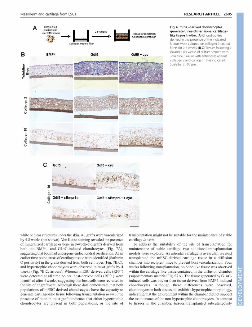
Formation of tissue in vitroTo evaluate further the potential of the BMP4- and Gdf5-treatedpopulations, cells from each were seeded onto membrane filters togenerate three-dimensional tissues with ECM that can be visualizedhistologically after several weeks (Fig. 6A). Within 2 weeks, bothpopulations generated cartilaginous tissue that showed metachromaticstaining with Toluidine Blue and expressed collagen 2 and focalcollagen 10 protein (Fig. 6B). Gdf5-treated chondrocytes consistentlygave rise to cartilaginous tissue that appeared thicker and staineddarker with Toluidine Blue than the tissue from BMP4-treated cells,indicating higher levels of proteoglycans. The presence of collagen 10in the tissue derived from the Gdf5-treated cells was somewhatunexpected, as the day 12 monolayers from which the tissue wasgenerated did not express significant levels of this gene.
In an attempt to reduce the hypertrophy and collagen 10expression in the Gdf5-treated population, we manipulatedadditional signaling pathways during the monolayer stage. As a firststep, we investigated the role of the Hedgehog (Hh) pathway as itis involved in GPC maturation (Long et al., 2001; Minina et al.,2001) and inhibition of the pathway promotes an AC phenotype inlimb explant cells (Lin et al., 2009). Addition of the Hh inhibitorcyclopamine (cyc) (Chen et al., 2002; Taipale et al., 2000) incombination with Gdf5 (Gdf+cyc) during monolayer culture did notsignificantly impact gene expression patterns, with the exception ofthose immediately responsive to Hh signaling (supplementarymaterial Fig. S5). Tissue derived from Gdf5+cyc-treated cells wasspatially organized with columnar chondrocytes at the basal layer,a collagen 2-rich tidemark, and a distinct superficial layer (Fig. 6B).Collagen 10 was still detected in the Gdf5+cyc-derived tissue, andwas localized to chondrocytes exhibiting a hypertrophic phenotypeat the basal surface of the tissue.
Interzone cells and ACs express the BMP inhibitors chordin andnoggin, and the BMP-binding ECM molecule asporin (Brunet et al.,1998; Tardif et al., 2006; Yasuhara et al., 2011), suggesting that thelevel of BMP activation might influence the development and/ormaintenance of this population. Bmp4 binds to both Bmpr1α andBmpr1β type I receptors, whereas Gdf5 signals primarily throughBmpr1β. Given these differences, we were able to specificallyinhibit endogenous Bmp4 signaling during Gdf5 treatment by theaddition of soluble Bmpr1α (sBmpr1α). sBmpr1α did notsignificantly affect gene expression patterns in the day 12monolayers (supplementary material Fig. S5). Tissues derived fromcells treated with Gdf5, Gdf5+cyc, Gdf5+sBmpr1α, andGdf5+sBmpr1α+cyc (hereafter referred to as G1αC) were analyzedafter 5 weeks (Fig. 6C). Collagen 10 expression was detected in thetissue generated from Gdf5-, Gdf5+cyc- and Gdf5+sBmpr1α-treated cells (Fig. 6C), indicating that sBmpr1α alone was notsufficient to prevent collagen 10 expression. However, treatment
2603RESEARCH ARTICLEMesoderm and cartilage from ESCs
Fig. 4. Chondrogenic potential segregates to the Bry–P+ fraction. (A) FACS analysis of noggin-induced aggregates on day 5. (B-D) Q-PCRanalysis of (B) Tcf15, Meox1, Nkx3.2, Tbx18, (C) Sox9 and (D) collagen 2expression in unsorted populations and in the Bry–P+ (d5P+) and Bry–P–
(d5P–) populations isolated from day 5 aggregates as indicated.Significance in d5P+-derived monolayers was compared with d5P–
cultures. E9.5 somites and E11.5 hindlimbs (HindL) were used as controls.(E,F) Morphology of monolayers derived from (E) d5P+ or (F) d5P–
populations following 11 days of culture. Scale bar: 100 μm. Error barsindicate s.e.m. (n=3-6). *P<0.05, **P<0.01, ***P<0.001.
DEVELO
PMENT

2604
with both inhibitors (G1αC) consistently resulted in cartilage-liketissue that expressed high levels of collagen 2 (supplementarymaterial Fig. S6), but virtually no collagen 10, suggesting that tissuecontaining non-hypertrophic chondrocytes was generated in vitro.By contrast, the BMP4-derived and other Gdf5-derived tissuesexpressed both collagen 2 and collagen 10, and as such representcartilage tissue comprising hypertrophic chondrocytes. Theseobservations indicate that through manipulation of the Hh, BMPand Gdf5 pathways, it is possible to specify two unique chondrocytepopulations that generate tissues in vitro that display characteristicsof articular (G1αC-treatment) and growth plate (BMP4-treatment)cartilage.
Potential of hypertrophic and non-hypertrophicmESC-derived chondrocytes in vivoGiven the potential of the mESC-derived chondrocytes to formtissue in vitro, we next sought to determine their ability to generatecartilage in vivo. To enable us to distinguish mESC-derived cellsfrom host cells, we used a GFP-Bry mESC line that constitutivelyexpresses the red fluorescent protein (RFP) (Luche et al., 2007).Bry–P+RFP+ cells isolated by fluorescence-activated cell sorting(FACS) from day 5 aggregates were cultured in the presence ofeither BMP4 or G1aC for 12 days. Following monolayer culture,cells were injected subcutaneously into immunodeficient mice.Developing tissue was easily identified after 2, 4 and 8 weeks as
RESEARCH ARTICLE Development 140 (12)
Fig. 5. Generation of hypertrophic and non-hypertrophic chondrocytes from mESC-derived chondrogenic mesoderm. (A,B) Protocol formonolayer culture for 4-12 days (A) and representative micrographs of chondrocytes (B) treated with BMP4 or Gdf5. Scale bar: 100 μm. (C-L) Q-PCRanalysis of (C) Sox9, (D) collagen 2, (E) collagen 10, (F) collagen 1, (G) Vegf, (H) osterix, (I) lubricin, (J) Wnt9a, (K) doublecortin and (L) Sostdc1 expressionduring monolayer culture at the indicated times. Primary ACs and adult rib cartilage were used as controls. Error bars indicate s.e.m. (n=3). *P<0.05,**P<0.01, ***P<0.001, #P=0.0546. Significance of each Gdf5-treated time point was compared with corresponding BMP4 time point.DEVELO
PMENT
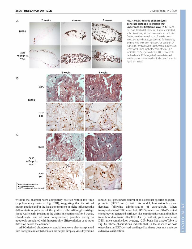
white or clear structures under the skin. All grafts were vascularizedby 4-8 weeks (not shown). Von Kossa staining revealed the presenceof mineralized cartilage or bone in 8-week-old grafts derived fromboth the BMP4- and G1αC-induced chondrocytes (Fig. 7A),suggesting that both had undergone endochondral ossification. At anearlier time point, areas of cartilage tissue were identified (SafraninO positivity) in the grafts derived from both cell types (Fig. 7B,C),and hypertrophic chondrocytes were observed in most grafts by 4weeks (Fig. 7B,C, arrows). Whereas mESC-derived cells (RFP+)were detected at all time points, host-derived cells (RFP–) wereidentified after 4 weeks, suggesting that host cells were recruited tothe site of engraftment. Although these data demonstrate that bothpopulations of mESC-derived chondrocytes have the capacity togenerate cartilage-like tissue following transplantation in vivo, thepresence of bone in most grafts indicates that either hypertrophicchondrocytes are present in both populations, or the site of
transplantation might not be suitable for the maintenance of stablecartilage in vivo.
To address the suitability of the site of transplantation formaintenance of stable cartilage, two additional transplantationmodels were explored. As articular cartilage is avascular, we nexttransplanted the mESC-derived cartilage tissue in a diffusionchamber into recipient mice to prevent host vascularization. Fourweeks following transplantation, no bone-like tissue was observedwithin the cartilage-like tissue contained in the diffusion chamber(supplementary material Fig. S7A). The tissue generated by G1αC-induced cells was thicker than tissue derived from BMP4-inducedchondrocytes. Although these differences were observed,chondrocytes in both tissues did exhibit a hypertrophic morphology,indicating that the environment within the chamber did not supportthe maintenance of the non-hypertrophic chondrocytes. In contrastto tissues in the chamber, tissues transplanted subcutaneously
2605RESEARCH ARTICLEMesoderm and cartilage from ESCs
Fig. 6. mESC-derived chondrocytesgenerate three-dimensional cartilage-like tissue in vitro. (A) Chondrocytesderived in the presence of the indicatedfactors were cultured on collagen 2-coatedfilters for 2-5 weeks. (B,C) Tissues following 2(B) and 5 (C) weeks of culture stained withToluidine Blue, or with antibodies againstcollagen 2 and collagen 10 as indicated.Scale bars: 100 μm.
DEVELO
PMENT
2606
without the chamber were completely ossified within this time(supplementary material Fig. S7B), suggesting that the site oftransplantation and/or the local environment or niche influences thedifferentiation potential of the grafted cells. Although cartilagetissue was clearly present in the diffusion chambers after 4 weeks,chondrocyte survival was compromised, possibly owing toapoptosis associated with hypertrophic differentiation or to poordiffusion across the chamber.
mESC-derived chondrocyte populations were also transplantedinto transgenic mice that contain the herpes simplex virus thymidine
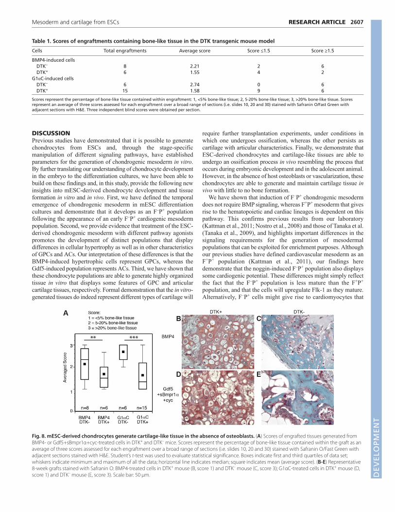
kinase (TK) gene under control of an osteoblast-specific collagen 1promoter (DTK+ mice). With this model, host osteoblasts aredepleted following administration of gancyclovir. Whentransplanted into DTK+ mice, both BMP4-treated and G1αC-treatedchondrocytes generated cartilage-like engraftments containing littleto no bone-like tissue after 8 weeks. By contrast, grafts in controlDTK– mice contained, on average, >20% bone-like tissue (Table 1;Fig. 8). These observations indicate that, in the absence of hostosteoblasts, mESC-derived cartilage-like tissue does not undergoextensive ossification.
RESEARCH ARTICLE Development 140 (12)
Fig. 7. mESC-derived chondrocytesgenerate cartilage-like tissue thatundergoes ossification in vivo. (A-C) BMP4-or G1αC-treated RFP.bry mESCs were injectedsubcutaneously at the mammary fat pad site.Grafts were harvested up to 8 weeks post-injection as indicated, processed for histologyand stained with von Kossa (A) or Safranin O(SafO; B,C, arrows) with Fast Green counterstain(chevrons). Immunohistochemistry for RFPindicates mESC-derived cells (B,C). Few host-derived cells (RFP-negative) were detectedwithin grafts (arrowheads). Scale bars: 1 mm inA; 50 μm in B,C.
DEVELO
PMENT
DISCUSSIONPrevious studies have demonstrated that it is possible to generatechondrocytes from ESCs and, through the stage-specificmanipulation of different signaling pathways, have establishedparameters for the generation of chondrogenic mesoderm in vitro.By further translating our understanding of chondrocyte developmentin the embryo to the differentiation cultures, we have been able tobuild on these findings and, in this study, provide the following newinsights into mESC-derived chondrocyte development and tissueformation in vitro and in vivo. First, we have defined the temporalemergence of chondrogenic mesoderm in mESC differentiationcultures and demonstrate that it develops as an F–P+ populationfollowing the appearance of an early F–P+ cardiogenic mesodermpopulation. Second, we provide evidence that treatment of the ESC-derived chondrogenic mesoderm with different pathway agonistspromotes the development of distinct populations that displaydifferences in cellular hypertrophy as well as in other characteristicsof GPCs and ACs. Our interpretation of these differences is that theBMP4-induced hypertrophic cells represent GPCs, whereas theGdf5-induced population represents ACs. Third, we have shown thatthese chondrocyte populations are able to generate highly organizedtissue in vitro that displays some features of GPC and articularcartilage tissues, respectively. Formal demonstration that the in vitro-generated tissues do indeed represent different types of cartilage will
require further transplantation experiments, under conditions inwhich one undergoes ossification, whereas the other persists ascartilage with articular characteristics. Finally, we demonstrate thatESC-derived chondrocytes and cartilage-like tissues are able toundergo an ossification process in vivo resembling the process thatoccurs during embryonic development and in the adolescent animal.However, in the absence of host osteoblasts or vascularization, thesechondrocytes are able to generate and maintain cartilage tissue invivo with little to no bone formation.
We have shown that induction of F–P+ chondrogenic mesodermdoes not require BMP signaling, whereas F+P+ mesoderm that givesrise to the hematopoietic and cardiac lineages is dependent on thispathway. This confirms previous results from our laboratory(Kattman et al., 2011; Nostro et al., 2008) and those of Tanaka et al.(Tanaka et al., 2009), and highlights important differences in thesignaling requirements for the generation of mesodermalpopulations that can be exploited for enrichment purposes. Althoughour previous studies have defined cardiovascular mesoderm as anF+P+ population (Kattman et al., 2011), our findings heredemonstrate that the noggin-induced F–P+ population also displayssome cardiogenic potential. These differences might simply reflectthe fact that the F–P+ population is less mature than the F+P+
population, and that the cells will upregulate Flk-1 as they mature.Alternatively, F–P+ cells might give rise to cardiomyocytes that
2607RESEARCH ARTICLEMesoderm and cartilage from ESCs
Table 1. Scores of engraftments containing bone-like tissue in the DTK transgenic mouse model
Cells Total engraftments Average score Score ≤1.5 Score ≥1.5
BMP4-induced cellsDTK– 8 2.21 2 6DTK+ 6 1.55 4 2
G1αC-induced cellsDTK– 6 2.74 0 6DTK+ 15 1.58 9 6
Scores represent the percentage of bone-like tissue contained within engraftment: 1, <5% bone-like tissue; 2, 5-20% bone-like tissue; 3, >20% bone-like tissue. Scoresrepresent an average of three scores assessed for each engraftment over a broad range of sections (i.e. slides 10, 20 and 30) stained with Safranin O/Fast Green withadjacent sections with H&E. Three independent blind scores were obtained per section.
Fig. 8. mESC-derived chondrocytes generate cartilage-like tissue in the absence of osteoblasts. (A) Scores of engrafted tissues generated fromBMP4- or Gdf5+sBmpr1α+cyc-treated cells in DTK+ and DTK– mice. Scores represent the percentage of bone-like tissue contained within the graft as anaverage of three scores assessed for each engraftment over a broad range of sections (i.e. slides 10, 20 and 30) stained with Safranin O/Fast Green withadjacent sections stained with H&E. Student’s t-test was used to evaluate statistical significance. Boxes indicate first and third quartiles of data set;whiskers indicate minimum and maximum of all the data; horizontal line indicates median; square indicates mean (average score). (B-E) Representative8-week grafts stained with Safranin O: BMP4-treated cells in DTK+ mouse (B, score 1) and DTK– mouse (C, score 3); G1αC-treated cells in DTK+ mouse (D,score 1) and DTK– mouse (E, score 3). Scale bar: 50 μm. D
EVELO
PMENT
2608
differ from those generated by F+P+ cells. Current studies are aimedat investigating this possibility. The emergence of the F–P+ cardiacmesoderm before the F–P+ chondrogenic mesoderm accuratelyrecapitulates the temporal allocation of these mesoderm fates in theearly embryo, once again demonstrating that lineage developmentin this in vitro model system faithfully mimics lineage developmentin the embryo (Kinder et al., 1999; Lawson et al., 1991). By takingadvantage of these temporal differences, it was possible to developstrategies to generate populations of highly enriched chondrocytes.
Access to a highly enriched population of chondrogenicmesoderm enabled us to investigate the signaling pathways thatregulate the specification of chondrocytes that display eitherhypertrophic growth plate-like or non-hypertrophic articular-likecharacteristics. Consistent with the findings of others using ESCsand our understanding of the regulation of the lineage in vivo(Hatakeyama et al., 2004; Tsumaki et al., 2002; Yoon et al., 2006),we found that BMP4 signaling did promote the development ofchondrocytes that display gene expression patterns indicative ofhypertrophic growth plate-like chondrocytes. When stimulated withGdf5, however, cells with gene expression profiles consistent withinterzone cells and ACs emerged. Lineage-tracing studies anddetailed expression analyses have shown that Gdf5 is expressed inthe emerging ACs as well as in the cells surrounding this population(Koyama et al., 2008; Rountree et al., 2004). These observationsare consistent with a role of this factor in inducing and/or promotingthe expansion of these chondrocytes. Although the ability togenerate chondrocyte populations with different characteristics byspecification with different factors supports the interpretation thatthey derive from distinct progenitors, the role of the environment inmaintaining distinct phenotypes was clearly demonstrated by the invivo studies presented in this report.
Bmp4 and Gdf5, both members of the TGFβ superfamily, havedifferent functions during the formation of synovial joints, cartilageand bone and, in this study, specify distinct subsets of mESC-derived chondrocytes that display different characteristics. Acomparison of Bmp4 and Gdf5 in embryonic limb micromassesdemonstrated that Bmp4 treatment induced chondrogenesis andcollagen 2 expression in all cells (within cartilage-like nodules andin internodulal spaces), whereas an increase in the number ofcartilage nodules was observed after Gdf5 treatment (Hatakeyamaet al., 2004). These effects are somewhat recapitulated in the mESC-derived monolayer cultures as BMP4 induced a homogeneouschondrocytes population, whereas Gdf5 treatment inducedpopulations of condensed nodules together with fibroblastic cells.The effects of both BMP4 and Gdf5 appear to be mediated throughBmpr1-dependent phosphorylation events as concurrent treatmentwith dorsomorphin (Yu et al., 2008) at high concentrations impairedcell survival and at lower concentrations prevented the upregulationof chondrogenic genes (supplementary material Fig. S8A-C).
When cultured on membrane filters, ESC-derived chondrocytepopulations generated cartilage-like tissue that was rich in collagensand proteoglycans and highly organized into zones of cartilagenormally found in articular joints. These findings are the firstdocumentation of tissue formation from ESC-derived chondrocyteswithout the requirement of co-culture or seeding onto a scaffold.Although our molecular analyses showed low levels of expressionof the hypertrophic marker collagen 10 in the Gdf5-inducedchondrocyte population, cartilage-like tissue generated from itclearly contained collagen 10-expressing hypertrophic cells. Bycontrast, populations specified in the presence of Gdf5 together withhedgehog- and Bmp4-specific inhibitors (G1αC treatment)generated tissue that showed markedly reduced numbers of collagen
10-expressing hypertrophic chondrocytes. These findings suggestthat low levels of endogenous Bmp4 and hedgehog signalingmaintain some hypertrophic chondrocytes in the Gdf5-treatedcultures and inhibition of these pathways during the chondrocytespecification step reduces the number of these contaminating cells.These observations are consistent with the known role of thesepathways in chondrocyte development in vivo. There is nodetectable hedgehog signaling in the region of AC developmentwhereas it is active in the growth plate (Long et al., 2001). Bmp4 isalso not expressed in mature ACs, which express chordin, a potentinhibitor of the pathway (Tardif et al., 2006). Given that Bmp4signaling is required for chondrocyte hypertrophy (Yoon et al.,2006) and is inhibited in non-hypertrophic chondrocytes undernormal conditions, it is possible that activation of the pathway innon-hypertrophic chondrocyte populations might initiate ahypertrophic response. Access to tissue with characteristics of ACswill provide a unique opportunity to identify and characterizefactors and signaling pathways that induce hypertrophy and possiblyplay a role in the initiation of OA pathogenesis.
Our analysis indicated that, whereas non-hypertrophic andhypertrophic chondrocyte-enriched populations and respectivecartilage-like tissues had been generated in vitro, grafts were ossifiedwhen subcutaneously transplanted in vivo. Although bone formationwas anticipated from the hypertrophic chondrocyte-derived grafts, itwas unexpected from the grafts generated with non-hypertrophicchondrocytes and tissues. It is possible that low numbers ofhypertrophic chondrocytes still contaminate the G1αC cultures andderivative tissues, which initiate ossification over time in vivo. Furtherenrichment of these populations will depend on the identification oflineage-specific markers and corresponding antibodies to allowisolation of non-hypertrophic (AC-like) and hypertrophic (GPC-like)chondrocytes by flow-cytometric approaches. Alternatively, theseobservations might indicate that the microenvironment of the graftwas incompatible with maintenance of stable cartilage. It is possiblethat BMPs or other factors expressed at the graft site might initiate aconversion of non-hypertrophic chondrocytes to become hypertrophicand display additional characteristics of GPCs. Transplantation intothe correct ‘niche’ in the joint space together with mechanical loadingmight be essential to maintain articular cartilage in vivo. Indeed,mESC-derived cartilage isolated from the host vasculature system bydiffusion chamber or cartilage tissues generated in the absence of hostosteoblasts were not ossified. These observations support thehypothesis that the correct niche would be required to support themaintenance of non-hypertrophic articular-like cartilage derived fromPSCs.
In summary, the findings presented here have established thedevelopmental program for chondrogenic mesoderm formationfrom mESCs and have identified regulatory pathways that play arole in the specification of this mesoderm to both hypertrophic andnon-hypertrophic chondrocytes. These advances enable us for thefirst time to generate cartilage-like tissue that has the characteristicsof articular and growth plate cartilage in vitro, providing a model forfurther investigating pathways that regulate these fates and/ormaintain this stable cartilage tissue under normal conditions, as wellas for identifying factors that may initiate changes indicative of theearly stages of disease such as OA. Access to highly enrichedpopulations of non-hypertrophic articular-like chondrocytes and tocartilage tissue derived from them provides the first opportunity tobegin to test the feasibility of developing cell and tissue replacementtherapy for the treatment of OA. The translation of these findings tohuman PSCs will be instrumental for the development of suchapplications for the treatment of human disease.
RESEARCH ARTICLE Development 140 (12)
DEVELO
PMENT
AcknowledgementsWe thank the Flow Cytometry Facility at the Hospital for Sick Children for cellsorting; Richard Renlund, Frank Guiliani and Sarah Wilson at the University ofToronto Division of Comparative Medicine for assistance in animal studies;Mona Reid, Clara Goodwin and Heather Whetstone for histological services;and members of the G.M.K. laboratory for critical reading of the manuscript.
FundingThis work was supported by a grant from the Canadian Institutes of HealthResearch (CIHR) [MOP 219710] to B.A.A. and G.M.K.
Competing interests statementThe authors declare no competing financial interests.
Supplementary materialSupplementary material available online athttp://dev.biologists.org/lookup/suppl/doi:10.1242/dev.087890/-/DC1
ReferencesAhmed, N., Gan, L., Nagy, A., Zheng, J., Wang, C. and Kandel, R. A. (2009).
Cartilage tissue formation using redifferentiated passaged chondrocytes invitro. Tissue Eng. Part A 15, 665-673.
Akiyama, H., Chaboissier, M. C., Martin, J. F., Schedl, A. and deCrombrugghe, B. (2002). The transcription factor Sox9 has essential roles insuccessive steps of the chondrocyte differentiation pathway and is requiredfor expression of Sox5 and Sox6. Genes Dev. 16, 2813-2828.
Archer, C. W., Dowthwaite, G. P. and Francis-West, P. (2003). Development ofsynovial joints. Birth Defects 69, 144-155.
Blumer, M. J., Longato, S., Schwarzer, C. and Fritsch, H. (2007). Bonedevelopment in the femoral epiphysis of mice: the role of cartilage canals andthe fate of resting chondrocytes. Dev. Dyn. 236, 2077-2088.
Brunet, L. J., McMahon, J. A., McMahon, A. P. and Harland, R. M. (1998).Noggin, cartilage morphogenesis, and joint formation in the mammalianskeleton. Science 280, 1455-1457.
Burgess, R., Rawls, A., Brown, D., Bradley, A. and Olson, E. N. (1996).Requirement of the paraxis gene for somite formation and musculoskeletalpatterning. Nature 384, 570-573.
Bussen, M., Petry, M., Schuster-Gossler, K., Leitges, M., Gossler, A. andKispert, A. (2004). The T-box transcription factor Tbx18 maintains theseparation of anterior and posterior somite compartments. Genes Dev. 18,1209-1221.
Chen, J. K., Taipale, J., Cooper, M. K. and Beachy, P. A. (2002). Inhibition ofHedgehog signaling by direct binding of cyclopamine to Smoothened. GenesDev. 16, 2743-2748.
Colnot, C. (2005). Cellular and molecular interactions regulating skeletogenesis.J. Cell. Biochem. 95, 688-697.
Dacquin, R., Starbuck, M., Schinke, T. and Karsenty, G. (2002). Mousealpha1(I)-collagen promoter is the best known promoter to drive efficient Crerecombinase expression in osteoblast. Dev. Dyn. 224, 245-251.
Dao, D. Y., Jonason, J. H., Zhang, Y., Hsu, W., Chen, D., Hilton, M. J. andO’Keefe, R. J. (2012). Cartilage-specific β-catenin signaling regulateschondrocyte maturation, generation of ossification centers, and perichondrialbone formation during skeletal development. J. Bone Miner. Res. 27, 1680-1694.
Darabi, R., Gehlbach, K., Bachoo, R. M., Kamath, S., Osawa, M., Kamm, K. E.,Kyba, M. and Perlingeiro, R. C. (2008). Functional skeletal muscleregeneration from differentiating embryonic stem cells. Nat. Med. 14, 134-143.
Fehling, H. J., Lacaud, G., Kubo, A., Kennedy, M., Robertson, S., Keller, G.and Kouskoff, V. (2003). Tracking mesoderm induction and its specification tothe hemangioblast during embryonic stem cell differentiation. Development130, 4217-4227.
Fletcher, R. B. and Harland, R. M. (2008). The role of FGF signaling in theestablishment and maintenance of mesodermal gene expression in Xenopus.Dev. Dyn. 237, 1243-1254.
Gadue, P., Huber, T. L., Paddison, P. J. and Keller, G. M. (2006). Wnt and TGF-beta signaling are required for the induction of an in vitro model of primitivestreak formation using embryonic stem cells. Proc. Natl. Acad. Sci. USA 103,16806-16811.
Gouon-Evans, V., Boussemart, L., Gadue, P., Nierhoff, D., Koehler, C. I., Kubo,A., Shafritz, D. A. and Keller, G. (2006). BMP-4 is required for hepaticspecification of mouse embryonic stem cell-derived definitive endoderm. Nat.Biotechnol. 24, 1402-1411.
Guo, S., Zhou, J., Gao, B., Hu, J., Wang, H., Meng, J., Zhao, X., Ma, G., Lin, C.,Xiao, Y. et al. (2010). Missense mutations in IHH impair Indian Hedgehogsignaling in C3H10T1/2 cells: implications for brachydactyly type A1, and newtargets for Hedgehog signaling. Cell. Mol. Biol. Lett. 15, 153-176.
Hatakeyama, Y., Tuan, R. S. and Shum, L. (2004). Distinct functions of BMP4and GDF5 in the regulation of chondrogenesis. J. Cell. Biochem. 91, 1204-1217.
Hirsinger, E., Jouve, C., Dubrulle, J. and Pourquié, O. (2000). Somite formationand patterning. Int. Rev. Cytol. 198, 1-65.
Hwang, N. S., Kim, M. S., Sampattavanich, S., Baek, J. H., Zhang, Z. andElisseeff, J. (2006). Effects of three-dimensional culture and growth factors onthe chondrogenic differentiation of murine embryonic stem cells. Stem Cells24, 284-291.
Hwang, Y. S., Polak, J. M. and Mantalaris, A. (2008). In vitro directchondrogenesis of murine embryonic stem cells by bypassing embryoid bodyformation. Stem Cells Dev. 17, 971-978.
Jukes, J. M., Both, S. K., Leusink, A., Sterk, L. M., van Blitterswijk, C. A. andde Boer, J. (2008). Endochondral bone tissue engineering using embryonicstem cells. Proc. Natl. Acad. Sci. USA 105, 6840-6845.
Kattman, S. J., Witty, A. D., Gagliardi, M., Dubois, N. C., Niapour, M., Hotta,A., Ellis, J. and Keller, G. (2011). Stage-specific optimization of activin/nodaland BMP signaling promotes cardiac differentiation of mouse and humanpluripotent stem cell lines. Cell Stem Cell 8, 228-240.
Kinder, S. J., Tsang, T. E., Quinlan, G. A., Hadjantonakis, A. K., Nagy, A. andTam, P. P. (1999). The orderly allocation of mesodermal cells to theextraembryonic structures and the anteroposterior axis during gastrulation ofthe mouse embryo. Development 126, 4691-4701.
Knosp, W. M., Saneyoshi, C., Shou, S., Bächinger, H. P. and Stadler, H. S.(2007). Elucidation, quantitative refinement, and in vivo utilization of theHOXA13 DNA binding site. J. Biol. Chem. 282, 6843-6853.
Koyama, E., Shibukawa, Y., Nagayama, M., Sugito, H., Young, B., Yuasa, T.,Okabe, T., Ochiai, T., Kamiya, N., Rountree, R. B. et al. (2008). A distinctcohort of progenitor cells participates in synovial joint and articular cartilageformation during mouse limb skeletogenesis. Dev. Biol. 316, 62-73.
Kramer, J., Hegert, C., Guan, K., Wobus, A. M., Müller, P. K. and Rohwedel, J.(2000). Embryonic stem cell-derived chondrogenic differentiation in vitro:activation by BMP-2 and BMP-4. Mech. Dev. 92, 193-205.
Kulesa, P. M. and Fraser, S. E. (2002). Cell dynamics during somite boundaryformation revealed by time-lapse analysis. Science 298, 991-995.
LaPrade, R. F., Bursch, L. S., Olson, E. J., Havlas, V. and Carlson, C. S. (2008).Histologic and immunohistochemical characteristics of failed articularcartilage resurfacing procedures for osteochondritis of the knee: a case series.Am. J. Sports Med. 36, 360-368.
Lawrence, R. C., Felson, D. T., Helmick, C. G., Arnold, L. M., Choi, H., Deyo, R.A., Gabriel, S., Hirsch, R., Hochberg, M. C., Hunder, G. G. et al. (2008).Estimates of the prevalence of arthritis and other rheumatic conditions in theUnited States. Part II. Arthritis Rheum. 58, 26-35.
Lawson, K. A., Meneses, J. J. and Pedersen, R. A. (1991). Clonal analysis ofepiblast fate during germ layer formation in the mouse embryo. Development113, 891-911.
Lin, A. C., Seeto, B. L., Bartoszko, J. M., Khoury, M. A., Whetstone, H., Ho, L.,Hsu, C., Ali, S. A. and Alman, B. A. (2009). Modulating hedgehog signalingcan attenuate the severity of osteoarthritis. Nat. Med. 15, 1421-1425.
Long, F., Zhang, X. M., Karp, S., Yang, Y. and McMahon, A. P. (2001). Geneticmanipulation of hedgehog signaling in the endochondral skeleton reveals adirect role in the regulation of chondrocyte proliferation. Development 128,5099-5108.
Luche, H., Weber, O., Nageswara Rao, T., Blum, C. and Fehling, H. J. (2007).Faithful activation of an extra-bright red fluorescent protein in ‘knock-in’ Cre-reporter mice ideally suited for lineage tracing studies. Eur. J. Immunol. 37, 43-53.
Mankoo, B. S., Skuntz, S., Harrigan, I., Grigorieva, E., Candia, A., Wright, C.V., Arnheiter, H. and Pachnis, V. (2003). The concerted action of Meoxhomeobox genes is required upstream of genetic pathways essential for theformation, patterning and differentiation of somites. Development 130, 4655-4664.
Minina, E., Wenzel, H. M., Kreschel, C., Karp, S., Gaffield, W., McMahon, A. P.and Vortkamp, A. (2001). BMP and Ihh/PTHrP signaling interact to coordinatechondrocyte proliferation and differentiation. Development 128, 4523-4534.
Murakami, S., Balmes, G., McKinney, S., Zhang, Z., Givol, D. and deCrombrugghe, B. (2004). Constitutive activation of MEK1 in chondrocytescauses Stat1-independent achondroplasia-like dwarfism and rescues theFgfr3-deficient mouse phenotype. Genes Dev. 18, 290-305.
Nakayama, N., Duryea, D., Manoukian, R., Chow, G. and Han, C. Y. (2003).Macroscopic cartilage formation with embryonic stem-cell-derivedmesodermal progenitor cells. J. Cell Sci. 116, 2015-2028.
Ng, A., Alman, B. and Grynpas, M. (2011). Development and validation of amouse model of adynamic bone disease (ABD). J. Bone Miner. Res. Suppl. 1.
Nostro, M. C., Cheng, X., Keller, G. M. and Gadue, P. (2008). Wnt, activin, andBMP signaling regulate distinct stages in the developmental pathway fromembryonic stem cells to blood. Cell Stem Cell 2, 60-71.
Ormestad, M., Astorga, J. and Carlsson, P. (2004). Differences in theembryonic expression patterns of mouse Foxf1 and -2 match their distinctmutant phenotypes. Dev. Dyn. 229, 328-333.
Pacifici, M., Koyama, E., Shibukawa, Y., Wu, C., Tamamura, Y., Enomoto-Iwamoto, M. and Iwamoto, M. (2006). Cellular and molecular mechanisms ofsynovial joint and articular cartilage formation. Ann. New York Acad. Sci. 1068,74-86.
2609RESEARCH ARTICLEMesoderm and cartilage from ESCs
DEVELO
PMENT
2610
Pelttari, K., Winter, A., Steck, E., Goetzke, K., Hennig, T., Ochs, B. G., Aigner,T. and Richter, W. (2006). Premature induction of hypertrophy during in vitrochondrogenesis of human mesenchymal stem cells correlates withcalcification and vascular invasion after ectopic transplantation in SCID mice.Arthritis Rheum. 54, 3254-3266.
Pelttari, K., Steck, E. and Richter, W. (2008). The use of mesenchymal stem cellsfor chondrogenesis. Injury 39 Suppl. 1, S58-S65.
Rountree, R. B., Schoor, M., Chen, H., Marks, M. E., Harley, V., Mishina, Y. andKingsley, D. M. (2004). BMP receptor signaling is required for postnatalmaintenance of articular cartilage. PLoS Biol. 2, e355.
Singh, M. K., Petry, M., Haenig, B., Lescher, B., Leitges, M. and Kispert, A.(2005). The T-box transcription factor Tbx15 is required for skeletaldevelopment. Mech. Dev. 122, 131-144.
Stadtfeld, M., Maherali, N., Breault, D. T. and Hochedlinger, K. (2008).Defining molecular cornerstones during fibroblast to iPS cell reprogrammingin mouse. Cell Stem Cell 2, 230-240.
Steinert, A. F., Ghivizzani, S. C., Rethwilm, A., Tuan, R. S., Evans, C. H. andNöth, U. (2007). Major biological obstacles for persistent cell-basedregeneration of articular cartilage. Arthritis Res. Ther. 9, 213.
Taipale, J., Chen, J. K., Cooper, M. K., Wang, B., Mann, R. K., Milenkovic, L.,Scott, M. P. and Beachy, P. A. (2000). Effects of oncogenic mutations inSmoothened and Patched can be reversed by cyclopamine. Nature 406, 1005-1009.
Takakura, N., Yoshida, H., Ogura, Y., Kataoka, H., Nishikawa, S. andNishikawa, S. (1997). PDGFR alpha expression during mouse embryogenesis:immunolocalization analyzed by whole-mount immunohistostaining usingthe monoclonal anti-mouse PDGFR alpha antibody APA5. J. Histochem.Cytochem. 45, 883-893.
Tam, P. P. and Tan, S. S. (1992). The somitogenetic potential of cells in theprimitive streak and the tail bud of the organogenesis-stage mouse embryo.Development 115, 703-715.
Tanaka, M., Jokubaitis, V., Wood, C., Wang, Y., Brouard, N., Pera, M., Hearn,M., Simmons, P. and Nakayama, N. (2009). BMP inhibition stimulates WNT-dependent generation of chondrogenic mesoderm from embryonic stemcells. Stem Cell Res. 3, 126-141.
Tardif, G., Pelletier, J. P., Hum, D., Boileau, C., Duval, N. and Martel-Pelletier,J. (2006). Differential regulation of the bone morphogenic protein antagonistchordin in human normal and osteoarthritic chondrocytes. Ann. Rheum. Dis.65, 261-264.
Tins, B. J., McCall, I. W., Takahashi, T., Cassar-Pullicino, V., Roberts, S.,Ashton, B. and Richardson, J. (2005). Autologous chondrocyte implantationin knee joint: MR imaging and histologic features at 1-year follow-up.Radiology 234, 501-508.
Tsumaki, N., Nakase, T., Miyaji, T., Kakiuchi, M., Kimura, T., Ochi, T. andYoshikawa, H. (2002). Bone morphogenetic protein signals are required forcartilage formation and differently regulate joint development duringskeletogenesis. J. Bone Miner. Res. 17, 898-906.
Visnjic, D., Kalajzic, I., Gronowicz, G., Aguila, H. L., Clark, S. H., Lichtler, A. C.and Rowe, D. W. (2001). Conditional ablation of the osteoblast lineage inCol2.3deltatk transgenic mice. J. Bone Miner. Res. 16, 2222-2231.
Yamashita, A., Krawetz, R. and Rancourt, D. E. (2008). Loss of discordant cellsduring micro-mass differentiation of embryonic stem cells into thechondrocyte lineage. Cell Death Differ. 16, 278-286.
Yasuhara, R., Ohta, Y., Yuasa, T., Kondo, N., Hoang, T., Addya, S., Fortina, P.,Pacifici, M., Iwamoto, M. and Enomoto-Iwamoto, M. (2011). Roles of beta-catenin signaling in phenotypic expression and proliferation of articularcartilage superficial zone cells. Lab. Invest. 91, 1739-1752.
Ying, Q. L., Nichols, J., Chambers, I. and Smith, A. (2003). BMP induction of Idproteins suppresses differentiation and sustains embryonic stem cell self-renewal in collaboration with STAT3. Cell 115, 281-292.
Yoon, B. S., Pogue, R., Ovchinnikov, D. A., Yoshii, I., Mishina, Y., Behringer, R.R. and Lyons, K. M. (2006). BMPs regulate multiple aspects of growth-platechondrogenesis through opposing actions on FGF pathways. Development133, 4667-4678.
Yu, P. B., Hong, C. C., Sachidanandan, C., Babitt, J. L., Deng, D. Y., Hoyng, S.A., Lin, H. Y., Bloch, K. D. and Peterson, R. T. (2008). Dorsomorphin inhibitsBMP signals required for embryogenesis and iron metabolism. Nat. Chem. Biol.4, 33-41.
Zhang, Y., Ryan, J. A., Di Cesare, P. E., Liu, J., Walsh, C. A. and You, Z. (2007).Doublecortin is expressed in articular chondrocytes. Biochem. Biophys. Res.Commun. 363, 694-700.
Zhang, Q., Cigan, A. D., Marrero, L., Lopreore, C., Liu, S., Ge, D., Savoie, F. H.and You, Z. (2011). Expression of doublecortin reveals articular chondrocytelineage in mouse embryonic limbs. Genesis 49, 75-82.
zur Nieden, N. I., Kempka, G., Rancourt, D. E. and Ahr, H. J. (2005). Inductionof chondro-, osteo- and adipogenesis in embryonic stem cells by bonemorphogenetic protein-2: effect of cofactors on differentiating lineages. BMCDev. Biol. 5, 1.
RESEARCH ARTICLE Development 140 (12)
DEVELO
PMENT
Copyright © 2022 FDOKUMEN