Aquaporin water channels AQP1 and AQP3, are expressed in equine articular chondrocytes
Upload
sacklerinstituteCategory
view
4download
0
Interleukin-1,8-modulated Gene Expression in ImmortalizedHuman ChondrocytesMary B. Goldring,* James R. Birkhead,* Lii-Fang Suen,* Rina Yamin,* Shuichi Mizuno,* Julie Glowacki,t Jack L. Arbiser,§and Jane F. Apperleyll*Arthritis Research Laboratory, Medical Services, Massachusetts General Hospital and Harvard Medical School, Charlestown,Massachusetts 02129; *Department of Orthopedic Surgery, Brigham and Women's Hospital and Harvard Medical School, Boston,Massachusetts 02115; 'Department of Dermatology, Massachusetts General Hospital and Harvard Medical School, Boston,Massachusetts 02114; and IlHammersmith Hospital, London W12 ONN, United Kingdom
Abstract
Immortalized human chondrocytes were established bytransfection of primary cultures of juvenile costal chondro-cytes with vectors encoding simian virus 40 large T antigenand selection in suspension culture over agarose. Stable celllines were generated that exhibited chondrocyte morphol-ogy, continuous proliferative capacity (> 80 passages) inmonolayer culture in serum-containing medium, and ex-
pression of mRNAs encoding chondrocyte-specific collagensII, IX, and XI and proteoglycans in an insulin-containingserum substitute. They did not express type X collagen or
versican mRNA. These cells synthesized and secreted extra-cellular matrix molecules that were reactive with mono-
clonal antibodies against type II collagen, large proteoglycan(PG-H, aggrecan), and chondroitin-4- and chondroitin-6-sulfate. Interleukin-1,8 (IL-1p) decreased the levels of typeII collagen mRNA and increased the levels of mRNAs forcollagenase, stromelysin, and immediate early genes (egr-1, c-fos, c-jun, and jun-B). These cell lines also expressedreporter gene constructs containing regulatory sequences
(-577/+3,428 bp) of the type II collagen gene (COL2A1)in transient transfection experiments, and IL-1p8 suppressedthis expression by 50-80%. These results show that immor-talized human chondrocytes displaying cartilage-specificmodulation by IL-1,3 can be used as a model for studyingnormal and pathological repair mechanisms. (J. Clin. Invest.1994. 94:2307-2316.) Key words: simian virus 40 large Tantigen * collagen - proteoglycan * cartilage * cytokine
Introduction
The chondrocyte, the only cell type in mature articular cartilage,is regarded as a terminally differentiated cell that maintains thecartilage-specific matrix phenotype under normal conditions oflow tumover. The collagen types II, IX, and XI are unique tothis tissue and interact with specific proteoglycans to form thehighly hydrated extracellular matrix of hyaline cartilage.Freshly isolated human chondrocytes express cartilage-specific
Address correspondence to Mary B. Goldring, Ph.D., Arthritis Research,Massachusetts General Hospital, 149 13th Street, The Navy Yard,Charlestown, MA 02129.
Receivedfor publication 6 June 1994 and in revisedform 22 August1994.
type II collagen and continue to do so for several days to weeksin primary culture ( 1, 2). During prolonged culture and serialsubculture in monolayer, these cells begin to express type I andtype III collagens. This "switch" can be accelerated by platingthe cells at low densities or by treating them with agents such asinterleukin- 1 (IL- I) (2, 3 ). The reexpression of the chondrocytephenotype in subcultured cells can be induced by suspensionculture on or within agarose or by use of serum-free definedmedium (4-6). Studies in embryonic quail and chick chondro-cytes demonstrated the potential of v-src and v-myc oncogenesto generate cell lines of high proliferative capacities that retainedat least some differentiated chondrocyte properties (7, 8). At-tempts to establish immortalized chondrocyte lines from murinesarcoma virus-transfected rat chondrocytes (9), simian virus40 (SV40)'-transfected rabbit chondrocytes (10), and humanchondrosarcomas (11, 12) have resulted in cells that expresscartilage-specific proteoglycans but little or no type II collagen.Chondrocyte lines have also arisen spontaneously from fetal ratcalvaria (13, 14).
Primary cultures of chondrocytes isolated from young ani-mals that maintain the cartilage-specific phenotype are easilyobtained and have been used widely to assess differentiatedchondrocyte functions. The use of chondrocytes of human originhas been more problematic, because the source of the cartilagecannot be controlled, sufficient numbers of cells are not readilyobtained from random operative procedures, and the phenotypicstability of adult human chondrocytes is lost more quickly uponexpansion in serial monolayer cultures than that of cells ofjuvenile human (1) or young or embryonic animal origin (15,16). The lack of a reproducible source of chondrocytes of hu-man origin has hampered progress in studies of cartilage func-tion relevant to human disease.
We have sought to develop chondrocyte lines that expressthe differentiated chondrocyte phenotype and display character-istic responses to cytokines that modulate this phenotype. IL- 1is thought to play a major role in inflammatory and destructiveprocesses associated with the breakdown of cartilage matrixin rheumatoid arthritis and osteoarthritis (17). This cytokinesuppresses the expression of cartilage-specific collagens (2, 18)and proteoglycans (19-21) in cultures of chondrocytes andintact cartilage. IL-1 may also have a role in the inappropriaterepair of cartilage matrix that accompanies inflammatory jointdisorders by stimulating the synthesis of noncartilage collagens(type I and type III) by chondrocytes (2, 3).
To obtain immortalized chondrocyte lines of human origin,we used a combination of strategies involving transfection with
1. Abbreviations used in this paper: CAT, chloramphenicol acetyl trans-ferase; CHX, cycloheximide; GAPDH, glyceraldehyde-3-phosphate de-hydrogenase; SV40, simian virus 40.
Immortalized Human Chondrocytes and Interleukin-l 2307
J. Clin. Invest.© The American Society for Clinical Investigation, Inc.0021-9738/94/12/2307/10 $2.00Volume 94, December 1994, 2307-2316

vectors containing SV40 DNA sequences, selection on agarose,expansion in serum-containing medium, and modulation of phe-notype in serum-free defined medium. Because type II collagenexpression is the most highly labile function of chondrocytecultures, we used this marker as the primary indicator of thedifferentiated phenotype. We established immortalized humanchondrocyte lines that express mRNAs encoding cartilage-spe-cific matrix proteins, including type II, type IX, and type XIcollagens and aggrecan, and that deposit an extracellular matrixwith reactivity to antibodies against type II collagen, large pro-teoglycan, and sulfated glycosaminoglycans. They also exhibitthe expected chondrocyte responses to IL-l1,, including de-creased expression of the endogenous or transiently transfectedtype II collagen gene and increased expression of metalloprotei-nase and immediate early genes.
Methods
Materials. Recombinant human IL-l1, (provided by Dr. J.-M. Dayer,Division of Immunology and Allergy, University Hospital, Geneva,Switzerland), thawed from frozen stock (2.5 ,uM in PBS containing 1mg/ml BSA), remained stable for several months when kept at 4°C andwas used at 5 pM unless stated otherwise. The a 1(11) procollagencDNA (KTh1330), the al (IX) procollagen cDNA (KTh123), and thea2(XI) procollagen cDNA (KThl81) probes were cloned using acDNA library prepared from our human juvenile costal chondrocytecultures (22, 23). The type I cDNA probe, Hf677, a 1,500-bp cDNAencoding part of the al (I) procollagen subunit, was provided by Dr.F. Ramirez (Mt. Sinai School of Medicine, New York) and Dr. D.Prockop (Thomas Jefferson University, Philadelphia, PA) (24). A hu-man aggrecan cDNA was provided by Dr. K. J. Doege (Shriner's Hospi-tal, Portland, OR) (25). Probes encoding the small proteoglycans bigly-can/PG-I and decorin/PG-II were provided by Dr. L. W. Fisher andDr. M. F. Young (National Institute of Dental Research, Bethesda, MD)(26, 27). The cDNA probe for human procollagenase (XHF,) wasprovided by Dr. H. J. Rahmsdorf (Kemforschungszentrum Karlsruhe,Karlsruhe, Germany) (28). An additional collagenase probe (pCOLL-4), a 1.5-kb XbaI cDNA fragment (29) that gave identical results andprobes for stromelysin (pSTR-33), a 1.8-kb PstI cDNA fragment, andglyceraldehyde-3-phosphate dehydrogenase (GAPDH) were providedby Dr. N. I. Hutchinson (Merck Sharp & Dohme Research Laboratories,Rahway, NJ) (29). The c-jun (30) and jun-B (31) cDNA probes wereprovided by Dr. D. Nathans (Johns Hopkins University School of Medi-cine, Baltimore, MD), and the v-fos cDNA (32) was provided by Dr.R. Bernards (Massachusetts General Hospital, Charlestown, MA). Theegr-1 cDNA (OC3.1) is the 3.1-kb EcoRI fragment from Dr. V. P.Sukhatme (Beth Israel Hospital, Boston, MA) (33).
Chondrocyte isolation and transfection. Human costal cartilage wasobtained from tissue discarded after surgical repair of pectus excavatum.Chondrocytes were isolated by dispersion with proteases and culturedin Dulbecco's modified Eagle's medium (DME; GIBCO BRL, Gaithers-burg, MD) containing 10% fetal calf serum (FCS), as described pre-viously (1-3). The immortalized human chondrocyte line, designatedC-20/A4, was derived from juvenile costal chondrocytes isolated froma specimen of rib cartilage from a 5-yr-old male. At day 10 of primaryculture, 10 confluent 75-cm2 flasks were transfected with origin-defec-tive SV40 (20 /Lg/flask of pMK/SV40 ori- plasmid DNA) (34) usingthe polybrene/DMSO method (35, 36), and two flasks were "mock"transfected. The cells were incubated at 37°C for 6 h with the plasmidDNA in DME containing 5% FCS and 10 ,g/ml polybrene. The mediumwas then removed and 30% DMSO in DME/5% FCS added for 3 min.The cells were washed once and incubated for 2 d in DME containing10% FCS. The medium was changed weekly, and foci appeared after2-3 wk in the SV40-transfected but not in the mock-transfected flasks.After 6-8 wk, foci (10-20 per flask) were removed with the tip of apasteur pipette, and each was placed in a 16-mm diameter well (24-well plate; Costar Corp., Cambridge, MA) containing 1 ml of DME
with 10% FCS. Confluent wells were obtained within 1 wk, and thecells from each well were passaged to 10-cm plates using trypsin-EDTA(GIBCO BRL). The cells were then passaged every week at a splitratio of 1:5 in DME containing 10% FCS. Of the 140 foci obtainedoriginally, 20 of the resulting cultures were subcultured more than 20passages to permit senescence of the parental cells.
The T/C-28 line was derived from primary cultures (day 5) isolatedfrom costal cartilage from a 15-yr-old female. We used the SV40 largeT antigen inserted in the retroviral neomycin-resistant pZipNeoSV(X)vector (37) and transfected using polybrene, as described previously(38). After selection of transfected cells in G418 (500 Mg/ml; GIBCOBRL), the uncloned cell populations were established through severalsubcultures in DME containing 10% FCS. They were then further se-lected by suspension culture above agarose (2-4 wk) (6) followed bymonolayer culture in DME/Ham's F12 (1 / 1, vol/vol) containing 10%FCS and cloned by limiting dilution. Over 40 clonal lines were estab-lished, most of which had the polygonal cobblestone morphology char-acteristic of chondrocytes.
Collagen synthesis. Subconfluent immortalized chondrocytes weretrypsinized and plated at a split ratio of 1:10 in DME with 10% FCSand incubated at 37°C for 3-5 d. Proteins were labeled with [3H]proline(> 20 Ci/mmol; Amersham Corp., Arlington Heights, IL) at 25 j.Ci/ml for a further 24 h in serum-free DME supplemented with 50 Mg/mlascorbate and 50 Mg/ml /3-aminoproprionitrile fumarate. Pepsin-resistantcollagen chains secreted into the culture medium were analyzed bySDS-PAGE (5% acrylamide) with and without delayed reduction in0.1% /3-mercaptoethanol to distinguish aI (III) from al (I or II) colla-gens, as described previously (1-3).
Immunocytochemistry. Cells were plated in plastic Lab-Tech 4-chamber slides (Nunc, Inc., Naperville, IL) at 6 x 10⁴ cells/chamberin DME/Ham's F12 containing 10% FCS and grown for 3-5 d tosubconfluence. The monolayer was rinsed with Dulbecco's Ca2+- andMg2+-free phosphate-buffered saline (PBS) and incubated with culturemedium containing 1% Nutridoma-SP (Boehringer Mannheim Bio-chemicals, Indianapolis, IN) and 25 Mg/ml ascorbic acid. At the end ofincubation periods of 1-8 d, the chambers were rinsed three times withPBS, and the cells were fixed with 2% paraformaldehyde in 0.1 Mcacodylate buffer, pH 7.4, for 2 h at 4°C. After two rinses with 0.1 Mcacodylate buffer, the following monoclonal antibodies were added tochambers on different slides: MAB1330 anti-human type II collagen(Chemicon International, Inc., Temecula, CA) and 2-B-I anti-humanlarge proteoglycan (Seikagaku America, Inc., Rockville, MD), whichhas been used to identify the protein core of proteoglycan in chondro-cytes and extracellular matrix in articular cartilage (39). Other chamberslides were incubated with chondroitinase ABC for 30 min at 37°Cbefore addition of monoclonal antibodies against chondroitin-4-sulfate(L/Di-4S), chondroitin-6-sulfate (ADi-6S), or chondroitin-0-sulfate(ADi-OS) (Seikagaku America, Inc.). The staining was visualized byincubation with a gold-conjugated secondary antibody using AuroprobeLM (Amersham Corp.) followed by silver enhancement with the In-tenSE Kit (Amersham Corp.). The monoclonal antibody pAb 416 (pur-chased as SV40 T Ag from Oncogene Science, Inc., Uniondale, NY)and fluorescein-labeled anti-mouse Ig (Kirkegaard & Perry Labora-tories, Inc., Gaithersburg, MD) were used to detect the expression ofthe SV40 large T antigen in the immortalized cells.
Analysis of RNA. Total RNA for Northern blots was extracted bythe acid guanidinium thiocyanate-phenol-chloroform method adaptedfrom Chomczynski and Sacchi (40) or a lithium chloride methodadapted from Cathala et al. (41). The final preparations gave yields of- 10 M.g of RNA per 1 x 10⁶ cells with the appropriate A260:A28o ratioof - 2.0. Total RNAs were separated on 0.8% agarose gels in thepresence of 2% formaldehyde. Northern blots were prepared on eithernitrocellulose or BAS-85 membranes (Schleicher & Schuell, Inc.,Keene, NH) as described previously (2, 42).
The cDNA inserts were excised from the plasmids using appropriaterestriction enzymes and labeled with [a-32P]dCTP (Dupont/New En-gland Nuclear Research Products, Boston, MA) by random primingusing an oligolabeling kit (Pharmacia LKB Biotechnology, Inc., Piscata-way, NJ). Blots were prehybridized in 50% formamide, 5x SSC, 5x
2308 Goldring et al.

Figure 1. (A) Phase-contrast mi-crograph of juvenile human costalchondrocytes expanded from a fo-cus selected after infection with or-igin-defective SV40. (B) After> 60 passages, the C-20/A4 linecontinued to grow well and main-tain chondrocyte morphology.
Denhardt's solution, and 0.3% SDS at the hybridization temperature,100 ng/ml DNA probe was added, and hybridization was carried outfor at least 16 h at 54°C with the collagen probes and 45°C with thenoncollagen probes. Staining with ethidium bromide and hybridizationwith the GAPDH cDNA probe were used to monitor uniform loadingof RNA on Northern blots.
Transient expression assay of type II collagen gene regulatory se-quences. The pCAT-B/4.0 construct contained a 4.0-kb PstI fragmentof the human type II collagen gene (COL2AI) (43), spanning -577bp of upstream promoter sequence to +3,428 bp downstream of themRNA start site, cloned into the PstI site of the pCAT'-Basic plasmid(Promega Corp., Madison, WI) immediately upstream of the chloram-phenicol acetyl transferase (CAT) reporter gene (42). Immortalizedhuman chondrocytes were seeded at 0.5 x 10⁶ cells per 10-cm plateand incubated overnight in DME/Ham's F12 containing 10% FCS.Fresh culture medium containing 10% FCS was added 2 to 3 h beforetransfection. The cells were transfected with 10 ,ug of plasmid DNAper 100-mm dish by the calcium-phosphate method (4 h) followed byglycerol shock (2 min) using the calcium phosphate mammalian celltransfection kit (5 Prime - 3 Prime, Inc., Boulder, CO). After washingtwice with serum-free medium, DME/Ham's F12 containing 1% Nutri-doma-SP and IL- 1/, was added. The cultures were incubated for a further42-48 h and harvested for CAT assay. Aliquots of cell extracts con-taining 25 /tg of protein (assayed with protein assay kit; Bio-Rad Labo-ratories, Richmond, CA) were analyzed for CAT activity by the fluordiffusion method (44, 45). First order slopes (Acpm per min) werederived from plots of activity versus reaction time.
Results
Immortalization ofhuman chondrocytes. Juvenile human chon-drocytes in early primary culture were used in these studies inan attempt to preserve the differentiated chondrocyte phenotype.We showed previously that these cells exhibit high levels oftype II collagen mRNA and synthesis of type II collagen withlittle or no detectable type I collagen expression at least throughthe first 2-4 wk of culture (1, 2). Therefore, chondrocytesisolated from two donors were transfected within 4-10 d ofprimary culture with different vectors containing DNA encodingSV40 large T antigen.
The plasmid pSVori -, containing origin-defective SV40 se-quences inserted into the pMK16 vector, had been used pre-viously to establish permanent human fibroblast lines (34, 46).Primary human chondrocytes at day 10 of culture weretransfected with pSVori- DNA. Transformed foci appeared inall flasks within 2 wk as densely packed clusters. The immortal-ized cells were obtained by selection of foci 4-8 wk aftertransfection and weekly subculture in medium containing 10%FCS to produce senescence of the parental cells (Fig. 1 A).
After - 20 wk of continuous culture, the pSVori --transformedchondrocytes entered a "crisis" phase corresponding to senes-cence in the nontransformed cells. Of the cultures that recov-ered, one of these, designated C-20/A4, continued to grow welland maintain a homogeneous polygonal morphology character-istic of chondrocytes rather than fibroblasts (Fig. 1 B). Thesecells were carried in culture medium containing 10% FCS andsubcultured weekly in 10-cm tissue culture plates; it was notnecessary to further select these cells by agarose suspensionculture. The C-20/A4 line maintained stable growth and mor-phology for > 80 passages.
Another set of immortalized human chondrocytes, desig-nated T/C-28, was derived by transfection of 5-d primary cul-tures with the retroviral vector pZipNeoSV(X) containing theSV40 large T antigen and the neomycin resistance gene (37).1 wk after the transfection, neomycin-resistant cells were se-lected by treatment with G418. In initial attempts, resistantcolonies that were picked randomly after 1 wk of treatmentwith G418 did not expand readily and generate stable cell lines,but underwent cell crisis within 4 mo. Successful establishmentof clonal cell lines derived from the T/C-28 cultures requiredthat they be established through several passages after G418selection. Several cell lines were established after direct cloningof neomycin-resistant cells by limiting dilution and culturedcontinuously on tissue culture plastic in medium containing10% FCS.
Generation of stable chondrocyte lines was accomplishedafter further selection of neomycin-resistant cells by suspensionculture on agarose-coated plates for 2-4 wk, as described forchick embryo chondrocytes by Castagnola et al. (6). The non-cloned T/C-28 chondrocytes at passage 20 (Fig. 2 A), 6 moafter transfection, were incubated with trypsin-EDTA, and theresulting cell suspension was washed, transferred to plates thathad been coated with 1% agarose, and incubated in culturemedium containing 10% FCS. The chondrocytes aggregatedand within 24 h began to form clumps that increased in sizeand number through the first week, as we have observed inagarose suspension cultures of normal human juvenile costalchondrocytes (our unpublished data). After 10-14 d of agaroseculture, the clumps began to disaggregate, become smaller, andform a single cell suspension. When this suspension was re-plated in tissue culture dishes (Fig. 2 B), a more homogeneousmonolayer of polygonal cells was generated that proliferatedreadily in culture medium containing 10% FCS and reachedconfluence within 1 wk. Confluent cultures formed nodules rem-iniscent of the original transformed foci (Fig. 2 C). These agar-
Immortalized Human Chondrocytes and Interleukin-J 2309

Figure 2. Phase-contrast micro-graphs of juvenile human costalchondrocytes after immortalizationwith pZipNeoSV40. (A) Neomycin-resistant T/C-28 cultures maintainedstable morphology and proliferativecapacity after 20 passages (1:5 splitratio). (B) 6 mo after transfection,T/C-28 chondrocytes were trans-ferred to suspension culture in dishescoated with 1% agarose for 10 d andreplated in tissue culture dishes. (C)A homogeneous monolayer ofpolygonal cells, designated T/C-28a, was generated that reached con-fluence within 1 wk. (D) After > 40passages, the T/C-28a line was rese-lected on agarose to generate the T/C-28a2 line and further subcultured> 20 passages.
ose-selected chondrocytes, designated T/C-28a, were passagedweekly at subconfluence in medium containing 10% FCS, andthey maintained stable morphology and proliferative capacitythroughout > 40 passages in monolayer culture. The doublingtime during 4 d of culture was 18-20 h in 10% FCS and 32 hin cells plated in 10% FCS but changed to 1% Nutridoma after24 h. Further cloning by limiting dilution of the agarose-selectedcells was accomplished easily after replating the cells on tissueculture plastic. After > 40 additional passages, the T/C-28acells were reselected on agarose to generate the T/C-28a2 line(Fig. 2 D). These immortalized chondrocyte lines retained thecapacity to produce immunoreactive T antigen throughout thevarious selection procedures (Fig. 3).
Expression of cartilage-specific phenotype by immortalizedhuman chondrocytes. These stable proliferating lines of immor-talized human chondrocytes (C-20/A4 and T/C-28) expresseddifferentiated cartilage-specific phenotype under defined condi-tions. In previous studies we showed that 10% FCS suppressedthe capacity of the nonimmortalized human costal chondrocytesto express regulatory sequences of the type II collagen gene intransient transfection experiments and that removal of serumand addition of an insulin-containing serum substitute was per-missive for expression of either endogenous type II collagen orthe transfected type II collagen gene regulatory sequences evenin subcultured chondrocytes (42). The levels of cartilage-spe-cific matrix mRNAs were compared in our immortalized chon-drocyte lines. The C-20/A4 and T/C-28a2 chondrocyte lines,as well as the C-28/12 and C-28/D8 clonal lines, could begrown and subcultured continuously in culture medium con-taining 10% FCS, but they exhibited optimal expression of typeII collagen mRNA only after one to several days of culture inserum-free defined medium containing 1% Nutridoma-SP (Fig.4 A). The cartilage-specific mRNAs encoding aggrecan and
collagen types IX and XI were also detected under these condi-tions (Fig. 4 B). These cells also expressed the mRNAs encod-ing link protein and the small proteoglycans biglycan/PG-I anddecorin/PG-II (Fig. 4 B). Expression ofmRNA encoding versi-can, a proteoglycan not found in cartilage, and type X collagen,a marker of the hypertrophic chondrocyte, was not observed onblots exposed for 4 wk under any of the conditions of culturetested. Type I collagen mRNA was not detected in cells incu-bated in Nutridoma unless they were treated with IL-1:3 (datanot shown).
Type II collagen synthesis, analyzed by SDS-PAGE as theal band comigrating with the al (I) collagen band of the rattail tendon collagen standard, was observed in the both the C-20/A4 and T/C-28a immortalized chondrocyte lines after cul-ture in 10% FCS and transfer to either 0.5% FCS (Fig. 5) orNutridoma-SP (not shown) before labeling with [3H]proline inthe absence of serum. The absence of the a2 (I) band in mediumfrom the T/C-28a chondrocytes indicated that the single bandcomigrating with the standard al (I) collagen band consistedprimarily of al collagen chains of type II collagen. The C-20/A4 chondrocytes synthesized very low levels of type I collagen,analyzed as the a2(I) band and quantitated as less than 2% ofthe al(I + II) collagen band by densitometric scanning ofmultiple gels. Furthermore, serum appeared to be permissivefor type I collagen synthesis, whereas the serum substitute andradiolabeling in the absence of serum permitted type II collagensynthesis. Cell layers from the T/C-28a2 chondrocytes alsoincorporated [35S ] sulfate into high molecular weight bands thatcomigrated with toluidine blue-stainable material on agarose/polyacrylamide gels indicative of large aggregating proteogly-can (Green, G., S. Chipman, J. Birkhead, 0. Troubetskoy, andM. Goldring, manuscript in preparation).
Immunocytochemical analyses revealed that the T/C-28a2
2310 Goldring et al.

Figure 3. Immunoreactive SV40 large Tantigen in immortalized human chondro-cytes. The T/C-28a2 chondrocytes wereplated on 4-chamber plastic slides in cul-ture medium containing 10% FCS andgrown to confluence for 4 d. After fixa-tion, the cells were permeabilized in0.1% Triton X- 100 for 5 min and stainedwith the PAb 416 monoclonal antibodythat recognizes specific antigenic deter-minants unique to the SV40 large T anti-gen.
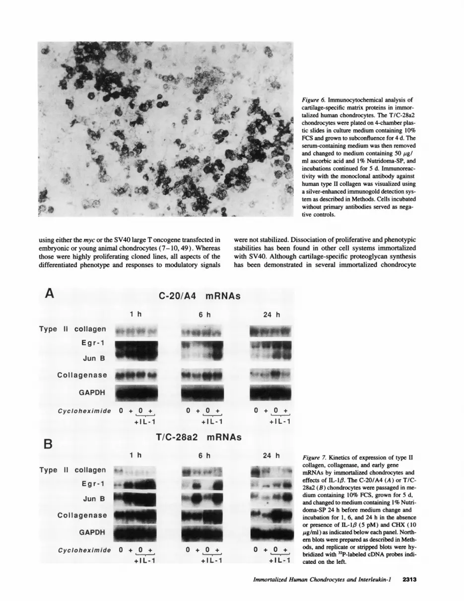
chondrocytes synthesized and deposited extracellular matrixmolecules immunoreactive with monoclonal antibodies againsttype II collagen, large proteoglycan, and chondroitin-4- and-6-sulfate. The data from three separate experiments are summa-rized in Table I. Pericellular and extracellular reactivity withthese antibodies was observed as early as 1 d after transfer tothe serum-free defined medium with maximal expression after5 d, as shown for the type II collagen antibody in Fig. 6. Cellsmaintained continuously in 10% FCS demonstrated no immuno-reactivity with these cartilage-specific monoclonal antibodies.In the absence of ascorbate, type II collagen immunoreactivityremained within the cells, and the staining for proteoglycan waspunctate rather than diffuse in the extracellular matrix. Stainingfor the nonspecific chondroitin-0-sulfate was not observed underany condition. The C-20/A4 line, which maintained a prolifera-tive state even in the presence of the serum substitute, did notdeposit significant amounts of immunostainable matrix.
Chondrocyte-specific responses to IL-1,6. Both the C-20/A4 and T/C-28a2 immortalized chondrocyte lines demon-strated responses to IL-pI similar to those described previouslyin normal human chondrocytes, including decreased expressionof cartilage-specific matrix genes and increased expression ofmatrix metalloproteinase and immediate early genes (2, 42,47). The time courses of expression of the a 1 (11) procollagengene, the matrix metalloproteinase gene, collagenase, and theimmediate early genes, egr-I and jun-B, and the kinetics oftheir stimulation or suppression by IL-I1/3 in the absence andpresence of cycloheximide (CHX) were compared in the C-20/A4 (Fig. 7 A) or the T/C-28a2 (Fig. 7 B) chondrocytes inparallel incubations up to 24 h. Type II collagen mRNA wasdetected in both immortalized chondrocyte lines and increasedwith time throughout the 24 h of incubation in 1% Nutridoma.At 1 h, IL-1/3 had no effect on type II collagen mRNA levels
in the C-20/A4 chondrocytes (Fig. 7 A), but a slight suppres-sion by IL-l: was evident in the T/C-28a2 chondrocytes atboth 1 and 6 h (Fig. 7 B). A > 60% suppression was achievedby 24 h in both chondrocyte lines. CHX suppressed type IIcollagen mRNA levels, particularly at 24 h, but it did not havea further suppressive effect in the presence of IL-1p6. The partialreversal by CHX of the suppression by IL-1,6 at 24 h wasobserved more consistently in the T/C-28a2 than in the C-20/A4 chondrocytes where type II collagen mRNA levels werecomparatively lower. These results are consistent with previousfindings in normal human chondrocytes that indicated that theinhibitory effect of IL- 1,/ on type II collagen synthesis was nota result of decreased a1(11) procollagen mRNA stabilitybut suggested that CHX may block the synthesis of an IL-1-induced repressor factor for al (II) procollagen genetranscription (42).
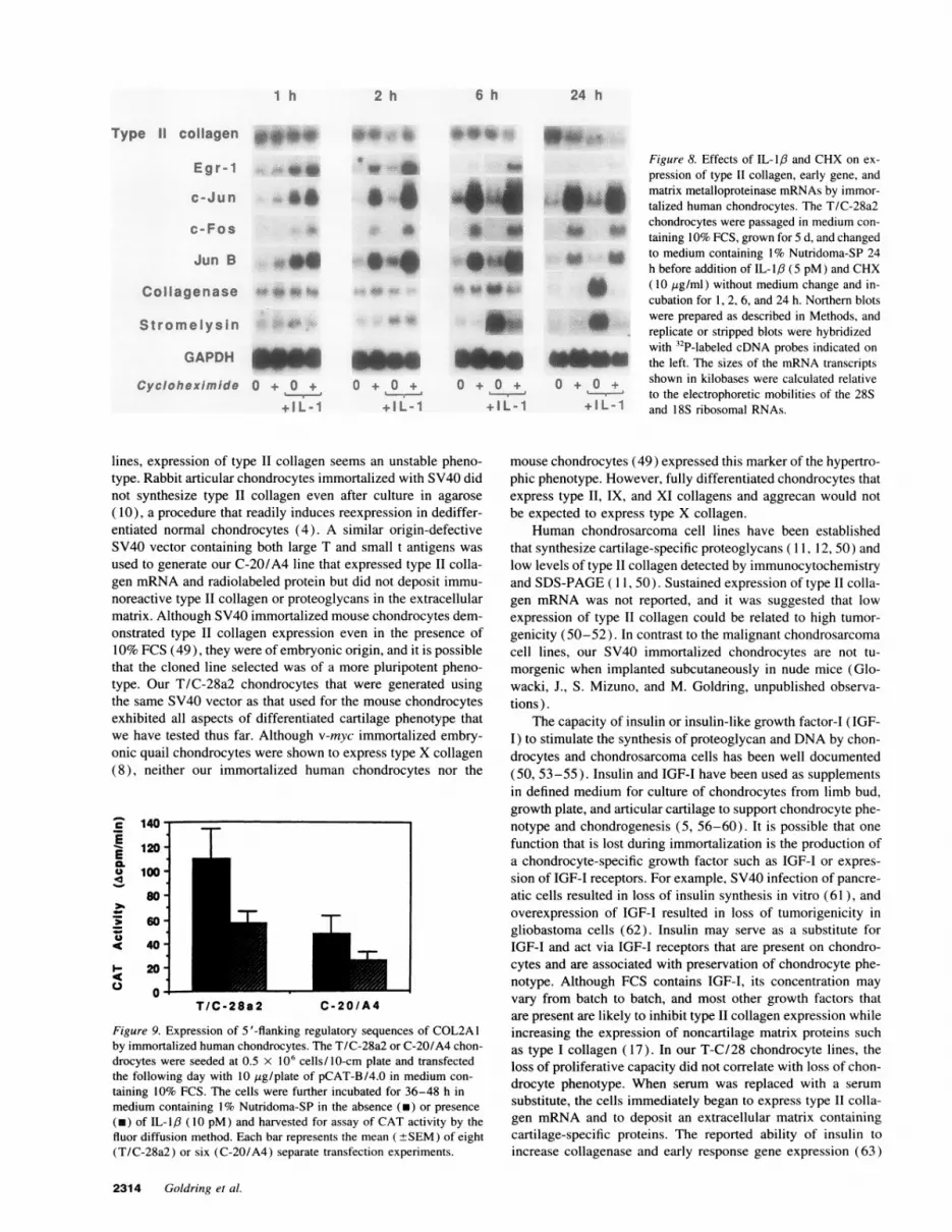
Addition of IL-1/3 to quiescent chondrocytes increased thelevels of egr-1, c-jun, c-fos, and jun-B mRNAs within 1 hwhich declined rapidly thereafter (Figs. 7 and 8). Incubationwith CHX augmented the levels of these mRNAs and prolongedthe IL-1,6-induced expression past 6 h. Constitutive expressionof collagenase mRNA was observed in both the C-20/A4 (Fig.7 A) and T/C-28a2 chondrocytes (Fig. 7 B); this was relativelystable at least up to 6 h but declined within 24 h. Stimulationof collagenase mRNA by IL-1,6 was apparent by 6 h, and themagnitude of this stimulation relative to the constitutive expres-sion increased after 24 h of incubation. The inhibitory effect ofCHX on the IL-1,8-3stimulated level of collagenase mRNA wasapparent after 24 h (Fig. 7). Lower constitutive expression ofboth collagenase and stromelysin mRNAs, as well as the earlygene mRNAs, was observed when the medium was not changedafter the initial 24 h in Nutridoma-containing medium, and CHXmore effectively blocked the effects of IL-1,6 (Fig. 8). These
Immortalized Human Chondrocytes and Interleukin-J 2311

AType 11 collagen 9 5 * * -5.5 kb
GAPDH * * *-1.3 kb
c/i,< 0 0
ci 'I. i
C., N\G G
T/C-28a2 mRNAs
.-
28S> w w* 28S>-
W8> 18S>
11 IX Xi Ag LP lb tcollagens c<
Figure 4. Expression of mRNAs encoding cartilage-specific collagensand other matrix proteins by immortalized chondrocytes cultured inserum-free defined medium. (A) The immortalized chondrocytes C-20/A4, T/C-28a2, C-28/D8, and C-28/12 were passaged in medium con-
taining 10% FCS, grown for 5 d, and changed to medium containing1% Nutridoma-SP 24 h before harvest of the cells for RNA extraction.Total RNAs (10 Mg/lane) were electrophoresed on a 0.8% agarose gelin the presence of 2% formaldehyde, blotted on nylon supported nitro-cellulose membranes, and hybridized with the 32P-labeled cDNA probesencoding al (II) procollagen and GAPDH. (B) Total RNAs from theT/C-28a2 chondrocytes incubated in 1% Nutridoma-SP for 24 h were
blotted and hybridized with the 32P-labeled cDNA probes indicated atthe bottom. The sizes of the mRNA transcripts in kilobases were: 5.5kb, type II collagen; 4.0 kb, type IX collagen; 7.5 kb, type XI collagen;9.5 kb, aggrecan (Ag); 2.5 kb, link protein (LP); 2.5 kb, biglycan; and1.9 and 1.6, decorin. Overexposed blots are shown for type IX collagenand link protein mRNAs. The positions of the 28S and 18S ribosomalRNAs are shown on the left.
.4?
.4
* _p .4 a1(1)
4a2(1)
1 2 3 4
Figure 5. SDS-PAGEanalysis of pepsin-resis-tant [3H ] proline-labeledcollagen in monolayercultures of chondrocytes.Lane 1, C-20/A4 immor-talized chondrocytes;lane 2, T/C-28a immor-talized chondrocytes; andlane 3, C-28 primarynonimmortalized chon-drocytes. The cells were
grown to subconfluencein medium containing10% FCS and transferredto serum-free culture me-
dium containing ascorbic acid for radiolabeling. Positions of the a I (I),a2(I), /3, and y chains of the rat tail tendon collagen standard (lane4) are identified by arrowheads.
Table I. Location and Time ofAppearance of Immunoreactivitiesin Immortalized Human Chondrocyte Cultures
Without ascorbic acid With ascorbic acid
Antibody Pericellular Extracellular Pericellular Extracellular
d d
Anti-large proteoglycan 1 1 3Anti-collagen II 3 - 2 8Anti-chondroitin sulfate 7 2 8
The T/C-28a2 chondrocytes were grown to subconfluence for 4 d inculture medium containing 10% FCS and transferred to medium con-taining 50 pg/ml ascorbic acid and 1% Nutridoma-SP. The results ofthree separate experiments are summarized as the days of appearanceof immunoreactivities after transfer from serum- to Nutridoma-con-taining medium. Note that in cultures incubated without ascorbic acid,no significant immunoreactivity was observed in the extracellular com-partment.
results indicate that the medium change at 0 h in the experimentshown in Fig. 7 may have increased constitutive levels of themetalloproteinase mRNAs and prevented early inhibition byCHX. No change in GAPDH mRNA with time or in responseto IL-18 was observed in these experiments.
Expression of regulatory sequences of the human type IIcollagen gene in immortalized chondrocytes. To determinewhether the immortalized chondrocytes could express regula-tory sequences of the type II collagen gene (COL2A1), weused a 4.0-kb fragment containing the promoter domain and aportion of intron 1 (spanning -577 to +3,428 bp) fused to theCAT reporter gene in transient expression experiments. Expres-sion of this construct, designated pCAT-B/4.0, was observedin both T/C-28a2 and C-20/A4 (Fig. 9) in the presence of 1%Nutridoma-SP. The expression of pCAT-B/4.0 was generallyhigher in the T/C-28a2 than in the C-20/A4 chondrocytes. IL-1/3 inhibited the expression by 30-80% in more than six sepa-rate experiments in each chondrocyte line. These results areconsistent with previous findings of expression of pCAT-B/4.0in nonimmortalized human costal chondrocytes cultured underthe same conditions but no expression in human fibroblasts(42). Nuclear extracts from these immortalized human chon-drocytes contain factors that bind specifically to sequenceswithin the COL2A1 promoter and are modulated by IL-1p3 (48).
Discussion
Our results demonstrate the generation of stable lines of immor-talized human chondrocytes that express the differentiated phe-notype under defined conditions. Different vectors, both con-taining DNA encoding SV40 large T antigen, and differenttransformation and selection protocols were used in derivingthe two chondrocyte lines that we have described in detail. Bothlines proliferated readily in monolayer culture in the presenceof 10% FCS and displayed a morphology similar to that innormal primary human chondrocyte cultures as described pre-viously (2). A serum substitute provided conditions that permit-ted expression of either the endogenous or introduced type IIcollagen gene, as we had found previously in the nonimmortal-ized juvenile costal chondrocyte cultures (42).
Immortalized chondrocyte lines were generated by others
2312 Goldring et al.
B
I v
Aft
st*4t
W
using either the myc or the SV40 large T oncogene transfected inembryonic or young animal chondrocytes (7-10, 49). Whereasthose were highly proliferating cloned lines, all aspects of thedifferentiated phenotype and responses to modulatory signals
Figure 6. Immunocytochemical analysis ofcartilage-specific matrix proteins in immor-talized human chondrocytes. The T/C-28a2chondrocytes were plated on 4-chamber plas-tic slides in culture medium containing 10%FCS and grown to subconfluence for 4 d. Theserum-containing medium was then removedand changed to medium containing 50 [gg/ml ascorbic acid and 1% Nutridoma-SP, andincubations continued for 5 d. Immunoreac-tivity with the monoclonal antibody againsthuman type H collagen was visualized usinga silver-enhanced immunogold detection sys-tem as described in Methods. Cells incubatedwithout primary antibodies served as nega-tive controls.
were not stabilized. Dissociation of proliferative and phenotypicstabilities has been found in other cell systems immortalizedwith SV40. Although cartilage-specific proteoglycan synthesishas been demonstrated in several immortalized chondrocyte
C-20/A4 mRNAs
1 h
Type 11 collagen
E g r - 1
Jun B
,s,.....
Collagenase
GAPDH
Cyclohexim ide
4.
O + O +
+ I L - 1
-IWE
O + O +I
+ IL-1
T/C-28a2 mRNAs
1 h
Type 11 collagen o4
E g r - 1
Jun B
Collagenase
GAPDH
Cycloheximide 0 + 0 +
+IL-1
6 h
4* #.4 .# .*
*6___-_ia
O + O +
sILI
24 h Figure 7. Kinetics of expression of type H
collagen, collagenase, and early gene
* mRNAs by immortalized chondrocytes andeffects of IL-1,6. The C-20/A4 (A) or T/C-
* ^ * 28a2 (B) chondrocytes were passaged in me-dium containing 10% FCS, grown for 5 d,and changed to medium containing 1% Nutri-
h'_ doma-SP 24 h before medium change andincubation for 1, 6, and 24 h in the absenceor presence of IL i,B (5 pM) and CHX (10
_ g/ml) as indicated below each panel. North-ern blots were prepared as described in Meth-
0 + 0 +ods, and replicate or stripped blots were hy-
L-.-e bridized with 32P-labeled cDNA probes indi-+ L- I cated on the left.
Immortalized Human Chondrocytes and Interleukin-1 2313
a,<
A6 h
4*
24 h
B
O + O +
+ I L - 1
4i - 'k
lw!..1
I v 00 t0111-4,"",10",-, 1%,
qdft !L
"W- li-
I
i
I
01-6dm..
2 h 6 h 24 h
Type 11 collagen *.**
Egr-1
c-Jun
_ _0_
c-Fos
Jun B 4.0Collagenase " *5
w #6 fto s
a~~ ~ ~ ~ ~ ~ ~ ~~,
004I*4-1. -;
Stromelysin d
GAPDHjAgCycloheximide O+ O + 0 ++ 0 +0+ 0
+IL-1 + I L-1 +IL-1
Figure 8. Effects of IL-1,8 and CHX on ex-pression of type II collagen, early gene, and
a. ^ matrix metalloproteinase mRNAs by immor-U'' W talized human chondrocytes. The T/C-28a2
chondrocytes were passaged in medium con-to * taining IO% FCS, grown for 5 d, and changed
to Z to medium containing 1% Nutridoma-SP 24h before addition of IL-1,6 (5 pM) and CHX
* ( 10 ,ig/ml) without medium change and in-cubation for 1, 2, 6, and 24 h. Northern blotswere prepared as described in Methods, andreplicate or stripped blots were hybridizedwith 32P-labeled cDNA probes indicated onthe left. The sizes of the mRNA transcripts
+ o + shown in kilobases were calculated relativeto the electrophoretic mobilities of the 28S
+ I L -1 and 18S ribosomal RNAs.
lines, expression of type II collagen seems an unstable pheno-type. Rabbit articular chondrocytes immortalized with SV40 didnot synthesize type II collagen even after culture in agarose(10), a procedure that readily induces reexpression in dediffer-entiated normal chondrocytes (4). A similar origin-defectiveSV40 vector containing both large T and small t antigens wasused to generate our C-20/A4 line that expressed type II colla-gen mRNA and radiolabeled protein but did not deposit immu-noreactive type II collagen or proteoglycans in the extracellularmatrix. Although SV40 immortalized mouse chondrocytes dem-onstrated type II collagen expression even in the presence of10% FCS (49), they were of embryonic origin, and it is possiblethat the cloned line selected was of a more pluripotent pheno-type. Our T/C-28a2 chondrocytes that were generated usingthe same SV40 vector as that used for the mouse chondrocytesexhibited all aspects of differentiated cartilage phenotype thatwe have tested thus far. Although v-myc immortalized embry-onic quail chondrocytes were shown to express type X collagen(8), neither our immortalized human chondrocytes nor the
c
EEal.U
I-c
:cJ
120-
100-
80-
60 -
40 -
20-
0-
T
7T
T/C-28a2 C-20/A4
Figure 9. Expression of 5 '-flanking regulatory sequences of COL2A1by immortalized human chondrocytes. The T/C-28a2 or C-20/A4 chon-drocytes were seeded at 0.5 x 10⁶ cells/10-cm plate and transfectedthe following day with 10 ag/plate of pCAT-B/4.0 in medium con-taining 10% FCS. The cells were further incubated for 36-48 h inmedium containing 1% Nutridoma-SP in the absence (m) or presence(m) of IL-1,6 (10 pM) and harvested for assay of CAT activity by thefluor diffusion method. Each bar represents the mean (±+SEM) of eight(T/C-28a2) or six (C-20/A4) separate transfection experiments.
mouse chondrocytes (49) expressed this marker of the hypertro-phic phenotype. However, fully differentiated chondrocytes thatexpress type II, IX, and XI collagens and aggrecan would notbe expected to express type X collagen.
Human chondrosarcoma cell lines have been establishedthat synthesize cartilage-specific proteoglycans ( 11, 12, 50) andlow levels of type II collagen detected by immunocytochemistryand SDS-PAGE ( 1 1, 50). Sustained expression of type II colla-gen mRNA was not reported, and it was suggested that lowexpression of type II collagen could be related to high tumor-genicity (50-52). In contrast to the malignant chondrosarcomacell lines, our SV40 immortalized chondrocytes are not tu-morgenic when implanted subcutaneously in nude mice (Glo-wacki, J., S. Mizuno, and M. Goldring, unpublished observa-tions).
The capacity of insulin or insulin-like growth factor-I (IGF-I) to stimulate the synthesis of proteoglycan and DNA by chon-drocytes and chondrosarcoma cells has been well documented(50, 53-55). Insulin and IGF-I have been used as supplementsin defined medium for culture of chondrocytes from limb bud,growth plate, and articular cartilage to support chondrocyte phe-notype and chondrogenesis (5, 56-60). It is possible that onefunction that is lost during immortalization is the production ofa chondrocyte-specific growth factor such as IGF-I or expres-sion of IGF-I receptors. For example, SV40 infection of pancre-atic cells resulted in loss of insulin synthesis in vitro (61), andoverexpression of IGF-I resulted in loss of tumorigenicity ingliobastoma cells (62). Insulin may serve as a substitute forIGF-I and act via IGF-I receptors that are present on chondro-cytes and are associated with preservation of chondrocyte phe-notype. Although FCS contains IGF-I, its concentration mayvary from batch to batch, and most other growth factors thatare present are likely to inhibit type II collagen expression whileincreasing the expression of noncartilage matrix proteins suchas type I collagen (17). In our T-C/28 chondrocyte lines, theloss of proliferative capacity did not correlate with loss of chon-drocyte phenotype. When serum was replaced with a serumsubstitute, the cells immediately began to express type II colla-gen mRNA and to deposit an extracellular matrix containingcartilage-specific proteins. The reported ability of insulin toincrease collagenase and early response gene expression (63)
2314 Goldring et al.
1 h

could also account for the constitutive levels of those mRNAsobserved in the early stages of our time course experiments.
During development, the expression of c-fos, c-jun, jun-B,and egr- 1 is found in hypertrophic chondrocytes in regions thatare destined to be replaced by bone, in prechondrocytes thatexpress an alternative type II collagen transcript (64), and ininterstitial zones surrounding cartilage (65-67). The potentialfor involvement of IL-1 in regulation of chondrocyte hypertro-phy and cartilage mineralization has been suggested by a studyin vitro using rabbit growth plate chondrocytes in which typeX collagen synthesis was inhibited by this cytokine (68). Avail-ability of immortalized human chondrocytes that can be manip-ulated in culture will provide a basis for developing a humanmodel of chondrogenesis in vitro. Our studies indicate that thesecells will also serve as excellent host cells for transfection ofchondrocyte-specific genes. Chondrocyte-specific responses toIL-1,6, as reported here, and to IFN-y (48) were similar tothose found previously in normal human chondrocyte cultures(12, 42, 47, 48). The mechanisms that control transcription ofthe late genes encoding collagens and metalloproteinases havenot been defined precisely, but may involve some of the earlygenes studied here. Our studies demonstrate the utility of theseimmortalized human chondrocytes for examining the molecularevents involved in responses of chondrocytes to inflammatorymediators.
Acknowledgments
The authors wish to acknowledge Dr. Dennis Brown, MassachusettsGeneral Hospital (MGH), for the fluorescence microscopy. We thankDr. Stephen M. Krane, MGH, for helpful discussions.
This work was supported by grants AR-34390, AR-03564, and AR-07258 from the National Institutes of Health, a Biomedical ResearchGrant from the Arthritis Foundation, a grant from OsteoArthritis Sci-ences, Inc., Cambridge, MA, and a fellowship from The Wellcome Trustto Dr. Apperley while at Children's Hospital, Boston, MA.
References
1. Goldring, M. B., L. J. Sandell, M. L. Stephenson, and S. M. Krane. 1986.Immune interferon suppresses levels of procollagen mRNA and type II collagensynthesis in cultured human articular and costal chondrocytes. J. Biol. Chem.261:9049-9055.
2. Goldring, M. B., J. Birkhead, L. J. Sandell, T. Kimura, and S. M. Krane.1988. Interleukin 1 suppresses expression of cartilage-specific types II and IXcollagens and increases types I and III collagens in human chondrocytes. J. Clin.Invest. 82:2026-2037.
3. Goldring, M. B., and S. M. Krane. 1987. Modulation by recombinantinterleukin 1 of synthesis of types I and III collagens and associated procollagenmRNA levels in cultured human cells. J. Biol. Chem. 262:16724-16729.
4. Benya, P. D., and J. D. Shaffer. 1982. Dedifferentiated chondrocytes reex-press the differentiated collagen phenotype when cultured in agarose gels. Cell.30:215-224.
5. Adolphe, M., B. Froger, X. Ronot, M. T. Corvol, and N. Forest. 1984. Cellmultiplication and type II collagen production by rabbit articular chondrocytescultivated in a defined medium. Exp. Cell Res. 155:527-536.
6. Castagnola, P., G. Moro, F. Descalzi-Cancedda, and R. Cancedda. 1986.Type X collagen synthesis during in vitro development of chick embryo tibialchondrocytes. J. Cell Biol. 102:2310-2317.
7. Alema, S., F. Tato, and D. Boettiger. 1985. myc and src oncogenes havecomplementary effects on cell proliferation and expression of specific extracellularmatrix components in definitive chondroblasts. Mol. Cell. Biol. 5:538-544.
8. Gionti, E., G. Pontarelli, and R. Cancedda. 1985. Avian myelocytomatosisvirus immortalizes differentiated quail chondrocytes. Proc. Natl. Acad. Sci. USA.82:2756-2760.
9. Horton, W. E. J., J. Cleveland, U. Rapp, G. Nemuth, M. Bolander, K.Doege, Y. Yamada, and J. R. Hassell. 1988. An established rat cell line expressingchondrocyte properties. Exp. Cell Res. 178:457-468.
10. Thenet, S., P. D. Benya, S. Demignot, J. Feunteun, and M. Adolphe. 1992.
SV40-immortalization of rabbit articular chondrocytes: alteration of differentiatedfunctions. J. Cell. Physiol. 150:158-167.
11. Takigawa, M., K. Tajima, H. 0. Pan, M. Enomoto, A. Kinoshita, F.Suzuki, Y. Takano, and Y. Mori. 1989. Establishment of a clonal human chondro-sarcoma cell line with cartilage phenotypes. Cancer Res. 49:3996-4002.
12. Block, J. A., S. E. Inerot, S. Gitelis, and J. H. Kimura. 1991. Synthesisof chondrocytic keratan sulphate-containing proteoglycans by human chondrosar-coma cells in long-term cell culture. J. Bone Jt. Surg. Am. Vol. 73:647-658.
13. Grigoriadis, A. E., J. N. M. Heersche, and J. E. Aubin. 1988. Differentia-tion of muscle, fat, cartilage, and bone from progenitor cells present in a bone-derived clonal cell population: effect of dexamethasone. J. Cell Bio. 106:2139-2151.
14. Bernier, S. M., and D. Goltzman. 1993. Regulation of expression of thechondrogenic phenotype in a skeletal cell line (CFK2) in vitro. J. Bone Miner.Res. 8:475-484.
15. Gerstenfeld, L. C., C. M. Kelly, M. Von Deck, and J. B. Lian. 1990.Comparative morphological and biochemical analysis of hypertrophic, non-hyper-trophic and 1,25 (OH )2D3 treated non-hypertrophic chondrocytes. Connect. TissueRes. 24:29-39.
16. Adams, S. L., K. M. Pallante, Z. Niu, P. S. Leboy, E. B. Golden, and M.Pacifici. 1991. Rapid induction of type X collagen gene expression in culturedchick vertebral chondrocytes. Exp. Cell Res. 193:190-197.
17. Goldring, M. B. 1992. Degradation of articular cartilage in culture: regula-tory factors. In Joint Cartilage Degradation: Basic and Clinical Aspects. J. F.Woessner, Jr., and D. S. Howell, editors. Marcel Dekker, Inc., New York. 281-345.
18. Tyler, J. A., and H. P. Benton. 1988. Synthesis of type II collagen isdecreased in cartilage cultured with interleukin 1 while the rate of intracellulardegradation remains unchanged. Collagen Relat. Res. 8:393-405.
19. Yaron, I., F. A. Meyer, J.-M. Dayer, I. Bleiberg, and M. Yaron. 1989.Some recombinant human cytokines stimulate glycosaminoglycan synthesis inhuman synovial fibroblast cultures and inhibit it in human articular cartilagecultures. Arthritis Rheum. 32:173-180.
20. Tyler, J. A. 1985. Articular cartilage cultured with catabolin (pig interleu-kin 1) synthesizes a decreased number of normal proteoglycan molecules. Bio-chem. J. 227:869-878.
21. Ikebe, T., M. Hirata, and T. Koga. 1988. Effects of human recombinanttumor necrosis factor-a and interleukin 1 on the synthesis of glycosaminoglycanand DNA in cultured rat costal chondrocytes. J. Immunol. 140:827-831.
22. Kimura, T., M.-G. Mattei, J. W. Stevens, M. B. Goldring, Y. Ninomiya,and B. R. Olsen. 1989. Molecular cloning of rat and human type IX collagencDNA and localization of the a1 (IX) gene on the human chromosome 6. Eur.J. Biochem. 179:71-78.
23. Kimura, T., K. S. E. Cheah, S. D. H. Chan, L. V. C. H. Lui, M.-G. Mattei,M. van der Rest, K. Ono, E. Solomon, Y. Ninomiya, and B. R. Olsen. 1989. Thehuman a2(XI) collagen (COLlIA2) chain. Molecular cloning of cDNA andgenomic DNA reveals characteristics of a fibrillar collagen with differences ingenomic organization. J. Biol. Chem. 264:13910-13916.
24. Chu, M.-L., J. C. Myers, M. P. Bernard, J.-F. Ding, and F. Ramirez. 1982.Cloning and characterization of five overlapping cDNAs specific for human a 1(I)collagen chain. Nucleic Acids Res. 10:5925-5934.
25. Doege, K. J., M. Sasaki, T. Kimura, and Y. Yamada. 1991. Completecoding sequence and deduced primary structure of the human cartilage largeaggregating proteoglycan, aggrecan. Human-specific repeats, and additional alter-natively spliced forms. J. Bio. Chem. 266:894-902.
26. Day, A. A., C. I. Ramis, L. W. Fisher, P. Gehron-Robey, J. D. Termine,and M. F. Young. 1986. Characterization of bone PG II cDNA and its relationshipto PG II mRNA from other connective tissues. Nucleic Acids Res. 14:9861-9876.
27. Fisher, L. W. H., A. M. Heegaard, U. Vetter, W. Vogel, W. Just, J. D.Termine, and M. F. Young. 1991. Human biglycan gene. Putative promoter,intron-exon junctions, and chromosomal localization. J. Biol. Chem. 266:14371 -14377.
28. Stephenson, M. L., M. B. Goldring, J. R. Birkhead, S. M. Krane, H. J.Rahmsdorf, and P. Angel. 1987. Stimulation of procollagenase synthesis parallelsincreases in cellular procollagenase mRNA in human articular chondrocytes ex-posed to recombinant interleukin 1/3 or phorbol ester. Biochem. Biophys. Res.Commun. 144:583-590.
29. MacNaul, K. L., N. Chartrain, M. Lark, M. J. Tocci, and N. I. Hutchinson.1990. Discoordinate expression of stromelysin, collagenase, and tissue inhibitorof metalloproteinases-1 in rheumatoid human synovial fibroblasts. Synergisticeffects of interleukin- 1 and tumor necrosis factor-alpha on stromelysin expression.J. Biol. Chem. 265:17238-17245.
30. Nakabeppu, Y., K. Ryder, and D. Nathans. 1988. DNA binding activitiesof three murine jun proteins: stimulation by Fos. Cell. 55:907-915.
31. Ryder, K., L. F. Lau, and D. Nathans. 1988. A gene activated by growthfactors is related to the oncogene v-jun. Proc. Natl. Acad. Sci. USA. 85:1487-1491.
32. Curran, T., W. P. MacConnell, F. van-Straaten, and I. M. Verma. 1983.Structure of the FBJ murine osteosarcoma virus genome: molecular cloning ofits associated helper virus and the cellular homolog of the v-fos gene from mouseand human cells. Mol. Cell. Biol. 3:914-921.
Immortalized Human Chondrocytes and Interleukin-I 2315
33. Sukhatme, V. P., X. Cao, C. Chang, C.-H. Tsai-Morris, D. Stamenkovich,P. C. P. Ferreira, D. R. Cohen, S. A. Edwards, T. B. Shows, T. Curran, M. M.LeBeau, and E. D. Adamson. 1988. A zinc finger-encoding gene coregulated withc-fos during growth and differentiation, and after cellular depolarization. Cell.53:37-43.
34. Small, M. B., Y. Gluzman, and H. L. Ozer. 1982. Enhanced transformationof human fibroblasts by origin-defective simian virus 40. Nature (Lond.).298:671-672.
35. Chaney, W. G., D. R. Howard, J. W. Pollard, S. Sallustio, and P. Stanley.1986. High frequency transfection of CHO cells using polybrene. Somatic CellMol. Genet. 12:237-244.
36. Arbiser, J. L., Z. K. Arbiser, and J. A. Majzoub. 1991. Regulation of geneexpression in choriocarcinoma by methotrexate and hydroxyurea. Endocrinology.128:972-978.
37. Williams, D. A., M. F. Rosenblatt, D. R. Beier, and R. D. Cone. 1988.Generation of murine stromal cell lines supporting hematopoietic stem cell prolif-eration by use of recombinant retrovirus vectors encoding simian virus 40 largeT antigen. Mol. Cell. Biol. 8:3864-3871.
38. Apperley, J. F., B. D. Luskey, and D. A. Williams. 1991. Retroviral genetransfer of human adenosine deaminase in murine hematopoietic cells: effect ofselectable marker sequences on long-term expression. Blood. 78:310-317.
39. Asari, A., S. Miyauchi, K. Miyazaki, A. Hamai, K. Horie, T. Takahashi, T.Sekiguchi, A. Machida, K. Kohno, and Y. Uchiyama. 1992. Intra- and extracellularlocalization of hyaluronic acid and proteoglycan constituents (chondroitin sulfate,keratan sulfate, and protein core) in articular cartilage of rabbit tibia. J. Histochem.Cytochem. 40:1693-1703.
40. Chomczynski, P., and N. Sacchi. 1987. Single step method of RNA isola-tion by acid guanidinium thiocyanate-phenol-chloroform extraction. Anal. Bio-chem. 162:156-159.
41. Cathala, G., J.-F. Savouret, B. Mendez, B. L. West, M. Karin, J. A.Martial, and J. D. Baxter. 1983. A method for isolation of intact, translationallyactive ribonucleic acid. DNA (NY). 2:329-335.
42. Goldring, M. B., K. Fukuo, J. R. Birkhead, E. Dudek, and L. J. Sandell.1994. Transcriptional suppression by interleukin-1 and interferon-y of type IIcollagen gene expression in human chondrocytes. J. Cell. Biochem. 54:85-99.
43. Ryan, M. C., M. Sieraski, and L. Sandell. 1990. The human type I1collagen gene: identification of an additional protein-coding domain and locationof potential regulatory sequences in the promoter and first intron. Genomics.8:41-48.
44. Neumann, C. A., C. A. Morency, and K. 0. Russian. 1987. A novelrapid assay for chloramphenicol acetyltransferase gene expression. Biotechniques.5:444-448.
45. Eastman, A. 1987. An improvement to the novel rapid assay for chloram-phenicol acetyltransferase gene expression. Biotechniques. 5:730-732.
46. Mumane, J. P., L. F. Fuller, and R. B. Painter. 1985. Establishment andcharacterization of a permanent pSV ori --transformed ataxia-telangiectasia cellline. Exp. Cell Res. 158:119-126.
47. Yamin, R., K. Fukuo, J. Birkhead, and M. B. Goldring. 1992. Regulationof early gene expression in human chondrocytes: specific induction of egr- 1mRNA by interleukin-l. J. Bone Miner. Res. 7:S 120.
48. Yamin, R., L. J. Sandell, and M. B. Goldring. 1992. Interleukin-l andinterferon-y suppress the expression of 5'-regulatory sequences in the type IIcollagen gene in human chondrocytes. Arthritis Rheum. 35:S49.
49. Mallein-Gerin, F., and B. R. Olsen. 1993. Expression of simian virus 40large T (tumor) oncogene in chondrocytes induces cell proliferation without lossof the differentiated phenotype. Proc. Natl. Acad. Sci. USA. 90:3289-3293.
50. Takigawa, M., H. 0. Pan, A. Kinoshita, K. Tajima, and Y. Takano. 1991.Establishment from a human chondrosarcoma of a new immortal cell line withhigh tumorigenicity in vivo, which is able to form proteoglycan-rich cartilage-like nodules and to respond to insulin in vitro. Int. J. Cancer. 48:717-725.
51. Cizdziel, P. E., J. Hosoi, J. C. Montgomery, R. W. Wiseman, and
J. C. Barrett. 1991. Loss of a tumor suppressor gene function is correlated withdownregulation of chondrocyte-specific collagen expression in Syrian hamsterembryo cells. Mol. Carcinog. 4:14-24.
52. Sandell, L. J., W. Raskind, J. R. Robbins, and E. U. Conrad. 1994.Characterization of a human chondrosarcoma cell line synthesizing abundantaggrecan and no fibrillar collagens or decorin. Orthop. Trans. 19:750.
53. Kato, Y., N. Nasu, T. Takase, Y. Daikuhara, and S. Suzuki. 1980. Aserum-free medium supplemented with multiplication-stimulating activity (MSA)supports both proliferation and differentiation of chondrocytes in primary culture.Exp. Cell Res. 125:167-174.
54. Stevens, R. L., S. P. Nissley, J. H. Kimura, M. M. Rechler, A. I. Caplan,and V. C. Hascall. 1981. Effects of insulin and multiplication-stimulating activityon proteoglycan biosynthesis in chondrocytes from the Swarm rat chondrosar-coma. J. Biol. Chem. 256:2045-2052.
55. Bohme, K., M. Conscience-Egli, T. Tschan, K. H. Winterhalter, and P.Bruckner. 1992. Induction of proliferation of hypertrophy of chondrocytes inserum-free culture: the role of insulin-like growth factor-I, insulin, or thyroxine.J. Cell Biol. 116:1035-1042.
56. Glaser, J. H., and H. E. Conrad. 1984. Properties of chick embryo chondro-cytes grown in serum-free medium. J. Biol. Chem. 259:6766-6772.
57. Hale, L. V., J. E. Hale, M. L. S. Kemick, Y. Ishikawa, and R. E. Wuthier.1986. Development of a new serum-free medium, USC-HCL, for growth andnormal phenotype in postembryonic chicken growth plate chondrocytes. In VitroCell. & Dev. Biol. 22:597-603.
58. Paulsen, D. F., and M. Solursh. 1988. Microtiter micromass cultures oflim-bud mesenchymal cells. In Vitro Cell. & Dev. Biol. 24:138-147.
59. Carrino, D. A., M. J. Kujawa, D. P. Lennon, and A. I. Caplan. 1989.Altered cartilage proteoglycans synthesized by chick limb bud chondrocytes cul-tured in serum-free defined medium. Exp. Cell Res. 183:62-71.
60. Rosselot, G., A. M. Reginato, and R. M. Leach. 1992. Development of aserum-free system to study the effect of growth hormone and insulinlike growthfactor-I on cultured postembryonic growth plate chondrocytes. In Vitro Cell. &Dev. Biol. 28A:235-244.
61. Niesor, E. J., C. B. Wollheim, D. H. Mintz, B. Blondel, A. E. Renold,and R. Weil. 1979. Establishment of rat pancreatic endocrine cell lines by infectionwith simian virus 40. Biochem. J. 178:559-568.
62. Trojan, J., B. K. Blossey, T. R. Johnston, S. D. Rudin, M. Tykocinski,and J. Ilan. 1992. Loss of tumorigenicity of rat glioblastoma directed by episome-based antisense cDNA transcription of insulin-like growth factor I. Proc. Natl.Acad. Sci. USA. 89:4874-4878.
63. Medema, R. H., R. Wubbolts, and J. L. Bos. 1991. Two dominant inhibi-tory mutants of p21 ' interfere with insulin-induced gene expression. Mol. Cell.Biol. 11:5963-5967.
64. Sandell, L. J., N. Morris, J. R. Robbins, and M. B. Goldring. 1991.Alternatively spliced type II procollagen mRNAs define distinct populations ofcells during vertebral development: differential expression of the amino-propep-tide. J. Cell Biol. 114:1307-1319.
65. McMahon, A. P., J. E. Champion, J. A. McMahon, and V. P. Sukhatme.1990. Developmental expression of the putative transcription factor Egr- 1 suggeststhat Egr-1 and c-fos are coregulated in some tissues. Development (Camb.).108:281-287.
66. Caubet, J. F., and J. F. Bernaudin. 1988. Expression of c-fos proto-oncogene in bone, cartilage, and tooth-forming tissues during mouse development.Biol. Cell. 64:101-104.
67. Wilkinson, D. G., S. Bhatt, R. P. Ryseck, and R. Bravo. 1989. Tissuespecific expression of c-jun and jun B during organogenesis in the mouse. Devel-opment (Camb.). 106:465-471.
68. Kato, Y., K. Nakashima, M. Iwamoto, H. Murakami, H. Hiranuma, T.Koike, F. Suzuki, H. Fuchihata, Y. Ikehara, M. Noshiro, and A. Jikko. 1993.Effects of interleukin- 1 on syntheses of alkaline phosphatase, type X collagen, and1,25-dihydroxyvitamin D3 receptor, and matrix calcification in rabbit chondrocytecultures. J. Clin. Invest. 92:2323-2330.
2316 Goldring et al.
Copyright © 2022 FDOKUMEN


![[- 200 [ PROVIDING MODULATED COMMUNICATION SIGNALS ]](https://static.fdokumen.com/doc/165x107/6328adc85c2c3bbfa804c60f/-200-providing-modulated-communication-signals-.jpg)