LEPROMATOUS AND TUBERCULOID LEPROSY: CLINICAL PRESENTATION AND CYTOKINE RESPONSES
Upload
independentCategory
view
1download
0
JOURNAL OF VIROLOGY, June 2002, p. 5646–5653 Vol. 76, No. 110022-538X/02/$04.00�0 DOI: 10.1128/JVI.76.11.5646–5653.2002Copyright © 2002, American Society for Microbiology. All Rights Reserved.
Cytokine-Sensitive Replication of Hepatitis B Virus in ImmortalizedMouse Hepatocyte Cultures†
Valérie Pasquetto,1‡ Stefan F. Wieland,1 Susan L. Uprichard,1 Marco Tripodi,2 andFrancis V. Chisari1*
Department of Molecular and Experimental Medicine, The Scripps Research Institute, La Jolla, California 92037,1 and Fondazione‘Istituto Pasteur-Cenci Bolognetti,’ Dipartimento di Biotechnologie Cellulari ed Ematologi, Università La Sapienza,
and I.N. Malattie Infettive, I.R.C.C.S. “L. Spallanzani,” Rome, Italy2
Received 5 December 2001/Accepted 12 February 2002
We have previously shown that alpha/beta interferon (IFN-�/�) and gamma interferon (IFN-�) inhibithepatitis B virus (HBV) replication by eliminating pregenomic RNA containing viral capsids from the hepa-tocyte. We have also shown that HBV-specific cytotoxic T lymphocytes that induce IFN-� and tumor necrosisfactor alpha (TNF-�) in the liver can inhibit HBV gene expression by destabilizing preformed viral mRNA. Inorder to further study the antiviral activity of IFN-�/�, IFN-�, and TNF-� at the molecular level, we soughtto reproduce these observations in an in vitro system. Accordingly, hepatocytes were derived from the livers ofHBV-transgenic mice that also expressed the constitutively active cytoplasmic domain of the human hepatocytegrowth factor receptor (c-Met). Here, we show that the resultant well-differentiated, continuous hepatocyte celllines (HBV-Met) replicate HBV and that viral replication in these cells is efficiently controlled by IFN-�/� orIFN-�, which eliminate pregenomic RNA-containing capsids from the cells as they do in the liver. Furthermore,we demonstrate that IFN-�, but not IFN-�/�, is capable of inhibiting HBV gene expression in this system,especially when it acts synergistically with TNF-�. These cells should facilitate the analysis of the intracellularsignaling pathways and effector mechanisms responsible for these antiviral effects.
Hepatitis B virus (HBV) is a hepatotropic DNA virus thatcauses acute and chronic hepatitis and hepatocellular carci-noma (5). We have previously shown that the intrahepaticinduction of alpha/beta interferon (IFN-�/�), gamma inter-feron (IFN-�), and tumor necrosis factor alpha (TNF-�) byvarious stimuli can inhibit HBV gene expression and/or HBVreplication in the livers of HBV-transgenic mice (3, 9, 10, 23).More recently, we demonstrated that similar noncytopathicantiviral events probably occur in the livers of chimpanzeesacutely infected with HBV (15).
Although it is now well documented that HBV replicationcan be inhibited by these cytokines (11, 19), in vivo systems areby nature difficult to manipulate and control. Therefore, stud-ies designed to determine which cytokines primarily inhibitHBV replication and to decipher the antiviral intracellularmechanism(s) they induce would be greatly facilitated by an invitro cell culture system that accurately reflects the cytokine-dependent control of HBV observed in vivo. Unfortunately,primary cultures of HBV-transgenic hepatocytes are not suit-able for these studies because they rapidly become less welldifferentiated in vitro. Recently, however, well-differentiatedimmortalized mouse hepatocyte cell lines were establishedfrom transgenic mice expressing the constitutively active cyto-plasmic domain of the human hepatocyte growth factor recep-tor (c-Met) in their livers (2). Cells prepared from these mice
are not transformed, and they can be induced to a stable,highly differentiated hepatocyte phenotype by dimethyl sulfox-ide (DMSO) (29).
In this report, we describe the establishment and character-ization of an in vitro cell culture system (HBV-Met) based onimmortalized, highly differentiated hepatocytes prepared frommice transgenic for both c-Met and HBV. We show that HBV-Met cells, induced to a highly differentiated hepatocyte phe-notype by DMSO, support HBV gene expression, HBV repli-cation, and virus secretion. Importantly, HBV replication inthis in vitro system is inhibited by the intracellular antiviralmechanism(s) induced by either IFN-�/� or IFN-�, but not byTNF-�. Moreover, longer treatment with IFN-� leads to areduction in the intracellular level of HBV transcripts, espe-cially when it acts synergistically with TNF-�.
MATERIALS AND METHODS
Transgenic mice. HBV-transgenic mouse lineage 1.3.46 (official designation,Tg[HBV 1.3 genome] Chi32) has been previously described (14). These micereplicate HBV in the hepatocyte from an integrated greater-than-genome-lengthHBV transcriptional template. The level of HBV replication in the livers of thesemice is comparable to that seen in the livers of infected patients with chronichepatitis, and there is no evidence of cytopathology (14). Cyto-Met transgenicmice which express the constitutively active cytoplasmic domain of the humanhepatocyte growth factor receptor (c-Met) in their livers have also been previ-ously described (1). HBV-Met double-transgenic mice were produced by matinghomozygous 1.3.46 mice with homozygous cyto-Met transgenic mice. F1 progenywere tested for HBV gene expression and replication by detection of hepatitis Be antigen (HBeAg) in the serum (EBK 125I RIA kit; DiaSorin Inc., Stillwater,Minn.) according to the manufacturer’s instructions.
Primary hepatocyte culture. Livers from 3-week-old F1 mice were perfusedand collagenase digested (collagenase D; Roche Molecular Biochemicals, Indi-anapolis, Ind.) as described previously (22). The cells were further purified overa 60% Percoll gradient (Pharmacia Biotech, Uppsala, Sweden). Hepatocyteviability was assessed by trypan blue dye exclusion to be �80%. The cells were
* Corresponding author. Mailing address: Department of Molecularand Experimental Medicine, The Scripps Research Institute, 10550 N.Torrey Pines Rd., La Jolla, CA 92037. Phone: (858) 784-8228. Fax:(858) 784-2160. E-mail: [email protected].
† This is manuscript no. 14574-MEM from the Scripps Research In-stitute.
‡ Present address: ViaCell, Worcester, MA 01605.
5646
plated at high density on collagen I-coated Biocoat dishes (Becton Dickinson,Franklin Lakes, N.J.) in RPMI 1640 (Gibco Invitrogen Corp., Carlsbad, Calif.)supplemented with 10% fetal calf serum (Gibco Invitrogen Corp.), 55 ng ofepidermal growth factor (Becton Dickinson)/ml, 16 ng of IGF-II (Calbiochem,San Diego, Calif.)/ml, 10 �g of insulin (Sigma, St. Louis, Mo.)/ml, and penicillin-streptomycin-glutamine (100�; liquid) (Gibco Invitrogen Corp.) (complete me-dium), as previously described (2a, 29). After overnight incubation, the majorityof the cells had attached. The semiconfluent cultures were washed and main-tained without transfer for several weeks.
HBV-Met cell lines and clones. All subsequent cell maintenance was per-formed using collagen-coated plasticware and complete RPMI medium as de-scribed above for the primary hepatocyte cultures. Individual epithelial cellislands were expanded slowly in dishes of increasing size and frozen after 10generations. Eight immortalized HBV-transgenic hepatocyte lines were estab-lished. One of these HBV-Met lines is characterized in the present work. Toobtain HBV-Met cell clones, serial dilutions of an early passage of HBV-Metcells were plated in collagen I-coated 96-well plates. Twelve cell clones wereestablished based on their hepatocyte-like morphology as described in Spagnoliet al. (29). These 12 clones were subsequently tested for HBV gene expression byNorthern blot analysis and for HBV replication by Southern blot analysis asdescribed below. Clone HBV-Met.4 was chosen for the studies presented in thiswork. Aliquots of 5 � 106 cells of early passages of the HBV-Met.4 cells werefrozen in 90% serum and 10% DMSO. Otherwise, HBV-Met.4 cells were prop-agated in complete medium until they reached confluence and then passaged ata 1:15 dilution. All experiments were performed with cells that underwent �40passages. For experiments, typically 0.5 � 106 to 1 � 106 HBV-Met.4 cells wereseeded into 60-mm-diameter dishes.
Cytokine treatment. Prior to each experiment, immortalized hepatocytes weregrown to confluence and subsequently kept in complete medium supplementedwith 2% DMSO for 10 days, at which time cytokine treatments were started.Recombinant murine IFN-� (mIFN-�; provided by M. Moriyama, Toray Indus-tries, Tokyo, Japan), recombinant mIFN-�, or recombinant mTNF-� (providedby S. Kramer, Genentech, South San Francisco, Calif.) was added at the indi-cated concentrations. During the whole time of DMSO addition and cytokinetreatment, the medium was replaced every other day with fresh medium, sup-plemented with DMSO and cytokines when necessary.
HBV DNA analysis. Cells were lysed in the culture dish by adding 500 �l ofDNA lysis buffer (50 mM Tris-HCl [pH 8.0], 20 mM EDTA, and 1% sodiumdodecyl sulfate). Samples were then digested overnight at 37°C with proteinaseK (1 mg/ml), and total DNA was extracted as described previously (14). Twentymicrograms of total DNA was analyzed by Southern blotting with a 32P-labeledfull-length HBV DNA probe after HinDIII digestion (14). All quantificationswere done with a Cyclone storage phosphor system (Packard Instrument Com-pany, Meriden, Conn.).
HBV DNA in the cell culture supernatant. Two days after medium change, 400�l of HBV-Met cell culture supernatant was collected and centrifuged at 14,000� g for 10 s to remove cell debris. The supernatant was then digested with 1 mgof proteinase K/ml in a total volume of 500 �l containing 50 mM Tris base (pH8.0) and 1% sodium dodecyl sulfate at 37°C overnight. Nucleic acids wereextracted by phenol-chloroform extraction and precipitated after the addition of10 �g of Escherichia coli tRNA with isopropanol. Nucleic acids were dissolved in30 �l of Tris-EDTA; 15 �l was loaded onto a 1.3% agarose gel (1� Tris-acetate-EDTA) and electrophoresed for 1 h at 5 V/cm. The gel was then blotted in 1.5M NaCl–0.5 M NaOH by vacuum blotting it (VacuGene; Amersham PharmaciaBiotech AB, Uppsala, Sweden) for 1 h onto a nylon membrane (Magnagraph;Osmonics Laboratory Products, Minnetonka, Minn.). Subsequent Southern blot-ting was performed as described previously (14). Alternatively, HBV in the cellculture supernatant was quantified by subjecting 0.5 �l of extracted nucleic acidsto HBV (genotype ayw)-specific TaqMan PCR in a 50-�l reaction volume usingTaqMan Universal PCR Master Mix (Applied Biosystems, Foster City, Calif.),200 nM upper primer HBV469U. The probe was labeled with fluorescein (6-FAM) at the 5 end and with the black hole quencher (BHQ-1; BioresearchTechnologies, Novato, Calif.) at the 3 end (5-CCCGTTTGTCCTCTAATTCC-3), 200 nM lower primer HBV569L (5-GTCCGAAGGTTTGGTACAGC-3), and 100 nM TaqMan probe HBV495P [5-6-FAMd(CTCAACAACCAGCACGGGACCA)BHQ-1-3]. Tenfold serial dilutions (108 to 100 copies) ofplasmid DNA containing a monomeric HBV insert were used as standards inparallel HBV-specific PCRs.
HBV RNA analysis. Total cellular RNA was isolated by the guanidine thiocy-anate method using standard protocols (6). Encapsidated RNA was extractedfrom cells grown in a 60-mm-diameter culture dish. Briefly, cells were scrapedfrom the dish into 1 ml of PBS and pelleted by brief centrifugation in a tabletopmicrotube centrifuge. The pelleted cells were lysed in 0.3 ml of lysis buffer (100
mM NaCl, 1 mM EDTA, 50 mM Tris base [pH 8.0], 0.5% NP-40). Nuclei werepelleted by centrifugation for 5 min at 12,000 � g and 4°C in a microtubecentrifuge. Encapsidated RNA in the supernatant was extracted as previouslydescribed (33). The RNA was dissolved in 50 �l of H2O, and 15 �l was used forNorthern blot analysis as described previously (14).
Immunohistochemical analysis. HBV-Met cells were grown in collagen I-coated Biocoat eight-chamber culture slides. The cells were fixed as previouslydescribed (13). The intracellular distribution of HBV core antigen (HBcAg) wasdetected by an avidin-biotin detection system as described elsewhere (13). Thismethod sequentially uses a primary rabbit anti-HBcAg antiserum (Dako, Carpin-teria, Calif.), a secondary biotin-conjugated goat antiserum specific for rabbitimmunoglobulin G [IgG(Fab)2; Sigma Chemical Co.], a streptavidin-horserad-ish peroxidase conjugate (Extravidin; Sigma), and 3-amino-9-ethyl carbazole(Shandon-Lipshaw, Pittsburgh, Pa.) as a coloring substrate.
RESULTS
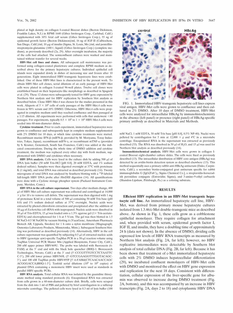
Efficient HBV replication in an HBV-Met transgenic hepa-tocyte cell line. An immortalized hepatocyte cell line, HBV-Met, was derived from primary mouse hepatocyte culturesisolated from 1.3.46/c-Met double-transgenic mice as describedabove. As shown in Fig. 1, these cells grow as a cobblestoneepithelial monolayer. They require collagen for attachmentand, when provided with serum, epidermal growth factor I,IGF II, and insulin, they have a doubling time of approximately24 h (data not shown). In the absence of DMSO, dividing cellsexpressed low levels of HBV RNA transcripts as measured byNorthern blot analysis (Fig. 2A, far left); however, no HBVreplicative intermediates were detectable by Southern blotanalysis of total cellular DNA (Fig. 2B, far left). Because it hasbeen shown that treatment of c-Met immortalized hepatocytecells with 2% DMSO induces hepatocellular differentiation(29), we incubated confluent monolayers of HBV-Met cellswith DMSO and monitored the effect on HBV gene expressionand replication for the next 18 days. Consistent with differen-tiation, cellular expression of the liver-specific gene for albu-min was observed to increase during DMSO treatment (Fig.2A, bottom), and this was accompanied by an increase in HBVtranscripts (Fig. 2A, days 2 to 18) and cytoplasmic HBV DNA
FIG. 1. Immortalized HBV-transgenic hepatocyte cell lines expressviral antigen. HBV-Met cells were grown to confluence and then cul-tured in 2% DMSO. After 10 days of DMSO treatment, HBV-Metcells were analyzed for intracellular HBcAg by immunohistochemistryin the absence (left panel) or presence (right panel) of HBcAg-specificprimary antibody as described in Materials and Methods.
VOL. 76, 2002 INHIBITION OF HBV REPLICATION BY IFNs IN VITRO 5647

replicative intermediates (Fig. 2B, days 2 to 18). HBV replica-tion reached a steady-state level after approximately 10 days ofDMSO treatment, which was comparable to the level of HBVreplication in the livers of the parental 1.3.46 HBV-transgenicmice (Fig. 2B, far right). Consistent with active replication,significant amounts of cytoplasmic HBcAg were detectable byimmunohistochemistry at this time (Fig. 1, right panel). Asviral replication increased, there was also a corresponding se-cretion of HBV virions, indicated by the presence of matureHBV DNA in the cell culture supernatant (Fig. 2C). Likewise,
secretion of HBsAg and HBeAg into the cell media was de-tected (Fig. 2C).
Inflammatory cytokines eliminate cytoplasmic HBV DNAreplicative intermediates from HBV-Met cells. To determinewhether HBV replication in the HBV-Met cells is inhibited byinflammatory cytokines, we treated the cultures with a pool ofcytokines and measured the level of HBV replicative interme-diates over time by Southern blot analysis. For these and allsubsequent experiments, confluent HBV-Met cells were incu-bated for 10 days with 2% DMSO, allowing HBV replication toreach steady-state levels (Fig. 3A, lane 1). The cultures werethen maintained in 2% DMSO alone or in DMSO plus a poolof mIFN-�, mIFN-�, and mTNF-� at 1,000 U/ml each. DNAwas extracted from the cells, and the level of intracellular HBVDNA intermediates was monitored beginning at 2 days andcontinuing up to 8 days after cytokine addition.
The morphology, cell count, and viability of the cells werenot affected by the cytokine treatment over the time courseof the experiment (data not shown). However, Fig. 3A showsthat cytokine treatment of the HBV-Met cells profoundlyreduces cytoplasmic HBV DNA replicative intermediates as
FIG. 2. Efficient HBV gene expression and replication in immor-talized HBV-transgenic hepatocytes. HBV-Met cells were grown toconfluence and then cultured in 2% DMSO for an additional 18 days.Total cellular RNA and DNA were harvested prior to and every 2 daysduring DMSO treatment. For comparison, total liver RNA and DNAfrom 1.3.46 HBV-transgenic mice were extracted and analyzed aspreviously described (14) (far right). (A) Northern blot analysis forHBV, GAPDH, and albumin gene expression. (B) Southern blot anal-ysis of intracellular HBV DNA replicative intermediates. (C) HBVvirion DNA was extracted from cell culture supernatant and analyzedby Southern blotting as described in Materials and Methods. Levels ofHBeAg and HBV surface antigen (HBsAg) secreted into the culturesupernatant are indicated at the bottom in nanograms per milliliter perday. Abbreviations: Tg, HBV transgene; DS, HBV dsDNA; SS, HBVssDNA.
FIG. 3. Inflammatory cytokines inhibit HBV replication in hepato-cyte cell cultures. HBV-Met cells were grown to confluence and thencultured in 2% DMSO. Starting at day 10 of DMSO treatment, theculture medium was replaced every other day (�2 to �8) by completemedium containing 2% DMSO. At the same time points, total cellularDNA was harvested and subjected to HBV-specific Southern blotanalysis. (A) Starting at day 10, the medium was supplemented with apool of mIFN-�, mIFN-�, and mTNF-� (1,000 U/ml each) at everymedium change. (B) Control cultures were treated exactly as describedfor panel A except for omitting cytokines from the culture medium.For abbreviations, see the legend to Fig. 2.
5648 PASQUETTO ET AL. J. VIROL.

early as 2 days after cytokine addition compared to un-treated cells harvested at the same time points (Fig. 3B). Asobserved after cytokine induction in HBV-transgenic mice(33), single-stranded DNA (ssDNA) is completely abol-ished, while trace amounts of double-stranded DNA(dsDNA) remained throughout the experiment (Fig. 3A).Therefore, in vitro cytokine treatment of HBV-Met cellsinduces an antiviral activity that appears to mimic the non-cytopathic clearance of HBV replication observed in vivo.
The cellular level of HBV transcripts is controlled by in-flammatory cytokines. We have previously shown that the in-trahepatic induction of cytokines in HBV-transgenic mice afterstimuli such as injection of virus-specific cytotoxic T-lympho-cytes (12), infection with murine cytomegalovirus (3), or per-sistent infection with lymphocytic choriomeningitis virus (9)suppresses HBV gene expression. Accordingly, the level ofHBV RNA was monitored in total RNA extracted from par-allel cell cultures in the experiment shown in Fig. 3. In DMSO-treated control cell cultures, the cellular content of HBV RNAdid not change significantly during the course of the experi-ment (Fig. 4B). In contrast, 2 days after cytokine addition, thelevels of the HBV 3.5- and 2.1-kb transcripts were reducedthreefold when normalized to glyceraldehyde-3-phosphate de-hydrogenase (GAPDH) mRNA (Fig. 4A; lane �2). With con-tinuous cytokine treatment, HBV RNA levels further de-creased over time (Fig. 4A; lanes �2 to �8). By day 8 ofcytokine treatment, the 3.5- and 2.1-kb HBV transcript levelswere reduced 30-fold and 5-fold, respectively (Fig. 4A, lane�8), compared to pretreatment levels. In summary, the mag-nitude and kinetics of the cytokine-mediated inhibition ofHBV replication and gene expression observed in the HBV-Met cell culture system are strikingly similar to our previousfindings in HBV-transgenic mice after injection of virus-spe-
cific cytotoxic T lymphocytes, where viral DNAs are clearedfirst, followed by a reduction in viral RNAs (12).
Inflammatory cytokines eliminate pgRNA-containing cap-sids from the hepatocyte cytoplasm. Previously, we have shownthat IFN-�/� induction in the livers of HBV-transgenic miceeliminates pregenomic RNA (pgRNA)-containing capsidsfrom the hepatocyte cytoplasm within 6 to 12 h without affect-ing the level of HBV-specific mRNA (33). In this set of exper-iments, we wanted to determine whether the cytokines affectthe same step(s) in HBV replication in the HBV-Met cellculture model. Therefore, we treated HBV-Met cells with cy-tokines as described in the legend to Fig. 3 and monitored theintracellular level of HBV transcripts, as well as encapsidatedHBV RNA isolated from cytoplasmic HBV capsids, every 3 hduring the first 24 h of the treatment.
Figure 5A shows the intracellular level of HBV RNA in theHBV-Met cells in the presence and absence of cytokine treat-ment at the indicated time points after cytokine addition.Quantification of the Northern blot signals revealed no changein total HBV RNA in the cells over the course of the experi-ment. In contrast, encapsidated HBV RNA is cleared from theHBV-Met cell cytoplasm as early as 6 h after cytokine addition,despite the fact that encapsidated HBV RNA is still increasingin the control samples harvested at the same time points (Fig.5B). The disappearance of encapsidated HBV RNA from thecells by 6 h indicates that the cytokines induce an intracellularmechanism that eliminates pgRNA-containing capsids veryrapidly. Furthermore, these results are consistent with a hy-pothesis proposed based on in vivo data (33), in which inflam-matory cytokines actively eliminate pgRNA-containing capsidsfrom the cell while the derivative HBV DNA replicative inter-mediates are passively cleared from the cells as a result ofcapsid maturation and viral export.
FIG. 4. Inflammatory cytokines inhibit HBV gene expression inhepatocyte cell cultures. HBV-Met cells were treated exactly as de-scribed in the legend to Fig. 3. Total RNA was extracted from the cellsat the indicated time points and analyzed for HBV and GAPDH geneexpression by Northern blotting. (A) Starting at day 10, the mediumwas supplemented with a pool of mIFN-�, mIFN-�, and mTNF-�(1,000 U/ml each) at every medium change. (B) Control cultures weretreated exactly as described for panel A except for omitting cytokinesfrom the culture medium. For abbreviations, see the legend to Fig. 2.
FIG. 5. Inflammatory cytokines eliminate pgRNA-containing cap-sids from the hepatocyte cytoplasm. HBV-Met cells were grown toconfluence and then cultured in 2% DMSO for an additional 10 days.At that time, a pool of mIFN-�, mIFN-�, and mTNF-� (1,000 U/mleach) was added to the culture medium. (A) Total cellular RNA wasisolated from untreated () and cytokine-treated (�) HBV-Met cellsat the indicated time points and analyzed for intracellular HBV andGAPDH transcripts by Northern blotting. (B) Encapsidated RNA wasextracted from cytoplasmic HBV capsids and subjected to HBV-spe-cific Northern blot analysis as described in Materials and Methods.
VOL. 76, 2002 INHIBITION OF HBV REPLICATION BY IFNs IN VITRO 5649

Establishment and characterization of HBV-Met clones. Toensure that the HBV-Met cell culture system reflects onlyevents that occur in hepatocytes and not nonparenchymal cellsthat might have contaminated the HBV-Met cell line, we es-tablished 12 clones that cytologically resembled hepatocytes(29). The clones were treated with DMSO for 10 days and thenanalyzed for HBV replication by Southern blot analysis of totalcellular DNA (Fig. 6). The levels of HBV replication variedconsiderably in different clones (Fig. 6), consistent with thedifferences in HBcAg expression in different cells in the HBV-Met cell line (Fig. 1, right panel). Quantitative real-time PCRof viral DNA in the cell culture supernatant collected at thesame time point showed that the amount of virus secreted fromthe cells reflects the level of intracellular HBV replication (Fig.6). For two clones (HBV-Met.4 and HBV-Met.7), it was alsoverified that HBV replication was equally sensitive to cytokinetreatment as in the cell lines (data not shown). All subsequentstudies described were performed with the HBV-Met cellclone no. 4 (HBV-Met.4) (Fig. 6, clone 4).
Both mIFN-� and mIFN-� induce a hepatocellular mecha-nism(s) that inhibits HBV replication. Clone HBV-Met.4 wasused to assess the individual effects of mIFN-�, mIFN-�, andmTNF-� on HBV replication. For these experiments, the cellswere grown and differentiated as described in the legend toFig. 2, and parallel dishes were treated with either mIFN-�(100 U/ml), mIFN-� (1,000 U/ml), mTNF-� (1,000 U/ml), or acombination of mIFN-� and mTNF-� (1,000 U/ml each). Totalcellular DNA was harvested at the indicated time points aftercytokine addition and analyzed for intracellular HBV DNAreplicative intermediates by Southern blot analysis.
Figure 7A, left, lane 4 shows that HBV replication in theHBV-Met.4 cells was reduced 18-fold at the level of ssDNAand reduced 5-fold at the level of dsDNA after only 1 day of100-U/ml mIFN-� treatment alone. HBV DNA continued to
decrease thereafter, by more than 90-fold at day 7 aftermIFN-� addition (Fig. 7A, lanes 5 to 8) compared to theuntreated control samples (Fig. 7A, lanes 1 to 3). Similarly,mIFN-� treatment of HBV-Met.4 cells resulted in a 25-foldinhibition of HBV DNA replication on day 7 (Fig. 7A, lane13), whereas HBV ssDNA and dsDNA were reduced only4-fold on day 1 (Fig. 7A, lane 9). This suggests that mIFN-� isless potent than mIFN-� in this system and that its antiviraleffect is delayed. In fact, titration experiments showed that aslittle as 10 U of mIFN-�/ml could inhibit HBV replicationwhereas at least 100 U of mIFN-�/ml was needed to induce anantiviral response (data not shown). Figure 8A, right, lanes 14to 18, shows that HBV DNA replication was not controlled bymTNF-� under these experimental conditions. When mTNF-�was combined with mIFN-�, HBV replication was inhibited(Fig. 7A, lanes 19 to 23), but the kinetics and extent of inhi-bition mirrored the antiviral activity seen with mIFN-� alone.Similarly, no other combination of these cytokines enhancedthe effect of the most potent one in the mixture at the level ofHBV replication (data not shown).
Synergistic antiviral activity of mIFN-� and mTNF-� onHBV gene expression. To determine which of the cytokines wasresponsible for the antiviral activity that inhibited HBV geneexpression as shown in Fig. 3, total cellular RNA isolated fromparallel dishes in the experiment shown in Fig. 7A was sub-jected to Northern blot analysis. To control for loading differ-ences, the membranes were also probed for GAPDH.
Although mIFN-� rapidly and profoundly reduced the levelof intracellular HBV DNA replicative intermediates (Fig. 7A,lanes 4 to 8), extended treatment with mIFN-� did not affectthe steady-state level of HBV transcripts (Fig. 7B, lanes 4 to 8).Prolonged treatment with mTNF-� also failed to reduce HBVmRNA levels (Fig. 7B, lanes 14 to 18), while mIFN-� caused aslight (twofold) reduction of HBV transcripts in the HBV-Met.4 cells by day 7 (Fig. 7B, lanes 9 to 13). Importantly, thecombination of mIFN-� and mTNF-� reduced the HBVmRNA content approximately 17-fold on day 6 (Fig. 7B, lanes19 to 22), suggesting that these two cytokines act synergisticallyto inhibit HBV gene expression. In contrast, the combinationof mTNF-� with mIFN-� did not inhibit HBV gene expression(data not shown).
The synergy between mIFN-� and mTNF-� was also evidentat the level of liver-specific cellular gene expression. As shownin Fig. 8, while GAPDH mRNA levels remained relativelyconstant under all conditions (Fig. 8, bottom panel), thesteady-state transcript level of several hepatocellular genesvaried depending on the particular cytokine administered. Asexpected, transcript levels for 2,5-oligoadenylate synthetase(OAS) were induced by mIFN-� (Fig. 8, lanes 1 to 4) and bymIFN-� (Fig. 8, lanes 5 to 7) but not by mTNF-� (Fig. 8, lanes8 to 10). However, the combination of mTNF-� and mIFN-�induced OAS expression to levels 5-fold higher than mIFN-�alone (Fig. 8, lanes 11 to 13). Similarly, expression of thechemokine Crg-2 was induced to 100-fold-higher levels by thecombination of mTNF-� and mIFN-� (Fig. 8, lanes 11 to 13)compared to mIFN-� alone (Fig. 8, lanes 5 to 7), whereasmTNF-� alone (Fig. 8, lanes 8 to 10), like mIFN-� alone, hadno effect (Fig. 8, lanes 2 to 4).
In the opposite manner, some hepatocellular genes were sub-ject to synergistic down-regulation by mTNF-� and mIFN-�. Ex-
FIG. 6. HBV replication in HBV-Met cell clones. HBV-Met cloneswere established by limiting dilution of the HBV-Met cell line. Indi-vidual clones were grown to confluence and then maintained in 2%DMSO for an additional 10 days. At that time, total DNA was ex-tracted from the cells and analyzed for HBV DNA replicative inter-mediates by Southern blotting. Viral DNA extracted from the cellculture supernatant was quantified by real-time PCR and is indicatedas viral copies per milliliter per day. For abbreviations, see the legendto Fig. 2.
5650 PASQUETTO ET AL. J. VIROL.

pression of the negative acute-phase gene for albumin was re-duced sevenfold after 6 days of mIFN-� treatment (Fig. 8, lane 7),while mIFN-� alone (Fig. 8, lanes 2 to 4) or mTNF-� alone (Fig.8, lanes 8 to 10) had no effect. The combination of mIFN-� andmTNF-�, however, enhanced albumin down-regulation 50-fold
(Fig. 8, lanes 11 to 13). The same pattern of change in geneexpression was seen for the liver transcription factor hepatocytenuclear factor 4 (HNF4) and C/EBP�, whose transcripts wereminimally reduced by mIFN-� alone, not reduced by mIFN-� ormTNF-�, and virtually eliminated by the combination of mIFN-�and mTNF-�.
DISCUSSION
In this report, we describe an immortalized HBV-transgenicmouse hepatocyte cell culture system that replicates HBV andis sensitive to the antiviral activities of cytokines, reflecting thebehavior of the virus and the cytokines in vivo (3, 4, 9, 11, 12,15, 33). Immortalized hepatocyte cultures were establishedfrom mice transgenic for HBV (14) and the constitutively ac-tive cytoplasmic fragment of the human hepatocyte growthfactor receptor (c-Met [1]). Cell lines immortalized by c-Methave been shown to display a liver-specific gene expressionprofile which can be further enhanced by cultivating the cells inthe presence of the well-known differentiation agent DMSO(2, 29, 35). Consistent with this finding, DMSO induced theexpression of hepatocyte-specific genes, such as those for al-bumin and transthyretin (data not shown), in our HBV-Metcells. Robust HBV gene expression was also dependent on
FIG. 7. Both type I and type II cytokines induce a hepatocellular mechanism(s) inhibiting HBV replication. The HBV-Met.4 clone was grown toconfluence and then maintained in 2% DMSO. After 10 days of culture in DMSO, parallel dishes were treated with either 100 U of mIFN-�/ml, 1,000U of mIFN-�/ml, 1,000 U of mTNF-�/ml, or a combination of mIFN-� and mTNF-� at 1,000 U/ml each. (A) Southern blot analysis for HBV DNAreplicative intermediates at the indicated time points during cytokine treatment or from untreated control cultures. (B) Total cellular RNA harvested atthe same time points and analyzed for HBV and GAPDH transcripts by Northern blot analysis. For abbreviations, see the legend to Fig. 2.
FIG. 8. Changes in cellular gene expression induced by inflammatorycytokines. Total cellular RNA from the experiment shown in Fig. 7 wassubjected to Northern blot analysis specific for the indicated genes. Tran-script levels at days 0, 2, 4, and 6 of cytokine treatment are shown.
VOL. 76, 2002 INHIBITION OF HBV REPLICATION BY IFNs IN VITRO 5651

DMSO-induced hepatocyte differentiation, perhaps due to theactivation of hepatocyte-specific transcription factors, whichhave been shown to regulate HBV promoters (18, 30). Nota-bly, however, HBV DNA replication does not seem to bedependent on hepatocyte differentiation, since it is easily sup-ported in nondifferentiated HBV-Met cells upon transfectionof constructs that express the viral transcripts from constitu-tively active promoters (data not shown). Consistent with thisfinding, it has recently been shown that expression of certainliver-specific transcription factors that transcriptionally acti-vate HBV allows for HBV replication in nonhepatic cells (31).Therefore, the strong restriction of HBV to the liver in vivo is,at least at one level, the consequence of the dependence ofHBV promoter and enhancer sequences on hepatocyte-spe-cific transcription factors.
One advantage of the in vitro HBV replication model systempresented here is the availability of a corresponding in vivosystem. Hence, we have shown that the profound cytokine-induced inhibition of HBV replication we observed in vitroquantitatively and qualitatively reflects the events that occurafter cytokine induction in vivo in the HBV-transgenic mousemodel (11, 12, 33). Additionally, our observations correspondwell with studies performed with duck HBV-infected duckhepatocytes (28). Taken together, the experiments presentedstrongly support the use of the HBV-Met cell line to study themechanisms responsible for the cytokine-dependent noncyto-pathic inhibition of HBV gene expression and replication pre-viously documented in vivo using HBV-transgenic mice (12,33).
Previously, we showed that HBV DNA replication in vivo inHBV-transgenic mice is regulated by inflammatory cytokines(3, 4, 9, 12, 16, 33) and that under certain conditions, HBVgene expression is also subject to cytokine control (3, 9, 12, 16).While it has been possible to some degree to attribute theseantiviral activities to specific cytokines in vivo (3, 4, 9, 12, 23),the HBV-Met cell culture system permits direct analysis of theindividual antiviral activities of these cytokines on both HBVDNA replication and gene expression. The results reveal sev-eral interesting new facts.
First, we have demonstrated that inflammatory cytokines actdirectly on hepatocytes to induce an intracellular mecha-nism(s) that inhibits HBV replication.
Second, we found that mIFN-�, as well as the universalIFN-�/� (chimeric human IFN-�A/D) alone (data not shown),can profoundly inhibit HBV DNA replication, while the levelof HBV transcripts is not affected during up to 6 days ofcytokine treatment. Although similar findings have been ob-tained by others (17), and IFN-�/� has been reported to inhibitHBV gene expression in other systems in vitro (7, 26, 27, 32),all previous studies have been performed with poorly differen-tiated, transformed hepatoma cell lines that may not reflect theintracellular events triggered by cytokines in well-differentiatedhepatocytes.
Third, we observed that, of the three cytokines tested, onlymIFN-� could independently reduce the steady-state level ofHBV transcripts in the HBV-Met cells. In Fig. 8 we show thatexpression of albumin, HNF4, and C/EBP� is also reduced inmIFN-�-treated hepatocytes. These results suggest that cyto-kine-induced changes in hepatocyte-specific transcription fac-tors known to control HBV gene expression (31) might be
responsible for the observed inhibition of HBV gene expres-sion, and further experiments are being done to determine theprecise mechanism mediating this inhibition.
Fourth, the mIFN-� effect on the steady-state level of HBVtranscripts was synergistically enhanced when HBV-Met cellswere simultaneously treated with mIFN-� and mTNF-�. Syn-ergistic antiviral activity of mIFN-� and mTNF-� has long beenrecognized (34). Indeed, synergistic activity of these cytokineshas been shown to affect early steps in herpes simplex virusreplication at the level of early gene transcription and transla-tion (8), while they inhibit murine cytomegalovirus late genetranscription and DNA replication (20). Our results add HBVto the list of viruses whose gene expression is synergisticallysuppressed by IFN-� and TNF-�. The synergistic activity ofthese cytokines on HBV-Met cells was also evident from theenhanced activation of the IFN-responsive genes, such as OASand chemokine Crg-2/IP-10, which has been previously shownto be synergistically induced by IFN-� and TNF-� (24; re-viewed in reference 25). At the same time, there was alsosynergy in the repression of albumin, HNF4, and C/EBP� geneexpression. The fact that in the HBV-Met cells efficient inhi-bition of HBV gene expression requires synergistic action ofcytokines whereas HBV DNA replication is efficiently con-trolled by mIFN-� alone or mIFN-� alone may explain why invivo HBV DNA replication is more sensitive to cytokine con-trol then HBV RNA.
Fifth, while it has been shown that IFN-� and TNF-� cansynergistically block adenovirus capsid formation (21), we donot find synergistic inhibition of HBV DNA replication, eventhough this occurs at the level of formation or stability ofpgRNA-containing capsids (Fig. 5) (33). In fact, mIFN-� is aspotent as mIFN-� in inhibiting HBV DNA replication, and wedo not observe any enhancement of this effect when mIFN-�and mTNF-� or mIFN-� are combined (data not shown).
Taken together, the HBV-Met cell lines should prove to bea very useful model system to study the effects of individualcytokines and other agents on HBV gene expression and rep-lication. Accordingly, we have recently performed gene profil-ing experiments on these cells to identify the hepatocellulargenes associated with the antiviral effects of these cytokines,and as a direct result of those studies, we are now using theHBV-Met cells to explore the molecular basis for the cytokine-induced antiviral effect (S. Wieland, M. Robek, and F. V.Chisari, unpublished data).
ACKNOWLEDGMENTS
V.P. and S.F.W. contributed equally to this work.This work was supported by grant CA40489 from the National In-
stitutes of Health (F.V.C.). V.P. was supported by a fellowship fromthe SKAGGS Institute. S.L.U. was supported by NIH fellowshipAI49670. M.T. was supported by the Associazione Italiana Ricerca sulCancro (AIRC), the Ministero della Sanita, and cofin-MIUR Italy.
We thank Toray Industries for providing the recombinant mIFN-�and Genentech for providing the recombinant mIFN-� and mTNF-�.We thank Luca Guidotti for valuable discussion and advice; CarlColburn, Amber Morris, and Angelina Eustaquio for excellent tech-nical assistance; and Andrea Achenbach for assistance with manuscriptpreparation.
REFERENCES
1. Amicone, L., M. A. Galimi, F. M. Spagnoli, C. Tommasini, V. De Luca, andM. Tripodi. 1995. Temporal and tissue-specific expression of the MET ORF
5652 PASQUETTO ET AL. J. VIROL.

driven by the complete transcriptional unit of human A1AT gene in trans-genic mice. Gene 162:323–328.
2. Amicone, L., F. M. Spagnoli, G. Spath, S. Giordano, C. Tommasini, S.Bernardini, V. De Luca, C. Della Rocca, M. C. Weiss, P. M. Comoglio, andM. Tripodi. 1997. Transgenic expression in the liver of truncated Met blocksapoptosis and permits immortalization of hepatocytes. EMBO J. 16:495–503.
2a.Bellovino, D., Y. Lanyau, I. Garaguso, L. Amicone, C. Cavallari, M. Tripodi,and S. Gaetani. 1999. MMH cells: an in vitro model for the study of retinol-binding protein secretion regulated by retinol. J. Cell. Physiol. 181:24–32.
3. Cavanaugh, V. J., L. G. Guidotti, and F. V. Chisari. 1998. Inhibition ofhepatitis B virus replication during adenovirus and cytomegalovirus infec-tions in transgenic mice. J. Virol. 72:2630–2637.
4. Cavanaugh, V. J., L. G. Guidotti, and F. V. Chisari. 1997. Interleukin-12inhibits hepatitis B virus replication in transgenic mice. J. Virol. 71:3236–3243.
5. Chisari, F. V., and C. Ferrari. 1995. Hepatitis B virus immunopathogenesis.Annu. Rev. Immunol. 13:29–60.
6. Chomczynski, P., and N. Sacchi. 1987. Single-step method of RNA isolationby acid guanidinium thiocyanate-phenol-chloroform extraction. Anal. Bio-chem. 162:156–159.
7. Davis, M. G., and R. W. Jansen. 1994. Inhibition of hepatitis B virus in tissueculture by alpha interferon. Antimicrob. Agents Chemother. 38:2921–2924.
8. Feduchi, E., M. A. Alonso, and L. Carrasco. 1989. Human gamma interferonand tumor necrosis factor exert a synergistic blockade on the replication ofherpes simplex virus. J. Virol. 63:1354–1359.
9. Guidotti, L. G., P. Borrow, M. V. Hobbs, B. Matzke, I. Gresser, M. B.Oldstone, and F. V. Chisari. 1996. Viral cross talk: intracellular inactivationof the hepatitis B virus during an unrelated viral infection of the liver. Proc.Natl. Acad. Sci. USA 93:4589–4594.
10. Guidotti, L. G., and F. V. Chisari. 1999. Cytokine-induced viral purging-rolein viral pathogenesis. Curr. Opin. Microbiol. 2:388–391.
11. Guidotti, L. G., and F. V. Chisari. 2001. Noncytolytic control of viral infec-tions by the innate and adaptive immune response. Annu. Rev. Immunol.19:65–91.
12. Guidotti, L. G., T. Ishikawa, M. V. Hobbs, B. Matzke, R. Schreiber, and F. V.Chisari. 1996. Intracellular inactivation of the hepatitis B virus by cytotoxicT lymphocytes. Immunity 4:25–36.
13. Guidotti, L. G., V. Martinez, Y. T. Loh, C. E. Rogler, and F. V. Chisari. 1994.Hepatitis B virus nucleocapsid particles do not cross the hepatocyte nuclearmembrane in transgenic mice. J. Virol. 68:5469–5475.
14. Guidotti, L. G., B. Matzke, H. Schaller, and F. V. Chisari. 1995. High-levelhepatitis B virus replication in transgenic mice. J. Virol. 69:6158–6169.
15. Guidotti, L. G., R. Rochford, J. Chung, M. Shapiro, R. Purcell, and F. V.Chisari. 1999. Viral clearance without destruction of infected cells duringacute HBV infection. Science 284:825–829.
16. Guilhot, S., L. G. Guidotti, and F. V. Chisari. 1993. Interleukin-2 downregu-lates hepatitis B virus gene expression in transgenic mice by a posttranscrip-tional mechanism. J. Virol. 67:7444–7449.
17. Hayashi, Y., and K. Koike. 1989. Interferon inhibits hepatitis B virus repli-cation in a stable expression system of transfected viral DNA. J. Virol.63:2936–2940.
18. Kosovsky, M. J., I. Qadri, and A. Siddiqui. 1998. The regulation of hepatitis
B virus gene expression: an overview of the cis- and trans-acting components,p. 21–50. In R. Koshy and W. H. Caselmann (ed.), Hepatitis B virus: mo-lecular mechanisms in disease and novel strategies for therapy. ImperialCollege Press, London, United Kingdom.
19. Lin, O. S., and E. B. Keeffe. 2001. Current treatment strategies for chronichepatitis B and C. Annu. Rev. Med. 52:29–49.
20. Lucin, P., S. Jonjic, M. Messerle, B. Polic, H. Hengel, and U. H. Koszi-nowski. 1994. Late phase inhibition of murine cytomegalovirus replication bysynergistic action of interferon-gamma and tumour necrosis factor. J. GenVirol. 75:101–110.
21. Mayer, A., H. Gelderblom, G. Kumel, and C. Jungwirth. 1992. Interferon-gamma-induced assembly block in the replication cycle of adenovirus 2:augmentation by tumour necrosis factor-alpha. Virology 187:372–376.
22. Mazier, D., R. L. Beaudoin, S. Mellouk, P. Druilhe, B. Texier, J. Trosper, F.Miltgen, I. Landau, C. Paul, O. Brandicourt, et al. 1985. Complete devel-opment of hepatic stages of Plasmodium falciparum in vitro. Science 227:440–442.
23. McClary, H., R. Koch, F. V. Chisari, and L. G. Guidotti. 2000. Relativesensitivity of hepatitis B virus and other hepatotropic viruses to the antiviraleffects of cytokines. J. Virol. 74:2255–2264.
24. Ohmori, Y., and T. A. Hamilton. 1995. The interferon-stimulated responseelement and a kappa B site mediate synergistic induction of murine IP-10gene transcription by IFN-� and TNF-�. J. Immunol. 154:5235–5244.
25. Paludan, S. R. 2000. Synergistic action of pro-inflammatory agents: cellularand molecular aspects. J. Leukoc. Biol. 67:18–25.
26. Rang, A., S. Gunther, and H. Will. 1999. Effect of interferon alpha onhepatitis B virus replication and gene expression in transiently transfectedhuman hepatoma cells. J. Hepatol. 31:791–799.
27. Romero, R., and J. E. Lavine. 1996. Cytokine inhibition of the hepatitis Bvirus core promoter. Hepatology 23:17–23.
28. Schultz, U., J. Summers, P. Staeheli, and F. V. Chisari. 1999. Elimination ofduck hepatitis B virus RNA-containing capsids in duck interferon-alpha-treated hepatocytes. J. Virol. 73:5459–5465.
29. Spagnoli, F. M., L. Amicone, M. Tripodi, and M. C. Weiss. 1998. Identifi-cation of a bipotential precursor cell in hepatic cell lines derived fromtransgenic mice expressing cyto-Met in the liver. J. Cell Biol. 143:1101–1112.
30. Tang, H., K. E. Banks, A. L. Anderson, and A. McLachlan. 2001. HepatitisB virus transcription and replication. Drug News Perspect. 14:325–334.
31. Tang, H., and A. McLachlan. 2001. Transcriptional regulation of hepatitis Bvirus by nuclear hormone receptors is a critical determinant of viral tropism.Proc. Natl. Acad. Sci. USA 98:1841–1846.
32. Tur-Kaspa, R., L. Teicher, O. Laub, A. Itin, D. Dagan, B. R. Bloom, andD. A. Shafritz. 1990. Alpha interferon suppresses hepatitis B virus enhanceractivity and reduces viral gene transcription. J. Virol. 64:1821–1824.
33. Wieland, S. F., L. G. Guidotti, and F. V. Chisari. 2000. Intrahepatic induc-tion of alpha/beta interferon eliminates viral RNA-containing capsids inhepatitis B virus transgenic mice. J. Virol. 74:4165–4173.
34. Wong, G. H., and D. V. Goeddel. 1986. Tumour necrosis factors alpha andbeta inhibit virus replication and synergize with interferons. Nature 323:819–822.
35. Zaret, K. S. 1996. Molecular genetics of early liver development. Annu. Rev.Physiol. 58:231–251.
VOL. 76, 2002 INHIBITION OF HBV REPLICATION BY IFNs IN VITRO 5653
Copyright © 2022 FDOKUMEN