A telomerase immortalized human proximal tubule cell line with a truncation mutation (Q4004X) in...
14
A Telomerase Immortalized Human Proximal Tubule Cell Line with a Truncation Mutation (Q4004X) in Polycystin-1 Brittney-Shea Herbert 1 , Brenda R. Grimes 1 , Wei Min Xu 7 , Michael Werner 7 , Christopher Ward 2 , Sandro Rossetti 2 , Peter Harris 2 , Elsa Bello-Reuss 6 , Heather H. Ward 4 , Caroline Miller 3 , Vincent H. Gattone II 3 , Carrie L. Phillips 5 , Angela Wandinger-Ness 4 , Robert L. Bacallao 7 * 1 Department of Medical and Molecular Genetics, Indiana University, Indianapolis, Indiana, United States of America, 2 Division of Nephrology, Mayo Clinic, Rochester, Minnesota, United States of America, 3 Department of Anatomy and Cell Biology, Indiana University, Indianapolis, Indiana, United States of America, 4 Department of Pathology, University of New Mexico, Albuquerque, New Mexico, United States of America, 5 Department of Pathology, Indiana University, Indianapolis, Indiana, United States of America, 6 Division of Nephrology and Hypertension, Texas Tech University, School of Medicine, Texas Tech University Health Science Center, Lubbock, Texas, United States of America, 7 Division of Nephrology, Richard L Roudebush VAMC and Indiana University, Indianapolis, Indiana, United States of America Abstract Autosomal dominant polycystic kidney disease (ADPKD) is associated with a variety of cellular phenotypes in renal epithelial cells. Cystic epithelia are secretory as opposed to absorptive, have higher proliferation rates in cell culture and have some characteristics of epithelial to mesenchymal transitions [1,2]. In this communication we describe a telomerase immortalized cell line that expresses proximal tubule markers and is derived from renal cysts of an ADPKD kidney. These cells have a single detectable truncating mutation (Q4004X) in polycystin-1. These cells make normal appearing but shorter cilia and fail to assemble polycystin-1 in the cilia, and less uncleaved polycystin-1 in membrane fractions. This cell line has been maintained in continuous passage for over 35 passages without going into senescence. Nephron segment specific markers suggest a proximal tubule origin for these cells and the cell line will be useful to study mechanistic details of cyst formation in proximal tubule cells. Citation: Herbert B-S, Grimes BR, Xu WM, Werner M, Ward C, et al. (2013) A Telomerase Immortalized Human Proximal Tubule Cell Line with a Truncation Mutation (Q4004X) in Polycystin-1. PLoS ONE 8(1): e55191. doi:10.1371/journal.pone.0055191 Editor: Eric Feraille, University of Geneva, Switzerland Received January 18, 2012; Accepted December 19, 2012; Published January 28, 2013 This is an open-access article, free of all copyright, and may be freely reproduced, distributed, transmitted, modified, built upon, or otherwise used by anyone for any lawful purpose. The work is made available under the Creative Commons CC0 public domain dedication. Funding: This work was supported by National Institutes of Health/NIDDK grants R21DK067246, R01DK050141 to RLB and AWN respectively, and the Indiana Genomics Initiative (INGEN) to BH and BG. INGEN of Indiana University is supported in part by Lilly Endowment Inc. Electron microscopy work was supported by a generous center grant from the PKD Foundation to VG. CW is supported by NIH R01 R01 grants DK59597, DK065056 grants. PH is supported by RO1 DK58816 grant from the NIH/NIDDK. BRG received funding support from the PKD Foundation and the IUSM Cytogenetics division. RLB has received funding support from the Department of Medicine at Indiana University. Mutation analysis was supported by the Mayo Translational Core which is supported by NIH P30 grant DK090726. URLs: www.niddk.nih.gov; www.pkdcure.org. The funders had no role in study design, data collection and analysis, decision to publish, or preparation of the manuscript. Competing Interests: The authors have declared that no competing interests exist. * E-mail: [email protected] Introduction Autosomal dominant polycystic kidney disease (ADPKD) accounts for ten percent of the dialysis population in the United States. The disease is characterized by numerous fluid filled cysts lined by a monolayer of epithelial cells. Mutations in PKD1 or PKD2 loci are responsible for most cases of adult polycystic kidney disease [3]. The genes code for polycystin-1 and 2 respectively and the two proteins interact via c-terminal domains [4]. Polycystin-1 is a multifunctional protein with motifs that mediate cell-cell interactions, cell-matrix attachments and the intracellular C- terminus has been shown to have transcription factor activity [3,5,6,7]. Polycystin-2 is also called Trpp2, a calcium channel that is a member of the Trpp channel family [8]. It forms a complex with polycystin-1 and has been shown to mediate calcium signaling upon mechanical stimulation of monocilia [9,10]. In a prior communication, another cell line with a point mutation in a transmembrane domain of polycystin-1 (DL2433) resulted in a lack of flow sensitive [Ca +2 ] i signaling [11]. These cells have normal levels of polycystin-1 but it fails to assemble in primary cilia [11]. In this communication, we describe an immortalized cystic epithelial cell line with a truncation mutation (Q4004X) in the PKD1 locus and this cell line was selected for proximal tubule markers. In addition, a cell line from an age-matched normal kidney was also created. Most ADPKD cell lines appear to have been derived cysts originating from collecting ducts. In the current study, we isolated cyst epithelial cells from an ADPKD kidney, immortalized the cells using telomerase and generated a stable cystic cell line that expresses proximal tubule markers. Identifica- tion of the PKD1 mutation revealed a germline Q4004X mutation. We have not identified possible somatic mutations. In addition to the ADPKD cell line, we also generated a cell line from an age-matched normal kidney. Both cell lines were immortalized with human telomerase and maintained in continuous passage for over 35 passages. Both the normal kidney cell line and the ADPKD cell line (PKD Q4004X) express some proximal tubule markers and have primary cilia. The cyst derived proximal tubule cell line expresses similar levels of polycystin-1 as compared to the normal proximal tubule cells. However, there is less uncleaved polycystin-1 in the cystic epithelial cells as compared to the normal human proximal tubule cell line and immuno gold decoration PLOS ONE | www.plosone.org 1 January 2013 | Volume 8 | Issue 1 | e55191
-
Upload
menninconsulting -
Category
Documents
-
view
0 -
download
0
Transcript of A telomerase immortalized human proximal tubule cell line with a truncation mutation (Q4004X) in...
A Telomerase Immortalized Human Proximal Tubule CellLine with a Truncation Mutation (Q4004X) in Polycystin-1Brittney-Shea Herbert1, Brenda R. Grimes1, Wei Min Xu7, Michael Werner7, Christopher Ward2,
Sandro Rossetti2, Peter Harris2, Elsa Bello-Reuss6, Heather H. Ward4, Caroline Miller3,
Vincent H. Gattone II3, Carrie L. Phillips5, Angela Wandinger-Ness4, Robert L. Bacallao7*
1 Department of Medical and Molecular Genetics, Indiana University, Indianapolis, Indiana, United States of America, 2 Division of Nephrology, Mayo Clinic, Rochester,
Minnesota, United States of America, 3 Department of Anatomy and Cell Biology, Indiana University, Indianapolis, Indiana, United States of America, 4 Department of
Pathology, University of New Mexico, Albuquerque, New Mexico, United States of America, 5 Department of Pathology, Indiana University, Indianapolis, Indiana, United
States of America, 6 Division of Nephrology and Hypertension, Texas Tech University, School of Medicine, Texas Tech University Health Science Center, Lubbock, Texas,
United States of America, 7 Division of Nephrology, Richard L Roudebush VAMC and Indiana University, Indianapolis, Indiana, United States of America
Abstract
Autosomal dominant polycystic kidney disease (ADPKD) is associated with a variety of cellular phenotypes in renal epithelialcells. Cystic epithelia are secretory as opposed to absorptive, have higher proliferation rates in cell culture and have somecharacteristics of epithelial to mesenchymal transitions [1,2]. In this communication we describe a telomerase immortalizedcell line that expresses proximal tubule markers and is derived from renal cysts of an ADPKD kidney. These cells have asingle detectable truncating mutation (Q4004X) in polycystin-1. These cells make normal appearing but shorter cilia and failto assemble polycystin-1 in the cilia, and less uncleaved polycystin-1 in membrane fractions. This cell line has beenmaintained in continuous passage for over 35 passages without going into senescence. Nephron segment specific markerssuggest a proximal tubule origin for these cells and the cell line will be useful to study mechanistic details of cyst formationin proximal tubule cells.
Citation: Herbert B-S, Grimes BR, Xu WM, Werner M, Ward C, et al. (2013) A Telomerase Immortalized Human Proximal Tubule Cell Line with a TruncationMutation (Q4004X) in Polycystin-1. PLoS ONE 8(1): e55191. doi:10.1371/journal.pone.0055191
Editor: Eric Feraille, University of Geneva, Switzerland
Received January 18, 2012; Accepted December 19, 2012; Published January 28, 2013
This is an open-access article, free of all copyright, and may be freely reproduced, distributed, transmitted, modified, built upon, or otherwise used by anyone forany lawful purpose. The work is made available under the Creative Commons CC0 public domain dedication.
Funding: This work was supported by National Institutes of Health/NIDDK grants R21DK067246, R01DK050141 to RLB and AWN respectively, and the IndianaGenomics Initiative (INGEN) to BH and BG. INGEN of Indiana University is supported in part by Lilly Endowment Inc. Electron microscopy work was supported by agenerous center grant from the PKD Foundation to VG. CW is supported by NIH R01 R01 grants DK59597, DK065056 grants. PH is supported by RO1 DK58816grant from the NIH/NIDDK. BRG received funding support from the PKD Foundation and the IUSM Cytogenetics division. RLB has received funding support fromthe Department of Medicine at Indiana University. Mutation analysis was supported by the Mayo Translational Core which is supported by NIH P30 grantDK090726. URLs: www.niddk.nih.gov; www.pkdcure.org. The funders had no role in study design, data collection and analysis, decision to publish, or preparationof the manuscript.
Competing Interests: The authors have declared that no competing interests exist.
* E-mail: [email protected]
Introduction
Autosomal dominant polycystic kidney disease (ADPKD)
accounts for ten percent of the dialysis population in the United
States. The disease is characterized by numerous fluid filled cysts
lined by a monolayer of epithelial cells. Mutations in PKD1 or
PKD2 loci are responsible for most cases of adult polycystic kidney
disease [3]. The genes code for polycystin-1 and 2 respectively and
the two proteins interact via c-terminal domains [4]. Polycystin-1
is a multifunctional protein with motifs that mediate cell-cell
interactions, cell-matrix attachments and the intracellular C-
terminus has been shown to have transcription factor activity
[3,5,6,7]. Polycystin-2 is also called Trpp2, a calcium channel that
is a member of the Trpp channel family [8]. It forms a complex
with polycystin-1 and has been shown to mediate calcium
signaling upon mechanical stimulation of monocilia [9,10]. In a
prior communication, another cell line with a point mutation in a
transmembrane domain of polycystin-1 (DL2433) resulted in a lack
of flow sensitive [Ca+2]i signaling [11]. These cells have normal
levels of polycystin-1 but it fails to assemble in primary cilia [11].
In this communication, we describe an immortalized cystic
epithelial cell line with a truncation mutation (Q4004X) in the
PKD1 locus and this cell line was selected for proximal tubule
markers. In addition, a cell line from an age-matched normal
kidney was also created. Most ADPKD cell lines appear to have
been derived cysts originating from collecting ducts. In the current
study, we isolated cyst epithelial cells from an ADPKD kidney,
immortalized the cells using telomerase and generated a stable
cystic cell line that expresses proximal tubule markers. Identifica-
tion of the PKD1 mutation revealed a germline Q4004X
mutation. We have not identified possible somatic mutations. In
addition to the ADPKD cell line, we also generated a cell line from
an age-matched normal kidney. Both cell lines were immortalized
with human telomerase and maintained in continuous passage for
over 35 passages. Both the normal kidney cell line and the
ADPKD cell line (PKD Q4004X) express some proximal tubule
markers and have primary cilia. The cyst derived proximal tubule
cell line expresses similar levels of polycystin-1 as compared to the
normal proximal tubule cells. However, there is less uncleaved
polycystin-1 in the cystic epithelial cells as compared to the normal
human proximal tubule cell line and immuno gold decoration
PLOS ONE | www.plosone.org 1 January 2013 | Volume 8 | Issue 1 | e55191

studies confirm that polycystin-1 fails to assemble in the cilia of the
PKD cell line. Finally, polycystin-2 is over-expressed in the PKD
cell line as compared to the normal proximal cells. The cystic
epithelial cells form cysts in 3D Matrigel cultures while normal
kidney cells do not. The immortalized ADPKD Q4004X cell line
will be a useful tool for the study of proximal tubule cyst formation
and comparative studies of its age and sex matched normal kidney
cell line.
Materials and Methods
Generation of an Immortalized Age and Sex MatchedNormal Kidney (NHPTK) and ADPKD Cell Lines
All animal and human studies including the acquisition of
human pathological samples to generate cell lines and recombi-
nant DNA work were approved by the Indiana University IACUC
(Institutional Animal Care and Use Committee), IBC (Institutional
Biosafety Committee) and IRB (Institutional Review Board)
respectively. No consent was obtained from human subjects
because all tissue samples, from which the cell lines were
generated, were pathological samples that arrived in the labora-
tory after pathology inspection. No identifying information was
collected other than age and sex of the patient from which the
sample was obtained. This protocol was approved under an
Expedited Review process administered by the Indiana University
IRB. PKD Q4004X cells were derived from an end-stage
polycystic kidney of a 57 year old male undergoing nephrectomy
for a renal transplant. Cells were isolated from cysts as previously
described [12]. To ensure that epithelial cells were derived from
cysts, cysts were dissected from the surface of the polycystic kidney
[13]. NHPTK cells were isolated from a 53 year old male whose
kidney was deemed unsuitable for transplantation. Cells were
maintained in culture using renal epithelia growth (REGM) media
(Lonza Walkersville, Walkersville, MD) supplemented with 2%
fetal calf serum and grown in 5% CO2 atmosphere at 37uC. Tissue
procurement was approved by the Institutional Review Board
(IRB) at Indiana University School of Medicine under an
expedited review (study number 0911-70). All cells were isolated
from kidneys without any identifying information other than age
and sex data.
Transduction of PKD Cells with Retroviral hTERTPrimary cell lines isolated as described above were infected with
retroviral hTERT (or an empty vector) as previously described
[14,15]. Briefly, amphotrophic PA317 retroviral packaging cells (a
generous gift to BH from Dr. Jerry Shay, UT-Southwestern)
containing either an empty vector (pLXSN) or hTERT were
propagated to collect supernatants [16]. Supernatants containing
released amphotropic retroviruses produced from confluent dishes
were filtered (pore size, 0.45 mm) and used to infect the kidney
cells. Infected cells were selected with 1 mg/ml G418 for two
passages and then maintained for at least 35 passages. A paired
uninfected primary cell line entered into senescence and failed to
grow any further at passage 6. Immortalized cells were analyzed
for hTERT expression and activity. In addition, a separate set of
NHPTK and PKD cells were also transduced with pBabepuro or
hTERT under a puromycin selection marker as described above.
The second set of cell lines was created to give researchers a choice
of selection markers in future experiments.
Telomeric Repeat Amplification Protocol (TRAP)Telomerase activity from cell extracts was analyzed by the
TRAP assay with the TRAP-eze Telomerase Detection kit
(Serologicals/Invitrogen) and established protocols [14–15]. Fol-
lowing PCR amplification of the in vitro TRAP reaction products,
the PCR products were run on a 10% non-denaturing acrylamide
gel. The gel was exposed without drying to a phosphor screen and
visualized on a Phosphor Imager using ImageQuant software
(Molecular Dynamics, Sunnyvale, CA). Telomerase activity was
estimated as the presence of a 6-bp telomerase-specific ladder; an
internal standard PCR control was also included as the product of
separate primers and is represented as a 36-bp band. Five hundred
cell equivalents of an H1299 lung cancer cell extract served as a
positive control for the TRAP assay; lysis buffer only served as a
negative control.
Immune Blot Analysis of Exogenous hTERT LevelsWhole cell lysates were prepared from logarithmically growing
cells using 2% sodium dodecyl sulfate (SDS) in 50 mM Tris-HCl.
Total protein concentration was determined using the BCA assay
(Pierce, Rockford, IL) according to manufacturer’s instructions.
Fifty micrograms of each sample were electrophoresed on a 10%
SDS polyacrylamide gel and transferred to a PVDF membrane
(Amersh, Arlington Heights, IL). The blots were incubated with
monoclonal antibodies to hTERT (IA4 antibody, a generous gift
from Geron Corporation) followed by anti-mouse IgG secondary
antibody coupled to horseradish peroxidase (Jackson ImmunoR-
esearch, West Grove, PA). After washing, the blots were exposed
to X-ray film (Kodak, Rochester, NY) using a chemiluminescent
substrate (Super Signal, Thermo Pierce, Rockford, IL).
Mutation AnalysisAnalysis of the mutation(s) in the ADPKD cell line was
performed by the Molecular Genetics and Proteomics Core at
the Mayo Translational PKD Center. Genomic DNA of both cell
lines was extracted from a cell pellet following the salting-out
method, as previously described [17]. Genomic DNA was PCR
amplified for all the coding exons of the PKD1 and PKD2 genes,
and the corresponding amplicons directly sequenced on both
strands following previously published protocols [17,18,19,20].
Briefly, the PKD2 gene and the single copy part of PKD1 were
amplified from genomic DNA by standard PCR. The duplicated
region of PKD1 was amplified as five, 3 to 9 Kb Long Range PCR
(LR-PCR) fragments, by use of primers that are either anchored in
the single copy DNA portion or mismatched with the Homologous
Genes sequence. LR-PCR fragments were amplified using the
rTth DNA polymerase (PE Applied Biosystems, Foster City, CA)
in the supplied DMSO containing buffer. Mutations were typically
confirmed in a second independent amplification.
Fluorescence Activated Cell SortingFACS sorting was performed on cells labeled with fluorescein
conjugated lotus tetraglobinus lectin (LTL) and rhodamine
conjugated dolicous biflorus agglutinin (DBA) purchased from
Vector Laboratories (Burlingame, CA). Cell were passaged as
described above and suspended in phosphate buffered saline
supplemented with 1 mg/ml fluorescein conjugated LTL for 30
minutes and 1 mg/ml rhodamine conjugated DBA. The cells were
washed three times with phosphate buffered saline and sorted with
a BD FACStar Plus Cell Sorter (BD Biosciences, San Jose, CA).
Isolated cells were then grown under the conditions described
above.
Cell Cycle AnalysisCell cycle analysis was performed as previously described [21].
Cells were passaged and suspended in phosphate buffered saline
Telomerase Immortalized Polycystic Kidney Cells
PLOS ONE | www.plosone.org 2 January 2013 | Volume 8 | Issue 1 | e55191
with 1 mM propidium iodide. DNA content was analyzed with a
BD FACscan Flow Cytometer (BD Biosciences, San Jose, CA).Population doubling analysis. Growth curves were calcu-
lated for normal kidney and PKDQ4004X based on split ratios for
passaging. Both telomerase immortalized cell lines are passaged
with a split of 1:6 flasks twice a week. Population doubling gain
was calculated as the log(cell number/number of cells plated)/
log2. Cells transfected with plasmids lacking a telomerase insert
routinely reached senescence by passage.
Measurement of Transepithelial ResistanceBoth telomerase immortalized normal and ADPKD proximal
cell lines selected with Lotus tetraglobinus were plated on 12 mm
diameter Costar Clear membrane filter supports and grown to
confluence for 5 days in the above described growth media
(Corning Costar, Cole Parmer, Vernon Hills, IL). Transepithelial
resistance was measured using an Epithelial Volt Ohm Meter with
chopstick electrodes (EVOM, World Precision Instruments,
Sarasota, FL). Each sample was measured in triplicate and
measurements were adjusted to a blank filter control. Measured
resistance was adjusted to account for the membrane surface area.
Antibodies and ReagentsAntibodies to NCB1 were generously supplied by I. Kurtz
(UCLA, Los Angeles, CA). NM002 and NM005 antiserum raised
against the 3rd cytoplasmic loop domain and the last 200 amino
acids from the c-terminal of polycystin-1 respectively, were
supplied by Angela Wandinger-Ness (University of New Mexico)
[22,23]. 7E12 monoclonal antibody raised against the LRR region
of polycystin-1 was provided by Christopher Ward [24]. Other
antibodies were purchased from commercial sources including;
anti-NHE-1 (BD Biosciences, San Jose, CA), anti-ZO-1 (Hybrid-
oma Bank, University of Iowa, Iowa City, IA), monoclonal anti-
actin (Millipore, Billerica, MA), anti-cytokeratin, anti-vimentin
(Sigma-Aldrich, St. Louis, MO) and anti-aquaporin 1 (Millipore,
Temmecula, Ca). All chemical supplies and buffers were of
reagent grade purchased from Thermo Fisher Scientific (Wal-
tham, MA). Human recombinant epidermal growth factor (EGF)
was purchased from Teva Pharmaceuticals (Teva, Israel). 8-
bromo-cyclic adenosine monophosphate (8-Br-cAMP) was pur-
chased from Sigma-Aldrich (St. Louis, MO).
Membrane PreparationCells were grown to confluence on 150 mm plates and washed
with ice cold phosphate buffered saline. Cells were scraped in ice
cold phosphate buffered saline supplemented with protease
inhibitor cocktail (Sigma, St. Louis, MO). After centrifuging at
14,000 rpm at 4uC for five minutes, the pellet was resuspended in
0.25 M sucrose, 10 mM Tris-Cl, pH 7.5, 0.2 mM CaCl2 with
protease inhibitors (Sigma, St. Louis, MO). Cell suspensions were
lysed by passing eight times through a Balch homogenizer cooled
to 4uC. Homogenates were diluted with 5X volumes of 0.25 M
sucrose, 10 mM Tris-Cl, pH 7.5, 1 mM EDTA supplemented
with protease inhibitors. The resultant suspension was centrifuged
at 4000g for five minutes at 4uC and transferred to a sucrose
cushion (1.02 M sucrose, 10 mM Tris-Cl, pH 7.5, 1 mM EDTA
with protease inhibitors) and centrifuged at 30,000g for 30 minutes
at 4uC in a TLS 55 rotor (Beckman, Pasadena, Ca). Cloudy
material observed at the interface of the sucrose cushion was
collected and centrifuged at 100,000g at 4uC for 45 minutes.
Membrane pellets were resuspended in 0.25 M sucrose, 10 mM
Tris-Cl, pH 7.5 supplemented with protease inhibitors. Mem-
brane fractions were assayed for protein concentration with BCA
Protein Assay (Thermo Fisher Scientific, Rockford, IL).
Isolation of ExosomesGrowth media containing only 1% bovine serum albumin as a
supplement was incubated with either PKD Q4004X or NHPTK
cells for 24 hours. Conditioned media was treated with protease
inhibitor cocktail tablets (Roche Diagnostics, Mannheim, Ger-
many). Similarly, first void human urine was collected and treated
with protease inhibitor cocktail tablets. Exosomes were isolated as
described by Gonzales et al. and Hogan et al. [25,26]. Purified
exosomes were resuspended with phosphate buffered saline and
the protein concentration of the suspension was determined using
a BCA Protein Assay (bicinchoninic assay, Thermo Fisher
Scientific, Rockford, IL) using bovine serum albumin as a protein
standard for the standard curve. Exosomes were snap frozen in
liquid nitrogen and stored at 280uC until used for immune blot
analysis.
Immunoblot and Immunofluorescence StudiesSerial dilutions were performed using all primary antibodies
when used for either immunostaining or immune blotting to
determine appropriate dilutions. Equivalent amounts of protein
were loaded onto SDS-polyacrylamide gels. For blots in which
polycystin-1 was to be studied, samples were loaded on 4–12%
gradient gels (Invitrogen, Carlsbad, CA) and run at 25 volts for 16
hours. Proteins were transferred to nitrocellulose at 25 volts for 4
hours at 4uC in 10 mM caps, pH 11.0, 0.01% SDS with 10%
methanol. All membranes were blocked with 3% calf serum
dissolved in tris buffered saline.
To analyze polycystin-1 expression in exosomes, 40 mg of total
protein was loaded per lane in a 3–8% gradient polyacrylamide gel
(Invitrogen, Carlsbad, CA) and the gel was run at 150 V for 90
minutes. High molecular weight protein standards (HiMark,
Invitrogen, Carlsbad, Ca) were loaded on the gels to assess
transfer and relative molecular weights. After running the samples,
gels were soaked in 2X transfer buffer with 0.02% SDS for 10
minutes. Transfer to nitrocellulose was performed in a tris-glycine
buffer with 0.01% SDS running the transfer at 24 V for 1 hour.
After blocking the blot with 1% non-fat dried milk (BioRad,
Hercules, CA) dissolved in tris-buffered saline with 0.1% Tween-
20, the blot was developed with monoclonal anti-polycystin-1
(7E12) and rabbit anti-mouse IgG1 (Southern Biotechnology,
Birmingham, AL). Blots were visualized with Super Signal
(Thermo Fisher Scientific, Rockford, IL).
To perform immune-staining studies, cells were grown on glass
cover slips and fixed with 2% paraformaldehyde dissolved in
phosphate buffered saline for 10 minutes. Fixation reactions were
quenched with 100 mM NH4Cl dissolved in phosphate buffer
saline. Samples were permeabilized with 2% saponin dissolved in
Tris-buffered saline (TBS) or with TBS with 0.1% Triton X-100.
Transmission Electron microscopyCells were grown on Thermanox cover slips (NUNC, Thermo
Fisher Scientific, Waltham, MA). After 10 days post plating they
were fixed with 4% Paraformaldehyde in 0.1 M phosphate buffer.
After fixation and rinsing in buffer pre-embedding immunostain-
ing was done. Cover slips were immersed in the primary antibody
overnight at 4̊C, rinsed the next day in buffer and placed in the
secondary antibody attached to 10 nm colloidal gold (AURION,
Hatfield, PA) for 2 hours. After several rinses in buffer the samples
were dehydrated through a graded series of ethyl alcohols and
embedded in Embed 812 (Electron Microscopy Sciences, Hatfield,
PA). Thin sections (70–90 nm) were cut, dried on grids and stained
for contrast using uranyl acetate. The grids were viewed with a
Tecnai G 12 Bio Twin transmission electron microscope (FEI,
Telomerase Immortalized Polycystic Kidney Cells
PLOS ONE | www.plosone.org 3 January 2013 | Volume 8 | Issue 1 | e55191

Telomerase Immortalized Polycystic Kidney Cells
PLOS ONE | www.plosone.org 4 January 2013 | Volume 8 | Issue 1 | e55191

Hillsboro, OR) and images taken with an AMT (Advanced
Microscopy Techniques, Danvers, MA) CCD camera.
Scanning Electron MicroscopyCell cultures grown on Thermanox coverslips (Nalge Nunc
International, Rochester, NY) were fixed with 2% paraformalde-
hyde/2% glutaraldehyde/0.1 M phosphate buffer. After initial
fixation, the specimens were rinsed with PBS (phosphate buffered
saline) followed by post fixation with 1% osmium tetroxide/0.1 M
phosphate buffer for 2 hours. After rinsing with PBS the specimens
were dehydrated using a series of graded ethyl alcohols from 70%–
100%. The specimens were then critical point dried (Samdri-790,
Tousimi, Rockville, MD). After drying, the specimens were
mounted on aluminum stubs with adhesive tabs and sputtered
coated for 3 minutes with gold/palladium (Polaron, Energy Beam
Sciences, Agawam, MA). The specimens were then viewed and
images taken with a JEOL 6390LV (Peabody, MA) scanning
electron microscope in SEI imaging mode, 5 KV, 12 mm working
distance, and spot size 30.
Measurement of Cell VolumeBoth cell lines were grown to confluence and then passaged with
trypsin-EDTA solution to create cell suspensions with a density of
100,000 cells/ml. Cell volume was measured with a Beckman
Coulter Counter (Miami, FL). Twelve samples from each cell line
were analyzed.
Growth in 3D CultureCells (PKD Q4004X and NHPTK) were added to growth
factor reduced Matrigel (GFR Matrigel, B&D Biosciences, Bed-
ford, MA) or type I collagen (Wako Chemical Co, Osaka, JP) at a
concentration of 3000 cells per ml and place in glass bottom plastic
culture dishes (Mat Tek Corp, Ashland, MA) or in 96 well plates.
Cultures were overlaid with culture media and incubated at 37uCin a 5% CO2 incubator. Culture media was replaced daily to
ensure adequate growth conditions. After cyst formation occurred,
usually after 14 days in culture, forskolin (5 mM) or forskolin plus
50 ng/ml IGF-1 was added to the culture to stimulate cyst
expansion.
Imaging of 3D cultures. Cell and matrix samples were fixed
in 4% paraformaldehyde dissolved in phosphate buffered saline for
30 minutes at room temperature. Fixation reactions were
quenched by incubating the samples with 100 mM NH4Cl
dissolved in phosphate buffered saline for 30 minutes. After
several washes with phosphate buffered saline, samples were
permeabilized with 0.1% Triton X-100 dissolved in phosphate
buffered saline. Samples were labeled with Bodipy phalloidin (Life
Technologies, Carlsbad, CA) and Hoechst 33342, washed with
0.1% Triton X-100, phosphate buffered saline, pH 7.4 and post-
fixed in 2% paraformaldehyde, phosphate buffered saline. Matrix
samples were removed from the wells and placed on glass bottom
plastic culture dishes (Mat Tek Corp, Ashland, MA). Confocal
images were collected as previously described using an Olympus
Flowview confocal microscope equipped for two photon confocal
microscopy [27].
Results
Primary cultured renal epithelial cell lines were developed from
pooled dissected cysts as previously described [28]. These cells
were obtained from a male in the fifth decade of life that had a
diagnosis of ADPKD. An age and sex matched primary culture
cell line isolated from a normal kidney was immortalized along
with the PKD cell line for comparison. Both primary cell lines
were sensitive to neomycin selection and following transduction
were neomycin resistant (Figure 1C and data not shown). We also
found that immortalization was successful without selection
because under our culture conditions, untransduced cells failed
to grow after passage 6. Growth failure at this passage is probably
due to senescence. Both western blot analysis and telomerase
activity assays confirmed that the cell lines over-expressed human
telomerase (Figure 1A and data not shown). Sequence analysis of
the PKD1 loci in the cystic cell line revealed a premature stop
codon that would result in a truncation of polycystin-1 at position
Q4004X (Figure 1B). This would eliminate the last 299 amino
acids from the carboxy terminal. No second mutation was detected
and there were no mutations detected in the PKD2 locus (data not
shown). Notably the height of the peak at position Q4004 (CRT,
red peak) is equivalent to the C peak (blue), suggesting that the
mRNA bearing the mutation is expressed at roughly equivalent
levels as the wild-type message (Figure 1B and data not shown).
Untransfected cells failed to grow in selection media (Figure 1C,
upper panel and data not shown) while the transfected cells formed
confluent monolayers in the selection media (Figure 1C, lower
panel).
Once the cells passed the point when we typically observed
senescence, we selected for expression of a proximal tubule marker
by performing FACs sorting on both normal and cystic epithelial
cell lines. After passaging the cells, fluorescein tagged lotus
tetraglobinus lectin (LTL) and rhodamine conjugated dolichous
biflourus was used to label cells in suspension. Control experiments
were performed using HK-2 cells and MDCK-II cells, a human
proximal tubule cells line and a mixed population cell line
respectively. The results of the fluorescence sort are shown in
figure 1 (D, E, and F). HK-2 cells had a fluorescent signature
Figure 1. Characterization of PKD Q4004X growth and selection. A: Telomerase activity assay and immune blot analysis of transduced anduntransduced cells. Left panel: Telomerase activity detected as labeled telomerase product telomeric DNA ladders. DNA ladders are only observed incells transduced with hTERT. Untransduced control cells or cells transduced with empty vector (pLXSN) have no detectable telomerase activity. Rightpanel: Exogenous telomerase expression confirmed by immune blot analysis. A 120 kDa band is observed in lysates made from PKD cells transfectedwith hTERT. B: Mutation detection in the PKD cell line. A C to T mutation resulting in a premature truncation at amino acid 4004 is shown. C:Phase contrast image of untransduced cells (upper image) and hTERT transfected PKD cells (lower image) maintained in selection media. D:Fluorescence activated cell sorting of LTL and DBA labeled HK2 cells. Fluorescence intensity at 575 nM/unit cell area versus fluorescenceintensity at 530 nM/unit cell area are plotted on the Y and X axis respectively. Since DBA was tagged with rhodamine, cells labeled with the DBAmarker are expected to register in the region demarcated by the circle. E: Fluorescence activated cell sorting of LTL and DBA labeled MDCK-II cells. Fluorescence intensity at 575 nM/unit cell area versus fluorescence intensity at 530 nM/unit cell area are plotted on the Y and X axisrespectively. Since DBA was tagged with rhodamine, cells labeled with the DBA marker are expected to register in the region demarcated by thecircle and shown in green. MDCK-II cells are a mixed population basted on this assay (compare red and green populations). F: Fluorescenceactivated cell sorting of LTL and DBA labeled PKD Q4004X cells. Fluorescence intensity at 575 nM/unit cell area versus fluorescence intensityat 530 nM/unit cell area are plotted on the Y and X axis respectively. Since DBA was tagged with rhodamine, cells labeled with the DBA marker areexpected to register in the region demarcated by the circle. Few cells have DBA labeling with greater than 99% of the cells showing LTL positivelabeling.doi:10.1371/journal.pone.0055191.g001
Telomerase Immortalized Polycystic Kidney Cells
PLOS ONE | www.plosone.org 5 January 2013 | Volume 8 | Issue 1 | e55191

comprised of predominantly equivalent signals from the FITC and
Rhodamine channels (Figure 1D). A small percentage of cells had
a high intensity rhodamine signal (Figure 1D, green dots in the
circled area). In contrast MDCK-II cells had a shifted fluorescent
signature (Figure 1E). The PKD Q4004X cell line has a
fluorescent lectin binding pattern most similar to the HK cells
(Figure 1F compare to 1D). Cells binding the LTL lectin were
collected and maintained in culture.
Figure 2A graphically depicts the population doublings and
extended life span of hTERT transduced cell lines for both the
normal human proximal tubule cell line (NHPTK) and the
Q4004X proximal PKD cell line. Both cell lines were found to
Figure 2. Cell Growth Characteristics of NHPTK and PKD Q4004X Cells. A: Population doublings of normal and cystic proximal tubule celllines. Graphical depiction of population doublings based on growth and passage number. Left panel NHPTK cell line population doubling, right panelPKD Q4004X cell line population doubling. B: Cell cycle analysis of telomerase immortalized PKD Q4004X and NHPTK cell lines. Left panelPKD Q4004X cells labeled with propidium iodide. Right panel NHPTK cells labeled with propidium iodide. Cells were passaged as described inmaterials and methods. Representative results are shown from 3 separate experiments. C: ZO-1 expression in PKD Q4004X cells. ZO-1 isexpressed at cell-cell borders typical of its expression pattern in renal epithelial cells.doi:10.1371/journal.pone.0055191.g002
Telomerase Immortalized Polycystic Kidney Cells
PLOS ONE | www.plosone.org 6 January 2013 | Volume 8 | Issue 1 | e55191

Telomerase Immortalized Polycystic Kidney Cells
PLOS ONE | www.plosone.org 7 January 2013 | Volume 8 | Issue 1 | e55191
have the same doubling rate. However, doubling times increased
when either cell line was plated at lower densities (data not shown).
The average cell volume of the PKD Q4004X cell line was 11.9%
greater than the NHPTK cell line as measured by Coulter
counters (83.3 femtoliters versus 79.2 femtoliters, N = 18, p,0.08).
This data suggests a trend toward significance but our studies
never achieved statistical significance. Cell cycle analysis in non-
synchronized cells revealed no significant differences between the
two cell lines (Figure 2B). Typically, the percentage of cells in G1
ranged between 68 and 72% in both cell lines (Figure 2B) and the
percentage of cell in S phase varied between 8 and 14% while the
percentage in G2 fluctuated between 12 and 16% with no clear
distinction between the two cell lines (Figure 2B and data not
shown).
When grown on filter supports, both normal and PKD cell lines
formed low resistance monolayers with trans-epithelial resistance
in the range of 40–80 ohms-cm2 after adjusting for the background
resistance of the filter supports (data not shown). Immunostaining
for ZO-1, a tight junction associated protein, showed a continuous
ring of staining at the apical region of the cells (Figure 2C). To
evaluate other epithelial characteristics we stained cells for
vimentin, cytokeratin 24, NHE-3, aquaporin-1 and NCB1. As
noted in figure 3, PKD Q4004X cells expressed cytokeratin,
NCB1 and aquaporin-1 but did not express NHE-3 (Figure 3
A,B,C and data not shown). Vimentin staining was maintained in
both cell lines even after 5 days in culture, suggesting that some
component of dedifferentiation or transdifferentiation had oc-
curred (Figure 3A). NCB1 staining revealed expression only in
intracellular compartments (Figure 3B, arrows) with no observable
membrane assembly of NCB1. In contrast, aquaporin-1 w, as
found on the plasma membranes on both NHPTK and PKD
Q4004X cells (Figure 3C, arrows). To confirm aquaporin-1
expression in both cell lines we performed an immune blot of
lysates from NHPTK and PKD Q4004X cells (Figure 3D). Under
our immune blot conditions using a gradient gel we found two
bands (29 kDa and ,33 kDa) in both cells corresponding to
monomeric aquaporin-1 and the glycosylated form of aquaporin-1
respectively [29].
Given the results of the PKD1 gene analysis we examined
expression of polycystins in the two cell lines to determine what the
effect of the truncation mutation has on polycystin-1 biogenesis.
Immunoblot analysis of cell lysates made with RIPA buffer failed
to show any significant differences in the molecular weight of
polycystin-1 using either anti-sera raised against the c-terminal 200
amino acids of polycystin-1 (NM005) or the amino terminal LRR
domain (7E12) (data not shown). Immune blots of membrane
preparations made from renal proximal tubule epithelial cells
(RPTEC), NHPTK cells or PKD cells did show differences in
polycystin-1 expression (Figure 4A). NM005 antiserum revealed
bands at approximately 480, 260, 248, 200 and 172 kDa in
RPTEC and NHPTK cells (Figure 4A, lanes 1 and 2). In the
NHPTK cells a strong band at 240 kDA is matched by a similar
dominant band in the PKDQ4004X cells (Figure 4 A compare
lanes 2 and 3). However little uncleaved polycystin 1 was observed
in the membrane preparation derived from PKDQ4004X cells
(Figure 4, lane 3) as compared to the levels observed in the
RPTEC and NHPTK cell lines (Figure 4, lanes 1,2). Additional
bands are also observed in the molecular weight range ,220 and
195 kDa range. These bands were observed in all repeat studies
and we observed in extracts or membrane preparations from the
PKDQ4004X line (data not shown). Below the top panel of
Figure 4A we show an actin immunoblot from the same
experiment demonstrating that the relative amount of protein
loaded per well was equivalent. Anti-LRR (7E12) (Figure 4A, lanes
4, 5 and 6) bound to five bands at relative molecular weights of
300, 260, 248, 200 and 172 kDa (Figure 4A, lanes 4, 5 and 6).
RPTEC cells strongly express polycystin-1 fragments of 300, 260,
248 and 172 kDa. In contrast NHPTK and PKDQ4004X express
bands at 260, 200 and 172 kDa (Figure 4A, Lanes 5 and 6).
PKDQ4004X cells express more of the 200 kDa fragment as
compared to NHPTK cells. However we did not observe a
truncated polycystin-1 fragment in the PKDQ4004X membrane
preparations as would be predicted from the mutation analysis. To
further evaluate the biogenesis of polycystin-1 in the PKDQ4004X
cell line we analyzed polycystin-1 expression in exosomes
preparations. As shown in Figure 4B, full-length polycystin-1
was observed in urine exosomes (Figure 4B, lane 1) and by
exosomes isolated from both NHPTK and PKD Q4004X cells
(Figure 4B, lanes 2–3 respectively). An addition band at ,55 kDa
is also observed in the exosomes fractions obtained from NHPTK
and PKD Q4004 cells. These bands are likely IgG heavy chains.
In lane 3, two additional high molecular weight bands are also
observed at ,350 and 240 kDa. More of this cleaved form of
polycystin-1 is observed in the exosomes isolated from PKD
Q4004X cells.
Co-staining cells with NM005 and 7E12 in sub-confluent cells
showed no difference in staining when cilia are absent (Figure 4 C,
D). The staining pattern is consistent with rough endoplasmic
reticulum and Golgi compartment staining. Polycystin-1 was
observed along the lateral membrane and a perinuclear membra-
nous compartment (Figure 4 C, D). In both cell lines, NM005 and
7E12 were 87% co-localized. Ultrastructural analysis with NM005
immuno gold labeling showed that primary cilia of the normal
kidney cells contained polycystin-1 (Figure 4E). In contrast, none
of the primary cilia examined in PKD Q4004X cells expressed
polycystin-1 (Figure 4F). Therefore, aside from the lack of cilia
staining, there were no other appreciable differences in polycystin-
1 localization between the NHPTK and PKD Q4004X cells.
Differences in polycystin localization revealed a lack of
polycystin-1 in monocilia in the PDK Q4004X line prompted
an examination of the cilia in both cell lines. This finding
prompted us to examine the two cell lines using scanning electron
microscopy. In figure 5 scanning electron photomicrographs are
shown with the same scale. NHPTK cells are smaller in size than
the PKD Q4004X cells. Furthermore we observed that NHPTK
cells have longer cilia (Figure 5 A and B). In addition on the
membrane surface of PKD Q4004X cells we saw surface
membrane blebs (Figure 5B, arrows) which were not observed
on NHPTK cells.
To further characterize polycystin expression, we examined
polycystin-2 expression in the two cell lines. Immune blot revealed
that polycystin-2 is expressed in greater amounts in the PKD
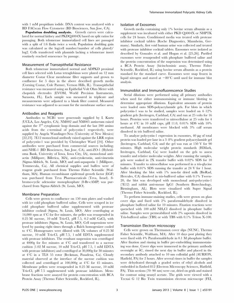
Figure 3. Immunostaining of PKD Q4004X and NHPTK cells. A: Vimentin and cytokeratin staining. Telomerase immortalized PKD Q4404X andNHPTK proximal tubule cells were maintained in culture for four days. After fixation and staining, samples were imaged with a wide-field microscope.Intermediate filament components, vimentin and cytokeratin are observed even after four days in culture. Bar = 10 mm. B: NCB1 staining.Telomerase immortalized PKD Q4004X and NHPTK proximal tubule cells were maintained in culture for three days. Cells were imaged with a wide-field microscope after fixation and staining. 60X objective. C: Aquaporin-1 expression. Immortalized PKD Q4004X and NHPTK proximal tubulecells were maintained in culture for three days. Cells were fixed and stained with anti-aquaporin-1. Images were collected with a wide-fieldmicroscope using a 60X oil objective (NA = 1.41).doi:10.1371/journal.pone.0055191.g003
Telomerase Immortalized Polycystic Kidney Cells
PLOS ONE | www.plosone.org 8 January 2013 | Volume 8 | Issue 1 | e55191

Telomerase Immortalized Polycystic Kidney Cells
PLOS ONE | www.plosone.org 9 January 2013 | Volume 8 | Issue 1 | e55191

Q4004X cell line. In lysates made 3 days after plating at a density
of 50,000 cell/cm2, a doublet of 130 kDa and a single band at
100 kDa was observed in both cell lines. The 130 kDa isoform is
expressed at higher levels in the cystic cell line. In lysates made
from cells grown in culture for 7 days, only the 100 kDa band was
observed and there is more polycystin-2 expression in the PKD
cell line (Figure 6). The observation that two forms of polycystin-2
are expressed in these cells suggests that there are at least two spice
variants expressed in these cell lines. A polycystin-2 spice variant
with an exon 7 deletion has been noted with a molecular weight of
103 kDa, a size in good agreement with the lower molecular
weight band seen in our blot [30]. Immune staining with the same
antibody was not robust under a variety of fixation and staining
conditions; however weak polycystin-2 staining was observed in an
intracellular compartment in both cell lines, consistent with a
rough endoplasmic reticulum staining pattern. Cilia staining were
not observed in either cell type (data not shown).
Finally, we examined the effect of growing both cell lines in 3D
matrix culture. Both PKD and NHPTK cells were plated on
collagen matrix at a density of 50,000 cell per cm2 and then
overlaid with collagen 24 hours after plating. Under identical
growth conditions, MDCK cells will form either cysts or tubules
depending on the presence of hepatocyte growth factor
[31,32,33,34]. The telomerase immortalized cell lines did not
form cysts when grown in collagen matrix (data not shown). Both
NHPTK and PKD Q4004X cells form tubules when overlaid with
type I collagen. However when PKD Q4004X cells were plated
with HK-2 cells at a ratio of 1:10, a few cysts were observed. In
contrast plating NHPTK cells with HK-2 cells did not cause cyst
formation (data not shown). PKD Q4004X cells formed many
cysts when plated in growth factor depleted Matrigel. Cysts were
observed within 14 days after plating (Figure 7A, C). Adding
forskolin or forskolin in combination with IGF-1 accelerated cyst
expansion (data not shown). In contrast NHPTK cells plated
under identical conditions never developed cysts or tubule arrays,
instead the cells formed small aggregates with intracellular
vacuoles observed at cell borders (Figure 7B).
Discussion
Autosomal dominant polycystic kidney disease is one of the
ciliopathies linked to renal cystic disease [35,36,37]. In APDKD
renal cyst formation can occur in any nephron segment but cystic
disease has been suggested to be predominantly arising from distal
segments [38]. Based on current evidence it is not clear how
mutations in either polycystin-1 or polycystin-2 genes lead to cyst
formation. A suggested mechanism proposed is the two-hit
hypothesis in which a second somatic mutation is required in
the normal allele in a cell that already has a germ line mutation
leading to a clonal expansion of cells that have mutations in both
alleles [39]. This mechanism readily accounts for the relatively
sparse generation of renal cysts.
The cell line developed in this communication was selected by
FACs for proximal tubule markers. Although the cells were grown
from a mixed population of isolated renal cysts, over 99% of the
cells were positive for LTL staining, suggesting that our culture
conditions select heavily for cells of proximal origin. It is also
possible cysts harvested solely from the surface of ADPKD kidneys
are of proximal origin. In four primary cell lines we have analyzed
by FACs we found that all of the lines were exclusively LTL
positive based on FACs analysis. One of the four cell lines was
selected for telomerase immortalization and genotyped based on
the fact that this PKD cell line could be paired with an age and sex
matched normal kidney cell line for further analysis. Since little is
known about the mechanisms of cyst formation by proximal
tubule cells the cell lines are a unique resource for the PKD
research community.
Telomerase was selected as an immortalization agent because
this method was considered less likely to induce secondary effects
on growth regulatory pathways that may be affected in the setting
of mutations in polycystin-1 or 2 function. Therefore analysis of
Myc, Src or Jak/Stat signaling pathways would be less likely to be
altered by the immortalization vector. Loghman-Adham et al
reported on the creation of cystic epithelial cell lines using SV40
temperature sensitive large T antigen to immortalize the cells.
They showed that at the non-permissive temperature, large T
antigen was degraded and that epithelial markers were up
regulated [40]. However it is not certain that downstream
signaling pathways or the pattern of gene expression is truly
representative of the de novo mutation in polycystin-1. Since large
T antigen expressing transgenic mice have cysts it is likely that
large T antigen is cystogenic [41]. Notably SV40 T antigen has
been shown to induce polyploidy in cells due to its interaction with
Bub1 this would suggest that these cell lines cannot be used to
examine genomic instability [42,43]. In contrast immortalization
with human telomerase is, based on our current understanding,
less likely to alter growth signaling responses or induces genomic
instability [44]. However a few papers have described alterations
in a growth regulatory signaling pathway associated with
telomerase over-expression [45,46]. For instance, Li et al.,
Figure 4. Polycystin-1 expression and staining in RPTEC, NHPTK and PKD Q4004X cells. A: Immune blot of membrane preparations fromRPTEC, NHPTK and PKD Q4004X cells. Membranes were isolated as described in materials and methods. Lanes 1, 2 and 3: Immune blot analysis usinganti polycystin-1 (NM 005). Lane 1 membrane preparation from renal proximal tubule renal epithelial cells (RPTEC). Lane 2 membrane preparationfrom telomerase immortalized normal kidney proximal tubule cells. Lane 3 membrane preparation from PKD Q4004X cells. Arrows point to bandsthat are unique to the PKDQ4004X cells (Lane 3). Lower panel: immune blot from the lower portion of the gel probed with anti-actin antibodies.Similar levels of actin are present in the blot. Lanes 4, 5 and 6: Immune blot analysis using anti-LRR polycystin-1 (7E12) used to probe the same blotafter stripping NM005. Lane 4 membrane preparation from RPTEC cells. Lane 5 membrane preparation from telomerase immortalized NHPTK cells.Lane 6 membrane preparation from telomerase immortalized PKD Q4004X cells. B: Immune blot of exosomes. Exosomes were isolated asdescribed in materials and methods section. Blot was probed with a monoclonal antibody raised against the leucine rich region of polycystin-1(7E12). Lane 1 exosomes from human urine. Lane 2 exosomes from NHPTK cells. Lane 3 exosomes from PKD Q4004X cells. Full length polycystin-1 isobserved in all sources of exosomes. More truncated polycystin-1 is observed in the exosomes derived from the PKD cell line. C,D: Immunestaining of polycystin-1 using 7E12 and NM 005 in telomerase immortalized kidney cells. Telomerase immortalized proximal tubule cells(C) or PKD Q4004X (D) were grown to confluence, fixed and stained with monoclonal anti-LRR (7E12) and polyclonal anti polycystin-1 (NM005). Theimage shown is a merged image in which 7E12 was labeled with Alexa 488 and NM 005 was labeled with rhodamine. Extensive co-localization of bothantibodies is observed in which a reticular peri-nuclear structure is labeled. Some areas of the reticulum appear to be labeled with only one of theantibodies. Pearson’s correlation coefficient was 88% for both cell lines (N = 3). Bar = 10 mm. E, F: Immuno-gold decoration of cilia. Telomeraseimmortalized proximal tubule (E) and PKD Q4004X (F) cells were grown to confluence and processed for immune gold decoration using antipolycystin-1 (NM 005). Gold decoration of cilia was only observed in the normal kidney cells (E). None of the cilia observed in PKD cells were positivefor immune gold staining (F).doi:10.1371/journal.pone.0055191.g004
Telomerase Immortalized Polycystic Kidney Cells
PLOS ONE | www.plosone.org 10 January 2013 | Volume 8 | Issue 1 | e55191

demonstrated up regulation of macrophage colony stimulating
factor-1 receptor (CSF1R) message and protein levels in hTERT-
immortalized human ovarian surface epithelial cells and in human
pancreatic epithelial cells [46]. In hTERT-immortalized human
ovarian surface epithelial cells stimulation of CSF1R with colony
stimulating factor-1 causes hTERT translocation into the nucleus
and c-Myc binding to the hTERT promoter region [46]. SiRNA
knockdown of CSF1R blocked these effects and inhibited cell
Figure 5. Scanning electron micrographs of NHPTK and PKD Q4004X cells. A: PKDQ4004X cells have sparse microvilli and short monocilia(arrows) as compared to NHPTK (arrow). Scale bar-10 mm is the same for both PKD Q4004X and NHPTK images. B: Apical membrane surface PKDQ4004X at high magnification. Membrane blebs are seen (arrows) which are roughly the size of exosomes. Bar = 5 mm.doi:10.1371/journal.pone.0055191.g005
Telomerase Immortalized Polycystic Kidney Cells
PLOS ONE | www.plosone.org 11 January 2013 | Volume 8 | Issue 1 | e55191

immortalization. This data suggests that hTERT immortalization
in renal epithelial cells may be modulated by other growth factor
signaling pathways. In other modes of immortalization, such as
adenovirus transduction, adenovirus mediated immortalization
involves transcriptional activation of hTERT by E1A acting
cooperatively with E1B activation of Ras [47].
Analysis of polycystin-2 expression in the PKD Q4004X-LTL
cell line as compared to the normal kidney cell lines revealed no
differences in sub cellular localization. However more polycystin-2
was noted in PKD Q4404X cells and two polycystin-2 isoforms
were expressed in both cell lines. Alternative splicing variants of
polycystin-2 has been described in brain and one splice variant
lacking exon 7 does not interact with polycystin1 [48]. By
comparison, we observed striking differences in polycystin-1
expression in the PKD Q4004X-LTL cells in that there was
Figure 6. Polycystin-2 expression in telomerase immortalizedproximal tubule cells. Both telomerase immortalized normal kidney(N) proximal and PKD Q4004X (P) cells were grown to confluence for 3or 7 days. Extracts were made with buffers containing non-ionicdetergent and equivalent amounts of protein (50 mg) were loaded ineach lane. Lanes labeled N and P represent lysates made fromtelomerase immortalized normal kidney proximal tubule and PKDQ4004X cells, respectively. Two bands at molecular weights 120 and100 kDa are seen and the PKD cells have higher expression levels atboth time points. At 7 days only the 100 kDa band is observed.doi:10.1371/journal.pone.0055191.g006
Figure 7. Cyst formation by PKDQ4004X cells grown in Matrigel. Normal and PKDQ4004X cells were plated at a density of 3000 cells per mlof Matrigel with culture media and grown in culture for 14 days. A: Phase contrast image of PKDQ4004X cells forming cysts in Matrigel matrix. B:Phase contrast image of normal kidney cells in Matrigel matrix. Bar = 50 microns. C: Fluorescence photomicrograph of PKD Q4004X cell cyst. Extendedfocus image of 5 image planes covering a z distance of 5 microns. Actin cytoskeleton is labeled with Bodipy phalloidin (green) and nuclei are labeledwith Hoechst 33342 (blue). A clear lumen (L) is surrounded by a layer of epithelial cells to form a cyst. Bar = 10 microns.doi:10.1371/journal.pone.0055191.g007
Telomerase Immortalized Polycystic Kidney Cells
PLOS ONE | www.plosone.org 12 January 2013 | Volume 8 | Issue 1 | e55191

significantly more uncleaved polycystin-1 present in these cells as
determined by immunoblot of membrane fractions. A current
hypothesis to explain the focal nature of cyst formation have
suggested that two hits are required for cytogenesis: the first
mutation being a germ-line transmitted mutation and the second
mutation is a spontaneous somatic mutation [49]. Alternatively,
but not necessary exclusionary, mutations in polycystin-1 affect
flow mediated calcium signaling mediated by mechanical defor-
mation of cilia [50,51]. In a separate communication, this cell line
has a defect in calcium signaling secondary to alterations in
purinogenic receptor function [52]. Four differences in polycystin-
1 biogenesis are noted in this cell line, first polycystin-1 was not
observed in monocilia, second anomalous sized bands were
observed in western blots probed with a polyclonal antibody
raised against 200 amino acids from the c-terminus of polycystin-1,
there is quantitatively more lower molecular weight forms of
polycystin-1 observed exosomes isolated from the PKD Q4004X-
LTL line and lastly there was less uncleaved full length polycystin-
1 observed in the PKD Q4004X-LTL cell line (figure 4A). One
potential interpretation of this finding is that the mutant form of
polycystin-1 is acting as a dominant negative that prevents
cleavage at the GPS site [53] or inhibits assembly into monocilia.
Only one allele of polycystin-1 was found to be mutated in this cell
line. While this finding is contrary to the prediction of the ‘‘two
hit’’ hypothesis, we cannot eliminate the possibility that an
undetected point mutation exists in the other polycystin-1 allele
[20,54]. It is also possible that since the cell line was created from a
mixed population of cysts, the second hit could not be identified
using current mutation detection techniques. Conversely if a
second hit does not exist in this cell line and the Q4004X mutation
acts as a dominant negative, then the cells are functionally
equivalent to cell lines with mutations in both HmPKD1 alleles
[53].
In conclusion, we have characterized a polycystic kidney renal
epithelial cell line that expresses proximal tubule markers. This cell
line appears to have a single truncation mutation at position
Q4004X and western blot analysis reveals that the cell line has
significant amounts of uncleaved polycystin-1. In three dimen-
sional cultures using type I collagen both cell lines (normal kidney
and PKD Q4004X) fail to form either tubules or cysts. But the
PKD Q4004X cell line does form cyst when co-cultured with HK
cells or when cultured in growth factor reduced Matrigel. This cell
line should be useful to further dissect the cellular phenotype
associated with mutations in HmPKD1 and factors required to
induce cyst formation in 3D culture.
Acknowledgments
The authors would like to thank Jason Byars, Nikki Collins and Janak
Bhavsar for their technical assistance, and Jerry Shay for hTERT reagents.
We also thank Darren Wallace for his helpful insights in the preparation of
this manuscript.
Author Contributions
Conceived and designed the experiments: BSH BRG PH VHG AWN
RLB. Performed the experiments: BSH BRG WMX MW CW SR EB-R
HHW CM VHG CLP AWN RLB. Analyzed the data: BSH BRG WMX
MW CW SR PH EB-R HHW CM VHG AWN RLB. Contributed
reagents/materials/analysis tools: BSH BRG SR CW PH EB-R AWN
RLB. Wrote the paper: B-SH AWN RLB.
References
1. Belibi FA, Reif G, Wallace DP, Yamaguchi T, Olsen L, et al. (2004) Cyclic AMP
promotes growth and secretion in human polycystic kidney epithelial cells.[see
comment]. Kidney International 66: 964–973.
2. Wilson PD, Sherwood AC (1991) Tubulocystic epithelium. Kidney International
39: 450–463.
3. Wilson PD, Wilson PD (2004) Polycystic kidney disease.[see comment]. New
England Journal of Medicine 350: 151–164.
4. Xu GM, Gonzalez-Perrett S, Essafi M, Timpanaro GA, Montalbetti N, et al.
(2003) Polycystin-1 activates and stabilizes the polycystin-2 channel. Journal of
Biological Chemistry 278: 1457–1462.
5. Boucher C, Sandford R (2004) Autosomal dominant polycystic kidney disease
(ADPKD, MIM 173900, PKD1 and PKD2 genes, protein products known as
polycystin-1 and polycystin-2). European Journal of Human Genetics 12: 347–
354.
6. Ong AC, Ward CJ, Butler RJ, Biddolph S, Bowker C, et al. (1999) Coordinate
expression of the autosomal dominant polycystic kidney disease proteins,
polycystin-2 and polycystin-1, in normal and cystic tissue. American Journal of
Pathology 154: 1721–1729.
7. Wilson PD (2001) Polycystin: new aspects of structure, function, and regulation.
Journal of the American Society of Nephrology 12: 834–845.
8. Koulen P, Cai Y, Geng L, Maeda Y, Nishimura S, et al. (2002) Polycystin-2 is an
intracellular calcium release channel.[see comment]. Nature Cell Biology 4:
191–197.
9. Vassilev PM, Guo L, Chen XZ, Segal Y, Peng JB, et al. (2001) Polycystin-2 is a
novel cation channel implicated in defective intracellular Ca(2+) homeostasis in
polycystic kidney disease. Biochemical & Biophysical Research Communications
282: 341–350.
10. Nauli SM, Zhou J, Nauli SM, Zhou J (2004) Polycystins and mechanosensation
in renal and nodal cilia. Bioessays 26: 844–856.
11. Xu C, Rossetti S, Jiang L, Harris PC, Brown-Glaberman U, et al. (2007) Human
ADPKD primary cyst epithelial cells with a novel, single codon deletion in the
PKD1 gene exhibit defective ciliary polycystin localization and loss of flow-
induced Ca2+ signaling. American Journal of Physiology - Renal Physiology
292: F930–945.
12. Charron AJ, Nakamura S, Bacallao R, Wandinger-Ness A (2000) Compromised
cytoarchitecture and polarized trafficking in autosomal dominant polycystic
kidney disease cells. Journal of Cell Biology 149: 111–124.
13. Carone FA, Jin H, Nakamura S, Kanwar YS (1993) Decreased synthesis and
delayed processing of sulfated glycoproteins by cells from human polycystic
kidneys. Laboratory Investigation 68: 413–418.
14. Lewis CM, Herbert BS, Bu D, Halloway S, Beck A, et al. (2006) Telomerase
immortalization of human mammary epithelial cells derived from a BRCA2mutation carrier. Breast Cancer Research & Treatment 99: 103–115.
15. Condon J, Yin S, Mayhew B, Wright WE, Shay JW, et al. (2002) Telomerase
immortalization of human myometrial cells. Biol Reprod 67: 506–514.
16. Felice B, Cattoglio C, Cittaro D, Testa A, Miccio A, et al. (2009) Transcription
Factor Binding Sites Are Genetic Determinants of Retroviral Integration in theHuman Genome. PLoS ONE 4: e4571.
17. Rossetti S, Consugar MB, Chapman AB, Torres VE, Guay-Woodford LM, et al.
(2007) Comprehensive molecular diagnostics in autosomal dominant polycystickidney disease. Journal of the American Society of Nephrology 18: 2143–2160.
18. Rossetti S, Strmecki L, Gamble V, Burton S, Sneddon V, et al. (2001) Mutation
Analysis of the Entire PKD1 Gene: Genetic and Diagnostic Implications.American Journal of Human Genetics 68: 46–63.
19. Rossetti S, Consugar MB, Chapman AB, Torres VE, Guay-Woodford LM, et al.
(2007) Comprehensive Molecular Diagnostics in Autosomal Dominant Polycys-tic Kidney Disease. Journal of the American Society of Nephrology 18: 2143–
2160.
20. Rossetti S, Strmecki L, Gamble V, Burton S, Sneddon V, et al. (2001) Mutation
analysis of the entire PKD1 gene: genetic and diagnostic implications. American
Journal of Human Genetics 68: 46–63.
21. Krishan A (1975) Rapid flow cytofluorometric analysis of mammalian cell cycle
by propidium iodide staining. J Cell Biol 66: 188–193.
22. Ward HH, Brown-Glaberman U, Wang J, Morita Y, Alper SL, et al. (2011) Aconserved signal and GTPase complex are required for the ciliary transport of
polycystin-1. Molecular Biology of the Cell 22: 3289–3305.
23. Boucher CA, Ward HH, Case RL, Thurston KS, Li X, et al. (2011) Receptorprotein tyrosine phosphatases are novel components of a polycystin complex.
Biochimica et Biophysica Acta 1812: 1225–1238.
24. Ong ACM, Harris PC, Davies DR, Pritchard L, Rossetti S, et al. (1999)
Polycystin-1 expression in PKD1, early-onset PKD1, and TSC2/PKD1 cystic
tissue. Kidney International 56: 1324–1333.
25. Hogan MC, Manganelli L, Woollard JR, Masyuk AI, Masyuk TV, et al. (2009)
Characterization of PKD protein-positive exosome-like vesicles. Journal of the
American Society of Nephrology 20: 278–288.
26. Gonzales PA, Pisitkun T, Hoffert JD, Tchapyjnikov D, Star RA, et al. (2009)
Large-scale proteomics and phosphoproteomics of urinary exosomes. Journal of
the American Society of Nephrology 20: 363–379.
27. Kher R, Sha EC, Escobar MR, Andreoli EM, Wang P, et al. (2011) Ectopic
expression of cadherin 8 is sufficient to cause cyst formation in a novel 3Dcollagen matrix renal tubule culture. Am J Physiol Cell Physiol 301: C99–C105.
Telomerase Immortalized Polycystic Kidney Cells
PLOS ONE | www.plosone.org 13 January 2013 | Volume 8 | Issue 1 | e55191

28. Carone FA, Nakamura S, Schumacher BS, Punyarit P, Bauer KD (1989) Cyst-
derived cells do not exhibit accelerated growth or features of transformed cellsin vitro. Kidney International 35: 1351–1357.
29. Maunsbach AB, Marples D, Chin E, Ning G, Bondy C, et al. (1997) Aquaporin-
1 Water Channel Expression in Human Kidney. Journal of American Society ofNephrology 8: 1–14.
30. Hackmann K, Markoff A, Qian F, Bogdanova N, Germino GG, et al. (2005) Asplice form of polycystin-2, lacking exon 7, does not interact with polycystin-1.
Human Molecular Genetics 14: 3249–3262.
31. McAteer JA, Evan AP, Gardner KD (1987) Morphogenetic clonal growth ofkidney epithelial cell line MDCK. Anatomical Record 217: 229–239.
32. Montesano R, Soriano JV, Pepper MS, Orci L (1997) Induction of epithelialbranching tubulogenesis in vitro. Journal of Cellular Physiology 173: 152–161.
33. Montesano R, Schaller G, Orci L (1991) Induction of epithelial tubularmorphogenesis in vitro by fibroblast-derived soluble factors. Cell 66: 697–711.
34. Mangoo-Karim R, Uchic ME, Grant M, Shumate WA, Calvet JP, et al. (1989)
Renal epithelial fluid secretion and cyst growth: the role of cyclic AMP. FASEBJournal 3: 2629–2632.
35. Pazour GJ, San Agustin JT, Follit JA, Rosenbaum JL, Witman GB (2002)Polycystin-2 localizes to kidney cilia and the ciliary level is elevated in orpk mice
with polycystic kidney disease. Current Biology 12: R378–380.
36. Ong ACM, Wheatley DN (2003) Polycystic kidney disease–the ciliaryconnection. Lancet 361: 774–776.
37. Pazour GJ, Rosenbaum JL, Pazour GJ, Rosenbaum JL (2002) Intraflagellartransport and cilia-dependent diseases. Trends in Cell Biology 12: 551–555.
38. Takakura A, Contrino L, Beck AW, Zhou J, Takakura A, et al. (2008) Pkd1inactivation induced in adulthood produces focal cystic disease. Journal of the
American Society of Nephrology 19: 2351–2363.
39. Watnick T, Germino GG (1999) Molecular basis of autosomal dominantpolycystic kidney disease. Seminars in Nephrology 19: 327–343.
40. Loghman-Adham M, Nauli SM, Soto CE, Kariuki B, Zhou J, et al. (2003)Immortalized epithelial cells from human autosomal dominant polycystic kidney
cysts. American Journal of Physiology - Renal Physiology 285: F397–412.
41. MacKay K, Striker LJ, Pinkert CA, Brinster RL, Striker GE (1987)Glomerulosclerosis and renal cysts in mice transgenic for the early region of
SV40. Kidney International 32: 827–837.42. Cotsiki M, Lock RL, Cheng Y, Williams GL, Zhao J, et al. (2004) Simian virus
40 large T antigen targets the spindle assembly checkpoint protein Bub1.Proceedings of the National Academy of Sciences of the United States of
America 101: 947–952.
43. Hein J, Boichuk S, Wu J, Cheng Y, Freire R, et al. (2009) Simian Virus 40 Large
T Antigen Disrupts Genome Integrity and Activates a DNA Damage Responsevia Bub1 Binding. Journal of Virology 83: 117–127.
44. Wieser M, Stadler G, Jennings P, Streubel B, Pfaller W, et al. (2008) hTERT
alone immortalizes epithelial cells of renal proximal tubules without changingtheir functional characteristics. American Journal of Physiology - Renal
Physiology 295: F1365–1375.45. Smith LL, Coller HA, Roberts JM, Smith LL, Coller HA, et al. (2003)
Telomerase modulates expression of growth-controlling genes and enhances cell
proliferation. Nature Cell Biology 5: 474–479.46. Li NF, Kocher HM, Salako MA, Obermueller E, Sandle J, et al. (2009) A novel
function of colony stimulating factor-1 receptor in hTERT immortalization ofepithelial cells. Oncogene 28: 773–780.
47. Glasspool RM, Burns S, Hoare SF, Svensson C, Keith WN, et al. (2005) ThehTERT and hTERC telomerase gene promoters are activated by the second
exon of the adenoviral protein, E1A, identifying the transcriptional corepressor
CtBP as a potential repressor of both genes. Neoplasia (New York) 7: 614–622.48. Hackmann K, Markoff A, Qian F, Bogdanova N, Germino GG, et al. (2005) A
splice form of polycystin-2, lacking exon 7, does not interact with polycystin-1.Human Molecular Genetics 14: 3249–3262.
49. Germino GG (1997) Autosomal dominant polycystic kidney disease: a two-hit
model. Hosp Pract 32: 81–82, 85–88, 91–82 passim.50. Nauli SM, Alenghat FJ, Luo Y, Williams E, Vassilev P, et al. (2003) Polycystins 1
and 2 mediate mechanosensation in the primary cilium of kidney cells.[com-ment]. Nature Genetics 33: 129–137.
51. Nauli SM, Rossetti S, Kolb RJ, Alenghat FJ, Consugar MB, et al. (2006) Loss ofpolycystin-1 in human cyst-lining epithelia leads to ciliary dysfunction. Journal of
the American Society of Nephrology 17: 1015–1025.
52. Xu C, Shmukler BE, Nishimura K, Kaczmarek E, Rossetti S, et al. (2009)Attenuated, flow-induced ATP release contributes to absence of flow-sensitive,
purinergic Cai2+ signaling in human ADPKD cyst epithelial cells. AmericanJournal of Physiology - Renal Physiology 296: F1464–1476.
53. Qian F, Boletta A, Bhunia AK, Xu H, Liu L, et al. (2002) Cleavage of
polycystin-1 requires the receptor for egg jelly domain and is disrupted byhuman autosomal-dominant polycystic kidney disease 1-associated mutations.
Proceedings of the National Academy of Sciences of the United States ofAmerica 99: 16981–16986.
54. Rossetti S, Chauveau D, Walker D, Saggar-Malik A, Winearls CG, et al. (2002)A complete mutation screen of the ADPKD genes by DHPLC. Kidney
International 61: 1588–1599.
Telomerase Immortalized Polycystic Kidney Cells
PLOS ONE | www.plosone.org 14 January 2013 | Volume 8 | Issue 1 | e55191