Estrogen directly and specifically downregulates NaPi-IIa through the activation of both estrogen...
13
CALL FOR PAPERS Sex and Gender Differences in Renal Physiology Estrogen directly and specifically downregulates NaPi-IIa through the activation of both estrogen receptor isoforms (ER and ER) in rat kidney proximal tubule Dara Burris, 1 Rose Webster, 1 Sulaiman Sheriff, 2 Rashma Faroqui, 1 Moshe Levi, 3 John R. Hawse, 4 and Hassane Amlal 1 1 Division of Nephrology and Hypertension, Department of Medicine, University of Cincinnati, Cincinnati, Ohio; 2 Department of Surgery, University of Cincinnati, Cincinnati, Ohio; 3 Division of Renal Diseases and Hypertension, Department of Medicine, University of Colorado Health Sciences Center, Denver, Colorado; and 4 Department of Biochemistry and Molecular Biology, Mayo Clinic, Rochester, Minnesota Submitted 9 July 2014; accepted in final form 5 January 2015 Burris D, Webster R, Sheriff S, Faroqui R, Levi M, Hawse JR, Amlal H. Estrogen directly and specifically downregulates NaPi-IIa through the activation of both estrogen receptor isoforms (ER and ER) in rat kidney proximal tubule. Am J Physiol Renal Physiol 308: F522–F534, 2015. First published January 21, 2015; doi:10.1152/ajprenal.00386.2014.—We have previously demon- strated that estrogen (E 2 ) downregulates phosphate transporter NaPi- IIa and causes phosphaturia and hypophosphatemia in ovariectomized rats. In the present study, we examined whether E 2 directly targets NaPi-IIa in the proximal tubule (PT) and studied the respective roles of estrogen receptor isoforms (ER and ER) in the downregulation of NaPi-IIa using both in vivo and an in vitro expression systems. We found that estrogen specifically downregulates NaPi-IIa but not NaPi- IIc or Pit2 in the kidney cortex. Proximal tubules incubated in a “shake” suspension with E 2 for 24 h exhibited a dose-dependent decrease in NaPi-IIa protein abundance. Results from OVX rats treated with specific agonists for either ER [4,4=,4;-(4-propyl-[1H]- pyrazole-1,3,5-triyl) trisphenol, PPT] or ER [4,4=,4-(4-propyl-[1H]- pyrazole-1,3,5-triyl) trisphenol, DPN] or both (PPT DPN), indi- cated that only the latter caused a sharp downregulation of NaPi-IIa, along with significant phosphaturia and hypophosphatemia. Lastly, heterologous expression studies demonstrated that estrogen down- regulated NaPi-IIa only in U20S cells expressing both ER and ER, but not in cells expressing either receptor alone. In conclusion, these studies demonstrate that rat PT cells express both ER and ER and that E 2 induces phosphaturia by directly and specifically targeting NaPi-IIa in the PT cells. This effect is mediated via a mechanism involving coactivation of both ER and ER, which likely form a functional heterodimer complex in the rat kidney proximal tubule. inorganic phosphate; postmenopause; sex steroids; proximal tubule; hypophosphatemia. SEVERAL CLINICAL STUDIES HAVE demonstrated that estrogen de- pletion (menopause) or repletion (hormone replacement ther- apy) is associated with changes in serum inorganic phosphate (P i ) levels in women. Indeed, clinical studies have shown that women treated with estrogen exhibit hypophosphatemia (2, 15, 18, 57, 59), and in some studies, a reduction in proximal tubular P i reabsorption was observed (12, 17, 59). Conversely, estrogen-depleted patients showed elevated plasma levels of P i and increased rate of proximal tubule P i reabsorption (2). Kidney and intestine play an important role in the control and maintenance of P i homeostasis (8, 18, 20, 40). P i enters the body through its absorption in the intestine via an apical sodium-dependent P i transporters NaPi-IIb encoded by Slc34a2 (18, 20). The involvement of kidney is more efficient in controlling P i metabolism, as it can reabsorb or excrete P i depending on the circulating levels of P i and the needs of the body. Indeed, most of filtered P i is immediately reabsorbed in the proximal tubule via the activity of several P i transporters. These transporters include NaPi-IIa (Slc34a1), NaPi-IIc (Slc34a3), and Pit-2 (Slc20a2), all of which are sodium- dependent (NaPi) and expressed in the brush-border membrane of the proximal tubule (see Refs. 9 and 40 for review). Recently, studies demonstrated that estrogen stimulates the expression of NaPi-IIb in the intestine (63) and inhibits phos- phate uptake in brush border membrane vesicles harvested from kidneys of estrogen-treated rats (6). Subsequently, we demonstrated that estrogen downregulates NaPi-IIa and causes hypophosphatemia and renal P i wasting in ovariectomized rats (23). We further demonstrated that the effect of estrogen on renal P i handling results from the downregulation of NaPi-IIa and is independent of parathyroid hormone (PTH) levels or changes in food intake (23). PTH and fibroblast growth factor 23 (FGF23) are known to regulate P i balance through the downregulation of P i transporters in the proximal tubule with subsequent phosphaturia in humans, as well as in experimental animals (7, 34, 52). Recent studies demonstrated that estrogen stimulates the synthesis and release of FGF23 from bone osteocytes, while decreasing the synthesis and secretion of PTH in the parathyroid gland of rat (11). The effect of estrogen on PTH appears to be indirect, as estrogen receptors ER or ER are not expressed in the parathyroid cells and rather results from the increase in the circulating levels of FGF23 and its action in the parathyroid gland (11). The majority of estrogen response is mediated through two different isoforms of estrogen receptors (ER), usually referred to as ER and ER, each encoded by a separate gene (33, 38, 39, 50, 58). Immunoblot studies have shown that ER and ER are expressed in the kidney of rat (61) and mice (29). Address for reprint requests and other correspondence: H. Amlal, Div. of Nephrology and Hypertension, 231 Albert Sabin Way, MSB Rm. G-259, Cincinnati, OH 45267-0585 (e-mail: [email protected]). Am J Physiol Renal Physiol 308: F522–F534, 2015. First published January 21, 2015; doi:10.1152/ajprenal.00386.2014. 1931-857X/15 Copyright © 2015 the American Physiological Society http://www.ajprenal.org F522
Transcript of Estrogen directly and specifically downregulates NaPi-IIa through the activation of both estrogen...

CALL FOR PAPERS Sex and Gender Differences in Renal Physiology
Estrogen directly and specifically downregulates NaPi-IIa through theactivation of both estrogen receptor isoforms (ER� and ER�) in rat kidneyproximal tubule
Dara Burris,1 Rose Webster,1 Sulaiman Sheriff,2 Rashma Faroqui,1 Moshe Levi,3 John R. Hawse,4
and Hassane Amlal11Division of Nephrology and Hypertension, Department of Medicine, University of Cincinnati, Cincinnati, Ohio; 2Departmentof Surgery, University of Cincinnati, Cincinnati, Ohio; 3Division of Renal Diseases and Hypertension, Department ofMedicine, University of Colorado Health Sciences Center, Denver, Colorado; and 4Department of Biochemistry andMolecular Biology, Mayo Clinic, Rochester, Minnesota
Submitted 9 July 2014; accepted in final form 5 January 2015
Burris D, Webster R, Sheriff S, Faroqui R, Levi M, HawseJR, Amlal H. Estrogen directly and specifically downregulatesNaPi-IIa through the activation of both estrogen receptor isoforms(ER� and ER�) in rat kidney proximal tubule. Am J Physiol RenalPhysiol 308: F522–F534, 2015. First published January 21, 2015;doi:10.1152/ajprenal.00386.2014.—We have previously demon-strated that estrogen (E2) downregulates phosphate transporter NaPi-IIa and causes phosphaturia and hypophosphatemia in ovariectomizedrats. In the present study, we examined whether E2 directly targetsNaPi-IIa in the proximal tubule (PT) and studied the respective rolesof estrogen receptor isoforms (ER� and ER�) in the downregulationof NaPi-IIa using both in vivo and an in vitro expression systems. Wefound that estrogen specifically downregulates NaPi-IIa but not NaPi-IIc or Pit2 in the kidney cortex. Proximal tubules incubated in a“shake” suspension with E2 for 24 h exhibited a dose-dependentdecrease in NaPi-IIa protein abundance. Results from OVX ratstreated with specific agonists for either ER� [4,4=,4�;-(4-propyl-[1H]-pyrazole-1,3,5-triyl) trisphenol, PPT] or ER� [4,4=,4�-(4-propyl-[1H]-pyrazole-1,3,5-triyl) trisphenol, DPN] or both (PPT � DPN), indi-cated that only the latter caused a sharp downregulation of NaPi-IIa,along with significant phosphaturia and hypophosphatemia. Lastly,heterologous expression studies demonstrated that estrogen down-regulated NaPi-IIa only in U20S cells expressing both ER� and ER�,but not in cells expressing either receptor alone. In conclusion, thesestudies demonstrate that rat PT cells express both ER� and ER� andthat E2 induces phosphaturia by directly and specifically targetingNaPi-IIa in the PT cells. This effect is mediated via a mechanisminvolving coactivation of both ER� and ER�, which likely form afunctional heterodimer complex in the rat kidney proximal tubule.
inorganic phosphate; postmenopause; sex steroids; proximal tubule;hypophosphatemia.
SEVERAL CLINICAL STUDIES HAVE demonstrated that estrogen de-pletion (menopause) or repletion (hormone replacement ther-apy) is associated with changes in serum inorganic phosphate(Pi) levels in women. Indeed, clinical studies have shown thatwomen treated with estrogen exhibit hypophosphatemia (2, 15,18, 57, 59), and in some studies, a reduction in proximaltubular Pi reabsorption was observed (12, 17, 59). Conversely,
estrogen-depleted patients showed elevated plasma levels of Pi
and increased rate of proximal tubule Pi reabsorption (2).Kidney and intestine play an important role in the control
and maintenance of Pi homeostasis (8, 18, 20, 40). Pi enters thebody through its absorption in the intestine via an apicalsodium-dependent Pi transporters NaPi-IIb encoded bySlc34a2 (18, 20). The involvement of kidney is more efficientin controlling Pi metabolism, as it can reabsorb or excrete Pi
depending on the circulating levels of Pi and the needs of thebody. Indeed, most of filtered Pi is immediately reabsorbed inthe proximal tubule via the activity of several Pi transporters.These transporters include NaPi-IIa (Slc34a1), NaPi-IIc(Slc34a3), and Pit-2 (Slc20a2), all of which are sodium-dependent (NaPi) and expressed in the brush-border membraneof the proximal tubule (see Refs. 9 and 40 for review).Recently, studies demonstrated that estrogen stimulates theexpression of NaPi-IIb in the intestine (63) and inhibits phos-phate uptake in brush border membrane vesicles harvestedfrom kidneys of estrogen-treated rats (6). Subsequently, wedemonstrated that estrogen downregulates NaPi-IIa and causeshypophosphatemia and renal Pi wasting in ovariectomized rats(23). We further demonstrated that the effect of estrogen onrenal Pi handling results from the downregulation of NaPi-IIaand is independent of parathyroid hormone (PTH) levels orchanges in food intake (23).
PTH and fibroblast growth factor 23 (FGF23) are known toregulate Pi balance through the downregulation of Pi transportersin the proximal tubule with subsequent phosphaturia in humans,as well as in experimental animals (7, 34, 52). Recent studiesdemonstrated that estrogen stimulates the synthesis and release ofFGF23 from bone osteocytes, while decreasing the synthesis andsecretion of PTH in the parathyroid gland of rat (11). The effectof estrogen on PTH appears to be indirect, as estrogen receptorsER� or ER� are not expressed in the parathyroid cells and ratherresults from the increase in the circulating levels of FGF23 and itsaction in the parathyroid gland (11).
The majority of estrogen response is mediated through twodifferent isoforms of estrogen receptors (ER), usually referredto as ER� and ER�, each encoded by a separate gene (33, 38,39, 50, 58). Immunoblot studies have shown that ER� andER� are expressed in the kidney of rat (61) and mice (29).
Address for reprint requests and other correspondence: H. Amlal, Div. ofNephrology and Hypertension, 231 Albert Sabin Way, MSB Rm. G-259,Cincinnati, OH 45267-0585 (e-mail: [email protected]).
Am J Physiol Renal Physiol 308: F522–F534, 2015.First published January 21, 2015; doi:10.1152/ajprenal.00386.2014.
1931-857X/15 Copyright © 2015 the American Physiological Society http://www.ajprenal.orgF522

Immunohistochemistry studies demonstrated that ER� is ex-pressed in both proximal tubule and distal tubule of both ratand mice kidney (29, 61). The segmental distribution of ER�in rat kidney was not examined in this study (61). In mice, ER�is also expressed in both proximal tubule and distal nephronsegments (29). However, the specificity of the antibodies usedin both studies was not demonstrated. Nevertheless, whetherestrogen receptors mediate directly or indirectly the effect ofestrogen on renal Pi transporters remains to be studied.
In the present studies, we examined the dose-response effectof estrogen on renal Pi handling and NaPi-IIa expression anddetermined the effects of estrogen on other apical Pi absorbingtransporters in the kidney proximal tubule. Further, we exam-ined whether the mRNA of ER� and ER� is expressed in thekidney proximal tubule cells and studied whether they mediatea direct effect of estrogen on NaPi-IIa expression using prox-imal tubular suspensions. Lastly, we determined the respectiveroles of ER� and ER� in estrogen-induced downregulation ofNaPi-IIa using both in vivo and in vitro experiments.
MATERIALS AND METHODS
The experiments performed in these studies were approved by theInstitutional Animal Care and Use Committee of the University ofCincinnati. Ovariectomized Sprague-Dawley rats were purchasedfrom Harlan (Harlan, Indianapolis, IN), housed two rats per cage withfree access to rat chow and distilled water, and maintained in atemperature-controlled room regulated on a 12:12-h light-dark cyclefor 1 wk before and during the following treatments.
Dose-Dependent Effects of Estrogen
OVX rats were placed in metabolic cages and allowed free accessto food and water, as previously described in our laboratory (23).After 3 or 4 days of adjustment to metabolic cages, rats wererandomly divided into several groups (n � 4 or 5 rats in each) andinjected subcutaneously with different doses of 17�-estradiol (0.5, 5,15, and 90 �g/100 g body wt/day) or vehicle (sesame oil). Theanimals were injected daily and euthanized after 3 days of treatment.
Respective Roles of Estrogen Receptors ER� or ER� UsingPharmacological Agonists
OVX rats were housed in metabolic cages and fed rodent chow anddistilled water and treated as follows: in a first set of the experiments,rats were divided into three groups (n � 4 rats in each) with twogroups injected subcutaneously with either 150 or 500 �g/100 g bodywt of PPT, a specific agonist of ER� (26, 27, 50, 54, 57), and the thirdgroup was injected with vehicle. Because it was expected that PPTwould cause a reduction in food intake, as previously shown by others(49, 50), the access of vehicle group to food was restricted to the sameamount consumed by PPT-treated rats at 500 �g/100 g body wt(pair-feeding). In a second set of experiments, rats were divided intothree groups (four rats in each) and injected subcutaneously witheither 150 or 500 �g/100 g body wt of DPN, a specific ER� agonist(26, 27, 50, 54, 57), or vehicle (sesame oil). In the last experiment, agroup of rats (n � 4) was injected subcutaneously with a combinationof PPT � DPN at 150 �g/100 g body weight each. In all of theseexperiments, rats were injected daily and killed after 3 days oftreatments. To confirm the findings of ER activation studies, the effectof PPT or DPN at 150 �g/100 g body wt vs. vehicle was reevaluatedusing another batch of four rats in each group, and NaPi-IIa proteinabundance was reexamined in the kidney cortex of these animals.
Our choice of 150 �g/100 g body wt of PPT or DPN is estimatedfrom studies that examined the dose-response effects of these com-pounds on food intake in OVX rats (26, 46, 50, 54, 57). Our doses are
slightly overestimated to account for the difference in the vehicle ofpreference, i.e., sesame oil in our studies vs. DMSO in Santollo’swork (50). To ascertain that PPT alone and DPN alone have no effecton renal phosphate handling, we have also studied the effects of ahigher dose of 500 �g/100 g body wt of these compounds on foodintake and renal Pi handling.
During each of the above studies, food and water intake and urinevolume were measured daily. At the end of each experiment, rats wereeuthanized and kidneys were removed. Slices of kidney cortex weredissected and snap frozen in liquid nitrogen and stored at �80°C fortotal RNA and membrane protein isolation. Urine chloride concentra-tion was measured using a chloridometer, and urine creatinine, Pi, andcalcium concentrations were determined using colorimetric assayskits (BioAssay Systems, Hayward, CA; or BioVision, Milpitas, CA).These parameters were measured in urine collected on the last day oftreatment. NH4
� concentration in culture media was measured aspreviously described and used in our laboratory (1).
Effect of Estrogen on NaPi-IIa Expression In Vitro Using ProximalTubular Suspension
Isolation of renal proximal tubule suspension. Proximal tubulesuspensions (PTS) were prepared from 200- to 250-g ovariectomizedSprague-Dawley rats using collagenase digestion protocol that hasbeen described and extensively used by others (14, 28, 31, 45, 46).This method was used to study the regulation of Na�/H� exchanger(NHE3) activity in freshly isolated proximal tubule suspensions (14,28, 31, 45, 46). We have also used PTS in our laboratory to study theactivity of basolateral Na�:HCO3
� cotransporter in the kidney proxi-mal tubule of control and K�-depleted rats (3). The passage of thefinal suspension through a 70-�m opening nylon mesh eliminatessmall fragments of distal tubules and isolated cells, while enrichingthe suspension with proximal tubule fragments, as shown in ourRT-PCR data depicted below (Fig. 3).
Expression of estrogen receptor isoforms in the proximal tubulecells. Total RNA isolated from proximal tubule suspensions andcortex slices was reverse transcribed and used as a DNA template toexamine the expression of ER� and ER� by PCR. Primers for cDNAsynthesis and PCR amplification of ER� are 5=-CCA TGACCAT-GACCCTTCACAC-3= (forward) and 5=-GAGCCTGGGAGTTCT-CAG ATG G-3=(reverse), corresponding to nucleotides 208 to 2026 ofrat ER� (accession no. NM_012689). The primers for ER� are5=-GGCTGAGCGACAACCAGTGGCTG-3= (forward) and 5=-GCGTGTGAGCATTCAGCATCTC-3= (reverse) corresponding tonucleotides 90 to 1779 of rat ER� (accession no. NM_012754). Theprimers for sodium glucose cotransporter SGLT1 are 5=-ATGGT-GTGGTGGCCGATTGG-3= (forward) and 5=-GTGTAGATGTC-CATGGTGAAGAG-3= (reverse) corresponding to nucleotides 362 to1379 of rat SGLT1 (accession no. NM_013033). The primers forneutral amino acid transporter SNAT3 or SN1 are 5=-CTGAA-GACGCCCAACACTG-3= (forward) and 5=-CAGAATGATGAT-GACGGATA-3= (reverse) corresponding to nucleotides 201 to 700 ofrat SNAT3 (accession no. 145776). The primers for epithelial calciumchannel (ECaC1 or TRPV5) are 5=-CCTCAAGTTCCGTGATGCC-3=(forward) and 5=-CATTAGCCAGCAGAAGCG-3= (reverse) corre-sponding to nucleotides 773 to 1211 of rat ECaC1 (accession no.XM_006236376.2). The primers for the Na�/Ca2� exchanger (NCX)are 5=-AATGAGCTTGGTGGCTTCACA-3= (forward) and 5=-CCGCCGATACAGCAGCAC-3= (reverse) corresponding to nucleo-tides 2113 to 2958 of the rat NCX (accession no. XM_008764435.1).Lastly, the primers used for GAPDH are 5=-CCCATCACCATCTTC-CAGGACC-3= (forward) and 5=-CCAGTGAGCTTCCCGTTCA-GC-3= (reverse) corresponding to nucleotides 286 to 758 of ratGAPDH (accession no. NM_017008.4). PCR products were resolvedon 1.2% agarose gel and visualized with ethidium bromide stainingusing Kodak Gel Logic 100 Imaging instrument and UV light tran-silluminator.
F523ESTROGEN DOWNREGULATES NAPI-IIA IN THE RAT KIDNEY
AJP-Renal Physiol • doi:10.1152/ajprenal.00386.2014 • www.ajprenal.org

Chronic effect of estrogen on NaPi-IIa expression in vitro usingproximal tubule suspensions. The approach used for these studies wasadapted from the method of shake medullary thick ascending limb(MTAL) suspension developed by Attmane-Elakeb et al. (4) and usedfor the long-term regulation of MTAL NKCC2/BSC1 (5, 30) andROMK (24) in rat kidney. Briefly, Proximal tubule suspensions weresuspended in aliquot of equal parts (volume) in 60-mm cell culturedishes containing phenol red- and serum-free DMEMF/12 mediumsupplemented with 400 UI/ml penicillin, 200 mg/ml streptomycin, 11mM NaHCO3, 5 mM leucine, 15 mM HEPES, pH adjusted to 7.40with tris(hydroxymethyl)-aminomethane (Tris). Dishes were thenplaced on the orbital shaker (Lab Line rotator orbital shaker, modelno. 1314), which is housed in a water-jacketed CO2 cell cultureincubator (model no. 2406; Shel Lab, Cornelius, OR) quipped with apath allowing the connection of the shaker to the electrical outlet.Tubule suspensions were, therefore, shaken at 37°C in a humid and95% O2-5% CO2 atmosphere in the presence of different doses ofestrogen (60, 300, or 600 nM) or its vehicle (ethanol) for up to 24 h.The viability of proximal tubule cells incubated in the above mediumwas verified using trypan blue exclusion method and by measuringtheir capacity to produce ammonium (NH4
�) over a 24-h period (SeeRESULTS below). At the end of treatment, PT suspensions were washedtwice with gentle centrifugation (50 g for 2 min) in the above Ringersolution and the suspension was then frozen in liquid nitrogen untilused for membrane fractions isolation.
Cloning and Generation of Rat Full-Length NaPi-IIa Plasmid andTransfection Studies
The rat kidney full-length NaPi-IIa plasmid was generated usingcommercial services (GeneCopoeia, Rockville, MD). Briefly, rat full-length NaPi-IIa sequence was generated from kidney cDNA templateby PCR using the forward primer 5=-CGAGCTGGAGCTGAGC-CATAGTCAAGGAC-3= and the reverse primer 5=-ATGATCAGGA-CAGATGCAACTTTTAAAACTTTTTC-3= of rat NaPi-IIa mRNAsequence (GenBank accession no. NM_0.13030.1). The PCR productwas subsequently constructed into the recombination scaffold ofshuttle vector. The sequence in shuttle clone was verified throughfull-length sequencing using the following primers: forward 5=-CAGCCTCCGGACTCTAGC-3= and reverse 5=-TAATACGACT-CACTATAGGG-3=, and then transferred into the expression vectorpEZ-M02 through recombination (GeneCopoeia). The plasmid con-taining full-length rat NaPi-IIa was amplified using Escherichia coli(Life Technologies, Carlsbad, CA) and purified using a plasmid Midikit (Qiagen, Valencia, CA). For transient transfection studies, U20Scells grown in 60-mm dishes were transfected with 2 �g of thefull-length rat NaPi-IIa cDNA construct using Lipofectamine (LifeTechnologies) or LyoVec (InvivoGen, San Diego, CA), according themanufacturer’s instructions. The transfected cells were incubated andtreated in appropriate media, as described below.
Effect of Estrogen on NaPi-IIa Expression In Vitro Using U20SCells Expressing ER� and/or ER�
We used human female U20S osteosarcoma cells stably expressingestrogen receptors ER� and/or ER� under the control of doxycyclineas previously described (37). U20S cells were cultured in DMEMF/12medium containing 10% FBS, 1 antibiotic/antimycotic, 5 mg/lblasticidin, and 500 mg/l zeocin and maintained at 37°C in a 5%CO2-95% O2 air incubator. For estrogen treatment, cells (70–80%confluence) were switched to a phenol red-free medium supplementedwith 10% charcoal-stripped serum and the same antibiotics describedabove and 24 h later, 100 ng/ml doxycycline was added to the cells.After an additional 24 h, cells were transfected with rat full-lengthNaPi-IIa plasmid, and treated in the same media with vehicle or 100nM estrogen. After 24 h of treatment, cells were harvested in RIPAbuffer containing 1 protease inhibitor cocktail and processed for
total protein lysates extraction. The protein concentrations were thendetermined using BCA kit (Thermo Scientific, Rockford, IL).
RNA Isolation and Northern Hybridization
Total cellular RNA was extracted from renal cortex by the methodof Chomczynski and Sacchi (15), and as previously described in ourlaboratory (1, 3, 23). Total RNA samples (30 �g/lane) were fraction-ated on a 1.2% agarose-formaldehyde gel and transferred to MagnaNT nylon membranes using 10 sodium chloride-sodium phosphate-EDTA as a transfer buffer. Hybridization was performed, according toChurch and Gilbert (16) and as previously used in our laboratory (1,3, 23). Specific probes for various Pi transporters were generated byRT-PCR using total RNA from rat kidney cortex.
Membrane Protein Isolation and Immunoblotting
Total cellular fraction containing plasma membrane and intracel-lular membrane proteins were prepared from the cortex, as previouslydescribed (1, 3, 23). Semiquantitative immunoblotting experimentswere performed, as previously described and used in our laboratory(1, 3, 23). Actin was used as a control constitutive protein for theequity in protein loading in all gels. Rabbit polyclonal NaPi-IIa,NaPi-IIc, and Pit2 antiserum were generated, characterized, and usedin Dr. Levi’s laboratory, as described before (10, 64).
Immunofluorescence
U20S cells expressing both ER� and ER� were grown to conflu-ence in four-well chamber slides in the above culture medium, aspreviously described (43) with some modifications. Briefly, cells weretransfected with rat full-length NaPi-IIa and treated with doxycyclinein the presence of vehicle or 100 nM estrogen as described above.After washing with TBS and fixation with 1% paraformaldehyde, cellswere blocked with TBS containing 10% normal goat serum (NGS) for1 h. Cells were then incubated with anti-NaPi-IIa antiserum diluted at1:50 in TBS containing 10% NGS overnight at 4°C. After washingwith TBS-containing Tween-20 (TBST), cells were incubated withAlexa Fluor 594 goat anti-rabbit IgG in TBS containing 10% NGS for1 h. Cells were then washed in TBST, and coverslips were mountedusing Fluoromount-G. The immunofluorescence was then revealed byconfocal microscopy at original magnification of 20.
Materials
32P-dCTP was purchased from Perkin Elmer (Boston, MA). Paperblotting nitrocellulose membranes used for Northern hybridizationwere purchased from Midwest Scientific (St. Louis, MO). 17�-Estradiol was purchased from Cayman Chemical. PPT and DPN wereobtained from Tocris (Tocris/R&D System, Minneapolis, MN). Highprime DNA-labeling kit was purchased from Roche (Roche Diagnos-tics, Indianapolis, IN). NaPi-IIa, NaPi-IIc, and Pit2 were provided byDr. Levi’s laboratory. Actin antibody was purchased from Santa CruzBiotechnology (Santa Cruz, Dallas, TX). Secondary antibodies don-key anti-rabbit and mouse anti-goat were purchased from ThermoScientific (Thermo Scientific, Rockford, IL). Progesterone and allother chemicals were purchased from Sigma Chemical (St. Louis,MO). Alexa Fluor 594 goat anti-rabbit IgG and Fluoromount werepurchased from Life Technologies (Grand Island, NY) and SouthernBiotech (Birmingham, AL). NGS was purchased from Vector Labo-ratories (Burlingame, CA).
Statistical Analysis
Semi-quantification of immunoblots and northern hybridizationband densities was determined by densitometry using a scanner(ScanJet ADF; Hewlett Packard, Palo Alto, CA) and UN-SCAN-ITgel software (Silk Scientific, Orem, UT) and Image Quant software(Molecular Dynamics, Sunnyvale, CA), respectively. Data were ex-pressed as absolute values or % of control (vehicle). Results were
F524 ESTROGEN DOWNREGULATES NAPI-IIA IN THE RAT KIDNEY
AJP-Renal Physiol • doi:10.1152/ajprenal.00386.2014 • www.ajprenal.org

presented as means SE. Statistical significance between control andexperimental groups was determined by Student’s unpaired t-test orone-way ANOVA, as needed using Sigma Plot software. P � 0.05was considered significant.
RESULTS
Dose-Response Effects of Estrogen on Food Intake, UrinaryChloride, and Pi Excretion, and on the Expression of NaPi-IIa in the Kidney Cortex of OVX Rats
In our previous study, we demonstrated that a single doseof estrogen 15 �g/100 g body wt administered once daily for3 days to OVX rats caused a significant downregulation ofNaPi-IIa and phosphaturia (23). In the present work, weexamined these effects in a dose-response study as describedin METHODS. The results shown in Fig. 1A indicate thatestrogen treatment is associated with a significant reductionin food intake at a lower dose of 0.5 �g/100 g body weight(P � 0.001 vs. vehicle; Fig. 1A) and further decreased at 15and 90 �g/100 g body wt (P � 0.0001 vs. vehicle, Fig. 1A).A parallel dose-dependent decrease in chloride/creatinine(Fig. 1B) and calcium/creatinine (Fig. 1C) excretion wasalso observed in response to estrogen. However, Pi/creati-nine excretion remained unchanged in response to all dosesof estrogen (Fig. 1D) despite a reduction Pi intake. Lastly,estrogen decreased serum Pi concentration significantly
(P � 0.05, Table 2) but did not change the circulating levelsof calcium (Table 1).
The expression of NaPi-IIa was then examined in the kidneycortex of vehicle or estrogen-treated OVX rats. The resultsshown in Fig. 2A indicate that estrogen caused a significantdecrease in the mRNA expression of NaPi-IIa at 5 �g/100 gbody wt (�45%, P � 0.02; Fig. 2A) and further decreased by63% (P � 0.001) and 52% (P � 0.001) at 15 and 90 �g/100g body wt, respectively, compared with vehicle (Fig. 2A). Datadepicted in Fig. 2B indicate that NaPi-IIa protein abundance issignificantly reduced by �22% (P � 0.003) at 5 �g/100 gbody wt by �52% at 15 and 90 �g/100 g body wt (P � 0.001)of estrogen, compared with vehicle (Fig. 2B).
Specificity of the Effects of Estrogen on Pi Transporters inthe Kidney
As described above, several transporters, including NaPi-IIa,NaPi-IIc, and Pit2, are involved in Pi reabsorption in theproximal tubule. The results shown in Table 2 indicate that themRNA expression levels and protein abundance of NaPi-IIcand Pit2 in the kidney cortex of OVX rats were not altered inresponse to 3 days of estrogen treatment (15 �g/100 g bodywt), compared with vehicle (P 0.05; Table 2). In addition,the mRNA expression level of Pit1 was also not affected byestrogen (Table 2).
02468
101214161820
0
5
10
15
20
25
30
Food
inta
ke (g
/24h
)
Cl-
/Cre
atin
ine
x 10
00(m
mol
/mg/
dl/d
ay)
0 0.5 5 15 90 0 0.5 5 15 90
**
****
A B
E2, μg/100 gm BW E2, μg/100 gm BW
0.0
0.1
0.2
0.3
0.4
0.5
0.6
0.7
Ca++
/Cre
atin
ine
0 0.5 5 15 90E2, μg/100 gm BW
C
**
0
2
4
6
8
10Pi
/Cre
atin
ine
0 0.5 5 15 90
NSNS
D
E2, μg/100 gm BW
Fig. 1. Dose-dependent effects of estrogen on food intake,chloride, and phosphate (Pi) excretion. Ovariectomized(OVX) rats were placed in metabolic cages and injecteddaily with vehicle (0) or indicated doses of estrogen for 3days. Food intake (A), chloride/creatinine excretion (B),calcium/creatinine excretion (C), and phosphate/creatinineexcretion (D). These parameters were measured on the thirdday of treatment. *P � 0.001, **P � 0.0001 vs. vehicle (0).¶P � 0.04 vs. E2 (0.5 �g/100 g body wt). n � 4 rats in eachgroup. NS, not significant vs. vehicle (0).
Table 1. Serum levels of inorganic phosphate and calcium
Vehicle-PFPPT, 150 �g/100 g
body wt VehicleDPN, 150 �g/100 g
body wt PPT � DPNEstrogen, 15 �g/100 g
body wt
Pi, mM 3.60 0.22 3.31 0.39 3.56 0.32 3.90 0.42 2.31 0.09* 2.63 0.08*Ca2�, mg/dl 7.72 1.93 9.46 1.28 10.43 0.85 13.0 1.42 10.40 0.80 10.32 0.43
Data are expressed as means SE. 4,4=,4�-(4-propyl-[1H]-pyrazole-1,3,5-triyl) trisphenol (PPT) is compared to pair-fed vehicle (vehicle-PF) Vehicle groupcombines vehicles for 2,3-bis(4-hydroxyphenyl)-propionitrile (DPN) and PPT�DPN and estrogen pooled in one vehicle group. Pi, inorganic phosphate. *P �0.05 compared with vehicle; n � 4 to 7 rats in each group.
F525ESTROGEN DOWNREGULATES NAPI-IIA IN THE RAT KIDNEY
AJP-Renal Physiol • doi:10.1152/ajprenal.00386.2014 • www.ajprenal.org

Expression of ER Isoforms in Whole Cortex and CorticalTubule Suspensions of Female Rat Kidney
The RT-PCR data depicted in Fig. 3 indicate the presence ofmRNA of both ER� and ER� in the kidney cortex of ovari-ectomized (OVX) rats (Fig. 3A). Both receptors are alsodetected in a preparation of cortical tubule suspension har-vested from cortex of OVX rat kidneys using collagenasedigestion, as described in METHODS. The cortical suspension isenriched with PTS, as indicated by increased mRNA expres-sion levels of SGLT1 and SNAT3 used as specific markers ofproximal tubule cells (Fig. 3B). As expected, distal nephronsegments are less abundant in PTS, as indicated by lowerexpression levels of calcium transporters ECaC1 and NCX(Fig. 3C), which are considered the specific makers of distaltubule segments.
Effect of Long-Term Estrogen Treatment on NaPi-IIaExpression in Cortical Tubular Suspensions In Vitro
Viability of proximal tubule suspensions. PTS incubated inserum- and phenol red-free DMEMF/12 for 24 h are viable, asindicated by the absence of dead cells, as determined by Trypanblue exclusion method. Further, PTS are metabolically highlyactive, as indicated by their ammoniagenic capacity assessed bymeasuring NH4
� production and accumulation in cultured media.Indeed, the results depicted in Fig. 4 indicate that NH4
� produc-tion increased from 0.35 0.008 mM at time 0 to 1.55 0.026mM (P � 0.00001; Fig. 4A) after an 18-h incubation period. Time0 is defined here as [NH4
�] in the medium before the addition ofPTS. After 18 h, PTS were washed by centrifugation and resus-pended in fresh serum-free medium and assayed again for NH4
�
production after 3- and 6-h incubation periods. The results indi-cate an increase in NH4
� production by PTS from 0.33 0.008mM at time zero to 0.56 0.015 mM (P � 0.0001 vs. time zero;Fig. 4B) and to 0.70 0.008 mM (P � 0.002 vs. 3 h; Fig. 4B)after 3- and 6-h incubation periods, respectively. These dataclearly demonstrate that PTS incubated for 24 h in a serum-freemedium are viable, and highly ammoniagenic, under baseline andunstimulated condition.
Chronic treatment of PTS by estrogen. PTS were freshlyisolated and incubated in serum- and phenol red-free DMEMF/12 inthe presence of 60, 300, and 600 nM estrogen or its vehicle for 24h. Membrane proteins were then extracted from these suspensionsand used for immunoblotting studies. The data depicted in Fig. 4show a significant reduction in NaPi-IIa protein abundance in PTSin response to 300 nM (�50%; P � 0.05) and 600 nM (�80%;
0 0.5 5 15 90
NaPi-IIaProtein
0 0.5 5.0 15 90
E2, μg/100gm BW
NaP
i-IIa
pro
tein
/ A
ctin
(arb
itrar
y un
it)
Actin
0 5 15 90
NaPi-IIamRNA
GAPDHmRNA
E2, μg/100gm BW
0
20
40
60
80
100
120
P<0.001
P<0.003NS
BA
E2, μg/100 gm BW
Longexposure
Shortexposure
90 kDa
42 kDa
0
20
40
60
80
100
120
NaP
i-IIa
mR
NA
/ 28S
rRN
A(a
rbitr
ary
unit)
0 5.0 15 90
P<0.02
P<0.001 P<0.001
E2, μg/100 gm BW
Fig. 2. Dose-response effect of estrogen on NaPi-IIa expression in the kidney. A, top: representative Northern blots showing long and short exposure ofNaPi-IIa mRNA (light bands) and GAPDH mRNA (dark band) in the renal cortex of rats injected daily for 3 days with indicated doses of estrogen vs.vehicle (0). GAPDH was used as a constitutive gene for the control of the equity of RNA loading into Northern gels. Bottom: corresponding densitometricanalysis showing the mean of NaPi-IIa mRNA-to-GAPDH mRNA ratio (n � 4 rat in each group). B, top: representative immunoblots showing theabundance of NaPi-IIa and actin proteins in membrane fractions isolated from renal cortex of vehicle- or estrogen-treated rats. B, bottom: averagedensitometric analysis of NaPi-IIa/actin bands (n � 4 rat in each group). Each lane was loaded with 30 �g of total RNA or 40 �g of membrane proteinsfrom a different rat. n � 4 rats in each group.
Table 2. Expression levels of NaPi-IIc, Pit2, and Pit1 in thekidney cortex
mRNA Protein
Vehicle EST Vehicle EST
NaPi-IIc 100 3.0 92 4.0 100 13 97 10Pit2 100 4.5 106 4.0 100 6.0 100 10Pit1 100 5.0 98 5.0 ND ND
Data are expressed as means SE; n � 4 rats in each group. Ovariecto-mized (OVX) rats were treated with estrogen (EST; 15 �g/100 g body weight)or vehicle for 3 days. The mRNA and protein expression of Pi transporterswere examined with Northern hybridization and immunoblotting, respectively.
F526 ESTROGEN DOWNREGULATES NAPI-IIA IN THE RAT KIDNEY
AJP-Renal Physiol • doi:10.1152/ajprenal.00386.2014 • www.ajprenal.org

P � 0.003) of E2, compared with vehicle (Fig. 4, C and D).Estrogen did not alter NaPi-IIa protein abundance in PTS at 60nM, compared with vehicle (P 0.05; Fig. 4D).
Effects of specific activation of ER� on food intake, Pi
excretion, and the expression of NaPi-IIa
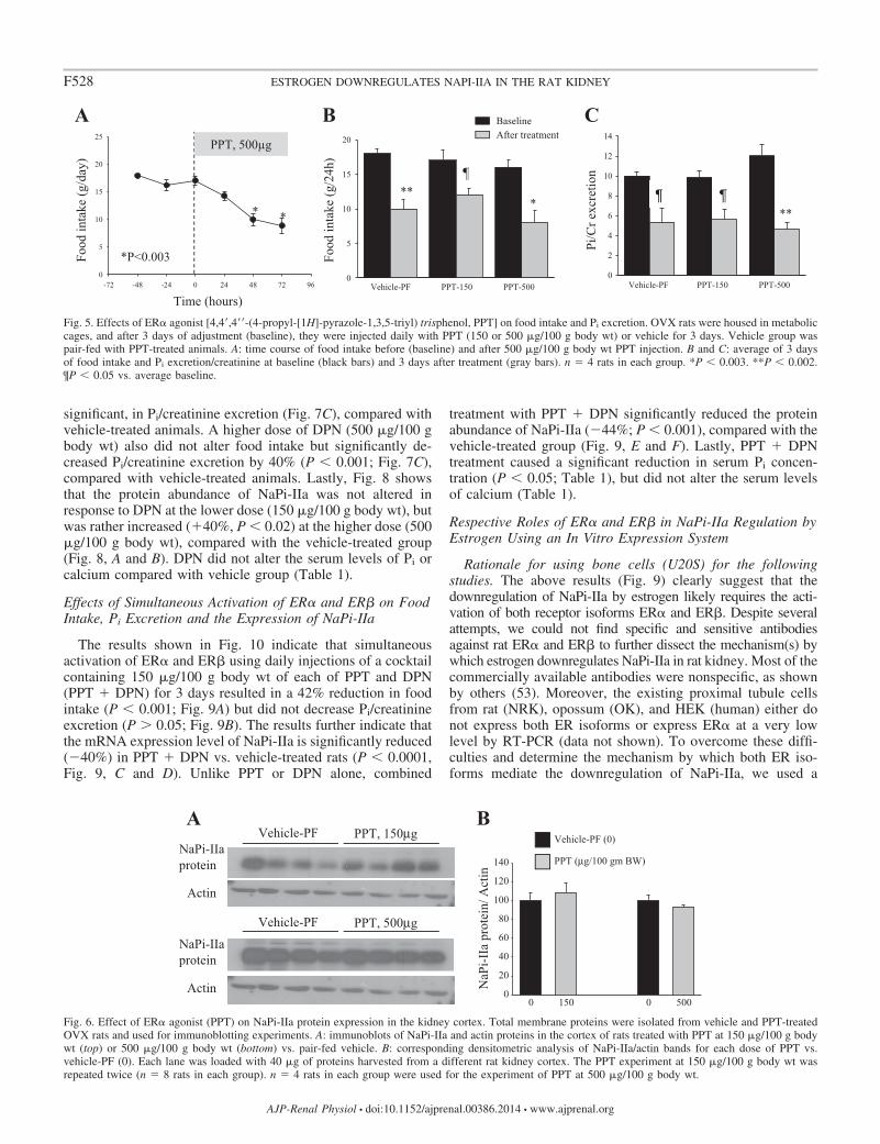
The results shown in Fig. 5 indicate that specific activationof ER� using 150 or 500 �g/100 g body wt of the ER�-specificagonist PPT resulted in a significant reduction in food intake(Fig. 5, A and B), which correlated with a significant reductionin urinary Pi/creatinine excretion (Fig. 5C), compared withbaseline levels. Both of these effects are comparable to thoseobtained in pair-fed and vehicle-treated animals. Note that food
intake and Pi/creatinine excretion at baseline or after PPTtreatment were not significantly different between the threegroups. Lastly, PPT did not alter the protein abundance ofNaPi-IIa in the kidney cortex at either dose used (P 0.05,Fig. 6, A and B), compared with the pair-fed vehicle group(vehicle-PF). PPT did not alter the serum levels of Pi orcalcium compared with vehicle-PF group (Table 1).
Effects of Specific Activation of ER� on Food Intake, Pi
Excretion, and the Expression of NaPi-IIa
The data depicted in Fig. 7 indicate that specific activation ofER� using 150 �g/100 g body wt of DPN did not affect foodintake (Fig. 7, A and B), but caused a decrease, albeit not
+ + L- -RT RT+ +- -L
ERα
ERβ
Sglt1
SNAT3
A B
+ +-LCortexPTSCortex PTSCortex PTS
NCX
ECaC
GAPDH GAPDH GAPDH
C
Fig. 3. Expression of ER� and ER� in the kidney proximal tubule. Kidneys were harvested from OVX rats, and total RNA was isolated from the cortex orproximal tubular suspension (PTS). RNA was reverse transcribed, and resulting cDNA was PCR amplified using specific primers for ER� (A, top) or ER� (A:bottom). Specific markers of proximal tubule cells SGLT1 (B, top) and SNAT3 (B: bottom) and distal tubule cells NCX (C, top) and ECaC1 (C, bottom) wereused to evaluate the enrichment of proximal tubule fragments in the tubular suspensions. GAPDH is used as a control for cDNA loading. In negative samples,the reverse transcriptase was omitted in the reverse transcription reaction (RT). L: 1 kb Plus DNA ladder. The ladders for GAPDH in A and for SNAT3 in Bwere rearranged to maintain consistency in the position of the ladder in the entire graph.
0.00.20.40.60.81.01.21.41.61.8
[NH
4+ ], m
M
0h 18h
P<0.00001
0.0
0.2
0.4
0.6
0.8
[NH
4+ ], m
M
0h 3h 6h
P<0.0001
P<0.002A B
Vehicle
NaPi-IIaprotein
Actin
E2, 600 nM
NaP
i-IIa
pro
tein
/Act
in
0
20
40
60
80
100
120
P<0.05
P<0.003
60 300 600
E2, nM
E2Vehicle (0)
0 0 0
CD
Fig. 4. Long-term effect of estrogen on NaPi-IIaprotein expression in proximal tubular suspension invitro. PTS was prepared and incubated in phenol red-and serum-free medium on a shaker in cell cultureincubator, as described in METHODS. Equal parts ofPTS were distributed in 100-mm culture dishes andincubated for 18 h under normal conditions. A: after18 h, NH4
� produced by PTS was measured in themedium and compared with its level in the mediumat time 0 (0 h), before the addition of PTS (n � 4dishes in each). B: after 18 h, PTS were washed bycentrifugation and resuspended in the same mediumfor up to 6 h. NH4
� was measured in the media after3 and 6 h and compared with its level at time 0 (n �4 dishes in each group). C and D: PTS were treatedwith vehicle (0.016% ethanol) or 60, 300, or 600 nMof E2 and incubated for 24 h. C: representativeimmunoblots showing the abundance of NaPi-IIa andactin proteins in membrane fractions isolated fromPTS treated with vehicle or 600 nM E2. D: averagedensitometric analysis of NaPi-IIa/actin bands foreach E2 dose vs. vehicle. Each lane was loaded with40-�g membrane proteins from a different 100-mmculture dish. n � 4 or 5 dishes for each group in twoseparate experiments. P � 0.05 and P � 0.003 vs. noestrogen (vehicle).
F527ESTROGEN DOWNREGULATES NAPI-IIA IN THE RAT KIDNEY
AJP-Renal Physiol • doi:10.1152/ajprenal.00386.2014 • www.ajprenal.org
significant, in Pi/creatinine excretion (Fig. 7C), compared withvehicle-treated animals. A higher dose of DPN (500 �g/100 gbody wt) also did not alter food intake but significantly de-creased Pi/creatinine excretion by 40% (P � 0.001; Fig. 7C),compared with vehicle-treated animals. Lastly, Fig. 8 showsthat the protein abundance of NaPi-IIa was not altered inresponse to DPN at the lower dose (150 �g/100 g body wt), butwas rather increased (�40%, P � 0.02) at the higher dose (500�g/100 g body wt), compared with the vehicle-treated group(Fig. 8, A and B). DPN did not alter the serum levels of Pi orcalcium compared with vehicle group (Table 1).
Effects of Simultaneous Activation of ER� and ER� on FoodIntake, Pi Excretion and the Expression of NaPi-IIa
The results shown in Fig. 10 indicate that simultaneousactivation of ER� and ER� using daily injections of a cocktailcontaining 150 �g/100 g body wt of each of PPT and DPN(PPT � DPN) for 3 days resulted in a 42% reduction in foodintake (P � 0.001; Fig. 9A) but did not decrease Pi/creatinineexcretion (P 0.05; Fig. 9B). The results further indicate thatthe mRNA expression level of NaPi-IIa is significantly reduced(�40%) in PPT � DPN vs. vehicle-treated rats (P � 0.0001,Fig. 9, C and D). Unlike PPT or DPN alone, combined
treatment with PPT � DPN significantly reduced the proteinabundance of NaPi-IIa (�44%; P � 0.001), compared with thevehicle-treated group (Fig. 9, E and F). Lastly, PPT � DPNtreatment caused a significant reduction in serum Pi concen-tration (P � 0.05; Table 1), but did not alter the serum levelsof calcium (Table 1).
Respective Roles of ER� and ER� in NaPi-IIa Regulation byEstrogen Using an In Vitro Expression System
Rationale for using bone cells (U20S) for the followingstudies. The above results (Fig. 9) clearly suggest that thedownregulation of NaPi-IIa by estrogen likely requires the acti-vation of both receptor isoforms ER� and ER�. Despite severalattempts, we could not find specific and sensitive antibodiesagainst rat ER� and ER� to further dissect the mechanism(s) bywhich estrogen downregulates NaPi-IIa in rat kidney. Most of thecommercially available antibodies were nonspecific, as shownby others (53). Moreover, the existing proximal tubule cellsfrom rat (NRK), opossum (OK), and HEK (human) either donot express both ER isoforms or express ER� at a very lowlevel by RT-PCR (data not shown). To overcome these diffi-culties and determine the mechanism by which both ER iso-forms mediate the downregulation of NaPi-IIa, we used a
Vehicle-PF PPT-1500
5
10
15
20
***
PPT-500
Food
inta
ke (g
/day
)
Time (hours)-72 -48 -24 0 24 48 72 96
0
5
10
15
20
25PPT, 500μg
*
*P<0.003
*
BaselineAfter treatment
A B
Pi/C
r exc
retio
n
Food
inta
ke (g
/24h
)
Vehicle-PF PPT-150 PPT-5000
2
4
6
8
10
12
14
**
C
Fig. 5. Effects of ER� agonist [4,4=,4==-(4-propyl-[1H]-pyrazole-1,3,5-triyl) trisphenol, PPT] on food intake and Pi excretion. OVX rats were housed in metaboliccages, and after 3 days of adjustment (baseline), they were injected daily with PPT (150 or 500 �g/100 g body wt) or vehicle for 3 days. Vehicle group waspair-fed with PPT-treated animals. A: time course of food intake before (baseline) and after 500 �g/100 g body wt PPT injection. B and C: average of 3 daysof food intake and Pi excretion/creatinine at baseline (black bars) and 3 days after treatment (gray bars). n � 4 rats in each group. *P � 0.003. **P � 0.002.¶P � 0.05 vs. average baseline.
0
20
40
60
80
100
120
140
Vehicle-PF PPT, 500μg
Vehicle-PF PPT, 150μgNaPi-IIaprotein
Actin NaP
i-IIa
pro
tein
/ Act
in
150 500
Actin
NaPi-IIaprotein
Vehicle-PF (0)
PPT (μg/100 gm BW)
A
0 0
B
Fig. 6. Effect of ER� agonist (PPT) on NaPi-IIa protein expression in the kidney cortex. Total membrane proteins were isolated from vehicle and PPT-treatedOVX rats and used for immunoblotting experiments. A: immunoblots of NaPi-IIa and actin proteins in the cortex of rats treated with PPT at 150 �g/100 g bodywt (top) or 500 �g/100 g body wt (bottom) vs. pair-fed vehicle. B: corresponding densitometric analysis of NaPi-IIa/actin bands for each dose of PPT vs.vehicle-PF (0). Each lane was loaded with 40 �g of proteins harvested from a different rat kidney cortex. The PPT experiment at 150 �g/100 g body wt wasrepeated twice (n � 8 rats in each group). n � 4 rats in each group were used for the experiment of PPT at 500 �g/100 g body wt.
F528 ESTROGEN DOWNREGULATES NAPI-IIA IN THE RAT KIDNEY
AJP-Renal Physiol • doi:10.1152/ajprenal.00386.2014 • www.ajprenal.org

human bone cell line (U20S), which stably expresses eitherER� alone, ER� alone, or both ER� and ER� under doxycy-cline control (37). Most importantly, ER� and ER� have beenshown to form a functional heterodimer complex, which me-diates the regulation of several target genes by estrogen inthese cells (37). These cells were transiently transfected withrat kidney full-length NaPi-IIa construct and used for thefollowing experiments.
Effects of estrogen on rat NaPi-IIa expression in U20S cells.First, Northern blotting experiment demonstrated that doxycy-cline treatment induced the expression of both ER� and ER�in U20S cells (Fig. 10, A and B). The induction of ER�precedes that of ER�, but their mRNA expression levels issignificantly higher after 48 h of doxycycline treatment (Fig.10, A and B). Second, immunoblotting experiment shows thatNaPi-IIa protein is expressed only in cells transfected with therat NaPi-IIa expression plasmid (Fig. 10C). NaPi-IIa protein inthese cells is expressed as a �160-kDa band, as previouslydescribed by other investigators in rat and mouse bone cells(35). Third, immunofluorescence studies depicted in Fig. 11shows that rat NaPi-IIa is expressed in the plasma membrane oftransfected and vehicle-treated U20S cells expressing bothER� and ER� (Fig. 11, top) and that the abundance of its
expression is significantly reduced in the presence of 100 nMestrogen (Fig. 11, bottom). Finally, immunoblotting experi-ments of U20S cells incubated with 100 nM estrogen or vehiclefor 24 h show that NaPi-IIa protein abundance was not alteredby E2 in U20S cells expressing ER� alone (P 0.05; Fig. 12,A and B) or ER� alone (P 0.05; Fig. 12, C and D). However,estrogen caused a sharp decrease in the abundance of NaPi-IIaprotein (�80%; P � 0.01) in cells expressing both ER� andER� (Fig. 12, E and F), compared with the vehicle-treatedgroup.
DISCUSSION
We have previously demonstrated that estrogen causes phos-phaturia and hypophosphatemia in ovariectomized rats (23).This effect is mediated through the downregulation of thebrush border sodium-dependent phosphate cotransporter NaPi-IIa at both the mRNA and protein levels (23). The presentstudies confirm the phosphaturic and hypophosphatemic ef-fects of estrogen and its suppression of NaPi-IIa expression ina dose-dependent manner (Figs. 1 and 2). Increased electrolytewasting despite decreased food intake is likely specific to Pi, aschloride excretion and calcium excretion correlated with re-
0
5
10
15
20
BaselineAfter treatment
-72 -48 -24 0 24 48 72 960
5
10
15
20
25
Food
inta
ke (g
/day
)
Time (hours)
DPNA B
Food
inta
ke (g
/24h
)
Vehicle DPN-150 DPN-5000
2
4
6
8
10
12
14
Vehicle DPN-150 DPN-500
Pi/C
r exc
retio
n
NS
*
C
Fig. 7. Effects of ER� agonist [2,3-bis(4-hydroxyphenyl)-propionitrile, DPN] on food intake and Pi excretion. OVX rats were housed in metabolic cages andafter 3 days of adjustment (baseline), they were injected daily with DPN (150 or 500 �g/100 g body wt) or vehicle for 3 days. A: time course of food intakebefore and after 150 �g/100 g body wt DPN injection. B: average of 3 days of food intake before (baseline, dark bars) or food consumed on the third day aftervehicle or DPN injection (gray bars). C: average of 3 days Pi excretion/creatinine excretion before (baseline, dark bars) and Pi excretion/creatinine excretion ofthe third day after vehicle or DPN injection (gray bars). n � 4 rats in each group. *P � 0.001 vs. baseline of DPN group.
Vehicle DPN, 150μg
Vehicle DPN, 500μg
0
20
40
60
80
100
120
140
160NaPi-IIaprotein
Actin150 500
NaP
i-IIa
pro
tein
/ Act
in
Actin
NaPi-IIaprotein
Vehicle (0)
DPN (μg/100 gm BW)
0 0
P<0.02
A B
Fig. 8. Effect of ER� agonist (DPN) on NaPi-IIa expression in the kidney cortex. Total membrane proteins were isolated from vehicle and DPN-treated OVXrats and used for immunoblotting experiments. A: immunoblots of NaPi-IIa and actin proteins in the cortex of rats treated with DPN at 150 �g/100 g body wt(top) or 500 �g/100 g body wt (bottom) vs. vehicle. B: corresponding densitometric analysis of NaPi-IIa/actin bands for each dose of DPN vs. vehicle (0). Eachlane was loaded with 40 �g proteins harvested from a kidney cortex of a different rat. The DPN experiment at 150 �g/100 g body wt was repeated twice (n �8 rats in each group). n � 4 rats in each group were used for the experiment of DPN at 500 �g/100 g body wt. P � 0.02 vs. vehicle (0).
F529ESTROGEN DOWNREGULATES NAPI-IIA IN THE RAT KIDNEY
AJP-Renal Physiol • doi:10.1152/ajprenal.00386.2014 • www.ajprenal.org

duction in food intake in response to estrogen (Fig. 1). Further,the present data clearly show that the phosphaturic effect ofestrogen is specific to the downregulation of NaPi-IIa, as theexpression of the other apical Pi-absorbing transporters in theproximal tubules (i.e., NaPi-IIc and Pit2) was not affected byestrogen (Table 2). We have used cortical total membranefractions to examine the expression of NaPi-IIa and NaPi-IIc inthese studies. Because both proteins are known to be down-regulated by membrane retrieval and lysosomal degradation(reviewed in Ref. 9), a downregulation of NaPi-IIc would have
been evident if targeted by estrogen in a chronic setting (i.e.,3-day treatment).
Our results demonstrate the presence of both estrogen re-ceptor isoforms (ER� and ER�) in the kidney cortex and in theproximal tubule suspension (Fig. 3). The level of expression ofthese receptors is identical between cortex and PTS prepara-tions (Fig. 3), as the expression of ER� and ER� in the wholecortex also includes the distal tubule where estrogen has beenreported to upregulate calcium-absorbing transporters, at least,in mice kidney (60). These data confirm the presence of ER�
0
20
40
60
80
100
120
Vehicle PPT + DPN
Vehicle PPT + DPN
02468
1012141618
0
1
2
3
4
5
6
Food
intake
(g/d
ay)
Urin
e Pi
/cre
atin
ine
PPT + DPNVehicle PPT + DPNVehicle
NaPi-IIaprotein
Actin
NaPi-IIamRNAGAPDHmRNA
NS
P<0.002
BA
C
E
0
20
40
60
80
100
120
PPT + DPNVehicle
PPT + DPNVehicle
F
NaP
i-IIa
/Act
inN
aPi-I
Ia/G
APD
H
mR
NA
P<0.0001
P<0.0001
D
Fig. 9. Effects of combined ER� and ER�agonists (PPT � DPN) on food intake, Pi ex-cretion, and NaPi-IIa expression. OVX ratswere placed in metabolic cages and injectedwith PPT � DPN (150 �g/100 g body wt each)or vehicle. Averages of food intake (A) andPi/creatinine excretion (B) of the last day oftreatment with Vehicle or PPT � DPN.C: Northern hybridization of NaPi-IIa andGAPDH mRNA. E: immunoblots of NaPi-IIaand actin proteins in the cortex of rats treatedwith PPT � DPN vs. vehicle. Correspondingdensitometric analysis of NaPi-IIa mRNA/GAPDH mRNA (D) and NaPi-IIa protein/actin(F). Each lane was loaded with 30 �g of totalRNA or 40 �g proteins harvested from a dif-ferent rat. n � 3 or 4 rats in each group. NS, notsignificant.
NaPi-IIa
NaPi-IIaprotein
Actin
- + + + - + + +Sham
160 kDa
42 kDa
28S rRNA
18S rRNA
ERαmRNA
ERβmRNA
A CB± Dox ± Dox
Fig. 10. Expression of ER� and ER� mRNA and NaPi-IIa protein in U20S cells. Northern hybridization of ER� (A), ER� (B), and ribosomal 28S-18S RNAin U20S cells treated with doxycycline or vehicle for indicated time. Each lane was loaded with 30 �g of total RNA isolated from three culture dishes (100 mm)pooled together in one sample. C: immunoblot showing NaPi-IIa protein (�160 kDa) and actin bands in lysates harvested from U20S cells transfected with ratNaPi-IIa expression construct or the empty vector (Sham). Each lane was loaded with 30 �g of proteins harvested from different culture dishes (60 mm) in eachgroup.
F530 ESTROGEN DOWNREGULATES NAPI-IIA IN THE RAT KIDNEY
AJP-Renal Physiol • doi:10.1152/ajprenal.00386.2014 • www.ajprenal.org

and ER� proteins in the proximal tubule of both mice and ratkidneys (29, 61). Estrogen receptors are ligand-regulated tran-scription factors that mediate the genomic effects of differentestrogenic molecules, including 17�-estradiol (22). Our data(Fig. 3) are also in agreement with several ligand-bindingstudies, which reported the presence of estrogen receptor inboth the cytosol and the nucleus of rat kidney cells (19, 25, 41).These receptors are expressed mainly in S1 and S2 segments ofrat proximal tubule (19). All together, these results wouldsuggest that estrogen likely acts directly through these recep-tors and downregulates NaPi-IIa in the proximal tubule cells.Our previous studies demonstrated that the downregulation ofNaPi-IIa and phosphaturia by estrogen are also produced inparathyroidectomized rats, indicating that the estrogen effecton NaPi-IIa is independent of the circulating levels of PTH(23). In addition to PTH, FGF23 is a potent phosphaturichormone, as it downregulates both NaPi-IIa and NaPi-IIcproteins in the proximal tubule (7, 34, 40, 52). Recent studiesdemonstrated that estrogen treatment increased FGF23 levelsin rats (11). However, our studies clearly demonstrate thatestrogen downregulates NaPi-IIa but does not alter NaPi-IIc orPit2 expression levels (Table 2) in OVX rats. Further, the invitro studies depicted in Figs. 4 and 11 clearly demonstrate thatestrogen directly downregulates NaPi-IIa in proximal tubulesuspensions harvested from OVX rat kidneys (Fig. 4), as wellas in U20S cells expressing both ER� and ER� and transfectedwith a rat NaPi-IIa expression construct (Figs. 11 and 12).Taken all together, these data indicate that FGF23 could not
fully account for the downregulation of NaPi-IIa and subse-quent phosphaturia in response to estrogen.
Altogether, this and previously published studies (6, 23)clearly define estrogen as a new phosphaturic hormone, whichspecifically targets NaPi-IIa in the rat kidney proximal tubule.This implies that estrogen is an important regulatory hormoneof Pi metabolism. This new role of estrogen also exists inhumans and is supported by abundant clinical data in theliterature. Indeed, it has been shown that estrogen replacementtherapy in postmenopausal women reduces bone resorption andprevents bone loss and osteoporosis (48). Such treatment isassociated with a reduction in the circulating levels of inor-ganic phosphate in postmenopausal women (12, 55, 56). Thephosphaturic effect of estrogen is not specific to female sub-jects, as a significant decrease in serum Pi levels was alsoreported in male patients with metastatic prostate cancertreated with estrogen (17). On the basis of our studies, it islikely that the hypophosphatemic effect of estrogen observed inthese patients results from the downregulation of NaPi-IIa inthe kidney with subsequent uncompensated phosphaturia.
Interestingly, the physiological significance of the phospha-turic effect of estrogen is not clear at this time. This effectappears to contradict the general view that this hormone isessential for bone health. Estrogen stimulates absorption ofcalcium from both the intestine and kidney and stimulatesabsorption of Pi from the intestine. Hence, it is possible thatestrogen promotes bone mineralization, but also increases renalPi wasting to offset the increase in its intestinal absorption and
Fig. 11. Immunofluorescence of rat NaPi-IIa inU20S cells. U20S cells were grown to confluenceand incubated in serum- and phenol red-free me-dium containing doxycycline. Cells were trans-fected with rat NaPi-IIa full-length construct andthen treated with vehicle (top) or 100 nM E2
(bottom) for 24 h. The immunofluorescence wasthen revealed by confocal microscopy at originalmagnification of 20 with the same settings (gain,brightness, zoom) between groups.
F531ESTROGEN DOWNREGULATES NAPI-IIA IN THE RAT KIDNEY
AJP-Renal Physiol • doi:10.1152/ajprenal.00386.2014 • www.ajprenal.org

maintain the blood level of Pi at a very narrow physiologicrange. With this coordinated action, estrogen will minimizeCa-PO4 precipitation in soft tissues and prevent vascular cal-cification and cardiovascular disease. This would suggest thatwomen are more prone to vascular calcification in postmeno-pausal life in the absence of HRT. Indeed, studies have shownthat there is an association between arterial calcification andosteoporosis in postmenopausal women (reviewed in Refs. 21and 32). Subsequent studies demonstrated that postmenopausalwomen treated with estrogen exhibited a decrease in the levelsof coronary artery calcification (36). However, the role of Pi
excretion in this protective effect of estrogen remains to bedemonstrated.
Lastly, we examined the respective roles of ER isoforms inestrogen-induced downregulation of NaPi-IIa and phosphaturiain OVX rats. Our results indicate that the specific activation ofER� with PPT caused a significant reduction in food intake(Fig. 5, A and B), as previously shown by others (57). How-ever, unlike estrogen, the decrease in food intake by PPTcorrelates with a significant reduction in urinary Pi excretion in(Fig. 5C). PPT treatment did not alter NaPi-IIa protein abun-dance (Fig. 6, A and B), indicating that its activity is main-tained during specific activation of ER�, which explains the
absence of phosphaturia in PPT vs. pair-fed vehicle animals(Fig. 5C). When ER� was specifically activated using DPN,food intake was not altered at any dose, whereas urinary Pi
excretion was rather reduced at a higher dose (Fig. 7). Theabundance of NaPi-IIa protein was unchanged (lower dose)and significantly increased at a higher dose of DPN (Fig. 8, Aand B). The increase in NaPi-IIa protein abundance/activitycould account for the reduction in urinary Pi excretion ob-served at a higher dose of DPN (Fig. 7C). However, when theanimals were treated with a combination of PPT � DPN(activation of both ER� and ER�, respectively), a significantreduction in food intake (Fig. 9A), phosphaturia (Fig. 9B), andsignificant downregulation of NaPi-IIa at both the mRNA (Fig.9, C and D) and protein levels (Fig. 9, E and F) were observed.These effects of PPT � DPN reproduce the exact effects ofestrogen on feeding, renal Pi handling, and NaPi-IIa expres-sion. The effects of estrogen on NaPi-IIa expression and Pi
handling are more pronounced than that of PPT � DPN,indicating that estrogen is also likely acting through otherpathways, including membrane-bound receptor, such asGPR30, in addition to nuclear receptors ER� and ER�. The invivo findings were confirmed by in vitro experiments showingthat estrogen decreased the expression of rat NaPi-IIa protein
Vehicle E2
NaPi-IIaprotein
Actin 0
20
40
60
80
100
120
140
ERα/β
E2Vehicle
NaP
i-IIa
pro
tein
/ Act
inP<0.01
NaPi-IIaprotein
Actin
Vehicle E2
0
20
40
60
80
100
120
140
160
ERα
NaP
i-IIa
pro
tein
/ Act
in
NaPi-IIaprotein
Actin
Vehicle E2
0
20
40
60
80
100
120
140
ERβ
NaP
i-IIa
pro
tein
/ Act
in
U20S cells
A B
C D
E FE2Vehicle
E2Vehicle
U20S cells
U20S cells
Fig. 12. Effects of estrogen on NaPi-IIa protein abundance in U20S cells expressing ER�, ER�, or ER� and ER� and transfected with rat NaPi-IIa construct.U20S cells were pretreated with doxycycline and transfected with rat NaPi-IIa full-length construct and then treated with E2 (100 nM) or vehicle for 24 h. TheImmunoblots show the abundance of NaPi-IIa and actin proteins (A, C, E) and the corresponding densitometric analysis of NaPi-IIa/actin (B, D, F) in cellsexpressing ER� alone (A, B) or ER� alone (C, D) or both ER� and ER� (E, F). Each lane is loaded with 30 �g of protein harvested from a different cell culturedish. Five different dishes were used for each group and in each experiment.
F532 ESTROGEN DOWNREGULATES NAPI-IIA IN THE RAT KIDNEY
AJP-Renal Physiol • doi:10.1152/ajprenal.00386.2014 • www.ajprenal.org

only in U20S cells expressing both ER� and ER� (Figs. 11 and12, E and F) but not in cells expressing either ER� alone (Fig.12, A and B) or ER� alone (Fig. 12, C and D). In varioustissues and cells, including U20S cells, ER� and ER� havebeen shown to function as a heterodimer complex and mediatethe regulation of a set of genes and cell functions that aredifferent from those regulated by ER homodimers (13, 37, 44,47). All together, these data clearly demonstrate, for the firsttime, that the effects of estrogen on NaPi-IIa and Pi transport inthe rat kidney proximal tubule require the activation of bothER� and ER�; whereas its effect on food intake is mediatedthrough the specific activation of ER�. Whether ER� and ER�form a functional heterodimer in the rat kidney proximal tubuleor activate synergistic pathways that downregulate NaPi-IIaremains to be determined when specific and sensitive antibod-ies for rat ER� and ER� become available (53). In ourprevious studies (23), we reported that ICI182,780, an inhibitorof ER� (42, 62), partially inhibited the anorexic effect ofestrogen but did not prevent the downregulation of NaPi-IIa byestrogen in OVX rats (23). In light of the present results, it islikely that the dose of ICI182,780 used in these studies was nothigh enough to completely block the anorexic effect of estro-gen or to prevent its downregulation of NaPi-IIa in the kidney.
In conclusion, our data demonstrate that estrogen specifi-cally downregulates NaPi-IIa but not NaPi-IIc or Pit2 in thekidney proximal tubule and causes phosphaturia in OVX rats.This effect is direct and independent of circulating factors, asshown by in vitro proximal tubular suspensions and U20S cellsexpressing rat kidney NaPi-IIa. The results further demonstratethat inhibition of food intake by estrogen is mediated solely byactivation of ER�, whereas the downregulation of NaPi-IIa andphosphaturia are mediated through the activation of both ER�and ER�.
ACKNOWLEDGMENTS
Parts of these studies were published in abstract form and presented at theannual meeting of the American Society of Nephrology in Atlanta, GA, inNovember 2013.
GRANTS
These studies were supported by National Institutes of Health GrantDK-083582 to H. Amlal.
DISCLOSURES
No conflicts of interest, financial or otherwise, are declared by the authors.
AUTHOR CONTRIBUTIONS
Author contributions: D.B., R.W., S.S., R.F., M.L., J.R.H., and H.A.performed experiments; D.B., R.W., S.S., R.F., and H.A. analyzed data; D.B.,R.W., S.S., R.F., and H.A. prepared figures; D.B., R.W., S.S., R.F., M.L.,J.R.H., and H.A. edited and revised manuscript; D.B., R.W., S.S., R.F., M.L.,J.R.H., and H.A. approved final version of manuscript; H.A. conception anddesign of research; H.A. interpreted results of experiments; H.A. draftedmanuscript.
REFERENCES
1. Abu Hossain S, Chaudhry FA, Zahedi K, Siddiqui F, Amlal H.Cellular and molecular basis of increased ammoniagenesis in potassiumdeprivation. Am J Physiol Renal Physiol 301: F969–F978, 2011.
2. Adami S, Gatti D, Bertoldo F, Bertoldo F, Rossini M, Fratta-Pasini A,Zamberlan N, Facci E, Lo Cascio V. The effects of menopause andestrogen replacement therapy on the renal handling of calcium. Osteopo-ros Int 2: 180–185, 1992.
3. Amlal H, Habo K, Soleimani M. Potassium deprivation upregulatesexpression of renal basolateral Na�-HCO3
� cotransporter (NBC-1). Am JPhysiol Renal Physiol 279: F532–F543, 2000.
4. Attmane-Elakeb A, Mount DB, Sibella V, Vernimmen C, Hebert SC,Bichara M. Stimulation by in vivo and in vitro metabolic acidosis ofexpression of rBSC-1, the Na�-K�(NH4�)-2Cl� cotransporter of the ratmedullary thick ascending limb. J Biol Chem 273: 33,681–33,691, 1998.
5. Attmane-Elakeb A, Sibella V, Moreau A, Vernimmen C, Feldmann G,Paillard M, Bichara M. Long-term shake suspension and membranevesicles of medullary thick ascending limb. Kidney Int 53: 439–447, 1998.
6. Beers KW, Thompson MA, Chini EN, Chini EN, Dousa TP. �-estradiolinhibits Na�-Pi cotransport across renal brush border membranes fromovariectomized rats. Biochem Biophys Res Commun 221: 442–445, 1996.
7. Bergwitz C, Jüppner H. Regulation of phosphate homeostasis by PTH,vitamin D, and FGF23. Annu Rev Med 61: 91–104, 2010.
8. Berndt TJ, Knox FG. Renal regulation of phosphate excretion. In: TheKidney, Physiology and Pathophysiology, edited by Seldin DW andGiebisch G. New York: Raven, p. 2511–2532, 1992.
9. Biber J, Hernando N, Forster I, Murer H. Regulation of phosphatetransport in proximal tubules. Pflügers Arch 458: 39–52, 2009.
10. Breusegem SY, Takahashi H, Giral-Arnal H, Giral-Arnal H, Wang X,Jiang T, Verlander JW, Wilson P, Miyazaki-Anzai S, Sutherland E,Caldas Y, Blaine JT, Segawa H, Miyamoto K, Barry NP, Levi, M.Differential regulation of the renal sodium-phosphate cotransporters NaPi-IIa, NaPi-IIc, and PiT-2 in dietary potassium deficiency. Am J PhysiolRenal Physiol 297: F350–F361, 2009.
11. Carrillo-López N, Román-García P, Rodríguez-Rebollar A, Fernán-dez-Martín JL, Naves-Díaz M, Cannata-Andía JB. Indirect regulationof PTH by estrogens may require FGF23. J Am Soc Nephrol 20: 2009–2017, 2009.
12. Castelo-Branco C, Martínez de Osaba MJ, Pons F, González-Merlo J.The effect of hormone replacement therapy on postmenopausal bone loss.Eur J Obstet Gynecol Reprod Biol 44: 131–136, 1992.
13. Chakraborty S, Willett H, Biswas PK. Insight into estrogen receptorbeta-beta and alpha-beta homo- and heterodimerization: A combinedmolecular dynamics and sequence analysis study. Biophys Chem 170:42–50, 2012.
14. Chalumeau C, du Cheyron D, Defontaine N, Kellermann O, PaillardM, Poggioli J. NHE3 activity and trafficking depend on the state of actinorganization in proximal tubule. Am J Physiol Renal Physiol 280: F283–F290, 2001.
15. Chomczynski P, Sacchi N. Single-step method of RNA isolation by acidguanidinium thiocyanate-phenol-chloroform extraction. Anal Biochem162: 156–159, 1987.
16. Church GM, Gilbert W. Genomic sequencing. Proc Natl Acad Sci USA81: 1991–1995, 1984.
17. Citrin DL, Elson P, Kies MS, Lind R. Decreased serum phosphate levelsafter high-dose estrogens in metastatic prostate cancer. Possible implica-tions. Am J Med 76: 787–793, 1984.
18. Danisi G, Murer H. Inorganic phosphate absorption in small intestine. In:Handbook of Physiology. The Gastrointestinal System: Intestinal Absorp-tion and Secretion, edited by Field M and Frizzel RA. Bethesda, MD: AmPhysiol Soc., 1991, p. 323–336.
19. Davidoff M, Caffier H, Schiebler TH. Steroid hormone binding recep-tors in the rat kidney. Histochemistry 69: 39–48, 1980.
20. Dennis VW. Phosphate homeostasis. In: Handbook of Physiology: RenalPhysiology, edited by Windhager, EE. Bethesda, MD: Am Physiol Soc.,sect 8, vol. II, 1992, p. 1785–1815.
21. Doherty TM, Uzui H, Fitzpatrick LA, Tripathi PV, Dunstan CR,Asotra K, Rajavashisth TB. Rationale for the role of osteoclast-like cellsin arterial calcification. FASEB J 16: 577–582, 2002.
22. Edwards DP. Regulation of signal transduction pathways by estrogen andprogesterone. Annu Rev Physiol 67: 335–376, 2005.
23. Faroqui S, Levi M, Soleimani M, Amlal H. Estrogen downregulates theproximal tubule type IIa sodium phosphate cotransporter causing phos-phate wasting and hypophosphatemia. Kidney Int 73: 1141–1150, 2008.
24. Gallazzini M, Attmane-Elakeb A, Mount DB, Hebert SC, Bichara M.Regulation by glucocorticoids and osmolality of expression of ROMK(Kir 1.1), the apical K channel of thick ascending limb. Am J PhysiolRenal Physiol 284: F977–F986, 2003.
25. Hagenfeldt Y, Eriksson HA. The estrogen receptor in the rat kidney.Ontogeny, properties and effects of gonadectomy on its concentration. JSteroid Biochem 31: 49–56, 1988.
F533ESTROGEN DOWNREGULATES NAPI-IIA IN THE RAT KIDNEY
AJP-Renal Physiol • doi:10.1152/ajprenal.00386.2014 • www.ajprenal.org

26. Harrington WR, Sheng S, Barnett DH, Petz LN, KatzenellenbogenJA, Katzenellenbogen BS. Activities of estrogen receptor alpha- andbeta-selective ligands at diverse estrogen responsive gene sites mediatingtransactivation or transrepression. Mol Cell Endocrinol 206: 13–22, 2003.
27. Harris HA, Katzenellenbogen JA, Katzenellenbogen BS. Characteriza-tion of the biological roles of the estrogen receptors, ER� and ER�, inestrogen target tissues in vivo through the use of an ER�-selective ligand.Endocrinology 143: 4172–4177, 2002.
28. Houillier P, Chambrey R, Achard JM, Froissart M, Poggioli J,Paillard M. Signaling pathways in the biphasic effect of angiotensin II onapical Na/H antiport activity in proximal tubule. Kidney Int 50: 1496–1505, 1996.
29. Irsik DL, Carmines PK, Lane PH. Classical estrogen receptors and ER�splice variants in the mouse. PLos One 8: e70926, 2013.
30. Karim Z, Attmane-Elakeb A, Sibella V, Bichara M. Acid pH increasesthe stability of BSC1/NKCC2 mRNA in the medullary thick ascendinglimb. J Am Soc Nephrol 14: 2229–2236, 2003.
31. Karim Z, Defontaine N, Paillard M, Poggioli J. Protein kinase Cisoforms in rat kidney proximal tubule: acute effect of angiotensin II. AmJ Physiol Cell Physiol 269: C134–C140, 1995.
32. Kiel DP, Kauppila LI, Cupples LA, Hannan MT, O’Donnell CJ,Wilson PW. Bone loss and the progression of abdominal aortic calcifi-cation over a 25 year period: the Framingham Heart Study. Calcif TissueInt 68: 271–276, 2001.
33. Kuiper GG, Enmark E, Pelto-Huikko M, Treuter E, Gustafsson JA.Cloning of a novel estrogen receptor expressed in rat prostate and ovary.Proc Natl Acad Sci USA 93: 5925–5930, 1996.
34. Kuro-o M. Klotho, phosphate and FGF-23 in ageing and disturbedmineral metabolism. Nat Rev Nephrol 9: 650–660, 2013.
35. Lundquist P, Murer H, Biber J. Type II Na�-Pi cotransporters inosteoblast mineral formation: regulation by inorganic phosphate. CellPhysiol Biochem 19: 43–56, 2007.
36. Manson JE, Allison MA, Rossouw JE, Carr JJ, Langer RD, Hsia J,Kuller LH, Cochrane BB, Hunt JR, Ludlam SE, Pettinger MB, GassM, Margolis KL, Nathan L, Ockene JK, Prentice RL, Robbins J,Stefanick ML, WHI and WHI-CACS Investigators. Estrogen therapyand coronary-artery calcification. N Engl J Med 356: 2591–2602, 2007.
37. Monroe DG, Secreto FJ, Subramaniam M, Getz BJ, Khosla S, Spels-berg TC. Estrogen receptor � and � heterodimers exert unique effects onestrogen- and tamoxifen-dependent gene expression in human U2OSosteosarcoma cells. Mol Endocrinol 19: 1555–1568, 2005.
38. Monroe DG, Spelsberg T. General mechanisms of steroid receptors andreceptor coregulator action. In: Advances in Molecular and Cell Biology,34th ed., edited by Miller V and Hay M, Amsterdam: Elsevier B.V., 2004,p. 39–48.
39. Monroe DG, Spelsberg TC. Gonadal steroids and receptors. In: Primeron the Metabolic Bone Diseases and Disorders of Mineral Metabolism,5th ed., edited by Favus MJ, Philadelphia: Lippincot, Williams andWilkins, 2004, p. 32–38.
40. Murer H, Forster I, Biber J. The sodium phosphate cotransporter familySLC34. Pflügers Arch 447: 763–767, 2004.
41. Murono EP, Kirdani RY, Sandberg AA. Specific estradiol-17� bindingcomponent in adult rat kidney. J Steroid Biochem 11: 1347–1351, 1979.
42. Nawaz Z, Lonard DM, Dennis AP, Smith CL, O’Malley BW. Protea-some-dependent degradation of the human estrogen receptor. Proc NatlAcad Sci USA 96: 1858–1862, 1999.
43. Patzer L, Hernando N, Ziegler U, Beck-Schimmer B, Biber J, MurerH. Ifosfamide metabolites CAA, 4-OH-Ifo and Ifo-mustard reduce apicalphosphate transport by changing NaPi-IIa in OK cells. Kidney Int 70:1725–1734, 2006.
44. Paulmurugan R, Tamrazi A, Massoud TF, Massoud TF, Katzenellen-bogen JA, Gambhir SS. In vitro and in vivo molecular imaging ofestrogen receptor � and � homo- and heterodimerization: exploration ofnew modes of receptor regulation. Mol Endocrinol 25: 2029–2040, 2011.
45. Poggioli J, Lazar G, Houillier P, Gardin JP, Paillard M. Acutevariations in extracellular pH modulate transduction pathways of PTH inrat proximal tubule. Am J Physiol Cell Physiol 263: C941–C947, 1992.
46. Poggioli J, Lazar G, Houillier P, Gardin JP, Achard JM, Paillard M.Effects of angiotensin II and nonpeptide receptor antagonists on transduc-tion pathways in rat proximal tubule. Am J Physiol Cell Physiol 263:C750–C758, 1992.
47. Powell E, Xu W. Intermolecular interactions identify ligand-selectiveactivity of estrogen receptor alpha/beta dimers. Proc Natl Acad Sci USA105: 19012–19017, 2008.
48. Riggs BL, Jowsey J, Kelly PJ, Jones JD, Maher FT. Effect of sexhormones on bone in primary osteoporosis. J Clin Invest 48: 1065–1072,1969.
49. Roesch DM. Effects of selective estrogen receptor agonists on food intakeand body weight gain in rats. Physiol Behav 87: 39–44, 2006.
50. Santollo J, Wiley MD, Eckel LA. Acute activation of ER� decreasesfood intake, meal size, and body weight in ovariectomized rats. Am JPhysiol Regul Integr Comp Physiol 293: R2194–R2201, 2007.
51. Schiavi SC, Kumar R. The phosphatonin pathway: new insights inphosphate homeostasis. Kidney Int 65: 1–14, 2004.
52. Silver J, Naveh-Many T. FGF23 and the parathyroid. Adv Exp Med Biol728: 92–99, 2012.
53. Snyder MA, Smejkalova T, Forlano PM, Woolley CS. Multiple ER �antisera label in ER � knockout and null mouse tissues. J NeurosciMethods 188: 226–234, 2010.
54. Stauffer SR, Coletta CJ, Tedesco R, Nishiguchi G, Carlson K, Sun J,Katzenellenbogen BS, Katzenellenbogen JA. Pyrazole ligands: struc-ture-affinity/activity relationships and estrogen receptor-�-selective ago-nists. J Med Chem 43: 4934–4947, 2000.
55. Stock JL, Coderre JA, Mallette LE. Effects of a short course of estrogenon mineral metabolism in postmenopausal women. J Clin EndocrinolMetab 61: 595–600, 1985.
56. Stock JL, Coderre JA, Posillico JT. Effects of estrogen on mineralmetabolism in postmenopausal women as evaluated by multiple assaysmeasuring parathyrin bioactivity. Clin Chem 35: 18–22, 1989.
57. Thammacharoen S, Geary N, Lutz TA, Ogawa S, Asarian L. Divergenteffects of estradiol and the estrogen receptor-� agonist PPT on eating andactivation of PVN CRH neurons in ovariectomized rats and mice. BrainRes 1268: 88–96, 2009.
58. Tremblay GB, Tremblay A, Copeland NG, Copeland NG, Gilbert DJ,Jenkins NA, Labrie F, Giguère V. Cloning, chromosomal location andfunctional analysis of the murine estrogen receptor �. Mol Endocrinol 11:353–365, 1997.
59. Uemura H, Irahara M, Yoneda N, Yasui T, Genjida K, Miyamoto KI,Aono T, Takeda E. Close correlation between estrogen treatment andrenal phosphate reabsorption capacity. J Clin Endocrinol Metab 85:1215–1219, 2000.
60. Van Abel M, Hoenderop JG, Dardenne O, St Arnaud R, Van Os CH,Van Leeuwen HJ, Bindels RJ. 1,25-dihydroxyvitamin D3-independentstimulatory effect of estrogen on the expression of ECaC1 in the kidney.J Am Soc Nephrol 13: 2102–2109, 2002.
61. Wells CC, Riazi S, Mankhey RW, Bhatti F, Ecelbarger C, Maric C.Diabetic nephropathy is associated with decreased circulating estradiollevels and imbalance in the expression of renal estrogen receptors. GendMed 2: 227–237, 2005.
62. Wijayaratne AL, McDonnell DP. The human estrogen receptor-� is aubiquitinated protein whose stability is affected differentially by agonists,antagonists, and selective estrogen receptor modulators. J Biol Chem 276:35,684–35,692, 2001.
63. Xu H, Uno JK, Inouye M, Inouye M, Xu L, Drees JB, Collins JF,Ghishan FK. Regulation of intestinal NaPi-IIb cotransporter gene expres-sion by estrogen. Am J Physiol Gastrointest Liver Physiol 285: G1317–G1324, 2003.
64. Zajicek HK, Wang H, Puttaparthi K, Halaihel N, Markovich D,Shayman J, Béliveau R, Wilson P, Rogers T, Levi M. Glycosphingo-lipids modulate renal phosphate transport in potassium deficiency. KidneyInt 60: 694–704, 2001.
F534 ESTROGEN DOWNREGULATES NAPI-IIA IN THE RAT KIDNEY
AJP-Renal Physiol • doi:10.1152/ajprenal.00386.2014 • www.ajprenal.org