Age-related decrease in aromatase and estrogen receptor (ERα and ERβ) expression in rat testes:...
11
Asian J Androl 2008; 10 (2): 177–187 . 177 . Abstract Aim: To examine the effects on rat aging of caloric restriction (CR1) and undernutrition (CR2) on the body and on testicular weights, on two enzymatic antioxidants (superoxide dismutase and catalase), on lipid peroxidation and on the expression of testicular aromatase and estrogen receptors (ER). Methods: CR was initiated in 1-month-old rats and carried on until the age of 18 months. Results: In control and CR2 rats an age-related decrease of the aromatase and of ER (α and β) gene expression was observed; in parallel a diminution of testicular weights, and of the total number and motility of epididymal spermatozo was recorded. In addition, aging in control and CR2 rats was accom- panied by a significant decrease in testicular superoxide dismutase, catalase activities, and an increase in lipid peroxidation level (thiobarbituric acid reactive substance), associated with alterations of spermatogenesis. Conversely, caloric restriction-treatment exerted a protective effect and all the parameters were less affected by aging. Conclusion: These results indicate that during aging, a low caloric diet (not undernutrition) is beneficial for spermatogenesis and likely improves the protection of the cells via an increase of the cellular antioxidant defense system in which aromatase/ ER could play a role. (Asian J Androl 2008 Mar; 10: 177–187) Keywords: aging; low caloric diet; rat testis; aromatase; estrogen receptors; antioxidants Age-related decrease in aromatase and estrogen receptor (ERα and ERβ) expression in rat testes: protective effect of low caloric diets* Khaled Hamden 1 , Dorothee Silandre 2 , Christelle Delalande 2 , Abdefattah El Feki 1 , Serge Carreau 2 1 Animal Ecophysiology, Faculty of Sciences, Sfax-3018, Tunisia 2 USC 2006 INRA- EA 2608, Biochemistry, Caen-14032, France . Original Article . DOI: 10.1111/j.1745-7262.2008.00343.x www.asiaandro.com © 2008, Asian Journal of Andrology, SIMM and SJTU. All rights reserved. Correspondence to: Dr Serge Carreau, University –Biochemistry, Esplanade de la Paix, 14032 Caen Cedex, France. Tel: +33-2-3156-5488 Fax: +33-2-3156-5120 E-mail: [email protected] *Preliminary reports were presented at the XIVth European Work- shop on Molecular and Cellular Endocrinology of the Testis. Bad Aibling, Germany, 22–26 April, 2006. Received 2007-05-10 Accepted 2007-08-23 1 Introduction During aging the diminution of the blood testoste- rone level parallels a decline of several physiological pa- rameters and among them, those of the reproductive func- tion [1]. Studies using the Brown Norway rat as a model have clearly shown that aging is associated with a func- tional deficit of the testicular cells [2]. A decrease in the number of germ cells and, consequently, a diminution of the daily sperm production have also been reported [3]. The loss of germ cells might be related to either a de- crease in the ability of the Sertoli cells to support the germ cell survival and differentiation, or to the reduced availability of the testosterone produced by Leydig cells [1], or to other unknown factors. Nutrition plays an
Transcript of Age-related decrease in aromatase and estrogen receptor (ERα and ERβ) expression in rat testes:...

Asian J Androl 2008; 10 (2): 177–187
.177.Tel: +86-21-5492-2824; Fax: +86-21-5492-2825; Shanghai, China
Abstract
Aim: To examine the effects on rat aging of caloric restriction (CR1) and undernutrition (CR2) on the body and ontesticular weights, on two enzymatic antioxidants (superoxide dismutase and catalase), on lipid peroxidation and onthe expression of testicular aromatase and estrogen receptors (ER). Methods: CR was initiated in 1-month-old ratsand carried on until the age of 18 months. Results: In control and CR2 rats an age-related decrease of the aromataseand of ER (α and β) gene expression was observed; in parallel a diminution of testicular weights, and of the totalnumber and motility of epididymal spermatozo was recorded. In addition, aging in control and CR2 rats was accom-panied by a significant decrease in testicular superoxide dismutase, catalase activities, and an increase in lipid peroxidationlevel (thiobarbituric acid reactive substance), associated with alterations of spermatogenesis. Conversely, caloricrestriction-treatment exerted a protective effect and all the parameters were less affected by aging. Conclusion:These results indicate that during aging, a low caloric diet (not undernutrition) is beneficial for spermatogenesis andlikely improves the protection of the cells via an increase of the cellular antioxidant defense system in which aromatase/ER could play a role. (Asian J Androl 2008 Mar; 10: 177–187)
Keywords: aging; low caloric diet; rat testis; aromatase; estrogen receptors; antioxidants
Age-related decrease in aromatase and estrogen receptor(ERα and ERβ) expression in rat testes: protective effect oflow caloric diets*
Khaled Hamden1, Dorothee Silandre2, Christelle Delalande2, Abdefattah El Feki1, Serge Carreau2
1Animal Ecophysiology, Faculty of Sciences, Sfax-3018, Tunisia2USC 2006 INRA- EA 2608, Biochemistry, Caen-14032, France
.Original Article .
DOI: 10.1111/j.1745-7262.2008.00343.xwww.asiaandro.com
© 2008, Asian Journal of Andrology, SIMM and SJTU. All rights reserved.
Correspondence to: Dr Serge Carreau, University –Biochemistry,Esplanade de la Paix, 14032 Caen Cedex, France.Tel: +33-2-3156-5488 Fax: +33-2-3156-5120E-mail: [email protected]*Preliminary reports were presented at the XIVth European Work-shop on Molecular and Cellular Endocrinology of the Testis. BadAibling, Germany, 22–26 April, 2006.Received 2007-05-10 Accepted 2007-08-23
1 Introduction
During aging the diminution of the blood testoste-
rone level parallels a decline of several physiological pa-rameters and among them, those of the reproductive func-tion [1]. Studies using the Brown Norway rat as a modelhave clearly shown that aging is associated with a func-tional deficit of the testicular cells [2]. A decrease in thenumber of germ cells and, consequently, a diminution ofthe daily sperm production have also been reported [3].The loss of germ cells might be related to either a de-crease in the ability of the Sertoli cells to support thegerm cell survival and differentiation, or to the reducedavailability of the testosterone produced by Leydig cells[1], or to other unknown factors. Nutrition plays an
.178.
Protective effect of caloric restriction during aging on rat testis
http://www.asiaandro.com; [email protected]
important role during aging and, indeed, caloric restric-tion delays the appearance of age-associated physiopatho-logical changes [1]. Therefore, caloric restriction in-duces some benefits on the longevity of rats [4]. Asreported, most of the steroidogenic enzyme activities aredecreased during aging [1]; nevertheless no informationregarding the aromatase status and estrogen’s role in agingis available. We have shown that rat testicular cells, in-cluding germ cells, are able to synthesize estrogens [5]and are also equipped with estrogen receptors, suggest-ing a putative role for these female hormones on sper-matogenesis [6]. Prokai et al. [7] show that estrogensexert various cellular actions, including an antioxidanteffect. Indeed, to protect against the adverse effects ofreactive oxygen species, mammalian cells are equippedwith various enzymatic and non-enzymatic antioxidantscavengers.
Therefore, the aim of the present study is: (i) toevaluate the expression of aromatase and of estrogenreceptor alpha and beta (ERα and ERβ) genes in themale rat testis during aging; and (ii) to analyze the im-pact of low caloric diets and undernutrition on theseparameters. To assess the fertility of these treated-animals, the epididymal sperm counts and the spermmotility have been evaluated. Finally, two major enzy-matic antioxidants, superoxide dismutase (SOD) andcatalase (CAT), as well as the lipid peroxidation(thiobarbituric acid reactive substance (TBAR) are de-termined to characterize some of the cellular adverseeffects targeted by aging.
2 Materials and methods
2.1 Animals and treatmentsMale Wistar rats, aged from 2 to 18 months, were
either fed ad libitum (control) or submitted to diets ofcaloric restriction (CR1) or undernutrition (CR2), as re-ported elsewhere [8, 9]. The CR1 and CR2 groupsreceived, respectively, 60% and 40% of the quantity offood given to the control rats (equivalent to 413 kCal/kgbody weight/day) and the restriction was initiated in ani-mals aged 4 weeks. All animals (six per group) wereobtained through Central Pharmacy, Tunis, Tunisia; theywere maintained at 24 ± 3ºC under a 12 h-controlled Light:Dark cycle, with access to water ad libidum. At theindicated age, the animals were weighted, killed bydecapitation, and the trunk blood was collected. Theserum was prepared by centrifugation (1 500 × g, 15 min,
4ºC) and the testes were removed, cleaned of fat andweighted; all these samples were stored at –80ºC untilused. The caudal region of the epididymides was savedto collect the spermatozoa. The handling of the animalswas approved by the local ethical committee for the careand use of laboratory animals.
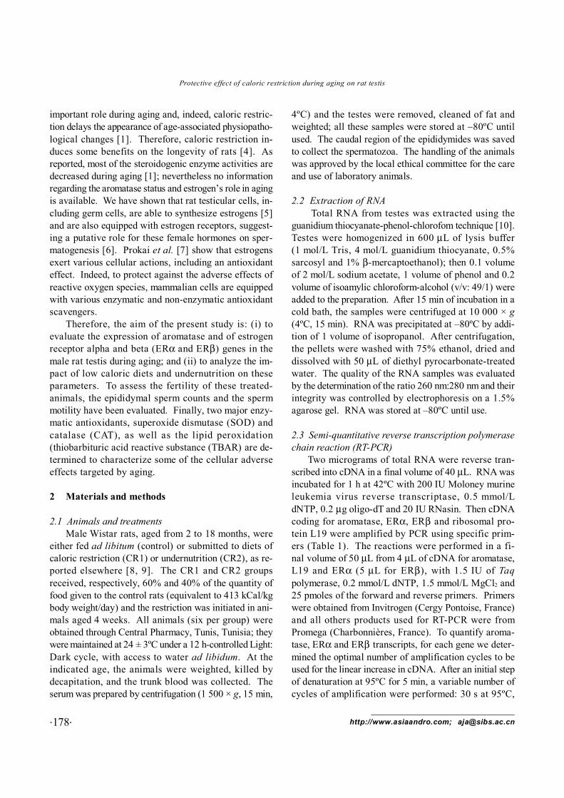
2.2 Extraction of RNA Total RNA from testes was extracted using the
guanidium thiocyanate-phenol-chlorofom technique [10].Testes were homogenized in 600 μL of lysis buffer(1 mol/L Tris, 4 mol/L guanidium thiocyanate, 0.5%sarcosyl and 1% β-mercaptoethanol); then 0.1 volumeof 2 mol/L sodium acetate, 1 volume of phenol and 0.2volume of isoamylic chloroform-alcohol (v/v: 49/1) wereadded to the preparation. After 15 min of incubation in acold bath, the samples were centrifuged at 10 000 × g(4ºC, 15 min). RNA was precipitated at –80ºC by addi-tion of 1 volume of isopropanol. After centrifugation,the pellets were washed with 75% ethanol, dried anddissolved with 50 μL of diethyl pyrocarbonate-treatedwater. The quality of the RNA samples was evaluatedby the determination of the ratio 260 nm:280 nm and theirintegrity was controlled by electrophoresis on a 1.5%agarose gel. RNA was stored at –80ºC until use.
2.3 Semi-quantitative reverse transcription polymerasechain reaction (RT-PCR)
Two micrograms of total RNA were reverse tran-scribed into cDNA in a final volume of 40 μL. RNA wasincubated for 1 h at 42ºC with 200 IU Moloney murineleukemia virus reverse transcriptase, 0.5 mmol/LdNTP, 0.2 μg oligo-dT and 20 IU RNasin. Then cDNAcoding for aromatase, ERα, ERβ and ribosomal pro-tein L19 were amplified by PCR using specific prim-ers (Table 1). The reactions were performed in a fi-nal volume of 50 μL from 4 μL of cDNA for aromatase,L19 and ERα (5 μL for ERβ), with 1.5 IU of Taqpolymerase, 0.2 mmol/L dNTP, 1.5 mmol/L MgCl2 and25 pmoles of the forward and reverse primers. Primerswere obtained from Invitrogen (Cergy Pontoise, France)and all others products used for RT-PCR were fromPromega (Charbonnières, France). To quantify aroma-tase, ERα and ERβ transcripts, for each gene we deter-mined the optimal number of amplification cycles to beused for the linear increase in cDNA. After an initial stepof denaturation at 95ºC for 5 min, a variable number ofcycles of amplification were performed: 30 s at 95ºC,

Asian J Androl 2008; 10 (2): 177–187
.179.Tel: +86-21-5492-2824; Fax: +86-21-5492-2825; Shanghai, China
30 s at 60ºC, and 45 s at 72ºC (Table 1), followed by afinal extension at 72ºC for 7 min. We have chosenL19 transcripts, which did not vary among the samples,to correct the difference in the quantities of total RNAused for reverse transcription [11]. The amplified cDNAfragments were run on a 2% agarose gel stained withethidium bromide, visualized under ultraviolet transillu-mination and analyzed with National Institutes of Health(NIH) software (http://rsb.info.nih.gov/nih-image).
2.4 Epididymal sperm countSpermatozoa were collected from an equal length of
the cauda epididymis of each rat by flushing with the samevolume (10 mL) of a medium containing 140 mmol/L NaCl,0.3 mmol/L KCl, 0.8 mmol/L Na2HPO4, 0.2 mmol/LKH2PO4 and 1.5 mmol/L D-glucose (pH 7.3). The col-lected samples were centrifuged at 100 × g for 2 min,and the pellets were re-suspended in 10 mL of freshmedium. An aliquot (100 μL) was mixed with an equalvolume of 1% Trypan blue, then the number of sperma-tozoa and the motility were determined [12].
2.5 Steroid determinations and measurements of anti-oxidant enzymatic activities
After homogenisation of testes in a phosphate buffer(1 g/2 mL), steroids were extracted by diethylether ac-cording to our reported method. The estradiol level wasthen measured by RIA using highly specific antibodiesfrom P.A.R.I.S (Compiègne, France). The intra-assayand inter-assay coefficients of variation were 8% and5% for estradiol. The lipid peroxidation was determinedin the homogenates from control and treated rat testesby quantification of the TBAR using the method appliedby Buege and Aust [13]. The superoxide dismutase ac-
tivity was assayed using the spectrophotometric methodof Marklund and Marklund [14]. The activity of cata-lase was measured using Aebi’s method [15]. The pro-tein level was determined using the method applied byLowry et al. [16].
For histological studies, pieces of testes were fixedin a Bouin’s solution for 24 h, then embedded in paraffin.Sections of 5 µm thickness were stained with hematoxy-lin-eosin and examined under a digital camera Olympusmicroscope (Olympus CX41; Olympus Industrial AmericaInc., Orangeburg, NY, USA).
2.6 Statistical analysisData are presented as mean ± SEM. The determina-
tions were performed using six animals per group andthe differences were examined using one-way analysisof variance (ANOVA) followed by the Scheffe test. Sig-nificance was accepted at P < 0.05 (StatView, SASInstitute, Cary, NC, USA).
3 Results
3.1 Body and testicular weightsFor the control rats, an increase in body weight was
observed between 2 and 15 months of age, which re-mained stable later. In the CR1 group body weight in-creased but more slowly, and from 4 months of age itwas significantly lower than that of the control rats. ForCR2 rats, we noticed a slight increase between 2 and12 months, then no variation was registered (Figure 1A).Compared to the controls, the body weight of CR2 ratswas statistically lower whatever the age and the decreasewas approximately 50% from 15 months of age. Whencompared to CR1 rats, the body weight of CR2 animals
Table 1. Sequences of primers. Size of fragments amplified and number of cycles used for amplification. Genbank accession number areindicated in parenthesis. PCR, polymerase chain reaction; S, sens primer; R, reverse primer.
Gene Primer SequenceOrientation and Size of PCR Number position product (bp) of cycle
Aromatase 5'-ARO GCTTCTCATCGCAGAGTATCCGG S (1555–1577) 290 33 (M33986) 3'-ARO CAAGGGTAAATTCATTGGGCTTGG R (1821–1844)ERα 5'-ERα AATTCTGACAATCGACGCCAG S (545–565) 345 29 (X61098) 3'-ERα GTGCTTCAACATTCTCCCTCCTC R (867–889)ERβ 5'-ERβ GAAGCTGAACCACCCAATGT S (1100–1120) 211 40 (U57439) 3'-ERβ CAGTCCCACCATTAGCACCT R (1291–1310)L19 5'-L19 GAAATCGCCAATGCCAACTC S (119–138) 290 24 (NM_031103) 3'-L19 ACCTTCAGGTACAGGCTGTG R (389–408)

.180.
Protective effect of caloric restriction during aging on rat testis
http://www.asiaandro.com; [email protected]
Figure 1. Body (A), testicular weights (B) and ratio testis/body weight (C) of two 18-month-old rats either fed ad libidum (control), orsubmitted to caloric restriction (CR1) or undernutrition (CR2) diet. The data are expressed as mean ± SEM (n = 6). a: control ratssignificantly different from 2-month-old rats; b: CR1 rats significantly different from the same age (control) rats; c: CR2 rats significantlydifferent from the same age (control) rats; d: CR1 rats significantly different from the same age (CR2) rats; e: CR1 rats significantly differentfrom 2-month-old CR1 rats; (f): CR2 rats significantly different from 2-month-old CR2 rats.
was also significantly lower at all ages, except at 2 and12 months and again, the difference was greater in ratsaged 15 months onwards.
In control rats, an increase in testicular weight wasnoticed until the age of 12 months, but in 18-month-oldrats the testicular weight was identical to that of 2-month-old animals (Figure 1B). For the CR1 group, a similarpattern was observed until the age of 15 months, thenthe testicular weight, before diminishing slightly. At 15and 18 months, the weight of testes in CR1 rats wassignificantly higher compared to that in the controls. ForCR2 rats, the testicular weights at all ages studied weresignificantly lower than those of their age-matchedcontrols, except at 2 months of age. Moreover, it wassignificantly lower compared to the CR1 animals what-ever the age (except at 2 months), especially from theage of 15 months (Figure 1B).
Testicular weight in relation to body weight decreasedwith age in control rats, whereas in the CR1 group asignificant augmentation was observed at 12 months(Figure 1C). It was also clear that relative testicularweight was significantly lower in 1-year-old CR2 ratsthan in CR1 animals.
3.2 Number and motility of epididymal spermatozoaIn the control CR1 and CR2 rats, an increase in the
number of spermatozoa was recorded from ages 2 to12 months; thereafter, a diminution was observed (39%
at 18 months compared to 1-year-old rats). In contrast,in CR2 rats no variations were noticed between 4 and18 months (in the older CR2 rats the amount of sperma-tozoa was identical to that of the controls). In 18-month-old CR1 rats, the number of spermatozoa was dimini-shed by 24% compared to 12-month-old animals. InCR1 rats aged of 15 and 18 months, the total number ofspermatozoa was significantly higher than that in thecontrol rats (Figure 2A).
Concerning the motility of spermatozoa, between 2and 4 months of age a sharp increase was observed incontrol CR1 and CR2 rats, although in 2-month-old CR2animals the number of motile spermatozoa was very low.In 1-year-old control rats and onwards the number ofmotile spermatozoa was lower (36%–47%) than in 4-month-old animals, whereas in CR1 rats no changes wereobserved with aging. In 18-month-old CR1 rats, thenumber of motile spermatozoa was significantly higher(35%) than in either controls or CR2 animals. In thatlater group, the motility was of the same magnitude as inthe controls starting from the age of 1 year (Figure 2B).
3.3 Histological changes in testes of control and treatedrats
The effects of aging, caloric restriction and under-nutrition were further analyzed through histological ex-amination of spermatogenesis (Figure 3). In 18-month-old controls and CR2 rats (Figure 3B and 3D), a deple-

Asian J Androl 2008; 10 (2): 177–187
.181.Tel: +86-21-5492-2824; Fax: +86-21-5492-2825; Shanghai, China
tion of germ cells was observed in comparison to 2-month- old rats (Figure 3A). In CR1 treated rats, sper-matogenesis proceeded normally and was similar to thatof 2-month-old rats (Figure 3C).
3.4 Aromatase expressionWe performed a semi-quantitative RT-PCR to deter-
mine whether the amount of aromatase transcripts in testiswas affected by aging and the caloric diets. As shown inFigure 4, an increase in the amount of aromatase tran-scripts was observed between 2 and 4 months in thecontrol and CR2 groups of animals (not for CR1), fol-lowed by a sharp and significant decrease from the ageof 12 months in control and CR2 animals (more than78% and 38%, respectively, compared to 4-month-oldrats). In contrast, in CR1 rats the diminutions were muchlower compared with the control group (Figure 4A and4B). From the age of 12 months onwards, the levels ofaromatase transcripts were approximately twice as highin both CR1 and CR2 rats compared to the control rats.Moreover, in 12- and 18-month-old CR1 rats, the amountof aromatase transcripts was greater and significantly
Figure 2. Total number (× 105) of spermatozoa in cauda epididymis (A) and measurement of their motility (B) in control, caloric restriction(CR1) and undernutrition (CR2) rats aged 2–18 months. Data are expressed as mean ± SEM (n = 8). a: control rats significantly differentfrom 2-month-old rats; b: CR1 rats significantly different from the same age (control) rats; c: CR2 rats significantly different from the sameage (control) rats; d: CR1 rats significantly different from the same age (CR2) rats; e: CR1 rats significantly different from 2-month-old CR1rats; f: CR2 rats significantly different from 2-month-old CR2 rats.
higher compared to CR2 rats (Figure 4B), and a signifi-cant decrease was observed only in 18-month-old ratscompared to young CR1 animals.
3.5 Age-related and caloric restriction-related changesof ERα and ΕRβ gene expression
The values of ERα/L19 and ERβ/19 ratio are re-ported in Figure 5. Between 2 and 4 months of age anincrease of the levels of ERα mRNAs was observed incontrol, CR1 and CR2 rats followed by a diminution ofthe amount of transcripts in older animals.
In the control group the decrease of the amount ofERα mRNA was significant at the age of one year (25%)and remained unchanged in the older rats. For CR1animals, the decrease of ERα mRNA was significant onlyin 18-month-old CR1 rats (Figure 5A). Moreover in CR1rats, the levels of ERα transcripts were higher comparedto control and CR2 rats starting from the age of12 months. In CR2 rats aged of 18 months the amountof ERα was similar to that of controls. Additionaly, inCR2 rats, ERα mRNA decreased significantly from 12-month-old rats compared to 2-month-old CR2 rats.

.182.
Protective effect of caloric restriction during aging on rat testis
http://www.asiaandro.com; [email protected]
Concerning the expression of ERβ (Figure 5B) in 1-year-old CR1 rats the amount of transcripts was signifi-cantly higher compared to that in control and CR2 animals,and it remained higher than in the two other groups at18 months of age. Whatever the age in CR1 animalsthere was no significant difference in the amount of ERβtranscripts.
3.6 Estradiol levels in blood and testesUntil 15 months of age, in control rats the serum
estradiol level was slightly but significantly lower than in2-month-old animals (Figure 6A). In CR1 rats, the es-tradiol concentrations were significantly lower comparedto that of the matched controls at 2 and 4 months of age.In CR2 rats an age-related decrease of serum estradiollevel was observed until 12 months of age, thereafter inolder rats the levels varied slightly. From the age of
4 months onwards the blood concentrations of estradiolwere significantly higher in CR1 compared to CR2 rats.
Age-related changes in testicular estradiol concen-tration was recorded (Figure 6B). In the three groups ofrats the estradiol levels were decreased by 70% at 12and 18 months of age compared to 4 month old animals.In 4-month-old CR1 rats the estradiol levels were notdetermined in that study; however in an other experi-mental group of rats breeded in the same conditions andused for an other protocol the testicular estradiol con-centrations were 20% lower than in control rats. In CR2rats the endogenous levels of estradiol were significantlylower from that of controls in 2 months, and from12 months they were even higher at the age of 15 and18 months.
3.7 SOD and CAT activities in testes of control and
Figure 3. Effect of aging (B), caloric restriction (CR1) (C) and undernutrition (CR2) (D) on the histological morphology of rat testes(magnification × 100). Control rat aged of 12 months (A) showing a regular course of spermatogenesis. Control rat aged 18 months (B) withalterations of spermatogenesis. In CR1 testes of 18-month-old rats a positive effect was observed with well-developed germ cells (3C). InCR2-testes of 18-month-old rats damages were observed in the seminiferous tubes (D). Bars = 140 µm.

Asian J Androl 2008; 10 (2): 177–187
.183.Tel: +86-21-5492-2824; Fax: +86-21-5492-2825; Shanghai, China
treated rats (Figure 7)In control and CR2 rats aged 15 and 18-months a
sharp and significant decrease of the activity of testicu-lar SOD and CAT was observed when compared to ei-ther 2 or 4 month old animals. In the 15-months-oldCR1 rats, no such decrease of the SOD or CAT activitieswas observed; in oldest rats these enzyme acitivities were
Figure 4. Effect of age on the expression of aromatase gene in testesof control, caloric restriction (CR1) and undernutrition (CR2) rats.(A): A representative signal of the amplification of aromatase orL19 gene obtained by reverse transcription polymerase chain reac-tion from total testis RNA of control (1), CR1 (2) and CR2 (3) ratat all ages studied (from 2 to 18 months). (B): The data are ex-pressed as mean ± SEM (n = 6). a: control rats significantly differ-ent from 2-month-old rats; b: CR1 rats significantly different fromthe same age (control) rats; c: CR2 rats significantly different fromthe same age (control) rats; d: CR1 rats significantly different fromthe same age (CR2) rats; e: CR1 rats significantly different from 2-month-old CR1 rats; f: CR2 rats significantly different from 2-month-old CR2 rats. AU, arbitrary unit-statistical analyses.
Figure 5. Expression of estrogen receptors alpha (ERα) (A) andERβ (B) genes in testes of rats aged 2–18 months either fed adlibitum (control), or under low caloric diets (caloric restriction [CR1]and undernutrition [CR2]). The data are expressed as mean ± SEM(n = 6). AU, arbitrary unit-statistical analyses; a: control rats sig-nificantly different from 2-month-old rats; b: CR1 rats significantlydifferent from the same age (C) rats; c: CR2 rats significantly dif-ferent from the same age (C) rats; d: CR1 rats significantly differentfrom the same age (CR2) rats; e: CR1 rats significantly differentfrom 2-month-old CR1 rats; f: CR2 rats significantly different from2-month-old CR2 rats.
diminished compared to the young control animals butstill remained higher than in either control or CR2 treatedrats.
3.8 Thiobarbituric level in testes of control and treatedrats
The testicular TBAR were significantly increased in

.184.
Protective effect of caloric restriction during aging on rat testis
http://www.asiaandro.com; [email protected]
the control and CR2 rats aged 18 months compared tothe 2-month-old animals (66% and 55%, respectively).When the animals were submitted to CR1 treatment, asignificant decrease in the TBAR levels by 16% and 27%was observed (Figure 8).
4 Discussion
Our results provide further evidence that aging incontrol and undernourished rats is accompanied by al-terations of some parameters concerned with male re-productive function together with a significant decrease(of 40%) in the relative testicular weight in the oldestcontrol rats. A loss of germ cells and, consequently, adecrease in daily sperm production was recorded duringaging in control and in undernourished rats, which likelyaccounts for the decrease in testicular weight, as reportedby Henkel et al. [3]. In parallel, a sharp decrease in thearomatase and estrogen receptor gene expression wasobserved during aging in the control rats. In addition, anage-related decline in testicular estrogen and testoster-one levels [1] has also been registered. In 18-month-old
Figure 6. Serum (A) and testicular (B) estradiol concentrations in the three groups of rats aged 2–18 months. The data are expressed as mean± SEM (n = 6). Statistical analyses as in legend of Figure 1. ND, not determined. a: control rats significantly different from 2-month-oldrats; b: CR1 rats significantly different from the same age (C) rats; c: CR2 rats significantly different from the same age (C) rats; d: CR1 ratssignificantly different from the same age (CR2) rats; e: CR1 rats significantly different from 2-month-old CR1 rats; f: CR2 rats significantlydifferent from 2-month-old CR2 rats.
controls and CR2 rats we recorded a significant diminu-tion of the SOD and catalase activities, together with anincrease in lipid peroxidation, associated with histologi-cal changes in the seminiferous tubules, and in agree-ment with a previous report [17]. In contrast, in CR1animals the aging effect on aromatase and ER gene ex-pression was much smaller than in control rats and,indeed, the CAT and SOD activities were higher.Therefore, caloric restriction without undernutrition (CR2)might exert a protective effect on the expression of botharomatase and estrogen receptor genes in rat testis, asreported by Chen et al. [1] for other enzymes involvedin the rat Leydig cell steroidogenic pathway. Using theBrown Norway rat to study the aging effects, Chen et al.[1] observed a diminution in the Leydig cell capacity tosynthesize testosterone and, because the aromatase ex-pression is also diminished, a decrease in estradiol out-put is obvious. Wang and Stocco [18] reported that anincrease in cyclooxygenase 2 is observed during agingand that the expression of StAR is dimished, which sug-gests that the testicular senescence affects various stepsof the steroid synthesis in the Leydig cells.

Asian J Androl 2008; 10 (2): 177–187
.185.Tel: +86-21-5492-2824; Fax: +86-21-5492-2825; Shanghai, China
In mammals, the ability of the testis to convert an-drogens into estrogens is well known [19]. It has beenrecently reported that estrogens can exert an antioxidantrole by scavenging free radicals and, therefore, they mightprevent any damage induced by these free radicals oncell protein and DNA contents [7]. Aromatase is presentin most of the cells of rodents [19], and taking into ac-count the widespread distribution of ER in these cells[6], the antioxidant effect of estrogens could be evoked.We have observed that the expression of aromatase andER (mainly ERβ) in the testicular tissue of the oldestCR1 rats is higher than in control animals, especially start-ing from the age of 12 months, which might well sug-gest a protecting effect of estrogens during aging. Vinaet al. [20] also demonstrate that estradiol as well asphytoestrogens are chemical antioxidants in vivo and areable to protect against aging by upregulating the expres-sion of antioxidants and longevity-related genes, such asglutathione peroxidase (GPx) and Mn-SOD, by the me-diation of ER. Indeed, Luo et al. [21] confirm the age-related decrease of rat Leydig cell antioxidant enzymesin terms of activities, protein levels and gene expressions.
The mechanisms concerned with the protective ef-fect of low diet are not clearly understood, but we might
Figure 7. Testicular antioxidant enzyme activity levels superoxide dismutase (SOD) (A) and catalase (CAT) (B) in control, caloricrestriction (CR1) and undernutrition (CR2) rats aged 2–18 months. Data are expressed as mean ± SEM (n = 8). a: control rats significantlydifferent from 2-month-old rats; b: CR1 rats significantly different from the same age (control) rats; c: CR2 rats significantly different fromthe same age (C) rats; d: CR1 rats significantly different from the same age (CR2) rats; e: CR1 rats significantly different from 2-month-oldCR1 rats; f: CR2 rats significantly different from 2-month-old CR2 rats.
Figure 8. Testicular thiobarbituric acid reactive substance (TBAR)level in control, caloric restriction (CR1) and undernutrition (CR2)rats aged 2–18 months. Data are expressed as mean ± SEM (n = 8).a: control rats significantly different from 2-month-old rats; b: CR1rats significantly different from the same age (C) rats; c: CR2 ratssignificantly different from the same age (control) rats; d: CR1 ratssignificantly different from the same age (CR2) rats; e: CR1 ratssignificantly different from 2-month-old CR1 rats; f: CR2 rats sig-nificantly different from 2-month-old CR2 rats.

.186.
Protective effect of caloric restriction during aging on rat testis
http://www.asiaandro.com; [email protected]
also evoke a decrease in lipid peroxidation (one of themechanisms by which oxygen free radicals can provokecell damage) by estrogens, which might be of benefit bydelaying the apparition of cell alterations caused by theaging process. The positive effect of estrogens on spermmaturation has been clearly demonstrated in the efferentducts and the proximal part of the rat epididymis [22].We showed that the number and the motility of sperma-tozoa in aged rats under CR1 is higher than in controls,which might suggest that the beneficial effect of a lowcaloric diet could be in part mediated by estrogens.Indeed, a positive role of estrogenic compounds on mouseand human sperm (i.e. capacitation and loss of acrosome)has been clearly demonstrated [23]. We have also re-ported a positive correlation between aromatase geneexpression and the motility of spermatozoa in humans[24].
Hence, a low caloric diet will help during aging toimprove protection of the cells against reactive oxygenspecies (ROS) via an increase of the cellular antioxidantdefense system in which estrogens are probablyconcerned, as shown by Urata et al. [25]. Our prelimi-nary data (Hamden et al., unpublished) supports the abovehypotheses because in rats submitted to CR1 diet or treatedwith either estradiol or phytoestrogen, the GPx, SODand catalase activities were similarly enhanced at the ageof 18 months. Therefore, caloric restriction protects themale gonad against the adverse effects of ROS by in-creasing the activity of some antioxidant enzymes.Moreover, these positive effects are further supportedby a low level of lipid peroxidation and estrogens mightbe one of the key hormones concerned in that process.
Acknowledgment
KH was a recipient of a fellowship from AgenceUniversitaire Française (Paris, France) and DS from RégionBasse-Normandie (Caen, France). That work was sup-ported by grants from the French Ministry of Educationand Research and from Région Basse Normandie.
References
1 Chen H, Hardy MP, Huhtaniemi I, Zirkin BR. Age-relateddecrease Leydig cell testosterone production in the brownNorway rat. J Androl 2004; 15: 551–7.
2 Luo L, Chen H, Zirkin BR. Leydig cell aging: steroidogenicacute regulatory protein (StAR) and cholesterol side-chaincleavage enzyme. J Androl 2001; 22:149–56.
3 Henkel R, Maass G, Jung A, Haidl G, Schill WB, SchuppeHC. Age-related changes in seminal polymorphonuclearelastase in men with asymptomatic inflammation of the geni-tal tract. Asian J Androl 2007; 9: 299–304.
4 Masoro EJ. Overview of caloric restriction and ageing. MechAgeing Dev 2005; 126: 913–22.
5 Bourguiba S, Chater S, Delalande C, Benahmed M, Carreau S.Regulation of aromatase gene expression in purified germ cellsof adult male rat: effects of TGFβ, TNFα, and cyclic AMP.Biol Reprod 2003; 69: 592–601.
6 Carreau S, Delalande C, Silandre D, Bourguiba S, Lambard S.Aromatase and estrogen receptor in male reproduction. MolCell Endocrinol 2006; 246: 65–8.
7 Prokai L, Prokai-Tatrai K, Perjési P, Simpkins JW. Mechanis-tic insights into the direct antioxidant effects of estrogens.Drug Dev Res 2006; 66: 118–25.
8 Chen H, Luo L, Liu J, Brown T, Zirkin BR. Aging and caloricrestriction: effects on Leydig cell steroidogenesis. Exp Gerontol2005; 40: 498–505.
9 Matecki S, Py G, Lambert K, Peyreigne C, Mercier J, PrefautC, Ramonatxo M. Effect of prolonged undernutrition on ratdiaphragm mitochondrial respiration. Am J Respir Cell MolBiol 2002; 26: 239–45.
10 Chomczynski P, Sacchi N. Single-step method of RNA isola-tion by acid guanidinuim thiocyanate-phenol-chloroformextraction. Anal Biochem 1987; 162: 156–9.
11 Tena-Sempere M, Barreiro ML, González LC, Gaytán F, ZhangFP, Caminos JE, et al. Novel expression and functional role ofghrelin in rat testis. Endocrinology 2002; 143: 717–25.
12 Alvarez JG, Storey BT. Assessment of cell damage caused byspontaneous lipid peroxidation in rabbit spermatozoa. BiolReprod 1984; 30: 323–31.
13 Buege JA, Aust SD. Microsomal lipid peroxidation. Meth-ods Enzymol 1984; 105: 302–10.
14 Marklund S, Marklund G. Involvement of the superoxideanion radical in the autoxidation of pyrogallol and convenientassay for superoxide dismutase. Eur J Biochem 1975; 47:469–74.
15 Aebi H. Catalase in vitro. Methods Enzymol 1984; 105:121–6.
16 Lowry OH, Rosebrough NJ, Farr AL, Randall RJ. Proteinmeasurement with Folin phenol reagent. J Biol Chem 1951;193: 265–75.
17 Borras C, Gambini J, Gomez-Cabrera MC, Sastre J, PallardóFV, Mann GE, et al. 17beta-oestradiol up-regulates longe-vity-related, antioxidant enzyme expression via the ERK1 andERK2 [MAPK]/NFkappaB cascade. Aging Cell 2005; 4: 113–8.
18 Wang X, Stocco DM. The decline in testosterone biosynthe-sis during male aging: a consequence of multiple alterations.Mol Cell Endocrinol 2005; 238: 1–7.
19 Carreau S, Lambard S, Delalande C, Denis-Galeraud I, BilinskaB, Bourguiba S. Aromatase expression and role of estrogens inmale gonads: a review. Reprod Biol Endocrinol 2003; 1: 35–40.
20 Vina J, Borras C, Gomez-Cabrera MC, Orr WC. Part of theseries: from dietary antioxidants to regulators in cellular sig-nalling and gene expression. Role of reactive oxygen species

Asian J Androl 2008; 10 (2): 177–187
.187.Tel: +86-21-5492-2824; Fax: +86-21-5492-2825; Shanghai, China
and (phyto)oestrogens in the modulation of adaptive responseto stress. Free Radic Res 2006; 40: 111–9.
21 Luo L, Chen H, Trush MA, Show MD, Anway MD, ZirkinBR. Aging and the Brown Norway rat Leydig cell antioxidantdefense system. J Androl 2006; 27: 240–7.
22 Hess RA. Estrogen in the adult male reproductive tract: Areview. Reprod Biol Endocrinol 2003; 1: 52–78.
23 Fraser LR, Adeoya-Osiguwa SA. The potential impact ofnovel investigational compounds on human fertility. Expert
Opin Investig Drugs 2006; 15: 1179–89.24 Galeraud-Denis I, Lambard I, Carreau S. Relationship be-
tween chromatin organization, mRNAs profile and human malegamete quality. Asian J Androl 2007; 9: 587–92.
25 Urata Y, Ihara Y, Murata H, Goto S, Koji T, Yodoi J, et al.17β-estradiol protects against oxidative stress-induced cell deaththrough the glutathione/glutaredoxin-dependent redox regula-tion of Akt in myocardiac H9c2 cells. J Biol Chem 2006; 281:13092–102.
ASIAN JOURNAL OF ANDROLOGYOriginal articles
Review articles
Mini-reviewi
Traditional/complementary
medicine
Shortcommunications
Clinical experiences
Letters to the Editor
WELCOME CONTRIBUTION