Expression levels of estrogen receptor beta in conjunction with aromatase predict survival in...
23
Expression Levels of Estrogen Receptor Beta in Conjunction with Aromatase Predict Survival in Non-Small Cell Lung Cancer Vei Mah 1,7 , Diana Marquez 2,7 , Mohammad Alavi 1 , Erin L. Maresh 1 , Li Zhang 1 , Nam Yoon 1 , Steve Horvath 4,5,6 , Lora Bagryanova 1 , Michael C. Fishbein 1,6 , David Chia 1,6 , Richard Pietras 2,6,8 , and Lee Goodglick 1,6,8 1 Department of Pathology and Laboratory Medicine, David Geffen School of Medicine at UCLA, Los Angeles, California, 90095, USA 2 Department of Medicine, David Geffen School of Medicine at UCLA, Los Angeles, California, 90095, USA 4 Department of Biostatistics, David Geffen School of Medicine at UCLA, Los Angeles, California, 90095, USA 5 Department of Human Genetics, David Geffen School of Medicine at UCLA, Los Angeles, California, 90095, USA 6 Jonsson Comprehensive Cancer Center, David Geffen School of Medicine at UCLA, Los Angeles, California, 90095, USA Abstract Estrogen signaling pathways may play a significant role in the pathogenesis of non-small cell lung cancers (NSCLC) as evidenced by the expression of aromatase and estrogen receptors (ERα and ERβ) in many of these tumors. Here we examine whether ERα and ERβ levels in conjunction with aromatase define patient groups with respect to survival outcomes and possible treatment regimens. Immunohistochemistry was performed on a high-density tissue microarray with resulting data and clinical information available for 377 patients. Patients were subdivided by gender, age and tumor histology, and survival data was determined using the Cox proportional hazards model and Kaplan-Meier curves. Neither ERα nor ERβ alone were predictors of survival in NSCLC. However, when coupled with aromatase expression, higher ERβ levels predicted worse survival in patients whose tumors expressed higher levels of aromatase. Although this finding was present in patients of both genders, it was especially pronounced in women ≥ 65 years old, where higher expression of both ERβ and aromatase indicated a markedly worse survival rate than that determined by aromatase alone. Conclusion: Expression of ERβ together with aromatase has predictive value for survival in different gender and age subgroups of NSCLC patients. This predictive value is stronger than each individual marker alone. Our results suggest treatment with aromatase inhibitors alone or combined with estrogen receptor modulators may be of benefit in some subpopulations of these patients. © 2011 Elsevier Ireland Ltd. All rights reserved. Correspondence: Lee Goodglick, Ph.D., Associate Professor, Department of Pathology and Laboratory Medicine, David Geffen School of Medicine, University of California, Los Angeles, 10833 Le Conte Ave; Box 951732, CHS, Los Angeles, California, 90095-1747, Phone: (310) 825-9134; Fax: (310) 267-2104, [email protected]. 7,8 Authors contributed equally Conflict of Interest Statement: None declared. Publisher's Disclaimer: This is a PDF file of an unedited manuscript that has been accepted for publication. As a service to our customers we are providing this early version of the manuscript. The manuscript will undergo copyediting, typesetting, and review of the resulting proof before it is published in its final citable form. Please note that during the production process errors may be discovered which could affect the content, and all legal disclaimers that apply to the journal pertain. NIH Public Access Author Manuscript Lung Cancer. Author manuscript; available in PMC 2012 November 1. Published in final edited form as: Lung Cancer. 2011 November ; 74(2): 318–325. doi:10.1016/j.lungcan.2011.03.009. NIH-PA Author Manuscript NIH-PA Author Manuscript NIH-PA Author Manuscript
-
Upload
independent -
Category
Documents
-
view
1 -
download
0
Transcript of Expression levels of estrogen receptor beta in conjunction with aromatase predict survival in...

Expression Levels of Estrogen Receptor Beta in Conjunctionwith Aromatase Predict Survival in Non-Small Cell Lung Cancer
Vei Mah1,7, Diana Marquez2,7, Mohammad Alavi1, Erin L. Maresh1, Li Zhang1, Nam Yoon1,Steve Horvath4,5,6, Lora Bagryanova1, Michael C. Fishbein1,6, David Chia1,6, RichardPietras2,6,8, and Lee Goodglick1,6,8
1Department of Pathology and Laboratory Medicine, David Geffen School of Medicine at UCLA,Los Angeles, California, 90095, USA2Department of Medicine, David Geffen School of Medicine at UCLA, Los Angeles, California,90095, USA4Department of Biostatistics, David Geffen School of Medicine at UCLA, Los Angeles, California,90095, USA5Department of Human Genetics, David Geffen School of Medicine at UCLA, Los Angeles,California, 90095, USA6Jonsson Comprehensive Cancer Center, David Geffen School of Medicine at UCLA, LosAngeles, California, 90095, USA
AbstractEstrogen signaling pathways may play a significant role in the pathogenesis of non-small cell lungcancers (NSCLC) as evidenced by the expression of aromatase and estrogen receptors (ERα andERβ) in many of these tumors. Here we examine whether ERα and ERβ levels in conjunction witharomatase define patient groups with respect to survival outcomes and possible treatmentregimens. Immunohistochemistry was performed on a high-density tissue microarray withresulting data and clinical information available for 377 patients. Patients were subdivided bygender, age and tumor histology, and survival data was determined using the Cox proportionalhazards model and Kaplan-Meier curves. Neither ERα nor ERβ alone were predictors of survivalin NSCLC. However, when coupled with aromatase expression, higher ERβ levels predictedworse survival in patients whose tumors expressed higher levels of aromatase. Although thisfinding was present in patients of both genders, it was especially pronounced in women ≥ 65 yearsold, where higher expression of both ERβ and aromatase indicated a markedly worse survival ratethan that determined by aromatase alone. Conclusion: Expression of ERβ together with aromatasehas predictive value for survival in different gender and age subgroups of NSCLC patients. Thispredictive value is stronger than each individual marker alone. Our results suggest treatment witharomatase inhibitors alone or combined with estrogen receptor modulators may be of benefit insome subpopulations of these patients.
© 2011 Elsevier Ireland Ltd. All rights reserved.Correspondence: Lee Goodglick, Ph.D., Associate Professor, Department of Pathology and Laboratory Medicine, David GeffenSchool of Medicine, University of California, Los Angeles, 10833 Le Conte Ave; Box 951732, CHS, Los Angeles, California,90095-1747, Phone: (310) 825-9134; Fax: (310) 267-2104, [email protected],8Authors contributed equallyConflict of Interest Statement: None declared.Publisher's Disclaimer: This is a PDF file of an unedited manuscript that has been accepted for publication. As a service to ourcustomers we are providing this early version of the manuscript. The manuscript will undergo copyediting, typesetting, and review ofthe resulting proof before it is published in its final citable form. Please note that during the production process errors may bediscovered which could affect the content, and all legal disclaimers that apply to the journal pertain.
NIH Public AccessAuthor ManuscriptLung Cancer. Author manuscript; available in PMC 2012 November 1.
Published in final edited form as:Lung Cancer. 2011 November ; 74(2): 318–325. doi:10.1016/j.lungcan.2011.03.009.
NIH
-PA Author Manuscript
NIH
-PA Author Manuscript
NIH
-PA Author Manuscript

KeywordsNSCLC; tissue microarray; aromatase; estrogen receptor; immunohistochemistry; prognosis
IntroductionLung cancer continues to be the leading cause of cancer mortality in both men and womenthroughout the world. According to the American Cancer Society, in the United States therewere an estimated 222,520 new cases of lung cancer and 157,300 deaths from the disease in2010 [1]. Survival rates for non-small cell lung cancer (NSCLC) continue to be very poorwith the 5-year survival rate for all stages at approximately 16% [1]. The search for effectivetreatment protocols remains elusive although newer agents such as epidermal growth factorreceptor (EGFR) inhibitors gefitinib and erlotinib may have beneficial effects insubpopulations of lung cancer patients with certain EGFR mutations [2,3,4,5].
There is increasing evidence to suggest that estrogens and estrogen signaling play asignificant role not only in normal lung development but also in lung cancerpathophysiology. In addition to known hormone responsive tissues, estrogen receptors (ERαand ERβ) are expressed in normal lung [6] and in a many non-small cell lung cancer cells[7,8,9,10,11,12,13,14,15]. The most biologically active estrogen, 17β-estradiol, is a mitogenfor NSCLC cells and stimulates gene transcription both in vitro and in vivo [9,16,17]. Inaddition, aromatase, the enzyme that catalyzes formation of estrogens, is expressed in manyNSCLCs and correlates with measures of estrogen production in these tumors [11,18]. Thus,localized production of estrogen may contribute to tumor promotion, whether throughincreased ER signaling or through the formation of oxidative metabolites of estrogen whichcan lead to formation of unstable DNA adducts and mutagenesis in the lung [19].
Previously we found that, in women 65 years and older with NSCLC, higher aromataselevels in tumor cells conferred a worse prognosis for survival [18]. In the retrospective studydescribed here, we utilized tissue microarray (TMA) technology to examine whether theexpression of ERα and ERβ correlated with lung cancer pathology and/or disease outcome.ERβ, but not ERα, showed significantly increased levels of expression with increasing tumorgrade. Neither ERα nor ERβ alone were predictors of survival in NSCLC. However, whencoupled with levels of aromatase expression, in tumors expressing higher levels ofaromatase, ERβ became a strong independent predictor of survival.
Materials and MethodsPatient material
A lung TMA was constructed with archival formalin-fixed, paraffin embedded lung tissuesamples as previously described [20,21,22]. All appropriate Institutional Review Board(IRB) and Health Insurance Portability and Accountability Act (HIPPA) regulations werefollowed. Tissues sampled include primary lung tumor, adjacent non-neoplastic lungparenchyma and metastatic lung carcinoma to lymph nodes and distant sites. Primary lungtumor specimens were derived from patients who underwent segmentectomies orlobectomies with clear surgical margins. All tumors were reviewed by at least twopathologists to confirm the diagnosis. At least three core tissue biopsies, each 0.6 mm indiameter, were taken from select, morphologically representative regions of each paraffinembedded lung tumor and precisely arrayed using a custom built instrument as previouslydescribed [20,21,22]. More specific details on the array are included in the Supplementaldata section.
Mah et al. Page 2
Lung Cancer. Author manuscript; available in PMC 2012 November 1.
NIH
-PA Author Manuscript
NIH
-PA Author Manuscript
NIH
-PA Author Manuscript

For this study, 377 of these patients had sufficient clinical and tissue staining information onprimary non-small cell lung tumors. Of these, 192 were women, and 185 were men. Of thecases, 226 were adenocarcinoma (127 women, 99 men), and 93 were squamous cellcarcinoma (36 women, 57 men). The remaining histologies included 30 large cellcarcinomas and small numbers of adenosquamous carcinoma, adenoid cystic carcinoma,mucoepidermoid carcinoma, carcinoid, and atypical carcinoid. The mean age of patients was65 years for men and 65.6 years for women. For smoking status, 320 were current or formersmokers and 47 were never smokers. No smoking history was available on the remainingpatients. Overall, 122 patients had received pre-operative treatment with eitherchemotherapy, radiation or both.
ImmunohistochemistryThe lung TMA was stained using standard two-step indirect immunohistochemistry similarto previous experiments [20,22]. Briefly, TMA sections were cut immediately prior to beingstained. After deparaffinization in xylenes, the sections were rehydrated in graded alcohols.Endogenous peroxidase was quenched with 3% hydrogen peroxide in methanol at roomtemperature (25° C). The sections were placed in a 95° C solution of 0.01M sodium citratebuffer pH 6.0 for antigen retrieval. Blocking nonspecific protein-binding sites was doneusing background sniper from Biocare Medical (catalog# BS966G/H/L) applied for 5minutes.
Primary antibody used for detecting ERβ was a mouse anti-ERβ-1 monoclonal antibody(clone PPG5/10, product #MCA1974ST, AbDSerotec, Raleigh, NC) against a syntheticpeptide derived from amino acid residues 516 - 530 at the C-terminus of ERβ isoform 1. Theprimary antibody was applied overnight at 4°C at 1:20 dilution. For ERα, primary rabbitanti-ERα antibody (1D5 obtained from Invitrogen/Zymed, Carlsbad, CA) was applied for 30minutes at room temperature at 1:800 dilution. For aromatase a goat anti- human CYP19antibody (C16, Santa Cruz Biotechnology) was used as previously described [18]. Detectionwas accomplished with the Dako Envision System, followed by chromogen detection withdiaminobenzidine (DAB). The sections were counterstained with Harris' haematoxylin,followed by dehydration through graded alcohol solutions and mounting. Positive controlsfor ERα and ERβ were from breast cancer cases. Positive controls for aromatase were frombreast and known positive lung cancer cases. Negative controls were identical array sectionsstained in the absence of the primary antibody. Semiquantitative scoring of antibody stainingon the TMA was performed by one pathologist (VM) and rechecked by a second (MA),without prior knowledge of clinical information. These scores were highly consistent with acorrelation coefficient of 0.97. Nuclear and cytoplasmic ERβ staining on array spots wasevaluated using staining intensity (0 = not detected, 1 = weak, 2 = moderate, and 3 = strong)and percentage of cells staining at each intensity level (0-100%). A final integrated value ofintensity and frequency was derived with the formula: [(3x) + (2y) + (1z)] / 100 where x, y,and z are % staining at intensity 3, 2, and 1, respectively. This value was used for comparingtissue staining.
Statistical analysisAnalyses were performed using the open source R software (http://www.R-project.org)including survival, Design, Hmisc and akima and lattice packages. Pooling criteria arediscussed in the Supplemental material section. ERβ expression differences among varioussubgroups was determined using the Wilcoxon signed rank test or Kruskal-Wallis rank sumtest. For dichotomized (high versus low of ERβ and aromatase) expression, the Fisher exacttest was used for analysis with categorical variables such as stage, grade and smokinghistory (Supplement, Table S3). Survival curves were calculated using the Kaplan-Meiermethod and comparisons were made using the log-rank test. The Cox proportional hazards
Mah et al. Page 3
Lung Cancer. Author manuscript; available in PMC 2012 November 1.
NIH
-PA Author Manuscript
NIH
-PA Author Manuscript
NIH
-PA Author Manuscript

model (univariate and multivariate) was used to determine the significance of other factorsrelated to survival. The proportional hazards assumption was verified using Schoenfeld anddfbeta residuals. LogRank and Fisher exact P-values were two-sided and a P < 0.05 wasconsidered significant.
ResultsBased on our previous work which showed aromatase expression in NSCLC stronglypredicting survival in women ≥ 65 years of age, we now examined ERα and ERβ expressionin this same setting. These two receptors are the most well characterized effectors ofestrogen signaling with possibilities for therapeutic interventions.
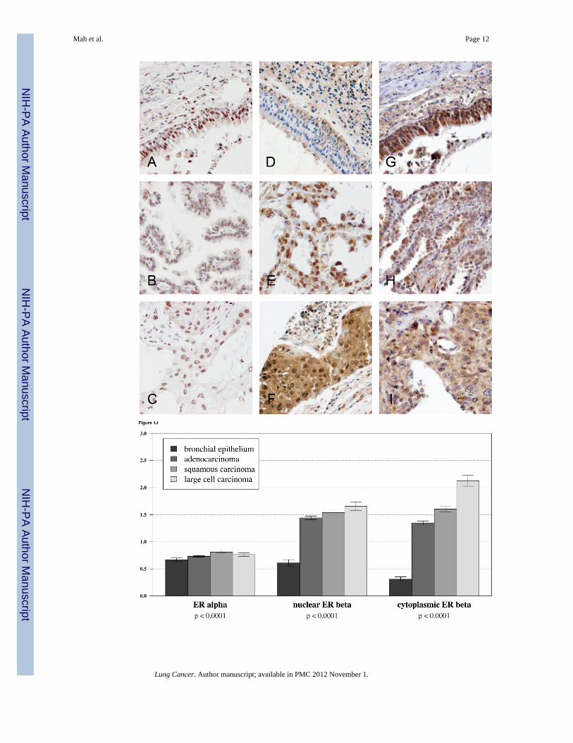
Expression profile of ERα and ERβ in NSCLCWe first considered ERα and ERβ expression separately in lung cancer samples using TMAtechnology. ERα expression in NSCLC was detected primarily in the nucleus and only veryweakly in the cytoplasmic compartment. Representative images are shown in Figures 1A-C.ERβ expression was also observed in both the nuclear and cytoplasmic compartments withrelatively strong cytoplasmic staining in some cases (Figure 1D-F) When we consideredexpression levels based on histologies, we observed that there was only slight difference inERα expression between non-malignant bronchial epithelial cells and any major subclass ofNSCLC (i.e., adenocarcinoma, squamous cell carcinoma, or large cell carcinoma) (Figure1J). However, both nuclear and cytoplasmic ERβ showed a markedly significant increase inexpression in NSCLC compared to bronchial epithelium (Figure 1J). For ERβ higher gradewas also associated with notably higher expression, in contrast to ERa where increase inexpression was much less prominent (Figure 1K).
Neither ERα nor ERβ expression alone predicts survival in patients with NSCLCWe next examined whether the expression levels of ERα or ERβ were predictive of diseasespecific survival. For ERβ we considered both nuclear and cytoplasmic expression. Usingboth univariate and multivariate Cox models, neither ERα nor ERβ alone was a predictor ofsurvival in NSCLC patients (P=0.57 for nuclear ERα; P=0.38 and 0.99 for cytoplasmic ERβand nuclear ERβ, respectively). This was the case with expression both as a continuousvariable or as a dichotomized variable (i.e., low versus high expression). We furtherstratified the population to examine whether ERα or ERβ (cytoplasmic or nuclear) was aprognostic marker in a subset of individuals. However, when we considered gender, stage,histopathology subtype, or smoking status, neither protein predicted outcome in anysubpopulation examined. We further examined whether expressions of these proteins incombination might predict outcome. The combined expression of ERα and ERβ was notpredictive of survival for individuals with NSCLC or any subpopulation that we examined.
ERβ plus aromatase expression predicts survival in NSCLCRecently, we and others observed that the enzyme aromatase was expressed in NSCLC cells[11,23,24,25] and that the expression levels were a powerful predictor of disease-specificdeath in women with NSCLC who were 65 years or older [18]. Aromatase is the finalenzyme in the biosynthesis of estrogens. Here we considered whether the combination ofaromatase plus ER expression would further segment the NSCLC population and prove tobe an even stronger predictor of survival outcome. High versus low expression of allmarkers was initially defined in a non-biased fashion by dichotomizing at the median value.When such an analysis was conducted, ERα added no predictive value compared toaromatase alone (data not shown). However, when we considered ERβ expression, we foundthat the combination of high ERβ and high aromatase predicted a significantly pooreroutcome in all individuals with NSCLC compared to individuals with higher levels of
Mah et al. Page 4
Lung Cancer. Author manuscript; available in PMC 2012 November 1.
NIH
-PA Author Manuscript
NIH
-PA Author Manuscript
NIH
-PA Author Manuscript

aromatase but relatively lower levels of ERβ (Figure 2A; P = 0.029). This observation wassimilar for both cytoplasmic and nuclear expression; however, cytoplasmic ERβ was aconsiderably stronger predictor as a continuous variable by the univariate Cox model (P=0.008; hazard ratio = 1.41). Notably, this observation was even stronger if we consideredpatients with aromatase expression above the 60th percentile (defined as high aromataseexpression) and dichotomized based on ERβ levels (Figure 2B; P=0.0098). Under theseconditions, ERβ also had a strong predictor of outcome as a continuous variable as well (P =0.005, hazard ratio = 1.48; see Supplement, Figure S1 and Table S1).
We further examined whether in individuals with high aromatase ERβ remained anindependent predictor of outcome when compared with additional clinical variables. Indeed,when we considered stage, age, and grade in a multivariate Cox model, of individuals whosetumors had higher aromatase levels, ERβ remained an independent predictor of survival(P=0.007, Table 1).
We further stratified the population by gender to test whether the combination of aromataseplus ERβ was a stronger predictor for women or men. For both men and women, thecombination of higher aromatase expression with elevated ERβ expression predicted a pooroutcome (Figures 3A and 3B; P = 0.019 and 0.030, respectively). However, only for womenwere the markers significant both as a continuous (P = 0.020) and a dichotomized variable.
As highlighted above, aromatase alone was a strong indicator of survival primarily inwomen who were 65 years and older [18]. Therefore, we further assessed men and womenby separating them into groups of those under or over 65. Stratifying the population by bothgender and age groups yielded slightly stronger differences for women 65 years of age orolder, with higher aromatase and ERβ predicting a shorter time course to death due todisease (P=0.003, Figure 3C). Although higher levels of ERβ in women under 65 and men65 and over with higher aromatase were also associated with poorer survival, this differencedid not reach significance in either group. In men under 65 with higher aromataseexpression, ERβ did not affect survival.
In individuals with higher ERβ expression, aromatase levels predict outcomeWe conducted a comparable analysis to the one described above with first dichotomizing ina non-biased fashion (50% percentile) the population by high versus low ERβ expression.Within the population with higher ERβ expression, individuals with lower aromataseexpression had a significantly higher probability of survival than those with higheraromatase levels (Figure 4A; P=0.001). This was similarly the case when aromatase was notdichotomized but considered as continuous variable using the univariate Cox model(P=0.001, hazard ratio = 1.88). Aromatase remained an independent predictor of survivalwhen stage, grade and age were taken into consideration (P=0.0001, Table 2). If the ERβ-high population was stratified by gender, aromatase expression predicted survivaldifferences more strongly in women (Figure 4B; P=0.013) than in men (Figure 4C;P=0.052).
DiscussionA mounting body of evidence from cell culture and mouse models has shown that estrogenand activation of the estrogen receptor pathways are important not only in lungembryogeneis [26,27] but also in lung cancer pathogenesis [9,28,29] Aromatase mediatedconversion of androstenedione to estradiol [30] may also be important for lung cancerprogression [29]. Evidence suggests that the majority of intratumoral estradiol is producedlocally by aromatase in the lung tumors themselves, possibly from circulating androgens[11]. The exact functions of estrogens in lung cancers however are not clear and gender-
Mah et al. Page 5
Lung Cancer. Author manuscript; available in PMC 2012 November 1.
NIH
-PA Author Manuscript
NIH
-PA Author Manuscript
NIH
-PA Author Manuscript

based differences also need to be further elucidated. In NSCLC cells, estradiol (E2) wasshown to increase cell proliferation in both in vitro and in vivo models [9,28,29,31]. Incontrast, agents which blocked estrogen synthesis (such as the aromatase inhibitorsanastrazole, exemestane) or interfered with receptor function (such as the pure antiestrogenfulvestrant) inhibited tumor xenograft proliferation [9,28,32]. Effects of estrogen in lungcancers may be mediated directly by receptor binding followed by nuclear localization andactivation of transcription or indirectly by extranuclear pathways that engage kinasesignaling to modulate transcription and tumor progression. In addition certain oxidativemetabolites of estrogen such as catechol estrogen-3,4-quinones can react with DNA to formdepurinating adducts, possibly leading to mutations that promote cancer initiation [33].
Gender related differences in lung cancer have been well documented and suggest a role forhormonal influences. Of non-smokers with lung cancer, a higher percentage are women[34]. Women have increased susceptibility to lung cancers from tobacco exposure but haveoverall better prognoses in some studies but not others [35,36,37,38]. In an analysis of theSEER database, a survival advantage for older (55-59 years) versus younger (40-49 years)women was seen for squamous cell carcinoma and bronchioloalveolar carcinoma suggestingpostmenopausal status might be advantageous at least for these histologic subtypes [16].Also recent studies from the Women's Health Initiative (WHI) and the Vitamins andLifestyle studies showed a possible increase in incidence and mortality from NSCLC inpost-menopausal women treated with combined estrogen and progesterone hormonereplacement therapy [39,40,41]. A somewhat analogous finding was reduced lung cancermortality in breast cancer patients who had been treated with anti-estrogens[42].
We have set out to profile key elements of the estrogen / ER signaling pathway inindividuals with lung cancer. Previous proteomics results showed that aromatase expressionlevels were a strong predictor of survival in women over 65 years of age with NSCLC.Lower levels of aromatase predicted a significantly longer survival. Here, we havecontinued to assess the estrogen signaling pathway by examining the expression levels andlocalization of ERα and ERβ. While ERβ in contrast to ERα displayed enhanced expressionwith increasing grade, neither ERα nor ERβ alone was predictive for survival in individualswith NSCLC nor any patient subgroup examined (gender, histology, stage, or smokingstatus). However, when we combined ERβ expression with aromatase expression, we foundthat higher levels of both proteins together predicted a significantly poorer survival outcome.While this observation held true for all individuals with NSCLC examined together, it wassomewhat stronger for women of age 65 years and older and weakest in men under 65 yearsof age.
ER Signaling PathwaysOf note, cytoplasmic levels of ERβ were a stronger predictor of outcome than nuclearexpression. Extranuclear estrogen receptor signaling may have considerable importancethrough interactions with tyrosine kinase receptors (EGFR and IGF1-R), MAP kinase and/orPI3/AKT kinase signaling [16,43,44]. In breast cancer cells, recent data suggest proline-glutamic acid-leucine-rich protein-1 (PELP1) couples ERs to signaling pathways such asSrc-MAPK, PI3K-Akt and EGFR-Stat3 [45].
ERs have also been found to cross-communicate with EGFR signaling in NSCLC cells suchthat combined targeting of ER and EGFR enhances antitumor effects [17,28,32,46,47].EGFR mutations are well documented in a subset of individuals with lung cancer; suchmutations are more prevalent in women in East Asian populations, never-smokers andindividuals with adenocarcinoma [48]. Recently, Nose et al. observed that in individualswith EGFR mutations, higher ERβ expression significantly correlated with lung cancer-freesurvival [49]. While we do not currently have the EGFR mutation status in the cohort we
Mah et al. Page 6
Lung Cancer. Author manuscript; available in PMC 2012 November 1.
NIH
-PA Author Manuscript
NIH
-PA Author Manuscript
NIH
-PA Author Manuscript

examined, the interplay between ER and EGFR is certainly an aspect that we are activelyexamining.
The observation that cytoplasmic levels of ERβ were more strongly predictive of survival inthe presence of high aromatase than nuclear ERβ levels could suggest that extranuclearsignaling, likely in concert with kinase signaling pathways, may have important factors inlung cancer progression. Indeed, it was recently reported that extranuclear ER forms arecritical in regulating invasion and metastatic progression in breast cancer [45]. It will beimportant to determine if similar functions are mediated by extranuclear ERs in lung cancer.
Niikawa et al. recently reported measurements of aromatase and estradiol within lungtumors [11]. They found that estradiol levels correlated with aromatase expression and thatmedian estradiol concentrations were significantly higher in NSCLCs than non-neoplasticlung tissues. In addition, they also observed that when tumors were dichotomized based onthe median level of estradiol, the group with both elevated estradiol and ER positivity,tended to have a worse prognosis. Although these earlier results did not quite reachstatistical significance, they indicated a trend consistent with our current report.
ER ImmunohistochemistryPublished data on both the presence and effects of ERα and ERβ on survival in lung canceris somewhat varied [11,12,13,14,15,17,28,32,50,51,52]. This might be due to a lack ofreproducibility in IHC with different anti-ER antibodies and diverse preparative methods[7,53,54] and/or to the relatively lower levels of ER proteins in lung tissue as compared tobreast tissue. Of note, there is currently no standardized IHC assay for the measure of ERs inlung cancer nor for breast cancer [54]. In addition, as is seen for EGFR, ethnic and regionalvariations in the several patient populations may account for different effects of estrogensignaling. Nevertheless, general trends are starting to emerge with overall higher expressionlevels of ERβ in lung cancer compared to ERα and a trend towards the former playing a rolein lung cancer pathobiology and reflecting disease aggressiveness [8,10,11,13,15,16,50]. Asone example, Hershberger et al. reported that ERβ agonists are highly effective in promotingproliferation of lung tumor cells [16]. This contrasts somewhat with what is observed inbreast cancers where ERα tends to predominate over ERβ expression [11,55].
ConclusionThe results presented here are consistent with our overall hypothesis that NSCLC cellshijack the estrogen signaling pathway. We predict that the mechanism of progression formost if not all NSCLCs involves dysfunction at one or more nodes in this pathway. It isinteresting to note that while aromatase was primarily predictive in women 65 years orolder, the combination of aromatase plus ERβ levels was a strong indicator of outcome inboth men and women. This observation could reflect hormonal differences as a function ofage. Estrogen levels in women decline after menopause while levels remain relativelyconstant in men throughout life. In postmenopausal women, the ovaries respond to highergonodotropin levels and therefore contribute significantly to the circulating pool oftestosterone and androgens. This appears to be maintained for at least ten years past theonset of menopause [56]. Levels of androgens fall in women during the reproductive years,then may continue to fall [57] or level off after approximately 65 years of age [58]. Effectsof other interacting proteins such as sex hormone-binding globulin (SHBG) may also play arole in their bioavailability. Androgens are the substrates used by aromatase for estrogensynthesis. As we continue to map branches of the ER signaling pathway with regard toexpression level and activation in men and women with NSCLC, we predict that differentnodes will be preferentially enhanced in different substrata of individuals. Suchcharacterization may provide useful clinical tools to determine disease aggressiveness as
Mah et al. Page 7
Lung Cancer. Author manuscript; available in PMC 2012 November 1.
NIH
-PA Author Manuscript
NIH
-PA Author Manuscript
NIH
-PA Author Manuscript
well as potential targets of therapeutic attack. We continue to explore both of thesepossibilities.
Supplementary MaterialRefer to Web version on PubMed Central for supplementary material.
AcknowledgmentsEarly Detection Research Network NCI CA86366 (L. Goodglick and D. Chia), the National Lung CancerPartnership (L. Goodglick), the Specialized Programs of Research Excellence in Lung Cancer grant P50-CA90388,NCI (R. Pietras and L. Goodglick), the Stiles Program in Oncology (R. Pietras), the Piansky Family Endowment(M.C. Fishbein).
References1. Jemal A, Siegel R, Xu J, Ward E. Cancer statistics, 2010. CA Cancer J Clin. 2010; 60:277–300.
[PubMed: 20610543]2. Tanaka T, Matsuoka M, Sutani A, Gemma A, Maemondo M, et al. Frequency of and variables
associated with the EGFR mutation and its subtypes. Int J Cancer. 20093. Lynch TJ, Bell DW, Sordella R, Gurubhagavatula S, Okimoto RA, et al. Activating mutations in the
epidermal growth factor receptor underlying responsiveness of non-small-cell lung cancer togefitinib. N Engl J Med. 2004; 350:2129–2139. [PubMed: 15118073]
4. Paez JG, Janne PA, Lee JC, Tracy S, Greulich H, et al. EGFR mutations in lung cancer: correlationwith clinical response to gefitinib therapy. Science. 2004; 304:1497–1500. [PubMed: 15118125]
5. Mitsudomi T, Kosaka T, Yatabe Y. Biological and clinical implications of EGFR mutations in lungcancer. Int J Clin Oncol. 2006; 11:190–198. [PubMed: 16850125]
6. Couse JF, Lindzey J, Grandien K, Gustafsson JA, Korach KS. Tissue distribution and quantitativeanalysis of estrogen receptor-alpha (ERalpha) and estrogen receptor-beta (ERbeta) messengerribonucleic acid in the wild-type and ERalpha-knockout mouse. Endocrinology. 1997; 138:4613–4621. [PubMed: 9348186]
7. Raso MG, Behrens C, Herynk MH, Liu S, Prudkin L, et al. Immunohistochemical expression ofestrogen and progesterone receptors identifies a subset of NSCLCs and correlates with EGFRmutation. Clin Cancer Res. 2009; 15:5359–5368. [PubMed: 19706809]
8. Abe K, Miki Y, Ono K, Mori M, Kakinuma H, et al. Highly concordant coexpression of aromataseand estrogen receptor beta in non-small cell lung cancer. Hum Pathol. 2009
9. Stabile LP, Davis AL, Gubish CT, Hopkins TM, Luketich JD, et al. Human non-small cell lungtumors and cells derived from normal lung express both estrogen receptor alpha and beta and showbiological responses to estrogen. Cancer Res. 2002; 62:2141–2150. [PubMed: 11929836]
10. Schwartz AG, Prysak GM, Murphy V, Lonardo F, Pass H, et al. Nuclear estrogen receptor beta inlung cancer: expression and survival differences by sex. Clin Cancer Res. 2005; 11:7280–7287.[PubMed: 16243798]
11. Niikawa H, Suzuki T, Miki Y, Suzuki S, Nagasaki S, et al. Intratumoral estrogens and estrogenreceptors in human non-small cell lung carcinoma. Clin Cancer Res. 2008; 14:4417–4426.[PubMed: 18579664]
12. Wu CT, Chang YL, Shih JY, Lee YC. The significance of estrogen receptor beta in 301 surgicallytreated non-small cell lung cancers. J Thorac Cardiovasc Surg. 2005; 130:979–986. [PubMed:16214508]
13. Kawai H, Ishii A, Washiya K, Konno T, Kon H, et al. Estrogen receptor alpha and beta areprognostic factors in non-small cell lung cancer. Clin Cancer Res. 2005; 11:5084–5089. [PubMed:16033821]
14. Ali G, Donati V, Loggini B, Servadio A, Dell'Omodarme M, et al. Different estrogen receptor betaexpression in distinct histologic subtypes of lung adenocarcinoma. Hum Pathol. 2008; 39:1465–1473. [PubMed: 18620727]
Mah et al. Page 8
Lung Cancer. Author manuscript; available in PMC 2012 November 1.
NIH
-PA Author Manuscript
NIH
-PA Author Manuscript
NIH
-PA Author Manuscript

15. Skov BG, Fischer BM, Pappot H. Oestrogen receptor beta over expression in males with non-smallcell lung cancer is associated with better survival. Lung Cancer. 2008; 59:88–94. [PubMed:17905466]
16. Hershberger PA, Stabile LP, Kanterewicz B, Rothstein ME, Gubish CT, et al. Estrogen receptorbeta (ERbeta) subtype-specific ligands increase transcription, p44/p42 mitogen activated proteinkinase (MAPK) activation and growth in human non-small cell lung cancer cells. J SteroidBiochem Mol Biol. 2009; 116:102–109. [PubMed: 19460433]
17. Stabile LP, Lyker JS, Gubish CT, Zhang W, Grandis JR, et al. Combined targeting of the estrogenreceptor and the epidermal growth factor receptor in non-small cell lung cancer shows enhancedantiproliferative effects. Cancer Res. 2005; 65:1459–1470. [PubMed: 15735034]
18. Mah V, Seligson DB, Li A, Marquez DC, Wistuba, et al. Aromatase expression predicts survival inwomen with early-stage non small cell lung cancer. Cancer Res. 2007; 67:10484–10490.[PubMed: 17974992]
19. Yager JD, Davidson NE. Estrogen carcinogenesis in breast cancer. N Engl J Med. 2006; 354:270–282. [PubMed: 16421368]
20. Kononen J, Bubendorf L, Kallioniemi A, Barlund M, Schraml P, et al. Tissue microarrays for high-throughput molecular profiling of tumor specimens. Nat Med. 1998; 4:844–847. [PubMed:9662379]
21. Seligson DB, Horvath S, Shi T, Yu H, Tze S, et al. Global histone modification patterns predictrisk of prostate cancer recurrence. Nature. 2005; 435:1262–1266. [PubMed: 15988529]
22. Seligson D, Horvath S, Huerta-Yepez S, Hanna S, Garban H, et al. Expression of transcriptionfactor Yin Yang 1 in prostate cancer. Int J Oncol. 2005; 27:131–141. [PubMed: 15942652]
23. Weinberg OK, Marquez-Garban DC, Fishbein MC, Goodglick L, Garban HJ, et al. Aromataseinhibitors in human lung cancer therapy. Cancer Res. 2005; 65:11287–11291. [PubMed:16357134]
24. Marquez-Garban DC, Chen HW, Goodglick L, Fishbein MC, Pietras RJ. Targeting aromatase andestrogen signaling in human non-small cell lung cancer. Ann N Y Acad Sci. 2009; 1155:194–205.[PubMed: 19250205]
25. Oyama T, Kagawa N, Sugio K, Uramoto H, Hatano O, et al. Expression of aromatase CYP19 andits relationship with parameters in NSCLC. Front Biosci. 2009; 14:2285–2292. [PubMed:19273201]
26. Patrone C, Cassel TN, Pettersson K, Piao YS, Cheng G, et al. Regulation of postnatal lungdevelopment and homeostasis by estrogen receptor beta. Mol Cell Biol. 2003; 23:8542–8552.[PubMed: 14612399]
27. Morani A, Barros RP, Imamov O, Hultenby K, Arner A, et al. Lung dysfunction causes systemichypoxia in estrogen receptor beta knockout (ERbeta-/-) mice. Proc Natl Acad Sci U S A. 2006;103:7165–7169. [PubMed: 16636272]
28. Pietras RJ, Marquez DC, Chen HW, Tsai E, Weinberg O, et al. Estrogen and growth factorreceptor interactions in human breast and non-small cell lung cancer cells. Steroids. 2005; 70:372–381. [PubMed: 15862820]
29. Hammoud Z, Tan B, Badve S, Bigsby RM. Estrogen promotes tumor progression in a geneticallydefined mouse model of lung adenocarcinoma. Endocr Relat Cancer. 2008; 15:475–483. [PubMed:18509000]
30. Simpson ER, Mahendroo MS, Means GD, Kilgore MW, Hinshelwood MM, et al. Aromatasecytochrome P450, the enzyme responsible for estrogen biosynthesis. Endocr Rev. 1994; 15:342–355. [PubMed: 8076586]
31. Hershberger PA, Vasquez AC, Kanterewicz B, Land S, Siegfried JM, et al. Regulation ofendogenous gene expression in human non-small cell lung cancer cells by estrogen receptorligands. Cancer Res. 2005; 65:1598–1605. [PubMed: 15735050]
32. Marquez-Garban DC, Chen HW, Fishbein MC, Goodglick L, Pietras RJ. Estrogen receptorsignaling pathways in human non-small cell lung cancer. Steroids. 2007; 72:135–143. [PubMed:17276470]
Mah et al. Page 9
Lung Cancer. Author manuscript; available in PMC 2012 November 1.
NIH
-PA Author Manuscript
NIH
-PA Author Manuscript
NIH
-PA Author Manuscript

33. Cavalieri E, Chakravarti D, Guttenplan J, Hart E, Ingle J, et al. Catechol estrogen quinones asinitiators of breast and other human cancers: implications for biomarkers of susceptibility andcancer prevention. Biochim Biophys Acta. 2006; 1766:63–78. [PubMed: 16675129]
34. Patel JD. Lung cancer in women. J Clin Oncol. 2005; 23:3212–3218. [PubMed: 15886308]35. Henschke CI, Miettinen OS. Women's susceptibility to tobacco carcinogens. Lung Cancer. 2004;
43:1–5. [PubMed: 14698531]36. Zang EA, Wynder EL. Differences in lung cancer risk between men and women: examination of
the evidence. J Natl Cancer Inst. 1996; 88:183–192. [PubMed: 8632492]37. Bach PB, Kattan MW, Thornquist MD, Kris MG, Tate RC, et al. Variations in lung cancer risk
among smokers. J Natl Cancer Inst. 2003; 95:470–478. [PubMed: 12644540]38. Bain C, Feskanich D, Speizer FE, Thun M, Hertzmark E, et al. Lung cancer rates in men and
women with comparable histories of smoking. J Natl Cancer Inst. 2004; 96:826–834. [PubMed:15173266]
39. Slatore CG, Chien JW, Au DH, Satia JA, White E. Lung cancer and hormone replacement therapy:association in the vitamins and lifestyle study. J Clin Oncol. 2010; 28:1540–1546. [PubMed:20159813]
40. Chlebowski RT, Anderson GL, Manson JE, Schwartz AG, Wakelee H, et al. Lung cancer amongpostmenopausal women treated with estrogen alone in the women's health initiative randomizedtrial. J Natl Cancer Inst. 2010; 102:1413–1421. [PubMed: 20709992]
41. Chlebowski RT, Schwartz AG, Wakelee H, Anderson GL, Stefanick ML, et al. Oestrogen plusprogestin and lung cancer in postmenopausal women (Women's Health Initiative trial): a post-hocanalysis of a randomised controlled trial. Lancet. 2009; 374:1243–1251. [PubMed: 19767090]
42. Rapiti, E.; Verkooijen, HM.; Fioretta, G.; Schubert, H.; Vinh-Hung, V., et al. Reduced LungCancer Mortality Risk among Breast Cancer Patients Treated with Anti-Estrogens. 2009.
43. Madak-Erdogan Z, Kieser KJ, Kim SH, Komm B, Katzenellenbogen JA, et al. Nuclear andextranuclear pathway inputs in the regulation of global gene expression by estrogen receptors. MolEndocrinol. 2008; 22:2116–2127. [PubMed: 18617595]
44. Fox EM, Andrade J, Shupnik MA. Novel actions of estrogen to promote proliferation: integrationof cytoplasmic and nuclear pathways. Steroids. 2009; 74:622–627. [PubMed: 18996136]
45. Chakravarty D, Nair SS, Santhamma B, Nair BC, Wang L, et al. Extranuclear functions of ERimpact invasive migration and metastasis by breast cancer cells. Cancer Res. 2010; 70:4092–4101.[PubMed: 20460518]
46. Pietras RJ, Marquez-Garban DC. Membrane-associated estrogen receptor signaling pathways inhuman cancers. Clin Cancer Res. 2007; 13:4672–4676. [PubMed: 17699844]
47. Liu M, Yang SC, Sharma S, Luo J, Cui X, et al. EGFR signaling is required for TGF-beta 1mediated COX-2 induction in human bronchial epithelial cells. Am J Respir Cell Mol Biol. 2007;37:578–588. [PubMed: 17600311]
48. Bell DW, Brannigan BW, Matsuo K, Finkelstein DM, Sordella R, et al. Increased prevalence ofEGFR-mutant lung cancer in women and in East Asian populations: analysis of estrogen-relatedpolymorphisms. Clin Cancer Res. 2008; 14:4079–4084. [PubMed: 18593984]
49. Nose N, Sugio K, Oyama T, Nozoe T, Uramoto H, et al. Association between estrogen receptor-beta expression and epidermal growth factor receptor mutation in the postoperative prognosis ofadenocarcinoma of the lung. J Clin Oncol. 2009; 27:411–417. [PubMed: 19064969]
50. Fasco MJ, Hurteau GJ, Spivack SD. Gender-dependent expression of alpha and beta estrogenreceptors in human nontumor and tumor lung tissue. Mol Cell Endocrinol. 2002; 188:125–140.[PubMed: 11911952]
51. Mollerup S, Jorgensen K, Berge G, Haugen A. Expression of estrogen receptors alpha and beta inhuman lung tissue and cell lines. Lung Cancer. 2002; 37:153–159. [PubMed: 12140138]
52. Radzikowska E, Langfort R, Giedronowicz D. Estrogen and progesterone receptors in non smallcell lung cancer patients. Ann Thorac Cardiovasc Surg. 2002; 8:69–73. [PubMed: 12027790]
53. Carder PJ, Murphy CE, Dervan P, Kennedy M, McCann A, et al. A multi-centre investigationtowards reaching a consensus on the immunohistochemical detection of ERbeta in archivalformalin-fixed paraffin embedded human breast tissue. Breast Cancer Res Treat. 2005; 92:287–293. [PubMed: 16155800]
Mah et al. Page 10
Lung Cancer. Author manuscript; available in PMC 2012 November 1.
NIH
-PA Author Manuscript
NIH
-PA Author Manuscript
NIH
-PA Author Manuscript

54. Speirs V, Green CA, Shaaban AM. Oestrogen receptor beta immunohistochemistry: time to get itright? J Clin Pathol. 2008; 61:1150–1151. author reply 1151-1152. [PubMed: 18820109]
55. Shaaban AM, Green AR, Karthik S, Alizadeh Y, Hughes TA, et al. Nuclear and cytoplasmicexpression of ERbeta1, ERbeta2, and ERbeta5 identifies distinct prognostic outcome for breastcancer patients. Clin Cancer Res. 2008; 14:5228–5235. [PubMed: 18698041]
56. Fogle RH, Stanczyk FZ, Zhang X, Paulson RJ. Ovarian androgen production in postmenopausalwomen. J Clin Endocrinol Metab. 2007; 92:3040–3043. [PubMed: 17519304]
57. Cappola AR, Ratcliffe SJ, Bhasin S, Blackman MR, Cauley J, et al. Determinants of serum totaland free testosterone levels in women over the age of 65 years. J Clin Endocrinol Metab. 2007;92:509–516. [PubMed: 17090636]
58. Davison SL, Bell R, Donath S, Montalto JG, Davis SR. Androgen levels in adult females: changeswith age, menopause, and oophorectomy. J Clin Endocrinol Metab. 2005; 90:3847–3853.[PubMed: 15827095]
Abbreviations
NSCLC non-small cell lung cancer
ER estrogen receptor
IHC immunohistochemistry
TMA tissue microarray
DAB diaminobenzidine
Mah et al. Page 11
Lung Cancer. Author manuscript; available in PMC 2012 November 1.
NIH
-PA Author Manuscript
NIH
-PA Author Manuscript
NIH
-PA Author Manuscript
Mah et al. Page 12
Lung Cancer. Author manuscript; available in PMC 2012 November 1.
NIH
-PA Author Manuscript
NIH
-PA Author Manuscript
NIH
-PA Author Manuscript

Figure 1.A-C: representative staining of ERα in bronchial epithelium, adenocarcinoma and squamouscarcinoma respectively; D-F: representative staining of ERβ in bronchial epithelium,adenocarcinoma and squamous carcinoma; G-I: representative staining of aromatase inbronchial epithelium, adenocarcinoma and squamous carcinoma; J: barplots of ERα andERβ in different tumor histologies; K: barplots of ERα and ERβ in different tumor gradesshow a significant increase in cytoplasmic levels of ERβ with increase in grade.
Mah et al. Page 13
Lung Cancer. Author manuscript; available in PMC 2012 November 1.
NIH
-PA Author Manuscript
NIH
-PA Author Manuscript
NIH
-PA Author Manuscript

Mah et al. Page 14
Lung Cancer. Author manuscript; available in PMC 2012 November 1.
NIH
-PA Author Manuscript
NIH
-PA Author Manuscript
NIH
-PA Author Manuscript

Fig 2.A: For patients with high aromatase expression (determined by staining intensity abovemedian levels) splitting cytoplasmic ERβ expression at the median level, the Kaplan-Meiersurvival curve shows significantly worse survival in those patients with high ERβ (hazardratio = 1.6, p-value = 0.029); B: Using above the 60th percentile as a cutoff to define higharomatase expression, low cytoplasmic ERβ (again median expression level) conferred aslightly better prognosis (p=0.0098, hazard ratio = 1.81).
Mah et al. Page 15
Lung Cancer. Author manuscript; available in PMC 2012 November 1.
NIH
-PA Author Manuscript
NIH
-PA Author Manuscript
NIH
-PA Author Manuscript

Mah et al. Page 16
Lung Cancer. Author manuscript; available in PMC 2012 November 1.
NIH
-PA Author Manuscript
NIH
-PA Author Manuscript
NIH
-PA Author Manuscript

Mah et al. Page 17
Lung Cancer. Author manuscript; available in PMC 2012 November 1.
NIH
-PA Author Manuscript
NIH
-PA Author Manuscript
NIH
-PA Author Manuscript

Fig 3.A: For women with high aromatase expression (staining intensity above the 60th percentilesplitting cytoplasmic ERβ expression at the midpoint), the Kaplan-Meier survival curveshows worse survival in those patients with high ERβ (hazard ratio = 2.18, p-value = 0.019);B: For men, findings were similar but slightly weaker (hazard ratio = 2.04, P = 0.030). C: Inwomen 65 and over, the findings were stronger than other population subgroups (p=0.003,hazard ratio = 3.25).
Mah et al. Page 18
Lung Cancer. Author manuscript; available in PMC 2012 November 1.
NIH
-PA Author Manuscript
NIH
-PA Author Manuscript
NIH
-PA Author Manuscript

Mah et al. Page 19
Lung Cancer. Author manuscript; available in PMC 2012 November 1.
NIH
-PA Author Manuscript
NIH
-PA Author Manuscript
NIH
-PA Author Manuscript

Mah et al. Page 20
Lung Cancer. Author manuscript; available in PMC 2012 November 1.
NIH
-PA Author Manuscript
NIH
-PA Author Manuscript
NIH
-PA Author Manuscript

Fig 4.A: For patients with high cytoplasmic ERβ expression (determined by staining intensityabove median levels) splitting aromatase expression at the median level, the Kaplan-Meiersurvival curve shows significantly worse survival in those patients with aromatase (hazardratio = 1.47, p-value = 0.001); B: For women with high cytoplasmic ERβ expression, againthe Kaplan-Meier survival curve again shows worse survival in those patients with higheraromatase (hazard ratio = 1.49, p-value = 0.013); C: For men, findings were similar butslightly weaker (hazard ratio = 1.38, P = 0.052).
Mah et al. Page 21
Lung Cancer. Author manuscript; available in PMC 2012 November 1.
NIH
-PA Author Manuscript
NIH
-PA Author Manuscript
NIH
-PA Author Manuscript

NIH
-PA Author Manuscript
NIH
-PA Author Manuscript
NIH
-PA Author Manuscript
Mah et al. Page 22
Table 1Multivariate Cox proportional hazards model for all patients with high aromatase[defined by higher than median levels], (n=190)
Variable Hazard Ratio(95% confidence interval)
P-value
Cytoplasmic ERβ mean intensity 1.48 (1.11 - 1.96) 0.0073
Stage 2.32 (1.88 - 2.87) <0.0001
Grade 0.93 (0.72 - 1.38) 0.1200
Age 1.05 (1.02 - 1.07) 0.0003
Lung Cancer. Author manuscript; available in PMC 2012 November 1.

NIH
-PA Author Manuscript
NIH
-PA Author Manuscript
NIH
-PA Author Manuscript
Mah et al. Page 23
Table 2Multivariate Cox proportional hazards model for all patients with high cytoplasmic ERβ[defined by higher than median levels], (n=189)
Variable Hazard Ratio(95% confidence interval)
P-value
Aromatase mean intensity 2.17 (1.46 - 3.23) 0.0001
Stage 2.13 (1.69 - 2.68) <0.0001
Grade 1.08 (0.81 - 1.45) 0.5767
Age 1.05 (1.02 - 1.08) 0.0005
Lung Cancer. Author manuscript; available in PMC 2012 November 1.
![In conjunction with Venus [planetary radar astronomy]](https://static.fdokumen.com/doc/165x107/631a4f09bb40f9952b01f2bc/in-conjunction-with-venus-planetary-radar-astronomy.jpg)