Conformational and Spectroscopic Behaviors of 2,4-xylyl isothiocyanate
Upload
independentCategory
view
1download
0
Pergamon ft. Steroid Biochem. Molec. Biol. Vol. 54, No. 5/6, pp. 201-210, 1995
Copyright © 1995 Elsevier Science Ltd 0 9 6 0 - 0 7 6 0 ( 9 5 ) 0 0 1 3 7 - 9 Printed in Great Britain. All rights reserved
0960-0760/95 $9.50 + 0.00
Relationship between Estrogen Structure and Conformational Changes in Estrogen
Receptor/DNA Complexes J. K. C h r i s t m a n , 1. S. N e h l s , 1 L. P o l i n 2 a n d S. C. B r o o k s 2 t
1Molecular Biology Program, Michigan Cancer Foundation and 2Department of Biochemistry, Wayne State University, School of Medicine, 540 Canfield, Detroit, MI 48201, U.S.A.
The effect of es t rogen s t ruc tu re on the con fo rma t ion of the complex f o r m e d with es t rogen receptor (ER) and the consensus es t rogen response e lement (EREc) has been examined with gel mobi l i ty shift assay. Pro te ins in MCF-7 cell extracts f o r m e d three dis t inct complexes with ERE. Only the slowest moving complex conta ined ER as ind ica ted by binding with an t i -ER ant ibodies H222 and D547. This ER-ERE complex dkisplayed i n c r e a s e d e lec t rophoret ic mobi l i ty when f o r m e d in the presence of es t radiol (E2) and bound radio labe led 16at-iodoestradiol. The ant ies t rogen ICI 164,384 decreased the mobi l i ty of the ER-ERE complex and blocked the effect of E 2. The resul ts r epor ted here indicate tha t the posi t ion and locat ion of hydroxyl groups on the es t ra t r iene nucleus is an i m p o r t a n t fac tor in de t e rmin ing the mobi l i ty of E R - E R E c (or a va r ian t ERE) in gel shift assays. The abi l i ty of E 2 analogs to cause con fo rma t iona l changes detectable as a l te red mobi l i ty was not d i rect ly re la ted ei ther to the i r b inding aff ini ty for ER or to the i r abil i ty to act ivate E 2 responsive genes. Al though several d ihydroxyes t rogens (estradiol-16~t, 1- and 2-hydroxyes t ra t r ien-17p-ol ) caused an increase in the mobi l i ty of the ER-ERE c, o ther l igands (estradiol-17~t, 4-hydroxyestratr iene-17fl-ol , 3-hydroxy es t ra t r iene , estratr ien-17fl-ol and 5-androsten-3fl, 1718-diol) with a capaci ty for act ivat ing at least some E2 responsive genes in MCF-7 cells had little or no effect. On the basis of these and previously publ ished results , it can be concluded tha t specific s t ruc tu re fea tures of estrogens are responsible for con fo rma t iona l changes of E R - E R E complexes detectable in gel-shift assays. F u r t h e r m o r e , the ident i f ied s t ruc tu ra l charac ter i s t ics of the l igand which are r equ i red for gel-shift are not the same as those previously repor ted to be essential for s t imula t ion of t r ansc r ip t iona l act ivi ty of ER.
J. Steroid Biochem. Molec. Biol., Vol. 54, No. 5/6, pp. 201-210, 1995
INTRODUCTION
Steroid hormones regulate gene transcription via re- ceptor binding [1]. Although the hormone-receptor complex is required for transactivation, the mech- anisms governing this phenomenon are not fully understood. Available information indicates that an important aspect of activation resides in conformational changes induced in the receptor by ligand binding [2].
Recently, Allan et al. have distinguished between the conformational changes necessary for DNA binding of the steroid-receptor complex and those directly related
*Present address: Depar tment of Biochemistry and Molecular Biology, Univers i ty of Nebraska Medical Center, Omaha, NE 68198-4525, U.S.A.
tCor re spondence to S. C. ]]rooks. Received 23 Feb. 1995; accepted 6 Apr. 1995.
to the creation of a transcriptionaly active form [3]. Ligand-free receptor is able to form a complex with the ERE in the absence of Mg z+ that is stable to electro- phoresis through native polyacrylamide gels [4]. How- ever, in presence of estradiol-17fl (Ez), this complex takes on a conformation that moves more rapidly in gels than the ligand-free receptor-ERE complex. It has been postulated that the conformational change in- duced in the estrogen receptor (ER) by Ez binding results in activation of the transactivation function of the ER [2, 4, 5]. A similar alteration in the ER-ERE complex is brought about when the non-steroid estro- gen DES is the ligand [4, 6]. However, in the presence of antiestrogenic ligands, the mobility of the ER-ERE complex is actually decreased (ICI 164,384, [6]; tamox- ifen, [7]). These findings indicate that even though antiestrogens do not interfere with the binding of ER
201

202 J .K. Christman et al.
to the ERE, they have markedly different effects on the conformation of the ER-ERE complex than estro- genic ligands. This suggests a relationship between conformation and the capacity of the ligand-ER-ERE complex to activate transcription [2, 4, 6].
In the studies reported here, we have assessed the effect of the ligand's structure on the mobility of ER-ERE complexes in native polyacrylamide gels and related this effect to our previously published results defining the capacity of these estrogen analogs to stimulate a series of estrogen responsive endogenous and transfected genes [8-10].
MATERIALS AND METHODS
Steroids and antibodies
The estrogens used in these investigations were either purchased (estratriene, estrone, estradiol-16a, estradiol-17a, estriol and E2) from Research Plus, Inc. (Bayonne, NJ) or synthesized in this laboratory. The A-ring isomers of E2 (1-, 2-, and 4-hydroxyestratriene- 17fl-ol) were synthesized according to published pro- cedures [11]. Synthesis of rnonohydroxyestrogens (3-hydroxyestratriene and estratriene-17fl-ol) has also been reported [12, 13]. Each estrogen analog was purified by thin layer chromatography and crys- tallization. The level of contaminants in each estro- gen was shown to be less than 1 part in 10,000 [8]. 5-Androstene-3fl,17fl-diol was purchased from Aldrich Chemical Co. (Milwaukee, WI) and purified as described by VanderKuur et al. [8]. The anti- estrogen 4-hydroxytamoxifen was a gift from Stuart Pharmaceutical (Division of ICI Americas, Inc., Wilmington, DE) and ICI 164,384 was kindly sup- plied by D.A.E. Wakeling, Imperial Chemical Industries (Alderly Park, England). 3,17fl-Estradiol- 16~-[12sI]iodo (2200 Ci/mmol) was obtained from Dupont NEN (Wilmington, DE).
Monoclonal antibodies directed against ER, H222 and D547, were gifts from Abbott Diagnostics Division (Abbott Laboratories, Abbott Park, IL). The antibody to P53 (Ab-I) was obtained from Oncogene Science (Uniondale, NY).
Cell culture
MCF-7 human breast cancer cells (subclone E3, [14]) were maintained at 37°C in phenol red-free, HEPES buffered Eagles modified MEM supplemented with 0.05/~g/ml gentamicin sulfate and 5% donor calf serum. Cells were plated in 75 cm 2 tissue culture flasks as described previously [15] and routinely passaged prior to reaching confluency. All experiments utilized cells derived from passages 168-197.
ER extraction
MCF-7 cells were grown to near confluence (~ 1.8 x 10⁷ cells/75 cm 2 flask). Cells were harvested by removing growth medium and then disrupting the
monolayer with a stream of warm (37°C) culture medium rapidly expelled through a 5"-long, 14-gauge cannula attached to a 25 ml syringe. Suspensions of single cells were obtained by repeated (10-15 strokes) aspiration and expulsion through the cannula. All subsequent operations were carried out at 4°C. Cells were collected by centrifugation at 800g and sus- pended in 2ml of TED buffer [10mM Tris-HC1, pH 7.4; 1.5 mM ethylene-diaminetetracetic acid (EDTA); 1 mM dithiothreitol (DTT), 5/~g/ml each of antipain dihydrochloride, leupeptin, chymostatin and pepstatin (Boehringer Mannheim, NY)]. The sus- pended cells were transferred to a 1 ml glass homogen- ization tube which was centrifuged at 600 g for 3 min. The pelleted cells were suspended in an equal volume of TED and lysed with 20 strokes of a teflon pestle. The homogenate was spun at 100,000 g for I h. The super- natant (cytosol), which contained a range of 3-5 pmol/ml of functional ER as determined by the dextran coated-charcoal binding assay [11] was stored at 4°C for use within 6 h.
Preparation of radiolabeled ERE
Complementary oligodeoxyribonucleotide strands containing a consensus ERE (GATCCAGGT- CACAGTGACCTGGGCCCG-27 bp) in 0.4 M Tris (pH 7.5) were annealed by heating to 90°C for 10 min, followed by slow cooling (70°C, 1 h; 60°C, 1 h; 50°C, 0.5 h; 37°C, 0.5 h). For end labeling with 32p, 900 ng of annealed ERE was incubated for 30 min at 37°C with 50 U T4 polynucleotide kinase and 100 #Ci 732p-ATP (sp. act. 3000 Ci/mmol) in a final volume of 50/~1 reaction buffer (buffer and enzyme were supplied by New England Biolabs). After stopping the reaction by addition of 2 #1 0.5 M EDTA, pH 8.0, radiolabeled DNA was separated from unreacted ATP by filtration through Sephadex G-50 equilibrated with TE buffer (10 mM Tris-HC1, pH 7.4; 1.5 mM EDTA). Radio- labeled DNA was precipitated with ethanol, collected by centrifugation, and resuspended in 200/~1 TE.
Complex formation
Conditions for optimal complex formation between ERE and ER in MCF-7 cytosol were determined empirically. To maintain the highest possible concen- tration of ER, reaction components were added directly to the cytosol at concentrations which did not increase its volume by more than 10%. In the standard reaction, 1.25 #g poly dI-dC, 1.8 ng (,,~ 105 cpm) 32P-labeled ERE and the indicated concentrations of estrogens were added to 25 pl or ER extract, mixed and incu- bated at 4°C for 18 h. All stock solutions of estrogens were prepared in ethanol at concentrations that would not increase the volume of the reaction to > 27.5 #1 or the concentration of ethanol in the reaction to >2%. Control reactions without estrogens contained an equivalent amount of ethanol.
Es t rogen R e c e p t o r - E R E C o m p l e x e s 203
b
¢
a
1 2 3 4 5 6 7 8 9 10 11 12 Fig. 1. Effect of E 2 and a n t i e s t r o g e n (ICI 164,384) on the mobi l i ty of MCF-7 pro te in -ERE~ complexes in a gel sh i f t assay. E x t r a c t s of MCF-7 cells were i n c u b a t e d with 32p-labeled EREc, E2 a n d / o r ICI 164,384 for 18 h at 4°C. The n u m b e r and mob i l i t y of complexes f o r m e d was ident ica l when ODNs were only added d u r i n g the final 60 m i n of incuLbation. 32P-radiolabeled ~X174 HaeIII was loaded in lanes 1, 7 and 11. Add i t ions to b ind ing r eac t ions were: 5 >: 10-7M E 2 (lane 2), 5 x 10 -aM E2 (lane 3), 5 x 10⁻⁹M E 2 ( lane 4), 5 x 10⁻¹°M E 2 ( lane 5), e thano l ( lane 6), 5 x 10 -6 M ICI 164,384 (lane 8), 2.5 x 10 -6 M ICI 164,384 + 5 x 10 -s M E 2 ( lane 9), 2.5 x 10 - 6 M ICI 164,384 + 5 x 10⁻⁹M E2 (lane 10), 5 x 10 -aM E 2 ( lane 12). P r o t e i n - E R E complexes a re ind ica ted (a, b a nd c). High levels of E 2 (> 10 -1° M) are shown to inc rease the in t ens i ty of complex -a . This o c c u r r e d in only 25%
of the gels r ega rd l e s s of the l igand (see o ther f igures) .
A
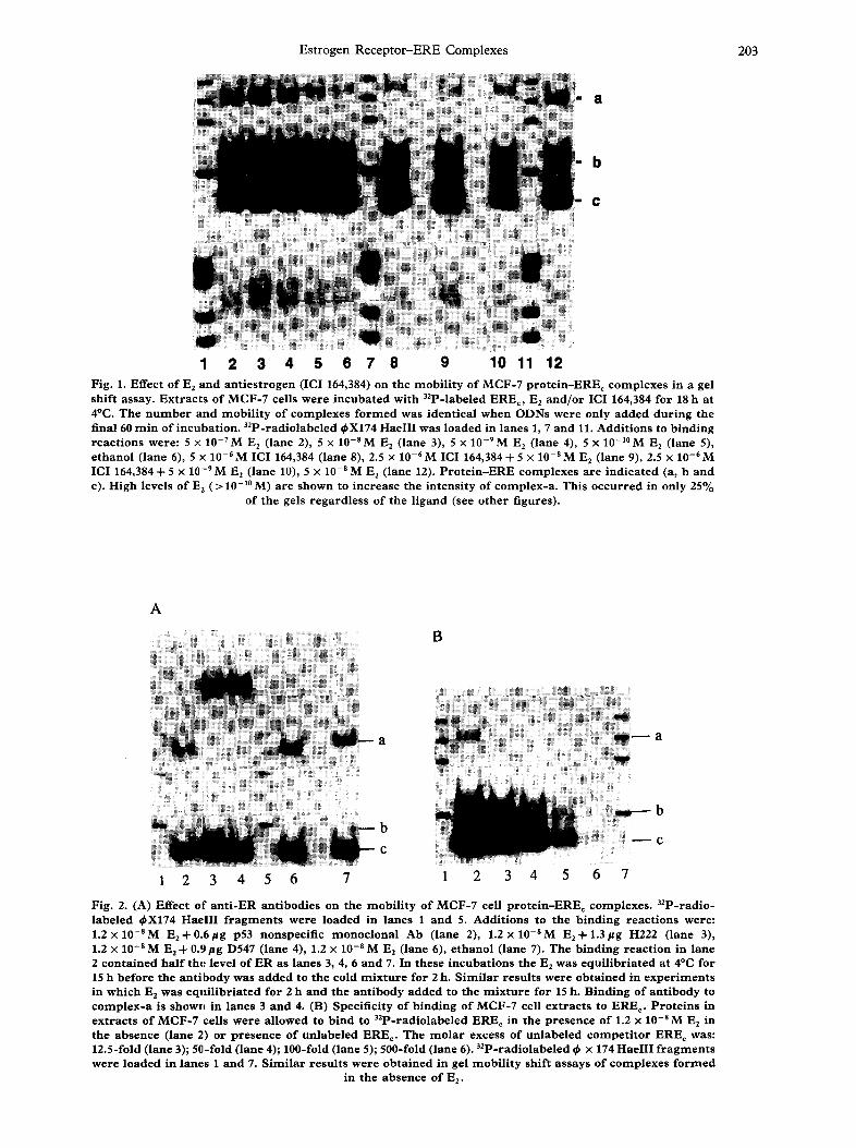
B
a
I
b
1 2 3 4 5 6 7 1 2 3 4 5
r b
~ C
6 7
Fig. 2. (A) Effect o f a n t i - E R an t ibodies on the mob i l i t y o f MCF-7 cell p r o t e i n - E R E c complexes . 32P-radio- labeled 0X174 HaeIII f r a g m e n t s were loaded in lanes 1 a nd 5. Addi t ions to the b ind ing r eac t ions were: 1.2 x 10-SM E 2 + 0 . 6 p g p53 nonspecif ic m o n o c l o n a l Ab (lane 2), 1.2 x 10-SM E2+ 1.3/tg H222 (lane 3), 1.2 x 10 -8 M E 2 + 0.9/lg D547 (lane 4), 1.2 x 10 -8 M E 2 ( lane 6), e thano l ( lane 7). The b ind ing r eac t ion in lane 2 con ta ined h a l f the level of ER as l anes 3, 4, 6 and 7. In these i nc uba t i ons the Ez was equ i l i b r i a t ed at 4°C for 15 h before the an t i body was added to the cold m i x t u r e for 2 h. S i m i l a r r e su l t s were ob ta ined in e x p e r i m e n t s in which E 2 was equ i l i b r i a t ed for 2 h and the an t i body a dde d to the m i x t u r e for 15 h. B ind ing of a n t i body to c o m p l e x - a is shown in lanes 3 a n d 4. (B) Specifici ty of b ind ing of MCF-7 cell ex t r ac t s to ERE c. P r o t e i n s in ex t r ac t s of MCF-7 cells were al lowed to b ind to 32P-radiolabeled EREc in the p re sence of 1.2 x 10 -8 M E z in the absence ( lane 2) or p r e sence of un l abe l ed EREc. The m o l a r excess of un l abe l ed c o m p e t i t o r EREc was: 12.5-fold ( lane 3); 50-fold ( lane 4); 100-fold ( lane 5); 500-fold ( lane 6). 32p-radiolabeled 0 x 174 HaeIII f r a g m e n t s were loaded in lanes 1 and 7. S i m i l a r r e su l t s were ob ta ined in gel mob i l i t y sh i f t a s says of complexes f o r m e d
in the absence of Ee.

204 J.K. Christman et al.
Gel shift assays
Native polyacrylamide gels (4% polyacrylamide, 14 x 16cm x 1.5mm) were prepared and pre-run (running buffer; 6.7 mM Tris, pH 8.0, 3.3 mM sodium acetate, pH5.2, 1raM EDTA) as described by Carthew et al. [16]. Immediately before loading, 6 #1 samples of each reaction mixture were brought to a final volume of 20/~1 with a concentration of 5mM Tris-HC1, pH 7.4; 0.5 mM D T T ; 100 mM KC1 and 1.5mM EDTA, 5% v/v glycerol. 32P-radiolabeled q~X174 HaeIII markers were diluted in the same manner for use as internal standards for measuring migration distances. Following electrophoresis for 3 h at 15°C with a current of 25 mA, the gels were placed on Whatman 3 mm paper, covered with plastic wrap and dried at 80°C under vacuum for 1 h. Autoradio- graphs were prepared by exposing Kodak X-OMAT AR film to the dried gel at - 70°C with Dupont Cronex Intensifying Screens.
R E S U L T S
Identification of E R - E R E complexes formed in the presence and absence of E 2
Whole cell extract of MCF-7 cells, prepared as described (Materials and Methods), contains a variety of proteins capable of binding to double-stranded oligodeoxyribonucleotides (ODNs) containing an ERE, even in the presence of a 700-fold excess (w/w) of non-specific competitor DNA (poly dI-dC). The three most abundant complexes formed with the con- census ERE (EREc) are readily separated by electro- phoresis in native 4% polyacrylamide gels (Fig. 1). Only the slowest moving of these complexes (complex- a), which has an electrophoretic mobility midway be- tween that of ds-DNA marker fragments 1353 and 1078 bp, displayed altered mobility when formed in the presence of E 2. At 10-~°M E2, complex-a migrated
A B m
e
1 2 1 2 3 4 5 6 Fig. 3. B ind ing of 16a[125I]iodo-3,17~-estradiol to MCF-7 cell ex t rac t p ro te in -ERE¢ complexes . (A) Un labe l ed ERE c (3.6 ng) i nc uba t e d wi th 50 ~1 ex t rac t con ta in ing 0.13 p m o l ER and 0 .14pmol 16a[12sI]iodo-3,17iS-estradiol (2200Ci /mmol ) . The rad io labe led 16a- iodoE2-ER-ERE c c omple x is shown in lane 1. The th ree h ighes t m o l e c u l a r weight ~bX174 HaeIII f rag- m e n t s can be seen in lane 2. (B) Al iquots of the s a m e MCF-7 cell ex t rac t con ta in ing 0.13 p m o l of ER a nd 3.5 ng 32P-radio- labeled ERE¢ i n c u b a t e d in the p resence of 1.2 x 10⁻⁸M E~ ( lanes 1 a nd 2) or in the absence of E 2 ( lanes 4 and 5). 32p-radiolabeled ~X174 HaeIII f r a g m e n t s were loaded in lanes 3 and 6. All deta i ls as in Fig. I. Only c o m p l e x - a is shown in this pho tog ra ph . The p h o t o g r a p h in pane l A was m a d e f r o m a digi t ized i m a g e p r o d u c e d on a Molecu la r D y n a m i c s Laser D e n s i t o m e t e r with c on t r a s t e n h a n c e d by use of d isplay func t ion of the 1-D Gel Analys i s sof tware f r o m Pro te in and
DNA I m a g e w a r e S y s t e m s ( H u n t i n g t o n Sta t ion, NY).
almost as rapidly as the 1078bp marker; at higher concentrations of E 2 (>i 10 -9 iV[), the mobility of com- plex-a slightly exceeded that of the 1078 bp DNA. An identical shift in mobility was observed when the ER-EREc complex was formed in the presence of the non-steroidal estrogen, DES at 10-SM (data not shown). The anti-estrogen ICI 164,384 caused a de- crease in the mobility of complex-a. At 10 -6 M , ICI 164,384 completely blocked the positive effect of 10-SM E2 on the mobility of complex-a. Since the migration of complexes-b and -c was unaffected by either E z or ICI 164,384, it is suggested that only complex-a was formed by binding of ER to ERE c. This was confirmed by adding monoclonal antibodies (mAbs) to ER to the reaction mixture. Antibodies to
A B
2 3 4 5 6 7 8 9 10 11 12 13 1 2 3 4 5 6 7 8 9 10 11 12 13 Fig. 4. (A) Effect of e s t r a t r i e n e and es t r a t r i en -17~-o l on the mobi l i ty o f the E R - E R E c c o m p l e x in the gel shift assay. E s t r o g e n - E R - E R E c c o m p l e x e s were f o r m e d as desc r ibed in "Ma te r i a l s a nd M e t h o d s " wi th the ind ica ted concentrat ions of es tratr iene , e s tratr ien-17~-o l or E 2 added. E 2 (1.2 x 10 -s M), lane 1, no l igand (ethanol) was ad d ed to lane 13. Es t r a t r i ene : 1.2 × 10 -s M, lane 3; 1.2 x 10 -6 M, lane 4; 1.2 x 10 -7 M, lane 5; 1.2 x 10 -s M, lane 6. Es t ra t r i en-17~-o l : 1.2 x 10⁻⁶M, lane 8; 1.2 x 10-TM, lane 9; 1.2 x 10-SM, lane 10; 1.2 x 10⁻⁹M, lane 11. 32p-radiolabeled ~X174 HaeIII f r a g m e n t s were run in lanes 2, 7 a nd 12. (B) Effect of 1 -hyd roxyes t r a t r i en -17~-o l an d 3-hydroxyes tra tr i ene on the mobi l i t y of the E R - E R E c c o m p l e x in the gel shift assay. The fo l lowing levels of 1-hydroxyestra t r ien-1718-ol were added to the incubations: 1 . 2 x l 0 - S M , lane 3; 1 .2x 10-6M, lane 4; 1.2 x 10 -7 M, lane 5; 1.2 × 10 -s M, lane 6. 3-Hydroxyes tra tr i ene was added to the incubat ions at the fo l lowing concentrat ions: 1.2x 10⁻⁶M, lane 8; 1.2 x 10-TM, lane 9; 1 . 2 x l 0 - S M , lane 10; 1 . 2 x 1 0 - g M , lane 11. E 2 (1.2 x 10 -s M) was added to i ncuba t ion in lane 1 a n d e thanol , lane 13. ~X174 HaeIII f r a g m e n t s were r u n in lanes
2, 7 an d 12. Only the three highest mo lecu lar we ight m a r k e r s and c o m p l e x - a a re shown in th i s f igure.

Estrogen Receptor-ERE Complexes 205
either the ligand binding domain (H222) or the hinge region of ER (D547) bound to proteins in complex-a with high enough affiniity to cause a marked decrease in its electrophoretic mobility [Fig. 2(A)]. The mAbs had no effect on migration of complexes-b and -c.
Additional evidence that complex-a was a specific complex between ER and ERE¢ was obtained by adding unlabeled ERE¢ to the reaction mixture. In this exper- iment the amount of radiolabel in all three complexes was diminished [Fig. 2(B)]. However, a 12.5-fold molar excess of unlabeled ERE~ was sufficient to completely block the binding of detectable amounts of radiolabeled ERE¢ while a 500-fold (900 ng) excess of unlabeled ERE¢ was required t:o displace radiolabeled ERE~ bound to proteins in complexes-b and -c. This result is consistent with the :identification of complex-a as a complex formed by specific binding between ER and the ERE¢. It also indicates some specificity of binding of ERE~ to the proteins in complexes-b and -c, since addition of as much as 10 #g of poly dI-dC reduced but did not completely block ERE¢ binding. Thus, it appears that these proteins must either be present in much higher concentration than ER or have a much higher capacity for DNA binding.
Taken together, these results reconfirmed previous reports that estrogenic compounds (E 2 and DES) had a different effect on the conformation of ER-ERE¢ complexes than antiestrogens [4-6]. However, they also suggested that compounds with greatly differing chemical structure (E2 and DES) could induce the same configurational change :in the ER-ERE¢ complex. Since it is obvious that ligand binding is not necessary for ER-ERE~ complex formation [17], one explanation for this result could be that the presence of ligand in the reaction mixture is sufficient to stably alter the confor-
mation of the complex and that continued binding of ligand is not necessary to maintain the altered confor- mation. Since our aim was to determine the effect of subtle structural alterations in ligand on the ER-ERE complex, it was important to determine whether these ligands catalyzed a conformational change without stable binding or whether they remained associated with the complex, thus retaining the possibility of affecting its ultimate conformation. To obtain ligand of sufficient specific activity and radiation energy to allow detection of ligand in complex-a, 16~-[125I]iodo-3,17fl - estradiol (2200 Ci/mmol) was added to the reaction mixture at 2.8nM. The presence of labeled 16~t- iodoestradiol was detected in a complex with the same mobility as ER-[32P]ERE¢ complex formed in the presence of 12nM E2 [compare Fig. 3(A) and (B)].
Effect of binding of structurally altered estrogens on the conformation of the E R - E R E complex
Removal of, or relocation of, the hydroxyl groups on E 2 had variable effects on the ability of ligand to alter the mobility of the ER-ERE c complex. Estratriene, a hydroxyl free estrogen, which possesses an affinity for ER too low to measure [8], did not cause an alteration in the mobility of complex-a until its concentration was 10-SM [Fig. 4(A)]. Even at this high concentration, the effect of estratriene was minor, causing complex-a to migrate only slightly faster than complex-a formed in the absence of ligand. Restoration of the D-ring alcoholic hydroxyl group on the estrogen nucleus (estratrien-17fl-ol) greatly increased its affinity for ER-binding (RBA=0.11, relative binding affinity compared to E 2 = l , Ka=3 .7 x 1 0 9 M - 1 ; ref. [8]). However, even at micromolar concentrations, the effect of this monohydroxyestrogen on the mobility of
A B
1 2 3 4 5 6 7 8 9 1 2 3 4 5 6 7 8
Fig. 5. (A) Effect o f 4 - h y d r o x y e s t r a t r i e n - 1 7 ~ - o l on t h e m o b i l i t y o f t h e E R - E R E v c o m p l e x in t h e gel s h i f t a s s a y . E x t r a c t s o f M C F - 7 ce l l s w e r e i n c u b a t e d w i t h 3ZP- rad io labe led EREv a n d e s t r o g e n as d e s c r i b e d in " M a t e r i a l s a n d M e t h o d s " w i t h t h e e x c e p t i o n t h a t y32p-ATP (6000 C i / m m o l ) a n d 8.2 n g E R E v w e r e u s e d in t h e p r o c e d u r e s . T h e fo l l owing l eve l s o f 4 - h y d r o x y e s t r a t r i e n - 1 7 ~ - o l w e r e a d d e d to t h e i n c u b a t i o n s : 1.1 x 1 0 - 5 M , l a n e 3; 1.1 x 1 0 - 6 M , l a n e 4; 1.1 x 10-T M, l a n e 5. E 2 w a s a d d e d to t h e i n c u b a t i o n s in f o l l owing c o n c e n t r a t i o n s : 1.1 x 1 0 - 9 M , l a n e IL; 1.1 x 1 0 - s i , l a n e 6; 1.1 x 1 0 - g i , l a n e 7; 1.1 x 10⁻¹°M, l a n e 8. E t h a n o l w a s a d d e d to t h e i n c u b a t i o n in l a n e 2 a n d 32P - r ad i o l abe l ed 0X174 H a e I I I f r a g m e n t s w e r e r u n in l a n e 9. (B) Ef fec t o f 2 - h y d r o x y e s t r a t r i e n - 1 7 p - o l on t h e m o b i l i t y o f t h e E R - E R E v c o m p l e x in t h e gel s h i f t a s s a y . E x t r a c t s o f M C F - 7 cel ls w e r e i n c u b a t e d w i t h 3 2 P - r a d i o l a b e l e d E R E v a n d e s t r o g e n as d e s c r i b e d in " M a t e r i a l s a n d M e t h o d s " w i t h t h e e x c e p t i o n t h a t ~,32P-ATP (6000 C i / m m o l ) a n d 1.1 n g E R E v w e r e u s e d in t h e p r o c e d u r e s . T h e fo l l owing leve ls o f 2 - h y d r o x y e s t r a l c r i e n - 1 7 ~ - o l w e r e a d d e d to t h e i n c u b a t i o n s : 1.1 x 1 0 - ~ M , l a n e 5; 1.1 x 1 0 - S M , l a n e 6; 1.1 x 1 0 - 9 M , l a n e 7; 1.1 × 1 0 - 1 ° i , l a n e 8. E 2 (1.1 x 1 0 - g M ) w a s a d d e d to t h e i n c u b a t i o n in l a n e 1. 32p- rad io - l a b e l e d 0 X 1 7 4 H a e I I I f r a g m e n t s w e r e r u n in l a n e s 2, 4 a n d 9 a n d e t h a n o l in l a n e 3. O n l y t h e t h r e e h i g h e s t
m o l e c u l a r w e i g h t m a r k e r s a n d c o m p l e x - a a r e s h o w n in t h i s f i gu re .
SBMB 54/5-~-B

206 J .K. Christman et al.
complex-a was minimal [Fig. 4(A)]. Similarly, micro- molar concentrations of a ligand with an RBA ap- proaching that of E2, 3-hydroxyestriene (RBA = 0.80), had no more than a minor effect on the migration of ER-ERE~ [complex-a, Fig. 4(B)] in the gel shift assay.
A-ring isomers of E2 also demonstrated variations in their effect on mobility of the ER-ERE~ complex. 1-Hydroxyestratrien-17fl-ol (RBA = 0.005) caused an increase in mobility of complex-a comparable to that
A
1 2 3 4 5 6 7 8 9 1011 12 13
B
1 2 3 4 5 6 7 8 9 1011 12 13
C
1 2 3 4 5 6 7 Fig. 6. (A) Ef fec t o f e s t r o n e a n d e s t r i o l o n t h e m o b i l i t y o f t h e E R - E R E c c o m p l e x in t h e ge l s h i f t a s s a y . T h e fo l l owing leve ls o f e s t r o n e w e r e a d d e d to i n c u b a t i o n s : 1.2 x 10 -SM, l a n e 2; 1.2 x 10⁻⁹ M, l a n e 3; 1.2 x 10⁻¹°M, l a n e 4; 1.2 x 10 -11 M, l a n e 5. V a r i o u s c o n c e n t r a t i o n s o f e s t r i o l w e r e a d d e d to t h e i n c u - b a t i o n s in t h e fo l l owi ng l anes : 1.2 x 10 - a M , l a n e 8; 1.2 x 1 0 - g M , l a n e 9; 1.2 x 10⁻¹°M, l a n e 10; 1.2 x 1 0 - ~ M , l a n e 11. E 2 (1.2 x 10 -SM) w a s a d d e d to t h e i n c u b a t i o n r u n in l a n e 7 a n d e t h a n o l in l a n e 12. 32P - r ad l o l abe l ed ~X174 H a e I I I f r a g m e n t s w e r e r u n in l a n e s 1, 6 a n d 13. (B) Ef fec t o f es t radiol -16~t a n d es t radiol -17~t on t h e m o b i l i t y o f t h e E R - E R E ¢ c o m p l e x in t h e gel s h i f t a s s ay . T h e fo l l owi ng leve ls o f es t radiol -16~t w e r e a d d e d to t h e i n c u b a t i o n s : 1.2 x 10 -8 M, l a n e 3; 1.2 x 1 0 - 9 M , l a n e 4; 1.2 x 10⁻¹° M, l a n e 5; 1.2 x 10-H M, l a n e 6. E s t r a d i o l 17~ w a s a d d e d to i n c u b a t i o n s a t t h e fo l l owing c o n c e n t r a t i o n s : 1.2 x 10 -SM, l a n e 8; 1.2 x 10⁻⁹M, l a n e 9; 1.2 x 10⁻¹°M, l a n e 10; 1.2 x 10⁻¹¹ M, l a n e 11. E 2 (1.2 x 1 0 - a M ) w a s a d d e d to t h e i n c u b a t i o n r u n in l a n e 13 a n d e t h a n o l in l a n e 1 . 3 2 P - r a d i o l a b e l e d ~ X 1 7 4 Hae I I I f r a g m e n t s w e r e r u n in l a n e s 2, 7 a n d 12. (C) Ef fec t o f 5 - a n d r o s t e n e - 3 , 1 7 p - d i o l on t h e m o b i l i t y o f E R - E R E c c o m p l e x in t h e gel s h i f t a s s ay . T h e fo l l owing c o n c e n t r a t i o n s o f 5 - a n d r o s t e n e - 3 , 1 7 ~ - d i o l w e r e a d d e d to t h e i n c u b a t i o n s : 1.2 x 10 -S M , l a n e 2; 1.2 x 1 0 - 6 M , l a n e 3; 1 . 2 x 1 0 - T M , l a n e 4; 1 . 2 x 1 0 - S M , l a n e 5. E 2 (1.2 x 10 -8 M) w a s a d d e d to t h e i n c u b a t i o n r u n in l a n e 7 a n d e t h a n o l in l a n e 1. 3 2 p - r a d i o l a b e l e d ~X174 H a e I I I f r a g m e n t s w e r e r u n in l a n e 6. O n l y t h e t h r e e h i g h e s t m o l e c u l a r w e i g h t
m a r k e r s a n d c o m p l e x - a a r e s h o w n in t h i s f i gu re .
induced by E 2 ( 1 0 -9 M ) when present at a micromolar concentration during equilibrium with the ER-ERE¢ complex prior to electrophoresis [Fig. 4(B)]. Relocation of the phenolic hydroxyl group of E2 to one or the other ortho positions on the A-ring had quite different effects on the conformation of the ER-ERE c complex. Even at the elevated concentration of 10 -5 M, 4-hydroxyestra- trien-17fl-ol (RBA = 0.07) had a minor effect on the mobility of the complex between ER and ERE~ (data not shown) or the variant pS2 EREv [Fig. 5(A)]. In contrast, 2-hydroxyestratrien-17fl-ol (RBA = 0.71), at concentrations of 10-7M or greater, increased the mobility of ER complexes with either ERE~ (data not shown) or EREv [Fig. 5(B)] to approx. 50% of that observed for the EE-ER-ERE¢ complex at 10-9M or greater.
Changes in the D-ring hydroxyl group also caused differential effects on the ability of ligand to mediate conformational changes in the ER-EREc complex. Estrone at 5 x 10⁻¹°M (RBA=0.22) and estriol (RBA = 0.17) at 5 × 10 -9 M both induced increases in mobility of complex-a comparable to that induced by E 2 [ 1 0 - 9 M ; Fig. 6(A)]. However, placement of the D-ring hydroxyl at position 17ct (estradiol-17ct, RBA = 0.22), brought about a complete loss of the ligand's ability to alter the mobility of complex-a [Fig. 6(B)], even though the binding affinity of this analog for ER was comparable to that of estrone and estriol. In contrast, placement of the D-ring hydroxyl at position 16ct (estradiol-16ct, RBA = 0.80), still al- lowed this E2 analog to bring about a measurable increase in the mobility of complex-a at concentrations as 10w as 5 x 10⁻⁹M [Fig. 6(B)].
The saturated A-ring of the active estrogen, 5-an- drosten-3fl,17fl-diol (RBA=0.007), prohibited this ligand from altering the mobility of the ERE-EREc complex in native polyacrylamide gels [Fig. 6(C)].
DISCUSSION
Identification of the E R - E R E complex
The experiments described herein confirm a pre- vious report that whole cell extracts of MCF-7 cells contain several proteins (i.e. those in complex a, b and c) which bind to and decrease the migration of ds- ODNs containing EREc and ERE v [18]. They further demonstrate that even though ER in these extracts had not been exposed to salt concentrations > 15 mM or temperatures >4°C during extraction and incubation with the ERE, complex formation occurred in the absence of added ligand. The ER-ERE complex was identified by: (1) its ability to undergo different confor- mational changes when formed in the presence of E2 or ICI 164,384; (2) the presence of ligand (16ct [125I]iodo- 3,17fl-estradiol) in the complex with altered mobility; (3) the ability of protein in the complex to bind

Estrogen Receptor-ERE Complexes 207
monoclonal Abs specific for ERE; and (4) the specific and high affinity binding of ERE¢ in the complex.
Since ER has been shown to bind as a dimer to the palindromic EREc in solution [18], complex-a may consist of an ER homodimer bound to ERE~. This supposition was supported by the observation that detectable levels of complex-a did not form until the concentration of ER in the binding reaction was greater than 1 nM (1.8-5.1 nM). Notides et al. [19, 20] have reported that the posifive cooperativity and Hill co- efficient obtained at ER concentrations between 1 and 10n M are characteristic of homodimerization of the activated ER. At concentrations below 0.3 nM, dimer formation occurs less readily. On the other hand, Furlow et al. [21] have demonstrated that 1 mol of EREc is complexed with 1 mol ER in these in vitro binding reactions. These authors conclude that the p ro te in -DNA complex is composed of an ER monomer or a heterodimer of ER and another protein. The data presented here demonstrate that ER was present in complex-a, but give no reformation as to whether it was present as a monomer, a dimer or in complex with another protein.
None of the characteristics expected of a pro- t e in -DNA complex containing ER were displayed by complexes-b and -c. These complexes did not bind ligand or undergo alterations in mobility in their pres- ence, nor did they bind either of the mAbs to ER (H222 or D527). Thus , although p ro te in -DNA complexes-b and -c were formed more readily with ERE than with non-specific D N A and were present at higher concen- trations than E R - E R E (complex-a), it is unlikely that ER is one of the proteins involved in their formation. DNA-pro te in cross-linking experiments (data not shown), indicated that the most abundant M C F - 7 protein bound to ERE c had an apparent molecular weight of approx. 70,000 and was not immunoprecip- itable by either of the.. anti-ER mAbs used in our studies. This suggests the possibility that either, or both complex-b and -c contain Ku protein. The ubiq- uitous protein NHP1 [22, 23], the PSE1 protein from K562 cells [24, 25] and the Ku protein from primate cells [26-28] all appear to bind to ERE with low affinity and may be similar or identical proteins. Ku is a hererodimer of 70 and 80 kDa proteins that make up a regulatory component controlling a kinase that phos- phorylates RNA polymerase II [28]. Ku protein, which neither enhances nor impairs interaction of ER and ERE, is present in whole cell extracts of M C F - 7 cells (F. E. Murdoch, pers. comm.).
Relation of gel mobility to transcription activation
It is assumed that the conformational alteration elicited upon binding 05 E2 to the ER-EREc complex is associated with this ligand's activation of transcrip- tion [2, 4]. This alteration brought about the maximal increase in the mobility of complex-a when E2 was
present in the in vitro binding reaction at concen- trations of at least 10-gM (Fig. 1). When E z is added to cultures of M C F - 7 cells, it causes maximal stimu- lation of growth rate at 10 -11M [15] and maximal expression of the estrogen responsive pS2 and cathep- sin D genes at 10 -1° M [9]. Peak stimulation of an ERE regulated transfected C A T reporter gene also occurs at an E 2 concentration of 10-1°M [10]. Thus , it appears that E2 activation of transcription in vivo occurs at concentrations (in the medium) 10-100-fold lower than those required for E2 to have maximal effect on the conformation of the E R - E R E c complex in vitro. How- ever, it should be noted that the native polyacrylamide gel electrophoresis system used to detect ER-EREc complexes does not " f reeze" the complexes in such a way as to allow separation of ER-ERE~ complexes with or without bound ligand. Rather, since the mobility of the complex gradually increased with increasing con- centration of ligand (Fig. 1), it appears that the mobil- ity of the complex depended on the percent of E R - E R E bound to ligand at equilibrium, i.e. a full shift in electrophoretic mobility will only be observed when, at equilibrium, most complexes contain ligand. If, as has been found in a number of systems, only a small fraction of ER need to bind E2 in order to activate transcription, but full occupancy of ER-ERE~ is needed to detect conformational changes by gel electro- phoresis, the basis for this discrepancy is evident. Nevertheless, it has been reported that nanomolar concentrations of E 2 are required to increase the rate of transcription from the vitellogenin promoter in vitro [29] compared to 10-100 pM concentrations which are effective with endogenous transfected genes [8-10]. Thus , the possibility remains that cellular concen- tration of estrogen or certain factors acting within the cell, but not in vitro, allow activation of genes by lower levels of E2.
Th e concept that l igand-induced alterations in the mobility of E R - E R E complexes detectable in gel shift assays do not always correlate with tran- scription activity has already been established through studies of interaction of E 2 and antiestrogens with ERs mutated in the ligand binding domain [30, 31]. The results reported here confirm this concept and demonstrate that agonist-induced increases in the mobility of E R - E R E complexes formed with a wtER depend on specific structural features of the ligand and that these structural features are not the same as those previously reported to be essential for stimulation of the transcriptional activation function of ER.
By comparing the results of the studies of the effect of estrogen analogs on the electrophoretic mobility of E R - E R E complexes reported herein with previous studies [8-10] of the effects of these analogs on gene expression in M C F - 7 cells (Table 1), several general conclusions can be reached.

208 J.K. Christman et al.
Table 1. Relationship between estrogen structure, gel mobility and gene stimulation
Gel s h i f t t G e n e s t imu la t i on~
E s t r o g e n R B A * c o m p l e x - a pS2 C a t h D
E s t r a t r i e n e < 0.001 - - - 3 - H y d r o x y e s t r a t r i e n e 0.800 - + + + +
Es t r a t r i en -17 f l -o l 0.110 - + + + +
E 2 1.000 + + + + + + 1 - H y d r o x y e s t r a t r i e n - 1 7 f l - o l 0.005 + + + + + +
2 - H y d r o x y e s t r a t r i e n - 17fl-ol 0.710 + + -
4 - H y d r o x y e s t r a t r i e n - 17fl-ol 0.070 - - - E s t r o n e 0.220 + + + + + +
Es t r io l 0.170 + + + + + +
Estradiol-16ct 0.800 + + + + + +
Estradiol-17ct 0 .220 - + + + +
5 -Andros tene-3f l ,17f l -d io l 0.007 - + + + +
*RBA, re la t ive b i n d i n g affinity; E 2 = 1 w i t h K , = 3.7 x 1 0 a M -~ [ref. 8].
t G e l sh i f t of c o m p l e x - a in the p resence of l i gand concen t ra t ions grea t e n o u g h to c o m p e n s a t e for r ecep to r affinity differences. N o shift , - ; 5 0 % shi f t o f E2, + ; sh i f t
equa l to tha t o f E 2, + + . :~Gene s t i m u l a t i o n by each l igand at a concen t r a t i on grea t e n o u g h to c o m p e n s a t e for
r ecep to r affini ty di f ferences [ref. 9]. G e n e t r ansc r ip t i on no t s t imu la t ed , - ; m o d e r a t e
level o f gene t r ansc r ip t i on , + ; ECs0 of gene t r a n s c r i p t i o n s t i m u l a t e d by l igand is equa l to tha t of E 2, + + . C a t h D = ca theps in D.
1. There was no direct relationship between the relative binding affinity of the analogs and their ability to alter the conformation of the ER-ERE complex as measured by migration in the gel shift assay. Similarly, although relative binding affinity of the analogs correlates with their ability to activate transcription of a transfected reporter gene regulated by an ERE upstream of a minimal T K promoter [10], it is not directly related to the capacity of an analog to activate transcription of some endogenous estrogen regulated genes. For example, estriol with an RBA of 0.17, maximally altered ER-ERE¢ conformation at 5 × 10⁻⁹M and pS2 transcription at 10-1°M [9], while 3-hy- droxyestratriene with an RBA of 0.80 activates pS2 transcription [9] at the same concentration as estriol but caused only minor conformational changes at micromolar concentrations.
2. Several analogs with excellent ability to activate transcription of endogenous genes (3-hydroxyes- tratriene, estradiol 17-~ and estratrien-17fl-ol; refs [8-10]) had little or no effect on ER-ERE¢ conformation as detected by gel electrophoresis.
3. None of the analogs with ability to alter ER-EREc conformation at concentrations < 1 0 - 6 M fail to activate transcription of the pS2 gene at nanomo- lar concentrations [9].
4. The presence, location and enantiomeric form of the hydroxyl groups on the estratriene nucleus were of importance in determining whether con- formational changes in ER-ERE complexes oc- cur, as well as the gene regulation activity of the ligand. Estratriene, with no hydroxyl groups, has extremely low binding affinity for ER and lacked the ability to cause either conformational change
in ER-ERE¢ complexes (above) or activation of E2 regulated genes [8-10]. Replacing one or the other of the hydroxyl groups of E 2 (3 or 17fl) on estratriene created ligands with minimal effect on ER-ERE¢ complex conformation even at micro- molar concentrations [Fig. 4(A and B)]. Never- theless, nanomolar levels of 3-hydroxyestratriene or estratrien-17fl-ol stimulate the accumulation of pS2 and cathepsin D mRNAs in MCF-7 cells to a level similar to that induced by 10-1°M E2 [9]. At micromolar concentrations, the binding affinity of these analogs is high enough to allow occupancy of all receptors in the binding reaction. Thus, if a conformational change was induced by these analogs, the conformation was different than that induced by E2. Clearly, efficient stimulation of estrogen responsive genes does not require the same ligand induced conformational change in the ER-ERE¢ complex as that mediated by E2 binding. These data also suggest that the confor- mational change induced by E2 required both the 3- and 17fl-hydroxyl groups.
In fact, a number of dihydroxyestrogens were capable of influencing the conformation of the ER-ERE¢ complex in the same way as E 2. Alterations in the position or oxidative state of D-ring oxygens on E2 yielded ligands which brought about the maximum gel-shift of complex-a at concentrations near 10-8M [Fig. 6(A and B)]. These estrogens (estrone, estriol and estradiol-16~) have been shown to actively stimulate estrogen responsive genes [8-10]. Only the estrogenic estradiol-17ct was ineffective in altering the confor- mation of ER-ERE¢ gel shift complexes. This suggests that the 17or-hydroxyl group did not interact with

Estrogen Receptor-ERE Complexes 209
ER, since this dihydroxyestrogen was as inefficient as 3-hydroxyestratriene in altering ER-ERE c complex conformation [Figs 4(B) and 6(B)] and equally effective in activating estrogen responsive genes [8-10]. The influence of a hydroxylated aromatic A-ring on the conformation of the ER-EREc complex was apparent from the result that the dihydroxyandrostene, 5- androsten-3fl,17fl-diol, did not display the capacity for increasing the mobility of complex-a [Fig. 6(C)]. Nevertheless, this steroid has been shown to stimu- late the accumulation oJ~ mRNAs for pS2 and cathepsin D in MCF-7 cells [9].
Location of the A-ring phenolic hydroxyl group had a dramatic influence on the conformation in- duced by binding of estrogen analogs to ER-ERE¢ complexes. Similarly, these isomers vary greatly in their ability to activate different E2 responsive genes [8-10]. A 1-hydroxyl group (1-hydroxyestratrien- 17fl-ol, RBA = 0.005), diminished the affinity of estra- trien-17fl-ol (RBA = 0.11). However, at micromolar concentrations, this analog affected the conformation of ER-ERE¢ in a manner that increased its mobility to the maximum obtained with E 2 at 10-9M [Fig. 4(B)]. This result suggests that 1-hydroxyestratrien-17fl-ol induced a conformational change in the ER-EREc similar to that induced by E2 at a concentration con- sistent with its reduced affinity for receptor. This A-ring isomer was also active in stimulating estrogen responsive genes at elevated concentrations [8-10].
2-Hydroxyestratrien-17fl-ol, which binds ER with an RBA of 0.71, didL not alter the migration of ER-ERE¢ or ER-ERE, [Fig. 5(B)] complexes until a concentration of >10--SM was achieved, and even then, failed to increase tlheir mobility to more than 50% that obtained with E2 ~t 10 -9 M. The effect on com- plexes with EREc and EREv (the response element of the pS2 gene) was identical. 2-Hydroxyestratrien-17fl- ol maximally stimulates accumulation of pS2 [9] in MCF-7 cells at 10 -s M. Even lower levels of this E 2 isomer (10 - ~ z M) stimul ate expression of an ERE-regu- lated CAT gene in a pla.smid with a minimal promoter [ 10]. In contrast, 2-hydtoxyestratrien- 17fl-ol is ineffec- tive in activating cathespin D expression in MCF-7 cells exposed to conce.ntrations as high as 10-7M [9].
The finding that 2-hydroxyestratrien-17fl-ol was unable to alter the mobility of complex-a to the same extent as E2, even when present at a concentration which should be sufficient to ensure binding to all ER-ERE complexes in the binding reaction mixture, suggests that the conformational change elicited by the 2-hydroxyl group in the ER-ERE complex was differ- ent to that brought about by the 3-hydroxyl group of E 2. The difference in response of the pS2 and CAT reporter genes and the absence of a response of the cathespin D gene to 2-hydroxyestratrien-17fl-ol may thus be the result of an inability of the ligand-ER-ERE complex to interact with transcriptional regulatory
factors necessary for expression of the cathespin D gene.
4-Hydroxyestratrien-17fl-ol did not affect the mobil- ity of complex-a (containing either ER-EREc or ER-EREv) at concentrations as high as 10-SM [Fig. 5(A)]. This isomer is also ineffective in regulating estrogen responsive genes in MCF-7 cells (pS2, cathes- pin-D and progesterone receptor; refs [8 and 9]). Nevertheless, 4-hydroxyestratrien-17fl-ol is capable of stimulating growth (maximal effect is a 3-fold stimu- lation at 10 -7 M , unpublished results from this labora- tory), transcription of an E2 responsive CAT gene [10] and synthesis of several as yet unidentified proteins in MCF-7 cells (10 -a M; ref. [32]). This indicates that a ligand bound ER-ERE with a conformation that differs from that of ER-ERE with bound E 2 is still capable of activating some endogenous ER genes. Taken together, the results from experiments with the three A-ring isomers demonstrate that there was no correlation between the ability of these isomers to increase the mobility of ER-EREc or ER-EREv complexes and their ability to activate at least some endogenous genes in MCF-7 cells.
The A-ring isomers of E2 possess a modulated posi- tioning of the electronegative isopotential above the A-ring [8]. We have postulated that this electronegative cloud (present above the unsubstituted aromatic A-ring and positioned differently above the A-ring isomers) may influence the position or conformation of the AF-2 in the estrogen binding domain of ER [8-10]. AF-2 is reported to form an amphoteric a-helix which has been proposed to lie near the A-ring of the bound estrogen ligand [33]. The negative side of the a-helix could conceivably be repulsed or distorted by the proximity of the electronegative isopotential above the A-ring of these analogs, resulting in a conformational change in the ER-ERE. The natural estrogen (E2) would produce a conformation favorable for the interaction with other transcriptional regulatory factors, whereas the A-ring isomers could either fail to alter ER-ERE conformation or yield conformational changes which do not interact effectively with certain of these factors.
In conclusion, our results clearly demonstrate that a dihydroxyestrogen ligand is required to induce a con- formational change in the ER-ERE that confers an equivalent increase in electrophoretic mobility to that obtained with E2. On the other hand, the finding that estrogen analogs active in stimulating expression of certain endogenous estrogen responsive genes did not cause conformational changes detectable in gel-shift assays, leaves open the question as to whether these analogs induced changes that are too subtle to be detected or whether some AF-2 interactions can occur without ligand-induced conformational changes. In either case, on the basis of the results presented here and in our previous reports [8-10, 32], it is reason- able to conclude that the conformational require- ments for productive interactions between the ligand

210 J . K . Christman et al.
b o u n d E R - E R E c o m p l e x a n d o t h e r t r a n s c r i p t i o n fac -
t o r s d e p e n d o n t h e n a t u r e o f t h e t r a n s c r i p t i o n f a c t o r s
i n v o l v e d .
Acknowledgements--These investigations were supported in part by NIH Grant CA44771, the Lloyd and Marilyn Smith Fund and institutional grants to the Michigan Cancer Foundation from the United Foundation of Greater Detroit. The authors are appreciative of the many discussions with Debra Skafar, PhD, which were helpful in accomplishing these investigations.
REFERENCES
1. Truss M. and Beato M.: Steroid hormone receptors: interaction with deoxyribonucleic acid and transcription factors. Endocrine Rev. 14 (1993) 459-479.
2. Beckman J. M., Allan G.F., Tsai S. Y., Tsai M-J., and O'Malley B. W.: Transcriptional activation by the estrogen receptor re- quires a conformational change in the ligand binding domain. Molec. Endocr. 7 (1993) 1266-1274.
3. Allan G. F., Tsai S. Y., Tsai M-J. and O'Malley B. W.: Ligand-dependent conformational changes in the progesterone receptor are necessary for events that follow D N A binding. Proc. Natn. Acad. Sci. U.S .A. 89 (1992) 11,750-11,754.
4. Brown M. and Sharp P. A.: Human estrogen receptor forms multiple protein-DNA complexes, ft. Biol. Chem. 265 (1990) 11,238-11,243.
5. Murdoch F. E. and Gorski J.: The role of ligand in estrogen receptor regulation of gene expression. Molec. Cell. Endocr. 78 (1991) C103--C108.
6. Sabbah M., Gouilleux F., Sola B., Redeuilh G. and Baulieu E-E.: Structural differences between the hormone and antihor- mone estrogen receptor complexes bound to the hormone re- sponse element. Proc. Natn. Acad. Sci. U.S .A. 88 (1991) 390-394.
7. Danielian P. S., White R., Hoare S. A., Fawell S. E. and Parker M. G.: Identification of residues in the estrogen receptor that confer differential sensitivity to estrogen and hydroxytamoxifen. Molec. Endocr. 7 (1993) 232-240.
8. VanderKuur J.A., Wiese T. and Brooks S. C.: Influence of estrogen structure on nuclear binding and progesterone receptor induction by the receptor complex. Biochemistry 32 (1993) 7002-7008.
9. Pilar M.J., Hafner M. S., Kral L. G. and Brooks S. C.: Differential induction of pS2 and cathepsin D mRNAs by structurally altered estrogens. Biochemistry 32 (1993) 7009-7015.
10. VanderKuur J. A., Hafner M. S., Christman J. K. and Brooks S. C.: Effects of estradiol-17p analogues on activation of estrogen response element regulated chloramphenicol acetyltransferase expression. Biochemistry 32 (1993) 7016-7021.
11. Palomino E., Heeg M. J., Horwitz J. P. and Brooks S. C.: Binding, X-ray and NMR studies of the three A-ring isomers of natural estradiol, ft. Steroid Biochem. Molec. Biol. 35 (1990) 219-229.
12. Horwitz J. P., Iyer V. K., Vardhan H. B., Corombos J. and Brooks S. C.: In vitro inhibition of estrogen sulfoconjugation by some 2- and 4-substituted estral,3,5(10)-trien-17~-ols, ft. Med. Chem. 29 (1986) 692-698.
13. Dannenbern H. and Kohler T.: Uber die durch Friedel-Crafts- Acetylierung yon A1,3,5(10)-Ostratrien-Verbindungen zugan- glichen amine. Chem. Ber. 97 (1964) 140-150.
14. Butler W. B., Berlinski P. J., Hillman R. M., Kelsey W. H. and Toenniges M. M.: Relation of in vitro properties to tumorigenic- ity for a series of sublines of the human breast cancer cell line MCF-7. Cancer Res. 46 (1986) 6639-6348.
15. Wiese T. E., Kral L. G., Dennis K. E., Butler W. B. and Brooks
S. C.: Optimization of estrogen growth response in MCF-7 cells. In Vitro Cell Dev. Biol. 28A (1992) 595-602.
16. Carthew R., Chodosh L. and Sharp P.: An RNA polymerase II transcription factor binds to an upstream element in the aden- ovirus major late promoter. Cell 43 (1985) 439-448.
17. Murdoch F. E., Meier D. A., Furlow J. D., Grunwald K. A. A. and Gorski J.: Estrogen receptor binding to a DNA response element in vitro is not dependent upon estradiol. Biochemistry 29 (1990) 8377-8385.
18. Kumar V. and Chambon P.: The estrogen receptor binds tightly to its responsive element as a ligand-induced homodimer. Cell 55 (1988) 145-156.
19. Notides A. C., Lerner N. and Hamilton D. E.: Positive cooper- ativity of the estrogen receptor. Proc. Natn. Acad. Sci. U.S .A. 78 (1981) 4926-4930.
20. Obourn J. D., Koszewski N. J. and Notides A. C.: Hormone- and DNA-binding mechanisms of the recombinant human estro- gen receptor. Biochemistry 32 (1993) 6229-6236.
21. Furlow J. D., Murdoch F. E. and Gorski J.: High affinity binding of the estrogen receptor to a DNA response element does not require homodimer formation or estrogen, ft. Biol. Chem. 268 (1993) 12,519-12,525.
22. Hughes M. J., Liang H. M., Jiricny J. and Jost J. P.: Purifi- cation and characterization of a protein from HeLa cells that binds with high affmity to the estrogen response element, GGTCAGCGTGACC. Biochemistry 28 (1989) 9137-9142.
23. Hughes M. J. and Jost J. P.: The ubiquitous nuclear protein, NHP1, binds with high affinity to different sequences of the chicken viteUogenin II gene. Nucl. Acid Res. 17 (1989) 8511-8520.
24. Knuth M. W., Gunderson S. I., Thompson N. E., Strasheim L. A. and Burgess R. R.: Purification and characterization of proximal sequence element-binding protein 1, a transcription activating protein related to Ku and TREF that binds proximal sequence element of the human U1 promoter..7. Biol. Chem. 265 (1990) 17,911-17,920.
25. Gunderson S. I., Knuth M. W. and Burgess R. R.: The human U 1 snRNA promoter correctly initiates transcription in vitro and is activated by PSE1. Genes Dev. 4 (1990) 2048-2060.
26. Chou C. H., Wang J., Knuth M. W. and Reeves W. H.: Role of a major autoepitope in forming the DNA binding site of the p70 (Ku) antigen. J. Exp. Med. 175 (1992) 1677-1684.
27. Wang J., Chou C. H., Blankson J., Satoh M., Knuth M. W., Eisenberg R. A., Pisetsky D. S. and Reeves W. H.: Murine monoclonal antibodies specific for conserved and non-conserved antigenic determinants of the human and murine Ku autoanti- gens. Molec. Biol. Res. 18 (1993) 15-28.
28. Dvir A., Peterson S. R., Knuth M. W., Lu H. and Dynan W. S.: Ku autoantigen is the regulatory component of a tem- plate-associated protein kinase that phosphorylates RNA poly- merase II. Proc. Natn. Acad. Sci. U.S.A. 89 (1992) 11,920-11,924.
29. Corthesy B., Hipskind R., Theulaz I. and Wahli W.: Estrogen- dependent in vitro transcription from the vitellogenin promoter in liver nuclear extracts. Science 239 (1988) 1137-1139.
30. Danielian P. S.,White R., Lees J. A. and Parker M. G.: Identification of a conserved region required for hormone depen- dent transcriptional activation by steroid hormone receptors. E M B O ft. 11 (1992) 1025-1033.
31. Pakdel F. and Katzenellenbogen B. S.: Human estrogen recep- tor mutants with altered estrogen and antiestrogen ligand discrimination, ft. Biol. Chem. 267 (1992) 3429-3437.
32. VanderKuur J. A. and Brooks S. C.: Effect of A-ring isomers of estradiol-17]~ on gene products in MCF-7 cells. Steroids 59 (1994) 548-554.
33. Goldstein R. A., Katzenellenbogen J. A., Luthey-Schulten Z. A., Seielstad D. A. and Wolynes P. G.: Three-dimensional model for the hormone binding domains of steroid receptors. Proc. Natn. Acad. Sci. U.S .A. 90 (1993) 9949-9953.
Copyright © 2022 FDOKUMEN