A novel steroidal inhibitor of estrogen-related receptor α (ERR
Upload
independentCategory
view
0download
0
HOXC6 is transcriptionally regulated via coordination of MLLhistone methylase and estrogen receptor under estrogenenvironment
Khairul I. Ansari1, Imran Hussain1, Bishakha Shrestha, Sahba Kasiri, and Subhrangsu S.Mandal*Department of Chemistry and Biochemistry The University of Texas at Arlington, 700 PlanetariumPlace, Arlington, Texas 76019
AbstractHomeobox containing gene HOXC6 is a critical player in mammary gland development, milkproduction and is overexpressed in breast and prostate cancer. We demonstrated that HOXC6 istranscriptionally regulated by estrogen (E2). HOXC6 promoter contains two putative estrogen-response elements (EREs), termed as ERE11/2 and ERE21/2. Promoter analysis using luciferasebased reporter assay demonstrated that both EREs are responsive to E2, ERE11/2 being moreresponsive than ERE21/2. Estrogen receptors, ERα and ERβ, bind to these EREs in an E2-dependent manner and antisense-mediated knockdown of ERs suppressed the E2-dependentactivation of HOXC6 expression. Similarly, knockdown of histone methylases, MLL2 and MLL3,decreased E2-mediated activation of HOXC6. However, depletion of MLL1 or MLL4 showed nosignificant effect. MLL2 and MLL3 were bound to the HOXC6 EREs in an E2-dependent manner.In contrast, MLL1 and MLL4 that were bound to the HOXC6 promoter in the absence of E2,decreased upon exposure to E2. MLL2 and MLL3 play key roles in histone H3K4-trimethylationand recruitment of general transcription factors and RNAP II in the HOXC6 promoter during E2-dependent transactivation. Nuclear receptor corepressors N-CoR and SAFB1 were bound in theHOXC6 promoter in absence of E2 and that binding were decreased upon E2-treatment indicatingtheir critical roles in suppressing HOXC6 gene expression under non-activated condition.Knockdown of either ERα or ERβ abolished E2-dependent recruitment of MLL2 and MLL3 intothe HOXC6 promoter demonstrating key roles of ERs in recruitment of these MLLs into HOXC6promoter. Overall, our studies demonstrated that HOXC6 is an estrogen-responsive gene andhistone methylases MLL2 and MLL3, in coordination with ERα and ERβ, transcriptionallyregulate HOXC6 in an E2-dependent manner.
IntroductionHomeobox (HOX) genes are group of evolutionarily conserved genes that play critical rolesin embryonic development.1,2 HOX genes also continue to be expressed at varying levelsthroughout postnatal life. There are 39 different HOX genes in human that are clustered infour different groups HOXA, B, C, and D and expression of each HOX gene is tightlyregulated.3 Recent studies demonstrate that HOX genes are associated with various
*Address correspondence to: Subhrangsu S. Mandal, 349 CPB, 700 Planetarium Place, Arlington, TX-76019; Tel: 817-272-3804; Fax:817-272-3808; [email protected] authors contributed equallyPublisher's Disclaimer: This is a PDF file of an unedited manuscript that has been accepted for publication. As a service to ourcustomers we are providing this early version of the manuscript. The manuscript will undergo copyediting, typesetting, and review ofthe resulting proof before it is published in its final citable form. Please note that during the production process errors may bediscovered which could affect the content, and all legal disclaimers that apply to the journal pertain.
NIH Public AccessAuthor ManuscriptJ Mol Biol. Author manuscript; available in PMC 2012 August 12.
Published in final edited form as:J Mol Biol. 2011 August 12; 411(2): 334–349. doi:10.1016/j.jmb.2011.05.050.
NIH
-PA Author Manuscript
NIH
-PA Author Manuscript
NIH
-PA Author Manuscript
oncogenic transformations.4-9 In particular, HOXC6, a critical player in mammary glanddevelopment and milk production, is expressed in osteosarcomas, medulloblastomas, as wellas carcinomas of the breast, lung, and prostate.10-16 HOXC6 regulates expression of BMP7(bone morphogenic protein 7), FGFR2 (fibroblast growth factor receptor 2), IGFBP3(insulin-like growth factor binding protein 3) and PDGFRA (platelet-derived growth factorreceptor α) in prostate cells and influences the Notch and Wnt signaling pathways invivo.13,15 HOXC6 regulates various genes including CD44 that are important for prostatebranching morphogenesis and bone metastasis of prostate cancer. Although, HOXC6 iscritical in so many hormonally regulated processes and diseases, the mechanism by which itmay be regulated is mostly unknown.
In general, mixed lineage leukemia (MLL) family of proteins are well known as masterregulators of HOX genes.17-19 MLLs are evolutionarily conserved trithorax family ofproteins that play critical roles during development.20 MLL1 is also well known to berearranged in leukemia.19-21 Biochemical studies demonstrate that MLLs are human histoneH3 lysine-4 (H3K4) specific methyl-transferases (HMTs) that are key players in geneactivation and epigenetics.17-19,21-40 There are several MLLs in human such as MLL1,MLL2, MLL3, MLL4, SET1A, and SET1B and each exists as multi-protein complexes withseveral common subunits such as ASH2, WDR5, RBBP5, CGBP, and DPY30.22,24,41,42
Although, different MLLs and SET1 possess similar enzymatic activities (H3K4-methylation) and are all critical players in gene activation, multiplicity of MLLs suggeststheir distinct roles beyond histone methylation. Recently, we and others showed that MLLsplay key roles in cell cycle regulation, stress response, and HOX gene regulation.28,38,43-49
Knockdown of MLL1 results in cell cycle arrest in G2/M phase.34 Beyond their roles inhistone H3K4-methylation, several MLLs are found to interact with nuclear hormonereceptors (including estrogen receptors), nuclear receptor coregulatory complexes and playcritical roles in regulation of hormone responsive genes.35,50-52
As HOXC6 expression is associated with various steroid hormone regulated developmentalprocesses and is over-expressed in various hormonally influenced carcinomas, we examinedif it is transcriptionally regulated by steroid hormone. Our studies demonstrated that HOXC6is an estrogen-responsive gene and histone methylases MLL2 and MLL3, along withestrogenreceptors (ERs), play critical roles in 17β estradiol (E2)-induced HOXC6expression.
Materials and MethodsCell culture, estrogen treatment and antisense experiments
Human choriocarcinoma placenta cells (JAR, ATCC) were grown and maintained in RPMI1640 supplemented with 10 % FBS, 2 mM L-glutamine and penicillin/streptomycin (100unit and 0.1 mg/mL respectively).52,53 Human breast cancer cells (MCF7) and ER negativeadenocarcinoma breast cell (MDA-MB-231) were maintained in DMEM supplemented with10 % FBS, 2 mM L-glutamine and penicillin/streptomycin (100 unit and 0.1 mg/mLrespectively). For the estrogen treatment, cells were grown and maintained (for at least 3rounds) in phenol red free DMEM-F-12 media (Sigma) supplemented with 10 % charcoalstripped FBS, 2 mM L-glutamine and penicillin/streptomycin (100 unit and 0.1 mg/mL,respectively). Cells were grown up to 70 % confluency, treated with varying concentrations(0 - 1000 nM) of 17β-estradiol (E2) and incubated for 8 h (or varying time points fortemporal studies) and then harvested for RNA and protein extraction.
For antisense experiments, JAR cells were grown up to 60 % confluency (60 mm plate) andtransfected with different antisense oligonucleotides (commercially synthesized from IDT)in FBS free media using ifect transfection reagent (MoleculA) and following manufacturer's
Ansari et al. Page 2
J Mol Biol. Author manuscript; available in PMC 2012 August 12.
NIH
-PA Author Manuscript
NIH
-PA Author Manuscript
NIH
-PA Author Manuscript
instruction. In brief, a cocktail of antisense and ifect transfection reagent was made in 300μL DMEM-F-12, applied to cells in presence of 1.7 mL supplement free medium, andincubated for 7 h. Then 2 mL media containing all supplements and 20 % charcoal strippedFBS were added and incubated for additional 48 h. Depending on the need, antisense treatedcells may have been exposed to 100 nM E2.
Antibodies were purchased from commercial sources as follows: MLL1 (Abgent, AP6182a);MLL2 (Abgent, AP6183a); MLL3 (Abgent, AP6184a); MLL4 (Sigma, AV33704);ERα(D-12) (Santa Cruz, sc-8005); ERβ(H-150) (Santa Cruz, sc-8974); H3K4-trimethyl(Upstate, 07-473; histone H3 (upstate, 07-499); H3K9-dimethyl(Upstate, 07-441); RNAPII(Abcam, 8WG16); TBP (Abcam, ab28175); TAF250 (Upstate, 05-500); N-CoR(C-20)(sc-1609); SAFB1 (Upstate, 05-588); β-actin (Sigma, A2066).
RNA/protein extracts, RT-PCR and western blotCells were harvested and collected by centrifugation at 500 g. The RNA and protein wereextracted as described previously.17,45 For the reverse transcriptase-PCR (RT-PCR), reversetranscription reactions were performed in a total volume of 25 μL containing 500 ng ofRNA, 2.4 μM of oligo dT (Promega), 100 units of MMLV reverse transcriptase, 1 × firststrand buffer (Promega), 100 μM each of dATP, dGTP, dCTP and dTTP (Invitrogen), 1 mMdithiothreitol (DTT), and 20 units of RNaseOut (Invitrogen). The cDNA was diluted to 100μL. PCR reactions were performed in a 10 μL reaction volume containing 5 μL dilutedcDNA and gene specific primer pairs (Table 1). Protein extracts were analyzed by westernblotting using antibodies against MLL1, MLL2, MLL3, MLL4, ERα, ERβ, and β-actin.Western blots were developed using alkaline phosphatase method.
Chromatin Immuno-precipitation (ChIP) experimentChIP assays were performed by using JAR cells and EZ Chip™ chromatin immuno-precipitation kit (Upstate) as described previously.17,34,45 In brief, JAR cells were treatedwith 100 nM E2 for varying time points, fixed in 4% formaldehyde, lysed in lysis buffer andsonicated to shear the chromatins. The fragmented chromatin was pre-cleaned with protein-G agarose beads and subjected to immuno-precipitation with antibodies specific to ERα,ERβ, MLL1, MLL2, MLL3, MLL4, RNAPII, histone H3, H3K4-trimethyl, H3K9-dimethyl,N-CoR, SAFB1, TBP, TAF250 or β-actin overnight. Immuno-precipitated chromatins werewashed and de-proteinized to obtain purified DNA fragments that were used as templates inPCR amplifications using various primers corresponding to different EREs of HOXC6promoter (Table 1).
Real Time RT-PCRFor gene expression analysis RNA was extracted from cells by using RNAGEM tissue plusRNA extraction kit (ZyGEM). The reverse transcription reactions were performed with 1 μgtotal RNA by using MMLV reverse transcriptase as mentioned above and the cDNA wasdiluted to 50μL final volume. The cDNA was amplified using SsoFast EvaGreen supermix(Bio-Rad) and primers as described in Table 1, using CFX96 real-time PCR detectionsystem. These results were analyzed using the CFX Manager. The real time PCR analysis ofthe ChIP DNA fragments were done with primers specific to ERE11/2 and ERE21/2 regionsof HOXC6 promoter. Each PCR reaction was done in triplicates.
Dual luciferase reporter assayHOXC6 promoter spanning ERE11/2-ERE21/2 regions (-1107 to +208 nt), and ERE11/2(alone, -184- to +208 nt), and ERE21/2 (alone -1107 to -697 nt) were cloned and insertedupstream of the promoter of firefly luciferase gene in pGL3-promoter vector (Promega)
Ansari et al. Page 3
J Mol Biol. Author manuscript; available in PMC 2012 August 12.
NIH
-PA Author Manuscript
NIH
-PA Author Manuscript
NIH
-PA Author Manuscript

(primers are listed in Table 1). JAR cells (4 × 10⁵ in 6 well plate) were co-transfected with1500 ng of these ERE containing luciferase reporter construct along with 150 ng of areporter plasmid containing renilla luciferase (pRLTk, Promega) as an internal transfectioncontrol using FuGENE6 transfection reagent. Control transfections were done using pGL3promoter vector without any ERE insertion or with a luciferase construct-containingsegment of HOXC6 promoter containing no ERE (non specific control, non-ERE). At 24 hpost transfection, cells were treated with 100 nM E2 and incubated for additional 8 h andthen subjected to luciferase assay using Dual luciferase reporter assay kit (Promega) asinstructed. Firefly luciferase activities were assayed and normalized to those of renillaluciferase. Each treatment was done in four replicates and the experiment was repeated atleast twice.
Statistical analysisEach experiment was done in 2-3 replicates and then cells were pooled (and treated as onesample), subjected to RNA extraction, RT-PCR and ChIP analysis and each experiment wasrepeated at least thrice (n=3). For luciferase assay each treatment was done in four replicatesand the experiment was repeated at least twice. The real time PCR analysis of such sampleswere done in three replicate reactions and repeated so in all three independent experiments(n = 3). Normally distributed data were analyzed by ANOVA and non-normally distributeddata were analyzed using student-t tests (SPSS) to determine the level of significancebetween individual treatments. The treatments were considered significantly different at P <0.05.
ResultsHOXC6 gene is transcriptionally regulated by estrogen
To examine if HOXC6 is transcriptionally regulated by estrogen, we treated JAR cells (ahuman placental choriocarcinoma origin) with varying concentrations of E2 and analyzed itsimpact on HOXC6 expression. Notably, JAR cell is a placental choriocarcinoma cell lineand placenta is known to produce various steroid hormones that are circulated to fetus aswell as the mother.54 JAR cells have been previously used for steroid hormone relatedstudies.55 Our analysis showed that JAR cells express both ERα and ERβ (data not shown).We isolated RNA from the E2-treated and control (not treated with E2) cells, reversetranscribed into cDNA and analyzed by PCR using primers specific to HOXC6. The cDNAwas also analyzed by real-time PCR for quantification. β-actin was used as control.Interestingly, we observed that HOXC6 expression was increased upon treatment with E2 ina concentration dependent manner (Fig. 1A). HOXC6 expression was about 4 fold higher in100 nM E2-treated JAR cells in comparison to control (compare lane 1 with 5, Fig. 1A).Temporal studies demonstrated that transcriptional activation of HOXC6 was increased withthe increase in incubation time with maxima at ~8 h and then decreased gradually (likelydue to squelching) (Fig. 1B). We also analyzed the E2-dependent expression of HOXC6 inadditional ER-positive breast cancer cell line MCF7 and an ER-negative breast cancer cellline MDA-MB-231. Our results showed that HOXC6 is also transcriptionally activated byE2 in a concentration dependent manner in MCF7, but not in ER-negative MDA-MB-231cells (Supplementary figure S1). The stimulation of HOXC6 in two independentsteroidogenic cell lines but not in the ER-negative cell suggested that it is an E2-responsivegene. As JAR cells showed more robust response to E2, we performed all mechanisticstudies in JAR cells.
HOXC6 promoter contains estrogen response elements (EREs)Estrogen-responsive genes are regulated via diverse mechanisms involving estrogenreceptors (ER) and various ER-coregulators.56 Commonly, upon binding to estrogen, ERs
Ansari et al. Page 4
J Mol Biol. Author manuscript; available in PMC 2012 August 12.
NIH
-PA Author Manuscript
NIH
-PA Author Manuscript
NIH
-PA Author Manuscript

get activated and then targeted to specific DNA sequence elements called estrogen responseelements (EREs) present in the promoter of estrogen-responsive genes leading to theirtranscriptional activation.57 As HOXC6 showed E2-dependent stimulation, we examined itspromoter sequence (up to -3000 nt) for the presence of any consensus EREs(GGTCAnnnTGACC). We found that HOXC6 promoter contains two ERE1/2 sites(GGTCA) located at –125 nt and –1143 nt regions located upstream of the transcriptionalstart site (Fig. 2A). Analysis of the neighboring sequences around these ERE1/2 sitesrevealed that ERE1/2 at -125 nt region has GGTCAnnTGACT sequence which has one basepair difference in the palindrome compared to the consensus full ERE(GGTCAnnnTGACC). Furthermore, the palindromic sequences are also separated by twonucleotides instead of three nucleotide separation in a typical full ERE. This analysissuggested that the ERE-sequence located at -125 nt region might be an imperfect ERE(termed as ERE11/2, Fig. 2A). The second ERE1/2 site located at -1143 nt regions has nosimilarity to a consensus full ERE (termed as ERE21/2, Fig. 2A).
To examine the potential involvement of HOXC6 promoter EREs in estrogen-response, wecloned the promoter region containing ERE11/2 and ERE21/2 and also each ERE separatelyin a luciferase based reporter construct, pGL3 (clones 1-3, Fig. 2A). A non-ERE sequencefrom the HOXC6 promoter was cloned as negative control (clone 4, Fig. 2A). Wetransfected each ERE-pGL3 constructs into JAR cells separately, then exposed to E2 (100nM for 8 h) and then analyzed the luciferase induction using a commercial luciferasedetection kit. We also cotransfected a renilla luciferase construct and analyzed the renillaexpression as an internal transfection control that was used for normalization of luciferaseexpression from ERE-pGL3 constructs in the absence and presence of E2. Our analysisshowed that transfection with control plasmid (empty pGL3) or with non-ERE plasmid(nonERE-pGL3) followed by treatment with E2, did not have any significant effect onluciferase induction (Fig. 2B). However, transfection with ERE11/2-ERE21/2-pGL3 (clone1), ERE11/2-pGL3 (clone 2), or ERE21/2-pGL3 (clone 3) constructs followed by exposure toE2, increased the luciferase induction by about 7.5, 5.6 and 2.3 fold respectively comparedto the control (Fig. 2B). The highest E2-response (luciferase activity) was observed for theconstruct that contain both EREs together. The higher E2-response of the ERE11/2-pGL3than ERE21/2-pGL3 is likely due to higher homology of the ERE11/2 with a consensus fullERE than just ERE-half site present in ERE21/2 region. Point mutations in ERE11/2(GGTCA to AATCA) keeping ERE21/2 intact (in clone 1) significantly decreased theluciferase activity (from 7.5 fold to 2.6 fold), while mutation in ERE21/2 keeping ERE11/2intact showed relatively less impact on luciferase induction (from 7.5 to 5.1 fold). Mutationfor both EREs simultaneously (TGACC to TGAAA) abolished the E2-dependent luciferaseinduction almost to basal level (Fig 2B). These observations suggest that ERE11/2 is majorregulator in E2-dependent regulation of HOXC6, though both EREs appear to haveinterdependent roles.
Estrogen receptors (ERs) are essential for E2-mediated activation of HOXC6As ERs are key players in transcriptional regulation of estrogen-sensitive genes56, weexamined roles of ERs in E2-mediated activation of HOXC6. We knocked down ERα andERβ separately using specific antisense oligonucleotides (Table 1) and then exposed the ER-knocked down cells to E2. A scramble antisense (with no homology to ERs) was used asnegative control. The knockdown efficiency of ERα and ERβ by respective antisense wasanalyzed at protein levels using western blot and as expected, application of either ERα orERβ antisense (9 μg) knocked down respective ER (lane 3 for ERα knockdown and lane 4for ERβ knockdown, Fig. 3A). RNA from ER-knocked down and E2-treated cells wasreverse transcribed and cDNA was PCR-amplified using primers specific to β-actin(control), ERs and HOXC6. Our results demonstrated that HOXC6 expression was increased
Ansari et al. Page 5
J Mol Biol. Author manuscript; available in PMC 2012 August 12.
NIH
-PA Author Manuscript
NIH
-PA Author Manuscript
NIH
-PA Author Manuscript

as expected upon exposure to E2 (lane 2, Fig. 3B, real-time quantification in panel C).Application of scramble antisense did not have any significant effects on E2-mediatedactivation of HOXC6 (lane 3, Figs. 3B and C). Interestingly, upon depletion of either ERαor ERβ, the E2-dependent activation of HOXC6 was suppressed (compare lanes 4 and 5with lanes 2-3, Figs. 3B-C). Combined knockdown of ERα and ERβ, further suppressed E2-dependent HOXC6 expression (lane 6, Figs. 3B-C). These results demonstrated that bothERα and ERβ play important roles in E2-dependent HOXC6 expression.
MLLs are essential in regulation of HOXC6 under estrogen environmentAs MLL histone methylases are key regulators of HOX genes and several MLLs areimplicated in estrogen-signaling via their interaction with ERs, we examined if MLLs areinvolved in E2-dependent activation of HOXC6 expression. We knocked down MLL1,MLL2, MLL3, and MLL4, independently by using specific antisense oligonucleotides(Table 1), then exposed the cells to E2 (100 nM for 8 h) and analyzed their impacts onHOXC6 expression using RT-PCR. The application of MLL-antisenses resulted in specificknockdown of respective MLLs both at mRNA (compare lanes 3 with lane 1, Figs. 4A-D forMLL1 to MLL4, respectively) and protein levels (data not shown). A scramble antisense(with no homology to MLLs) was used as negative control. As seen in figure 4A, theapplication of MLL1-antisense specifically knocked down MLL1 but not β-actin (control)(compare lanes 1 and 3, Fig. 4A). MLL1-knockdown has no significant effect on E2-mediated activation of HOXC6 (compare lane 2 and 3, Fig. 4A). Interestingly, theapplication of MLL2-antisense not only knocked down MLL2 but also suppressed E2-induced expression of HOXC6 (compare lane 3 with lanes 1 and 2, Fig. 4B, real-timequantification is in the bottom panel). Similar to MLL2, knockdown of MLL3 also resultedin suppression of E2-mediated activation of HOXC6 (Fig. 4C, real-time quantification in thebottom panel). However, similar to MLL1, knockdown of MLL4 did not show anysignificant effect on E2-dependent HOXC6 expression (compare lane 3 with lanes 1 and 2,Fig. 4D). These observations demonstrated that MLL2 and MLL3 play critical roles in E2-mediated activation of HOXC6.
ERs and MLLs bind to HOXC6 promoter in an E2-dependent mannerAs HOXC6 promoter contains two ERE1/2 sites and ERs are involved in E2-dependentstimulation of HOXC6, we analyzed the in vivo bindings of ERα and ERβ to the HOXC6promoter EREs in the absence and presence of E2 using chromatin immuno-precipitation(ChIP) assay. In brief, JAR cells were treated with E2 (100 nM for 8 h), fixed withformaldehyde, sonicated to shear the chromatin and then subjected to ChIP with antibodiesfor ERα, ERβ and β-actin (control). The immuno-precipitated DNA fragments were PCR-amplified using primers spanning ERE11/2 and ERE21/2 regions of HOXC6 promoter (Fig5A-B). The real-time PCR quantifications of the ChIP DNA fragments are shown in figure5C. A promoter segment (-4299 to -3984 nt) containing no ERE site was used as control(non-ERE). As seen in figure 5B-C, no significant binding of β-actin was observed inERE11/2, ERE21/2 and non-ERE regions irrespective of E2. However, the binding of ERαand ERβ were enhanced in both ERE11/2 and ERE21/2 in an E2-dependent manner (comparelane 1 with 2, and 3 with 4, Figs. 5B-C). No significant binding of ERs was observed in non-ERE region (lane 5 and 6, Fig. 5B). These observations suggested that ERα and ERβ areboth associated with E2-mediated activation of HOXC6 via binding to ERE regions.
As MLL2 and MLL3 were found to be critical in E2-mediated activation of HOXC6, weexamined the E2-dependent binding of different MLLs (MLL1-4) in the HOXC6 promoterusing ChIP assay with antibodies specific to different MLLs. These analysis demonstratedthat binding of MLL2 and MLL3 was increased in both ERE11/2 and ERE21/2 in presence ofE2 (compare lanes 1 with 2 for binding in ERE11/2, and 3 with 4 for binding in ERE21/2
Ansari et al. Page 6
J Mol Biol. Author manuscript; available in PMC 2012 August 12.
NIH
-PA Author Manuscript
NIH
-PA Author Manuscript
NIH
-PA Author Manuscript

regions, Figs. 5D, real-time PCR analysis of the ChIP DNA samples is shown in panel 5E).E2-dependent binding of MLL2 and MLL3 are more robust in the ERE11/2 region incomparison to the ERE21/2 (compare lanes 1 and 2 with 3 and 4, Figs. 5D-E). In contrast toMLL2 and MLL3, binding of MLL1 and MLL4 were not enhanced in presence of E2,instead decreased level of binding of MLL1 (to ERE21/2) and MLL4 (to ERE11/2) wereobserved in presence of E2 (Figs. 5D-E). These results demonstrated further that, in additionto ERs, MLL2 and MLL3 play critical role in E2-dependent activation of HOXC6.
To further confirm the E2-dependent binding of ERs and MLLs to HOXC6 promoter, weanalyzed their binding pattern in a time-dependent manner using ChIP assay with ER andMLL antibodies. ChIP DNA samples were PCR-amplified using real-time PCR and plotted(Fig. 6, agarose gel analysis of the PCR products are shown in the supplementary figure S2).In agreement with our above studies, we observed that binding of ERα and ERβ wasenhanced in both ERE11/2 and ERE21/2 regions in presence of E2 in a temporal manner(Figs. 6A-B). ERα and ERβ enrichments were observed as early as 15 min and increasedwith time reaching saturation within 2-3 hrs, (Figs. 6A-B). Recruitment of MLL2 and MLL3were also enhanced in both ERE11/2 and ERE21/2 regions in presence of E2, though kineticsof their recruitment to different EREs was different (Figs. 6C-D). In the ERE11/2 region, theenhanced recruitment of MLL3 was observed as early as 15 min after E2 treatment and thenreached to saturation within 2 hr and this kinetics appeared to be very similar to recruitmentof ERα and ERβ in the ERE11/2 region (Fig. 6C). However, the E2-dependent binding ofMLL2 to the ERE11/2 was delayed to 4 hr post E2-treatment and then increased and reachedto saturation at around 6-8 hr (Fig. 6C). However, in the ERE21/2 region, similar to thekinetics of recruitment of ERs, E2-dependent recruitment of MLL2 and MLL3 was initiallyincreased and then reached to saturation within 2-3 hr (Fig. 6D). Interestingly however, inagreement with our observation in figure 5, significant amount of constitutive binding ofMLL4 to ERE11/2 and MLL1 to ERE21/2 regions were observed and these bindings weregradually decreased in a time dependent manner in presence of E2 (Figs. 6C and D). Nobinding of MLL1 to the ERE11/2 and MLL4 to the ERE21/2 were observed irrespective ofpresence of E2 (Figs. 6C and D). These studies further suggested that MLL2 and MLL3along with ERα and ERβ, play key roles in E2-dependent activation of HOXC6, whileMLL1 and MLL4 might be involved in basal transcription of HOXC6.
As E2-treatment enhanced recruitment of MLL histone methylases onto HOXC6 EREs, weanalyzed if H3K4-trimethylation level at the HOXC6 promoter is also enhanced uponexposure to E2. We observed that H3K4-trimethylation level and RNA polymerase II(RNAPII) recruitment were increased in the ERE regions of HOXC6 promoter in a time-dependent manner as a function of E2, while the net level of histone H3 at the HOXC6promoter region remained almost constant (Figs. 6E-F). Notably, we also observed thepresence of relatively low amount of H3K9-dimethylation marks present in HOXC6 EREsand these marks decreased upon treatment with E2.
It is known that general transcription factor TFIID interacts with trimethylated histoneH3K4 and facilitates the pre-initiation complex (PIC) assembly at the gene promoter.58 Weexamined if components of TFIID are also concomitantly recruited in the HOXC6 promoterwith increase in H3K4-trimethylation and RNAPII in presence of E2. Our ChIP analysisshowed that along with enrichment in H3K4-trimethylation and RNAPII level, there isincreased recruitment of TBP (TATA binding protein, component of TFIID) and TAF250(TBP-associated factor 250) in the HOXC6 promoter EREs in presence of E2 (lane 2, Fig.7A-B). Knockdown of either MLL2 or MLL3 decreased the E2-dependent enrichment ofTBP, TAF250, H3K4-trimethylation and RNAPII (lanes 4-5, Figs. 7A-B), indicating criticalroles of MLL2 and MLL3 histone methylases in PIC assembly at the HOXC6 promoterduring E2-mediated gene activation.
Ansari et al. Page 7
J Mol Biol. Author manuscript; available in PMC 2012 August 12.
NIH
-PA Author Manuscript
NIH
-PA Author Manuscript
NIH
-PA Author Manuscript

To understand if any corepressor is involved in maintaining transcriptionally repressed stateof HOXC6 in absence of E2, we examined the binding of N-CoR (nuclear receptorcorepressor) and SAFB1 (scaffold attachment factors B1), the two well known nuclearreceptor corepressor59-61 using ChIP assay. Interestingly, we found that both N-CoR andSAFB1 were bound to the HOXC6 promoter (in both EREs) in the absence of E2 and theirbinding was gradually decreased upon exposure to E2 (Figs. 7C-D, quantification in therespective bottom panels).
MLL2 and MLL3 are recruited to the HOXC6 promoter in an ER-dependent mannerERs are well known to bind directly to EREs of estrogen-responsive genes via their ownDNA binding domain. Notably, MLLs (MLL1-4) also have several DNA binding domainsand these DNA binding domains may facilitate their direct binding with the promoter.22
Alternatively, MLLs may be recruited to the promoter via interactions with ERs or otherassociated proteins. Notably, MLL2 and MLL3 have multiple LXXLL domains (NR boxes)and are previously reported to interact with ERα in presence of estrogen.35,50,51 Weexamined if MLL2 and MLL3 that are involved in E2-mediated activation of HOXC6 bindto HOXC6 EREs directly or their bindings are dependent on ERs. To examine this, weknocked down ERα and ERβ separately, then exposed the cells to E2 (100 nM for 8 h) andanalyzed the status of MLL2 and MLL3 recruitment to ERE11/2 and ERE21/2 regions ofHOXC6 promoter in the absence and presence of E2 (Fig. 8). Our results demonstrated thatbinding of MLL2 and MLL3 were increased in both ERE11/2 and ERE21/2 regions inpresence of E2 (lanes 1, 2 and 5, 6, Fig. 8). However, knockdown of either ERα or ERβ,decreased (or even abolished) the recruitment of MLL2 and MLL3 onto both the EREs(compare lanes 3-4 with lane 2 and 7-8 with 6, Fig. 8). These results demonstrated thatbinding of both MLL2 and MLL3 to the HOXC6 promoter (in presence of E2) is dependentupon ERα and ERβ.
DiscussionHOX genes are critical players in the development and diseases and therefore, understandingtheir roles and regulation is important.3 Vast bodies of literature exist that address thefunctions of different HOX genes during embryonic development in various types oforganism. Increasing amounts of evidence suggest that beyond their critical roles indevelopment, various HOX genes are misregulated and overexpressed in variety of diseaseincluding breast and prostate cancer.3 HOX genes are potential target for novel biomarkerdevelopment and targeted gene therapy.4-9 In spite of their roles in development and disease,little is known about the mechanism by which these HOX genes may be expressed andregulated in different types of tissues or in cancer cells. Increasing amounts of studiesindicate that HOX genes (especially HOXA genes) are potentially regulated by steroidhormones and may be misregulated upon exposure to endocrine disruptors.62-65 In ourstudies we focused towards understanding the regulatory mechanism of HOXC6 especiallyin presence of steroid hormone estrogen. HOXC6 is expressed in various steroidogenictissues.10,13,66 HOXC6 homozygous mutant female mice showed the absence of epithelialcells in thoracic mammary gland.4,12 In mammary glands of ovariectomized animals,HOXC6 transcript levels are substantially elevated compared to glands from intact virginmice, indicating a link between ovarian hormones and HOXC6 expression.4,12,15 HOXC6expression is associated with osteosarcomas, medulloblastomas, breast and prostatecarcinomas.4,10-16 Our studies demonstrated that HOXC6 gene is transcriptionally activatedupon exposure to E2 in human breast cancer (MCF7) and placental choriocarcinoma celllines (JAR).
We have also investigated the molecular mechanism by which E2 regulates HOXC6 geneexpression. Sequence analysis revealed that HOXC6 promoter contains two putative EREs,
Ansari et al. Page 8
J Mol Biol. Author manuscript; available in PMC 2012 August 12.
NIH
-PA Author Manuscript
NIH
-PA Author Manuscript
NIH
-PA Author Manuscript

within first 3000 bp upstream of transcription start site. ERE11/2 which is located at -5 nt, isa nearly complete full ERE, whereas ERE21/2 (at -1023 nt) region is a ERE half-site.Luciferase based reporter analysis demonstrated that both ERE11/2 and ERE21/2 areresponsive to E2, ERE11/2 being more responsive than ERE21/2. Mutation of ERE11/2resulted in significant loss in E2-dependent luciferase induction, in comparison to mutationin the ERE21/2 (Fig 2). ERE11/2 and ERE21/2 appeared to have interdependent E2-responsein luciferase induction suggesting their potential coordination during E2-mediated geneactivation. The enhanced E2-response of ERE11/2 over ERE21/2 further suggests that theERE11/2 is potentially an imperfect full ERE. Notably, genes with imperfect EREs are wellknown to be regulated by estrogen and estrogen receptors.63,67-69 Though it is obvious thatERE-pGL3 are artificial constructs and do not represent a native chromatin environment ofHOXC6 promoter present in the cell nucleus, the induction of luciferase activity upon E2-expsoure suggested that ERE11/2 and ERE21/2 sequences of HOXC6 promoter areresponsive to estrogen.
Antisense-mediated knockdown experiments demonstrated that both ERα and ERβ areinvolved in E2-mediated activation of HOXC6. Generally, ERs bind to the EREs ofestrogen-responsive genes as a function of estrogen. Depending on the target gene and celltypes, ERα and ERβ, can form homo- and heterodimers prior to binding to the responseelements on the target gene promoters. ChIP analysis demonstrated that both ERα and ERβbind to the ERE11/2 as well as ERE21/2 (Fig. 5) although binding to the ERE11/2 is slightlymore efficient than ERE21/2. Some amount of constitutive binding of ERβ is also observedin the ERE11/2 region in the absence of E2, which may have implication in regulationHOXC6 under basal environment (Fig. 5A). Temporal studies (Fig. 6) also demonstratedthat binding of ERs to ERE11/2 takes places at earlier time points than ERE21/2. The higherand faster response of ERE11/2 than ERE21/2 towards ERs binding is likely due to thedifference in imperfect full ERE (ERE11/2) versus ERE-half site (ERE21/2). Importantly, inthe transient transfection based luciferase assay (Fig. 2), we also observed higher sensitivityof ERE11/2 towards E2-exposure than ERE21/2.
During E2-mediated gene regulation, in addition to ER, various other activators andcoactivators (commonly known as ER-coregulators) participate in the process and bind tothe promoter of estrogen-sensitive genes leading to their activation.57 Diverse arrays of ER-coregulators have been discovered and many of them contain enzymatic activities (such asacetyl-transferase activity) that presumably modify chromatin, lead to structural changes andchromatin remodeling resulting in transcription activation.56,70 Recent studies demonstratedthat histone methylases MLL2, MLL3, and MLL4, act as co-activators for ERs in regulationof E2-responsive genes.32,35,50,52 Notably proteins containing LXXLL (NR box) are knownto interact with nuclear hormone receptors (NRs) and play critical roles in ligand-dependentgene activation.22 Sequence analysis of the MLLs showed that MLL1 contains only oneLXXLL domain that remains buried in its globular domain.22,71 Whereas MLL2, MLL3,and MLL4 contain at least four NR-boxes indicating their more facile interaction withERs.22,71 Our studies (Fig. 4) demonstrated that, antisense-mediated knockdown of MLL2and MLL3 resulted in downregulation of E2-dependent activation of HOXC6. However,knockdown of MLL1 and MLL4 had no significant effect in this process. ChIP analysis(Figs. 5-6) demonstrated that, MLL2, and MLL3 were bound to the ERE11/2 and ERE21/2regions of the HOXC6 promoter in an E2-dependent manner. These results demonstratedthat MLL2 and MLL3 play critical roles in E2-mediated activation of HOXC6. In contrast toMLL2 and MLL3, we observed binding of some amount of MLL1 (to ERE21/2) and MLL4(in the ERE11/2) even in the absence of E2 (Figs. 5-6) and these bindings of MLL1 andMLL4 were decreased upon addition of E2. Thus, these observations suggest that, upontranscription activation by E2, there may be an exchange of basal transcription factors (suchas MLL1 and MLL4 in this case) with the factors that are associated with activated
Ansari et al. Page 9
J Mol Biol. Author manuscript; available in PMC 2012 August 12.
NIH
-PA Author Manuscript
NIH
-PA Author Manuscript
NIH
-PA Author Manuscript
transcription (such as MLL2 and MLL3). These observations further indicate that MLL1 andMLL4 may be involved in E2-independent basal transcriptional regulation and maintenanceof HOXC6 expression, while MLL2 and MLL3 are critical for E2-dependent transcriptionactivation of HOXC6.
ERs are well known for binding to the EREs of estrogen-responsive genes via their DNAbinding domains56 However, the recruitment of MLL2 and MLL3 to the HOXC6 promotercould have different options. Both MLL2 and MLL3 contain DNA binding domains in theirN-terminus which may lead to their direct binding to the promoter, though it may notdepend on estrogen. Otherwise, these MLLs may interact with ERs via their NR-boxesleading to their recruitment onto the ERE regions via ERs. Our results (Fig. 8) demonstratedthat independent knockdown of both ERα and ERβ resulted in decreased binding of MLL2and MLL3 into the EREs of HOXC6 suggesting their ER-dependent mode of binding ofMLLs. Notably, both ERα and ERβ are known to regulate ER-responsive genes eitherindependently or in combination.56 JAR cells do express both ERα and ERβ, and both ERsare involved in E2-mediated activation of HOXC6. So it is likely that MLL2 and MLL3interact with ERα and ERβ and bind to EREs that facilitate the recruitment of MLL2 andMLL3 into the HOXC6 promoter leading to HOXC6 transactivation.
What could be the potential roles of MLL2 and MLL3 in E2-mediated HOXC6 activation?MLL2 and MLL3 are both histone H3K4-specific methyl-transferases and H3K4-trimethylation is critical for transcription activation. Analysis (Fig. 6E-F) of the H3K4-trimethylation status in HOXC6 promoter demonstrated that similar to MLL2 and MLL3,the level of H3K4-trimethylation is increased in the HOXC6 promoter upon exposure to E2.This finding suggest that MLL2 and MLL3 may be acting as the histone H3K4-trimethylases that help in promoter opening (via recruitment of other chromatin remodelers)and recruitment of general transcription factors (GTFs) including RNAPII, leading totranscription activation. Indeed our results (Figs. 7A-B) demonstrated that along withenrichment of H3K4-trimethylation and RNAP II recruitment, binding of TFIID componentssuch as TBP and TAF250 were increased upon treatment with E2 and these bindings weredecreased upon knockdown of either MLL2 or MLL3 indicating key roles of MLL2 andMLL3 in E2-dependent histone H3K4-trimethylation, recruitment of GTFs, RNAPII andassembly of transcription pre-initiation complexes.
Furthermore, we observed that histone methylases MLL2 and MLL3 are actively exchangedwith MLL1 and MLL4 upon treatment with E2 and ER-binding causing the transition frombasal to activated transcription state of HOXC6. The obvious question is “Does HOXC6remain repressed in the absence of E2?” To address this we analyzed the binding of two wellknown nuclear receptor corepressors N-CoR and SAFB1 in the absence and presence of E2.Notably, SAFB1/2 and N-CoR function as ER corepressor, they directly interact with eachother as well as with ER, and repress transcription.59-61,72 Our studies demonstrated thatindeed N-CoR and SAFB1 were bound to both ERE11/2 and ERE21/2 in the absence of E2(Figs. 7C-D). Binding of both N-CoR and SAFB1 was decreased upon treatment with E2 ina time dependent manner while the binding of ERα was increased. These observationssuggested that HOXC6 transcription was originally repressed by co-repressors in theabsence of E2 and this repression was relived in the presence of E2 which is mediated viabinding of ERs and various ER-coactivators including MLL2 and MLL3. Constitutivebinding of ERs (as observed in Fig 5A) may be responsible for the recruitment of N-CoRand SAFB1 corepressors in the HOXC6 promoter in absence of E2. The level of MLL2 andMLL3 binding to HOXC6 EREs in presence of E2 did not seem to be affected significantlyby MLL1 or MLL4 knockdown, though the binding of MLL2 (to ERE11/2) is slightlyincreased in the absence of E2 (see supplementary figure S3). We also examined the level ofhistone H3K9-methylation in the HOXC6 promoter ERE regions in the absence and
Ansari et al. Page 10
J Mol Biol. Author manuscript; available in PMC 2012 August 12.
NIH
-PA Author Manuscript
NIH
-PA Author Manuscript
NIH
-PA Author Manuscript
presence of E2. H3K9-methylation is usually associated with transcriptionally repressedchromatin or silenced chromatin.73 Our ChIP analysis showed that levels of H3K9-dimethylation were relatively low in both ERE11/2 and ERE21/2 regions in the absence ofE2, this level was further decreased upon addition of E2. These observations suggested thatH3K9-methylation is at least partially responsible for transcriptional repression (basaltranscription) of HOXC6 in the absence of E2. The detailed mechanism of transcriptionalrepression, functional interaction of NCoR, SAFB1 with the HOXC6 promoter and differenthistone modification states and their coordination with MLL1 and MLL4 still need to beinvestigated. In addition, we also can not explain why there is an exchange of MLL1 andMLL4 with MLL2 and MLL3 upon E2-expsoure, even though MLL1 and MLL4 could havedone the histone methylation job, interaction with ERs, and promoter opening. It may behypothesized that MLL2 and MLL3, in addition to their histone methylation activities,specifically interact with and recruit various other ER-coregulators that are specific toHOXC6 gene expression and regulation.
Notably, HOXC6 is expressed in various steroidogenic tissues and overexpressed inhormone sensitive breast and prostate cancers indicating critical roles of steroid hormone intranscriptional regulation of HOXC6.4,10,15 In contrast, increased expression of HOXC6 inmammary glands of ovariectomized female mice indicates potential negative regulation ofthis gene by ovarian hormone.5,74 These observations suggest that HOXC6 expression couldboth be positively and negatively regulated by steroid hormones and which is likelydependent on tissue type. Our studies demonstrated that HOXC6 is transcriptionallyactivated by estrogen in breast (MCF7) as well as placental choriocarcinoma (JAR) cells andhistone methylases MLL2 and MLL3, in coordination with ERα and ERβ, play critical rolesin E2-induced HOXC6 expression.
Supplementary MaterialRefer to Web version on PubMed Central for supplementary material.
AcknowledgmentsWe thank Saoni Mandal and other lab members for helpful discussions. This work was supported in parts by grantsfrom NIH (1R15 ES019129-01) and Texas-ARP (00365-0009-2006).
References1. Lappin TR, Grier DG, Thompson A, Halliday HL. HOX genes: seductive science, mysterious
mechanisms. Ulster Med J. 2006; 75:23–31. [PubMed: 16457401]2. Yu BD, Hess JL, Horning SE, Brown GA, Korsmeyer SJ. Altered Hox expression and segmental
identity in Mll-mutant mice. Nature. 1995; 378:505–8. [PubMed: 7477409]3. Alexander T, Nolte C, Krumlauf R. Hox genes and segmentation of the hindbrain and axial
skeleton. Annu Rev Cell Dev Biol. 2009; 25:431–56. [PubMed: 19575673]4. Chen H, Sukumar S. HOX genes: emerging stars in cancer. Cancer Biol Ther. 2003; 2:524–5.
[PubMed: 14614319]5. Friedmann Y, Daniel CA, Strickland P, Daniel CW. Hox genes in normal and neoplastic mouse
mammary gland. Cancer Res. 1994; 54:5981–5. [PubMed: 7954431]6. Makiyama K, Hamada J, Takada M, Murakawa K, Takahashi Y, Tada M, Tamoto E, Shindo G,
Matsunaga A, Teramoto K, Komuro K, Kondo S, Katoh H, Koike T, Moriuchi T. Aberrantexpression of HOX genes in human invasive breast carcinoma. Oncol Rep. 2005; 13:673–9.[PubMed: 15756441]
7. Maroulakou IG, Spyropoulos DD. The study of HOX gene function in hematopoietic, breast andlung carcinogenesis. Anticancer Res. 2003; 23:2101–10. [PubMed: 12894584]
Ansari et al. Page 11
J Mol Biol. Author manuscript; available in PMC 2012 August 12.
NIH
-PA Author Manuscript
NIH
-PA Author Manuscript
NIH
-PA Author Manuscript

8. Nunes FD, de Almeida FC, Tucci R, de Sousa SC. Homeobox genes: a molecular link betweendevelopment and cancer. Pesqui Odontol Bras. 2003; 17:94–8. [PubMed: 12908068]
9. Rhoads K, Arderiu G, Charboneau A, Hansen SL, Hoffman W, Boudreau N. A role for Hox A5 inregulating angiogenesis and vascular patterning. Lymphat Res Biol. 2005; 3:240–52. [PubMed:16379594]
10. Bodey B, Bodey B Jr. Siegel SE, Kaiser HE. Immunocytochemical detection of the homeobox B3,B4, and C6 gene products in breast carcinomas. Anticancer Res. 2000; 20:3281–6. [PubMed:11062754]
11. Castronovo V, Kusaka M, Chariot A, Gielen J, Sobel M. Homeobox genes: potential candidates forthe transcriptional control of the transformed and invasive phenotype. Biochem Pharmacol. 1994;47:137–43. [PubMed: 7906121]
12. Garcia-Gasca A, Spyropoulos DD. Differential mammary morphogenesis along the anteroposterioraxis in Hoxc6 gene targeted mice. Dev Dyn. 2000; 219:261–76. [PubMed: 11002345]
13. McCabe CD, Spyropoulos DD, Martin D, Moreno CS. Genome-wide analysis of the homeobox C6transcriptional network in prostate cancer. Cancer Res. 2008; 68:1988–96. [PubMed: 18339881]
14. Miller GJ, Miller HL, van Bokhoven A, Lambert JR, Werahera PN, Schirripa O, Lucia MS,Nordeen SK. Aberrant HOXC expression accompanies the malignant phenotype in humanprostate. Cancer Res. 2003; 63:5879–88. [PubMed: 14522913]
15. Ramachandran S, Liu P, Young AN, Yin-Goen Q, Lim SD, Laycock N, Amin MB, Carney JK,Marshall FF, Petros JA, Moreno CS. Loss of HOXC6 expression induces apoptosis in prostatecancer cells. Oncogene. 2005; 24:188–98. [PubMed: 15637592]
16. Waltregny D, Alami Y, Clausse N, de Leval J, Castronovo V. Overexpression of the homeoboxgene HOXC8 in human prostate cancer correlates with loss of tumor differentiation. Prostate.2002; 50:162–9. [PubMed: 11813208]
17. Ansari KI, Mishra BP, Mandal SS. Human CpG binding protein interacts with MLL1, MLL2 andhSet1 and regulates Hox gene expression. Biochim Biophys Acta. 2008; 1779:66–73. [PubMed:18082152]
18. Guenther MG, Jenner RG, Chevalier B, Nakamura T, Croce CM, Canaani E, Young RA. Globaland Hox-specific roles for the MLL1 methyltransferase. Proc Natl Acad Sci U S A. 2005;102:8603–8. [PubMed: 15941828]
19. Hess JL. MLL: a histone methyltransferase disrupted in leukemia. Trends Mol Med. 2004; 10:500–7. [PubMed: 15464450]
20. Ernst P, Mabon M, Davidson AJ, Zon LI, Korsmeyer SJ. An Mll- dependent Hox program driveshematopoietic progenitor expansion. Curr Biol. 2004; 14:2063–9. [PubMed: 15556871]
21. Meyer C, Kowarz E, Hofmann J, Renneville A, Zuna J, Trka J, Ben Abdelali R, Macintyre E, DeBraekeleer E, De Braekeleer M, Delabesse E, de Oliveira MP, Cave H, Clappier E, van Dongen JJ,Balgobind BV, van den Heuvel-Eibrink MM, Beverloo HB, Panzer-Grumayer R, Teigler-SchlegelA, Harbott J, Kjeldsen E, Schnittger S, Koehl U, Gruhn B, Heidenreich O, Chan LC, Yip SF,Krzywinski M, Eckert C, Moricke A, Schrappe M, Alonso CN, Schafer BW, Krauter J, Lee DA,Zur Stadt U, Te Kronnie G, Sutton R, Izraeli S, Trakhtenbrot L, Lo Nigro L, Tsaur G, Fechina L,Szczepanski T, Strehl S, Ilencikova D, Molkentin M, Burmeister T, Dingermann T, Klingebiel T,Marschalek R. New insights to the MLL recombinome of acute leukemias. Leukemia. 2009;23:1490–9. [PubMed: 19262598]
22. Ansari KI, Mishra BP, Mandal SS. MLL histone methylases in gene expression, hormone signalingand cell cycle. Front Biosci. 2009; 14:3483–95. [PubMed: 19273288]
23. Canaani E, Nakamura T, Rozovskaia T, Smith ST, Mori T, Croce CM, Mazo A. ALL-1/MLL1, ahomologue of Drosophila TRITHORAX, modifies chromatin and is directly involved in infantacute leukaemia. Br J Cancer. 2004; 90:756–60. [PubMed: 14970849]
24. Crawford BD, Hess JL. MLL core components give the green light to histone methylation. ACSChem Biol. 2006; 1:495–8. [PubMed: 17168535]
25. Dou YL, Milne TA, Tackett AJ, Smith ER, Fukuda A, Wysocka J, Allis CD, Chait BT, Hess JL,Roeder RG. Physical association and coordinate function of the H3K4 methyltransferase MLL1and the H4K16 acetyltransferase MOF. Cell. 2005; 121:873–885. [PubMed: 15960975]
Ansari et al. Page 12
J Mol Biol. Author manuscript; available in PMC 2012 August 12.
NIH
-PA Author Manuscript
NIH
-PA Author Manuscript
NIH
-PA Author Manuscript

26. Glaser S, Schaft J, Lubitz S, Vintersten K, van der Hoeven F, Tufteland KR, Aasland R,Anastassiadis K, Ang SL, Stewart AF. Multiple epigenetic maintenance factors implicated by theloss of Mll2 in mouse development. Development. 2006; 133:1423–1432. [PubMed: 16540515]
27. Hanson RD, Hess JL, Yu BD, Ernst P, van Lohuizen M, Berns A, van der Lugt NM, ShashikantCS, Ruddle FH, Seto M, Korsmeyer SJ. Mammalian Trithorax and polycomb-group homologuesare antagonistic regulators of homeotic development. Proc Natl Acad Sci U S A. 1999; 96:14372–7. [PubMed: 10588712]
28. Hsieh JJ, Cheng EH, Korsmeyer SJ. Taspase1: a threonine aspartase required for cleavage of MLLand proper HOX gene expression. Cell. 2003; 115:293–303. [PubMed: 14636557]
29. Issaeva I, Zonis Y, Rozovskaia T, Orlovsky K, Croce CM, Nakamura T, Mazo A, Eisenbach L,Canaani E. Knockdown of ALR (MLL2) reveals ALR target genes and leads to alterations in celladhesion and growth. Mol Cell Biol. 2007; 27:1889–903. [PubMed: 17178841]
30. Lee J, Saha PK, Yang QH, Lee S, Park JY, Suh Y, Lee SK, Chan L, Roeder RG, Lee JW. Targetedinactivation of MLL3 histone H3-Lys-4 methyltransferase activity in the mouse reveals vital rolesfor MLL3 in adipogenesis. Proc Natl Acad Sci U S A. 2008; 105:19229–34. [PubMed: 19047629]
31. Lee JH, Skalnik DG. CpG-binding protein is a nuclear matrix- and euchromatin-associated proteinlocalized to nuclear speckles containing human trithorax. Identification of nuclear matrix targetingsignals. J Biol Chem. 2002; 277:42259–67. [PubMed: 12200428]
32. Lee S, Kim DH, Goo YH, Lee YC, Lee SK, Lee JW. Crucial roles for interactions betweenMLL3/4 and INI1 in nuclear receptor transactivation. Mol Endocrinol. 2009; 23:610–9. [PubMed:19221051]
33. Milne TA, Hughes CM, Lloyd R, Yang Z, Rozenblatt-Rosen O, Dou Y, Schnepp RW, Krankel C,Livolsi VA, Gibbs D, Hua X, Roeder RG, Meyerson M, Hess JL. Menin and MLL cooperativelyregulate expression of cyclin-dependent kinase inhibitors. Proc Natl Acad Sci U S A. 2005;102:749–54. [PubMed: 15640349]
34. Mishra BP, Ansari KI, Mandal SS. Dynamic association of MLL1, H3K4 trimethylation withchromatin and Hox gene expression during the cell cycle. FEBS J. 2009; 276:1629–40. [PubMed:19220463]
35. Mo R, Rao SM, Zhu YJ. Identification of the MLL2 complex as a coactivator for estrogen receptoralpha. J Biol Chem. 2006; 281:15714–20. [PubMed: 16603732]
36. Nakamura T, Mori T, Tada S, Krajewski W, Rozovskaia T, Wassell R, Dubois G, Mazo A, CroceCM, Canaani E. ALL-1 is a histone methyltransferase that assembles a supercomplex of proteinsinvolved in transcriptional regulation. Mol Cell. 2002; 10:1119–28. [PubMed: 12453419]
37. Ruthenburg AJ, Wang WK, Graybosch DM, Li HT, Allis CD, Patel DJ, Verdine GL. Histone H3recognition and presentation by the WDR5 module of the MLL1 complex. Nature Structural &Molecular Biology. 2006; 13:704–712.
38. Takeda S, Chen DY, Westergard TD, Fisher JK, Rubens JA, Sasagawa S, Kan JT, Korsmeyer SJ,Cheng EH, Hsieh JJ. Proteolysis of MLL family proteins is essential for taspase1-orchestrated cellcycle progression. Genes Dev. 2006; 20:2397–409. [PubMed: 16951254]
39. Yokoyama A, Wang Z, Wysocka J, Sanyal M, Aufiero DJ, Kitabayashi I, Herr W, Cleary ML.Leukemia proto-oncoprotein MLL forms a SET1-like histone methyltransferase complex withmenin to regulate Hox gene expression. Mol Cell Biol. 2004; 24:5639–49. [PubMed: 15199122]
40. Cosgrove MS, Patel A. Mixed lineage leukemia: a structure-function perspective of the MLL1protein. Febs J. 277:1832–42. [PubMed: 20236310]
41. Dou Y, Milne TA, Ruthenburg AJ, Lee S, Lee JW, Verdine GL, Allis CD, Roeder RG. Regulationof MLL1 H3K4 methyltransferase activity by its core components. Nat Struct Mol Biol. 2006;13:713–9. [PubMed: 16878130]
42. Patel A, Dharmarajan V, Cosgrove MS. Structure of WDR5 bound to mixed lineage leukemiaprotein-1 peptide. J Biol Chem. 2008; 283:32158–61. [PubMed: 18829459]
43. Deng LW, Chiu I, Strominger JL. MLL 5 protein forms intranuclear foci, and overexpressioninhibits cell cycle progression. Proc Natl Acad Sci U S A. 2004; 101:757–62. [PubMed:14718661]
Ansari et al. Page 13
J Mol Biol. Author manuscript; available in PMC 2012 August 12.
NIH
-PA Author Manuscript
NIH
-PA Author Manuscript
NIH
-PA Author Manuscript
44. Hsieh JJD, Ernst P, Erdjument-Bromage H, Tempst P, Korsmeyer SJ. Proteolytic cleavage of MLLgenerates a complex of N- and C-terminal fragments that confers protein stability and subnuclearlocalization. Molecular and Cellular Biology. 2003; 23:186–194. [PubMed: 12482972]
45. Ansari KI, Hussain I, Das HK, Mandal SS. Overexpression of human histone methylase MLL1upon exposure to a food contaminant mycotoxin, deoxynivalenol. Febs J. 2009; 276:3299–3307.[PubMed: 19438726]
46. Libura J, Ward M, Solecka J, Richardson C. Etoposide-initiated MLL rearrangements detected athigh frequency in human primitive hematopoietic stem cells with in vitro and in vivo long-termrepopulating potential. Eur J Haematol. 2008; 81:185–95. [PubMed: 18510699]
47. Moneypenny CG, Shao J, Song Y, Gallagher EP. MLL rearrangements are induced by low dosesof etoposide in human fetal hematopoietic stem cells. Carcinogenesis. 2006; 27:874–81. [PubMed:16377807]
48. Chantrain CF, Sauvage D, Brichard B, Dupont S, Poirel HA, Ameye G, De Weer A, VandenbergheP, Detaille T, Anslot C, de Clety SC, Vermylen C. Neonatal acute myeloid leukemia in an infantwhose mother was exposed to diethylstilboestrol in utero. Pediatr Blood Cancer. 2009; 53:220–2.[PubMed: 19405140]
49. Schnyder S, Du NT, Le HB, Singh S, Loredo GA, Vaughan AT. Estrogen treatment induces MLLaberrations in human lymphoblastoid cells. Leuk Res. 2009; 33:1400–4. [PubMed: 19264358]
50. Dreijerink KM, Mulder KW, Winkler GS, Hoppener JW, Lips CJ, Timmers HT. Menin linksestrogen receptor activation to histone H3K4 trimethylation. Cancer Res. 2006; 66:4929–35.[PubMed: 16651450]
51. Lee S, Lee DK, Dou Y, Lee J, Lee B, Kwak E, Kong YY, Lee SK, Roeder RG, Lee JW.Coactivator as a target gene specificity determinant for histone H3 lysine 4 methyltransferases.Proc Natl Acad Sci U S A. 2006; 103:15392–7. [PubMed: 17021013]
52. Ansari KI, Kasiri S, Hussain I, Mandal SS. Mixed lineage leukemia histone methylases playcritical roles in estrogen-mediated regulation of HOXC13. FEBS J. 2009; 276:7400–11. [PubMed:19922474]
53. Ansari KI, Kasiri S, Grant JD, Mandal SS. Apoptosis and anti-tumour activities of manganese(III)-salen and -salphen complexes. Dalton Trans. 2009:8525–31. [PubMed: 19809727]
54. Strauss JF 3rd, Martinez F, Kiriakidou M. Placental steroid hormone synthesis: unique features andunanswered questions. Biol Reprod. 1996; 54:303–11. [PubMed: 8788180]
55. Wadsack C, Hrzenjak A, Hammer A, Hirschmugl B, Levak-Frank S, Desoye G, Sattler W, MalleE. Trophoblast-like human choriocarcinoma cells serve as a suitable in vitro model for selectivecholesteryl ester uptake from high density lipoproteins. Eur J Biochem. 2003; 270:451–62.[PubMed: 12542695]
56. Nilsson S, Gustafsson JA. Estrogen receptor action. Crit Rev Eukaryot Gene Expr. 2002; 12:237–57. [PubMed: 12641394]
57. Nilsson S, Makela S, Treuter E, Tujague M, Thomsen J, Andersson G, Enmark E, Pettersson K,Warner M, Gustafsson JA. Mechanisms of estrogen action. Physiol Rev. 2001; 81:1535–65.[PubMed: 11581496]
58. Vermeulen M, Mulder KW, Denissov S, Pijnappel WW, van Schaik FM, Varier RA, Baltissen MP,Stunnenberg HG, Mann M, Timmers HT. Selective anchoring of TFIID to nucleosomes bytrimethylation of histone H3 lysine 4. Cell. 2007; 131:58–69. [PubMed: 17884155]
59. Jiang S, Meyer R, Kang K, Osborne CK, Wong J, Oesterreich S. Scaffold attachment factorSAFB1 suppresses estrogen receptor alpha-mediated transcription in part via interaction withnuclear receptor corepressor. Mol Endocrinol. 2006; 20:311–20. [PubMed: 16195251]
60. Oesterreich S, Zhang Q, Hopp T, Fuqua SA, Michaelis M, Zhao HH, Davie JR, Osborne CK, LeeAV. Tamoxifen-bound estrogen receptor (ER) strongly interacts with the nuclear matrix proteinHET/SAF-B, a novel inhibitor of ER-mediated transactivation. Mol Endocrinol. 2000; 14:369–81.[PubMed: 10707955]
61. Shibata H, Spencer TE, Onate SA, Jenster G, Tsai SY, Tsai MJ, O'Malley BW. Role of co-activators and co-repressors in the mechanism of steroid/thyroid receptor action. Recent ProgHorm Res. 1997; 52:141–64. discussion 164-5. [PubMed: 9238851]
Ansari et al. Page 14
J Mol Biol. Author manuscript; available in PMC 2012 August 12.
NIH
-PA Author Manuscript
NIH
-PA Author Manuscript
NIH
-PA Author Manuscript

62. Taylor HS. Endocrine disruptors affect developmental programming of HOX gene expression.Fertil Steril. 2008; 89:e57–8. [PubMed: 18308065]
63. Block K, Kardana A, Igarashi P, Taylor HS. In utero diethylstilbestrol (DES) exposure alters Hoxgene expression in the developing mullerian system. Faseb J. 2000; 14:1101–8. [PubMed:10834931]
64. Daftary GS, Taylor HS. Endocrine regulation of HOX genes. Endocr Rev. 2006; 27:331–55.[PubMed: 16632680]
65. Taylor HS, Arici A, Olive D, Igarashi P. HOXA10 is expressed in response to sex steroids at thetime of implantation in the human endometrium. J Clin Invest. 1998; 101:1379–84. [PubMed:9525980]
66. Chen H, Sukumar S. Role of homeobox genes in normal mammary gland development and breasttumorigenesis. J Mammary Gland Biol Neoplasia. 2003; 8:159–75. [PubMed: 14635792]
67. Lane DB, Rutherford TJ, Taylor HS. HOXA10 expression in endometrial adenocarcinoma.Tumour Biol. 2004; 25:264–9. [PubMed: 15627890]
68. Akbas GE, Song J, Taylor HS. A HOXA10 estrogen response element (ERE) is differentiallyregulated by 17 beta-estradiol and diethylstilbestrol (DES). J Mol Biol. 2004; 340:1013–23.[PubMed: 15236964]
69. Lalmansingh AS, Uht RM. Estradiol regulates corticotropin-releasing hormone gene (crh)expression in a rapid and phasic manner that parallels estrogen receptor-alpha and -betarecruitment to a 3',5'-cyclic adenosine 5'-monophosphate regulatory region of the proximal crhpromoter. Endocrinology. 2008; 149:346–57. [PubMed: 17947358]
70. Lonard DM, O'Malley BW. Expanding functional diversity of the coactivators. Trends BiochemSci. 2005; 30:126–32. [PubMed: 15752984]
71. Ansari KI, Mandal SS. Mixed lineage leukemia: roles in gene expression, hormone signaling andmRNA processing. FEBS J. 2010; 277:1790–1804. [PubMed: 20236313]
72. Baniahmad A. Nuclear hormone receptor co-repressors. J Steroid Biochem Mol Biol. 2005; 93:89–97. [PubMed: 15860250]
73. Bannister AJ, Kouzarides T. Histone methylation: recognizing the methyl mark. MethodsEnzymol. 2004; 376:269–88. [PubMed: 14975312]
74. Duverger O, Morasso MI. Role of homeobox genes in the patterning, specification, anddifferentiation of ectodermal appendages in mammals. J Cell Physiol. 2008; 216:337–46.[PubMed: 18459147]
Ansari et al. Page 15
J Mol Biol. Author manuscript; available in PMC 2012 August 12.
NIH
-PA Author Manuscript
NIH
-PA Author Manuscript
NIH
-PA Author Manuscript

Research highlights▶ HOXC6 is transcriptionally regulated by estrogen (E2). ▶ Histone methylases MLL2and MLL3, along with estrogen-receptors, play critical roles in E2-mediated activation ofHOXC6. ▶ MLL1 and MLL4, and NR-corepressors (N-CoR, SAFB1), are bound to theHOXC6 promoter in the absence of E2 and are replaced with MLL2 and MLL3 duringE2-mediated gene activation.
Ansari et al. Page 16
J Mol Biol. Author manuscript; available in PMC 2012 August 12.
NIH
-PA Author Manuscript
NIH
-PA Author Manuscript
NIH
-PA Author Manuscript

Figure 1.Effect of estrogen on HOXC6 gene expression. (A) JAR cells (grown in phenol red freemedia) were treated with varying concentrations of E2. RNA from the control and E2-treated cells was isolated, converted to cDNA and analyzed by PCR using primers specificto HOXC6. β-actin was used as a loading control. The cDNA was also analyzed by real-timePCR and expression of HOXC6 (relative to β-actin) is plotted in the right panel. (B) JARcells were treated with 100 nM E2 for varying time periods (0-24 h) and reverse transcribed-PCR products were analyzed in agarose gel and quantified using real-time PCR (rightpanel). Each experiment was repeated at least thrice. Bars indicate standard errors (p<0.05).
Ansari et al. Page 17
J Mol Biol. Author manuscript; available in PMC 2012 August 12.
NIH
-PA Author Manuscript
NIH
-PA Author Manuscript
NIH
-PA Author Manuscript

Figure 2.HOXC6 promoter EREs and their estrogen-response: (A) HOX gene promoter EREs(termed as ERE11/2 and ERE21/2, locations and the neighboring sequences are shown).HOXC6 promoter regions spanning ERE11/2 to ERE21/2, ERE11/2 (alone), ERE21/2 (alone)and a non-ERE regions were cloned (clones 1-4) into a luciferase based reporter construct,pGL3, used for transfection and reporter assay. In the mutant pGL3 constructs, clone 1 usedfor mutation of either ERE11/2 or ERE21/2 alone or both ERE11/2 and ERE21/2simultaneously. For mutations, the GG of ERE11/2 and CC of ERE21/2 were mutated to AA.(B) Luciferase based reporter assay. ERE11/2-pGL3 and ERE21/2-pGL3 constructs weretransfected into JAR cells for 24 h. Control cells were treated with empty pGL3 vector and
Ansari et al. Page 18
J Mol Biol. Author manuscript; available in PMC 2012 August 12.
NIH
-PA Author Manuscript
NIH
-PA Author Manuscript
NIH
-PA Author Manuscript

non-ERE-pGL3. A renilla luciferase construct was also co-transfected along with ERE-pGL3 constructs as an internal transfection control. Cells were then treated with 100 nM E2and subjected to luciferase assay by using dual-Glo Luciferase Assay System (Promega).The luciferase activities (normalized to renilla activity) in presence of E2 over untreatedcontrols were plotted. The experiment with four replicate treatments was repeated at leasttwice. Bars indicate standard errors.
Ansari et al. Page 19
J Mol Biol. Author manuscript; available in PMC 2012 August 12.
NIH
-PA Author Manuscript
NIH
-PA Author Manuscript
NIH
-PA Author Manuscript

Figure 3.Effect of depletion of ERα and ERβ on E2 induced expression of HOXC6. (A) Assessmentof ERα and ERβ antisense-mediated knockdown of respective ERs. JAR cells weretransfected with ERα, ERβ or scramble antisense (9 μg each) for 48 h and proteins wereanalyzed by western blot using ERα, ERβ, and β-actin antibodies. (B-C) Effects of ERα andERβ knockdown on E2-mediated activation of HOXC6. JAR cells were transfected withERα, ERβ or scramble antisense (9 μg each) for 48 h separately and treated with E2 (100nM) for additional 8 h. RNA was isolated and subjected to reverse transcriptase-PCRanalysis by using primers specific to HOXC6, ERα, and β-actin (loading control). PCRproducts were analyzed in agarose gel and quantified using real-time PCR (panel C). Lane 1:
Ansari et al. Page 20
J Mol Biol. Author manuscript; available in PMC 2012 August 12.
NIH
-PA Author Manuscript
NIH
-PA Author Manuscript
NIH
-PA Author Manuscript

control cells, lane 2: cells were exposed to 100 nM E2. Lane 3-5: cells were initiallytransfected with scramble, ERα, and ERβ antisenses separately followed by exposure to E2.Lane 6: Cells were transfected with a mixture (1:1) of ERα and ERβ antisenses followed byexposure to E2. Real-time quantification of cDNA (showing the relative level of HOXC6expression) is shown in the bottom panel. Each experiment was repeated at least thrice (n =3). Bars indicate standard errors.
Ansari et al. Page 21
J Mol Biol. Author manuscript; available in PMC 2012 August 12.
NIH
-PA Author Manuscript
NIH
-PA Author Manuscript
NIH
-PA Author Manuscript

Figure 4.Effect of depletion of MLL1, MLL2, MLL3, and MLL4 on E2-induced expression ofHOXC6. JAR cells were transfected with 5 μg (2 × 10⁶ cells) of MLL1, MLL2, MLL3, andMLL4 specific phosphorothioate antisense oligonucleotides separately. Control cells weretreated with a phosphorothioate scramble antisense with no homology with MLL1, MLL2,MLL3, and MLL4 genes. The antisense-treated cells were incubated for 48 h followed bytreatment with 100 nM E2 for 8 h. RNA was isolated from treated and control cells andsubjected to reverse transcriptase-PCR by using primers specific to HOXC6 along withMLL1, MLL2, MLL3, and MLL4. β-actin was used as control. The PCR products wereanalyzed by agarose gel and quantified. Real-time PCR quantification of the cDNA showingthe relative levels of respective MLL and HOXC6 expression are shown in the respectivebottom panel. (A) Effects of MLL1 knockdown. (Top) Lane 1: control cells; lane 2: cellsthat were initially transfected with 5 μg of scramble antisense followed by exposure to E2.Lanes 3: cells were initially transfected with MLL1 antisense and then treated with E2.Expression levels of MLL1 and HOXC6 (relative to actin, average of three replicateexperiments, n = 3) were quantified using real-time PCR and plotted in the bottom panel.(B-D) These figures show the effects of knockdown of MLL2, MLL3, and MLL4,respectively, in the similar manner as shown for MLL1 in panel A.
Ansari et al. Page 22
J Mol Biol. Author manuscript; available in PMC 2012 August 12.
NIH
-PA Author Manuscript
NIH
-PA Author Manuscript
NIH
-PA Author Manuscript

Figure 5.E2-dependent recruitment of ERs and MLLs in the ERE regions of HOXC6 promoter. (A)Scheme showing positions of ChIP PCR primers. (B-C) Recruitment of ERs: JAR cells weretreated with 100 nM E2 for 8 h and subjected to ChIP assay using antibodies specific to ERαand ERβ. β-actin antibody was used as control IgG. The immuno-precipitated DNAfragments were PCR-amplified using primers specific to ERE11/2 and ERE21/2 of HOXC6promoter. Primer specific to a promoter sequence containing no ERE (non-ERE) was usedas control. ChIP DNA fragments were analyzed by real-time PCR and shown in the panel B.Each experiment was repeated at least thrice. Bars indicate standard errors. (D-E)Recruitment of MLLs (MLL1-MLL4): JAR cells were treated with 100 nM E2 for 8 h andsubjected to ChIP assay using antibodies specific to MLL1, MLL2, MLL3 and MLL4. ChIPDNA fragments were PCR-amplified using primers specific to ERE11/2 and ERE21/2 ofHOXC6 promoter. ChIP DNA fragments were analyzed by real-time PCR and shown inpanel D. Each experiment was repeated at least thrice. Bars indicate standard errors.
Ansari et al. Page 23
J Mol Biol. Author manuscript; available in PMC 2012 August 12.
NIH
-PA Author Manuscript
NIH
-PA Author Manuscript
NIH
-PA Author Manuscript

Figure 6.Dynamics of recruitments of ERs and MLLs onto HOXC6 promoter: Cells were treated with100 nM E2 for varying time periods (0 - 8 h) and then subjected to ChIP assay usingantibodies specific to ERα, ERβ, MLL1, MLL2, MLL3, MLL4, H3k4-trimethyl and RNApolymerase II. Immuno-precipitated DNA fragments were PCR-amplified using primersspecific to ERE11/2 and ERE21/2 of HOXC6 promoter respectively, quantified and plotted.(A-B) Recruitment of ERα and ERβ in the ERE11/2 and ERE21/2. (C-D) Recruitment ofMLL1-4 in the ERE11/2 and ERE21/2. (E-F) Recruitment of RNA pol II (RNAP II) and levelof histone H3 (control), H3K4-trimethylation and H3K9-dimethylation. Each experimentwas repeated at least thrice. Bars indicate standard errors.
Ansari et al. Page 24
J Mol Biol. Author manuscript; available in PMC 2012 August 12.
NIH
-PA Author Manuscript
NIH
-PA Author Manuscript
NIH
-PA Author Manuscript
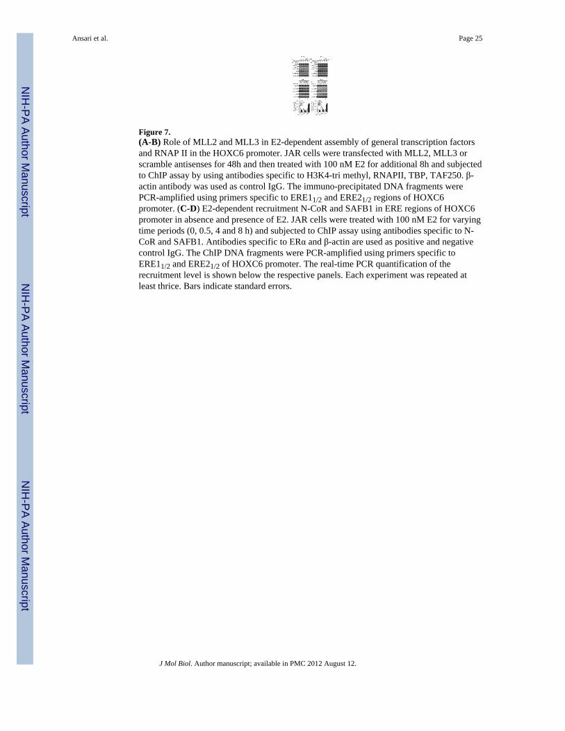
Figure 7.(A-B) Role of MLL2 and MLL3 in E2-dependent assembly of general transcription factorsand RNAP II in the HOXC6 promoter. JAR cells were transfected with MLL2, MLL3 orscramble antisenses for 48h and then treated with 100 nM E2 for additional 8h and subjectedto ChIP assay by using antibodies specific to H3K4-tri methyl, RNAPII, TBP, TAF250. β-actin antibody was used as control IgG. The immuno-precipitated DNA fragments werePCR-amplified using primers specific to ERE11/2 and ERE21/2 regions of HOXC6promoter. (C-D) E2-dependent recruitment N-CoR and SAFB1 in ERE regions of HOXC6promoter in absence and presence of E2. JAR cells were treated with 100 nM E2 for varyingtime periods (0, 0.5, 4 and 8 h) and subjected to ChIP assay using antibodies specific to N-CoR and SAFB1. Antibodies specific to ERα and β-actin are used as positive and negativecontrol IgG. The ChIP DNA fragments were PCR-amplified using primers specific toERE11/2 and ERE21/2 of HOXC6 promoter. The real-time PCR quantification of therecruitment level is shown below the respective panels. Each experiment was repeated atleast thrice. Bars indicate standard errors.
Ansari et al. Page 25
J Mol Biol. Author manuscript; available in PMC 2012 August 12.
NIH
-PA Author Manuscript
NIH
-PA Author Manuscript
NIH
-PA Author Manuscript

Figure 8.Roles of ERα and ERβ on E2-dependent recruitment of MLL2 and MLL3. JAR cells weretransfected with ERα and ERβ antisense for 48 h followed by exposure to E2 (100 nM foradditional 8 h). Cells were harvested and subjected to ChIP assay using anti-MLL2 and anti-MLL3 antibodies. The immuno-precipitated DNA fragments were PCR-amplified usingprimer specific to ERE11/2 and ERE21/2 regions of HOXC6 promoter and subjected to real-time PCR quantification and plotted (panel B)
Ansari et al. Page 26
J Mol Biol. Author manuscript; available in PMC 2012 August 12.
NIH
-PA Author Manuscript
NIH
-PA Author Manuscript
NIH
-PA Author Manuscript

NIH
-PA Author Manuscript
NIH
-PA Author Manuscript
NIH
-PA Author Manuscript
Ansari et al. Page 27
Table 1
Primers used for cloning, RT-PCR, ChIP, and antisense experiments
Primers Forward primer (5′- 3′) Reverse primer (5′ - 3′)
PCR primer
β-actin AGAGCTACGAGCTGCCTGAC GTACTTGCGCTCAGGAGGAG
HOXC6 CAGACCCTGGAACTGGAGAA CTTCCCGCTTTTCCTCTTTT
HOXC6- ERE11/2 TTTTTCCCCCTTCCTGACAT GCCTTTACCTGGTCGGTCTA
HOXC6- ERE21/2 AGCCTCATAGCTCAGGTCCA CCAGAAAGAGAAGGCTGGTG
HOXC6-non-ERE TATGAGGGGAGCTGAGCAAT CCCTCGCACACAGATACACA
MLL1 GAGGACCCCGGATTAAACAT GGAGCAAGAGGTTCAGCATC
MLL2 AGGAGCTGCAGAAGAAGCAG CAGCCAAACTGGGAGAAGAG
MLL3 CATATGCACGACCCTTGTTG ACTGCTGGATGTGGGGTAAG
MLL4 CCCTCCTACCTCAGTCGTCA CAGCGGCTACAATCTCTTCC
ERα AGCACCCTGAAGTCTCTGGA GATGTGGGAGAGGATGAGGA
ERβ AAGAAGATTCCCGGCTTTGT TCTACGCATTTCCCCTCATC
Cloning primer
HOXC6- ERE11/2 CCACCAAACCAGTTCCCTTA* ATCATAGGCGGTGGAATTGA*
HOXC6- ERE21/2 AGCCTCATAGCTCAGGTCCA* CTCCTTCTCAGGACCCCTCT*
HOXC6-Non-ERE TATGAGGGGAGCTGAGCAAT* CCCTCGCACACAGATACACA*
Antisense
MLL1 antisense TGCCAGTCGTTCCTCTCCAC**
MLL2 antisense ACTCTGCCACTTCCCGCTCA**
MLL3 antisense CCATCTGTTCCTTCCACTCCC**
MLL4 antisense CCTTCTCTTCTCCCTCCTTGT**
ERα antisense CATGGTCATGGTCAG**
ERβ antisense GAATGTCATAGCTGA**
Scramble antisense CGTTTGTCCCTCCAGCATCT**
*Flanked by appropriate restriction sites
**Phosphorothioate antisense oligonucleotide.
J Mol Biol. Author manuscript; available in PMC 2012 August 12.
Copyright © 2022 FDOKUMEN